


Languages
Pages
Legal

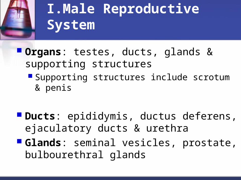
I.Male Reproductive System
Organs: testes, ducts, glands & supporting structures Supporting structures include scrotum & penis
Ducts: epididymis, ductus deferens, ejaculatory ducts & urethra
Glands: seminal vesicles, prostate, bulbourethral glands
Figure 23.1
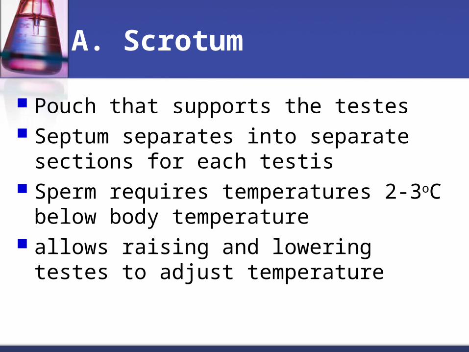
A. Scrotum
Pouch that supports the testes Septum separates into separate sections for
each testis Sperm requires temperatures 2-3oC below
body temperature allows raising and lowering testes to adjust
temperature
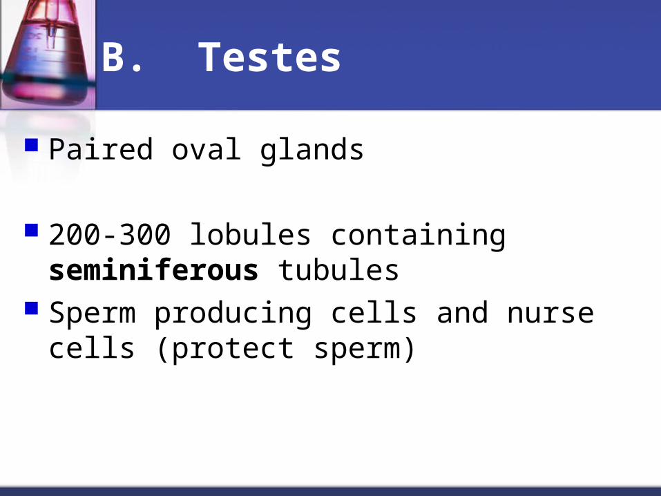
B. Testes
Paired oval glands
200-300 lobules containing seminiferous tubules
Sperm producing cells and nurse cells (protect sperm)
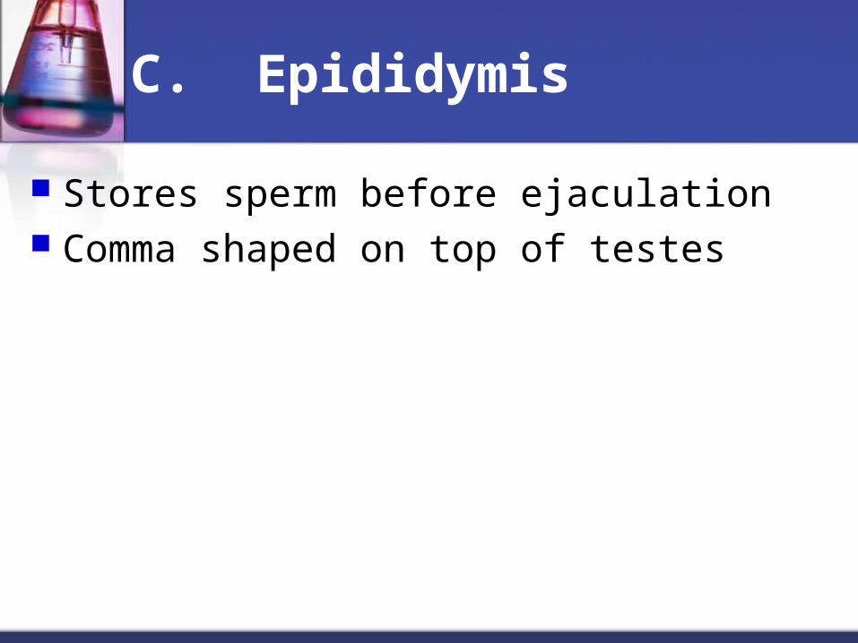
C. Epididymis
Stores sperm before ejaculation Comma shaped on top of testes
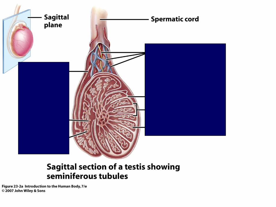
Figure 23.2a
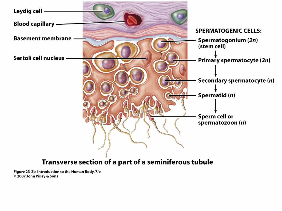
Figure 23.2b
D. Spermatogenesis
Occurs in seminiferous tubules Cell types involved: spermatagonia, sertoli cells & interstitial
cells (leydig cells)
move into->epididymis
Spermatogenesis stages
Takes ~65-75 days from first division to release
~300 million /day Life span ~ 48 hrs in female tract
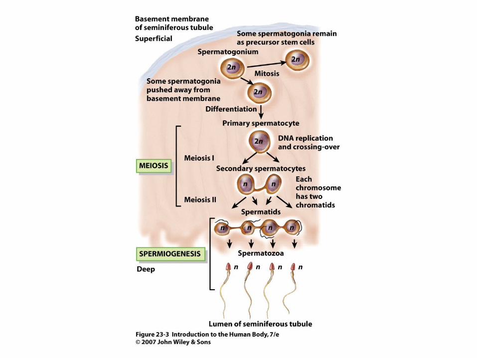
Figure 23.3
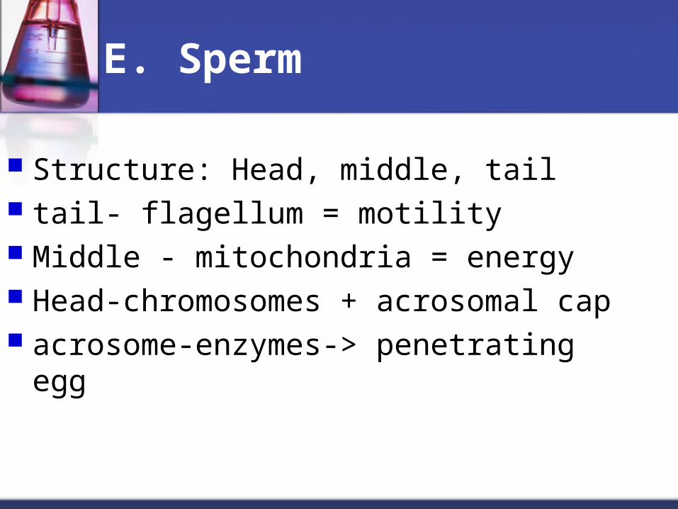
E. Sperm
Structure: Head, middle, tail tail- flagellum = motility Middle - mitochondria = energy Head-chromosomes + acrosomal cap acrosome-enzymes-> penetrating egg
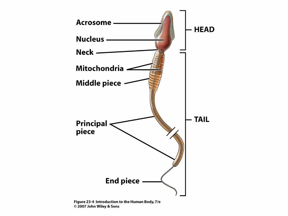
Figure 23.4
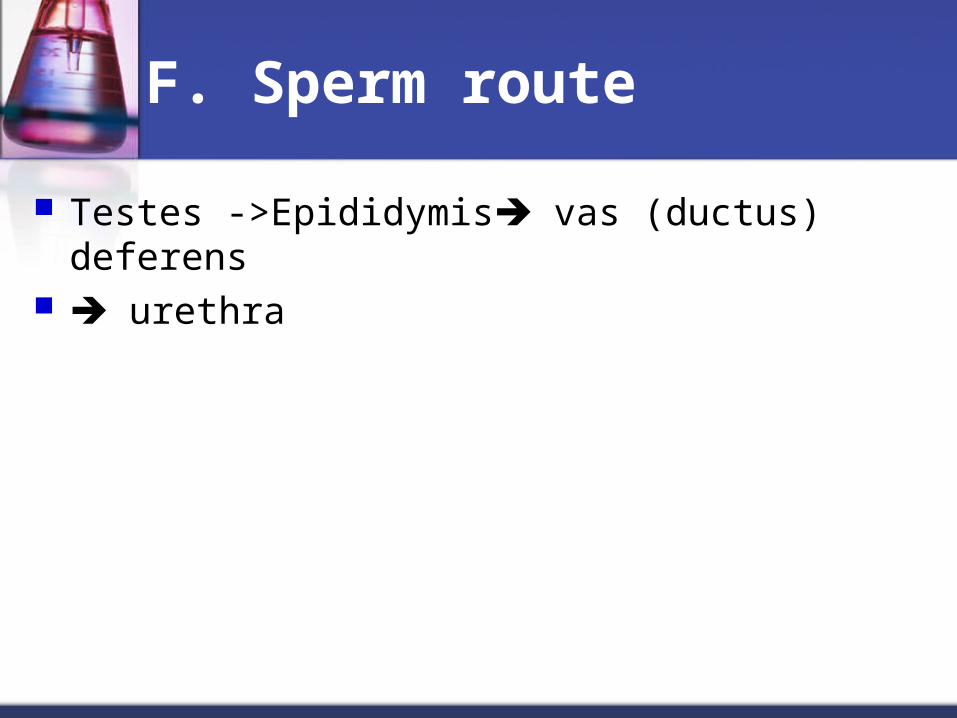
F. Sperm route
Testes ->Epididymis vas (ductus) deferens urethra
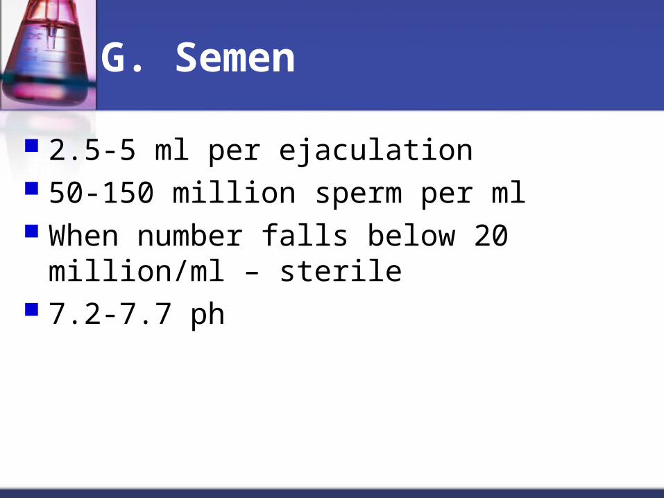
G. Semen
2.5-5 ml per ejaculation 50-150 million sperm per ml When number falls below 20 million/ml –
sterile 7.2-7.7 ph
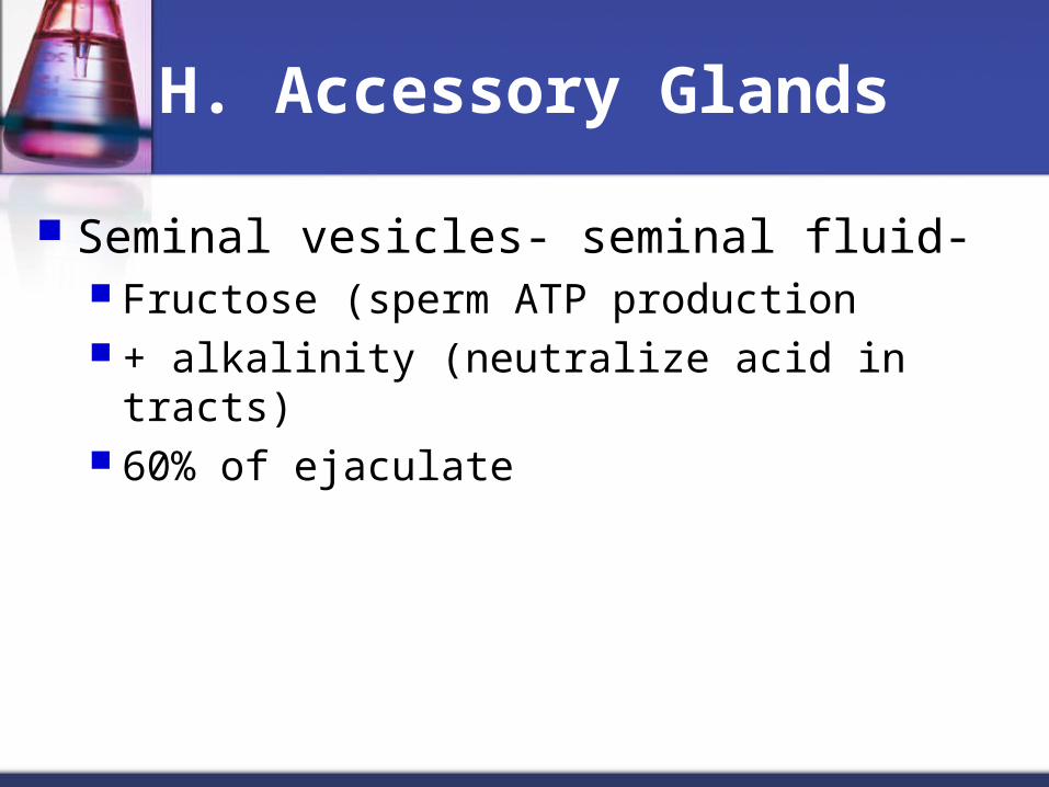
H. Accessory Glands
Seminal vesicles- seminal fluid- Fructose (sperm ATP production + alkalinity (neutralize acid in tracts) 60% of ejaculate
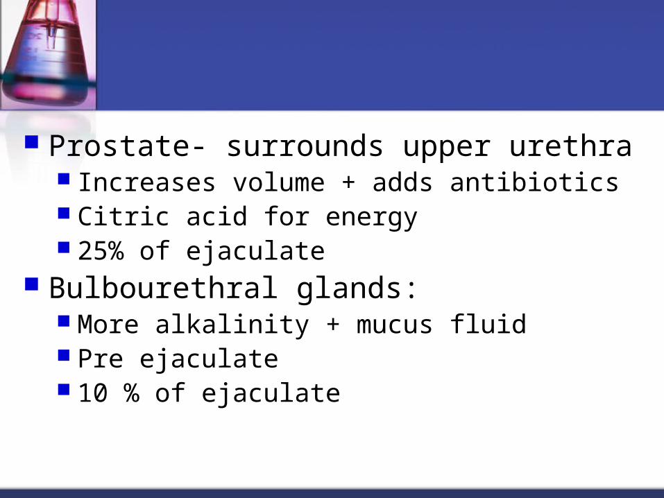
Prostate- surrounds upper urethra Increases volume + adds antibiotics Citric acid for energy 25% of ejaculate
Bulbourethral glands: More alkalinity + mucus fluid Pre ejaculate 10 % of ejaculate
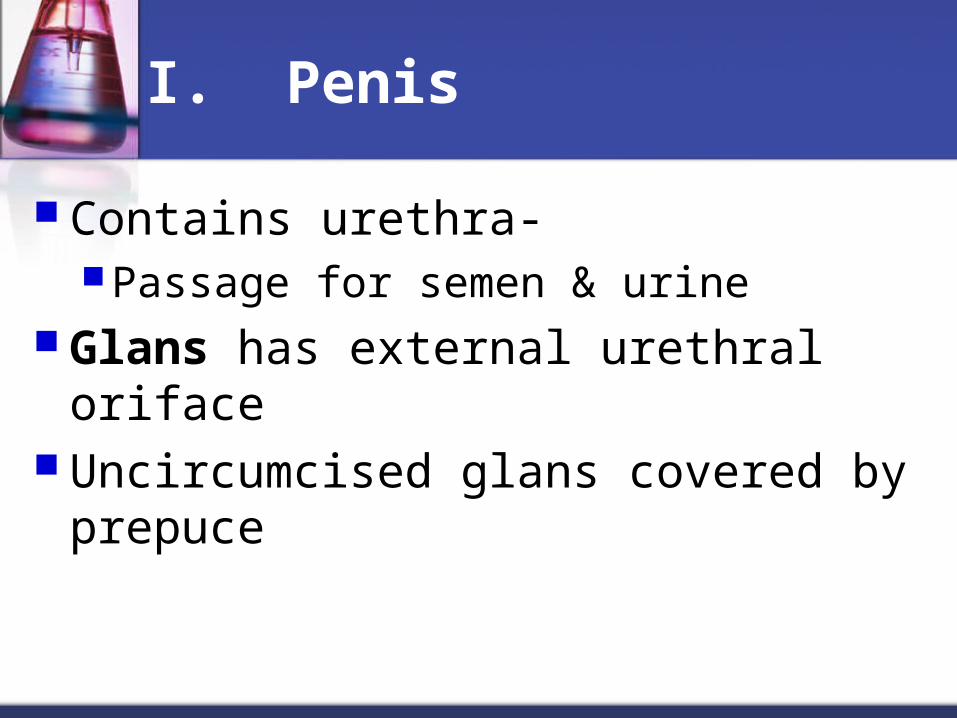
I. Penis
Contains urethra-Passage for semen & urine
Glans has external urethral oriface Uncircumcised glans covered by
prepuce
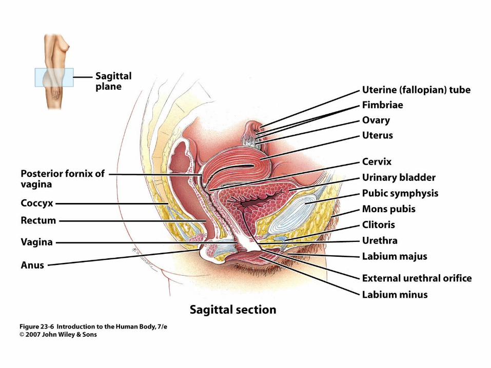
Figure 23.6
II.Female Reproductive System
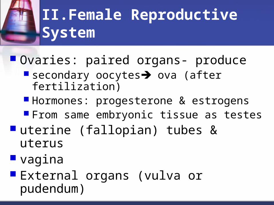
Ovaries: paired organs- produce secondary oocytes ova (after fertilization) Hormones: progesterone & estrogens From same embryonic tissue as testes
uterine (fallopian) tubes & uterus vagina External organs (vulva or pudendum)
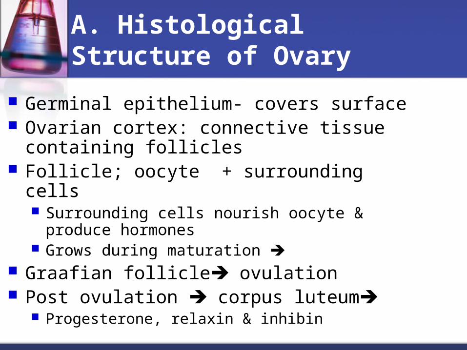
A. Histological Structure of Ovary
Germinal epithelium- covers surface Ovarian cortex: connective tissue containing
follicles Follicle; oocyte + surrounding cells
Surrounding cells nourish oocyte & produce hormones
Grows during maturation Graafian follicle ovulation Post ovulation corpus luteum
Progesterone, relaxin & inhibin

Figure 23.6
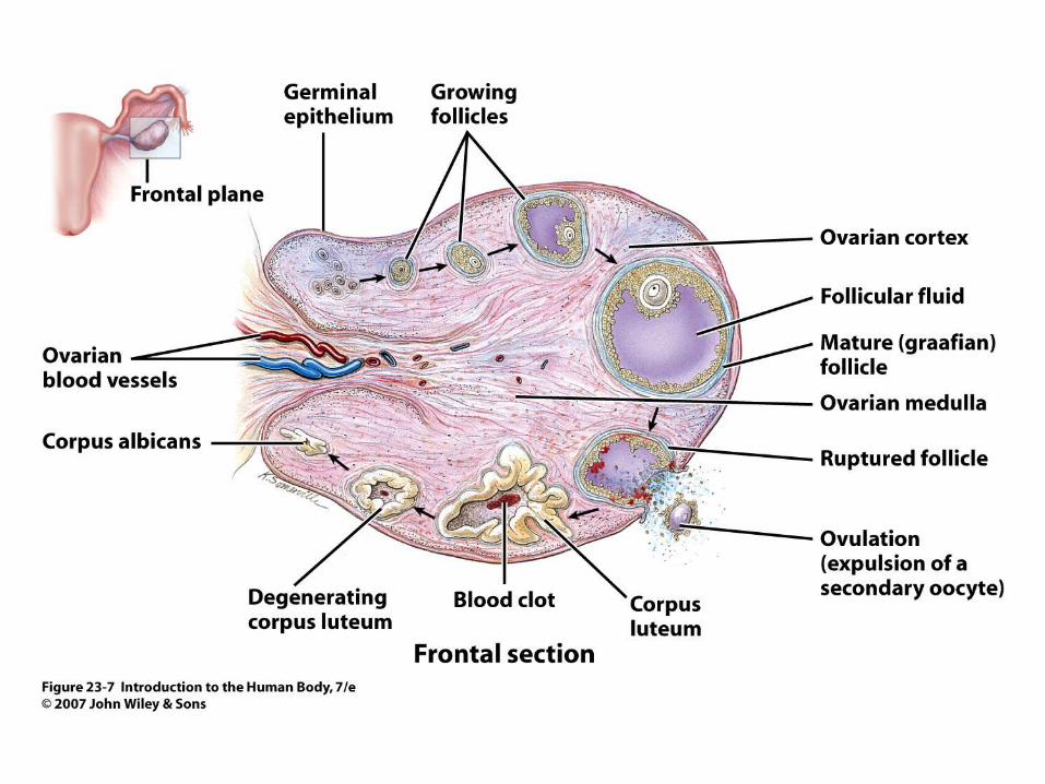
Figure 23.7
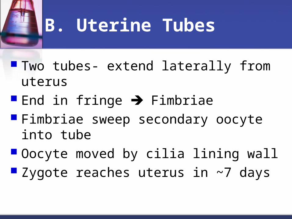
B. Uterine Tubes
Two tubes- extend laterally from uterus End in fringe Fimbriae Fimbriae sweep secondary oocyte into tube Oocyte moved by cilia lining wall Zygote reaches uterus in ~7 days

C. Uterus
Pathway for sperm & site of implantation Fundus -Dome-shaped area above tubes= Body – tapering central portion Cervix- narrow opening into vagina Uterine cavity- interior of body
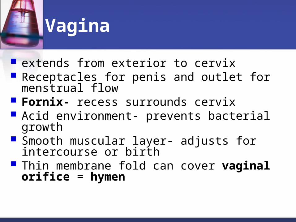
Vagina
extends from exterior to cervix Receptacles for penis and outlet for menstrual flow Fornix- recess surrounds cervix Acid environment- prevents bacterial growth Smooth muscular layer- adjusts for intercourse or
birth Thin membrane fold can cover vaginal orifice =
hymen
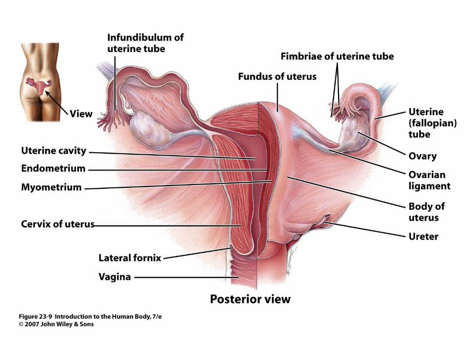
Figure 23.9
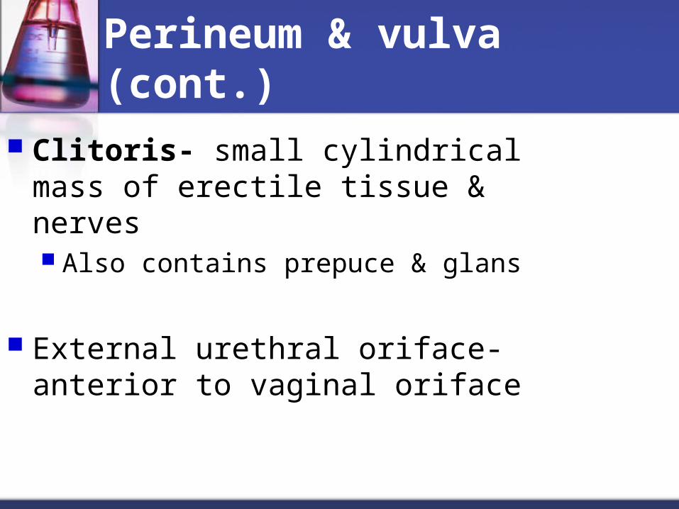
Perineum & vulva (cont.)
Clitoris- small cylindrical mass of erectile tissue & nerves Also contains prepuce & glans
External urethral oriface- anterior to vaginal oriface
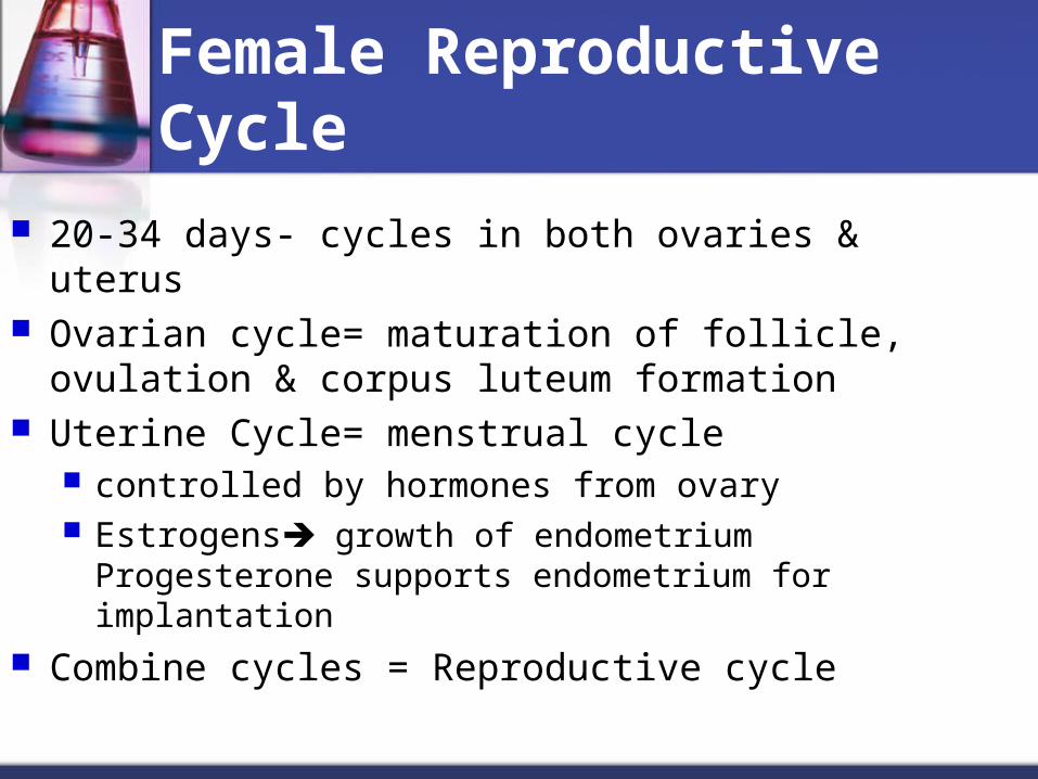
Female Reproductive Cycle
20-34 days- cycles in both ovaries & uterus Ovarian cycle= maturation of follicle, ovulation &
corpus luteum formation Uterine Cycle= menstrual cycle
controlled by hormones from ovary Estrogens growth of endometrium Progesterone
supports endometrium for implantation Combine cycles = Reproductive cycle
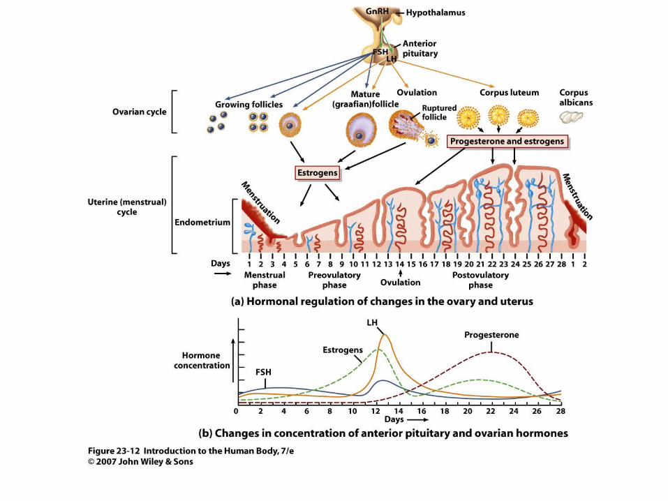
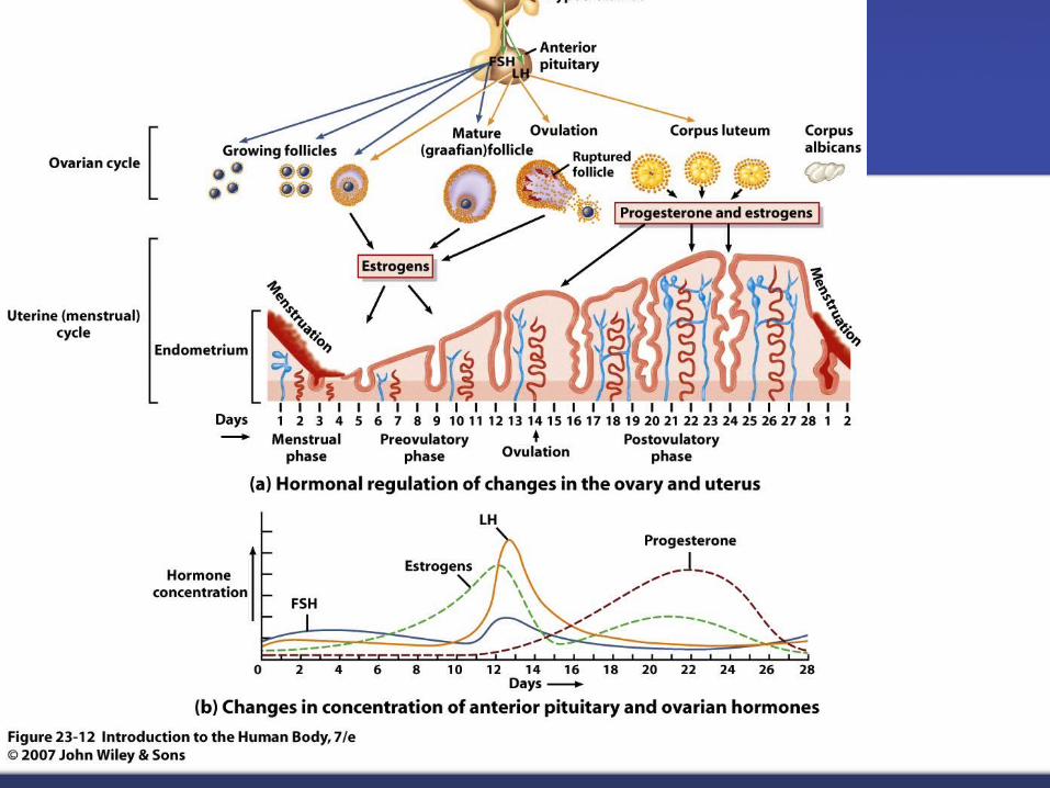
Figure 23.12
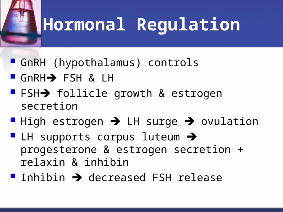
Hormonal Regulation
GnRH (hypothalamus) controls GnRH FSH & LH FSH follicle growth & estrogen secretion High estrogen LH surge ovulation LH supports corpus luteum progesterone &
estrogen secretion + relaxin & inhibin Inhibin decreased FSH release
Gonadotropin releasing hormone= GnRH
Comes from hypothalamus and stimulates the release of FSH from pituitary.
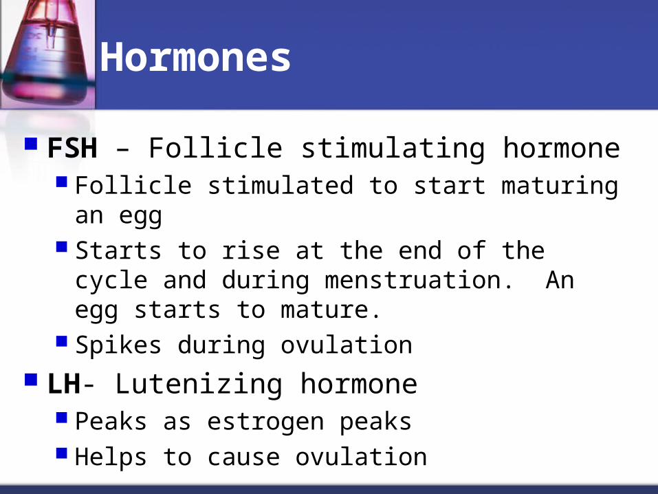
Hormones
FSH – Follicle stimulating hormone Follicle stimulated to start maturing an egg Starts to rise at the end of the cycle and during
menstruation. An egg starts to mature. Spikes during ovulation
LH- Lutenizing hormone Peaks as estrogen peaks Helps to cause ovulation
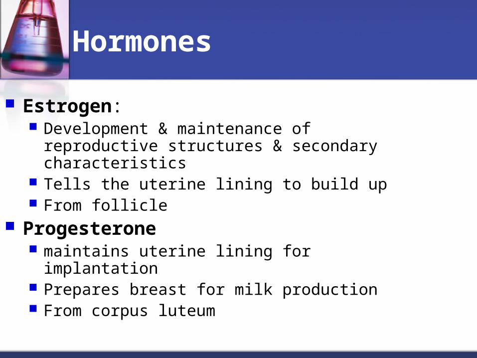
Hormones
Estrogen: Development & maintenance of reproductive
structures & secondary characteristics Tells the uterine lining to build up From follicle
Progesterone maintains uterine lining for implantation Prepares breast for milk production From corpus luteum
hCG- human chorionic gonadotropin Made by embryo Tells corpus luteum to keep making
progesterone to maintain lining
Relaxin= relaxes uterus- inhibits myometrium
Inhibin- inhibits FSH release
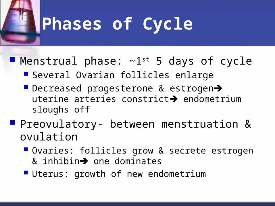
Phases of Cycle
Menstrual phase: ~1st 5 days of cycle Several Ovarian follicles enlarge Decreased progesterone & estrogen uterine arteries
constrict endometrium sloughs off
Preovulatory- between menstruation & ovulation Ovaries: follicles grow & secrete estrogen & inhibin one
dominates Uterus: growth of new endometrium
Phases of Cycle (cont.)
Ovulation Release of 2o oocyte with LH surge
Postovulatory- Ovaries: follicle collapses corpus luteum
(luteal phase) If no fertilization FSH & LH corpus
albicans & decreased Progesterone menstruation
Phases of Cycle (cont.)
If fertilization & division human chorionic Gonadotrophin (hCG) stimulates corpus luteum secretion
Uterus: Progesterone & estrogens complete development of uterus for implantation
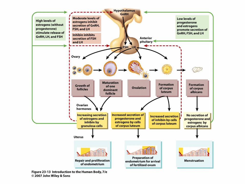
Figure 23.13
Top Related