

Languages
Pages
Legal


7/27/10
1
Fig 1. Symptom-based diagnosis of allergic rhinitis
Fig 2. Differential diagnosis of rhinitis / rhinosinusitis
SUBGROUPS RHINITIS Allergic rhinitis Intermittent / persistent
Mild / moderate / severe Occupational
Infectious rhinitis Viral Bacterial Non-viral non-bacterial (Protozoa / fungi)
Non-allergic non-infectious rhinitis
Drug-induced (! blokkers / vasodilators / contraceptives / aspirin / NSAID) Hormonal (hypothyroidism / pregnancy) NARES (some have local IgE production) Occupational (LMW agents / irritants) Atrophic / rhinitis of the elderly Idiopathic
RHINOSINUSITIS Acute / chronic Mild / moderate / severe

7/27/10
2
Fig 3. Inspection of the nose showing distortion of the anatomy at the level of the nasal entry (in cleft lip patients)
Courtesy of P. Hellings
Fig 4. Inspection of the nose showing alar collapse during inspiration
Courtesy of P. Hellings
Oral breathing! Nasal inspiration!

7/27/10
3
Fig 5. Tip elevation test for evaluation of improved breathing by restoration of normal tip support
nasal !obstruction!
good nasal!flow!
Courtesy of P. Hellings
Fig 6. Anterior rhinoscopy allowing the evaluation of mucosal and/or anatomic pathology at the anterior part of the nasal cavity

7/27/10
4
Fig 7. Mirror test for evaluation of the condensate of expired air on a cold metal instrument or mirror
Fig. 8 Nasal endoscopy
Courtesy of P. Delaere, Leuven

7/27/10
5
Fig. 10 Diagnostic algorithm for the diagnosis of AR
Fig. 10 Practical approaches for nasal provocation test

7/27/10
6
Fig. 11 Skin prick test with evaluation of wheal and flare reaction on the skin at the site of allergen deposition
Fig. 12 Peak nasal inspiratory flow measurement

7/27/10
7
Fig. 13 Active anterior rhinomanometry
Fig. 14 Acoustic rhinometry
7/27/10
8
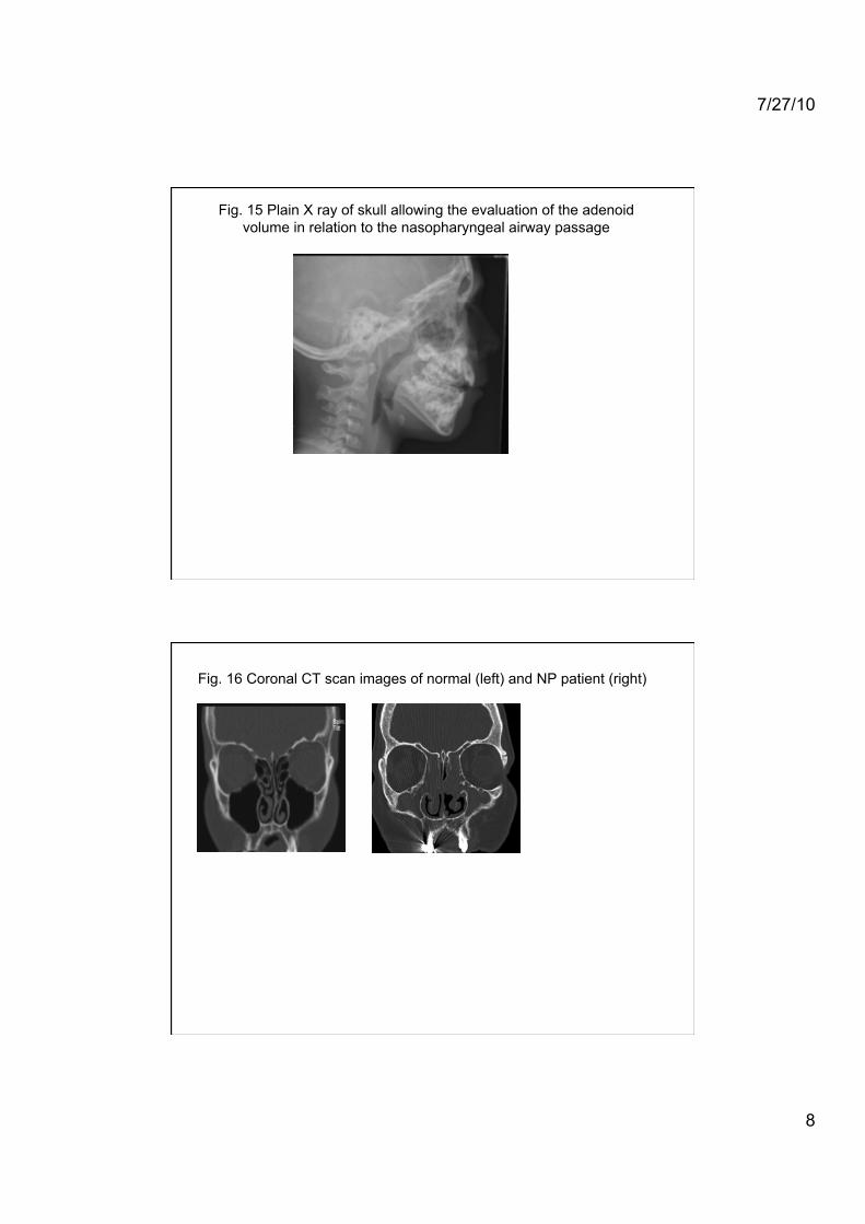
Fig. 15 Plain X ray of skull allowing the evaluation of the adenoid volume in relation to the nasopharyngeal airway passage
Fig. 16 Coronal CT scan images of normal (left) and NP patient (right)

7/27/10
9
Fig. 18 Occupational vs work-exacerbated rhinitis
Fig. 18 Diagnostic algorithm for occupational rhinitis

7/27/10
10
Fig. 19 University of Pennsylvania Smell Identification Test (UPSIT)
Fig. 20 Connecticut Chemosensory Clinical Research Center (CCCRC)

7/27/10
11
Fig. 21 Sniffin’ Sticks
Fig. 22 Barcelona Smell Test (BAST)-24
Top Related