


Languages
Pages
Legal


Department of Life Sciences and ChemistryRoskilde UniversityDK-4000 Roskilde
Denmark
Anne Karin Rasmussen
Ph.D. Thesis
December 2001
Identification of Factors Interacting withhMSH2 and hMLH1 in the Fetal Liver
and Investigations of how MitochondrialDysfunction Creates a Mutator Phenotype

Identification of factors interacting with
hMSH2 and hMLH1 in the fetal liver
and Investigations of how Mitochondrial
Dysfunction creates a mutator phenotype
Anne Karin Rasmussen
Ph.D. thesis
December 2001
Department of Life Sciences and Chemistry Roskilde University DK-4000 Roskilde
Denmark

Preface This Ph.D. thesis is based on work carried out at Department of Life Sciences and
Chemistry, Roskilde University, Denmark in Dr. Lene Juel Rasmussen's laboratory and at
Johns Hopkins Oncology Center, Baltimore, MD, USA in Dr. Keshav K. Singh's laboratory
from 1998 to 2001.
On the beginning, my Ph.D. research focus area was primarily the DNA mismatch repair
pathway. However, after a stay at Johns Hopkins the scope of my thesis ended up being
more comprehensive. In the Ph.D. thesis I am also addressing the following issues:
molecular mechanisms associated with DNA damage and DNA repair, and mutagenesis in
mitochondria.
Front cover: HeLa cells stained with MitoTracker Red
1

Acknowledgements
First, I wish to express my gratitude to Dr. Lene Juel Rasmussen for critical reading of my
manuscripts and thesis and particularly for being an extraordinarily positive, dedicated and
stimulating supervisor.
Secondly, I wish to give my thanks to Dr. Keshav Singh for letting me work in his laboratory
at Johns Hopkins and for widening my intellectual horizon in the field of mutagenesis in
mitochondria. Furthermore, I want to thank him for the critical reading of my manuscripts
and for being an inspiring supervisor.
Thirdly, I want to express my thanks to Lene Markussen and Gerda Olesen for excellent
technical assistance.
In addition, I want to give my thanks to all my colleagues and friends at Department of Life
Sciences and Chemistry at Roskilde University and at Johns Hopkins Oncology Center. A
special thanks to Rob Delsite and Barbara Sigala for technical support and to Anne Lützen
and Jonas Andreasen for critical reading of my thesis and for creating a cheerful atmosphere.
Finally, I want to express my gratitude to my dearest family and boyfriend for all the love
and support they gave me while completing my Ph.D.
Anne Karin Rasmussen
Copenhagen 2001
2

Index
PUBLICATIONS/MANUSCRIPTS.................................................................................................................. 5 STRUCTURE ...................................................................................................................................................... 5
ABBREVIATIONS............................................................................................................................................. 6
SUMMARY ......................................................................................................................................................... 7
SAMMENDRAG (SUMMARY IN DANISH).................................................................................................. 8
1. ANALYSIS OF HUMAN DNA MISMATCH REPAIR.............................................................................. 9 1.1 INTRODUCTION ........................................................................................................................................... 9 1.2 DNA MISMATCH REPAIR........................................................................................................................... 9
MMR in Escherichia coli .............................................................................................................................. 9 Mismatch recognition - in eukaryotes......................................................................................................... 10 The step after mismatch recognition - in eukaryotes .................................................................................. 11 Excision....................................................................................................................................................... 14
1.3 THE HUMAN EXONUCLEASE 1 ................................................................................................................. 14 The first subfamily – XPG........................................................................................................................... 14 The second subfamily – FEN1..................................................................................................................... 15 The third subfamily – EXO1........................................................................................................................ 15 Interactions with hEXO1............................................................................................................................. 16 Expression/localization of hEXO1 .............................................................................................................. 16 Mutator gene............................................................................................................................................... 17 DNA exonuclease activity ........................................................................................................................... 17 3' → 5' exonuclease .................................................................................................................................... 18
1.4 METHODS.................................................................................................................................................. 19 Principles of the two-hybrid system ............................................................................................................ 19
1.5 RESULTS & DISCUSSION........................................................................................................................... 21 Two-hybrid screening with hMSH2 as a bait.............................................................................................. 23 Two-hybrid screening with hMLH1 as a bait.............................................................................................. 24 Importin α ................................................................................................................................................... 26
2. REPAIR OF MITOCHONDRIAL DNA .................................................................................................... 28 2.1 INTRODUCTION ......................................................................................................................................... 28 2.2 BASE EXCISION REPAIR ........................................................................................................................... 29
Repair of uracil ........................................................................................................................................... 29 Repair of oxidative damage ........................................................................................................................ 30 Removal of 8-oxoguanine............................................................................................................................ 30 Removal of adenine paired with 8-oxoguanine........................................................................................... 31 Removal of oxidized pyrimidines ................................................................................................................ 31 Removal of 3-methyladenine....................................................................................................................... 32 Other BER factors....................................................................................................................................... 32 Mismatch repair.......................................................................................................................................... 34 Direct repair ............................................................................................................................................... 34
2.3 DIRECT REPAIR: O6-METHYLGUANINE-DNA METHYLTRANSFERASE.................................................. 34 MGMT function........................................................................................................................................... 35 Repair of O4-methylthymine........................................................................................................................ 36 The methylation tolerant phenotype............................................................................................................ 37 Mitochondrial MGMT activity .................................................................................................................... 39 MGMTs intracellular localization .............................................................................................................. 39
3. MITOCHONDRIAL DYSFUNCTION VERSUS GENETIC STABILITY OF THE NUCLEAR DNA............................................................................................................................................................................ 41
3.1 INTRODUCTION ......................................................................................................................................... 41 3.2 MITOCHONDRIAL BIOLOGY AND GENETICS ........................................................................................... 41 3.3 REACTIVE OXYGEN SPECIES ................................................................................................................... 43
3
3.4 EFFECTS OF MITOCHONDRIAL ROS PRODUCTION ................................................................................ 45 Inhibition of mitochondrial oxidative phosphorylation............................................................................... 45 Cytosolic superoxide dismutase (sod1) mutants ......................................................................................... 45 Mitochondrial superoxide dismutase (sod2) mutants ................................................................................. 46
3.5 MITOCHONDRIAL DYSFUNCTION CONTRIBUTES TO NDNA MUTATIONS .............................................. 48 SECTION TWO................................................................................................................................................ 54
PAPER I ........................................................................................................................................................... 54 MANUSCRIPT II .............................................................................................................................................. 67 MANUSCRIPT III............................................................................................................................................. 91 MANUSCRIPT IV ........................................................................................................................................... 108
4. CONCLUDING REMARKS ..................................................................................................................... 130
REFERENCES................................................................................................................................................ 134
4

Publications/Manuscripts
The present thesis is based on one original publication (I), one submitted manuscript (III),
and two manuscripts in preparation (II, IV). The manuscripts referred to in the thesis are
identified by the following roman numerals:
I. Lene Juel Rasmussen, Merete Rasmussen, Byung-In Lee, Anne Karin Rasmussen,
David M. Wilson III, Finn Cilius Nielsen, and Hanne Cathrine Bisgaard.
Identification of factors interacting with hMSH2 in the fetal liver utilizing the yeast
two-hybrid system. In vivo interaction through the C-terminal domains of hEXO1
and hMSH2 and comparative expression analysis. Mutation Research (2000) 460,
41-52.
II. Anne Karin Rasmussen, Keshav K. Singh, and Lene Juel Rasmussen.
Characterization of O6-MeG DNA Methyltransferase (MGMT) protein in repairing
human mitochondrial DNA. Manuscript in preparation.
III. Anne Karin Rasmussen, Aditi Chatterjee, Lene Juel Rasmussen and Keshav K.
Singh. Mitochondria as determinant of genetic stability in Saccharomyces cerevisiae.
Nature Genetics (submitted).
IV. Anne Karin Rasmussen, Lene Juel Rasmussen, and Keshav K. Singh.
Mitochondrial dysfunction suppresses the mutator phenotype of Saccharomyces
cerevisiae superoxide dismutase deficient cells. Manuscript in preparation.
Structure The thesis is divided into two sections:
1. Section one is a general introduction where important results from my research are
included. The section is divided into three parts: The first part is an introduction to the
publication (I). The second part is an introduction to manuscript II. The third part is an
introduction to manuscripts III and IV.
2. Section two consists of one published paper and three manuscripts.
5
Abbreviations
A Adenine hUNG1 Human mitochondrial UNG AD Activation Domain hUNG2 Human nuclear UNG ANT Adenine Nucleotide Translocator 3-MeA 3-methyladenine AP Apurinic/apyrimidinic 3-MeG 3-methylguanine ATP Adenosine triphosphate MGMT O6-methylguanine-DNA
methyltransferase BD Binding Domain MMR Mismatch repair BER Base Excision DNA Repair MNNG N-methyl-N’-nitro-N-
nitrosoguanidine C Cysteine MNU Methylnitrosourea CAT Catalase MPG N-methylpurine-DNA glycosylase Complex I NADH dehydrogenase or NADH:ubiquinone
oxidoreductase Msh1 Mitochondrial homolog of the E. coli
MutS Complex II Succinate dehydrogenase mtDNA Mitochondrial DNA Complex III Ubiquinol:cytochome c oxidoreductase nDNA Nuclear DNA Complex IV Cytochrome c oxidase NER Nucleotide Excision Repair Complex V ATP synthase NLS Nuclear Localization Signal CoQ Coenzyme Q, also called ubiquinone NPC Nuclear pore complexes DNA Deoxyribonuceic acid O2
- Superoxide radical E. coli Escherichia coli O4-MeT O4-methylthymine FEN1 Flap endonuclease-1, S. cerevisiae rad27
homolog O6-MeG O6-methylguanine
G Guanine 8-oxoG 7,8-dihydro-8-oxoguanine GGR Global genome repair PCNA Proliferating Cell Nuclear Antigen GPx Glutathione peroxidase POL G Polymerase γ H2O2 Hydrogen peroxide Pol ε DNA polymerases (Pol II) hAPE1 Human AP endonuclease Pol δ DNA polymerases (Pol III) hEXO1 Human exonuclease 1 rho- Mutations in the mitochondrial
genome hMLH1-hMLH3
E. coli MutL homolog complex rho0 Lack of mitochondrial genome
hMLH1-hPMS1
E. coli MutL homolog complex ROS Reactive oxygen species
hMLH1-hPMS2
E. coli MutL homolog complex S. cerevisiae Saccharomyces cerevisiae
hMSH2-hMSH3
E. coli MutS homolog complex, recognizes insertion/deletions
SOD1 Nuclear superoxide dismutase
hMSH2-hMSH6
E. coli MutS homolog complex, recognizes base:base mispairs and insertion/deletions
SOD2 Mitochondrial superoxide dismutase
hMYH Glycosylase, Human MutY Homolog SSB Single-strand binding protein HNPCC Hereditary Non-Polyposis Colon Cancer T Thymine hNTH1 Glycosylase, Human Nth homolog TCR Transcription coupled repair hOGG1 8-OxoGuanine DNA Glycosylase UNG Uracil-DNA Glycosylase HO• Hydroxyl radical XPG S. cerevisiae Rad2 homolog
6

Summary
Increased spontaneous mutation frequency is associated with increased cancer risk.
However, the relative contribution of spontaneous endogenous mutagenesis to
carcinogenesis is not known today. Defects in the postreplication DNA mismatch repair
(MMR) pathway are recognized to increase spontaneous mutations. Mutations in MMR
genes cause hereditary non-polyposis colon cancer.
In an effort to identify unidentified genes involved in MMR and tissue-specific MMR-
associated factors, we employed the yeast two-hybrid system, using the human hMSH2 as
bait and a human fetal liver cDNA library as prey. We demonstrated that hMSH2 interacts
with a human 5’ → 3’ exonuclease 1 (hEXO1).
Data presented in this thesis also support the conclusion that mitochondrial dysfunction leads
to spontaneous nuclear DNA damage. We employed the yeast Saccharomyces cerevisiae as
a model system to investigate a potential link between mitochondrial activity and genomic
instability. Mitochondrial dysfunction and genetic instability are characteristic features of
cancer cells. Furthermore, mitochondrial dysfunction is a key feature of aging due to
accumulation of mutations in mtDNA. Our studies in a yeast model system suggest that
mitochondria contain some intrinsic properties that control the generation of the mutator
phenotype associated with cancer cells. We hypothesize that cancer cells by losing their
mitochondrial function create a mutator phenotype.
Given the importance of maintaining the integrity of the mitochondrial genome we have
found that it might be valuable to further investigate the molecular processes and
components responsible for mtDNA repair. It has recently been recognised that base
excision DNA repair (BER) is operating in the mitochondria, however, knowledge about
other repair pathways is still very limited. We decided to investigate O6-methylguanine-
DNA methyltransferase (MGMT) because of the fact that its sub-cellular localization has not
been determined. We determined that it was localized to nucleus but not to mitochondria in
HeLa and breast epithelial cells.
7

Sammendrag (Summary in Danish)
En øget frekvens af spontane mutationer er forbundet med øget risiko for cancer. Hvor stor
en del af cancinogenese som skyldes spontan endogen mutagenese, er endnu ikke kendt. Fejl
i det postreplikative DNA mismatch repair (MMR) system har imidlertid vist sig at forøge
den spontane mutationsfrekvens, da mutationer i MMR-generne kan forårsage hereditær
non-polypos colon cancer.
For at finde uidentificerede gener, som er involveret i MMR, samt vævsspecifikke MMR-
associerede faktorer, gjorde vi brug af gær two-hybrid systemet. Vi brugte hMSH2 som
“bait” og et humant føtalt lever cDNA bibliotek som “prey”. Vi viste, at hMSH2 interagerer
med en human 5’ → 3’ exonuclease 1 (hEXO1).
Endvidere indeholder afhandlingen resultater, som understøtter konklusionen om, at
mitokondriel dysfunktion fører til spontan nukleær DNA skade. Vi brugte Saccharomyces
cerevisiae som modelsystem til at undersøge en mulig sammenhæng mellem mitokondriers
aktivitet og genomisk stabilitet. Mitokondriel dysfunktion og reduceret genetisk stabilitet er
karakteristiske egenskaber for cancer celler. Yderligere er mitokondriel dysfunktion
forbundet med aldring på grund af akkumulering af mutationer i mtDNA. Vore studier i et
gær modelsystem indikerer, at mitokondrier har egenskaber som kan styre udviklingen af
den mutator fænotype der er forbundet med cancer celler. På baggrund af dette opstiller vi en
hypotese om, at cancer celler ved at miste deres mitokondrie funktion kan frembringe en
mutator fænotype.
Givet vigtigheden af at opretholdeholde/reparere det mitokondrielle genom for at forhindre
mitokondriel dysfunktion, fandt vi det værdifuldt yderligere at undersøge de molekylære
processer og komponenter ansvarlige for mtDNA reparation. Det er i dag anerkendt at base
excision DNA repair (BER) virker i mitokondrierne. Viden om andre reparationssystemer i
mitokondrierne er dog stadig meget begrænset. Vi undersøgte O6-methylguanine- DNA
methyltransferase (MGMT) subcellulære lokalisation, og fandt at MGMT i bryst epithelial-
celler er lokaliseret til kernen, men ikke til mitokondrierne.
8
Analysis of human DNA Mismatch Repair
1. Analysis of Human DNA Mismatch Repair
1.1 Introduction The postreplication DNA mismatch repair (MMR) pathway is responsible for the
maintenance of DNA fidelity upon replication (Buermeyer et al., 1999; Kolodner &
Marsischky, 1999; Harfe & Jinks-Robertson, 2000; Jiricny et al., 2000). MMR captures
errors in the newly synthesized DNA strand that are missed by the polymerase proofreading
and lowers the mutation frequency by a factor of 100-1000-fold as compared to MMR
deficient cells (Bhattacharyya et al., 1994; Eshleman et al., 1995). In humans, accumulation
of mutations is a critical step in carcinogenesis. Loss of a single allele of one of the mismatch
repair proteins causes Hereditary Non-Polyposis Colon Cancer (HNPCC), a form of cancer
that accounts for 1-5% of all cases of colon cancer (Lynch & de la Chapelle, 1999;
Peltomäki, 2001). HNPCC is caused by inherited mutations in MMR genes. Most HNPCC
families have germline mutations in the hMSH2 (2p22-p21) and hMLH1 (3p21) genes.
Mutations in the other known MMR genes hMSH6 (2p16), hMSH3 (5q11-q12), hPMSH2
(7p22), hPMSH1 (2q31), and hMLH3 (14q24) are either rare or non-existent in HNPCC
families (Peltomäki & Vasen, 1997; Kolodner & Marsischky, 1999; Wood et al., 2001).
Some HNPCC families fail to display mutations in known MMR genes. Therefore, we argue
that these HNPCC families must harbor mutations in yet unidentified genes that are involved
in MMR. In an attempt to identify such genes, we employed the yeast two-hybrid system,
using the human MMR proteins hMSH2 (Rasmussen et al., 2000) or hMLH1 as bait and a
fetal liver matchmaker cDNA library as prey.
1.2 DNA Mismatch Repair
MMR in Escherichia coli
Mechanisms and functions of mismatch correction are best understood in Escherichia coli,
therefore a short introduction to MMR in E. coli is given. MMR is directed by the state of
adenine methylation of GATC sequences. Since DNA adenine methylation (Dam) occurs
after replication, an unmethylated newly synthesized strand is temporarily paired with a
fully methylated parental strand, which provides a strand discrimination signal for MMR in
9

Analysis of human DNA Mismatch Repair
E. coli (Lahue & Modrich, 1989). Initiation of MMR occurs via mismatch recognition and
binding of a MutS homodimer followed by binding of a MutL homodimer. In E. coli MutL
serves to couple mismatch recognition with downstream MMR events. Interactions between
MutL, MutS, and ATP are believed to result in translocation of the MutS-MutL complex
away from the mispair, leading to the activation of the MutH endonuclease (Allen et al.,
1997; Hall & Matson, 1999). After activation, MutH introduces a nick in the nascent strand
of the nearest hemi-methylated GATC sequences. MutL helps to load DNA helicase II
(UvrD) at the nicked GATC site and UvrD unwinds DNA from the nick toward and past the
mismatch (Hall et al., 1998). Removal of the error-containing DNA strand is facilitated by
one of four single-stranded, DNA-specific exonucleases (RecJ, ExoI, ExoVII, ExoX)
depending on the polarity of the reaction (Viswanathan & Lovett, 1998; Burdett et al.,
2001). The resulting single-stranded gap, is stabilized by single-strand binding protein
(SSB) and filled by DNA polymerase III holoenzyme. The remaining nick is closed by
DNA ligase and Dam methyltransferase finishes the MMR pathway in E. coli by
methylating the newly synthesized strand (Rasmussen et al., 1998).
Mismatch recognition - in eukaryotes
In eukaryotes, mispaired bases in DNA are recognized by the heterodimeric complexes,
MSH2-MSH6 and MSH2-MSH3 (MutS homologs) (Acharya et al., 1996; Guerrette et al.,
1998, Genschel et al., 1998). Analysis of mismatch binding specificities of the human
hMSH2-hMSH6 and hMSH2-hMSH3 complexes showed that they were overlapping but
not identical. The hMSH2-hMSH6 complex recognizes base:base mispairs and
insertion/deletion mispairs of up to 8 unpaired bases. In comparison, hMSH2-hMSH3 has a
high affinity for insertion/deletions of 2-8 unpaired bases, weak affinity for single-nucleotid
insertion/deletion mispairs, and do not bind base:base mispairs (Drummond et al., 1997;
Genschel et al., 1998). The predominant DNA-binding protein in the hMSH2-hMSH6
complex appears to be hMSH6 when binding to a mismatched oligonucleotide (Matton et
al., 2000). Hence, the hMSH2-hMSH6 complex appears to provide the predominant
mismatch-binding activity in human cells (Genschel et al., 1998; Marra et al., 1998). The
hMSH3 protein is believed to compete with hMSH6 for the available hMSH2, as the
interacting regions of hMSH2 with hMSH3 and hMSH6 are identical (Guerrette et al.,
1998). This prediction is supported by two independent findings: extracts prepared from
HCT15 cells, which lack hMSH6, contain approximately three-fold higher levels of
hMSH2-hMSH3 complex compaired to MMR proficient cells (Genschel et al., 1998); and
10

Analysis of human DNA Mismatch Repair
cells overexpressing hMSH3 preferentially form hMSH2-hMSH3 complexes (Marra et al.,
1998). Thus, hMSH3 interacts with all hMSH2 to form the hMSH2-hMSH3 complex,
making cells functionally deficient in hMSH2-hMSH6 complex. Consequently, these cells
lack base:base mispair correction (Drummond et al., 1997; Marra et al., 1998).
Table 1.1 • DNA mismatch repair proteins (Kolodner & Marsischky, 1999; Burdett et al., 2001; Wood et al., 2001) E. coli S. cerevisia H. sapiens MutS
Msh2-Msh6
hMSH2-hMSH6
Function Recognizes single-nucleotide and insertion/deletion mispairs.
Msh2-Msh3 hMSH2-hMSH3 Recognizes insertion/deletion mispairs.
MutL Mlh1-Pms1 hMLH1-hPMS2
Mlh1-Mlh2 hMLH1-hPMS1
Mlh1-Mlh3 hMLH1-hMLH3
Couples mismatch recognition with downstream MMR events in E. coli. The function in eukaryotes is unclear.
MutH not identified not identified Endonuclease; nicks hemimethylated GATC sequences.
UvrD not identified not identified Helicase; facilitates DNA unwinding
RecJ & ExoVII not identified not identified 5’→ 3’single-stranded DNA exonucleases
ExoI & ExoX not identified not identified 3’→ 5’ single-stranded DNA exonucleases
not identified Exo1
hEXO1 5’→ 3’ DNA exonucleases that have a preference for degrading double-stranded DNA
The step after mismatch recognition - in eukaryotes
Except for the initial mismatch recognition step, relatively little is known about the MMR
mechanism in humans. However, human MutL homologs have been identified (table 1.1 &
figure 1.1). The human MutL complexes consist of three different heterodimers: hMLH1-
hPMS2, hMLH1-hPMS1 and hMLH1-hMLH3.
A glutathione-S-transferase (GST) fusion protein assay has shown that the interactions
between hMLH1 and hPMS2 (Guerrette et al., 1999), hMLH1 and hPMS1 (Kondo et al.,
2001), and hMLH1 and hMSH3 (Lipkin et al., 2000) all are mediated through the same C-
terminal region of hMLH1 (Kondo et al., 2001). This fact could imply that hPMS2, hPMS1
and hMSH3 are competing for hMLH1.
11

Analysis of human DNA Mismatch Repair
To date, the exact biochemical roles of hMLH1-hPMS1 and hMLH1-hMSH3 complexes
have not been determined. Only hMLH1-hPMS2 has been shown to be involved in MMR (Li
& Modrich, 1995; Nicolaides et al., 1995), although, hMLH1 seems to have greater affinity
for hPMS1 than for hPMS2, when measured in the yeast two-hybrid assay (Räschle et al.,
1999) and hPMS1 has been shown to be mutated in one HNPCC family (Nicolaides et al.,
1994).
Overproduction of an hMLH3 N-terminal deletion protein is associated with a microsatellite
instability phenotype (Lipkin et al., 2000). Furthermore, results obtained in S. cerevisiae
imply that the Mlh1-Mlh3 heterodimer repairs insertion/deletion mispairs, most likely in
cooperation with the Msh2-Msh3 heterodimer (Flores-Rozas & Kolodner, 1998). These
results suggest that the hMLH1-hMLH3 complex can substitute for the hMLH1-hPMS2
complex in the repair of insertion/deletion loops recognized by the hMSH2-hMSH3 complex
(figure 1.1).
GT
Repair of base-basemismatches
Insertion/deletionloop repair
A A A A
hMSH6hMSH2
hMLH1hPMS2
hMSH3hMSH2
hMLH1hPMS2
hMSH3hMSH2
hMLH1hMLH3
?
Figure 1.1: Protein complexes involved in MMR and their diverse functions. Base:base mispairs are only recognized by hMSH2-hMSH6/hMLH1-hPMS2 complexes. Whereas insertion/deletion loops are recognized by hMSH2-hMSH6/hMLH1-hPMS2 and hMSH2-hMSH3/hMLH1-hPMS2. Studies in yeast suggest that hMSH2-hMSH3/ hMLH1-hMLH3 recognize insertion/deletion loops but this has not yet been proven in human, therefore the question mark.
As in E. coli, the human MutL homologs have been shown to interact with the mismatch
recognition complex; hMSH2, hMSH6, hMLH1, and either hPMS2 or hPMS1 (but not both
together) have been co-precipitated from HeLa nuclear extracts in the absence of added ATP
12

Analysis of human DNA Mismatch Repair
(Matton et al., 2000). The exact function of the MLH heterodimers as well as the signal for
strand discrimination is not clearly defined in eukaryotes. However, it is known that strand-
specific mismatch repair in human cells can be directed by a single strand nick in the DNA
helix (Holmes et al., 1990; Thomas et al., 1991).
It has been demonstrated that PCNA (Proliferating Cell Nuclear Antigen)1 is required for
initiation of repair as well as for resynthesis in MMR (Umar et al. 1996; Gu et al., 1998).
Studies (two-hybrid screens) in yeast have shown that Mlh1 interacts with the replication
accessory factor PCNA, suggesting that Mlh1 heterodimers serve as an interface between
Msh heterodimers (mismatch recognition) and DNA replication components (Umar et al.,
1996). The involvement of PCNA in MMR initiation has been supported by observations,
which demonstrated that hMSH2, hMLH1, hPMS2, and PCNA can be co-
immunoprecipitated from HeLa nuclear extracts in the presence of double-stranded circular
DNA, Mg2+ and ATP. Furthermore, PCNA could not be precipitated from either hMSH2-
defective LoVo or hMLH1-defective H6 cells or if ATP was eliminated from the
immunoprecitation step suggesting that formation of the complex requires functional hMSH2
and hMLH1 proteins (Gu et al., 1998). Other research has suggested that PCNA plays an
important role in steps proceeding mismatch recognition. Bowers et al. (2001) demonstrated
that following mismatch recognition, PCNA could disrupt ternary complexes in yeast
composed of Msh2-Msh6, Mlh1-Pms1 and linear mismatched substrate (Bowers et al.,
2001).
Genetic analyses in yeast also indicate that mutations in PCNA can create mutator
phenotypes (increase frameshifts in simple sequence repeats) consistent with disrupted
MMR, possibly as the result of defects in strand discrimination (Johnson et al., 1996;
Kokoska et al., 1999; Chen et al., 1999). Recent work in yeast has suggested that PCNA-
Msh2-Msh6 interactions play a key role in facilitating specific binding of Msh2-Msh6 to
mispairs and/or that the specific activity of Msh2-Msh6 is increased by the interaction with
PCNA (Flores-Rozas et al., 2000). The interaction between PCNA and Msh2-Msh6 seems to
be mediated by a specific PCNA-binding site present in Msh6 (Flores-Rozas et al., 2000).
Therefore, PCNA seems to be implicated in steps in all phases of mismatch recognition.
PCNA interacts with a number of DNA factors, including DNA polymerases (Pol δ, Pol ε),
1 Proliferating cell nuclear antigen (PCNA) is a replication accessory factor encoded by the essential gene POL30 in Saccharomyces cerevisiae. It is a homotrimeric ring-shaped protein that serves as an accessory factor for DNA polymerase δ (Pol δ) and DNA polymerase ε (Pol ε). DNA-bound PCNA forms a sliding clamp that tethers Pol δ and Pol ε to template DNA and thus promotes processive DNA synthesis (Chen et al., 1999).
13
Analysis of human DNA Mismatch Repair
DNA endonucleases (FEN1, XPG), DNA ligases (Ligase 1), and methyltransferases (DNA-
(cytosine-5) methyltransferase), indicating that it may have multiple roles in DNA repair
(Gary et al., 1999). However, it is still not clear how PCNA is involved in the MMR
pathway.
Excision
One of the interacting proteins we found in the two-hybrid screening with hMSH2 as a bait
was the human exonuclease 1 (hEXO1) (Rasmussen et al., 2000). Therefore, the excision
step in MMR will be described in greater detail in the next section.
1.3 The Human Exonuclease 1 The human exonuclease 1 (hEXO1) belongs to a family of nucleases with structure-specific
nuclease activity that is conserved from phage to human (Lieber, 1997; Ceska & Sayers,
1998). The strong sequence homologies are limited to two discrete regions, designated to the
N (N-terminal) and I (internal) regions that comprise the catalytic domain responsible for
exo- and endonuclease activities. Based on sequence comparisons, positioning of the N-
terminal and internal regions, and their biochemical and biological functions, this nuclease
family can be divided into three subfamilies (Lee & Wilson III, 1999).
One subfamily includes human XPG (xeroderma pigmentosum group G) and its S. cerevisiae
(Rad2) and S. pombe (Rad13) homologs. Another consists of FEN1 (flap endonuclease-1), S.
cerevisiae (Rad27) and S. pombe (Rad2) homologs. The third subfamily includes human
exonuclease 1 (hEXO1), S. cerevisiae (Exo1) and S. pombe (Exo1).
The first subfamily – XPG
The XPG proteins possess both endonuclease and 5’→ 3’ exonuclease activities and are
known to operate in Nucleotide Excision Repair (NER) (Habraken et al., 1994). Two NER
subpathways exist; repair of damage that blocks elongating RNA polymerases, transcription
coupled repair (TCR), and repair of lesions over the entire genome, referred to as global
genome repair (GGR). In both the TCR and GGR pathways the XPG and ERCC1-XPF
proteins are responsible for cleaving 3' and 5' respectively of the damaged strand which
occurs after the damage is detected and a region around the damage site has been opened.
Cleavages with XPG and ERCC1-XPF result in the removal of 24-32 nucleotides containing
the lesion. The resulting gap is filled in by the combined action of DNA polymerase δ or ε,
14

Analysis of human DNA Mismatch Repair
PCNA, single-strand binding protein (RPA) and ligase. In mammalian cells, at least 25
polypeptides are required for the NER process (de Laat et al., 1999; Hoeijmakers, 2001).
Mutations in the XPG gene have been found to lead to the human disorder xeroderma
pigmentosum, characterized by a hypersensitivity to sunlight and an increased likelihood of
developing skin cancer (van Steeg & Kraemer, 1999).
The second subfamily – FEN1
The FEN1-like proteins exhibit a 5’→ 3’ flap-specific exo/endonuclease that plays an
important role in multiple DNA metabolic processes. The flap-specific endonuclease activity
is required for branched DNA structures produced by DNA polymerase strand displacement
during lagging strand DNA synthesis, or as intermediates during DNA recombination
(Harrington & Lieber, 1994a, 1994b). The 5’nuclease function of FEN1 is responsible for the
excision of Okazaki fragments, (FEN1 excises the final 5’-terminal ribonucleotide at the
RNA-DNA junction) (Bambara et al., 1997) and for long-patch base excision repair (Kim et
al., 1998).
In S. cerevisiae, a deletion of the FEN1 homolog Rad27 results in sensitivity to the alkylating
agent methylmethane sulfonate, modest sensitivity to ultraviolet light, increased spontaneous
chromosome instability, and temperature sensitivity (Johnson et al., 1995; Reagan et al.,
1995; Vallen & Cross, 1995). These phenotypes are consistent with participation of FEN1 in
both DNA replication and repair.
The third subfamily – EXO1
The human exonuclease 1 (hEXO1) gene consists of 14 exons, and is transcribed to yield a 3-
kb mRNA. The hEXO1 gene is located on chromosome 1 (1q42-43) (Schmutte et al., 1998;
Tishkoff et al., 1998; Wilson III et al., 1998).
There exist two forms of exonuclease 1, hEXO1a/HEX1 and hEXO1b (Schmutte et al., 1998;
Tishkoff et al., 1998; Wilson III et al., 1998). The hEXO1a/HEX1 protein is 803 amino acids
long, whereas the hEXO1b protein is 846 amino acids. This difference in length arises from
alternatively spliced RNA transcripts involving only C-terminal content outside of the
nuclease domain (Schmutte et al., 1998; Tishkoff et al., 1998).
The ratio between hEXO1a/HEX1 and hEXO1b ESTs, found during database searches, was
1:6, which suggests that hEXO1b is the more abundant species (Tishkoff et al., 1998). The
DNA sequences of the hEXO1 two-hybrid clones we isolated showed homology to the C-
terminal region of hEXO1b (Rasmussen et al., 2000).
15

Analysis of human DNA Mismatch Repair
It should be noted that hEXO1 will be used as nomenclature for human exonuclease 1 when
hEXO1a/HEX1 and hEXO1b are not compared. Interactions with hEXO1
The S. cerevisiae and human exonucleases 1 (Exo1 & hEXO1) interact with mismatch repair
protein Msh2/hMSH2 as demonstrated by the two-hybrid system and immuno-
coprecipitation, suggesting that hEXO1 may play a role in MMR (Tishkoff et al., 1997;
Schmutte et al., 1998; Tishkoff et al., 1998; Rasmussen et al., 2000). The interaction is
mediated through C-terminal domains for both the S. cerevisiae and human exonucleases 1
(Tishkoff et al., 1997; Rasmussen et al., 2000). We have shown that hMSH2 interacts with
both forms of human exonuclease 1, suggesting that the interacting domain is located
between exons 8 and 13 (∼amino acids 384-870) (Rasmussen et al., 2000). Recently,
Schmutte et al. (2001) suggested that the carboxy-terminal amino acids 603-846 in hEXO1
are the specific interacting region with hMSH2 (Schmutte et al., 2001).
Like hMSH2, the hMLH1 protein interacts with hEXO1 through the C-terminal domain of
the hEXO1, as demonstrated by the two-hybrid system and via in vitro pull-down assay
(Jäger et al., 2001; Schmutte et al., 2001). Two other MMR proteins, hMSH6 and hPMS2,
have been shown not to interact with hEXO1 in the two-hybrid system (Rasmussen et al.,
2000; Jäger et al., 2001). However, the N-terminal (amino acids 129-390) of hEXO1 seems
to interact with the N-terminal of hMSH3 (Schmutte et al., 2001). This result indicates that
hEXO1 and hMSH3 proteins interact with same region of hMSH2. The interacting region of
hMSH2 with hEXO1 are amino acids 261-671 of hMSH2 (261-669aa are essential for
interaction and 261-600aa stabilize the interaction) and the interaction region between
hMSH2 and hMSH3 are amino acids 378-625 of hMSH2. (Schmutte et al., 2001).
Expression/localization of hEXO1
We have shown that hMSH2 and hEXO1 are co-expressed at high levels in fetal liver, adult
testis and thymus. hEXO1 transcripts are expressed in the fetal tissue of liver, spleen and
kidney but not in adult liver, spleen, and kidney tissue, suggesting a role for hEXO1 in
development of these tissues. Northern Blot analysis revealed that hEXO1 is highly expressed
in several liver cancer cell lines as well as in colon and pancreas adenocarcinomas but not in
the corresponding non-neoplastic tissue (Rasmussen et al., 2000).
16

Analysis of human DNA Mismatch Repair
Sub-cellular localization of hEXO1 was restricted to the nucleus of murine NIH3T3 cells
transfected with YFP-hEXO1b plasmids (Jäger et al., 2001), indicating that hEXO1 has no
function in the mitochondria.
Mutator gene
Disruption of EXO1 increased the mutation rate in S. cerevisiae cells, indicating a role for
EXO1 in DNA repair (Tishkoff et al., 1997; Tran et al., 1999). However, S. cerevisiae exo1
mutants show a moderately lower mutator phenotype than that caused by mutations in MSH2
(Tishkoff et al., 1997; Tran et al., 1999). This suggests that there are additional exonucleases
involved in MMR in S. cerevisiae.
FEN1 may be another exonuclease involved in MMR. It has been observed that rad27 and
exo1 mutations are lethal in combination with one another, unlike the corresponding single
mutants (Tishkoff et al., 1997; Gary et al., 1999). Overexpressions of Exo1 or hEXO1
proteins suppress both the temperature sensitive and the spontaneous mutator phenotype of
rad27 mutants. (Tishkoff et al., 1997; Qiu et al., 1999).
hEXO1 remains unverified as a colon cancer predisposition gene. However, Wu et al. (2001)
have detected germline hEXO1 variants in HNPCC families. All hEXO1 variants were
identified in families in which no germline hMSH2, hMLH1, and hMSH6 mutations had been
found. One hEXO1 variant found in a family with HNPCC resulted in a truncated protein that
was 106 amino acids shorter than the wild-type gene product detected (Wu et al., 2001).
Because hEXO1 interacts with hMSH2 through its C-terminal, the shorter gene product could
have functional consequences for MMR.
DNA exonuclease activity
All of the Exo1 homologous (S. cerevisiae, S. pombe, and human) posses a 5’→ 3’ double-
stranded DNA exonuclease activity (Szankasi & Smith, 1995; Tishkoff et al., 1997; Qiu et
al., 1999). The hEXO1 and Exo1 proteins act both on single-stranded DNA (ssDNA) and
double-stranded DNA (dsDNA) substrate but have prefererence for dsDNA. The human
hEXO1 does not discriminate between RNA and DNA substrates in vitro (Qiu et al., 1999).
This suggests that the lethality of the exo1rad27 double mutants are caused by inability to
remove primer RNAs during lagging strand DNA synthesis. As mentioned earlier, the
function of removing RNA primers of the lagging DNA strand is known to be performed by
the FEN1/Rad27 nuclease in eukaryotes (Bambara et al., 1997). However, the efficiency of
17
Analysis of human DNA Mismatch Repair
hEXO1 to remove RNAs indicates that it may also be involved in removal of RNA primers
during lagging strand DNA synthesis.
A detailed analysis of the nuclease domain (HEX1-N2) of hEXO1 showed that HEX1-N2 has
5’flap and pseudo flap-like structure-specific endonuclease activities analogous to FEN1 (Lee
& Wilson, 1999). However, neither HEX1-N2 nor FEN1 were active on 3’-flap structures
(Lee & Wilson, 1999). An analysis revealed that HEX1-N2 degrades blunt duplex substrate
at a rate roughly 7-fold faster than ssDNA, but the analysis did not show specific
endonuclease activity at 10-base pair bubble-like structures, G:T mismatches, or U:G
mismatches (Lee & Wilson, 1999).
Both hEXO1 and FEN1 appear to be directed by their C-terminal domains. As mentioned
earlier hEXO1 associates with hMSH2 and hMLH1 through a C-terminal interaction, a
physical association that may direct hEXO1 to sites of mispaired nucleotides. The C-terminus
of FEN1 has been identified as the PCNA binding sequence (Gary et al., 1997; Warbrick et
al., 1997). PCNA stimulates FEN1 nuclease activity and PCNA stabilizes FEN1 on a DNA
substrate (Tom et al., 2000). These properties suggest that FEN1 and PCNA interact during
the course of DNA replication, DNA repair, or both.
Gomes & Burgers (2000) have found that protein-protein contacts between FEN1 and PCNA
differ depending on whether the proteins are in complex with DNA or not (Gomes &
Burgers, 2000). In the absence of DNA, FEN1 interacts with PCNA mainly through the
InterDomain Connector Loop (IDCL). However, when PCNA encircles the DNA, the C-
terminal domain of PCNA rather than its IDCL is important for binding FEN1 (Gomes &
Burgers, 2000). This interesting bimodal interaction between PCNA and FEN1 may represent
a model for several other proteins with the PCNA-binding motif (Q1XX[ILM]4XXF7[FY]8).
The PCNA-binding motif has been identified in a large number of proteins involved in DNA
metabolic processes, such as DNA methylation (MCMT, cytosine-5-methyltransferase), NER
(XPG endonuclease), base excision repair (hMYH glycosylase), MMR (hMSH3 and hMSH6)
and cell cycle control (p21) (Warbrick, 1998; Tsurimoto, 1999; Boldogh et al., 2001;
Kleczkowska et al., 2001).
3' → 5' exonuclease
Strand-specific mismatch repair in HeLa cell extracts can be directed by a single strand nick
in a heteroduplex and the mismatch correction is independent of whether the nick occurs 3' or
5' to the mispair (Fang & Modrich, 1993). This suggests that the human MMR possesses a
18

Analysis of human DNA Mismatch Repair
bidirectional capability. Polymerase δ and ε are responsible for lagging and leading DNA
strand replication. The 3' → 5' proofreading exonuclease activity of both polymerase δ and ε
has been proposed to be functionally redundant with Exo1 in S. cerevisiae (Tran et al., 1999).
Yeast strains harboring inactivation of exo1 and the 3' → 5' proofreading exonuclease
function of polymerase ε led to an increase in the mutation rate of up to 55-fold over that
found for either single mutant (Tran et al., 1999). Furthermore, yeast strains with mutation in
the 3' → 5' proofreading exonuclease of DNA polymerase δ in combination with a deletion of
exo1 or msh2 are lethal. These findings indicate that 5' → 3' activity of Exo1 and the 3' → 5'
exonuclease activity of DNA polymerase δ participate in a bi-directional MMR (Tran et al.,
1999).
1.4 Methods We employed the yeast two-hybrid system in order to identify new MMR proteins. The yeast
two-hybrid system is a genetic assay designed to detect protein-protein interactions in vivo
and has been used with great success to identify new partners in multi-protein complexes
(Fields and Song, 1989; Chien et al., 1991).
Principles of the two-hybrid system
The yeast two-hybrid system relies on the structure of particular transcription factors that
have two physically separable domains. One domain (the Binding Domain) interacts with the
DNA at an upstream activation site. The second domain (the Activation Domain) binds to the
basal transcription apparatus and activates transcription. The MATCHMAKER GAL4 two-
hybrid system (CLONTECH) utilizes the yeast GAL4 transcriptional activator which is
required for expression of genes encoding proteins involved in galactose metabolism. In the
two-hybrid system, the two GAL4 domains are separately fused to proteins, and the
recombinant hybrid proteins are expressed in yeast. If the two hybrid proteins interact, the
two GAL4 domains (BD and AD) will be in close proximity and will be able to activate
transcription of reporter genes (e.i. HIS and lacZ) (Bai & Elledge, 1997).
19
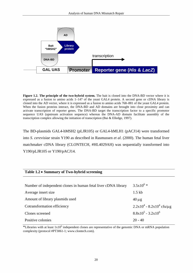
Analysis of human DNA Mismatch Repair
GAL UAS Promoter Reporter gene (His & LacZ)
transcription
Bait“hMSH2”
DNA-BD
Libraryprotein
AD
Figure 1.2. The principle of the two-hybrid system. The bait is cloned into the DNA-BD vector where it is expressed as a fusion to amino acids 1–147 of the yeast GAL4 protein. A second gene or cDNA library is cloned into the AD vector, where it is expressed as a fusion to amino acids 768–881 of the yeast GAL4 protein. When the fusion proteins interact, the DNA-BD and AD domains are brought into close proximity and can activate transcription of reporter genes. The DNA-BD target the transcription factor to a specific promotor sequence UAS (upstream activation sequence) whereas the DNA-AD domain facilitate assembly of the transcription complex allowing the initiation of transcription (Bai & Elledge, 1997). The BD-plasmids GAL4-hMSH2 (pLJR105) or GAL4-hMLH1 (pACJ14) were transformed
into S. cerevisiae strain Y190 as described in Rasmussen et al. (2000). The human fetal liver
matchmaker cDNA library (CLONTECH, #HL4029AH) was sequentially transformed into
Y190/pLJR105 or Y190/pACJ14.
Table 1.2 • Summary of Two-hybrid screening
Number of independent clones in human fetal liver cDNA library
3.5x10⁶ *
Average insert size 1.5 kb
Amount of library plasmids used 40 µg
Cotransformation efficiency 2.2x10⁴ - 8.2x10⁴ cfu/µg
Clones screened 8.8x10⁵ - 3.2x10⁶
Positive colonies 20 - 40
*Libraries with at least 1x10⁶ independent clones are representative of the genomic DNA or mRNA population complexity (protocol #PT3061-1; www.clontech.com).
20

Analysis of human DNA Mismatch Repair
Interactors were selected on synthetic dextrose minimal medium (SD) lacking tryptophan (to
maintain the GAL4 binding domain plasmids), leucine (to maintain the GAL4 activation
domain plasmids), and histidine (to identify peptides capable of assembling a functional
GAL4 transcription factor), and supplemented with 30 mM 3-amino-1, 2, 4-triazole (3-AT).
The plates were incubated at 30oC for 7-10 days and approximately 700 positive clones
(minimum size ∼ 1 mm) for each screen were picked from SD/-TRP/-LEU/-HIS + 3-AT
plates and screened for β-galactosidase activity to verify positive interactions (for more
method details see Rasmussen et al., 2000). The total number of positive interactions after β-
galactosidase screening was therefore only ∼ 30 positive clones per screen (table 1.2).
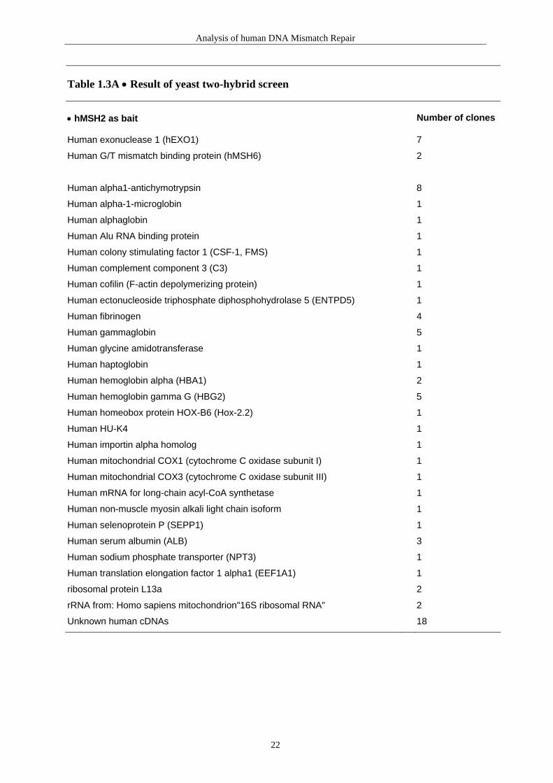
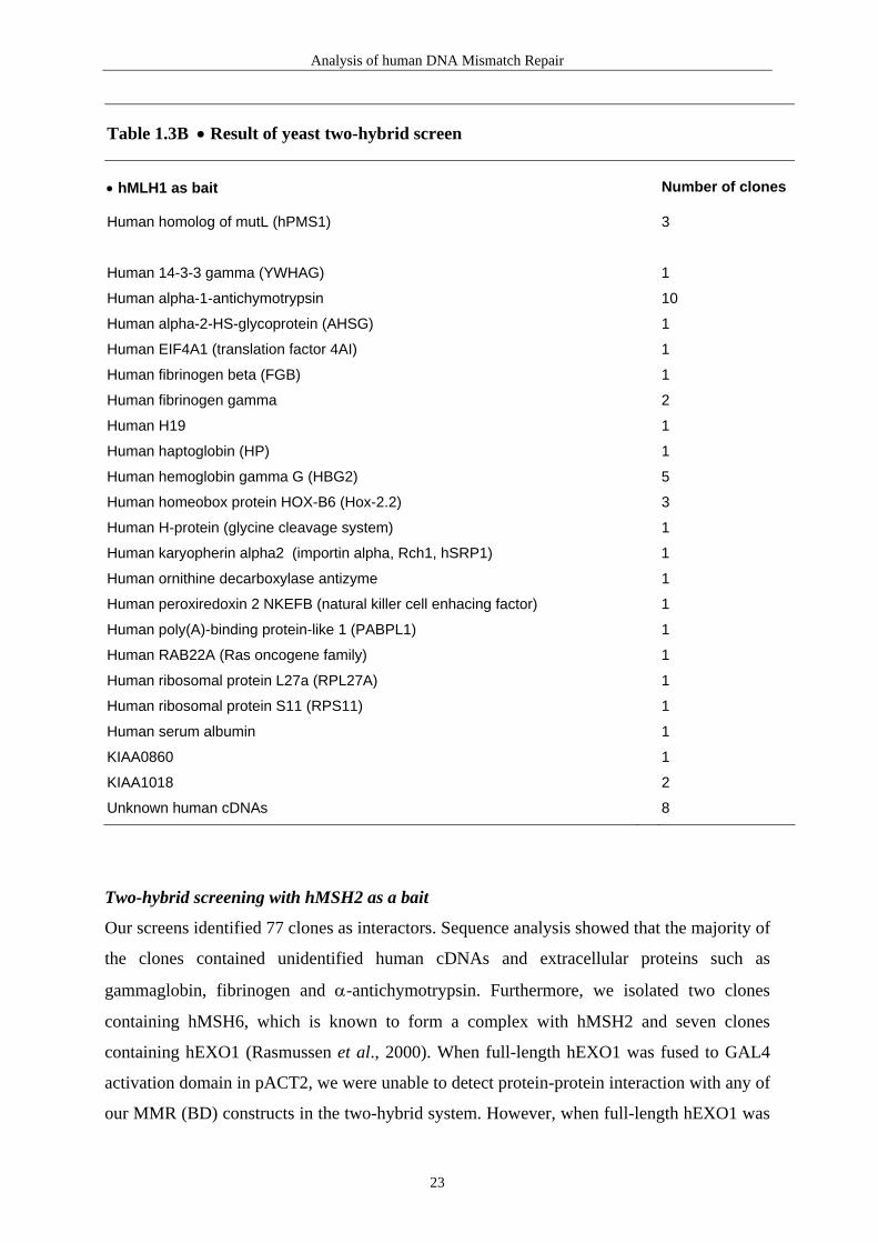
1.5 Results & Discussion Library screenings with BD-hMSH2 and BD-hMLH1 were done twice for each MMR
protein. The results presented in table 1.3A and 1.3B are a summary of four independent
screens. A hMSH2-hMSH6 binding domain vector was also constructed by inserting the
hMSH2 coding sequence into the SalI site of the pBridge binding domain vector
(CLONTECH). The human hMSH6 coding sequence was inserted into the NotI site. The
hMSH6 protein was in this way conditionally expressed from the PMet25 promoter. Because
hMSH2 binds to a mismatch DNA sequence in complex with hMSH6, one could expect
hMSH6 to work as a bridge protein that stabilizes a weak interaction between hMSH2 and
another MMR protein, or as a modifier of hMSH2 or another MMR protein. However, a
library screen using this construct gave no positive interactions.
21
Analysis of human DNA Mismatch Repair
Table 1.3A • Result of yeast two-hybrid screen • hMSH2 as bait
Number of clones
Human exonuclease 1 (hEXO1) 7
Human G/T mismatch binding protein (hMSH6) 2
Human alpha1-antichymotrypsin 8
Human alpha-1-microglobin 1
Human alphaglobin 1
Human Alu RNA binding protein 1
Human colony stimulating factor 1 (CSF-1, FMS) 1
Human complement component 3 (C3) 1
Human cofilin (F-actin depolymerizing protein) 1
Human ectonucleoside triphosphate diphosphohydrolase 5 (ENTPD5) 1
Human fibrinogen 4
Human gammaglobin 5
Human glycine amidotransferase 1
Human haptoglobin 1
Human hemoglobin alpha (HBA1) 2
Human hemoglobin gamma G (HBG2) 5
Human homeobox protein HOX-B6 (Hox-2.2) 1
Human HU-K4 1
Human importin alpha homolog 1
Human mitochondrial COX1 (cytochrome C oxidase subunit I) 1
Human mitochondrial COX3 (cytochrome C oxidase subunit III) 1
Human mRNA for long-chain acyl-CoA synthetase 1
Human non-muscle myosin alkali light chain isoform 1
Human selenoprotein P (SEPP1) 1
Human serum albumin (ALB) 3
Human sodium phosphate transporter (NPT3) 1
Human translation elongation factor 1 alpha1 (EEF1A1) 1
ribosomal protein L13a 2
rRNA from: Homo sapiens mitochondrion"16S ribosomal RNA" 2
Unknown human cDNAs 18
22
Analysis of human DNA Mismatch Repair
Table 1.3B • Result of yeast two-hybrid screen • hMLH1 as bait
Number of clones
Human homolog of mutL (hPMS1) 3
Human 14-3-3 gamma (YWHAG) 1
Human alpha-1-antichymotrypsin 10
Human alpha-2-HS-glycoprotein (AHSG) 1
Human EIF4A1 (translation factor 4AI) 1
Human fibrinogen beta (FGB) 1
Human fibrinogen gamma 2
Human H19 1
Human haptoglobin (HP) 1
Human hemoglobin gamma G (HBG2) 5
Human homeobox protein HOX-B6 (Hox-2.2) 3
Human H-protein (glycine cleavage system) 1
Human karyopherin alpha2 (importin alpha, Rch1, hSRP1) 1
Human ornithine decarboxylase antizyme 1
Human peroxiredoxin 2 NKEFB (natural killer cell enhacing factor) 1
Human poly(A)-binding protein-like 1 (PABPL1) 1
Human RAB22A (Ras oncogene family) 1
Human ribosomal protein L27a (RPL27A) 1
Human ribosomal protein S11 (RPS11) 1
Human serum albumin 1
KIAA0860 1
KIAA1018 2
Unknown human cDNAs 8
Two-hybrid screening with hMSH2 as a bait
Our screens identified 77 clones as interactors. Sequence analysis showed that the majority of
the clones contained unidentified human cDNAs and extracellular proteins such as
gammaglobin, fibrinogen and α-antichymotrypsin. Furthermore, we isolated two clones
containing hMSH6, which is known to form a complex with hMSH2 and seven clones
containing hEXO1 (Rasmussen et al., 2000). When full-length hEXO1 was fused to GAL4
activation domain in pACT2, we were unable to detect protein-protein interaction with any of
our MMR (BD) constructs in the two-hybrid system. However, when full-length hEXO1 was
23

Analysis of human DNA Mismatch Repair
fused to the GAL4 binding domain in pAS2, we could detect interactions with MMR (AD)
proteins in the two-hybrid assay. This could explain why we failed to isolate any full-length
cDNAs of hEXO1 in our two-hybrid screen (Rasmussen et al., 2000).
Three clones containing COX1, COX3 and 16S ribosomal RNA were shown to form a
complex with hMSH2. However, these interactions are probably artifacts as COX1 and
COX3 are subunits of cytochrome c oxidase (Complex III) of the electron transport chain. It
has not yet been determined if MMR is active in human mitochondria. In yeast a MutS
homolog (Msh1) of the MMR pathway has been identified in mitochondria (Reenan &
Kolodner, 1992). Our two-hybrid screenings with hMSH2 and hMLH1 did not indicate the
presence of mitochondria specific MMR proteins, although we can not exclude that they can
be found among the unidentified human cDNAs.
Two-hybrid screening with hMLH1 as a bait
The screening with hMLH1 was performed as described previously for hMSH2 (Rasmussen
et al., 2000). The GAL4 DNA-BD was fused to full-length hMLH1 and verified by
sequencing. Again the sequence analysis of the hMLH1 interactors showed that the majority
of the clones contained unidentified human cDNAs and proteins such as fibrinogen and α-
antichymotrypsin (table 1.3B). Furthermore, we isolated three clones containing hPMS1,
which is known to form a complex with hMLH1. To find hPMS1 as the interacting partner
with hMLH1 at first surprised us, as hPMS2 has been shown to be approximately 10-fold
more abundant in HeLa nuclear extract than hPMS1 (Räschle et al., 1999). However, the
same authors demonstrated that the affinity of hMLH1 for hPMS1, measured in the yeast
two-hybrid system, was greater than for hPMS2 (Räschle et al., 1999). Therefore, a greater
affinity for hPMS1 could explain why we only isolated this gene in our two-hybrid screen.
Given that hPMS2 might compete with hPMS1 for the available hMLH1, we decided to
determine the relative amounts of hPMS1 in human tissues. We used a human RNA master
blot (figure 1.3) and a human multiple tissue RNA master blot (figure 1.4) from CLONTECH
to characterize the expression pattern of hPMS1 (as described in Rasmussen et al., 2000). We
found that hPMS1 is predominantly expressed in fetal liver and adult liver, but also in
pancreas, kidney, testis and appendix (figure 1.3). The hMLH1-hPMS1 complex could
therefore predominate in fetal liver and account for why we isolate hPMS1 and not hPMS2
when using the two-hybrid screen. It is tempting to speculate that hPMS1 is tissue specific
and that the hMLH1-hPMS1 complex plays an important role in DNA repair in liver.
24

Analysis of human DNA Mismatch Repair
hPMS1 β-actin
1 2 3 4 5 6 7 8
Awholebrain
amygdala(brain)
caudatenucleus
cere-bellum
cerebralcortex
frontallobe
hippo-campus
medullaoblongata
Boccipital
lobeputamen substantia
nigratemporal
lobethalamus nucleus
accumbeusspinalcord
Cheart aorta skeletal
musclecolon bladder uterus prostate stomach
Dtestis ovary pancreas pituitary
glandadrenalgland
thyroidgland
salivarygland
mammarygland
Ekidney liver small
intestinespleen thymus peripheral
leukocytelymphnode
bonemarrow
Fappendix lung trachea placenta
Gfetalbrain
fetalheart
fetalkidney
fetalliver
fetalspleen
fetalthymus
fetallung
Figure 1.3. Expression profile of hPMS1. The RNA master blot (CLONTECH #7770-1) was hybridized with hPMS1 or β-actin control probes.
Kb
9.57.5
4.4
2.4
1.35
1 2 3 4 5 6 7 8 9 10 11 12
Figure 1.4. Hybridization of multiple tissue Northern blot with hPMS1 probe. Each lane contains following human tissues. Lane 1: brain. Lane 2: heart. Lane 3: skeletal muscle. Lane 4: colon. Lane 5: thymus. Lane 6: spleen. Lane 7: kidney. Lane 8: liver. Lane 9: small intestine. Lane 10: placenta. Lane 11: lung. Lane 12: peripheral blood leukocyte (CLONTECH #7780-1).
25

Analysis of human DNA Mismatch Repair
Importin α
In the hMSH2 and the hMLH1 two-hybrid screens we found interaction with an importin α
homolog and importin-α, respectively. Importin-α is also known as importin 58/Srp1/Rch1/
Kap60/karyopherin α (Jans et al., 1998).
All passive and active transport into and out of the nucleus occurs through the nuclear pore
complexes (NPCs) present in the nuclear envelope. Molecules smaller than approximately 60
kDa may passively diffuse through the NPCs into the nucleus, but import of larger molecules
requires specific transport signals (Görlich, 1998). Targeting of many proteins to the nucleus
is determined by the presence of a Nuclear Localization Signal (NLS), a short sequence
containing one or two clusters of basic amino acid residues. (Köhler et al., 1999). An NLS-
bearing protein, often termed the "cargo", is delivered to the nucleus by association with a
heterodimer, formed by importin α and importin β (also called karyopherin- α and -β).
Importin α recognizes the NLS, while importin β is responsible for docking to the NPC and
translocation through the pore (Görlich, 1998; Jans et al., 1998; 2000). Translocation into the
nucleus is terminated at the nuclear side of the NPC by disassembly of the trimeric NLS-
protein/importin α/β complex. Association of importin β with the protein Ran, a Ras-related
GTPase may trigger the dissociation (Jans et al., 2000).
Higher eukaryotes possess more than one form of importin-α (at least 6 forms for humans)
(Köhler et al., 1999). The larger number of distinct importin α isoforms in higher mammals
implies that there is specialization in their cellular role, and that different isoforms could bind
unique target proteins. Consistent with this is the fact that neither any single human importin-
α nor any triple combinations of three analyzed importin-α homologues were able to
complement a S. cerevisiae SRP1 mutant (S. cerevisiae has only one gene for importin-α)
(Nachury et al., 1998).
The hMSH2 human mismatch repair protein has a weak nuclear signal (Boulikas, 1997). A
protein processing a single weak NLS is more likely to be retained in the cytoplasm after a
mutation at the NLS peptide than a protein with two or more NLSs (Boulikas, 1997).
Mutations at the weak NLS of the hMSH2 gene, or dysfunction of translocation proteins like
importin-α may result in cytoplasmic retention that again could lead to dysfunction of the
MMR pathway. Hampered translocation of the MMR proteins could thus be a possible
explanation why so many HNPCC families fail to display mutations in the known MMR
genes. We hope to do more research regarding translocation of MMR proteins in the future.
26

Analysis of human DNA Mismatch Repair
Potential NLSs for MMR proteins are listed in table 1.4 by using the PSORT software
programs (Nakai et al., 1992; http://psort.nibb.ac.jp/).
Table 1.4 • Prediction of nuclear localization signal (NLS) MMR protein Potential NLSs Reference hMSH2
EKHEGKHQKLL at 422
(Boulikas et al., 1997)
hMSH3
RRKP at 3 RRKK at 76 RKKR at 77 KKRP at 78 KKRK at 704 KRKR at 1091 PVKKKVK at 87 PLIKKRK at 701 RRKKRPLENDGPVKKKV at 76 RKKRPLENDGPVKKKVK at 77 KKRPLENDGPVKKKVKK at 78 KRPLENDGPVKKKVKKV at 79
(PSORT; Nakai et al., 1992)
hMSH6 KKRR at 246 RKRK at 298 KRKR at 299 HRRR at 382 RRRP at 383 RKRKRMVTGNGSLKRKS at 298 KRKRMVTGNGSLKRKSS at 299 RKRMVTGNGSLKRKSSR at 300 KRMVTGNGSLKRKSSRK at 301
(PSORT; Nakai et al., 1992)
hMLH1 PRKR at 469 RKRH at 470 KRHR at 471 PRRR at 496 PRKRHRE at 469 PRRRIIN at 496
(PSORT; Nakai et al., 1992)
hPMS1 RPRK at 869 KRAIEQESQMSLKDGRK at 636
(PSORT; Nakai et al., 1992)
hPMS2 PNTKRFK at 574
(PSORT; Nakai et al., 1992)
hEXO1 KRPR at 418 KRKH at 775 PIKRKLI at 290
(PSORT; Nakai et al., 1992)
27

Repair of Mitochondrial DNA
2. Repair of Mitochondrial DNA
2.1 Introduction Within all mammalian cells there are two distinct genomes, one located in the nucleus
(nDNA) and the other in the mitochondria (mtDNA). Although each human somatic cell has
several hundred to thousand mitochondria (and 1-10 mtDNA copies per mitochondrion) the
amount of the mtDNA is only roughly 1% of the total DNA in the cell due to the small size
of mtDNA compared to nDNA (Bestwick, 1982; Wallace et al., 1998). The mammalian
mtDNA retains only 22 tRNAs, 12S rRNA and 16S rRNA genes necessary for the
mitochondrial protein synthesis as well as 13 polypeptide genes (Anderson et al., 1981). All
13 polypeptides are part of the 87 structural polypeptide subunits, all of which are
components of the respiratory chain. The respiratory chain is composed of five multisubunit
enzymes whose components are encoded by both the nuclear and mitochondrial genomes
(Wallace, 1999).
Unlike nDNA mammalian mtDNA contains very few noncoding sequences, no introns and it
is unprotected by histones. Therefore, damage to mtDNA can be expected to have greater
impact on cell function than damage to nuclear DNA, as the probability of damaging coding
sequences in mtDNA is much higher. Thus, it has been reported that the rate of point
mutations is higher in mtDNA compared to nDNA in human tissues (Khrapko et al., 1997).
In addition mtDNA point mutations and mtDNA rearrangement mutations (deletions and
insertions) have now been recognized to play a critical role in numerous human disorders
which prove the importance of maintaining the integrity of the mitochondrial genome (Kang,
1998; Kogelnik et al., 19982; Pulkes & Hanna, 2001; Wallace, 1999).
For this reason DNA repair in mitochondria could be expected to be very efficient.
However, early investigations of removal of UV-induced pyrimidine dimers in mtDNA led
to the conclusion that mitochondria accumulate DNA damage because these organelles are
deficient in repair activity (Clayton et al., 1974; Prakash et al., 1975). This finding, in
combination with very limited interest in this research area, led to the belief that rather than
repairing damage in mtDNA, cells simply destroyed the injured genomes and replaced
them by replicating existing, undamaged mtDNA
2Variations in the human mitochondrial genomes are updated on http://www.gen.emory.edu/mitomap.html (Kogelnik et al., 1998).
28

Repair of Mitochondrial DNA
(LeDoux et al., 1999). It has later been confirmed that UV-induced pyrimidine dimers are
not repaired in mitochondria (LeDoux et al., 1992; Pascucci et al., 1997), which suggest
absence of nucleotide excision repair. However, recent evidence showed that mitochondria
are indeed able to repair their genomes (Croteau et al., 1999; Marcelino & Thilly, 1999).
Unfortunately the knowledge about the repair mechanisms operating in the mitochondria is
still limited.
To gain a better understanding of the molecular processes and components responsible for
DNA repair in the human mitochondrion we have investigated the sub-cellular localization
of O6-methylguanine-DNA methyltransferase (MGMT) (Rasmussen et al., II).
2.2 Base Excision Repair Several observations support that mitochondria have Base Excision DNA Repair (BER)
pathways that are responsible for the removal of simple lesions in DNA (Croteau et al.,
1999; Marcelino & Thilly, 1999). Removal of a damaged base by the BER pathway is
initiated by DNA glycosylase that cleave the N-glycosylic bond between the base and the
deoxyribose-phosphate backbone (Lindahl & Wood, 1999). The resulting abasic site is
cleaved 5’ by an apurinic/apyrimidinic (AP) endonuclease that generates a 3’OH group,
which can be extended by a DNA polymerase, but not ligated before a 5’ terminal
deoxyribose-phosphate residue is removed. The removal of the abasic sugar residues is done
by a lyase. Finally, the gap is filled by DNA polymerase and the ends rejoined by DNA
ligase (Lindahl, 2000). Several distinct DNA glycosylases have been identified both in
nuclei and mitochondria in human cells (table 2.1).
Repair of uracil
Uracil-DNA Glycosylase (UNG or UDG) removes uracil bases in DNA. An uracil base in
DNA can occur as a result of either misincorporation of dUTP instead of dTTP into a newly
synthesized DNA, or deamination of cytosine to uracil (Slupphaug et al., 1995).
Deamination of cytosine to uracil, unless repaired before the next round of replication, will
result in a GC → AT transition mutation (Slupphaug et al., 1995). Studies have
demonstrated the presence of both nuclear- and mitochondrial-associated UNG (Anderson &
Friedberg, 1980; Slupphaug et al., 1993; Otterlei et al., 1998). The human mitochondrial
(hUNG1) and nuclear (hUNG2) forms have identical catalytic domains, but very different N-
29

Repair of Mitochondrial DNA
terminal sequences. The two forms are both generated from the hUNG gene using two
promoters, and making use of an exon specific for the N-terminal end of the nuclear form,
and alternative splicing (Nilsen et al., 1997; Otterlei et al., 1998).
Repair of oxidative damage
Both purine and pyrimidine residues in DNA are sensitive to reactive oxygen species (ROS)
(Croteau & Bohr, 1997). The most common purine lesion is 8-hydroxyguanine also called
7,8-dihydro-8-oxoguanine (8-oxoG) (Steenken & Jovanovic, 1997; Burrows & Muller,
1998). 8-oxoG is a highly mutagenic base derivative, which base-pairs with adenine as well
as cytosine, causing G → T transversion mutations (Cheng et al., 1992). The mitochondrial
respiratory chain produces superoxide (Wallace, 1999) which can be converted to hydroxyl
radicals via hydrogen peroxide (Imlay & Linn, 1988). The hydroxyl radical is the main
species of active oxygen that attacks the guanine base (Kasai et al., 1984). Because mtDNA
is subjected to a relatively high amount of oxidative damage and because the strand
containing 28 of the 37 mitochondrial genes is rich in guanine (Bianchi et al., 2001), it
seems that mitochondria would need efficient DNA repair mechanisms to remove oxidative
damage from its DNA.
Removal of 8-oxoguanine
An early study revealed that 8-oxoG can pair with all four normal bases (Kuchino et al.,
1987) but it was later shown that 8-oxoG perferentially pairs with C or A during in vitro
DNA synthesis (Shibutani et al., 1991). In E. coli two DNA glycosylases, encoded by mutM
(Fpg) and mutY genes, function to prevent mutagenesis by removing 8-oxoG paired with
cytosine and adenine (Bailly et al., 1989; Tchou et al., 1991; Michaels et al., 1992).
The human MutM homolog, hOGG1 (8-OxoGuanine DNA Glycosylase) excise 8-oxoG
preferentially when it is paired with C, followed by 8-oxoG:T and 8-oxoG:G, but it has no
detectable 8-oxoG:A-specific strand cleavage (Hazra et al., 1998). The glycosylase activity
of hOGG1 is accompanied with apurinic/apyrimidinic (AP) lyase that cleaves the AP site via
β-elimination (Hazra et al., 1998).
Human OGG1 localizes both to the nucleus and mitochondria (Takao et al., 1998; Nishioka
et al., 1999). Seven alternatively spliced forms of hOGG1 mRNAs have been identified
(Nishioka et al., 1999). All these splice forms of hOGG1 contain a putative mitochondrial
targeting signal (MTS) at the common N-terminal region. A nuclear localization signal
(NLS) was only found in the C-terminal end of hOGG1-1a (Nishioka et al., 1999). Human
30

Repair of Mitochondrial DNA
OGG1-1a, which has been found to have a weak MTS, localized predominantly to the
nucleus. When the NLS is deleted, the protein is targeted to mitochondria (Nishioka et al.,
1999). When a strong MTS is fused upstream to the hOGG1-1a gene it is selectively targeted
to the mitochondria (Dobson et al., 2000).
Interestingly, results from mice liver have revealed that mitochondrial 8-oxoG glycosylase
activity increased with age. In contrast no age-associated changes were found for nuclear 8-
oxoG glycosylase activity or mtUDG activity (de Souza-Pinto et al., 2001). This result
suggests that the mitochondrial OGG1 glycosylase is up-regulated during the aging process.
Removal of adenine paired with 8-oxoguanine
The Human MutY Homolog, hMYH has been shown to excise adenine mispaired with
guanine and 8-oxoG as well as 2-hydroxyadenine paired with 8-oxoG in double-stranded
oligonucleotides (Ohtsubo et al., 2000). This glycosylase has been detected in nucleus and
mitochondria (Takao et al., 1998; 1999; Ohtsubo et al., 2000; Boldogh et al., 2001).
Boldogh et al. (2001) found that the levels of hMYH in the nucleus was increased 3- to 4-
fold during progression of the cell cycle and reached maximum levels in S phase, suggesting
a cell cycle-dependent regulation of expression and/or subcellular targeting. However, there
was no evidence that the cytoplasmic or mitochondrial levels of hMYH decreased as nuclear
levels increased (Boldogh et al., 2001).
It has been suggested that 8-oxoG:C is more efficiently repaired in mitochondria than 8-
oxo:A. Miyako et al. (2000) detected inefficient cleavage of human mtDNA by the E. coli 8-
oxoG:C specific MutM protein but could report that human mtDNA was cleaved by the 8-
oxoG:A specific MutY protein, suggesting that 8-oxoG accumulates as an 8-oxoG:A pair but
not as an 8-oxoG:C pair.
Removal of oxidized pyrimidines
Oxidized pyrimidines, such as thymine glycol, 5-hydroxycytosine and 5,6-dihydrouracil
(DHU), are excised by the hNTH1 glycosylase (a homolog of E. coli endonuclease III, Nth)
(Ikeda et al., 1998; Takao et al., 1998; Lindahl & Wood, 1999). Thymine glycol is only
slightly mutagenic but it can block progression of both DNA and RNA polymerases
(Stierum et al., 1999). The DHU DNA lesion derives from cytosine by deamination. Its
opposite base should be guanine, but DHU is also able to mispair with adenine during DNA
replication. hNTH1 cleaves A or G opposite DHU with the same rate (Ikeda et al., 1998).
The human NTH1 has, similar to hOGG1, combined glycosylase and AP lyase activity and
31

Repair of Mitochondrial DNA
this enzyme is located to both nucleus and mitochondria (Tomkinson et al., 1990; Takao et
al., 1998; Mol et al., 1999).
Removal of 3-methyladenine
The N-methylpurine-DNA glycosylase, MPG (MDG, AAG, APNG) gene is coding for a
human glycosylase, which removes 3-methyladenine (3-MeA) as well as 3-methylguanine
(3-MeG), 7-methylguanine (7-MeG), N6-ethenoadenine, hydroxanthine, guanine, and 8-
oxoG (Pendlebury et al., 1994; Wyatt et al., 1999; Bouziane et al., 2000). The MPG activity
has so far only been reported from nuclear extracts.
Table 2.1 • Enzymes identified in repair of mitochondrial DNA in human cells Gene
Full Name
Substrates/Activity DNA glycosylases: major altered base released
AP lyase activity
Base excision repair (BER) hUNG1 Uracil-DNA glycosylase U no hOGG1 8-oxoguanine DNA glycosylase 1 8-oxoG opposite C yes hMYH MutY homolog (E.coli) A opposite 8-oxoG or G no hNTH1 Endonuclease three homolog 1(E.coli) T-glycol yes Other BER factors hMTH1 MutT homolog (E.coli) Hydrolyzes 8-oxo-dGTP to 8-oxo-dGMP hLIG3 DNA Ligase III Ligation hPOLG
Polymerase-gamma Replication/repair polymerase
Other BER factors
In cells, the deoxyribonucleotide pools are also subjected to oxidative damage. dGTP can be
converted to 8-oxo-dGTP and incorporated into nascent DNA strands opposite adenine. The
MutT enzyme in E. coli hydrolyzes 8-oxo-dGTP to 8-oxo-dGMP, and thereby prevents
misincorporation of the damaged base into DNA (Maki & Sekiguchi, 1992). The human
MutT homolog, hMTH1 (for mutT homologue) is present in the cytoplasm as well as in the
mitochondrial matrix (Kang et al., 1995; Nakabeppu, 2001). The hMTH1 gene produces by
alternative transcription inititation and splicing, seven different mRNAs. One of these
mRNA’s is imported into mitochondria when fused to green fluorescent protein, GFP
(Nakabeppu, 2001). The human MTH1 protein has wide substrate specificity as it has been
32

Repair of Mitochondrial DNA
reported to hydrolyze 8-oxo-dGTP, 8-oxoG, 8-oxo-dATP and 2-OH-dATP (Nakabeppu,
2001).
A major human AP endonuclease, hAPE1 (alternative titles; human apurinic endonuclease 1
(HAP1), apurinic/apyrimidinic exonuclease (APEX), apurinic/apyrimidinic endonuclease/
redox effector factor (APE/REF-1)) plays a central role in BER (Evans et al. 2000). It
initiates repair by hydrolyzing AP sites in DNA produced either spontaneously or after
removal of bases in DNA by DNA glycosylases (i.e. UNG). Alternatively, it can act as a 3'-
phosphoesterase after the AP lyase reaction of DNA glycosylases/AP lyases (i.e. hOGG1)
(Izumi et al., 2000). Takao et al. (1998) examined the subcellular localization of hAPE1 and
found that it was only localized to the nucleus (Takao et al., 1998). Furthermore, Prieto-
Alamo & Laval (1999) found no increased hAPE1 activity in the mitochondrial fraction of
Chinese hamster ovary (CHO-9) cells transfected with a plasmid expressing the human
APE1 compared to a 4.5 fold activity increase in nuclear fractions. However, the APE1
homolog in S. cerevisiae, the Apn1 enzyme has been shown to localize in both nucleus and
mitochondria. The Pir1 (a cell wall protein) is required for the localization of Apn1 to
mitochondria (Vongsamphanh et al., 2001). Results of two different studies have also shown
that APE1 is present in mitochondria of rat thyroid FRTL-5 cells and rat pleural mesothelial
cells (Fung et al., 1998; Tell et al., 2001). However, the contribution of the mitochondrial
AP lyase activity, to repair of abasic sites, and the processing of 3'-unsaturated sugar-
phosphate generated by e.i. hOGG1 AP lyase activity, still remain to be established in
human mitochondria.
Human mitochondrial DNA is replicated by polymerase γ. Polymerase γ (POLG) is a
heterodimer composed of a 140-kD catalytic subunit (POLG1) and a smaller accessory
subunit (POLG2) (Schmitt & Clayton, 1993). Because polymerase γ is the only known DNA
polymerase in human mitochondria, it is expected to participate in DNA replication and
repair in this organelle (Longley et al., 1998). The human polymerase γ has been shown to
posess both 3'→5' exonuclease proofreading activity and lyase activity (Schmitt & Clayton,
1993; Longley et al., 1998). The human polymerase γ fills single nucleotide gaps and
produces a substrate that can be ligated after action of uracil-DNA glycosylase and AP-
endonuclease (Longley et al., 1998).
33

Repair of Mitochondrial DNA
A mitochondrial DNA ligase has been identified. The human DNA ligase III gene encodes
both a nuclear and a mitochondrial protein and DNA ligase III plays an essential role in the
maintenance of mtDNA in mammalian cells (Lakshmipathy & Campbell, 1999; 2001).
Mismatch repair
The DNA mismatch repair (MMR) removes errors in the newly synthesized DNA strand that
are missed by the polymerase proofreading (see Analysis of human DNA Mismatch Repair).
It is not yet known if there is an active post-replication MMR system in human mitochondria
but it has been indicated that hMSH6 is not involved in repair of mismatches in the mtDNA.
Human hMSH6 deficient lymphoblastoid cells have a higher spontaneous mutation rate in
the nuclear genome but a similar spontaneous mutation rate in mitochondrial DNA when
compared to a parental mismatch repair proficient cell line (Marcelino et al., 1998;
Marcelino & Thilly, 1999).
In yeast, a component of the MMR pathway (Msh1) has been identified in mitochondria
(Reenan & Kolodner, 1992). The Msh1 protein is a homolog of the E. coli MutS mismatch
binding protein (Reenan & Kolodner, 1992). Inactivation of the MSH1 gene resulted in large
scale mtDNA rearrangements suggesting that Msh1 is involved in repair of mtDNA (Reenan
& Kolodner, 1992).
Direct repair
We have investigated the sub-cellular localization of O6-methylguanine-DNA methyl-
transferase (MGMT) in human mitochondria (Rasmussen et al., II). Therefore, the MGMT
protein will be described in greater detail in the next section.
2.3 Direct Repair: O6-Methylguanine-DNA Methyltransferase O6-methylguanine (O6-MeG) is generated endogenously by reactive cellular catabolites and
S-adenosylmethionine, which normally acts as a methyl donor in the synthesis of 5-
methylcytosine but occasionally causes methylation at other sites (Rossman & Goncharova,
1998; Lindahl & Wood, 1999).
Alkylating agents produce various kinds of alkylated purine and pyrimidine bases in DNA,
3-methyladenine (3-MeA), O6-methylguanine (O6-MeG) and O4-methylthymine (O4-MeT),
among other adducts (Lindahl & Sedgwick, 1988). 3-MeA has been shown to block DNA
replication (Larson et al., 1985) and is excised by 3-methyladenine glycosylase (MPG). O6-
34

Repair of Mitochondrial DNA
MeG and O4-MeT DNA lesions are mutagenic and carcinogenic (Loveless, 1969; Swann,
1990). However, O4-MeT is formed less frequently than O6-MeG. The proportion of
alkylation at each site is ~ 0.1 % for O4-MeT and 3-6% for O6-MeG of total alkylation after
reaction with Dimethylnitrosamine (DMN), Methylnitrosourea (MNU) or 1,2-
Dimethylhydrazine (SDMH) (Singer & Dosanjh, 1990).
During DNA replication, O6-MeG can pair with thymine leading to G:C → A:T transition
mutations while O4-MeT can pair with guanine, generating A:T → G:C transition mutations
(Singer & Dosanjh, 1990; Dosanjh et al., 1991; 1993, Altshuler et al., 1996). Both
prokaryotic and eukaryotic DNA alkyltransferases remove methyl groups from the O6
position of the guanine. cDNAs coding for the repair protein from E. coli (ada, Sedgwick,
1983; ogt, Potter et al., 1987), S. cerevisiae (MGT1, Xiao et al., 1991), mouse (Shiota et al.,
1992), rat (Rahden-Staron & Laval, 1991) and human (MGMT, Tano et al., 1990; Rydberg
et al., 1990; Hayakawa et al., 1990) have all been cloned and characterized. Furthermore,
DNA methyltransferase has been identified in 28 different species (Pegg, 2000). Although
the amino acid sequences of these proteins are homologous, there are only 8 absolutely
invariant residues in these sequences (Gly109, Asn137, Pro144, Cys145, His146, Arg147, Lys165,
and Glu172, numbered according to the human MGMT sequence) (Xu-Welliver et al., 2000).
MGMT function
Human O6-MethylGuanine-DNA MethylTransferase (MGMT, also known as AGT) repairs
O6-alkylguanine (O6-MeG, O6-ethylguanine & O6-butylguanine) in double-stranded DNA in
a single step. MGMT removes alkyl groups from the O6 position of guanine to a cysteine
acceptor residue in MGMT without removing the damaged base (Pegg, 2000). The cysteine
residue is located at position Cys145 in MGMT in a highly conserved sequence -PCHR-
(Pegg, 2000). It has been shown that mutation of the cysteine acceptor site leads to a
complete loss of MGMT activity (Harris et al., 1992; Crone & Pegg, 1993; Hazra et al.,
1997; Edara et al., 1999). Once bound to the cysteine acceptor site, the alkyl group
permanently inactivates MGMT. Therefore, the number of O6-alkylG that can be repaired is
equal to the number of active MGMT molecules – MGMT is “suicidal” in the repair of O6-
alkylG residues in DNA (Pegg, 1990; 2000). Even though MGMT is not specific for methyl
groups, the rate of the reaction decreases as the size of the adduct increases (Pegg & Byers,
1992).
Mutational analysis of the MGMT protein showed that deletion of 7 amino acids from the N
terminus (codons 1-7) or 28 amino acids from the C terminus (codons 180-207) result in
35

Repair of Mitochondrial DNA
specific activity comparable to that found for the full-length MGMT (Hazra et al., 1997).
Mutant MGMT with deletion of codon 1-10 or 1-19 were shown to be active in protecting
cells from a methylating agent N-methyl-N’-nitro-N-nitrosoguanidine (MNNG) suggesting
that they are able to repair methylated DNA in vivo (Crone et al., 1996). However, the
mutant MGMT proteins were less effective compared to the wild-type MGMT and highly
unstable with half-lives of 48-90 min compared to a half-live of > 720 min for wild-type
MGMT. Interestingly, we found that deletion of codons 2 to 18 resulted in a complete
absence of MGMT in nucleus and accumulation in cytoplasm (Rasmussen et al., II). This
suggests that deletion of the first 18 codons interfere with stability and translocation to
nucleus. However, our results indicate that this mutant MGMT protein provide the same
level of protection to killing by MNNG as MGMT wild-type (Rasmussen et al., II). It has
been found by site-directed mutagenesis that the fully conserved residues Asn137, His146,
Arg147 and Glu172 are critical for maintaining the MGMT stability. Proteins with mutations in
these residues were highly unstable with half-lives of 15-60 min (Crone et al., 1996).
Furthermore, the residues Arg128 and Tyr114 seemed to be involved in DNA binding and
catalytic activity (Kanugula et al., 1995; Goodtzova et al., 1998).
Repair of O4-methylthymine
Although E. coli Ogt has been shown to repair O4-MeT it is still not fully established if the
human MGMT can repair O4-MeT (Sassanfar et al., 1991). The rate constants for MGMT
acting on O6-MeG is much higher than O4-MeT (O6-MeG = 1x10⁹ M-1min-1 and O4-MeT =
1.8x10⁵ M-1min-1 (Zak et al., 1994). Repair of O6-MeG by MGMT in vivo is 1000-5000-fold
greater than for O4-MeT (Samson et al., 1997; Encell & Loeb, 2000). However, one study
has shown that human MGMT expressed in E. coli suppresses MNNG-induced A:T → G:C
mutations indicating that MGMT repairs O4-MeT DNA lesions (Kooistra et al., 1999). Other
studies showed that expression of MGMT in a methyltransferase-deficient E. coli strain
(lacking Ade and Ogt) failed to suppress A:T → G:C mutations, but actually increased O4-
MeT mutations (Samson et al., 1997, Edara et al., 1999). Samson et al. (1997) suggested
that MGMT binds to O4-MeT lesions but repairs it so slowly that it interferes with the repair
of this lesion by NER and consequently increases the frequency of A:T → G:C mutations.
The mammalian NER pathway has been shown to repair both O6-MeG and O4-MeT lesions
(Bronstein et al., 1991; 1992, Huang et al., 1994, Kein et al., 1994). However, the ability to
serve as substrates for NER increases with the size of adduct and is poor for methyl groups
36

Repair of Mitochondrial DNA
(Pegg & Byers, 1992). It is still unknown whether NER is the predominant pathway for
removal of O4-MeT lesions in human cells or serves as a back-up repair system for removing
O6-MeG lesions in cells with limited MGMT activity.
The methylation tolerant phenotype
Even though MGMT is widely expressed, the intracellular MGMT levels vary between
different tissues and also between different individuals. The liver and spleen have the highest
MGMT activities and the nervous system and mammary gland the lowest (Myrnes et al.,
1983; Yarosh et al., 1985; Pegg & Byers, 1992; Karran & Bignami, 1994). Most tumor cells
express high level of MGMT, while about 15-20% of human tumor cell lines are deficient in
MGMT expression (Sklar et al., 1981; Tsujimura et al., 1987; Yu et al., 1999). Cells lacking
MGMT activity (Mex- or Mer- cells) are more sensitive to mutagenesis and killing by
alkylating agents (Karran & Bignami, 1994).
Alkylating agents were among the first cytotoxic drugs used in the treatment of cancer and
are still used clinically against a number of tumors (Kleibl & Margison, 1998). A class of
anti-tumor alkylating agents as the chloroethylnitrosourea (CENU) is clinically used to treat
a number of tumors, including brain neoplasms, malignant melanoma, advanced lymphomas,
and gastrointestinal carcinomas (Kreklau et al., 1999). However, certain tumors are resistant
to alkylating chemotherapeutic agents and this resistance are known to be correlated with the
level of MGMT in the tumors (Pegg & Byers, 1992). Another mechanism of cellular
resistance to methylating agents is an ability to ignore the presence of O6-MeG lesions in
DNA. This tolerance of DNA damage is due to the loss of MMR, even if MGMT is not
expressed or expressed at low levels (Carethers et al., 1996; Karran & Hampson, 1996;
Hampson et al., 1997; Umar et al., 1997; Dosch et al., 1998; Kawate et al., 1998, Humbert
et al., 1999).
There is increasing evidence that tumor cells could acquire resistance to methylating drugs
by loss of MMR activity. A lack of MMR results in poor ability of the cell to detect DNA
damage and activate apoptosis, thus increasing the mutation rate throughout the genome
(Fink et al., 1998). This implies that genotoxic and cytotoxic effects of O6-MeG are
mediated by MMR. In vitro studies have demonstrated that the mammalian hMSH2-hMSH6
complex can recognize O6-MeG: T and O6-MeG: C (Duckett et al., 1996; Ceccotti et al.,
1996; Mu et al., 1997; Christmann & Kaina, 2000). Although, the mechanisms of MMR-
mediated response to base damage are not known, two models have been proposed as
explanations.
37

Repair of Mitochondrial DNA
1. In one model, the MMR system recognizes the mismatched bases opposite the damaged
base but since there is no perfect complementary match for O6-MeG, the polymerase will
again fail to find an ideal partner and incorporate thymine or cytosine opposite O6-MeG
on the newly synthesized strand. The repeated attempts to repair the mismatch will result
in a methylated template strand that is single-stranded for much of the time and a newly
synthesized strand that contains persisting DNA termini. The killing effect of the lesion
is expected to come during the following cycle of DNA replication, when the replication
fork arrives at a discontinuity and thus generates a double-strand break that will then
trigger apoptosis (Galloway et al., 1995; Karran & Hampson, 1996; Kaina et al., 1997).
2. In the second model, MMR proteins function as the sensors of DNA damage, and
generate a signal capable of activating apoptosis (Modrich, 1997; Fink et al., 1998).
The p53 protein functions as a transcription factor that regulates the expression of several
genes involved in cell cycle control, DNA damage repair, and apoptosis. Therefore, p53 is
the classic signal transduction molecule thought to respond to DNA breaks and gaps (Nelson
& Kastan, 1994; Huang et al., 1996; Vogelstein et al., 2000). However, MNNG induced
apoptosis seems to be p53-independent (Hickman & Samson, 1999). Moreover, Christmann
& Kaina (2000) have shown that after exposure of cells to MNNG, hMSH2, hMSH6 and
hPMS2 proteins are translocated from the cytoplasm into the nucleus, leading to an increased
nuclear MMR protein level immediately after exposure. This translocation was independent
of the p53 status of the cells and was first observed at high dosage level of the mutagen in
cells expressing MGMT (>25 µM MNNG; 750 fmol/mg protein MGMT; Christmann &
Kaina, 2000). In order to explore a possible link between p53 and MGMT we expressed the
MGMT-GFP fusion protein in cell lines with different p53 status. One cell line was wild
type for p53, one expressed very low levels of the p53 protein, and one cell line was
completely deficient for p53 activity. We found no difference in subcellular localization of
MGMT suggesting that MGMT translocation is independent of the p53 status. In all three
cell lines the MGMT-GFP fusion protein was exclusively present in the nucleus (Rasmussen
et al. II).
Treatment of human cells with MNNG concentrations that increased the mutation rate of
nuclear genes showed no induction of mitochondrial mutations (Mita et al., 1988).
Marcelino et al. (1998) observed that to induce MNNG mutations above a very high
spontaneous background in mtDNA, one needed to treat MNNG-resistant (MT1) cells with
an amount of MNNG (4 µM) that would kill all normal parental cells. Therefore, they
38

Repair of Mitochondrial DNA
concluded that it is unlikely that in vivo mutations are induced in mtDNA by exogenous
mutagens in normal human cells (Khrapko et al., 1997; Marcelino et al., 1998).
Mitochondrial MGMT activity
Two groups have reported mitochondrial MGMT activity in rat liver (Myers et al., 1988;
Satoh et al., 1988). The kinetics of removal of O6-MeG was similar in nuclear and
mitochondrial DNA (Myers et al., 1988). Furthermore, they found very slow removal of O6-
butyl-2'-deoxyguanine in mtDNA but fast removal in nDNA which is consistent with the
absence of NER in mitochondria (Myers et al., 1988). Satoh et al. (1988) reported the
removal of O6-ethyl-2'-deoxyguanosine (O6-EtG) from mtDNA and nDNA in rat liver
following exposure to N-ethyl-N-nitrosourea in vivo. Longer alkyl groups including ethyl- is
known to be repaired by MGMT and NER (Pegg, 2000), indicating, that O6-EtG is removed
by an alkyltransferase mechanism in the mitochondria. Additionally, N-methyl-N-nitrosourea
(MNU) induced mutations were repaired in mitochondria from Chinese hamster ovary
(CHO) cells (LeDoux et al., 1992).
These results obtained in rodents suggest that an alkyltransferase mechanism is operating
within the mitochondrion in mammalian species. However, extrapolation of results obtained
with rodents to human cells may not be straightforward. Even MGMTs from mammalian
species have different substrate specificity. For eksample rat MGMT repairs O6-EtG much
better than human MGMT (Liem et al., 1994). Mitochondrial MGMT activity has not yet
been reported from human cells and the intracellular localization seems ambiguous.
MGMTs intracellular localization
Most immunostaining data have shown that MGMT is a nuclear protein (Ayi et al., 1992,
Lim & Li, 1996; Belanich et al., 1996). However, there are reports on cytosolic appearance
indicating that MGMT could be present in nucleus, cytoplasm and mitochondria (Ayi et al.,
1992; Ishibashi et al., 1994a,b; Belanich et al., 1996). To access the actual localization of
the MGMT protein we studied exogenous MGMT-GFP expressed as fusion protein in a
human breast epithelial cell line (MCF12A) and detected the subcellular localization by
fluorescence microscopy. We found that MGMT localized mainly in the nucleus. We also
observed a weak cytoplasmic staining but no mitochondrial localization. Our results
therefore suggest that MGMT is not present in mitochondria of human cells (Rasmussen et
al., II). In order to test whether nuclear MGMT was recruited to mitochondria upon DNA
damage of mtDNA by alkylating agents we treated cells expressing MGMT-GFP with
39
Repair of Mitochondrial DNA
MNNG. The MGMT-GFP fusion protein was detected in nucleus but not in mitochondria
indicating that nuclear MGMT is not recruited to mitochondria after DNA damage by
MNNG (Rasmussen et al., II). This suggests that the alkyltransferase mechanism which
seems to be operate within the mitochondrion in mammalian species is not MGMT in human
breast epithelial cells.
40
Mitochondrial Dysfuntion versus Genetic Stability of the nuclear DNA
3. Mitochondrial Dysfunction versus Genetic Stability of the Nuclear DNA
3.1 Introduction Mitochondrial diseases are severely debilitating and characteristically complex in nature.
They can affect any organ in the body at any age. Usually the mitochondrial diseases are
inherited through the mother but they can also be sporadic or induced by the environment.
Mitochondrial dysfunction is found in diseases as diverse as cancer, infertility, diabetes,
heart diseases, blindness, deafness, kidney disease, liver disease, stroke, and migraine.
Mitochondrial dysfunction is also involved in aging and neurodegenerative diseases such as
Parkinson and Alzheimer dementia (Wallace, 1999; DiMauro & Schon, 2001). To date,
there is no cure for mitochondrial diseases.
We used Saccharomyces cerevisiae as a model system to explore the role of mitochondrial
dysfunction on genetic stability of the nuclear DNA (Rasmussen et al., III) as well as the role
of mitochondrial activity in oxidative DNA damage and repair of the nuclear genome
(Rasmussen et al., IV)
3.2 Mitochondrial Biology and Genetics The mitochondria are surrounded by a double membrane, a smooth outer membrane, and a
"highly folded" inner membrane (termed cristae). Depending on source and conformational
state, cristae can vary from simple tubular structures to more complex lamellar structures
that are connected to the inner boundary membrane (Frey & Mannella, 2000; Perkins &
Frey, 2000). The inner membrane of the mitochondrion contains the proteins and enzymes of
the electron transport chain (Complexes I-IV) and Complex V that are responsible for
phosphorylation of ADP to ATP (adenosine diphosphate to adenosine trisphosphate). The
inner compartment is called the matrix, which contains the many enzymes of Krebs cycle
(the citric acid cycle), the pathway of fatty acid oxidation, and the mitochondrial DNA
(Scheffler, 1999).
In mammalians, electron transfer from NADH donated to Complex I (NADH dehydrogenase
or NADH:ubiquinone oxidoreductase) or electrons from succinate (donated to Complex II,
succinate dehydrogenase) are transfered to coenzyme Q, also called ubiquinone or CoQ.
41

Mitochondrial Dysfuntion versus Genetic Stability of the nuclear DNA
Regardless of whether CoQ receives its electrons from Complex I or II, it shuttles them to
Complex III (ubiquinol:cytochome c oxidoreductase). From Complex III the electrons move
through cytochrome c and Complex IV (cytochrome c oxidase or COX), which passes the
electrons directly to oxygen to form water. As electrons transverse complex I, III, and IV,
protons (H+) are moved from the mitochondrial matrix across the inner membrane into the
inter-membrane space. This creates an electrochemical proton gradient (∆Ψ) that is positive
and acidic on the outside and negative and alkaline on the mitochondrial matrix side. The
proton gradient across the inner-membrane is used to drive the condensation of ADP and Pi
(inorganic phosphate) to generate ATP by allowing the protons to flow back through
Complex V (ATP synthase or F1F0ATPase). Under normal circumstances, the inner
membrane is impermeable to protons, leaving Complex V as the only route whereby protons
can return to the matrix (figure 3.1). The ATP is exchanged for ADP across the
mitochondrial inner membrane by the Adenine Nucleotide Translocator, ANT (Wallace,
1999; Saraste, 1999). In fungi, Complex I is very similar to its counterpart in other
eukaryotes except for S. cerevisiae, Saccharomyces carlsbergii and Kluyveromyces lactis
which do not contain Complex I.
The mammalian Complex I is a NADH dehydrogenase involved in the oxidation of intra-
mitochondrial NADH produced by the citric acid cycle, and its NADH-binding site faces the
mitochondrial matrix. S. cerevisiae has three NADH dehydrogenases; Nde1 and Nde2 which
both face the intermembrane space (referred to as external) and Ndi1 an internal NADH
dehydrogenase like the mammalian Complex I (Joseph-Horne et al., 2001). Ndi1 is believed
to be attached to the inner membranes on the matrix side (de Vries & Grivell, 1988; de Vries
et al., 1992). It has been shown that when Ndi1 was expressed in a Complex I-deficient
Chinese hamster cell, it restored the capacity for respiration and oxidative phosphorylation in
these cells (Seo et al., 1998). Furthermore Ndi1 was incorporated into mitochondria in
human embryonic kidney cells where Complex I was still present. Overexpression of Ndi1 in
cells led to decreased coupling of NADH oxidation to ATP synthesis (ADP/Oxygen
consumption ratio down from 2.4 to 1.8), while succinate oxidation was unchanged. These
results suggest that the Ndi1 protein from S. cerevisiae complements the mammalian
Complex I consisting of 43 peptides (Wallace, 1999). However, Ndi1 does not translocate
protons, but only feeds electrons to the electron transport chain (Joseph-Horne et al., 2001).
Apart from the lack of Complex I in S. cerevisiae the electron transport and proton transfer
proceed via the same complexes as in mammalians (figure 3.1)
42

Mitochondrial Dysfuntion versus Genetic Stability of the nuclear DNA
3.3 Reactive Oxygen Species Mitochondria generate more than 80% of cellular energy in the form of ATP and are
therefore regarded as the "powerhouse" of the cell (Kang et al., 1998). However,
mitochondrial respiration is also the major endogenous source of Reactive Oxygen Species
(ROS), including superoxide radical (O2-), hydrogen peroxide (H2O2) and the hydroxyl
radical (HO•) (Cadenas & Davies, 2000). Under normal physiological conditions, in vitro
evidence indicates that electrons "leak" from the electron transport chain, converting about
1-2% of oxygen molecules into O2-, a more reactive form of oxygen (Boveris & Chance
1973; Boveris, 1977; Papa, 1996; Loft & Poulsen, 1996). Research in ROS production from
mitochondria indicate that Complex I (Robinson, 1998) and Complex III (Turrens et al.,
1985; Nohl & Jordan 1986) play a major role in production of superoxides however O2- may
also arise from Complex II (Ishii et al., 1998) in mammalian cells. Under normal metabolic
conditions, Complex III seems to be the main site of O2- production (Finkel, 2001).
Superoxide itself can attack enzymes containing the catalytically iron-sulfur [4Fe-4S]
tetranuclear cluster, such as Complex I, Complex II and the Krebs cycle enzyme aconitase,
resulting in release of iron and decreased mitochondrial ATP production (Flint et al., 1993;
Gardner, 1995, Melov et al., 1999; Cadenas & Davies, 2000). Hence, mitochondria are
particularly sensitive to oxidative stress. As we are using S. cerevisiae as model system it
should be noted that the Ndi1 is a polypeptide enzyme with no iron-sulfur cluster and
therefore not attacked by O2- (de Vries & Grivell, 1988).
ROS is reduced by intracellular antioxidant enzymes including superoxide dismutase,
glutathione peroxidase and catalase. Superoxide is simultaneously reduced and oxidized
(dismutated) to form hydrogen peroxide and oxygen by superoxide dismutase (SOD).
Eukaryotes, including S. cerevisiae, hold a manganese containing superoxide dismutase
(MnSOD, product of the SOD2 gene) in the matrix of the mitochondria, and a copper-and
zinc containing form (CuZnSOD, product of the SOD1 gene) in the nuclear and cytoplasmic
matrixes. Further, mammalians have an extracellular CuZn superoxide dismutase (SOD3)
(Crapo et al., 1992; Lindenau et al., 2000; Srinivasan et al., 2000).
43

Mitochondrial Dysfuntion versus Genetic Stability of the nuclear DNA
II III IV VNADH-
dehydrogenaseSuccinate
dehydrogenasecomplex
Cytochrome bc1Complex
Cytochrome coxidase complex
ATP synthasecomplex
“I”II
III IV V
O2
e- e-
O2- H2O2
H2O
HO•Fe2+SOD2
GPx Oxidative DamageLipid Peroxidation
2GSH
GR
GSSG
CoQ
Figure 3.1. Mitochondrial respiratory chain and reactions involved in removal of ROS in mitochondria The respiratory enzyme complexes involved in oxidative phosphorylation are NADH-dehydrogenase (Complex I in mammalian and Ndi1 in S. cerevisiae); succinate dehydrogenase (Complex II); ubiquinol:cytochrome c oxidoreductase (Complex III); cytochrome c oxidase (Complex IV); ATP synthase (Complex V) in both mammalian and S. cerevisiae. Complex I and II in mammalian cells and Complex II in S. cerevisiae contains 4Fe-4S centers. When the respiratory chain is inhibited, the electrons accumulate in the early stages of the respiratory chain (Complex I and coenzyme Q), were they can be donated to molecular oxygen to give superoxide. Superoxide (O2
-) is converted to hydrogen peroxide (H2O2) by mitochondrial superoxide dismutase (SOD2). H2O2 is converted to water (H2O) by glutathione peroxide (GPx1 in mammalian). Ferrous ions (Fe2+), released e.i. during O2
- attack of enzymes with 4Fe-4S centers, can participate in a Fenton reaction and convert H2O2 into hydroxyl radical (HO•) (Wallace, 1999; Joseph-Horne et al., 2001). GSH (reduced glutathione); GR (glutathione reductase); GSSG (oxidized glutathione).
Hydrogen peroxide produced by SOD2 in the mitochondria is reduced to water (H2O) by
glutathione peroxidase (GPx), in a reaction that converts reduced glutathione (GSH) to
oxidized glutathione (GSSG) (figure 3.1). Five GPx isoenzymes have been found in
mammalian cells. GPx1, which are located in the mitochondria and cytosol and Gpx4 (or
PHGPx, a phospholipid hydroperoxide) are found in most tissues. Cytosolic Gpx2 and
extracellular Gpx3 are poorly detected in most tissues except for the gastrointestinal tract
and kidney, respectively (de Haan et al., 1998). Hydrogen peroxide can also be reduced to
44

Mitochondrial Dysfuntion versus Genetic Stability of the nuclear DNA
H2O by catalase (CAT), located primarily in peroxisomes (especially in liver) and in
cytoplasm (erythrocyte) (Bai & Cederbaum, 2001). Catalase has only been found in
mitochondria of rat heart (Radi et al., 1991). In the presence of transition metal ions (Fe2+,
Cu+) hydrogen peroxide can be converted by the Fenton reaction to the highly damaging
hydroxyl radical, which can cause degradation of most biological macromolecules, e.g.
peroxidation of lipids, oxidation of sugars and of protein thiols, DNA base damage, and
strand breakage of nucleic acids (Bai et al., 1999).
3.4 Effects of Mitochondrial ROS Production Inhibition of mitochondrial oxidative phosphorylation
Inhibition of mitochondrial oxidative phosphorylation in mice not only reduces energy
production, but also increases mitochondrial ROS production. Mitochondria from normal
mice have an increased H2O2 production after antimycin A inhibition (Esposito et al.,
1999). Antimycin A is not a direct producer of H2O2 but an inhibitor of Complex III. Thus,
the inhibition results in a substantial increase in superoxide production (Raha et al., 2000)
which is converted into H2O2 by SOD2.
Also, mitochondria from adenine nucleotide translocator Ant1 (-/-) mice were shown to
have the same high level of H2O2 as normal mitochondria treated with antimycin A. This
suggests that the absence of ANT1 blocks the exchange of ADP and ATP across the
mitochondrial inner membrane, thus inhibiting oxidative phosphorylation which is
resulting in increased oxidative stress. Furthermore, tissues with high oxidative stress
(skeletal muscle and heart) were showed to increase the level of ROS detoxification
enzymes, SOD2 and GPx protein in Ant1 (-/-) mice. Although the antioxidant activities
were increased, a very high level of mtDNA rearrangement was observed in the Ant1 (-/-)
mice heart but not in skeletal muscle. It was suggested that the different levels of mtDNA
rearrangement in the two tissues were a result of lower induction of SOD2 in the heart than
in skeletal muscle (Esposito et al., 1999).
Cytosolic superoxide dismutase (sod1) mutants
By analyzing null SOD1 and SOD2 mutations in different species it has been suggested
that ROS could be involved in limiting life span. Yeast cells deficient in the SOD1 gene are
viable but grow poorly in oxygen and the mutation frequencies are increased in nDNA in
sod1∆ mutant cells compared to the wild type parental strain (Gralla & Valentine, 1991;
45

Mitochondrial Dysfuntion versus Genetic Stability of the nuclear DNA
Longo et al., 1997, Rasmussen et al., IV). It has been shown that SOD1 activity in motor-
neurons is an important factor in ageing and lifespan of Drosophila (Parkes et al., 1998).
Sod1 null mutants of Drosophila have a very short lifespan (the adult lifespan is shortened
by 85-95%). Overexpression of human SOD1 in motor neurons of a wild type Drosophila
extended the adult life span by up to 40% (Parkes et al., 1998).
Mutations in the SOD1 gene are also linked to Familial Amyotrophic Lateral Sclerosis
(FALS) a disorder resulting from degeneration and death of motor neurons. 15-20% of
FALS patients have been found to harbour mutations in SOD1. Transgenic mice that
express a mutant SOD1 allele develop a motor neuron degeneration that parallels most
aspects of ALS (Gurney et al., 1994; Bruijn et al., 1997). Whereas, knockout mice
deficient in the entire SOD1 gene live to adulthood and show no overt motor defect
(Reaume et al., 1996). This result demonstrates that the loss of SOD1 function is not, by
itself, sufficient to kill motor neurons in vivo. It has been suggested that FALS is a
consequence of reduction in SOD1 dismutation activity leading to oxidative damage (loss-
of-function) or a gain-of-function as the mutant SOD1 protein seems to increase the ability
to generate hydroxyl radicals (Yim et al., 1996; Bogdanov et al., 1998; Liu et al., 1999).
The results obtained with SOD1-/- knockout mice suggest that FALS SOD1 acquires or
enhances toxic property.
Mitochondrial superoxide dismutase (sod2) mutants
Yeast sod2∆ mutants have a less dramatic phenotype than sod1∆ mutants when grown in
glucose media although sod2∆ mutants are oxygen-sensitive and grow poorly in carbon
sources that require respiration for their metabolism (Liu et al., 1992; Longo et al., 1996).
In contrast to sod2∆ mutants in yeast, an acute toxicity of mitochondrial O2- has been
found in SOD2-/- mice. While mice, deficient in cytosolic SOD1 activity were viable
(Reaume et al., 1996), inactivation of the mitochondrial SOD2 was lethal early in life. Two
models of SOD2 knockout mice have been reported. SOD2tm1Cje mutant mice in which
exon 3 has been deleted (Li et al., 1995) and SOD2m1BCM mutants which are missing exon
1 and 2 (Lebovitz et al., 1996). The homozygous SOD2 tm1Cje mutant mice die within the
first 10 days of life with a dilated cardiomyopathy, accumulation of lipid in liver and
skeletal muscle, and metabolic acidosis (Li et al., 1995). The homozygous SOD2m1BCM
mutant mice survived up to 18 days and showed motor disturbance, central nervous system
46

Mitochondrial Dysfuntion versus Genetic Stability of the nuclear DNA
injury, and extensive mitochondrial injury within cells with requirements of high levels of
oxidative metabolism, including cardiac myocytes, neurons, hepatocytes, and
hematopoietic cells (Lebovitz et al., 1996).
In the SOD2 tm1Cje (-/-) mutant mice, the mitochondria's O2- toxicity was shown to result in
dramatic reductions in the activities of mitochondrial enzymes containing iron-sulfur
centers, including the Krebs cycle enzyme aconitase and respiratory chain enzymes of
Complex I and II. Hence, the dilated cardiomyopathy observed in SOD2 (-/-) mice (Li et
al., 1995) was caused by blocking of the Krebs cycle and respiratory chain giving energy
starvation in the heart (Melov et al., 1999). The increased mitochondrial oxidative stress of
the SOD2 (-/-) mice also resulted in high levels of oxidative damage to DNA. However, it
was not determined whether it was nDNA, mtDNA damage or both (Melov et al., 1999).
Measurement of the lipid peroxidation levels in heterozygous SOD2 tm1Cje (+/-) mutants and
SOD2 (+/+) control mice revealed that young SOD2 (+/-) and control animals had the same
levels of lipid peroxidation in the liver mitochondria. However, lipid peroxidation peaked
for SOD2 (+/-) at middle age and declined in old SOD2 (+/-) animals, whereas lipid
peroxidation of SOD2 (+/+) mice first peaked at old age (Kokoszka et al., 2001). Apoptosis
seemed to be able to explain the loss of mitochondrial lipid peroxidation from middle-age
to old age in SOD2 (+/-) mice. TUNEL staining showed that old SOD2 (+/-) mouse livers
had three times more apoptotic hepatocytes than old SOD2 (+/-) mouse livers. It was
therefore suggested that cells with a high number of damaged mitochondria was destructed
by apoptosis (Kokoszka et al., 2001).
It has also been demonstrated that Caenorhabditis elegans life span was increased 54% by
treatment with SOD/catalase mimetics. In addition, treatment with these compounds could
restore a mev-1 mutant life span to normal. The mev-1 gene, encodes the cytochrome b
subunit of succinate dehydrogenase (Complex II) and the mutant increases mitochondrial
ROS production which shortens the worms life span by 37% (Melov et al., 2000).
The just mentioned models showed that ROS generation by mitochondria can be damaging
to mitochondria themself but also to the length of life span of the animal. High ROS
production can increase the percentage of mutant mtDNA, which decreases the
mitochondrial energetic capacity and increases ROS production as well as the propensity for
apoptosis. The tissues most sensitive to mitochondrial dysfunction are brain, heart, skeletal
muscle, endocrine system and kidney (Wallace, 1999).
47

Mitochondrial Dysfuntion versus Genetic Stability of the nuclear DNA
ROS generates a variety of DNA lesions including modified bases, abasic sites, and single
strand breaks. If left unrepaired, these damages may contribute to a number of degenerative
processes, including aging and cancer. Association to cancer has been established because
one of the major base lesions formed upon oxidative attack to DNA, 7,8-dihydro-8-
oxoguanine (8-oxoG), was present at higher level in both lung and breast tumor tissue
compared to normal tissue (Malins et al., 1991; Olinski et al., 1992). However, an important
question is whether ROS generated in mitochondria contributes to induce mutations in
nDNA?
3.5 Mitochondrial Dysfunction Contributes to nDNA Mutations Hydroxyl radicals are extremely unstable with an estimated half-live of only 10-9 seconds
(Pryor, 1986) whereas H2O2 is freely diffusible and relatively long-lived and may therefore
efflux out of the mitochondria, and into the cytoplasm (Finkel & Holbrook, 2000; Bai &
Cederbaum, 2001). Diffusion of H2O2 across the mitochondrial membrane has been
suggested by Bai et al. (1999). They showed that catalase overexpressed in the
mitochondria of HepG2 cells protected the cells from cytotoxicity of H2O2. Similarly,
overexpression of cytosolic catalase protected HepG2 cells from cytotoxicity of antimycin
A. Bai et al. (1999) showed that H2O2 diffuses into the mitochondria and that damage to
the mitochondria could be an important factor contributing to H2O2 toxicity. Similarly,
cytosolic catalase protected the cells against antimycin A induced H2O2 toxicity,
suggesting that mitochondrial produced H2O2 diffused into the cytosol.
We used the yeast S. cerevisiae as a model system to investigate a potential link between
mitochondrial activity and genomic instability. We generated strains that were either
impaired in mitochondrial activity due to mutations in the mitochondrial genome (rho-) or
strains absolutely deficient in mitochondrial activity due to lack of mitochondrial genome
(rho0). We assayed these strains for spontaneous mutations and found that spontaneous
mutation frequencies, measured as nuclear mutational events, were significantly higher in
both rho- and rho0 strains compared to the wild type strains (Rasmussen et al., III)
To investigate whether the increased spontaneous mutation frequencies observed in the rho0
and rho- strains were a result of defective respiratory function in these cells, we measured the
effect of disrupting the respiratory chain function in S. cerevisiae using various
48

Mitochondrial Dysfuntion versus Genetic Stability of the nuclear DNA
mitochondrial inhibitors. We demonstrated that all drugs caused an increase in nuclear
spontaneous mutation frequencies but that antimycin A had the most profound impact. This
indicates that mitochondrial ROS production can cause nuclear mutations. Further, our
results show that mitochondrial function and more specifically complex III activity is critical
for maintaining genomic integrity (Rasmussen et al., III). The results correspond with in
vitro studies indicating that Complex III may be responsible for more than 80% of ROS
produced in S. cerevisiae (Chance et al., 1979). However, not all types of inhibition of the
respiratory chain result in mutations in nDNA. We observed that mutations in COQ3 and
COX6 did not affect spontaneous mutation frequencies and we therefore concluded that
inhibition of these specific gene products is not involved in mitochondrial-mediated
mutagenesis (figure 3.2).
0
5
10
15
20
25
wild type coq3 cox6
can
1r (x1
0-8ce
lls)
• Spontaneous mutation frequency
Strain can1r (x10-8) Fold increase
wild typeHansen BY4741
11.2 ± 5.6 1
coq3∆ 16.7 ± 4.2 1.5cox6∆ 11.9 ± 6.2 1.1
Figure 3.2. Mutation frequencies in S. cerevisiae Hansen BY4741 wild type, coq3∆, and cox6∆ (Research Genetics, Inc., Huntsville, AL). The COQ3 gene encodes an O-methyltransferase required for two steps in the biosynthetic pathway of ubiquinone (coenzyme Q) - coq3 mutants do not synthesize coenzyme Q (Jonassen & Clarke, 2000). The cox6 gene encodes subunit VI of Complex IV. S. cerevisiae cytochrome c oxidase is composed of nine different subunits. The three largest (cox1, cox2, and cox3) are encoded by mitochondrial genes the other six are encoded by nuclear genes (de Vries and Marres, 1987; Foury et al., 1998). Strains deficient in cox6 activity show residual cytochrome c activity corresponding to 7% of the activity found in wild type mitochondria (de Vries and Marres, 1987). It has been demonstrated that respiration plays a role in ROS production as well as for
viability in SOD deficient yeast strains. S. cerevisiae deficient in Sod2 activity failed to
grow normally in hyperoxia (doubling time 10.8 h.). In contrast, sod2 null mutants with
complete absence of electron transport (rho0) grew normally in hyperoxia (doubling time
2.8h.). In addition, the viability was partially restored by mutations (cox6) which disrupt
the electron transport chain (doubling time 5.6 h.). Lack of respiration (coq3 mutant) also
prevents viability loss of sod1 null mutants and sod1sod2 double null mutants (Guidot et
al., 1993; Longo et al., 1996).
49
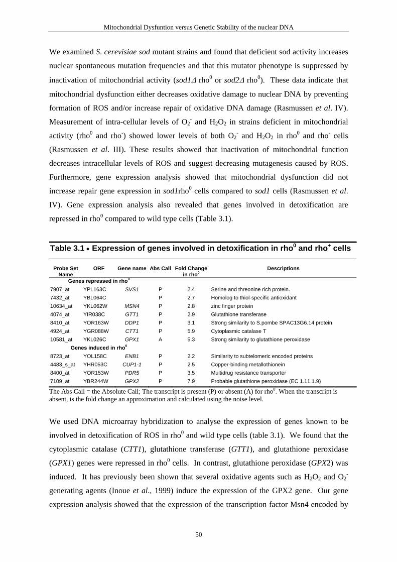
Mitochondrial Dysfuntion versus Genetic Stability of the nuclear DNA
We examined S. cerevisiae sod mutant strains and found that deficient sod activity increases
nuclear spontaneous mutation frequencies and that this mutator phenotype is suppressed by
inactivation of mitochondrial activity (sod1∆ rho0 or sod2∆ rho0). These data indicate that
mitochondrial dysfunction either decreases oxidative damage to nuclear DNA by preventing
formation of ROS and/or increase repair of oxidative DNA damage (Rasmussen et al. IV).
Measurement of intra-cellular levels of O2- and H2O2 in strains deficient in mitochondrial
activity (rho0 and rho-) showed lower levels of both O2- and H2O2 in rho0 and rho- cells
(Rasmussen et al. III). These results showed that inactivation of mitochondrial function
decreases intracellular levels of ROS and suggest decreasing mutagenesis caused by ROS.
Furthermore, gene expression analysis showed that mitochondrial dysfunction did not
increase repair gene expression in sod1rho0 cells compared to sod1 cells (Rasmussen et al.
IV). Gene expression analysis also revealed that genes involved in detoxification are
repressed in rho0 compared to wild type cells (Table 3.1).
Table 3.1 • Expression of genes involved in detoxification in rho0 and rho+ cells
Probe Set
Name
ORF
Gene name
Abs Call
Fold Change
in rho0
Descriptions
Genes repressed in rho0 7907_at YPL163C SVS1 P 2.4 Serine and threonine rich protein. 7432_at YBL064C P 2.7 Homolog to thiol-specific antioxidant 10634_at YKL062W MSN4 P 2.8 zinc finger protein 4074_at YIR038C GTT1 P 2.9 Glutathione transferase 8410_at YOR163W DDP1 P 3.1 Strong similarity to S.pombe SPAC13G6.14 protein 4924_at YGR088W CTT1 P 5.9 Cytoplasmic catalase T 10581_at YKL026C GPX1 A 5.3 Strong similarity to glutathione peroxidase
Genes induced in rho0 8723_at YOL158C ENB1 P 2.2 Similarity to subtelomeric encoded proteins 4483_s_at YHR053C CUP1-1 P 2.5 Copper-binding metallothionein 8400_at YOR153W PDR5 P 3.5 Multidrug resistance transporter 7109_at YBR244W GPX2 P 7.9 Probable glutathione peroxidase (EC 1.11.1.9)
The Abs Call = the Absolute Call; The transcript is present (P) or absent (A) for rho0. When the transcript is absent, is the fold change an approximation and calculated using the noise level.
We used DNA microarray hybridization to analyse the expression of genes known to be
involved in detoxification of ROS in rho0 and wild type cells (table 3.1). We found that the
cytoplasmic catalase (CTT1), glutathione transferase (GTT1), and glutathione peroxidase
(GPX1) genes were repressed in rho0 cells. In contrast, glutathione peroxidase (GPX2) was
induced. It has previously been shown that several oxidative agents such as H2O2 and O2-
generating agents (Inoue et al., 1999) induce the expression of the GPX2 gene. Our gene
expression analysis showed that the expression of the transcription factor Msn4 encoded by
50

Mitochondrial Dysfuntion versus Genetic Stability of the nuclear DNA
the MSN4 gene was repressed in rho0 cells. Msn4 and Msn2 proteins regulate the expression
of gene products that are induced by stress conditions through the stress response element
(STRE) promoter sequence (Gasch et al., 2000; Causton et al., 2001). Msn4 and Msn2
proteins have been shown to bind upstream of the STRE element (Görner et al., 1998). A
known target for this transcription factor is the CTT1 gene (Gasch et al., 2000). Our results
indicate that rho0 cells experience stress although the nature of this stress is unknown at this
time.
These data indicate that absence of mitochondrial respiration is an important factor for the
viability as well as for suppression of nuclear spontaneous mutations in sod1rho0 mutant
cells. Our results suggest that rho0 cells have a lower production of ROS which results in
lower nuclear spontaneous mutation frequency even when Sod1 or Sod2 are absent.
Since the level of ROS is lower in rho0 cells one would expect that rho0 cells had lower
spontaneous mutation frequencies than wild type but this is not the case. Interestingly we
show that rho0 cells had (2.6 fold) statistically significant higher spontaneous mutation
frequencies than wild type. Gene expression analysis showed that REV1 expression is
upregulated in rho0 strains. The REV1 gene encodes a deoxycytidyl transferase activity
involved in error-prone translesion DNA synthesis (TLS) that inserts dCMP opposite an
abasic site or a strand break in the template, thereby producing a terminus that can be
extended by polymerase zeta (Pol ξ). Polymerase ξ is composed of two subunits encoded by
REV3 and REV7. TLS is mutagenic because Rev1 preferentially inserts cytosine opposite an
abasic site in yeast (Lawrence and Hinkle, 1996; Nelson et al., 1996; Lawrence and Maher,
2001). We examined rev1rho0, rev3rho0 and rev7rho0 strains to determine if inactivation of
TLS could eliminate the mutator phenotype observed in rho0 cells. Our results show that
mitochondrial-mediated mutator phenotype depend on functional Rev1, Rev3 and Rev7
proteins and that the mutator phenotype of rho0 cells is suppressed by preventing
Rev1/Rev3/Rev7-dependent translesion synthesis (Rasmussen et al., III).
It has been shown that 3-methyladenine DNA glycosylase encoded by MAG1 in S.
cerevisiae, which removes a variety of alkylated bases and generates abasic sites, increases
spontaneous mutation frequency by 600-fold when overexpressed in S. cerevisiae. This
mutator phenotype is also dependent on the Rev1/Rev3/Rev7 TLS bypass pathway (Glassner
et al., 1998).
51

Mitochondrial Dysfuntion versus Genetic Stability of the nuclear DNA
Our genetic evidence therefore suggests that mitochondrial dysfunction contributes to
spontaneous DNA damage, which is fixed in the genome as mutation by error-prone
Rev1/Rev3/Rev7 TLS pathway, which results in cell survival but genetic instability.
Interestingly, one of the profound features of cancer cells is their defective mitochondrial
function. A role for mitochondria in tumorigenesis was hypothesized when it was found that
most tumors up-regulate glycolysis and therefore seems to be more dependent upon
glycolysis for energy production, than mitochondrial oxidative phosphorylation (Warburg,
1956).
Furthermore, cancer cell lines (bladder, head, neck, colorectal and pancreatic) and lung
primary tumors have been observed to have high frequency of homoplasmic mutations of
mtDNA. The mitochondrial mutations are present in each of the hundreds of mitochondrial
genomes in the tumor cell and in virtually all of the cells of the tumor (Polyak et al., 1998;
Fliss et al., 2000; Jones et al., 2001). Homoplasmic mutations of mtDNA indicate that a
single cell with a mutant mitochondrial genome has acquired a selective growth advantage
during tumor evolution, allowing it to become the predominant cell type in the tumor cell
population. Homoplasmy also indicates that each mutant mitochondrial genome has a
replicative advantage in the particular mitochondria in which it occurred, and that this
mitochondrion has selectively proliferated over other mitochondria in the same cell (Polyak
et al., 1998; Jones et al., 2001).
Our studies using S. cerevisiae with depleted mitochondria suggest that cancer cells by
losing their mitochondrial function create a mutator phenotype (Rasmussen et al., II). Figure
3.3 provides a model for how mitochondrial dysfunction generates a mutator phenotype in
two different ways:
1. Inhibition of the respiratory chain increases mitochondrial ROS production.
Mitochondrial reactive oxygen species damage the mtDNA and the nDNA if
mitochondrial H2O2 diffuses to the nucleus and is converted to HO• which can cause
DNA damage.
2. Mitochondrial dysfunction leads to nuclear DNA damage. The DNA damage activates
the Rev1/Rev3/Rev7 error-prone translesion DNA synthesis pathway that contributes to
cell survival, but at the expense of higher spontaneous mutation frequency.
52

Mitochondrial Dysfuntion versus Genetic Stability of the nuclear DNA
Nuclear MutationsNuclear Mutations
Increased levels of DNA damage?(Imbalanced repair of DNA damage?)
Increased levels of DNA damage?(Reactive oxygen species (ROS))
Suppressor gene inactivation Oncogene activation
Uncontrolled proliferation
TumorTumor
Mitochondrial dysfunctionMitochondrial dysfunction1 2
Figure 3.3. Mitochondrial dysfunction creates a mutator phenotype. Our results suggest that mitochondrial dysfunction can create a mutator phenotype in at least two ways. 1) Mitochondrial dysfunction caused by inhibition of the respiratory chain increases mitochondrial ROS production and if the ROS level is higher than the endogenous antioxidant capacity, ROS might difusse as H2O2 to the nucleus. In nucleus H2O2 can be converted to HO• which causes DNA damage. If the DNA damage is left unrepaired mutations in nDNA will occur and may lead to mutations that enhances cancer risk. 2) Mitochondrial dysfunction leads to nuclear DNA damage. The nature of this spontaneous nuclear DNA damage is unknown at this time, but might be caused by imbalanced repair as seen when Mag1 is overexpressed (Glassner et al., 1998). However, the DNA damage activates the Rev1/Rev3/Rev7 TLS pathway, which contributes to cell survival, but at the expense of higher spontaneous mutation frequency, which may lead to enhanced cancer risk.
53

Paper I
Section two
Paper I
Lene Juel Rasmussen, Merete Rasmussen, Byung-In Lee, Anne Karin Rasmussen, David
M. Wilson III, Finn Cilius Nielsen, and Hanne Cathrine Bisgaard. Identification of factors
interacting with hMSH2 in the fetal liver utilizing the yeast two-hybrid system. In vivo
interaction through the C-terminal domains of hEXO1 and hMSH2 and comparative
expression analysis. Mutation Research (2000) 460, 41-52.
54

Manuscript II
Acknowledgements
This work was supported by grants from the Danish Cancer Society (LJR), Danish Natural
Science Council (LJR), Carlsberg Foundation (LJR), Denmark-America Foundation
(AKR), Frimodt-Heineke Foundation (AKR), Frederik and Marie Beyers Fellowship
(AKR), Christian and Ottilia Brorsons Fellowship (AKR), Grosserer L.F. Foghts
Foundation (AKR), Julie von Müllens Foundation (AKR), Grosserer Sven Hansen and
Hustru Ina Hansens Foundation (AKR), Anna and Dagny Hjerrilds Foundation (AKR).
81

Ž .Mutation Research 460 2000 41–52www.elsevier.comrlocaterdnarepair
Community address: www.elsevier.comrlocatermutres
Identification of factors interacting with hMSH2 in the fetal liverutilizing the yeast two-hybrid system
In vivo interaction through the C-terminal domains of hEXO1 andhMSH2 and comparative expression analysis
Lene Juel Rasmussen a,), Merete Rasmussen a, Byung-In Lee b,Anne Karin Rasmussen a, David M. Wilson III b, Finn Cilius Nielsen c,
Hanne Cathrine Bisgaard a
a Department of Life Sciences and Chemistry, Roskilde UniÕersity, DK-4000 Roskilde, Denmarkb Biology and Biotechnology Research Program, L-452, Lawrence LiÕermore National Laboratory, LiÕermore, CA 94551, USA
c Department of Clinical Biochemistry, Rigshospitalet, DK-2100 Copenhagen, Denmark
Received 29 November 1999; received in revised form 21 February 2000; accepted 13 March 2000
Abstract
Ž .Mutations in DNA mismatch repair MMR genes have been shown to segregate with Hereditary NonpolyposisŽ .Colorectal Cancer HNPCC . However, because many HNPCC families fail to display mutations in known MMR genes, we
argued that changes in other components of the MMR pathway may be responsible. The increasing number of proteinsreported to interact in the MMR pathway suggests that larger complexes are formed, the composition of which may differamong cell types and tissues. In an attempt to identify tissue-specific MMR-associated factors, we employed the yeasttwo-hybrid system, using the human hMSH2 as bait and a human fetal liver library as prey. We demonstrate that hMSH2
X X Ž .interacts with a human 5 –3 exonuclease 1 hEXO1rHEX1 and that this interaction is mediated through their C-terminaldomains. The hMSH6 protein does not interact with hEXO1 in the two-hybrid system. Dot-blot analysis of multiple tissueRNA revealed that hMSH2 and hEXO1 are coexpressed at high levels in fetal liver as well as in adult testis and thymus.Northern blot analysis also revealed that hEXO1rHEX1 is highly expressed in several liver cancer cell lines as well as incolon and pancreas adenocarcinomas, but not in the corresponding non-neoplastic tissue. q 2000 Elsevier Science B.V. Allrights reserved.
Keywords: Mismatch repair; hMSH2; hEXO1; Two-hybrid; Cancer
) Corresponding author. Tel.: q45-46-74-27-28; fax: q45-46-74-30-11.
Ž .E-mail address: [email protected] L.J. Rasmussen .
1. Introduction
Changes in the efficiency of DNA repair andrecombination activities can be associated with pre-
w xdisposition to cancer 1 . The finding that Hereditary
0921-8777r00r$ - see front matter q 2000 Elsevier Science B.V. All rights reserved.Ž .PII: S0921-8777 00 00012-4

( )L.J. Rasmussen et al.rMutation Research 460 2000 41–5242
Ž .Nonpolyposis Colorectal Cancer HNPCC familiesfrequently harbor mutations in DNA mismatch repairŽ .MMR genes has generated widespread interest in
w xthis research area 2–4 . At the time of writing,germline mutations in at least four genes, hMSH2,hMLH1, hPMS1 and hPMS2, all homologs of bacte-rial components involved in MMR, have been foundin HNPCC patients. Although no biochemical rolehas to date been proposed for hPMS1, the other three
w xproteins have been shown to participate in MMR 3 .In addition, MMR proteins might play importantroles in other pathways of DNA metabolism such asDNA recombination and cell cycle checkpoint sig-
w xnaling 4 .The development of an assay to study MMR in
cell-free extracts of bacteria has led to a detailedunderstanding of the mechanism underlying the re-pair process and, on the basis of these in vitro
w xstudies, a model for MMR has been proposed 5 ,w xreviewed in Refs. 4,6 . Based on homology between
eukaryotic and prokaryotic MMR proteins, it hasbeen hypothesized that the mechanism underlyinghuman MMR is similar. Subsequently, it has beenshown that eukaryotic MMR proteins interact withpolypeptides involved in DNA replication, DNA re-combination, and DNA nucleotide excision repairŽ . w xNER 7–17 . A number of these genes have beeninactivated giving rise to complex phenotypes, suchas defects in MMR and recombination or in NER
w xand recombination 18–20 . These findings suggestthat DNA repair proteins are components of largerprotein complexes, and that at least some of theseproteins function in several distinct processes. Theincreasing number of polypeptides reported to inter-act with proteins in the MMR pathway suggests thatthese complexes are composed of a larger number ofsubunits, and also that the composition of such com-plexes differs among cell types and tissues.
In the present communication, we have employedthe yeast two-hybrid system in an attempt to identifyproteins that interact with the human MMR proteinhMSH2 in vivo, using hMSH2 as bait. We chose ahuman fetal liver cDNA library as prey since weexpected MMR-associated proteins to be highly ex-pressed in an organ containing rapidly proliferatingcells. We report that hMSH2 interacts with the geneproducts of both hMSH6 and hEXO1rHEX1. Wewere unable to detect binding between hMSH6 and
hEXO1 in the two-hybrid assay. Our results suggestthat the interaction between hMSH2 and hEXO1 ismediated via the C-terminal domains of these pro-teins. Expression analysis showed that high levels ofthis exonuclease are indeed coexpressed with hMSH2in fetal liver as well as in adult testis and thymus.Coexpression, although at lower levels, is also foundin colon, small intestine, bone marrow, placenta,fetal kidney, fetal spleen and fetal thymus. Furtherexpression analysis revealed high levels of hEXO1transcripts in liver cancer cell lines and in colon andpancreas adenocarcinomas, but not in the corre-sponding non-neoplastic tissue.
2. Material and methods
2.1. Human cell lines
ŽThe cell lines HepG2 human hepatocellular car-. Ž .cinoma , SK-Hep1 human liver adenocarcinoma ,Ž .Huh-7 human hepatocellular carcinoma , Chang
Ž . Ž .human liver , and WRL68 human liver embryowere purchased from European Collection of Cell
Ž .Cultures Wiltshire, UK . All cell lines were main-Žtained as monolayer cultures in DMEM Gibco, Life
. ŽTechnologies supplemented with 10% FBS Gibco,.Life Technologies .
2.2. Plasmids
Plasmid pLJR105 was constructed by insertingthe human hMSH2 coding sequence into the NcoI
Žsite of the pAS2 binding domain vector CLON-.TECH . The hMSH2 gene was obtained on a 2.9-kbp
w xfragment from pCite-1-hMSH2 21 kindly providedŽby Dr. Josef Jiricny Institute of Medical Radiobiol-
.ogy of the University of Zurich, Switzerland . ThehMSH2 bait was sequenced to confirm that thehMSH2 protein was in frame with the GAL4 bindingdomain. Plasmid pLJR112 contains a C-terminaltruncation of the hMSH2 gene and was constructed
Žby deleting a 1-kb fragment between the BlpI bp. Ž .1796 in hMSH2 and Sal I located in the vector
sites in pLJR105. This truncation eliminates the C-terminal 374 amino acids of the hMSH2 protein andincludes elimination of the conserved region. The
Ž . Ž .HEX1 C-terminal and HEX1 N-terminal plasmids
( )L.J. Rasmussen et al.rMutation Research 460 2000 41–52 43
were constructed by inserting either a 1182- or a1355-bp fragment into the NcoI and BamHI sites of
Ž .the pACT2 activation domain vector CLONTECH .The fragments were amplified by PCR using primerscontaining additional NcoI or BamHI sites. Primerpairs 5X-GGCACCATGGGGATACAGGGAT-3X, 5X-CGGGATCCTCACTTCAATTGTGGGGCATCT-3X
and 5 X-G G C A C C A T G G C C T G C C C A T T -CAAGAAGT-3X, 5X-CGGGATCCTCAGAATTTTT-TAAATCCAA-3X were used for amplification of theN-terminal and C-terminal parts of thehEXO1arHEX1 protein, respectively.
2.3. Two-hybrid techniques
Ž .The GAL4-hMSH2 pLJR105 bait plasmid wasŽtransformed into S. cereÕisiae strain Y190 MATa ,
ura3-52, his3-200, lys2-801, ade2-101, trp1-901,leu 2 -3 , 1 1 2 , g a l4 D , g a l8 0 D , cyh r 2 ,LYS2::GAL1 -HIS3 -HIS3, URA3::GAL1 -UAS TATA UAS
w xGAL1 -lacZ 22 . The resulting strain was subse-TATA
quently transformed with a human fetal liver match-Ž .maker cDNA library CLONTECH, aHL4029AH
and hMSH2 interactors were selected on syntheticŽ . w xdextrose minimal medium SD 23 lacking trypto-
Ž . Žphan to maintain pLJR105 , leucine to maintain the. ŽGAL4 activation domain plasmids , and histidine to
identify peptides capable of assembling a functional.GAL4 transcription factor and supplemented with
Ž . Ž25 mM 3-amino-1, 2, 4-triazole 3-AT SD-HIS-.TRP-LEUq25 mM 3-AT . 3-AT is a competitive
inhibitor of the S. cereÕisiae His3 protein and isused to lower the background growth of the reporterstrain due to leaky expression of the HIS3 gene. Theplates were incubated at 308C for 7–10 days and atotal of 53 positive clones, Y1–Y53, were screenedfor b-galactosidase activity on SD-TRP-LEUq60mgrml 5-bromo-4-chloro-3-indolyl-b-D-galactosideŽ .X-gal plates to verify positive interactions. Theseclones were rescued for the GAL4 binding domain
Ž .plasmid pLJR105 by repeated growth on SD-LEUplates. The rescued clones were tested for lack oflacZ expression on SD-LEUqX-gal plates to elimi-nate false positive clones.
2.4. DNA sequencing
DNA sequencing was performed directly on yeastcolonies grown on SD-HIS-TRP-LEU plates by
touching the colony with a sterile pipette tip. TheŽpipette tip was rinsed in incubation buffer 1.2 M
sorbitol; 100 mM sodium phosphate, pH 7.4; 2.5.mgrml zymolase and incubated at 378C for 5 min.
Sequence inserts were amplified by PCR using theMATCHMAKER AD LD-Insert Screening Am-
Ž .plimer Set CLONTECH . The PCR amplified frag-ments were purified using a Qiaquick PCR purifica-
Ž .tion kit Qiagen and sequenced. Database searcheswere performed using the BLAST algorithm
.http:rrwww.ncbi.nlm.nih.govrBLASTr .
2.5. Northern blot analysis
Human RNA Master Blot containing 100–500 ngŽ .q Ž .poly A RNArdot Human RNA Master Blot was
purchased from CLONTECH. For detection of tran-Ž .scripts in human cells aliquots 20 mg of total RNA
Ž TM .RNAqueous , Ambion were run on 1%agaroser0.2 M formaldehyde gels and immobilizedonto nylon membranes. All probes were labeled by
Žrandom primer extension Rediprime DNA labeling. wsystem, Amersham Pharmacia Biotech and a-
32 x Ž .P dCTP Amersham Pharmacia Biotech using aPCR product as template. For the detection of exonu-clease homologs, the blots were probed with either a914-bp fragment containing the N-terminal region of
Ž .hEXO1arHEX1 N-terminal probe or a 946-bp frag-ment containing the C-terminal region of
Ž .hEXO1arHEX1 C-terminal probe . These probeswere generated by PCR primers 5X-GGTGCCA-CATGCGATCTCTGAG-3X and 5X-TCCAATCC-
X Ž .CACGCAGTGATGAC-3 HEX1 N-terminal probeand 5X-GAGTGTAAGCACTCCACCTAGG-3X and
X X Ž5 -CCCGGCTTGTTCTCGGCATTAT-3 HEX1 C-.terminal probe . For the detection of hMSH2, the
blots were probed with a 1054-bp fragment contain-ing the N-terminal region, which shows limited ho-mology to other known MutS homologs. This probewas generated by PCR primers 5X-GTGCGCTTCTT-TCAGGGCATGC-3X and 5X-TTATTCAGCAAGG-
X Ž .CAGCCAGAG-3 hMSH2 probe . All membraneswere hybridized with 1=106 cpmrml of 32 P-labeledprobes. The Human RNA Master Blot was prehy-bridized for 30 min at 658C and hybridized overnightat 658C in ExpressHybw solution according to the
Ž .manufacturer CLONTECH . All other membraneswere prehybridized for 30 min at 688C and hy-

( )L.J. Rasmussen et al.rMutation Research 460 2000 41–5244
bridized for 90 min at 688C in QuikHybw Hybridiza-Ž .tion Solution Stratagene . Blots were washed twice
at room temperature for 15 min and once at 608CŽ .with wash solution 1= SSC, 0.1% SDS . Images
were obtained using autoradiography. GAPDH andb-actin probes provided by the manufacturerŽ .CLONTECH were prepared as described aboveand used to correct for differences in loading.
2.6. b-galactosidase assays
b-galactosidase specific activity was assayed onthree separate transformants of each strain. In brief,colonies were inoculated into 5 ml of SD-LEU-TRPmedium and grown overnight at 308C with vigorousshaking. The next day, 2 ml of overnight cultureswere inoculated into 2 ml of YPD medium and thecultures grown for 3–5 h at 308C with vigorousshaking. Samples were prepared and assayed forb-galactosidase activity essentially as described
Žby CLONTECH Protocol a PT1020-1,.www.clontech.com .
3. Results
3.1. Identification of hMSH2-interacting proteinpartners
To define the mechanism of human MMR and toidentify missing components of this complex pro-cess, we set up a two-hybrid screen in S. cereÕisiae,using as bait GAL4 fused to the entire open readingframe of human hMSH2 and as prey GAL4 activa-tion-tagged human fetal liver matchmaker cDNA
Ž .library. Our initial screen identified 53 Y1–Y53clones as interactors. Sequence analysis showed thatthe majority of the clones contained either yetunidentified human cDNAs or proteins such as g-globulin, fibrinogen, albumin, haptoglobin, repeatregions, ferritin, and a-chymotrypsin but that Y5,Y10, Y47 and Y50 contained a carboxyl-terminalregion of the human homologue of S. cereÕisiae
w x Žexonuclease EXO1, termed hEXO1b 10, 11 Fig..1 . We also isolated two clones containing hMSH6,
w xwhich is known to form a complex with hMSH2 4 ,confirming to us that hMSH2 is able to form acomplex with human MMR proteins in yeast.
The DNA sequences of the hEXO1 clones weŽ .isolated Y5, Y10, Y47 and Y50 showed a perfect
match to the 3X-end of hEXO1b, which is dissimilarŽ .to the hEXO1arHEX1 sequence Fig. 1 . Further-
more, we observed several DNA sequence polymor-phisms in the hEXO1 clones that could reflect hu-
Ž .man population variation Fig. 1 . The C to T baseŽ .pair change bp 2488 in Y5, Y10 and Y47 causes a
Ž . Ž .proline P to leucine L codon change, whereas theŽ . Ž .G to A bp 1279 in Y50 causes an arginine R toŽ .histidine H codon change. The Y50 clone contains
Ž .also an A to G base pair change bp 1983 , whichŽ . Ž .results in a lysine K to glutamic acid E codon
change. Furthermore, Y50 contains a deletion ofŽ .CAG bp 1486 resulting in a deletion of an alanine
Ž .A residue. All the clones we isolated appear tointeract equally well with hMSH2 in the two-hybridassay, indicating that these amino acid residues arenot crucial for hMSH2-hEXO1 interaction. The Y5,Y10 and Y47 clones contained an additional 127-bp
Ž .sequence Fig. 1 , which corresponds to 59171–Ž59048 bp of the genomic DNA sequence Genbank
.AC004783 . This sequence is located upstream ofŽ . w xthe first exon Exon 1 in hEXO1arHEX1 12 . For
simplicity, we have named this 127-bp DNA se-quence for Exon 0. We do not think that Y5, Y10and Y47 represent true splice variants of hEXO1bsince we were only able to detect transcripts corre-sponding to full-length hEXO1 using Northern blot
Ž .analysis data not shown .
3.2. Interaction domains of hMSH2 and hEXO1
It has been shown that purified full-lengthhEXO1b interacts with hMSH2, in vitro, in an im-
w xmunoprecipitation assay 10 . We used two-hybridanalysis to characterize the interaction betweenhMSH2 and hEXO1 in vivo and to define the re-gion of association. We fused the full-lengthhEXO1arHEX1 and hEXO1b cDNAs to the GAL4activation domain in pACT2 and tested for interac-tion with our GAL4–hMSH2 binding domain plas-
Ž .mid pLJR105 . We were unable to detect protein–protein interactions with any of our full-length ex-
Žonuclease 1 constructs in the two-hybrid assay Fig..2A . However, when we switched the inserts around
and instead fused hMSH2 to the GAL4 activationdom ain in pA C T2, and hEXO 1b and

( )L.J. Rasmussen et al.rMutation Research 460 2000 41–52 45
w x Ž .Fig. 1. Exonuclease 1 gene structures. The full-length hEXO1arHEX1 and hEXO1b 10–12 coding regions comprise 13 exons II–XIV .Ž X .The shorter variants named Y5, Y10, Y47 and Y50 comprise eight exons VII –XIV . These clones contain only a part of exon VII that we
refer to as exon VIIX. Solid lines represent exon sequences containing the N and I regions thought to be required for exonuclease activity.Shaded boxes in hEXO1arHEX1 and hEXO1b represent the predicted coding regions. DGA to stop indicates the two base pairs deletion ofGA that causes a frameshift in hEXO1arHEX1 compared with hEXO1b. The Y5, Y10 and Y47 clones contain an additional 127-bp DNA
Ž . Ž .sequence exon 0 corresponding to 59171–59048 bp of the genomic DNA sequence Genbank AC004783 .
hEXO1arHEX1 to the GAL4 binding domain inpAS2, we could detect an interaction in the two-hy-
Ž .brid assay Fig. 2A, Table 1 . One reason for thisŽdiscrepancy could be that the GAL4 activation do-
.main -hEXO1 fusion proteins are structurally differ-ent from the native proteins and therefore unable to
Ž .interact with the GAL4 binding domain –hMSH2fusion protein. This would also explain why wefailed to isolate any full-length cDNAs of hEXO1bor hEXO1arHEX1 in our initial two-hybrid screen.
W e constructed truncated peptides ofhEXO1arHEX1 such that the protein was dividedinto two regions containing either the 451 aminoacids N-terminal or the 388 amino acids C-terminal
Ž .part of full-length hEXO1arHEX1 Fig. 1 . We alsoconstructed a C-terminal truncation of hMSH2 todelete the conserved region of the protein that isnecessary for interaction with hMSH3 and hMSH6w x24 . We did not observe any interaction betweenhMSH2 and the N-terminal region of hEXO1 in the
Ž .two-hybrid test Fig. 2B . Neither did we observeinteraction between the hMSH2 N-terminal regionand any of the exonuclease clones we isolated in the
Ž .two-hybrid screen data not shown . However, wecannot rule out that the N-terminal region of hEXO1makes contact to hMSH2 in the native protein. Inter-estingly, we did observe an interaction between
Ž .hMSH2 and Y5 hEXO1b C-terminal , as well as

( )L.J. Rasmussen et al.rMutation Research 460 2000 41–5246
Fig. 2. Interaction of hMSH2 with hEXO1arHEX1 and hEXO1bŽ . Ž .Y5 . A Strains containing various plasmids were streaked in
Ž .horizontal rows on either SD-LEU-TRPqX-gal upper panel orŽ . Ž .SD-LEU-TRP-HISq45 mM 3-AT lower panel plates. B Strains
containing various plasmids were streaked on SD-LEU-TRPqX-gal plate. The plasmids contained in each strain tested are indi-cated at the top of each column and at the left of each row. No
Ž .insert: pAS2, SNF4: pACT2 containing SNF4 control , and SNF1:Ž .pAS2 containing SNF1 control . BD: binding domain. AD: acti-
vation domain.
Table 1Specific b-galactosidase activity assay for protein interactions inthe two-hybrid assay
Specific b-galactosidaseaactivity
Ž . Ž .Y5 AD qhMSH2 BD 6.3Ž . Ž .Y5 AD qhMSH2-N BD 0
Ž . Ž .HEX1-N AD qhMSH2 BD 0Ž . Ž .HEX1-N AD qhMSH2-N BD 0Ž . Ž .HEX1-C AD qhMSH2 BD 0.3Ž . Ž .HEX1-C AD qhMSH2-N BD 0Ž . Ž .hMSH2 AD qHEX1 BD 17.7Ž . Ž .hMSH2 AD qhEXO1b BD 12.8
Ž . Ž .SNF4 AD qSNF1 BD 3.1Ž . Ž .pACT2 AD qpAS2 BD 0
aSpecific b-galactosidase activity was determined as describedin Materials and methods.
Žbetween hMSH2 and hEXO1arHEX1 C-terminal. Ž . Žregion Fig. 2B, Table 1 . Our Y5 hEXO1b C-.terminal clone did not contain the full-length
hEXO1b cDNA, but instead contained an insert en-coding the carboxyl half of hEXO1b protein begin-ning at amino acid 302. These data argue that thecarboxyl-terminal region of hMSH2 contacts the car-boxyl-terminal domains of both hEXO1arHEX1 andhEXO1b. Thus, the hMSH2-interacting region is out-
Ž .side the N and I regions Fig. 1 thought to bew xrequired for exonuclease activity 12,25,26 . Our re-
sults complement the finding that S. cereÕisiaeyMSH2 interacts with the carboxyl-terminal region
w xof yEXO1 outside the N and I regions 16 .hEXO1arHEX1 differs from hEXO1b by two nu-
Ž .cleotides 2616-AG-2617 at the boundary of exonŽ .14 Fig. 1 . This frame-shift results in the truncation
of the hEXO1arHEX1 polypeptide by 43 aminoacids. We show that hMSH2 interacts with bothforms of human exonuclease 1, suggesting that theinteracting domain is located between exons 8 and
Ž .13 Fig. 1 . Interestingly, from data shown in Fig. 2it appears that hMSH6 does not interact with hEXO1in our two-hybrid assay.
3.3. Expression of exonuclease and hMSH2 in hu-man tissues
We used a human RNA master blot to character-ize the expression pattern of hEXO1 and to look for

( )L.J. Rasmussen et al.rMutation Research 460 2000 41–52 47
tissues predominately expressing the C-terminal tran-script of hEXO1. One of the DNA probes contained
only the C-terminal region of hEXO1arHEX1 andshould detect all hEXO1 splice variants identified to
Fig. 3. Expression profiles of human exonuclease 1 and hMSH2. The RNA master blot was hybridized with hMSH2, hEXO1arHEX1N-terminal, hEXO1arHEX1 C-terminal or b-actin control probes.

( )L.J. Rasmussen et al.rMutation Research 460 2000 41–5248
date. The second probe contained the N-terminalregion of hEXO1arHEX1, which would be specificfor full-length hEXO1arHEX1 and hEXO1b mR-NAs, but would not detect the C-terminal transcriptsŽ .Fig. 1 . We reasoned that by comparing the expres-sion patterns obtained with these two probes, wewould be able to identify tissues that express onlyC-terminal fragment. Since we did not observe anydifferences in expression results, it would appear thatno tissue preferentially expresses the C-terminal part
Ž .of hEXO1 Fig. 3 . We do not know whether theC-terminal part of hEXO1 is naturally present andhas a specific function in the cell or whether itsoccurrence is a result of inefficient DNA synthesisduring the construction of the matchmaker cDNAlibrary, but presume the latter to be true based on thefact that shorter transcripts were not observed. Bothprobes detected transcripts prominently expressed infetal liver, adult testis and thymus, but we alsoobserved significant expression in adult colon, smallintestine, bone marrow, placenta, fetal kidney, fetal
Ž .spleen and fetal thymus Fig. 3 . Interestingly, wedid not detect any expression of hEXO1 mRNA inadult liver, spleen, and kidney, indicating a specificrole for hEXO1 in the development of these tissues.
ŽSince hMSH2 interacts with exonuclease 1 Fig..2 , we compared the expression patterns of hMSH2
with that of hEXO1 to identify tissues where theexonuclease could play a role in DNA repair androrrecombination. We found that hMSH2 is expressed
Ž .in all tissues investigated Fig. 3 . Fig. 3 shows thathMSH2 and hEXO1 are coexpressed at higher levelsin fetal liver, adult testis, thymus, and at lower levelsin bone marrow, placenta, fetal spleen, fetal kidneyand fetal thymus.
3.4. Expression of exonuclease and hMSH2 in hu-man liÕer cancer cell lines
We have shown that hEXO1 is expressed in theŽ .fetal liver but not in the adult liver Fig. 3 . We thus
examined the expression of the hMSH2 and hEXO1Ž .genes in various human liver cell lines Fig. 4 :
Ž .HepG2 human hepatocellular carcinoma , SK-Hep1Ž . Žhuman liver adenocarcinoma , Huh-7 human hep-
. Ž .atocellular carcinoma , Chang human liver , andŽ .WRL68 human liver embryo . As expected, both
hEXO1 and hMSH2 were highly expressed in the
Fig. 4. Expression of human exonuclease 1 and hMSH2 in livercancer cell lines. Membranes containing total RNA isolated fromhuman adult liver, fetal liver, and various liver cell lines wereprobed with hMSH2, hEXO1a r HEX1 N-terminal domain,hEXO1arHEX1 C-terminal domain and GAPDH control probes.The lower panel of the figure shows the 28S ribosomal RNA bandof the ethidium bromide stained gel. The human liver cell lines aredescribed in Materials and methods.
Ž .fetal liver but not in the adult liver Fig. 4 . Onereason for failing to detect any hMSH2 expression inthe adult liver in this experiment could be that the
Ž .qblot shown in Fig. 3 contains poly A mRNA whilethe blot in Fig. 4 contains total RNA. However,since hMSH2 is preferentially expressed in prolifer-
w xating cells 27–29 , high transcript levels in thenormally quiescent adult liver would be unexpected.When the hEXO1 probes were used, we could onlydetect a single transcript of approximately 3 kb,
Žwhich corresponds to full-length hEXO1 Fig. 4 and.data not shown . This result suggests that hEXO1
mRNA is unlikely spliced to any of the shorter formsidentified in the two-hybrid screens and that theC-terminal clones likely do not exist. A 3-kb tran-script was detected using the hMSH2 probe, whichcorresponds to the full-length hMSH2 cDNA. Inter-

( )L.J. Rasmussen et al.rMutation Research 460 2000 41–52 49
estingly, we find that both hEXO1 and hMSH2 arehighly expressed in the liver cancer cell lines HepG2,SK-Hep1 and Huh7 as well as in the fetal-like Changand WRL68 cells.
3.5. Expression of exonuclease and hMSH2 in tumortissue
Our results revealed that hEXO1 is expressedpreferentially in tissue containing proliferating cellpopulations as well as in cell lines established from
Ž .human liver tumors Figs. 3 and 4 . This led us toinvestigate the expression of hEXO1 in human tu-mors from colon and pancreas. Interestingly, wefound that hEXO1 expression is indeed upregulatedin samples isolated from colon and pancreas adeno-
Ž .carcinomas compared to normal tissue Fig. 5 . Theseresults support the idea of hEXO1 being preferen-tially expressed in proliferating cells. Interestingly,
Fig. 5. Expression of human exonuclease 1 and hMSH2 in colonand pancreas adenocarcinomas. Membranes containing total RNAisolated from human adenocarcinomas and corresponding non-neoplastic tissue were probed with hMSH2, hEXO1arHEX1C-terminal domain and GAPDH control probes. The lower panelof the figure shows the 28S ribosomal RNA band of the ethidiumbromide stained gel. Colorectal and pancreatic adenocarcinomaand normal colon and pancreatic tissue were collected duringsurgery and immediately frozen in liquid nitrogen. All tumorswere verified by histological examination. Normal tissues fromcolon and pancreas were also examined histologically and foundto be free of tumor cells. The local ethics committee approved theuse of tumor and normal tissue.
we found that hMSH2 expression is upregulated inŽthe colon tumors but not in the pancreas tumors Fig.
.5 . These results may imply a defect in MMR activ-ity in the pancreas tumors due to the low levels ofhMSH2 protein.
4. Discussion
Eukaryotic proteins are often components of largercomplexes. It is therefore easy to imagine that thedisruption of a particular protein–protein interactionmight affect several different cellular processes. Inaddition, many eukaryotic genes show tissue-specificexpression and it is conceivable that the compositionof protein complexes varies among different celltypes and tissues. In agreement with this model, ithas been shown that mice carrying disruptions ofspecific MMR genes display a variety of phenotypesranging from tissue-specific tumorigenesis to sterilityw x20,30–35 . This is consistent with the evidence thatimplicates MMR proteins in processes other thanpostreplicative MMR. Homologous and homeolo-gous recombination, transcription coupled repair orcell cycle control following DNA damage are exam-
w xples of such pathways 2,4,6 . It is possible, evenlikely, that MMR proteins will interact with differentpartners in each of these alternative pathways. Identi-fication of these partners will help elucidate themechanisms of these complex processes.
In an attempt to identify tissue-specific MMR-as-sociated factors, we employed the yeast two-hybridsystem, using the human hMSH2 as bait and ahuman fetal liver library as prey. We demonstratethat hMSH2 interacts with hEXO1 through theirC-terminal domains. Another MutS homolog hMSH6does not interact with hEXO1 in the two-hybridsystem. It has previously been shown that the humanexonuclease 1 is expressed in a wide variety of
w xtissues 11 . Our comparative expression analysisrevealed that hMSH2 and hEXO1 are coexpressed inthe fetal liver, adult testis and thymus as well as incolon, small intestine, and in the fetal tissues ofkidney and spleen but not in the corresponding adulttissues. hEXO1rHEX1 is highly expressed in sev-eral liver cancer cell lines as well as in colon andpancreas adenocarcinomas, but not in the corre-sponding non-neoplastic tissue.

( )L.J. Rasmussen et al.rMutation Research 460 2000 41–5250
4.1. Interaction between hMSH2 and human exonu-clease 1
The human MMR protein hMSH2 forms het-erodimers with hMSH3 or with hMSH6, which func-tion in the substrate recognition step during MMR.In the subsequent step, these bound heterodimersinteract with the hMLH1rhPMS2 complex. Al-though the following events of the MMR process arenot clear, it is anticipated based on the bacterialmodel that the four-protein complex will recruit DNAhelicase and exonuclease activities, which mediatethe displacement and the degradation of the mis-match-containing strand, respectively. The positionof the strand break and the initiation site of theexonucleolytic process with respect to the mismatchwould determine the nature of the exonuclease. Thus,repair of mismatches with upstream-positionedstrand-discrimination nicks will require a 5X–3X ex-
w xonuclease such as EXO1 16 . Conversely, MMRinvolving downstream nicks would require 3X–5X ex-onucleases to mediate the degradation process. In thepresent study, we show that hMSH2 interacts withthe human 5X–3X exonuclease hEXO1. We are cur-rently investigating whether the hMSH2rhEXO1complex functions in MMR or other pathways suchas recombination, where hEXO1 has already been
w xshown to play a role 11,16 .
4.2. ComparatiÕe expression analysis of hEXO1
Tumorigenesis in HNPCC patients is primarilyrestricted to the proximal colon and endometrium.We found that the hMSH2 and hEXO1 genes arecoexpressed, although at low levels, in colon andsmall intestine. Due to the likely redundancy be-tween exonucleases in the MMR process, it wouldappear unlikely to anticipate that mutations in thehEXO1 gene will segregate with HNPCC. Humanpopulation studies monitoring the association ofhEXO1 gene defects with cancer or the constructionof a mEXO1 knockout mouse should clarify if thisexonuclease plays any role in carcinogenesis. Ourexpression profiles of hMSH2 and hEXO1 also re-vealed that these genes are coexpressed in humantestis and bone marrow, suggesting a role in meiosisand hematopoiesis likely due to their function in
w xrecombination 36 . Along these lines, we have also
shown that both hEXO1 and hMSH2 are highlyexpressed in the fetal liver; a site of hematopoiesisduring fetal development. However, in addition tosupporting hematopoiesis during embryonic develop-ment, the fetal liver is also characterized by rapiddivision of fetal hepatocytes. We have shown thathEXO1 and hMSH2 are not expressed in normaladult liver where cell turnover is normally very slow.However, both genes are highly expressed in theliver cancer cell lines HepG2, SK-Hep1 and Huh7 aswell as in the fetal-like Chang and WRL68 cells.Furthermore, we showed that hEXO1 is highly ex-pressed in colon and pancreas adenocarcinomas butnot in the corresponding non-neoplastic tissues. Theseresults suggest a role for hEXO1 in cellular pro-
w xcesses other than meiosis and hematopoiesis 36 .Such processes could be MMR, recombinational re-pair or replication, which are expected to be active inproliferating cells such as fetal hepatocytes and can-cer cells. Current studies are examining for potentialassociations of hEXO1 with factors from these cellu-lar activities. The fact that hEXO1 is specificallyexpressed in liver cancer cells as well as in colonand pancreas adenocarcinomas could make the ex-pression of this gene a useful marker to diagnosethese cancers.
Acknowledgements
We thank Lene Markussen and Gerda D. Olesenfor expert technical assistance. We are grateful to Dr.Josef Jiricny for very thoughtful suggestions andcritical reading of the manuscript. This work wassupported by grants from the Danish Cancer SocietyŽ .LJR and HCB , Danish Medical Research CouncilŽ . Ž .LJR , Danish Natural Research Council HCB , and
Ž .Arvid Nilssons Foundation MR .
References
w x1 C. Lengauer, K.W. Kinzler, B. Vogelstein, Genetic instabili-Ž .ties in human cancers, Nature 396 1998 643–649.
w x2 J. Jiricny, Eukaryotic mismatch repair: an update, Mutat.Ž .Res. 409 1998 107–121.
w x Ž .3 J. Jiricny, Replication errors: cha lle nging the genome,Ž .EMBO J. 17 1998 6427–6436.
w x4 P. Modrich, R. Lahue, Mismatch repair in replication fi-delity, genetic recombination, and cancer biology, Annu.
Ž .Rev. Biochem. 65 1996 101–133.

( )L.J. Rasmussen et al.rMutation Research 460 2000 41–52 51
w x5 P. Modrich, Mechanisms and biological effects of mismatchŽ .repair, Annu. Rev. Genet. 25 1991 229–253.
w x6 L.J. Rasmussen, L. Samson, M.G. Marinus, Dam-directedDNA mismatch repair, in: M.F. Hoekstra, J.A. NickoloffŽ .Eds. , DNA Damage and Repair: Biochemistry, Geneticsand Cell Biology vol. 1 Humana Press, Clifton, NJ, 1998, pp.205–228.
w x7 M.E. Budd, J.L. Campbell, A yeast replicative helicase,Dna2 helicase, interacts with yeast FEN-1 nuclease in carry-
Ž .ing out its essential function, Mol. Cell. Biol. 17 19972136–2142.
w x8 R.E. Johnson, G.K. Kovvali, L. Prakash, S. Prakash, Re-quirement of the yeast RTH1 5X to 3X exonuclease for the
Ž .stability of simple repetitive DNA, Science 269 1995 238–239.
w x9 D.X. Tishkoff, N. Filosi, G.M. Gaida, R.D. Kolodner, Anovel mutation avoidance mechanism dependent on S. cere-Õisiae RAD27 is distinct from DNA mismatch repair, Cell 88Ž .1997 253–263.
w x10 C. Schmutte, R.C. Marinescu, M.M. Sadoff, S. Guerrette, J.Overhauser, R. Fishel, Human exonuclease 1 interacts with
Ž .the mismatch repair protein hMSH2, Cancer Res. 58 19984537–4542.
w x11 D.X. Tishkoff, N.S. Amin, C.S. Viars, K.C. Arden, R.D.Kolodner, Identification of a human gene encoding a homo-logue of Saccharomyces cereÕisiae EXO1, an exonucleaseimplicated in mismatch repair and recombination, Cancer
Ž .Res. 58 1998 5027–5031.w x12 D.M. Wilson III, J.P. Carney, M.A. Coleman, A.W. Adam-
son, M. Christensen, J.E. Lamerdin, Hex1: a new humanRad2 nuclease family member with homology to yeast ex-
Ž .onuclease 1, Nucleic Acids Res. 26 1998 3762–3768.w x13 R.E. Johnson, G.K. Kovvali, S.N. Guzder, N.S. Amin, C.
Holm, Y. Habraken, P. Sung, L. Prakash, S. Prakash, Evi-dence for involvement of yeast proliferating cell nuclear
Ž .antigen in DNA mismatch repair, J. Biol. Chem. 271 199627987–27990.
w x14 Y.-L. Lin, M.K.K. Shivji, C. Chen, R. Kolodner, R.D.Woods, A. Dutta, The evolutionarily conserved zinc fingermotif in the largest subunit of human replication protein A isrequired for DNA replication and mismatch repair but not for
Ž .nucleotide excision repair, J. Biol. Chem. 273 1998 1453–1461.
w x15 A. Umar, A.B. Buermeyer, J.A. Simon, D.C. Thomas, A.B.Clark, R.M. Liskay, T.A. Kunkel, Requirement for PCNA inDNA mismatch repair at a step preceding DNA resynthesis,
Ž .Cell 87 1996 65–73.w x16 D.X. Tishkoff, A.L. Boerger, P. Bertrand, N. Filosi, G.M.
Gaida, M.F. Kane, R.D. Kolodner, Identification and charac-terization of Saccharomyces cereÕisiae EXO1, a gene encod-ing an exonuclease that interacts with MSH2, Proc. Natl.
Ž .Acad. Sci. U.S.A. 94 1997 7487–7492.w x17 P. Bertrand, D.X. Tishkoff, N. Filosi, R. Dasgupta, R.D.
Kolodner, Physical interaction between components of DNAmismatch repair and nucleotide excision repair, Proc. Natl.
Ž .Acad. Sci. U.S.A. 95 1998 14278–14283.w x18 D. Melton, A.M. Ketchen, F. Nunez, S. Bonatti-Abbon-
dandolo, A. Abbondandolo, S. Squires, R. Johnson, Cellsfrom ERCC1-deficient mice show increased genome instabil-ity and a reduced frequency of S-phase-dependent illegiti-mate chromosome exchange but a normal frequency of ho-
Ž .mologous recombination, J. Cell Sci. 111 1998 395–404.w x19 G. Weeda, I. Donker, J. de Witt, H. Morreau, R. Janssens,
C.J. Vissers, A. Nigg, H. van Steeg, D. Bootsma, H. Hoeij-makers, Disruption of mouse ERCC1 results in a novel repairsyndrome with growth failure, nuclear abnormalities and
Ž .senescence, Curr. Biol. 7 1997 427–439.w x20 N. de Wind, M. Dekker, A. Berns, M. Radman, H. te Riele,
Inactivation of the mouse Msh2 gene results in mismatchrepair deficiency, methylation tolerance, hyperrecombination,
Ž .and predisposition to cancer, Cell 82 1995 321–330.w x21 F. Palombo, P. Gallinari, I. Iaccarino, T. Lettieri, M. Hughes,
A. D’Arrigo, O. Truong, J.J. Hsuan, J. Jiricny, GTBP, a160-kilodalton protein essential for mismatch-binding activ-
Ž .ity in human cells, Science 268 1995 1912–1914.w x22 J.W. Harper, G.R. Adami, N. Wei, K. Keyomarski, S.J.
Elledge, The p21 Cdk-interacting protein Cip1 is a potentŽ .inhibitor of G1 cyclin-dependent kinases, Cell 75 1993
805–816.w x23 M.D. Rose, F. Winston, P. Hieter, Methods in Yeast Genet-
ics: A Laboratory Course Manual, Cold Spring Harbor Lab.Press, Plainview, NY, 1990.
w x24 S. Guerrette, T. Wilson, S. Gradia, R. Fishel, Interaction ofhuman hMSH2 with hMSH3 and hMSH2 with hMSH6:explanation of mutations found in hereditary nonpolyposis
Ž .colorectal cancer, Mol. Cell. Biol. 18 1998 6616–6623.w x25 F.A. Digilio, A. Pannuti, J.C. Lucchesi, M. Furia, L.C.
Polito, Tosca: a Drosophila gene encoding a nuclease specifi-cally expressed in the female germline, Dev. Biol. 178Ž .1996 90–100.
w x26 P. Fiorentini, K.N. Huang, D.X. Tishkoff, R.D. Kolodner,L.S. Symmington, Exonuclease 1 of Saccharomyces cere-Õisiae functions in mitotic recombination in vivo and in
Ž .vitro, Mol. Cell. Biol. 17 1997 2764–2773.w x27 F.S. Leach, K. Polyak, M. Burrell, K.A. Johnson, D. Hill,
M.G. Dunlop, A.H. Wyllie, P. Peltomaki, A. de la Chapelle,¨S.R. Hamilton, K.W. Kinzler, B. Vogelstein, Expression ofthe human mismatch repair gene hMSH2 in normal and
Ž .neoplastic tissues, Cancer Res. 56 1996 235–240.w x28 G. Marra, C.L. Chang, L.A. Laghi, D.P. Chauhan, D. Young,
Ž .C.R. Boland, Expression of human MutS homolog 2 hMSH2Ž .protein in resting and proliferating cells, Oncogene 13 1996
2189–2196.w x29 T.M. Wilson, A. Ewel, J.R. Duguid, J.N. Eble, M.K. Lescoe,
R. Fishel, M.R. Kelley, Differential cellular expression of thehuman MSH2 repair enzyme in small and large intestine,
Ž .Cancer Res. 55 1995 5146–5150.w x30 S.M. Baker, C.E. Bronner, L. Zhang, A.W. Plug, M. Ro-
batzek, G. Warren, E.A. Elliott, J. Yu, T. Ashley, N. Arn-heim, R.A. Flavell, R.M. Liskay, Male mice defective in theDNA mismatch repair gene PMS2 exhibit abnormal chromo-
Ž .some synapsis in meiosis, Cell 82 1995 309–319.w x31 S.M. Baker, A.M. Plug, T.A. Prolla, C.E. Bronner, A.C.
Harris, X. Yao, D.-M. Christie, C. Monell, N. Arnheim, A.

( )L.J. Rasmussen et al.rMutation Research 460 2000 41–5252
Bradley, T. Ashley, R.M. Liskay, Involvement of mouseMlh1 in DNA mismatch repair and meiotic crossing over,
Ž .Nat. Genet. 13 1996 336–342.w x32 W. Edelmann, P.E. Cohen, M. Kane, K. Lau, B. Morrow, S.
Bennett, A. Umar, T. Kunkel, G. Cattoretti, R. Chaganti,J.W. Pollard, R.D. Kolodner, R. Kucherlapati, Meiotic
Ž .pachytene arrest in MLH1-deficient mice, Cell 85 19961125–1134.
w x33 T.A. Prolla, S.M. Baker, A.C. Harris, J.-L. Tsao, X. Yao,C.E. Bronner, B. Zheng, M. Gordon, J. Reneker, N. Arn-heim, D. Shibata, A. Bradley, R.M. Liskay, Tumour suscep-tibility and spontaneous mutation in mice deficient in Mlh1,Pms1 and Pms2 DNA mismatch repair, Nat. Genet. 18Ž .1998 276–279.
w x34 A.H. Reitmair, R. Schmits, A. Ewel, B. Bapat, M. Redston,A. Mitri, P. Waterhouse, H.-W. Mittrucker, A. Wakeham, B.¨Liu, A. Thomason, H. Griesser, S. Gallinger, W.G. Ball-hausen, R. Fishel, T.W. Mak, MSH2 deficient mice areviable and susceptible to lymphoid tumours, Nat. Genet. 11Ž .1995 64–70.
w x35 A.H. Reitmair, M. Redston, J.C. Cai, T.C.Y. Chuang, M.Bjerknes, H. Cheng, K. Hay, S. Gallinger, B. Bapat, T.W.Mak, Spontaneous intestinal carcinomas and skin neoplasms
Ž .in Msh2-deficient mice, Cancer Res. 56 1996 3842–3849.w x36 B.-I. Lee, M. Shannon, L. Stubbs, D.M. Wilson III, Expres-
Ž .sion specificity of the mouse exonuclease 1 mExo1 gene,Ž .Nucleic Acids Res. 27 1999 4114–4120.
Manuscript II
Manuscript II Anne Karin Rasmussen, Keshav K. Singh, and Lene Juel Rasmussen. Characterization of
O6-MeG DNA Methyltransferase (MGMT) protein in repairing human mitochondrial DNA.
Manuscript in preparation.
67

Manuscript II
Characterization of O6-MeG DNA Methyltransferase (MGMT) protein in
repairing human mitochondrial DNA
Anne Karin Rasmussen1,2, Keshav K. Singh2, and Lene Juel Rasmussen1
1Department of Life Sciences and Chemistry, Roskilde University, 4000 Roskilde,
Denmark, 2Johns Hopkins Oncology Center, Bunting-Blaustein Cancer Research Building,
1650 Orleans Street, Baltimore MD 21231-1000, USA.
Corresponding author:
Lene Juel Rasmussen
Department of Life Sciences and Chemistry
Roskilde University
4000 Roskilde
Denmark
Phone: (+45) 46742728
Fax: (+45) 46743011
Email: [email protected]
68

Manuscript II
Abstract
DNA repair is essential for maintaining the integrity of the genetic material. A number of
DNA repair mechanisms have been well characterized for the nuclear DNA. However,
little is known about DNA repair in mitochondria. Mutations in mitochondrial DNA
(mtDNA) are involved in the pathogenesis of a variety of diseases including cancer, heart
disease, diabetes, and a number of degenerative diseases. Furthermore, accumulation of
mutations in mtDNA is a common feature of aging cells. Human mtDNA is extremely rich
in guanine, therefore, repair of modified guanine is important for proper mitochondrial
function. Certain intracellular or exogenous DNA damaging agents are known to frequently
modify guanine to O6-MeG in DNA. Generation of O6-MeG is mutagenic because it can
mispair with thymine resulting in G:C → A:T transition mutations. The DNA repair protein
O6-Methylguanine-DNA Methyltransferase (MGMT) removes methyl groups from the O6
position of guanine (O6-MeG) in the nuclear DNA. In order to gain understanding of the
role of MGMT in mitochondria, we investigated (1) whether MGMT contains
mitochondrial localization signals and is transported to mitochondria, (2) whether MGMT,
when expressed in mitochondria can repair O6-MeG DNA lesions in this organelle, and (3)
whether MGMT when expressed in mitochondria can protect cells from apoptosis induced
by the DNA damaging agents which modify guanine in mtDNA.
69

Manuscript II
Introduction
Mammalian cells contain two distinct genomes: a nuclear (nDNA) and a mitochondrial
(mtDNA). Despite each human somatic cell contains 100 to 1000 mitochondria, that each
contains 1 to 10 mitochondrial genomes, the amount of the mtDNA is only estimated to
account for 1% of total DNA in the cell (Wallace et al., 1998; Bestwick, 1982).
In contrast to nDNA the mammalian mitochondrial genome contains few noncoding
sequences, no introns and it is not protected by histones. Thus, damage to mtDNA is
expected to have great impact on the function of this organelle. Accordingly, it has been
reported that the rate of point mutations is elevated in mtDNA compared to nDNA
(Khrapko et al., 1997). Point mutations in and genomic rearrangements of mtDNA have
been recognized to play a critical role in numerous human disorders underscoring the
importance of maintaining the integrity of the mitochondrial genome (Pulkes and Hanna,
2001; Wallace, 1999; Kogelnik et al., 19983).
Given the importance of maintaining the mitochondrial genome intact for
organelle activity, efficient DNA repair systems in mitochondria would be expected. Even
so, early investigations showed that UV-induced pyrimidine dimers are not removed from
mtDNA, a result that led to the conclusion that mitochondria are deficient in DNA repair
(Clayton et al., 1974; Prakash et al., 1975). These findings encouraged the model that
rather than repairing DNA damage in mtDNA, cells simply eliminate damaged genomes
and replace them by replicating existing undamaged mitochondrial genomes (LeDoux et
al., 1999). It was later confirmed that UV-induced pyrimidine dimers are not repaired in
mitochondria (LeDoux et al., 1992; Pascucci et al., 1997) suggesting that mitochondria do
not have a functional nucleotide excision repair (NER) pathway. However, recent
research has shown that at least base excision repair (BER) activities are operational in
mitochondria showing that this organelle does indeed repair damaged mtDNA (Croteu et
al., 1999; Sawyer et al., 1999). But the knowledge about the mechanisms underlying
DNA repair in the mitochondria is still limited.
DNA repair alkyltransferases, responsible for removing methyl groups from the O6
position of guanine, have been identified in both prokaryotic and eukaryotic organisms. The
genes coding for these DNA repair alkyltransferases have been cloned from Escherichia
coli (ada, Sedgwick, 1983; ogt, Potter et al., 1987), Saccharomyces cerevisiae (MGT1,
70

Manuscript II
Xiao et al., 1991), mouse (MGMT, Shiota et al., 1992), rat (MGMT, Rahden-Staron and
Laval, 1991) and human (MGMT, Tano et al., 1990; Rydberg et al., 1990; Hayakawa et al.,
1990). To date most research has focused on the role of DNA repair alkyltransferases in
repair of nDNA but mitochondrial MGMT activity has been reported in rat liver (Myers et
al., 1988; Satoh et al., 1988).
A mitochondrial DNA repair alkyltransferase has been partially purified from rat
liver mitochondria and it was shown that this protein is similar in size to the 23 kDa DNA
repair protein found in nuclei (Myers et al., 1988). DNA damage caused by the methylating
agent N-methyl-N-nitrosourea (MNU) is repaired in mitochondria from Chinese Hamster
Ovary (CHO) cells (LeDoux et al., 1992) suggesting that an alkyltransferase repair
mechanism is active in mammalian mitochondria. However, mitochondrial MGMT activity
has not been identified in human cells and the intracellular localization seems ambiguous.
To gain a better understanding of the molecular processes and components
responsible for DNA repair of methylation damage in the human mitochondrion we have
investigated the localization of human MGMT. We found that full-length MGMT localized
to the nucleus when expressed as a GFP fusion protein. This nuclear localization was
independent of p53 status and we did not observe any MGMT protein in mitochondria in
cells treated with MNNG. Our results suggest that the N-terminal region (codons 2-18) is
required for directing the MGMT protein to nucleus. Expression of the MGMT protein
lacking codons 2-18 in the mitochondria protected the cells from the cytotoxic effect of
MNNG but had no effect on survival of cells treated with the oxidizing agent Menadione.
3Variations in the human mitochondrial genomes are updated on http://www.gen.emory.edu/mitomap.html (Kogelnik et al., 1998).
71

Manuscript II
Materials & Methods
Cell Cultures
Human MCF12A (breast, mammary gland, normal, non-tumorigenic epithelial) cells were
obtained from ATCC. MCF12A cells were maintained in DMEM/F12 growth medium
(Gibco/Life Technologies) supplemented with 10% Horse Serum (Gibco/Life
Technologies), 1% Penicillin-Streptomycin (Gibco/Life Technologies), 100 ng/ml Cholera
Toxin (Sigma), 500 ng/ml Hydrocortisone (Sigma), 10 µg/ml Insulin (Sigma), and 20 ng/ml
EGF (Epidermal Growth Factor, PeproTech) at 37oC in a humidified 95% air-5% CO2
atmosphere. HCT116 p53-/- (colorectal carcinoma) human cells were obtained from Dr.
Bert Vogelstein (Johns Hopkins University) and maintained in McCoy's 5A medium
(Gibco/Life Technologies) supplemented with 10% fetal bovine serum (Gibco/Life
Technologies), 1% Penicillin-Streptomycin (Gibco/Life Technologies) at 37oC in a
humidified 95% air-5% CO2 atmosphere. HeLa (HSL2, cervical carcinoma) cells were
maintained as previously described (Singh et al., 1999) in DMEM (Gibco/Life
Technologies) supplemented with 10% FBS (Gibco/Life Technologies), 1% Penicillin-
Streptomycin (Gibco/Life Technologies) and 4 ng/ml uridine (Gibco/Life Technologies) at
37oC in a humidified 95% air-5% CO2 atmosphere. Construction of plasmids
A DNA sequence of MGMT lacking the terminal 23 amino acids (codon 185 to 207) was
cloned into the Eco47III and SacI sites of pEGFP-N2 (Clontech). Oligonucleotides were
used to PCR amplify a MGMT fragment (∆MGMT) lacking codons 2 to 18 and 185 to 207.
The 5' primer: 5'GAAGATCTATGCTGGAG CTGTCTGGTT3' contains a BglII site and
the 3' primer, 5'TTGAGCTC CCTCCCAAGCCTG3' contains a SacI site. The ∆MGMT
fragment was cloned into the BglII and SacI sites of the pEGFP-N2 vector. The pShooter
vectors pEF/myc/mito and pEF/myc/mito/GFP (control) (Invitrogen) containing the
mitochondrial targeting sequence (MSVLTPLLLRGLTGSARRLPVPRAKIHSL) are
designed to express and target a recombinant protein to the mitochondria in mammalian
cells. Plasmid pEF/myc/mito-MGMT plasmid was constructed by inserting the complete
coding sequence of human MGMT into the XhoI and NotI sites of pEF/myc/mito. The
fragment containing full-length MGMT was amplified by PCR using the primers: (5'
primer) 5'CCGCTCGAGATGGACAAGGATTGTGA3' containing a XhoI site and (3'
72

Manuscript II
primer) 5'AAGCGGCCGCTCAGTTTCGGCCAGCA3' containing a NotI site. The DNA
sequences of all constructs were confirmed by DNA sequencing.
Transfection
Cell cultures were transfected using either Lipofectamine2000 or Lipofectin
(Gibco/LifeTechnologies) according to manufacturer’s guidelines. For fluorescence
microscopy exponentially growing cells were counted and diluted, and 3 x 10⁵ cells were
seeded in 2 ml culture medium in glass bottom dishes (BioSoft International, Amsterdam,
The Netherlands) one day prior to treatment. On the day of treatment cells were transfected
with 3.5 µg DNA of each construct and incubated at 37oC in a humidified 95% air-5% CO2
atmosphere for 24 hours before they were assayed for MGMT-GFP expression.
Alternatively, exponentially growing cells were counted and diluted, and 7 x 10⁴ cells were
seeded in 2 ml culture medium without serum in tissue culture dishes containing coverslips
one day prior to treatment. On the day of treatment cells were transfected with 2 µg DNA
and incubated at 37oC in a humidified 95% air-10% CO2 atmosphere for 48 hours before
they were assayed. In these experiments the growth medium was changed every 18 hours.
MitoTracker staining
For detection of mitochondria, MitoTracker Red (CMXros, Molecular Probes, Eugene, OR)
was added to the growth medium of the transfected cells. Cells grown in glass bottom
dishes were stained with 500 nM of MitoTracker Red for 20 min at 37oC in a humidified
95% air-5% CO2 atmosphere. Cells grown on coverslips were stained with 50 nM of
MitoTracker Red for 30 min in a humidified 95% air-5% CO2 atmosphere before fixation
with 3.7% formaldehyde in Hank's balanced salt solution. Cells were treated with 20 µM
MNNG (N-methyl-N’-nitro-N-nitrosoguanidine) (Sigma) for 2 or 3 hours. After 1 hour and
45 min of MNNG treatment the growth medium was changed to medium containing both
MNNG and MitoTracker Red and incubated further. The cells were visualized using a LSM
510 confocal laser scanning microscope (Zeiss) or a Zeiss-Axiovert 135 TY Inverted
microscope equipped with PXL camera (SENSYS Phptometrics).
Cell proliferation assay
The Cell Titer 96TN AQueous nonradioactive cell proliferation assay kit (Promega) was
used to assay cell survival. Exponentially growing cells were counted, diluted, and seeded
73

Manuscript II
in 100 µl of culture medium in 96-well microtiter plates at a cell density of 1 x 10⁴ cell/well
one day prior to treatment. On the day of treatment cells were transfected with 0.32 µg
DNA/well in 125 µl growth medium Cells were transfected with either pEF/myc/mito/GFP
(control), pEF/myc/mito-MGMT, pEGFPN2 (control), MGMT-GFP or ∆MGMT-GFP.
Cells were incubated for 2-3 days at 37oC in a humidified 95% air-5% CO2 atmosphere
before treatment with 0, 13.6, 54.4, 108.8, and 217.6 µM MNNG or 100, 400 or 1000 µM
Menadione (Sigma). Cells were incubated for 3 days and on the third day cell survival was
evaluated by the cell proliferation assay according to manufacturer’s guidelines. The data
represent an average of two experiments where each data point represents an average of ten
independent measurements.
74

Manuscript II
Results
The human MGMT protein localizes to nucleus
DNA repair alkyltransferases have been identified in both nuclear and cytoplasmic fractions
of eukaryotic cells (Ayi et al., 1992; Lee et al., 1992; Brent et al., 1993; Ishibashi et al.,
1994a,b; Lim and Li, 1996). Cytoplasmic appearance of MGMT indicates that this repair
protein could be present in mitochondria. To access the actual localization of the MGMT
protein we studied exogenous MGMT-GFP expressed as fusion protein in human breast
cell line MCF12A and detected the subcellular localization by fluorescence microscopy.
We found that MGMT protein is mainly localized in the nucleus (Figure 2), which is
supported by most of the immunostaining analysis reported (Ayi et al., 1992, Lim and Li,
1996; Belanich et al., 1996). However, we observed no mitochondrial localization
concluded from the results of Mito Tracker Red staining of the cells (Figure 2). We did not
observe any difference in subcellular localization of MGMT when we compared fixed cells
(Figure 2B) with viable cells (Figure 2C). Our control experiment expressing the GFP alone
showed the expected diffuse localization (Figure 2A). These results reveal that the human
MGMT, when expressed as a GFP fusion protein, localizes in the nucleus. The results
presented in Figure 2 also show that the codons 184 to 207 of the C-terminal part of
MGMT is not required for proper nuclear localization of the protein as these were deleted
in our construct.
We deleted codons 2 to 18 of MGMT in order to investigate the importance of this region
in subcellular localization of MGMT. The ∆MGMT-GFP construct, lacking codons 2 to 18
of the N-terminal region and codons 184 to 207 of the C-terminal region of MGMT, was
exogenously expressed in MCF12A cells. Interestingly, we found that deletion of codons 2
to 18 resulted in weak nucleus staining and an accumulation of ∆MGMT in cytoplasm
(Figure 2D). We observed weak fluorescence with several fluorescence forming dots within
the cytosol. However, the granular dots was not identical with the mitochondrial Mito
Tracker Red staining, although we can not exclude that any cytoplasmic ∆MGMT protein is
located in mitochondria because of the uniform weak fluorescence around the granular dots
(Figure 2D). Our data suggest the N-terminal domain or more specifically codons 2 to 18
are essential for proper nuclear localization of MGMT.
75

Manuscript II
The localization of MGMT is independent of p53 status
The p53 protein has been detected inside the mitochondria but the majority of
mitochondrial-associated p53 is found on the surface of this organelle (Sansome et al.,
2001). Furthermore, the p53 protein has been shown to reduce transcription of the MGMT
gene in human fibroblasts (Harris et al. 1996). Both p53 and MGMT play roles in DNA
damage recognition and repair. The p53 protein is believed to detect DNA damage and
activate one or more cell cycle checkpoints that allow repair of damaged DNA. In contrast,
there has been no evidence so far that MGMT is involved in activation of cell cycle
checkpoints but a role in repair of DNA damage is well established. Previous studies by
Balanich et al. (1996) have shown that MGMT in HT29 colon cancer cells were localized
to nucleus and cytoplasm (cell lines defective in p53, Gayet et al., 2001), indicating that
MGMT could be present in nucleus, cytoplasm and mitochondria in p53 defective cells. In
order to explore a possible link between p53 and MGMT we expressed the MGMT-GFP
fusion protein in two cell lines differing in p53 status. One cell line is completely deficient
in p53 activity (HCT116 p53-/-) and the other expresses very low levels of the p53 protein
(HeLa HSL2). We found no difference in subcellular localization of MGMT (Figure 3)
suggesting that MGMT translocation is independent of the p53 status. In both cell lines the
MGMT-GFP fusion protein was exclusively present in the nucleus.
Alkylating agents have no effect on the localization of MGMT
It has been shown that mtDNA is damaged by alkylating agents and that O6Me-G DNA
lesions are repaired in mitochondria (Satoh et al., 1988; LeDoux et al., 1992). However,
our data suggest that MGMT locates to the nucleus and no MGMT protein is detected in
mitochondria of untreated cells (Figure 2). In order to test if nuclear MGMT is recruited to
mitochondria upon DNA damage of mtDNA by alkylating agents we treated cells
expressing MGMT-GFP with MNNG. MCF12A cells were treated with 20 µM MNNG for
3 hours and cells were analyzed by fluorescence microscopy. The MGMT-GFP fusion
protein was detected in nucleus but not in mitochondria (Figure 4) indicating that nuclear
MGMT is not recruited to mitochondria after DNA damage by MNNG.
Effect of MGMT expression in protecting cells from alkylating agents
Research from other groups has shown that O6-Me-G and O6-Et-G lesions are repaired in
mitochondria (Satoh et al., 1988; LeDoux et al., 1992). Our data suggest that MGMT does
76
Manuscript II
not play a role in repair of O6Me-G DNA lesions in mitochondria. We constructed a set of
plasmids in order to (1) investigate the cytotoxic effect of alkylation damage in cells
expressing MGMT in mitochondria and (2) investigate the role of the cytoplasmic ∆MGMT
protein in protecting cells after alkylation damage. MCF12A cells were transfected with the
pEF/myc/mito-MGMT construct, which directs the fusion protein to mitochondria, as well
as the pEF/myc/∆MGMT-GFP and pEF/myc/MGMT-GFP constructs. Surprisingly,
expression of mito-MGMT increased sensitivity to killing by MNNG whereas expression
of ∆MGMT-GFP provided the same level of protection to killing by MNNG as MGMT-
GFP (Figure 5). We also treated cells with Menadione, which introduce oxidative damage
to DNA. These lesions are not repaired by MGMT serving as a control for the effects on
cell survival observed after transfection with the plasmids and MNNG treatment are
specific for alkylation damage. We did not observe any difference in cell survival after
treatment with Menadione (Figure 6). Our results indicate that the presence of MGMT in
mitochondria increases sensitivity to MNNG. Conversely the cytoplasmic ∆MGMT protein
protects cells from killing by MNNG at high doses.
77

Manuscript II
Discussion
MGMT is a small protein of 207 amino acids and a molecular weight of 21.7 kDa
(Hayakawa et al., 1990; Rydberg et al., 1990). It has been proposed that small non-nuclear
proteins, with a molecular weight less than 40-60 kDa can passively equilibrate between the
cytosol and the nucleus (Weis, 1998). The small size of the MGMT protein and its DNA
binding property may allow apparent mobilization to nuclear DNA by passive diffusion and
DNA binding. However, when bacterial methyltransferase (ada; 39 kDa) is expressed in
mammalian cells it fails to accumulate in the nucleus of NIH-3T3 cells (Dumenco et al.,
1989). Therefore, it seems unlikely that the “diffusion” model of the entry into the nucleus
for this protein operates. It has been suggested that the sequence -KLLKVVK- (codons
101-107) shares some homology to the nuclear location signal PKKKRKV (Ayi et al.,
1992; Weis, 1998). However, mutants of this basic –KLLKVVK- region of MGMT were
DNA binding and repair deficient but entirely nuclear (Lim and Li, 1996). MGMT
expressed as β-gal fusion protein, which is too large (130 kDa) for the passive diffusion
into the nucleus, was shown to be present in the nucleus (Lim and Li, 1996). We show that
a MGMT-GFP fusion protein localizes to the nucleus suggesting that MGMT can target the
GFP into the nucleus. We also show that the MGMT protein does not localize to
mitochondria when fused to GFP.
Comparison of the structures and sequences of bacterial (Ada) with the human
MGMT proteins reveals that the similarity between these proteins is highest in the C-
terminal region (MGMT amino acids 92 to 174 and Ada amino acids 94 to 175)(Wibley et
al., 2000). A divergent N-terminal region could therefore explain nuclear-targeting
differences between Ada and MGMT. It has been suggested that the residues Glu45 to
Gly55 form a "hydrophobic handle" that may serve to anchor MGMT to other proteins
(Wibley et al., 2000). The conformation of residues Glu45 to Gly55 is stabilized by the first
β-strand (residues Lys8 to Ser14) in MGMT. We showed that a deletion of the first 18
amino acids is sufficient to keep MGMT in the cytoplasm. Mutant proteins of MGMT
carrying deletions of either codons 1 to 10 or 1 to 19 were active in protecting cells from
killing by MNNG (Crone et al., 1996). These results suggest that these mutant proteins are
active in repair of alkylation DNA damage in vivo. However, it was shown that the mutant
MGMT proteins were less effective in repair compared to wild type MGMT as well as
highly unstable with half-lives of 48-90 min compared to a half-live of > 720 min for the
78

Manuscript II
wild type MGMT. We found that the ∆MGMT-GFP construct lacking codons 2 to 18 of the
N-terminus resulted in weak fluorescence from nuclear and several bright granular dots in
the cytosol, indicating degradation in lysosome and therefore a shorter half-live than
MGMT-GFP. Interestingly, it has been shown that both N- and C-terminal deletions of the
MGMT protein (deletion of codons: 1-30; 1-60; 1-90; 1-120; 1-150; 1-180, 165-180; 59-
207 or 31-207) affect the nuclear localization of MGMT (Lim and Li, 1996). In summary
these observations indicate that the structure of MGMT is of great importance for
subcellular localization.
We re-evaluated possible targeting signal for nucleus and mitochondria by using the
PredictNLS, MitoProt and PSORT software programs4. PredictNLS finds experimentally
known nuclear localization signals in a given protein. MitoProt calculates the N-terminal
protein region that can support a Mitochondrial Targeting Sequence (MTS) and the
cleavage site (Claros and Vincens, 1996) and PSORT finds both MTS and NLS sequences
(Nakai et al., 1992). Neither of these programs identified any nuclear localization signal or
Mitochondrial Targeting Sequence (MTS) in the MGMT protein (data not shown).
The majority of proteins, which are synthesized in the cytoplasm and imported to the
mitochondria, contain a MTS in the N-terminal region of the protein. The sequence is
diverse in both length (20 to 60 amino acid residues) and composition. However, the major
feature appears to be residues with abundant positive charges, very little if any negative
charges, and frequent hydroxylated residues which can form an amphipathic a-helix
(Neupert, 1997). A fraction of mitochondrial proteins lacks this targeting sequence, for
instance, APE1 which has been shown to locate to rat mitochondria (Tell et al., 2001). In
fact a large number of outer and inner membrane proteins, as well as proteins that reside in
the intermembrane space, have internal targeting signals and no MTS (Neupert, 1997). The
conclusion of the re-evaluation of possible targeting signals is, therefore, that MGMT
seems to lack both MTS and experimentally known nuclear signals.
Overexpression of the simple glycosylase MPG (AAG) increases sensitivity to alkylating
agents as a result of accumulation of cytotoxic AP sites (Limp-Foster and Kelley, 2000;
Coquerelle et al., 1995). This is supported by the finding that overexpression of MPG in the
human breast carcinoma cells does not alter the activities of the proteins immediately
downstream in the BER pathway (i.e. APE1) (Kreklau et al., 2001). Increased sensitivity to
alkylating agents is also observed when MPG is overexpressed in the mitochondria
4 PredictNLS (http://cubic.bioc.columbia.edu/predictNLS) & MitoProt (www.mips.biochem.mpg.de/cgi-bin/proj/medgen/mitofilter) & PSORT (http://psort.nibb.ac.jp)
79

Manuscript II
(personal communication with Dr. Mark R. Kelley, Department of Pediatrics -
Hematology/Oncology, Indiana University School of Medicine, Indianapolis, IN, USA,
submitted to JBC). It was concluded that overexpression of MPG results in imbalance of
the proteins involved in BER. MGMT is a direct repair protein and it is unlikely to cause
imbalance in downstream components of a pathway. However, it has been suggested that
MGMT binds to O4-MeT DNA lesions, but the repair is slow, resulting in hindrance of
NER of this lesion (Samson et al., 1997). This could explain the increased sensitivity to
MNNG treatment in cells overexpressing the mito-MGMT protein in mitochondria (Figure
6). However; so far no NER activity has been identified in mitochondria. Alternatively,
MGMT blocks the mitochondrial DNA replication by binding to O4-MeT DNA lesions.
In summary, we have shown that MGMT when fused to GFP does not localize to
mitochondria but to the nucleus. This nuclear localization is independent of the p53 status
of the cells. We show that deletion of the first 18 amino acids is sufficient to keep MGMT
in the cytoplasm and overexpression of MGMT in mitochondria increases sensitivity to the
alkylating agent MNNG.
80

Manuscript II
References
Ayi TC, Loh KC, Ali RB, Li BF. Intracellular localization of human DNA repair enzyme methylguanine-DNA methyltransferase by antibodies and its importance. Cancer Res. 1992;52(23):6423-30. Belanich M, Randall T, Pastor MA, Kibitel JT, Alas LG, Dolan ME, Schold SC Jr, Gander M, Lejeune FJ, Li BF, White AB, Wasserman P, Citron ML, Yarosh DB. Intracellular Localization and intercellular heterogeneity of the human DNA repair protein O(6)-methylguanine-DNA methyltransferase. Cancer Chemother Pharmacol. 1996;37(6):547-55. Bestwick RK, Moffett GL, Mathews CK. Selective expansion of mitochondrial nucleoside triphosphate pools in antimetabolite-treated HeLa cells. J Biol Chem. 1982;257(16):9300-4. Brent TP, von Wronski MA, Edwards CC, Bromley M, Margison GP, Rafferty JA, Pegram CN, Bigner DD. Identification of nitrosourea-resistant human rhabdomyosarcomas by in situ immunostaining of O6-methylguanine-DNA methyltransferase. Oncol Res. 1993;5(2):83-6. Claros MG, Vincens P. Computational method to predict mitochondrially imported proteins and their targeting sequences. Eur J Biochem. 1996;241(3):779-86. Clayton DA, Doda JN, Friedberg EC. The absence of a pyrimidine dimer repair mechanism in mammalian mitochondria. Proc Natl Acad Sci U S A. 1974;71(7):2777-81. Coquerelle T, Dosch J, Kaina B. Overexpression of N-methylpurine-DNA glycosylase in Chinese hamster ovary cells renders them more sensitive to the production of chromosomal aberrations by methylating agents--a case of imbalanced DNA repair. Mutat Res. 1995;336(1):9-17. Crone TM, Goodtzova K, Pegg AE. Amino acid residues affecting the activity and stability of human O6-alkylguanine-DNA alkyltransferase. Mutat Res. 1996;363(1):15-25. Croteau DL, Stierum RH, Bohr VA. Mitochondrial DNA repair pathways. Mutat Res. 1999;434(3):137-48. Review. Dumenco LL, Warman B, Hatzoglou M, Lim IK, Abboud SL, Gerson SL. Increase in nitrosourea resistance in mammalian cells by retrovirally mediated gene transfer of bacterial O6-alkylguanine-DNA alkyltransferase. Cancer Res. 1989;49(21):6044-51. Gayet J, Zhou XP, Duval A, Rolland S, Hoang JM, Cottu P, Hamelin R. Extensive characterization of genetic alterations in a series of human colorectal cancer cell lines. Oncogene. 2001;20(36):5025-32. Harris LC, Remack JS, Houghton PJ, Brent TP. Wild-type p53 suppresses transcription of the human O6-methylguanine-DNA methyltransferase gene. Cancer Res. 1996;56(9):2029-32. Hayakawa H, Koike G, Sekiguchi M. Expression and cloning of complementary DNA for a human enzyme that repairs O6-methylguanine in DNA. J Mol Biol. 1990;213(4):739-47.
82
Manuscript II
Ishibashi T, Nakabeppu Y, Sekiguchi M. Artificial control of nuclear translocation of DNA repair methyltransferase. J Biol Chem. 1994;269(10):7645-50. Ishibashi T, Nakabeppu Y, Kawate H, Sakumi K, Hayakawa H, Sekiguchi M. Intracellular localization and function of DNA repair methyltransferase in human cells. Mutat Res. 1994;315(3):199-212. Khrapko K, Coller H, Andre P, Li XC, Foret F, Belenky A, Karger BL, Thilly WG. Mutational spectrometry without phenotypic selection: human mitochondrial DNA. Nucleic Acids Res. 1997;25(4):685-93. Kogelnik AM, Lott MT, Brown MD, Navathe SB, Wallace DC. MITOMAP: a human mitochondrial genome database--1998 update. Nucleic Acids Res. 1998;26(1):112-5. Kreklau EL, Limp-Foster M, Liu N, Xu Y, Kelley MR, Erickson LC. A novel fluorometric oligonucleotide assay to measure O(6)-methylguanine DNA methyltransferase, methylpurine DNA glycosylase, 8-oxoguanine DNA glycosylase and abasic endonuclease activities: DNA repair status in human breast carcinoma cells overexpressing methylpurine DNA glycosylase. Nucleic Acids Res. 2001;29(12):2558-66 LeDoux SP, Wilson GL, Beecham EJ, Stevnsner T, Wassermann K, Bohr VA. Repair of mitochondrial DNA after various types of DNA damage in Chinese hamster ovary cells. Carcinogenesis. 1992;13(11):1967-73. LeDoux SP, Driggers WJ, Hollensworth BS, Wilson GL. Repair of alkylation and oxidative damage in mitochondrial DNA. Mutat Res. 1999;434(3):149-59. Review. Lee SM, Rafferty JA, Elder RH, Fan CY, Bromley M, Harris M, Thatcher N, Potter PM, Altermatt HJ, Perinat-Frey T, et al. Immunohistological examination of the inter- and intracellular distribution of O6-alkylguanine DNA-alkyltransferase in human liver and melanoma. Br J Cancer. 1992;66(2):355-60. Lim A, Li BF. The nuclear targeting and nuclear retention properties of a human DNA repair protein O6-methylguanine-DNA methyltransferase are both required for its nuclear localization: the possible implications. EMBO J. 1996;15(15):4050-60. Limp-Foster M, Kelley MR. DNA repair and gene therapy: implications for translational uses. Environ Mol Mutagen. 2000;35(2):71-81. Review. Myers KA, Saffhill R, O'Connor PJ. Repair of alkylated purines in the hepatic DNA of mitochondria and nuclei in the rat. Carcinogenesis. 1988;9(2):285-92. Nakai K, Kanehisa M. A knowledge base for predicting protein localization sites in eukaryotic cells, Genomics. 1992; 14, 897-911. Neupert W. Protein import into mitochondria. Annu Rev Biochem. 1997;66:863-917. Review. Pascucci B, Versteegh A, van Hoffen A, van Zeeland AA, Mullenders LH, Dogliotti E. DNA repair of UV photoproducts and mutagenesis in human mitochondrial DNA. J Mol Biol. 1997;273(2):417-27.
83

Manuscript II
Potter PM, Wilkinson MC, Fitton J, Carr FJ, Brennand J, Cooper DP, Margison GP. Characterisation and nucleotide sequence of ogt, the O6-alkylguanine-DNA-alkyltransferase gene of E. coli. Nucleic Acids Res. 1987;15(22):9177-93. Prakash L. Repair of pyrimidine dimers in nuclear and mitochondrial DNA of yeast irradiated with low doses of ultraviolet light. J Mol Biol. 1975;98(4):781-95. Pulkes T, Hanna MG. Human mitochondrial DNA diseases. Adv Drug Deliv Rev. 2001;49(1-2):27-43. Review. Rahden-Staron I, Laval F. cDNA cloning of the rat O6-methylguanine-DNA-methyltransferase. Biochem Biophys Res Commun. 1991;177(2):597-602. Rydberg B, Spurr N, Karran P. cDNA cloning and chromosomal assignment of the human O6-methylguanine-DNA methyltransferase. cDNA expression in Escherichia coli and gene expression in human cells. J Biol Chem. 1990;265(16):9563-9. Samson L, Han S, Marquis JC, Rasmussen LJ. Mammalian DNA repair methyltransferases shield O4MeT from nucleotide excision repair. Carcinogenesis. 1997;18(5):919-24. Sansome C, Zaika A, Marchenko ND, Moll UM. Hypoxia death stimulus induces translocation of p53 protein to mitochondria. Detection by immunofluorescence on whole cells. FEBS Lett. 2001;488(3):110-5. Satoh MS, Huh N, Rajewsky MF, Kuroki T. Enzymatic removal of O6-ethylguanine from mitochondrial DNA in rat tissues exposed to N-ethyl-N-nitrosourea in vivo. J Biol Chem. 1988;263(14):6854-6. Sawyer DE, Van Houten B. Repair of DNA damage in mitochondria. Mutat Res. 1999;434(3):161-76. Review. Sedgwick B. Molecular cloning of a gene which regulates the adaptive response to alkylating agents in Escherichia coli. Mol Gen Genet. 1983;191(3):466-72. Shiota S, von Wronski MA, Tano K, Bigner DD, Brent TP, Mitra S. Characterization of cDNA encoding mouse DNA repair protein O6-methylguanine-DNA methyltransferase and high-level expression of the wild-type and mutant proteins in Escherichia coli. Biochemistry. 1992;31(7):1897-903. Singh KK, Russell J, Sigala B, Zhang Y, Williams J, Keshav KF. Mitochondrial DNA determines the cellular response to cancer therapeutic agents. Oncogene. 1999;18(48):6641-6. Tano K, Shiota S, Collier J, Foote RS, Mitra S. Isolation and structural characterization of a cDNA clone encoding the human DNA repair protein for O6-alkylguanine. Proc Natl Acad Sci U S A. 1990;87(2):686-90. Tell G, Crivellato E, Pines A, Paron I, Pucillo C, Manzini G, Bandiera A, Kelley MR, Di Loreto C, Damante G. Mitochondrial localization of APE/Ref-1 in thyroid cells. Mutat Res. 2001;485(2):143-52. Wallace DC, Brown MD, Melov S, Graham B, Lott M. Mitochondrial biology, degenerative diseases and aging. Biofactors. 1998;7(3):187-90. Review.
84

Manuscript II
Wallace DC. Mitochondrial diseases in man and mouse. Science. 1999;283(5407):1482-8. Review. Weis K. Importins and exportins: how to get in and out of the nucleus. Trends Biochem Sci. 1998;23(5):185-9. Review. Wibley JE, Pegg AE, Moody PC. Crystal structure of the human O(6)-alkylguanine-DNA alkyltransferase. Nucleic Acids Res. 2000;28(2):393-401. Xiao W, Derfler B, Chen J, Samson L. Primary sequence and biological functions of a Saccharomyces cerevisiae O6-methylguanine/O4-methylthymine DNA repair methyltransferase gene. EMBO J. 1991;10(8):2179-86.
85

Manuscript II
Legends to figures
Figure 1. Nucleotide sequence of human MGMT cDNA. Letters in bold indicate the
sequences deleted in full-length and truncated MGMT expression plasmids. A highlighted
box marks the cysteine acceptor residue surrounded by the 3 conserved amino acids.
Figure 2. Subcellular localization of MGMT in MCF12A cells. (A) GFP-control,
MCF12A transfected with pEGFP-N2 vector. (B) Detection of fixed cells transfected with
MGMT fused to GFP. (C) Detection of viable cells transfected with MGMT fused to GFP.
(D) Detection of viable cells transfected ∆MGMT-GFP construct lacking codon 2-18 of the
N-terminus.
Figure 3. Subcellular localization in HeLa and HTC116 (p53-/-) cells. (A) Detection of
HeLa cells transfected with fusion product of MGMT and GFP. (B) Detection of HTC116
(p53-/-) cells transfected with MGMT-GFP.
Figure 4. Localization of MGMT in MCF12A cells after 20 µM MNNG treatment for
3 hours.
Figure 5. Survival following methylation. MCF12A cells transfected with Mito-MGMT,
MGMT-GFP and ∆MGMT-GFP were treated with different doses of MNNG (0, 13.6, 54.4,
108.8 or 217.6 µM) before the viability of the cells were determined. Each data point
represents an average number of ten measurements from one representative experiment.
Figure 6. Survival following oxidative damage. MCF12A cells transfected with Mito-
MGMT, MGMT-GFP and ∆MGMT-GFP were treated with different doses of Menadione
(0, 100, 400 or 1000 µM) before the viability of the cells was determined. Each data point
represents an average number of ten measurements from one representative experiment.
86

Manuscript II
Figure 1
Codons 2-18 are deleted from ∆MGMT-GFP ATG GAC AAG GAT TGT GAA ATG AAA CGC ACC ACA CTG GAC AGC CCT TTG GGG AAG CTG GAG CTG Met Asp Lys Asp Cys Glu Met Lys Arg Thr Thr Leu Asp Ser Pro Leu Gly Lys Leu Glu Leu 1 10 20 TCT GGT TGT GAG CAG GGT CTG CAC GAA ATA AAG CTC CTG GGC AAG GGG ACG TCT GCA GCT GAT Ser Gly Cys Glu Gln Gly Leu His Glu Ile Lys Leu Leu Gly Lys Gly Thr Ser Ala Ala Asp 30 40 GCC GTG GAG GTC CCA GCC CCC GCT GCG GTT CTC GGA GGT CCG GAG CCC CTG ATG CAG TGC ACA Ala Val Glu Val Pro Ala Pro Ala Ala Val Leu Gly Gly Pro Glu Pro Leu Met Gln Cys Thr 50 60 GCC TGG CTG AAT GCC TAT TTC CAC CAG CCC GAG GCT ATC GAA GAG TTC CCC GTG CCG GCT CTT Ala Trp Leu Asn Ala Tyr Phe His Gln Pro Glu Ala Ile Glu Glu Phe Pro Val Pro Ala Leu 70 80 CAC CAT CCC GTT TTC CAG CAA GAG TCG TTC ACC AGA CAG GTG TTA TGG AAG CTG CTG AAG GTT His His Pro Val Phe Gln Gln Glu Ser Phe Thr Arg Gln Val Leu Trp Lys Leu Leu Lys Val 90 100 GTG AAA TTC GGA GAA GTG ATT TCT TAC CAG CAA TTA GCA GCC CTG GCA GGC AAC CCC AAA GCC Val Lys Phe Gly Glu Val Ile Ser Tyr Gln Gln Leu Ala Ala Leu Ala Gly Asn Pro Lys Ala 110 120 GCG CGA GCA GTG GGA GGA GCA ATG AGA GGC AAT CCT GTC CCC ATC CTC ATC CCG TGC CAC AGA Ala Arg Ala Val Gly Gly Ala Met Arg Gly Asn Pro Val Pro Ile Leu Ile Pro Cys His Arg 130 140 145 GTG GTC TGC AGC AGC GGA GCC GTG GGC AAC TAC TCC GGA GGA CTG GCC GTG AAG GAA TGG CTT Val Val Cys Ser Ser Gly Ala Val Gly Asn Tyr Ser Gly Gly Leu Ala Val Lys Glu Trp Leu 150 160 CTG GCC CAT GAA GGC CAC CGG TTG GGG AAG CCA GGC TTG GGA GGG AGC TCA GGT CTG GCA GGG Leu Ala His Glu Gly His Arg Leu Gly Lys Pro Gly Leu Gly Gly Ser Ser Gly Leu Ala Gly 170 180 GCC TGG CTC AAG GGA GCG GGA GCT ACC TCG GGC TCC CCG CCT GCT GGC CGA AAC TGA Ala Trp Leu Lys Gly Ala Gly Ala Thr Ser Gly Ser Pro Pro Ala Gly Arg Asn Stop 190 200 Codons 185-207 are deleted from MGMT-GFP and ∆MGMT-GFP
87

Manuscript II
Figure 2
MitoTracker A) GFP-Control
GFP
Overlay
B) MGMT-GFP
C) MGMT-GFP
D) ∆MGMT-GFP
88

Manuscript II
Figure 3
MitoTracker A) HeLa
GFP
Overlay
B) p53-/-
Figure 4
MitoTracker GFP Overlay
89

Manuscript II
Figure 5
1
10
100
0 50 100 150 200 250
Mito-MGMT MGMT-GFP Delta-MGMT-GFP
% S
urvi
val
MNNG (µM)
Figure 6
1
10
100
0 50 100 150 200 250 300 350 400
Mito-MGMT MGMT-GFP Delta-MGMT-GFP
% S
urvi
val
Menadione (µM)
90

Manuscript III
Manuscript III
Anne Karin Rasmussen, Aditi Chatterjee, Lene Juel Rasmussen and Keshav K. Singh.
Mitochondria as determinant of genetic stability in Saccharomyces cerevisiae. Nature
Genetics (submitted).
91
Manuscript III
Mitochondria as determinant of genetic stability in
Saccharomyces cerevisiae
Anne Karin Rasmussen1,2, Aditi Chatterjee1, Lene Juel Rasmussen2 and Keshav K. Singh1*
1Johns Hopkins Oncology Center 1650 Orleans Street, Baltimore MD 21231 USA
2Department of Life Sciences and Chemistry Roskilde University, 4000 Roskilde, Denmark
*Corresponding Author: Keshav K Singh, Johns Hopkins Oncology Center Bunting-Blaustein Cancer Research Building 1650 Orleans Street, Room 143 Baltimore, MD 21231 E-mail: [email protected]: 410-614-5128 Fax: 410-502-7234
92

Manuscript III
Accumulation of mitochondrial mutations is a consistent phenotype of
cancer cells (1-2). Another hallmark of cancer cells is the generation of a mutator
phenotype, which results in a rapid accumulation of mutations that, drives tumor
development (3). To date, it is not clear how mitochondrial dysfunction impacts
on the genetic stability of nuclear genome. Using Saccharomyces cerevisiae as a
model organism we analyzed the consequences of disrupting mitochondrial
function on genetic stability of the nuclear genome. In strains of yeast lacking
mitochondrial DNA (rho0), and those with mitochondrial mutations (rho-), we
tested the instability of the nuclear genome by measuring the frequency of
canavanine resistant colonies. We demonstrate that mitochondrial dysfunction
induces mutations in the nuclear genome. We also demonstrate that REV1, REV3
or REV7 gene products, implicated in error-prone translesion DNA synthesis (TLS)
(4) mediate the genetic instability of the nuclear genome arising as a result of
mitochondrial dysfunction. Our results in the yeast model provide a direct link
between mitochondrial dysfunction and genomic instability, which has important
implications in human cancer and aging.
93

Manuscript III
Besides generating ATP, mitochondria perform multiple functions that are
essential to the cell (5). Mitochondrial dysfunction is one of the most common
and profound phenotypes of many human cancers (6-14). Mitochondrial
dysfunction is also involved in aging, adaptive mutation, pathogenesis of many
childhood and adult mitochondrial diseases (7, 16-20). Aging studies and studies
conducted in human cancer cells have reported the accumulation of mutations in
mitochondrial DNA (mtDNA) (2, 5, 11-12, 16-19). In this paper, we investigated
the effect of mitochondrial dysfunction on the integrity of the nuclear genome.
We generated yeast strains that were either impaired in mitochondrial activity
due to mutations in the mitochondrial genome (rho-) or strains deficient in
mitochondrial functions due to the absence of the entire mitochondrial genome
(rho0) (Figure 1). To these cells, we applied a mutation detection assay that
measures the stability of the nuclear genome (21). The CAN1 gene of S.
cerevisiae encodes a transmembrane amino acid transporter that renders the cell
sensitive to lethal arginine analogue, canavanine. Any inactivating mutation in
this gene results in a canavanine resistant phenotype (CANR) (15). We calculated
the frequency of canavanine resistant colonies as a measure of spontaneous
nuclear mutational events in rho0 and rho- strains. We found that, compared to
wild type cells, nuclear mutational events were significantly higher in both rho-
and rho0 strains (Figure 2A).
Both rho0 and rho- cells lack proper mitochondrial function. Therefore, one
possible explanation for the increased spontaneous mutation frequencies
observed in the rho0 and rho- strains is that these cells exhibit defective
respiratory function. To test if respiratory dysfunction played a role in increased
rate of mutagenesis in rho- and rho0 cells, we disrupted mitochondrial respiration
with Oligomycin, Antimycin A, and potassium cyanide, inhibitors of mitochondrial
function (22). These inhibitors influence different steps in mitochondrial-mediated
electron transport. Oligomycin binds directly to mitochondrial ATP synthase and
inhibits both electron transfer and ATP production (complex V). Antimycin A is a
specific inhibitor of the quinone reduction site, binds to the bc1 complex and
blocks electron flow at complex III. Potassium cyanide blocks electron flow at
complex IV (22). Analysis of nuclear mutational events in the strain treated with
94
Manuscript III
each of these inhibitors showed that mutation frequency increased in cells
exposed to the mitochondrial inhibitors (Figure 2B). Of all the mitochondrial
inhibitors used in this study, Antimycin A had the most profound effect indicating
that complex III activity is critical for maintaining genomic integrity (Figure 2B). It
is noteworthy, that of all the mitochondrial inhibitors, only Antimycin A treatment
induces many of the same genes induced in rho0 cells (23). Together, our results
provide evidence that mitochondrial dysfunction generates a mutator phenotype
in the nuclear genome.
It has been hypothesized that mitochondrial dysfunction could increase the
level of reactive oxygen species (ROS) (1, 18-19). Elevated level of reactive
oxygen species could lead to loss of genomic integrity. Therefore, we measured
superoxide and hydrogen peroxide levels in rho0 and rho- cells. Our results show
that both complete loss of the mitochondrial genome and mutations in mtDNA
result in lower levels of superoxide (O2-) and H2O2 (Figure 3 A and B). Thus, ROS
does not appear to play a major role in the mitochondria-mediated mutagenesis.
Mitochondria are intimately involved in pyrimidine biosynthesis (24). It is
conceivable that impairment of nucleotide biosynthesis due to mitochondrial
dysfunction contributes to mutagenesis of the nuclear genome.
Yeast cells are known to modulate the expression of nuclear genes in
response to mitochondrial dysfunction (25). In fact, mitochondria-to-nucleus
communication has been studied extensively in yeast cells lacking mitochondrial
function (25-26). Previous studies have demonstrated that up-regulation of
certain nuclear genes involves activation and nuclear translocation of
heterodimeric basic helix-loop-helix factors Rtg1 and Rtg3p in response to
mitochondrial dysfunction. A related protein Rtg2 is also involved in mitochondria-
to-nucleus communication (25). To identify genes, whose expression profiles
change as a result of impairment of mitochondrial function, we conducted
microarray analysis (data not shown). We compared gene expression of cells
lacking mitochondrial function (rho0) with that of wild type cells (rho+).
Interestingly we found that among other genes, the expression of REV1, which is
involved in error-prone translesion DNA synthesis (TLS) (27), was upregulated.
TLS enables cells to repair DNA lesions that escape the vigilance of the generally
95

Manuscript III
efficient DNA repair systems (27). TLS occurs when the replication machinery,
upon encountering a lesion, has, or some how acquires, the ability to copy the
damaged template directly by incorporating a nucleotide opposite the modified
base. TLS is potentially mutagenic because it often incorporates incorrect
nucleotides (27). TLS has significance in tumorigenesis since mutations induced
during replication of damaged nucleotides are believed to cause cancer (3).
In S. cerevisiae, the three proteins, Rev1, Rev3 and Rev7 constitute the
major components of TLS. The REV1 gene product possesses deoxycytidyl
transferase activity whereas Rev3 and Rev7 proteins are the subunits of DNA
polymerase zeta (27). We examined whether these three gene products play a
role in determining the mitochondria-mediated mutator phenotype. We
determined mitochondria-mediated mutator phenotype in strains with null
mutations in REV1, REV3 or REV7 genes. Our results clearly showed that the
mutator phenotype is eliminated in cells lacking REV1, REV3, or REV7 genes
(Figure 4). These results strongly suggest that the mitochondria-mediated
mutator phenotype is dependent on functional Rev1, Rev3 and Rev7 proteins.
The REV1, REV3 and REV7 genes are conserved between yeast and humans.
Based on our observations in yeast, it is conceivable that human Rev1p, Rev3p
and Rev7p proteins are involved in mitochondria-mediated mutagenesis.
The data presented in this paper supports the following conclusion for the
role of mitochondrial dysfunction in genetic instability. Mitochondrial dysfunction
leads to spontaneous nuclear DNA damage. The nature of this spontaneous
nuclear DNA damage is unknown at this time. In order to avoid cell death due to
spontaneously damaged nuclear DNA, cells activate the error-prone TLS pathway
that contributes to cell survival but genetic instability.
Mitochondrial dysfunction and genetic instability are both characteristic
features of cancer cells (1-3, 32). Mitochondrial dysfunction due to accumulation
of mutations in mtDNA is also a key feature of aging (16-19). Our studies using a
yeast model system suggest that mitochondria contain some intrinsic properties
that control the mutator phenotype associated with cancer cells. Our study
provides, for the first time, a link between cancer and aging and mitochondrial
dysfunction.
96

Manuscript III
Methods
Media and Strains. Growth media were prepared as described in (29). The S.
cerevisiae strains used in this study are derived from RKY3109 (15) provided by R.
D. Kolodner (Ludwig Institute for Cancer Research, La Jolla, CA).
Generation of rho- and rho0 strains. Strains with mitochondrial mutations
(rho-) or strains lacking mtDNA (rho0) were generated by treatment of strains with
ethidium bromide (29). The mutants were selected as cells unable to form colonies
on yeast extract-peptone-glycerol (YPG) plates. In rho0 cells the loss of mtDNA was
verified by DAPI (4,6-diamidino-2-phenylindole) staining and rho- status was
verified by genetic crossing with rho0 cells.
Generation of yeast rev null mutants. Mutations in REV1, REV3 and REV7
genes (rho0rev1, rho0rev3 and rho0rev1) were introduced by one-step gene
deletion/replacement in YAKR144 (table 1). The plasmids pSF3-REV1∆::URA3,
pYPG101-REV3∆::URA3 and pYPG102-REV7∆::URA3 used to generate the S.
cerevisiae REV null mutants. The null plasmids were obtained from Dr. C.
Lawrence (Department. of Biochemistry and Biophysics, University of Rochester,
NY).
Determination of spontaneous mutation frequency. Mutation frequencies
were determined using the fluctuation test method essentially as described in (30).
Briefly, for each strain five to seven independent cultures were grown in yeast
extract-pentose-dextose (5 ml, YPD). Each experiment was repeated at least
twice. Appropriate dilutions were plated on YPD plates to determine the number
of viable cells. The frequency of forward mutation at the CAN1 gene locus was
determined from the number of canavanine resistant colonies that grew on
selective minimal dropout plates (SD/-Arg + 60 µg/ml canavanine). The colonies
were counted after incubation at 30oC for 3-5 days. The data were analyzed by
using the method of the median (30).
97

Manuscript III
Inhibition of respiratory activity. Wild type strain (RKY3109) was grown in
YPD media overnight before plating the cells on YPD plates and SD/-Arg +
canavanine plates containing either water (control), oligomycin (5 µg/ml),
Antimycin A (0.02 mM), potassium cyanide (0.5 mM). After incubation at 30oC for
3-5 days the colonies were counted and the data were analyzed by the method of
the median (30).
Flow cytometric analysis of intracellular levels of free radicals. To
determine superoxide anion generation, dihydroethidium (DHE, Molecular Probes,
Eugene, OR) was added to early exponential phase cultures of either wild type
(RKY3109), rho- (YAKR145) or rho0 (YAKR144). The cultures were grown in YPD
medium to A600 = 0.4 - 0.5. The cells were washed twice in water and the cells
were incubated in 1 ml 50 mM Tris pH 7.5 containing 40 µg/ml DHE for 2 hrs
before they were analyzed by flow cytometry. The H2O2-sensitive dye 5-(and-6)-
chloromethyl-2’,7’-dichlorodihydrofluorescein diacetate (CM-H2DCFDA, Molecular
Probes) was used to measure peroxide production in the cells. CM-H2DCFDA
staining was conducted by adding the dye (10 µM) to the growth medium (1 ml)
and incubated for 20 hrs. For sample analysis, an aliquot (100 µl) of the CM-
H2DCFDA-stained yeast cultures were suspended in 900 µl of 50 mM Tris pH 7.5
before they were analyzed by flow cytometry (31).
98
Manuscript III
Acknowledgement
We thank Dr. Richard Kolodner for the yeast strain RKY3109. Research in
our laboratories is supported by grants from National Institutes of Health and
American Heart Association (KKS) and from Danish Cancer Society, Danish
Research Council, Carlsberg Foundation (LJR). We thank the members of our
laboratory for critical reading of this manuscript.
99

Manuscript III
References
1. Wallace DC. Mitochondrial diseases in man and mouse. Science 283:1482-1488 (1999) 2. Bianchi NO, Bianchi MS, Richard SM. Mitochondrial genome instability in human cancers. Mutat Res 488:9-23 (2001) 3. Loeb LA. A mutator phenotype in cancer. Cancer Res 61:3230-3239 (2001) 4. Lawrence CW and Maher VM. Mutagenesis in eukaryotes dependent on DNA polymerase zeta and Rev1p. Phil Trans R. Soc London 356:41-46 (2001) 5. Singh KK Mitochondrial DNA mutation in aging disease and cancer. Springer, New York, NY USA (1998). 6. Warburg O. On the origin of cancer cells. Science 123:309-314 (1956). 7. Warburg O. Metabolism of tumours. Translated by F. Dickens, Constable, London (1930) 8. Pedersen PL. Mitochondrial events in the life and death of animal cells: a brief overview. J Bioenerg Biomembr 31:291-304 (1999) 9. Duchesne, J. A unifying biochemical theory of cancer, senescence and maximal life span. J Theor Biol; 66:137-145 (1977). 10. Hartung, J. Might cancer be a failed response to renegade mitochondria? J Theor Biol 94:173-178 (1982). 11. Polyak K, Li Y, Zhu H, Lengauer C, Willson JK, Markowitz SD, Trush MA, Kinzler KW, Vogelstein B.Somatic mutations of the mitochondrial genome in human colorectal tumours. Nat Genet 20:291-293 (1998). 12. Fliss MS, Usadel H, Caballero OL, Wu L, Buta MR, Eleff SM, Jen J, Sidransky D. Facile detection of mitochondrial DNA mutations in tumors and bodily fluids. Science 2000 287:2017-2019 (2000). 13. Baggetto, L.G. Role of mitochondria in carcinogenesis. Europ J Cancer 1992; 29:156-169 (1992). 14. McCabe, E.R. Role of mitochondria in oncogenesis. Biochem Med Metab Biol 47:105-107 (1992). 15. Cotton, D.W., Rogers, S. Aging, cancer, and mitochondrial deterioration. Lancet 341:281-282 (1993).
100

Manuscript III
16. Richter, C. Oxidative damage to mitochondrial DNA and its relationship to aging. Int J Biochem Cell Biol; 27:647-653 (1995). 17. Shigenaga, M.K., Hagen, T.M., Ames, B.N. Oxidative damage and mitochondrial decay in aging. Proc Natl Acad Sci USA 91:10771-10778 (1994). 18. Ames, B.N., Shigenaga, M.K., Hagen, T.M. Mitochondrial decay in aging. Biochim Biophys Acta 1271:165-70 (1995). 19. Wallace, D.C., Shoffner, J.M., Trounce, I., Brown, M.D., Ballinger, S.W., Corraldebrinski, M., Horton, T., Jun, A.S., Lott, M.T. Mitochondrial-DNA mutations in human degenerative diseases and aging. Biochim Biophys Acta 1271:141-151 (1995). 20. Heidenreich E, Wintersberger U. Replication-dependent and selection-induced mutations in respiration-competent and respiration-deficient strains of Saccharomyces cerevisiae. Mol Gen Genet 260:395-400 (1998). 21. Ni, T. T., Marsischky, G. T. & Kolodner, R. D. MSH2 and MSH6 are required for removal of adenine misincorporated opposite 8-oxo-guanine in S. cerevisiae. Mol. Cell 4, 439-444 (1999). 22. Grant CM, MacIver FH, Dawes IW. Mitochondrial function is required for resistance to oxidative stress in the yeast Saccharomyces cerevisiae. FEBS Lett 410:219-22 (1997) 23. Epstein CB, Waddle JA, Hale W 4th, Dave V, Thornton J, Macatee TL, Garner HR, Butow RA. Genome-wide responses to mitochondrial dysfunction Mol Biol Cell 12:297-308 (2001) 24. Loffler M, Jockel J, Schuster G, Becker C. Dihydroorotat-ubiquinone oxidoreductase links mitochondria in the biosynthesis of pyrimidine nucleotides. Mol Cell Biochem 174:125-129 (1997). 25. Sekito T, Thornton J, Butow RA. Mitochondria-to-nuclear signaling is regulated by the subcellular localization of the transcription factors Rtg1p and Rtg3p. Mol Biol Cell 11:2103-2115 (2000) 26. Liu, Z. & Butow, R.A. A transcriptional switch in the expression of yeast tricarboxylic acid cycle genes in response to a reduction or loss of respiratory function. Mol. Cell. Biol. 19, 6720-6728 (1999). 27. Lawrence CW, Hinkle DC. DNA polymerase zeta and the control of DNA damage induced mutagenesis in eukaryotes. Cancer Surv 28:21-31 (1996). 28. Baynton K, Bresson-Roy A, Fuchs RP. Distinct roles for Rev1p and Rev7p during translesion synthesis in Saccharomyces cerevisiae. Mol Microbiol 34:124-133 (1999).
101

Manuscript III
29. Sherman, F., Fink, G.R.& Hicks, J.B. Methods in Yeast Genetics: A Laboratory Manual (Cold Spring Harbor Laboratory Press, Plainview, NY, (1994). 30. Von Borstel, R. C. Measuring spontaneous mutation frequencies in yeast. Methods Cell Biol. 20, 1-24 (1978). 31. Madeo F, Frohlich E, Ligr M, Grey M, Sigrist SJ, Wolf DH, Frohlich KU (1999) Oxygen stress: a regulator of apoptosis in yeast. J Cell Biol 145:757-767 (1999). 32. Cotton DW, Rogers S. Ageing, cancer, and mitochondrial deterioration. Lancet 341:281-282 (1993)
102
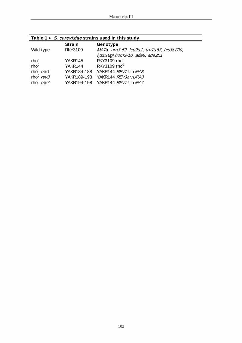
Manuscript III
Table 1 • S. cerevisiae strains used in this study Strain Genotype Wild type
RKY3109 MATa, ura3-52, leu2∆1, trp1∆63, his3∆200, lys2∆Bgl,hom3-10, ade8, ade2∆1
rho- YAKR145 RKY3109 rho-
rho0 YAKR144 RKY3109 rho0
rho0 rev1 YAKR184-188 YAKR144 REV1∆::URA3 rho0 rev3 YAKR189-193 YAKR144 REV3∆::URA3 rho0 rev7 YAKR194-198 YAKR144 REV7∆::URA7
103
Manuscript III
Figure Legends: Figure 1:
Strains with mtDNA mutations (rho-) or strains lacking mtDNA (rho0) were generated by treatment of strains with ethidium bromide (29). The mutants were selected as cells unable to form colonies on yeast extract-peptone-glycerol (YPG) plates. Wild type and rho- cells show mtDNA after cells were stained with DAPI (4,6-diamidino-2-phenylindole). DAPI stains both the nuclear and mtDNA. The rho0
cells show nuclear DNA staining but they do not show punctuate staining indicating loss of mtDNA. The rho- status was verified by genetic crossing with rho0 cells.
Fluorescence was examined using a DAPI optimized filter on Zeiss-Axiovert 135 TV inverted microscope equipped with a PXl camera (SEBSYS photometrica). Figure 2 A and B:
Strains were tested in CAN1R assay as described in Methods. The average median frequency in each assay is presented. CAN1S mutation assay detects frequency of CAN1S to CAN1R forward mutations. A: Frequency of mutation in CAN1 gene in yeast strains defective in mitochondrial function. B: Frequency of mutation in CAN1 gene after respiratory inhibition. Inhibition of respiration was carried out as described in material methods. The data presented are an average of at least 10 independent cultures. Figure 3 A and B:
Production of reactive oxygen species in yeast strains lacking mitochondrial function. A) superoxide and B) hydrogen peroxide. Free radical production was measured as described in Methods. Superoxide was measured by using dihydroethidium (DHE) and 5-(and-6)-chloromethyl-2’,7’-dichlorodihydrofluorescein diacetate (CM-H2DCFDA) was used to measure H2O2. Figure 4:
Mutation frequency in yeast strain lacking rev1, rev3 or rev7 gene expression. Strains were tested in CAN1R assay as described in Methods. The average median frequency in each assay is presented. CAN1S mutation assay detects frequency of CAN1S to CAN1R forward mutations. The data presented are an average of at least 10 independent cultures.
104

Manuscript III
Figure 1
rho0 rho-Wild type rho0 rho-Wild type rho0 rho-Wild type
Figure 2 A
0
10
20
30
40
50
60
WT rho- rho0
Mut
atio
n fr
eque
ncy
per1
08 cells
105
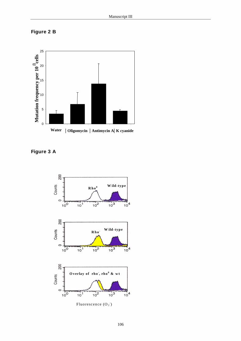
Manuscript III
Figure 2 B
0
5
10
15
20
25
Water | Oligomycin | Antimycin A| K cyanide
Mut
atio
n fr
eque
ncy
per
108 ce
lls
Figure 3 A
F luo rescence (O 2-)
R h o 0 W ild -typ e
W ild -typ eR h o -
O verlay o f rh o -, rh o 0 & w t
106
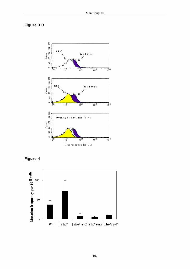
Manuscript III
Figure 3 B
F lu o re sc e n c e (H 2 O 2 )
O v e r la y o f r h o -, r h o 0 & w t
R h o -
R h o 0
W ild -ty p e
W ild -ty p e
Figure 4
0
50
100
WT | rho0 | rho0 rev1 | rho0 rev3 | rho0 rev7
Mut
atio
n fr
eque
ncy
per
108
cells
107

Manuscript IV
Manuscript IV
Anne Karin Rasmussen, Lene Juel Rasmussen, and Keshav K. Singh. Mitochondrial
dysfunction suppresses the mutator phenotype of Saccharomyces cerevisiae superoxide
dismutase deficient cells. Manuscript in preparation.
108

Manuscript IV
Mitochondrial dysfunction suppresses the mutator phenotype of
Saccharomyces cerevisiae superoxide dismutase deficient cells
Anne Karin Rasmussen1,2, Lene Juel Rasmussen1, and Keshav K. Singh2
1Department of Life Sciences and Chemistry, Roskilde University, 4000 Roskilde,
Denmark, 2Johns Hopkins Oncology Center, Bunting-Blaustein Cancer Research Building,
1650 Orleans Street, Baltimore MD 21231-1000, USA.
Corresponding author:
Lene Juel Rasmussen, Department of Life Sciences and Chemistry, Roskilde University,
4000 Roskilde, Denmark, Phone: (+45) 46742728, Fax: (+45) 46743011, Email: [email protected]
Keshav K. Singh, Johns Hopkins Oncology Center, Bunting-Blaustein Cancer Research
Building, 1650 Orleans Street, Room 143, Baltimore, MD 21231, USA, Phone: 410-614-
5128, Fax: 410-502-7234, Email: [email protected]
109
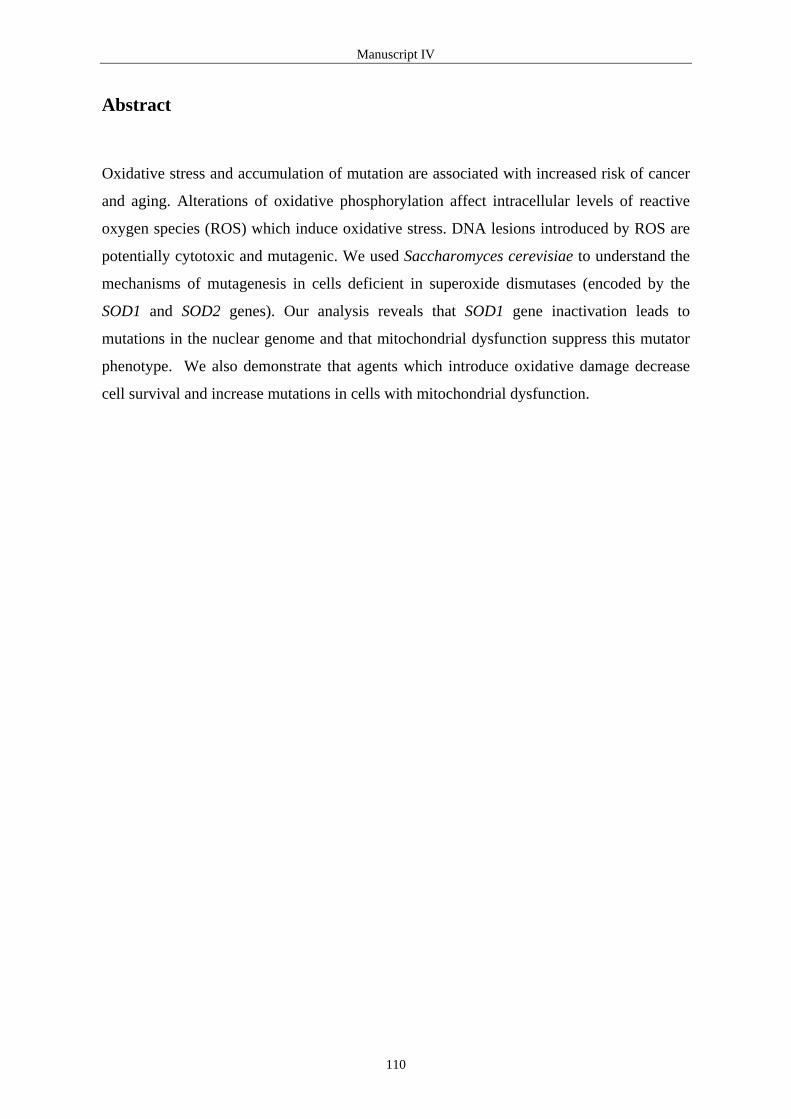
Manuscript IV
Abstract
Oxidative stress and accumulation of mutation are associated with increased risk of cancer
and aging. Alterations of oxidative phosphorylation affect intracellular levels of reactive
oxygen species (ROS) which induce oxidative stress. DNA lesions introduced by ROS are
potentially cytotoxic and mutagenic. We used Saccharomyces cerevisiae to understand the
mechanisms of mutagenesis in cells deficient in superoxide dismutases (encoded by the
SOD1 and SOD2 genes). Our analysis reveals that SOD1 gene inactivation leads to
mutations in the nuclear genome and that mitochondrial dysfunction suppress this mutator
phenotype. We also demonstrate that agents which introduce oxidative damage decrease
cell survival and increase mutations in cells with mitochondrial dysfunction.
110

Manuscript IV
Introduction
Mitochondria generate more than 80% of cellular energy in the form of ATP. In addition,
these organelles carry out other cellular functions, such as respiration and heme, lipid,
amino acid and nucleotide biosynthesis (Kang et al., 1998). Mitochondrial respiration is a
major endogenous source of reactive oxygen species (ROS), including superoxide radical
(O2-), hydrogen peroxide (H2O2), and hydroxyl radical (HO•) (Cadenas and Davies, 2000).
These ROS also arise from external environmental factors such as drugs, radiation, and
heavy metals (Cadenas, 1989). As a result, ROS cause damage to proteins, lipids, and
nucleic acids and thereby compromise cell viability. Both prokaryotic and eukaryotic cells
have developed defense mechanisms against the lethal and mutagenic effect of ROS.
Superoxide dismutases (SOD) are enzymes that eliminate O2- and thus protect cells from
damage induced by free radicals (Halliwell and Gutterridge, 1985; Fridovich, 1995;
Oberley and Buettner, 1979). ROS are also reduced by intracellular antioxidant enzymes
such as glutathione peroxidase (GPx) and catalase (CAT) as well as some antioxidant
molecules like glutathione (Raha & Robinson, 2001; Jamieson, 1998). Therefore, the
balance between ROS production and antioxidant defenses determines the degree of
oxidative stress.
Superoxide is the primary oxygen free radical produced by the mitochondrial respiratory
chain. However, O2- is rapidly dismutated to H2O2 by either Sod2 (mitochondria) or Sod1
(nucleus, cytoplasm, and lysosomes) (Srinivasan et al., 2000). Hydrogen peroxide is
reduced to water by CAT and GPx (Wallace, 1998). Saccharomyces cerevisiae cells
deficient in Sod1 activity show a variety of phenotypes such as growth inhibition under
aerobic conditions, sensitivity to killing by menadione or freeze-thawing, decreased
viability in stationary phase, auxothropy for the amino acids methionine and lysine as well
as increased spontaneous mutation rates under aerobic growth conditions. In contrast,
cells deficient in Sod2 activity have a less dramatic phenotype when grown in glucose
media but are oxygen-sensitive and grow poorly on carbon sources that require respiration
for metabolization. The phenotype of the sod1sod2 double mutant strain includes all
phenotypes of the single mutants (Gralla and Valentine, 1991; Liu et al., 1992; Longo et
al., 1996; Park et al., 1998).
111

Manuscript IV
Oxidative phosphorylation generates ROS which suggests a role for mitochondrial
activity in cell survival and mutagenesis. Guidot et al. (1993) found that S. cerevisiae
cells deficient in the mitochondrially-located superoxide dismutase (Sod2) failed to grow
normally in hyperoxia. In contrast, strains deficient in both Sod2 and mitochondrial
activity (rho0) grew normally in hyperoxia. In addition, the viability was partially
restored by mutations, which disrupt the electron transport chain. Lack of respiration also
prevents viability loss of sod1 and sod1sod2 double mutant strains (Longo et al., 1996).
These data imply that mitochondrial function and respiration play an important role in
viability of sod mutant strains. Studies have shown that rho0 strains as well as strains
carrying deletions in their mitochondrial DNA (rho-), are more sensitive to killing by O2-
and H2O2 than wild type cells (Flatter-O'Brien et al., 1993; Jamieson, 1992; Collinson and
Dawes, 1992; Grant et al., 1997).
Mammalian cells respond to oxidative stress by increasing antioxidant enzymes, which are
capable of detoxifying reactive oxygen species (Akashi et al., 1995; Brambilla et al., 1997;
Esposito et al., 1999; de Hann et al., 1998). However, there exists tissue specificity in the
cellular response to oxidative stress. Skeletal muscle dramatically induced SOD2
expression whereas heart only increased SOD2 expression modestly upon oxidative stress.
In contrast, expression of GPx1 was induced similarly in both tissues. In S. cerevisiae, the
expression of both superoxide dismutases, SOD1 and SOD2, glutathione peroxidase GPX2,
CTT1 encoding the cytosolic catalase T, and glutathione reductase GLR, which catalyses
reduction of glutathione disulfide (GSSG) to glutathione (reduced form, GSH) are induced
in response to oxidative stress (Galiazzo et al., 1987; Gasch et al., 2000; Grant et al., 1996;
Inoue et al., 1999; Marchler et al., 1993).
In this paper we examined the role of Sod1 and Sod2 in mitochondria-mediated
mutagenesis in S. cerevisiae. We show that cells deficient in Sod activity increase
spontaneous mutation levels in the nuclear genome and that this mutator phenotype is
suppressed by inactivation of mitochondrial activity. In order to characterize the role of
mitochondrial activity in mutagenesis and cell survival we treated wild type and rho0 cells
with various drugs that affect intracellular levels of ROS. We show that both H2O2 and
menadione increase spontaneous mutations in nuclei; but that rho0 cells are 10-20 fold
more sensitive than wild type cells to mutations introduced by these agents. Furthermore,
both H2O2 and menadione decrease survival of rho0 cells indicating that cells deficient in
112

Manuscript IV
mitochondrial activity are impaired in repair of DNA damage introduced by these agents.
We used DNA microarray hybridization analysis to further characterize the cellular
response to oxidative stress. We compared gene expression patterns in sod1 and sod1rho0
mutant strains. We did not detect any increase in the expression of the majority of DNA
repair genes in sod1rho0 cells compared to sod1 cells. However, gene expression analysis
revealed that genes involved in DNA repair are induced in sod1 compared to wild type
cells.
113

Manuscript IV
Materials and Methods Media and strains. Growth media were prepared as described in Sherman et al., 1994.
The S. cerevisiae strains used in this study are derived from RKY3109 (Ni et al., 1999) and
provided by Dr. Richard Kolodner (Ludwig Institute for Cancer Research, La Jolla, CA) or
strain DL1 (D273-10B) (Paul et al., 1989). The strains used in this study are listed in Table
1.
Generation of yeast strains. Mutations in the superoxide dismutase genes (sod1 and sod2)
were introduced by one-step gene deletion/replacement in RKY3109. The plasmids pUC-
SOD1∆::URA3-A and pG-SOD2∆::TRP1-A were provided by Dr. Gralla (Department of
Chemistry and Biochemistry, University of California, Los Angeles, CA). The sod1 mutant
strain YAKR129 was generated using the SOD1::URA3 deletion/disruption cassette as
described in (Gralla and Valentine, 1991). The sod2 mutant strain was generated similarly
using the SOD2::TRP1 deletion/disruption cassette (Marres et al., 1985). The sod deletion
derivatives were verified by PCR analysis (data not shown).
Generation of rho- and rho0 strains. Strains with mitochondrial mutations (rho-) or
strains lacking mtDNA (rho0) were generated by treatment of wild type, sod1 and sod2
strains with ethidium bromide (Sherman et al., 1994). The mutants were selected as cells
unable to form colonies on extract-pentose-glycerol (YPG) plates. The loss of mtDNA was
verified by DAPI (4,6-diamidino-2-phenylindole) staining (data not shown).
Determination of spontaneous mutation frequency. Mutation frequency was determined
using fluctuation test method described in (von Borstel, 1978). For each strain, five to
seven independent cultures were grown in 5 ml yeast extract-pentose-dextrose (YPD).
Appropriate dilutions were plated on YPD agar plates to determine the number of viable
cell in the cultures. The frequency of forward mutation at the CAN1 gene locus was
determined from the number of canavanine resistant colonies that grew on selective
minimal dropout plates (SD/-Arg + 60 mg/liter canavanine). The colonies were counted
after incubation at 30oC for 3-5 days for wild type and rho0 cells and for 11-14 days for sod
mutant strains. The data were analyzed by the method of the median (Lea 1949, von
Borstel, 1978). Each experiment was repeated at least twice.
114

Manuscript IV
Drug treatment. Overnight cultures grown in YPD medium were diluted into fresh
growth medium at A600 of 0.15 and incubated until cell density reached A600 of 0.4 - 0.5.
Aliquots of 5 ml were treated with various concentrations of drugs at 30oC with vigorous
shaking for 1 hr. The concentrations of the drugs were: H2O2 (0, 2 and 4 mM), 4-NQO (4-
Nitroquinole N-oxide) (0, 2 and 4 µg/ml), adriamycin (0, 20 and 40 µg/ml), menadione
sodium bisulfite (0, 10 and 20 mM), and diamide (0, 25 and 50 mM). Relative cell survival
was determined by immediately diluting the samples into sterile H2O and plating on YPD
plates. Mutation frequency was determined from the number of canavanine resistant
colonies by the method of the median (von Borstel, 1978). The yeast cells were collected
by centrifugation, washed in water before plating on SD/-Arg + canavanine plates. All
reagents were obtained from Sigma Chemical Co. (St Louis, MO) with the exception of
adriamycin (Johns Hopkins Hospital, MD).
Gene array analysis. Total RNA was isolated from log-phase S. cerevisiae strains
according to manufactures guide lines using RNeasy (QIAGEN). Total RNA (5 µg) was
converted into double stranded cDNA by GIBCO BRL's SuperScript Choice system for
cDNA synthesis (LifeTechnologies) and a T7-(dT)24 oligomer provided by Research
Genetics (Huntsville, AL). Double stranded cDNA was purified by phenol/chloroform
extraction and ethanol precipitation. In vitro transcription was performed with T7 RNA
polymerase following the instructions from BioArray high yield RNA transcript Labeling
kit from Enzo (distributed by Affymetrix). The Biotin-labeled cRNA was purified on an
affinity resin (RNeasy mini cleanup, QIAGEN) and the amount of labeled cRNA was
determined by measuring absorbance at 260 nm and using the convention that 1 OD at 260
nm corresponds to 40 µg/ml RNA. The Yeast Genome S98 Array (Affymetrix) containing
approximately 6,400 open reading frames (ORFs) of S. cerevisiae genome was used for
gene expression analysis. Hybridization, reading and analysis were performed by Research
Genetics (Huntsville, AL). The classification of genes into functional groups was done as
described in MIPS database, Munich Information Center for Protein Sequences (Mewes et
al., 1997; http://mips.gsf.de).
115

Manuscript IV
Results and Discussion
Mitochondrial dysfunction prevents mutations in the nuclear genome in superoxide
dismutase deficient cells (Sod1 and Sod2) deficient cells.
Saccharomyces cerevisiae coq3sod1 double mutant deficient in superoxide dismutase
activity and oxidative phosphorylation show increased viability compared to the superoxide
deficient sod1 mutant alone (Longo et al., 1996). This indicates that free radicals generated
during oxidative phosphorylation in mitochondria are cytotoxic and that superoxide
dismutases protect the cell from the lethal effect of mitochondrial-mediated damage. In
order to characterize the interaction between mitochondrial-mediated mutagenesis and
repair in more detail we measured spontaneous mutation frequencies. We applied a
mutation detection assay that measures the stability of the nuclear genome. The CAN1
gene of S. cerevisiae encodes a transmembrane amino acid transporter that renders the cell
sensitive to the lethal arginine analogue, canavanine. Any inactivating mutation in this
gene results in a canavanine resistant phenotype (CANR). We calculated the frequency of
canavanine resistant colonies as a measure of spontaneous nuclear mutational events in
sod1 and sod2 mutant strains (Table 2). As expected, based on earlier work (Gralla and
Valentine, 1991; Longo et al., 1997), mutation frequencies are increased in the sod1 mutant
strain compared to the wild type parental strain (Table 2). Inactivation of the sod2 gene
increased mutation frequencies about 2-fold, which is similar to the mutator phenotype
generated by inhibiting mitochondrial activity (rho0 or rho-) (Rasmussen et al., (III)). The
increased mutation frequencies observed in superoxide dismutase deficient cells were
suppressed by inactivation of mitochondrial activity (sod1rho0 and sod2rho0) (Table 2).
These data suggest that the mutator phenotype observed in superoxide dismutase deficient
cells is dependent on mitochondrial activity.
Differential expression of repair genes in superoxide dismutase deficient strains.
We used genome analysis by DNA microarray hybridization analysis to identify DNA
repair transcripts differentially expressed as a result of mitochondrial dysfunction and
superoxide dismutase deficiency (Table 3). We found that a large number of DNA repair
genes acting in various DNA repair pathways are induced by the sod1 mutation (Table 3).
The differentially expressed genes included not only DNA repair genes known to repair
oxidative DNA damage (NTG2), but also genes involved in repair of other kinds of DNA
116

Manuscript IV
damage such as base excision repair (MAG1), direct reversal repair (MGT1), nucleotide
excision repair (RAD10, RAD14, RAD16, RAD26 and RAD28), recombinational repair
(RAD50, RAD52, RAD59, SGS1, MUS81, and PSO1), mismatch repair (MSH3), and
meiotic recombination (MSH4). It has been shown that the transcript of MAG1, encoding a
3-methyladenine DNA glycosylase, is induced after treatment with the methylating agent
methylmethane sulfonate (MMS) as well as with the oxidizing agents H2O2 and menadione
(Jelinsky and Samson, 1999; Gasch et al., 2001). The induction of DNA repair genes
observed in the sod1 mutant strain suggests that one or more shared transcription factors
responsible for expression of DNA repair genes are activated in response to endogenous
DNA damage in superoxide dismutase deficient cells.
Further analysis of gene expression patterns revealed that several checkpoint genes were
induced in the sod1 mutant strain (Table 3). The MEC3 gene is a checkpoint gene that
together with RAD9, RAD17, RAD24 and DDC1 are required for detection of DNA damage
and activation of the checkpoint pathways (Elledge, 1996, Zhou and Elledge, 2000).
Induction of MEC3 and RAD9 suggest that sod1 mutant cells generate DNA damage that is
detected by the checkpoint pathways.
We show in Table 2 that the mutator phenotype of the sod1 mutant strain is suppressed by
inactivating mitochondrial activity (sod1rho0). We compared gene expression patterns of
the sod1 mutant strain with the sod1rho0 mutant strain in order to identify DNA repair
genes involved in this suppression. Interestingly, only seven DNA repair genes were
differentially expressed (Table 4). One of these is the OGG1 gene encoding the 8-
oxoguanine DNA glycosylase. Both purine and pyrimidine residues in DNA are sensitive
to ROS-mediated DNA damage (Croteau and Bohr, 1997). However, the most common
purine DNA lesion is the mutagenic 8-hydroxyguanine (8-oxoG) (Steenken and Jovanovic,
1997; Burrows and Muller, 1998). We have recently shown that complete loss of the
mitochondrial genome (rho0) results in lower levels of ROS (Rasmussen et al., (III)).
Therefore, it is tempting to speculate that the sod1 mutant strain induces Ogg1 in response
to high levels of ROS.
117

Manuscript IV
Cells deficient in mitochondrial activity are sensitive to H2O2 and menadione induced
DNA damage.
Studies have shown that rho- and rho0 cells, which are respiratorically deficient, are more
sensitive to the cytotoxic effect of ROS (Flatter-O'Brien et al., 1993; Jamieson, 1992;
Collinson and Dawes, 1992; Grant et al., 1997). We treated exponentially growing wild
type, rho-, and rho0 cells with various drugs that affect intracellular levels of ROS to
determine the degree of sensitivity and mutation frequencies for each drug (Figure 1).
Adriamycin (Doxorubicin) is an anti-tumor drug that can inhibit DNA replication by direct
intercalation between DNA base pairs (Singal et al., 1997). Single electron donors such as
NADPH cytochrome P450 reductase can reduce the quinoid ring in adriamycin to
semiquinone. In the presence of molecular oxygen the semiquinone radicals are rapidly
oxidized to the parent compound in a process, which generate O2- (Olson and Mushlin,
1990). NADPH-cytochrome P450 reductase catalyzes the reduction of menadione leading
to the formation of semiquinones that is readily autooxidized generating O2- (Lind et al.,
1982). 4-nitroquinoline (4-NQO) is metabolized in vivo to the carcinogenic 4-
hydroxyaminoquinoline 1-oxide (4-HAQO), which is oxidized generating H2O2 (Hozumi,
1969). However, 4-NQO also undergoes redox cycling to generate superoxide, which can
be converted to H2O2 and form oxidative DNA damage such as 8-oxoG and DNA strand
breaks (Nunoshiba and Demple, 1993; Yamamoto et al., 1993). In addition, acylated 4-
HAQO reacts with DNA to form stable quinoline-purine monoadducts at the N2 of guanine
and N6 of adenine and 4-NQO is therefore also known as a "UV-mimetic" (Ramotar et al.,
1998). Finally, diamide induces oxidation of cysteine SH-residues of proteins present in
mitochondrial membranes and this oxidation also induces formation of O2- (de Souza and
Geibel, 1999).
As expected, based on earlier work (Flatter-O'Brien et al., 1993; Jamieson, 1992; Collinson
and Dawes, 1992; Grant et al., 1997), rho0 cells are more sensitive to killing by H2O2 and
menadione compared to wild type. We showed that these agents at the same time increased
spontaneous mutations in rho0 cells (Figure 1A and 1B, Table 5). In contrast,
mitochondrial dysfunction mediated resistance to killing by 4-NQO and adriamycin as well
as increased 4-NQO-induced mutations (Figure 1D and 1E, Table 5). A phenotype not only
observed for yeast as human HeLa rho0 cells have been shown to be more resistant to
adriamycin than HeLa rho+ cells (Singh et al., 1999). We did neither detect any difference
in cell survival nor any difference in spontaneous mutations in rho0 cells after treatment
with diamide (Figure 1C, Table 5). These results suggest that antioxidant and/or repair
118

Manuscript IV
defense against damage generated by H2O2 and menadione is impaired in rho0 cells.
However, the data also indicate that the mutagenic and cytotoxic effect of 4-NQO and
adriamycin can be separated in rho0 cells.
The transcript of REV1 is induced in rho0 cells compared to wild type cells (Rasmussen et
al., III). The Rev1 protein is involved in error-prone translesion synthesis and possesses a
transferase activity, which incorporate dCMP opposite abasic site in the DNA template
(Lawrence and Maher, 2001). The Rev1 protein is also involved in bypass of a T-T (6-4)
UV photoproduct (Nelson et al., 2000). Thus, the enzymatic nature of this Rev1p
function is not yet fully understood. We are currently testing a Rev1-model to see if Rev1
can bypass DNA lesions introduced by 4-NQO (UV-mimetic) but not H2O2 and
menadione.
119

Manuscript IV
Acknowledgements
We thank Dr. Richard Kolodner for the yeast strain RKY3109. Research in our
laboratories is supported by grants from National Institutes of Health and American Heart
Association (KKS), Danish Cancer Society (LJR), Danish Natural Science Council (LJR),
Carlsberg Foundation (LJR), Denmark-America Foundation (AKR), Frimodt-Heineke
Foundation (AKR), Frederik and Marie Beyers Fellowship (AKR), Christian and Ottilia
Brorsons Fellowship (AKR), Grosserer L.F. Foghts Foundation (AKR), Julie von Müllens
Foundation (AKR), Købmand Sven Hansen and Hustru Ina Hansens Foundation (AKR),
and Anna and Dagny Hjerrilds Foundation (AKR). We thank the members of our
laboratory for the critical reading of this manuscript.
120

Manuscript IV
References Akashi M, Hachiya M, Paquette RL, Osawa Y, Shimizu S, Suzuki G. Irradiation increases manganese superoxide dismutase mRNA levels in human fibroblasts. Possible mechanisms for its accumulation. J Biol Chem. 1995;270(26):15864-9. Brambilla L, Cairo G, Sestili P, O'Donnel V, Azzi A, Cantoni O. Mitochondrial respiratory chain deficiency leads to overexpression of antioxidant enzymes. FEBS Lett. 1997;418(3):247-50. Cadenas E. Biochemistry of oxygen toxicity. Annu Rev Biochem. 1989;58:79-110. Cadenas E, Davies KJ. Mitochondrial free radical generation, oxidative stress, and aging. Free Radic Biol Med. 2000;29(3-4):222-30. Collinson LP, Dawes IW. Inducibility of the response of yeast cells to peroxide stress. J Gen Microbiol. 1992;138 ( Pt 2):329-335. Croteau DL, Bohr VA. Repair of oxidative damage to nuclear and mitochondrial DNA in mammalian cells. J Biol Chem. 1997;272(41):25409-12. de Haan JB, Bladier C, Griffiths P, Kelner M, O'Shea RD, Cheung NS, Bronson RT, Silvestro MJ, Wild S, Zheng SS, Beart PM, Hertzog PJ, Kola I. Mice with a homozygous null mutation for the most abundant glutathione peroxidase, Gpx1, show increased susceptibility to the oxidative stress-inducing agents paraquat and hydrogen peroxide. J Biol Chem. 1998;273(35):22528-36. de Souza Pereira R, Geibel J. Direct observation of oxidative stress on the cell wall of Saccharomyces cerevisiae strains with atomic force microscopy. Mol Cell Biochem. 1999;201(1-2):17-24. Elledge SJ. Cell cycle checkpoints: preventing an identity crisis. Science. 1996;274(5293):1664-72. Esposito LA, Melov S, Panov A, Cottrell BA, Wallace DC. Mitochondrial disease in mouse results in increased oxidative stress. Proc Natl Acad Sci U S A. 1999;96(9):4820-5. Flattery-O'Brien J, Collinson LP, Dawes IW. Saccharomyces cerevisiae has an inducible response to menadione which differs from that to hydrogen peroxide. J Gen Microbiol. 1993;139:501-7. Galiazzo F, Schiesser A, Rotilio G. Glutathione peroxidase in yeast. Presence of the enzyme and induction by oxidative conditions. Biochem Biophys Res Commun. 1987;147(3):1200-5. Gasch AP, Spellman PT, Kao CM, Carmel-Harel O, Eisen MB, Storz G, Botstein D, Brown PO. Genomic expression programs in the response of yeast cells to environmental changes. Mol Biol Cell. 2000;11(12):4241-57.
121

Manuscript IV
Gasch AP, Huang M, Metzner S, Botstein D, Elledge SJ, Brown PO. Genomic expression responses to dna-damaging agents and the regulatory role of the yeast atr homolog mec1p. Mol Biol Cell. 2001;12(10):2987-3003. Gralla EB, Valentine JS. Null mutants of Saccharomyces cerevisiae Cu,Zn superoxide dismutase: characterization and spontaneous mutation rates. J Bacteriol. 1991;173(18):5918-20. Grant CM, MacIver FH, Dawes IW. Glutathione is an essential metabolite required for resistance to oxidative stress in the yeast Saccharomyces cerevisiae. Curr Genet. 1996;29(6):511-5. Grant CM, MacIver FH, Dawes IW. Mitochondrial function is required for resistance to oxidative stress in the yeast Saccharomyces cerevisiae. FEBS Lett. 1997;410(2-3):219-22. Guidot DM, McCord JM, Wright RM, Repine JE. Absence of electron transport (Rho 0 state) restores growth of a manganese-superoxide dismutase deficient Saccharomyces cerevisiae in hyperoxia. Evidence for electron transport as a major source of superoxide generation in vivo. J Biol Chem. 1993;268(35):26699-703. Halliwell B, Gutterridge JMC. Free Radicals in Biology and Medicine. Oxford University Press, New York, 1985. Hozumi M. Production of hydrogen peroxide by 4 hydroxyaminoquinoline 1-oxide. Gann. 1969;60(1):83-90. Inoue Y, Matsuda T, Sugiyama K, Izawa S, Kimura A. Genetic analysis of glutathione peroxidase in oxidative stress response of Saccharomyces cerevisiae. J Biol Chem. 1999;274(38):27002-9. Jamieson DJ. Saccharomyces cerevisiae has distinct adaptive responses to both hydrogen peroxide and menadione. J Bacteriol. 1992;174(20):6678-81. Jamieson DJ. Oxidative stress responses of the yeast Saccharomyces cerevisiae. Yeast. 1998;14(16):1511-27. Jelinsky SA, Samson LD. Global response of Saccharomyces cerevisiae to an alkylating agent. Proc Natl Acad Sci U S A. 1999;96(4):1486-91. Kang D, Takeshige K, Sekiguchi M, Singh KK. Mitochondrial DNA mutations in aging, disease and cancer. Springer-Verlag and R.G. Landes Company (ed. Keshav K. Singh) 1998;1-15 Lawrence CW, Maher VM. Mutagenesis in eukaryotes dependent on DNA polymerase zeta and Rev1p. Philos Trans R Soc Lond B Biol Sci. 2001;356(1405):41-6. Lea DE, Coulson CA. The distribution of numbers of mutants in bacterial populations. J Genet. 1949; 49:264-85.
122

Manuscript IV
Lind C, Hochstein P, Ernster L. DT-diaphorase as a quinone reductase: a cellular control device against semiquinone and superoxide radical formation. Arch Biochem Biophys. 1982;216(1):178-85. Liu XF, Elashvili I, Gralla EB, Valentine JS, Lapinskas P, Culotta VC. Yeast lacking superoxide dismutase. Isolation of genetic suppressors. J Biol Chem. 1992;267(26):18298-302. Longo VD, Gralla EB, Valentine JS. Superoxide dismutase activity is essential for stationary phase survival in Saccharomyces cerevisiae. Mitochondrial production of toxic oxygen species in vivo. J Biol Chem. 1996;271(21):12275-80. Longo VD, Ellerby LM, Bredesen DE, Valentine JS, Gralla EB. Human Bcl-2 reverses survival defects in yeast lacking superoxide dismutase and delays death of wild-type yeast. J Cell Biol. 1997;137(7):1581-8. Marchler G, Schuller C, Adam G, Ruis H. A Saccharomyces cerevisiae UAS element controlled by protein kinase A activates transcription in response to a variety of stress conditions. EMBO J. 1993;12(5):1997-2003. Marres CA, Van Loon AP, Oudshoorn P, Van Steeg H, Grivell LA, Slater EC. Nucleotide sequence analysis of the nuclear gene coding for manganese superoxide dismutase of yeast mitochondria, a gene previously assumed to code for the Rieske iron-sulphur protein. Eur J Biochem. 1985;147(1):153-61. Nelson JR, Gibbs PE, Nowicka AM, Hinkle DC, Lawrence CW. Evidence for a second function for Saccharomyces cerevisiae Rev1p. Mol Microbiol. 2000;37(3):549-54. Mewes HW, Albermann K, Heumann K, Liebl S, Pfeiffer F. MIPS: a database for protein sequences, homology data and yeast genome information. Nucleic Acids Res. 1997;25(1):28-30 Ni TT, Marsischky GT, Kolodner RD. MSH2 and MSH6 are required for removal of adenine misincorporated opposite 8-oxo-guanine in S. cerevisiae. Mol Cell. 1999;4(3):439-44. Nunoshiba T, Demple B. Potent intracellular oxidative stress exerted by the carcinogen 4-nitroquinoline-N-oxide. Cancer Res. 1993;53(14):3250-2. Oberley LW, Buettner GR. Role of superoxide dismutase in cancer: a review. Cancer Res. 1979;39(4):1141-9. Olson RD, Mushlin PS. Doxorubicin cardiotoxicity: analysis of prevailing hypotheses. FASEB J. 1990;4(13):3076-86. Park JI, Grant CM, Davies MJ, Dawes IW. The cytoplasmic Cu,Zn superoxide dismutase of saccharomyces cerevisiae is required for resistance to freeze-thaw stress. Generation of free radicals during freezing and thawing. J Biol Chem. 1998;273(36):22921-8.
123

Manuscript IV
Paul MF, Velours J, Arselin de Chateaubodeau G, Aigle M, Guerin B. The role of subunit 4, a nuclear-encoded protein of the F0 sector of yeast mitochondrial ATP synthase, in the assembly of the whole complex. Eur J Biochem 1989;185(1):163-71 Raha S, Robinson BH. Mitochondria, oxygen free radicals, and apoptosis. Am J Med Genet. 2001;106(1):62-70. Ramotar D, Belanger E, Brodeur I, Masson JY, Drobetsky EA. A yeast homologue of the human phosphotyrosyl phosphatase activator PTPA is implicated in protection against oxidative DNA damage induced by the model carcinogen 4-nitroquinoline 1-oxide. J Biol Chem. 1998;273(34):21489-96. Sherman F, Fink GR, Hicks JB. Methods in Yeast Genetics: A Laboratory Manual Cold Spring Harbor Laboratory Press, Plainview, NY. 1994. Singal PK, Iliskovic N, Li T, Kumar D. Adriamycin cardiomyopathy: pathophysiology and prevention. FASEB J. 1997;11(12):931-6. Srinivasan C, Liba A, Imlay JA, Valentine JS, Gralla EB. Yeast lacking superoxide dismutase(s) show elevated levels of "free iron" as measured by whole cell electron paramagnetic resonance. J Biol Chem. 2000;275(38):29187-92. Von Borstel, R. C. Measuring spontaneous mutation frequencies in yeast. Methods Cell Biol. 1978 (20):1-24 Wallace DC, Melov S. Radicals r'aging. Nat Genet. 1998;19(2):105-6. Yamamoto K, Inoue S, Kawanishi S. Site-specific DNA damage and 8-hydroxydeoxyguanosine formation by hydroxylamine and 4-hydroxyaminoquinoline 1-oxide in the presence of Cu(II): role of active oxygen species. Carcinogenesis. 1993;14(7):1397-401. Zhou BB, Elledge SJ. The DNA damage response: putting checkpoints in perspective. Nature. 2000;408(6811):433-9.
124

Manuscript IV
Table 1. Saccharomyces cerevisiae strains used in this study Strain Genotype wild type
RKY3109 MATa, ura3-52, leu2∆1, trp1∆63, his3∆200, lys2∆Bgl, hom3-10, ade8, ade2∆1
rho- YAKR145 RKY3109 rho-
rho0 YAKR144 RKY3109 rho0
sod1 YAKR129 RKY3109 sod1::URA3 sod1 rho0 YAKR163-166 RKY3109 rho0
sod2 YAKR133 RKY3109 sod2::TRP1 sod2 rho0 YAKR135 RKY3109 rho0
sod1 sod2 YAKR137 RKY3109, sod1::URA3, sod2::TRP1 wild type DL1 MATα, met rho0 DL1 rho0 DL1 rho0
Table 2. Spontaneous mutation frequencies in superoxide deficient strains Strain
can1r (x10-8) Fold increase
Wild type 15.3 ± 3.7 1 rho- 34.0 ± 5.0 2.2 rho0 39.9 ± 11.7 2.6 sod2 30.5± 5.6 2.0 sod2 rho0 6.4 ± 2.4 0.4 can1r (x10-6) wild type 1.3 ± 0.3 1 sod1 165.5 ± 5.1 127.3 sod1 rho0 8.9 ± 1.8 6.8 Spontaneous mutation frequency in rho-, rho0, sod2, sod2 rho0, sod1 and sod1 rho0 is significantly different (P < 0.01) from wild type, as determined by t-Test: Two-Sample Assuming Equal Variances.
125
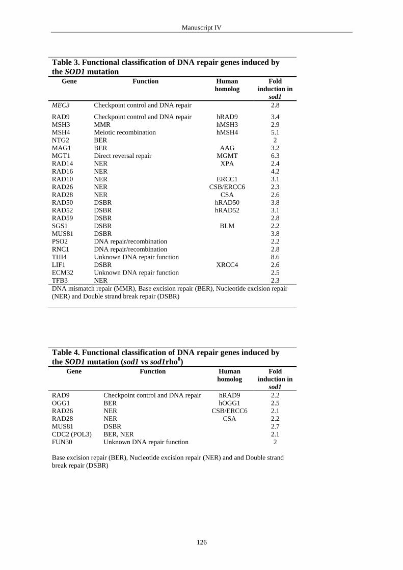
Manuscript IV
Table 3. Functional classification of DNA repair genes induced by the SOD1 mutation
Gene Function Human homolog
Fold induction in
sod1 MEC3 Checkpoint control and DNA repair 2.8
RAD9 Checkpoint control and DNA repair hRAD9 3.4 MSH3 MMR hMSH3 2.9 MSH4 Meiotic recombination hMSH4 5.1 NTG2 BER 2 MAG1 BER AAG 3.2 MGT1 Direct reversal repair MGMT 6.3 RAD14 NER XPA 2.4 RAD16 NER 4.2 RAD10 NER ERCC1 3.1 RAD26 NER CSB/ERCC6 2.3 RAD28 NER CSA 2.6 RAD50 DSBR hRAD50 3.8 RAD52 DSBR hRAD52 3.1 RAD59 DSBR 2.8 SGS1 DSBR BLM 2.2 MUS81 DSBR 3.8 PSO2 DNA repair/recombination 2.2 RNC1 DNA repair/recombination 2.8 THI4 Unknown DNA repair function 8.6 LIF1 DSBR XRCC4 2.6 ECM32 Unknown DNA repair function 2.5 TFB3 NER 2.3 DNA mismatch repair (MMR), Base excision repair (BER), Nucleotide excision repair (NER) and Double strand break repair (DSBR) Table 4. Functional classification of DNA repair genes induced by the SOD1 mutation (sod1 vs sod1rho0)
Gene Function Human homolog
Fold induction in
sod1 RAD9 Checkpoint control and DNA repair hRAD9 2.2 OGG1 BER hOGG1 2.5 RAD26 NER CSB/ERCC6 2.1 RAD28 NER CSA 2.2 MUS81 DSBR 2.7 CDC2 (POL3) BER, NER 2.1 FUN30 Unknown DNA repair function 2 Base excision repair (BER), Nucleotide excision repair (NER) and and Double strand break repair (DSBR)
126

Manuscript IV
Table 5. Spontaneous mutation frequency (x10-8) Spontaneous mutation frequency (x10-7)
H2O2 (mM)
Menadione (mM)
Diamide (mM)
4-NQO (µg/ml)
Adriamycin (µg/ml) Conc. 0 2 4 0 10 20 0 25 50 0 2 4 0 20 40
wt 2.5 6.8 4.0 3.6 1.7 2.4 1.6 4.2 2.2 5.6 34.8 39.2 5.0 6.4 9.7
rho0 3.8 87.6 85.4 6.1 11.6 4.2 5.9 5.8 27.5 158.1 214.8 71.3 115.1 95.3
127

Manuscript IV
Figure Legends
Figure 1. Cell survival of wild type and rho0 cells after drug treatment. Percent
survival is expressed relative to the untreated control culture (100%). Cells were treated
with various concentrations of (A) H2O2, (B) menadione, (C) diamide, (D) 4-NQO, and (E)
adriamycin. Each data point represents an average of duplicate measurements from one
representative experiment. Closed symbols: wild type. Open symbols: rho0.
Figure 1.
0.1
1
10
100
0 20 40
1
10
100
0 2 4
Dose H2O2 (mM)
% S
urvi
val
(A)
1
10
100
1000
0 10 20
Dose menadione (mM)
(B)
1
10
100
0 25 50
(C)
0.1
1
10
100
0 2 4
(D)
(E)
% S
urvi
val
Dose diamide (mM) Dose 4-NQO (µg/ml)
Dose adriamycin (µg/ml)
% S
urvi
val
% S
urvi
val
% S
urvi
val
128

Manuscript IV
Supplemental materials
Table • Genes repressed in DL1 vs sod1∆ cells Probe
Set Name ORF Gene
name AC
Fold Change in DL1
Descriptions
DNA Damage Checkpoint 10048_at YLR288C MEC3 P 2.8 Involved in checkpoint control and DNA repair. 6274_at YDR217C RAD9 P 3.4 Cell cycle arrest protein. DNA Mismatch Repair
6787_at YCR092C MSH3 P 2.9 MutS homolog, forms a complex with Msh2p to repair insertion-deletion mispairs.
Meiotic recombination 5385_at YFL003C MSH4 A 5.1 MutS homolog, localizes to discrete sites on meiotic chromosomes, meiosis
specific protein. Base Excision Repair
Oxidative DNA Repair 8609_at YOL043C NTG2 P 2 Endonuclease III-like glycosylase 2. DNA Alkylation Repair 5582_at YER142C MAG1 A 3.2 3-methyladenine DNA glycosylase. Direct Repair
6663_at YDL200C MGT1 A 6.3 6-O-methylguanine-DNA methylase. Nucleotide Excision Repair
9428_at YMR201C RAD14 P 2.4 Human xeroderma pigmentosum group A DNA repair gene homolog. 7250_at YBR114W RAD16 P 4.2 Radiation repair protein, putative DNA helicase. 9749_at YML095C RAD10 A 3.1 Endonuclease (with Rad1p) that degrades single-stranded DNA for repair. 9750_at YML095C RAD10 A 3.2 Questionable ORF 11009_at YJR035W RAD26 P 2.3 DNA-dependent ATPase, homologous to human Cockayne syndrome B
gene ERCC6, that is a putative helicase. 6444_at YDR030C RAD28 P 2.6 Protein involved in the same pathway as Rad26p, has beta-transducin (WD-
40) repeats. Recombination Repair
9099_at YNL250W RAD50 P 3.8 Protein contains a purine-binding domain, two heptad repeats and a hydrophobic tail, Rad50p interacts with Mre11p and Xrs2p in two-hybrid and immunoprecipitation analyses\; it co-localizes to spots with Mre11p and Xrs2p in a rad50s background.
9678_at YML032C RAD52 A 3.1 Questionable ORF. 6539_at YDL059C RAD59 P 2.8 A mutation in this gene results in RADiation sensitivity and recombination
defects, which are general properties of the RAD52 epistasis group mutants. rad59 is epistatic to rad52 for its repair and recombination defects. The RAD59 gene product has homology to the Rad52 protein.
9461_at YMR190C SGS1 P 2.2 Has DNA helicase signature motifs. 6083_at YDR386W MUS81 A 3.8 MMS and UV Sensitive\; Mus81p and Rad54p are found together in a
complex from whole-cell extracts. 9491_at YMR137C PSO2 P 2.2 Interstrand crosslink repair protein. Other/unknown DNA Repair Function
10528_at YKR056W RNC1 P 2.8 Endo-exonuclease yNucR. 4844_at YGR144W THI4 A 8.6 Component of the biosynthetic pathway producing the thiazole precursor of
thiamine. 5107_at YGL090W LIF1 P 2.6 Ligase Interacting Factor 1\; physically interacts with DNA ligase 4 protein. 5524_at YER176W ECM32 P 2.5 DNA Helicase I. 6019_at YDR460W TFB3 P 2.3 TFIIH subunit Tfb3 \; similar to mammalian CAK subunit.
AC = the Absolute Call; The transcript is present (P) or absent (A) for DL1. When the transcript is absent is the fold change an approximation and calculated using the noise level.
129

Concluding Remarks
4. Concluding Remarks
Increased spontaneous mutation frequency is associated with higher risk of cancer.
However, the relative contribution of spontaneous endogenous mutagenesis to
carcinogenesis is not yet known. Among the normal cellular metabolic processes that lead
to elevated spontaneous mutation rates are (Jackson et al., 1998; Rossman & Goncharova,
1998; Rasmussen et al., III):
• DNA polymerase errors,
• Depurination, which gives rise to abasic sites,
• Deamination of deoxycytidine residues (C → U and 5-methyl-C → T),
• Methylation of DNA (O6-methylguanine),
• Damage by oxygen free radicals,
• and mitochondrial dysfunction.
Thus, both a main repair pathway and one or more backup pathways can generally remove
these lesions. For example the main repair pathway for eliminating mutations, caused by
the deoxyguanosine oxidation product 8-oxoG, is BER (hOGG1, hMYH and hMTH).
However, studies in S. cerevisiae have shown that MSH2-MSH6-dependent MMR is
involved in repairing 8-oxoG:A mispairs and 8-oxoG:C base pairs. These results suggest
that MMR is functioning as a backup pathway under high oxidative stress (Ni et al., 1999).
The MMR pathway is also involved in repair of O6-MeG and O6-MeG-triggered cell death
when MGMT repair capacity is saturated (see 2.3 Direct Repair: O6-methylguanine-DNA
methyl-transferase). Furthermore, it has been suggested that the hMLH1 protein, from the
MMR pathway, has an impact on the apoptosis pathway after oxidative stress. The
mitochondria were suggested to be involved in this apoptosis pathway because increased
mitochondrial permeability and cytochrome c release after H2O2 treatment were observed
preferentially in hMLH proficient cell lines (Hardman et al., 2001).
Therefore, it seems like inactivation or dysregulation of the DNA protection system are
important for generating a mutator phenotype. Its importance in human disease is supported
by the discovery that defects in several kinds of DNA repair are known to raise
spontaneous mutation frequency i.e. mutations in MMR genes cause hereditary non-
polyposis colon cancer, HNPCC (Hoeijmakers, 2001).
130

Concluding Remarks
In an attempt to identify new MMR genes, we employed the yeast two-hybrid system, using
the human mismatch repair protein hMSH2 (Rasmussen et al., 2000) or hMLH1 as bait and
a fetal liver cDNA library as prey. We demonstrated that hMSH2 interacts with
exonuclease hEXO1 suggesting that hEXO1 plays a role in MMR. Futhermore, we showed
that hMSH2 and EXO1 are co-expressed at high levels in fetal liver but not in adult liver
where cell turnover is normally very slow. The hEXO1 protein was highly expressed in
several liver cancer cell lines as well as in colon and pancreas adenocarcinomas, but not in
the corresponding non-neoplastic tissue. These results support a role for hEXO1 in cellular
processes such as MMR, recombination repair or replication, which are expected to be
active in proliferating cells (Rasmussen et al., 2000).
The mismatch repair protein hMLH1 is known to interact with hPMS1. However, the exact
biochemical role for the hMLH1-hPMS1 complex has not yet been determined. We showed
that hMLH1 interacts with hPMS1 in the yeast two-hybrid system using hMLH1 as bait and
fetal liver cDNA library as prey. Dot-blot analysis of multiple tissue RNA revealed that
hPMS1 is predominantly expressed in fetal liver, adult liver, pancreas, kidney, testis and
appendix. Unfortunately, we do not have the corresponding dot-blot analysis for hPMS2.
However, it is tempting to speculate that hPMS1 is tissue specific and that the hMLH1-
hPMS1 complex plays an important role in DNA repair in liver. Furthermore, it could be
interesting to compare hPMS1 and hPMS2 expression levels in different cancer cell lines
with non-neoplastic tissue to investigate if any up or down regulation will occur in the
cancer cells.
Mutations in MMR genes are known to cause spontaneous mutations. Defects in
translocation of repair proteins could be a way to inactivate the DNA protection system. We
present preliminary data showing that hMSH2 and hMLH1 interact with an importin-α
homolog and importin-α, respectively. Importin-α is part of the translocation system to the
nucleus. Protein-protein interaction and nuclear localization studies with HNPCC-hMSH2
mutants or HNPCC-hMLH1 mutants together with importin-α and the importin-α homolog
could be a way to determine if mutations in MMR mutant genes found in HNPCC patients
interfere with the translocation system.
131

Concluding Remarks
Another model to inactivate the DNA repair system is oxidative damage. The
mitochondrial theory of aging postulates that accumulation of oxidative damage in the
mtDNA leads to mitochondrial dysfunction that results in energy deficiencies in old cells.
Declined mitochondrial energy capacity could in turn lead to lack in repair (Ames, 1989).
Our results in S. cerevisiae showed that mitochondrial dysfunction caused significantly
higher spontaneous mutation frequencies in nDNA compared to wild type and this was not
due to increased levels of ROS in the cells. However, we demonstrated that disruption of
the respiratory chain function using various mitochondrial inhibitors caused an increase in
spontaneous mutation frequencies, indicating that increased mitochondrial ROS production
can diffuse to the nucleus where it causes DNA damage. Furthermore, in rho0 cells we
found an up-regulation of Rev1 compared to wild type and we showed that mitochondrial-
mediated mutator phenotype of rho0 cells is suppressed by preventing Rev1/Rev3/Rev7-
dependent translesion synthesis (Rasmussen et al., III). Even though we do not know today
how mitochondrial dysfunction leads to spontaneous nuclear DNA damage, mitochondrial
dysfunction elevates the spontaneous mutation frequencies.
The nuclear (Sod1) and mitochondrial (Sod2) superoxide dismutases eliminate the
superoxide anion and thus protect cells from damage induced by free radicals. We find that
strains deficient in SOD activity increase spontaneous mutations and that this mutator
phenotype is suppressed both in sod1∆rho0 and sod2∆rho0 mutant strains. Our results
suggest that mitochondrial dysfunction either reduces oxidative damage to DNA by
preventing formation of ROS and/or increases repair of oxidative DNA damage. We
compared gene expression patterns of the sod1 mutant strain with the sod1rho0 mutant
strain in order to identify DNA repair genes involved in this suppression. This analysis
suggested that sod1rho0 cells did not increase repair response compared to sod1 cells. This
infers that removal of mitochondrial function reduces intracellular levels of ROS and
consequently decreases mutagenesis caused by oxidative damage to DNA (Rasmussen et
al., IV).
We found that both H2O2 and menadione treatments reduced cell survival of rho0 cells but
at the same time increased spontaneous mutations in these cells compared to wild type.
Gene expression analysis showed that the induced antioxidant enzymes in rho0 cells were
different from the ones induced in the wild type. Implying that defense against oxidative
132

Concluding Remarks
damage generated by H2O2 and Menadione is different in rho0 cells. These results
emphasize the significance of mitochondrial activity in controlling cell death as well as
mutational events following DNA damage. Proteins involved in such control mechanisms
have been identified and include checkpoint proteins such as the human tumor suppressor
protein p53. It is therefore tempting to consider mitochondria as a new guardian of genomic
stability.
Given the fact that the integrity of the mtDNA is important it could be beneficial to
understand the molecular processes and components responsible for mtDNA repair. Most
DNA glycosylases that have been identified in the nucleus have also been identified in
mitochondria, which indicates that BER is operating in mitochondria.
It has not yet been determined if mitochondrial DNA is repaired by MMR. Our two-hybrid
screenings with hMSH2 or hMLH1 as baits and a fetal liver cDNA library as prey gave no
answers to this question. However, we found that full-length MGMT, when expressed as a
GFP fusion protein in human MCF12A (breast) epithelial cells, were localized to the nuclei
but not to the mitochondria (Rasmussen et al., II). These data suggest that the
alkyltransferase mechanism, which seems to be operating within the mitochondrion in
mammalian species, is not MGMT in human breast epithelial cells.
Expression of the mitochondrial genes are required to maintain proper function of the
organelle, suggesting that even a slight alteration of mitochondrial function may have
profound effects. Interestingly, one of the profound features of cancer cells is their
defective mitochondrial function (Warburg, 1956; Polyak et al., 1998; Fliss et al., 2000).
133

References
References
Acharya S, Wilson T, Gradia S, Kane MF, Guerrette S, Marsischky GT, Kolodner R, Fishel R. hMSH2 forms specific mispair-binding complexes with hMSH3 and hMSH6. Proc Natl Acad Sci U S A. 1996;93(24):13629-34. Allen DJ, Makhov A, Grilley M, Taylor J, Thresher R, Modrich P, Griffith JD. MutS mediates heteroduplex loop formation by a translocation mechanism. EMBO J. 1997;16(14):4467-76. Altshuler KB, Hodes CS, Essigmann JM. Intrachromosomal probes for mutagenesis by alkylated DNA bases replicated in mammalian cells: a comparison of the mutagenicities of O4-methylthymine and O6-methylguanine in cells with different DNA repair backgrounds. Chem Res Toxicol. 1996;9(6):980-7. Ames BN. Endogenous oxidative DNA damage, aging, and cancer. Free Radic Res Commun. 1989;7(3-6):121-8. Anderson CT, Friedberg EC. The presence of nuclear and mitochondrial uracil-DNA glycosylase in extracts of human KB cells. Nucleic Acids Res. 1980;8(4):875-88. Anderson S, Bankier AT, Barrell BG, de Bruijn MH, Coulson AR, Drouin J, Eperon IC, Nierlich DP, Roe BA, Sanger F, Schreier PH, Smith AJ, Staden R, Young IG. Sequence and organization of the human mitochondrial genome. Nature. 1981;290(5806):457-65. Ayi TC, Loh KC, Ali RB, Li BF. Intracellular localization of human DNA repair enzyme methylguanine-DNA methyltransferase by antibodies and its importance. Cancer Res. 1992;52(23):6423-30. Bai C, Elledge SJ. Searching for interaction proteins with the Two-Hybrid system I (The yeast Two-Hybrid system ed. Bartel PL, Fields S) Oxford University Press, Inc.,New York, NY. 1997:11-28 Bai J, Rodriguez AM, Melendez JA, Cederbaum AI. Overexpression of catalase in cytosolic or mitochondrial compartment protects HepG2 cells against oxidative injury. J Biol Chem. 1999;274(37):26217-24. Bai J, Cederbaum AI. Mitochondrial catalase and oxidative injury. Biol Signals Recept. 2001;10(3-4):189-99. Bailly V, Verly WG, O'Connor T, Laval J. Mechanism of DNA strand nicking at apurinic/apyrimidinic sites by Escherichia coli [formamidopyrimidine]DNA glycosylase. Biochem J. 1989;262(2):581-9. Bambara RA, Murante RS, Henricksen LA. Enzymes and reactions at the eukaryotic DNA replication fork. J Biol Chem. 1997;272(8):4647-50. Belanich M, Randall T, Pastor MA, Kibitel JT, Alas LG, Dolan ME, Schold SC Jr, Gander M, Lejeune FJ, Li BF, White AB, Wasserman P, Citron ML, Yarosh DB. Intracellular Localization and intercellular heterogeneity of the human DNA repair protein O6-methylguanine-DNA methyltransferase. Cancer Chemother Pharmacol. 1996;37(6):547-55. Bestwick RK, Moffett GL, Mathews CK. Selective expansion of mitochondrial nucleoside triphosphate pools in antimetabolite-treated HeLa cells. J Biol Chem. 1982;257(16):9300-4. Bhattacharyya NP, Skandalis A, Ganesh A, Groden J, Meuth M. Mutator phenotypes in human colorectal carcinoma cell lines. Proc Natl Acad Sci U S A. 1994;91(14):6319-23. Bianchi NO, Bianchi MS, Richard SM. Mitochondrial genome instability in human cancers. Mutat Res. 2001;488(1):9-23. Bogdanov MB, Ramos LE, Xu Z, Beal MF. Elevated "hydroxyl radical" generation in vivo in an animal model of amyotrophic lateral sclerosis. J Neurochem. 1998;71(3):1321-4.
134

References
Boldogh I, Milligan D, Lee MS, Bassett H, Lloyd RS, McCullough AK. hMYH cell cycle-dependent expression, subcellular localization and association with replication foci: evidence suggesting replication-coupled repair of adenine:8-oxoguanine mispairs. Nucleic Acids Res. 2001;29(13):2802-9. Boulikas T. Nuclear import of DNA repair proteins. Anticancer Res. 1997;17(2A):843-63. Bouziane M, Miao F, Bates SE, Somsouk L, Sang BC, Denissenko M, O'Connor TR. Promoter structure and cell cycle dependent expression of the human methylpurine-DNA glycosylase gene. Mutat Res. 2000;461(1):15-29. Boveris A. Mitochondrial production of superoxide radical and hydrogen peroxide. Adv Exp Med Biol. 1977;78:67-82. Boveris A, Chance B. The mitochondrial generation of hydrogen peroxide. General properties and effect of hyperbaric oxygen. Biochem J. 1973;134(3):707-16. Bowers J, Tran PT, Joshi A, Liskay RM, Alani E. MSH-MLH complexes formed at a DNA mismatch are disrupted by the PCNA sliding clamp. J Mol Biol. 2001 Mar 9;306(5):957-68. Bronstein SM, Cochrane JE, Craft TR, Swenberg JA, Skopek TR. Toxicity, mutagenicity, and mutational spectra of N-ethyl-N-nitrosourea in human cell lines with different DNA repair phenotypes. Cancer Res. 1991;51(19):5188-97. Bronstein SM, Skopek TR, Swenberg JA. Efficient repair of O6-ethylguanine, but not O4-ethylthymine or O2-ethylthymine, is dependent upon O6-alkylguanine-DNA alkyltransferase and nucleotide excision repair activities in human cells. Cancer Res. 1992;52(7):2008-11. Bruijn LI, Becher MW, Lee MK, Anderson KL, Jenkins NA, Copeland NG, Sisodia SS, Rothstein JD, Borchelt DR, Price DL, Cleveland DW. ALS-linked SOD1 mutant G85R mediates damage to astrocytes and promotes rapidly progressive disease with SOD1-containing inclusions. Neuron. 1997;18(2):327-38. Buermeyer AB, Deschenes SM, Baker SM, Liskay RM. Mammalian DNA mismatch repair. Annu Rev Genet. 1999;33:533-64. Burdett V, Baitinger C, Viswanathan M, Lovett ST, Modrich P. In vivo requirement for RecJ, ExoVII, ExoI, and ExoX in methyl-directed mismatch repair. Proc Natl Acad Sci U S A. 2001;98(12):6765-70. Burrows CJ, Muller JG. Oxidative nucleobase modifications leading to strand scission. Chem. Rev. 1998;98:1109-51. Cadenas E, Davies KJ. Mitochondrial free radical generation, oxidative stress, and aging. Free Radic Biol Med. 2000;29(3-4):222-30. Carethers JM, Hawn MT, Chauhan DP, Luce MC, Marra G, Koi M, Boland CR. Competency in mismatch repair prohibits clonal expansion of cancer cells treated with N-methyl-N'-nitro-N-nitrosoguanidine. J Clin Invest. 1996;98(1):199-206. Ceccotti S, Aquilina G, Macpherson P, Yamada M, Karran P, Bignami M. Processing of O6-methylguanine by mismatch correction in human cell extracts. Curr Biol. 1996;6(11):1528-31. Ceska TA, Sayers JR. Structure-specific DNA cleavage by 5' nucleases. Trends Biochem Sci. 1998;23(9):331-6. Chance B, Sies H, Boveris A. Hydroperoxide metabolism in mammalian organs. Physiol Rev. 1979;59(3):527-605. Cheng KC, Cahill DS, Kasai H, Nishimura S, Loeb LA. 8-Hydroxyguanine, an abundant form of oxidative DNA damage, causes G→T and A→C substitutions. J Biol Chem. 1992;267(1):166-72.
135

References
Chen C, Merrill BJ, Lau PJ, Holm C, Kolodner RD. Saccharomyces cerevisiae pol30 (proliferating cell nuclear antigen) mutations impair replication fidelity and mismatch repair. Mol Cell Biol. 1999;19(11):7801-15. Chien CT, Bartel PL, Sternglanz R, Fields S. The two-hybrid system: a method to identify and clone genes for proteins that interact with a protein of interest. Proc Natl Acad Sci U S A. 1991;88(21):9578-82. Christmann M, Kaina B. Nuclear translocation of mismatch repair proteins MSH2 and MSH6 as a response of cells to alkylating agents. J Biol Chem. 2000;275(46):36256-62. Clayton DA, Doda JN, Friedberg EC. The absence of a pyrimidine dimer repair mechanism in mammalian mitochondria. Proc Natl Acad Sci U S A. 1974;71(7):2777-81. Crapo JD, Oury T, Rabouille C, Slot JW, Chang LY. Copper,zinc superoxide dismutase is primarily a cytosolic protein in human cells. Proc Natl Acad Sci U S A. 1992;89(21):10405-9. Crone TM, Pegg AE. A single amino acid change in human O6-alkylguanine-DNA alkyltransferase decreasing sensitivity to inactivation by O6-benzylguanine. Cancer Res. 1993;53(20):4750-3. Crone TM, Goodtzova K, Pegg AE. Amino acid residues affecting the activity and stability of human O6-alkylguanine-DNA alkyltransferase. Mutat Res. 1996;363(1):15-25. Croteau DL, Bohr VA. Repair of oxidative damage to nuclear and mitochondrial DNA in mammalian cells. J Biol Chem. 1997;272(41):25409-12. Croteau DL, Stierum RH, Bohr VA. Mitochondrial DNA repair pathways. Mutat Res. 1999;434(3):137-48. de Haan JB, Bladier C, Griffiths P, Kelner M, O'Shea RD, Cheung NS, Bronson RT, Silvestro MJ, Wild S, Zheng SS, Beart PM, Hertzog PJ, Kola I. Mice with a homozygous null mutation for the most abundant glutathione peroxidase, Gpx1, show increased susceptibility to the oxidative stress-inducing agents paraquat and hydrogen peroxide. J Biol Chem. 1998;273(35):22528-36. de Laat WL, Jaspers NG, Hoeijmakers JH. Molecular mechanism of nucleotide excision repair. Genes Dev. 1999;13(7):768-85. de Souza-Pinto NC, Hogue BA, Bohr VA. DNA repair and aging in mouse liver: 8-oxodG glycosylase activity increase in mitochondrial but not in nuclear extracts. Free Radic Biol Med. 2001;30(8):916-23. de Vries S, Marres CA. The mitochondrial respiratory chain of yeast. Structure and biosynthesis and the role in cellular metabolism. Biochim Biophys Acta. 1987;895(3):205-39. de Vries S, Grivell LA. Purification and characterization of a rotenone-insensitive NADH:Q6 oxidoreductase from mitochondria of Saccharomyces cerevisiae. Eur J Biochem. 1988;176(2):377-84. De Vries S, Van Witzenburg R, Grivell LA, Marres CA. Primary structure and import pathway of the rotenone-insensitive NADH-ubiquinone oxidoreductase of mitochondria from Saccharomyces cerevisiae. Eur J Biochem. 1992;203(3):587-92. DiMauro S, Schon EA. Mitochondrial DNA mutations in human disease. Am J Med Genet. 2001;106(1):18-26. Dobson AW, Xu Y, Kelley MR, LeDoux SP, Wilson GL. Enhanced mitochondrial DNA repair and cellular survival after oxidative stress by targeting the human 8-oxoguanine glycosylase repair enzyme to mitochondria. J Biol Chem. 2000;275(48):37518-23. Dosanjh MK, Galeros G, Goodman MF, Singer B. Kinetics of extension of O6-methylguanine paired with cytosine or thymine in defined oligonucleotide sequences. Biochemistry. 1991;30(49):11595-9.
136

References
Dosanjh MK, Menichini P, Eritja R, Singer B. Both O4-methylthymine and O4-ethylthymine preferentially form alkyl T.G pairs that do not block in vitro replication in a defined sequence. Carcinogenesis. 1993;14(9):1915-9. Dosch J, Christmann M, Kaina B. Mismatch G-T binding activity and MSH2 expression is quantitatively related to sensitivity of cells to methylating agents. Carcinogenesis. 1998;19(4):567-73. Drummond JT, Genschel J, Wolf E, Modrich P. DHFR/MSH3 amplification in methotrexate-resistant cells alters the hMutSalpha/hMutSbeta ratio and reduces the efficiency of base-base mismatch repair. Proc Natl Acad Sci U S A. 1997;94(19):10144-9. Duckett DR, Drummond JT, Murchie AI, Reardon JT, Sancar A, Lilley DM, Modrich P. Human MutSalpha recognizes damaged DNA base pairs containing O6-methylguanine, O4-methylthymine, or the cisplatin-d(GpG) adduct. Proc Natl Acad Sci U S A. 1996;93(13):6443-7. Edara S, Kanugula S, Pegg AE. Expression of the inactive C145A mutant human O6-alkylguanine-DNA alkyltransferase in E.coli increases cell killing and mutations by N-methyl-N'-nitro-N-nitrosoguanidine. Carcinogenesis. 1999;20(1):103-8. Encell LP, Loeb LA. Enhanced in vivo repair of O(4)-methylthymine by a mutant human DNA alkyltransferase. Carcinogenesis. 2000;21(7):1397-402. Eshleman JR, Lang EZ, Bowerfind GK, Parsons R, Vogelstein B, Willson JK, Veigl ML, Sedwick WD, Markowitz SD. Increased mutation rate at the hprt locus accompanies microsatellite instability in colon cancer. Oncogene. 1995;10(1):33-7. Esposito LA, Melov S, Panov A, Cottrell BA, Wallace DC. Mitochondrial disease in mouse results in increased oxidative stress. Proc Natl Acad Sci U S A. 1999;96(9):4820-5. Evans AR, Limp-Foster M, Kelley MR. Going APE over ref-1. Mutat Res. 2000;461(2):83-108. Fang WH, Modrich P. Human strand-specific mismatch repair occurs by a bidirectional mechanism similar to that of the bacterial reaction. J Biol Chem. 1993;268(16):11838-44. Fields S, Song O. A novel genetic system to detect protein-protein interactions. Nature. 1989;340(6230):245-6. Fink D, Aebi S, Howell SB. The role of DNA mismatch repair in drug resistance. Clin Cancer Res. 1998;4(1):1-6. Finkel T, Holbrook NJ. Oxidants, oxidative stress and the biology of ageing. Nature. 2000;408(6809):239-47. Flint DH, Tuminello JF, Emptage MH. The inactivation of Fe-S cluster containing hydro-lyases by superoxide. J Biol Chem. 1993;268(30):22369-76. Fliss MS, Usadel H, Caballero OL, Wu L, Buta MR, Eleff SM, Jen J, Sidransky D. Facile detection of mitochondrial DNA mutations in tumors and bodily fluids. Science. 2000;287(5460):2017-9. Flores-Rozas H, Kolodner RD. The Saccharomyces cerevisiae MLH3 gene functions in MSH3-dependent suppression of frameshift mutations. Proc Natl Acad Sci U S A. 1998;95(21):12404-9. Flores-Rozas H, Clark D, Kolodner RD. Proliferating cell nuclear antigen and Msh2p-Msh6p interact to form an active mispair recognition complex. Nat Genet. 2000;26(3):375-8. Foury F, Roganti T, Lecrenier N, Purnelle B. The complete sequence of the mitochondrial genome of Saccharomyces cerevisiae. FEBS Lett. 1998;440(3):325-31. Frey TG, Mannella CA. The internal structure of mitochondria. Trends Biochem Sci. 2000;25(7):319-24. Fung H, Kow YW, Van Houten B, Taatjes DJ, Hatahet Z, Janssen YM, Vacek P, Faux SP, Mossman BT.
137

References
Asbestos increases mammalian AP-endonuclease gene expression, protein levels, and enzyme activity in mesothelial cells. Cancer Res. 1998;58(2):189-94. Galloway SM, Greenwood SK, Hill RB, Bradt CI, Bean CL. A role for mismatch repair in production of chromosome aberrations by methylating agents in human cells. Mutat Res. 1995;346(4):231-45. Gardner PR, Raineri I, Epstein LB, White CW. Superoxide radical and iron modulate aconitase activity in mammalian cells. J Biol Chem. 1995;270(22):13399-405. Gary R, Ludwig DL, Cornelius HL, MacInnes MA, Park MS. The DNA repair endonuclease XPG binds to proliferating cell nuclear antigen (PCNA) and shares sequence elements with the PCNA-binding regions of FEN-1 and cyclin-dependent kinase inhibitor p21. J Biol Chem. 1997;272(39):24522-9. Gary R, Park MS, Nolan JP, Cornelius HL, Kozyreva OG, Tran HT, Lobachev KS, Resnick MA, Gordenin DA. A novel role in DNA metabolism for the binding of Fen1/Rad27 to PCNA and implications for genetic risk. Mol Cell Biol. 1999;19(8):5373-82. Genschel J, Littman SJ, Drummond JT, Modrich P. Isolation of MutSbeta from human cells and comparison of the mismatch repair specificities of MutSbeta and MutSalpha. J Biol Chem. 1998;273(31):19895-901. Glassner BJ, Rasmussen LJ, Najarian MT, Posnick LM, Samson LD. Generation of a strong mutator phenotype in yeast by imbalanced base excision repair. Proc Natl Acad Sci U S A. 1998;95(17):9997-10002. Gomes XV, Burgers PM. Two modes of FEN1 binding to PCNA regulated by DNA. EMBO J. 2000;19(14):3811-21. Goodtzova K, Kanugula S, Edara S, Pegg AE. Investigation of the role of tyrosine-114 in the activity of human O6-alkylguanine-DNA alkyltranferase. Biochemistry. 1998;37(36):12489-95. Gralla EB, Valentine JS. Null mutants of Saccharomyces cerevisiae Cu,Zn superoxide dismutase: characterization and spontaneous mutation rates. J Bacteriol. 1991;173(18):5918-20. Gu L, Hong Y, McCulloch S, Watanabe H, Li GM. ATP-dependent interaction of human mismatch repair proteins and dual role of PCNA in mismatch repair. Nucleic Acids Res. 1998;26(5):1173-8. Guerrette S, Wilson T, Gradia S, Fishel R. Interactions of human hMSH2 with hMSH3 and hMSH2 with hMSH6: examination of mutations found in hereditary nonpolyposis colorectal cancer. Mol Cell Biol. 1998;18(11):6616-23. Guerrette S, Acharya S, Fishel R. The interaction of the human MutL homologues in hereditary nonpolyposis colon cancer. J Biol Chem. 1999;274(10):6336-41. Guidot DM, McCord JM, Wright RM, Repine JE. Absence of electron transport (Rho 0 state) restores growth of a manganese-superoxide dismutase-deficient Saccharomyces cerevisiae in hyperoxia. Evidence for electron transport as a major source of superoxide generation in vivo. J Biol Chem. 1993;268(35):26699-703. Gurney ME, Pu H, Chiu AY, Dal Canto MC, Polchow CY, Alexander DD, Caliendo J, Hentati A, Kwon YW, Deng HX, et al. Motor neuron degeneration in mice that express a human Cu,Zn superoxide dismutase mutation. Science. 1994;264(5166):1772-5. Görlich D. Transport into and out of the cell nucleus. EMBO J. 1998;17(10):2721-7. Habraken Y, Sung P, Prakash L, Prakash S. A conserved 5' to 3' exonuclease activity in the yeast and human nucleotide excision repair proteins RAD2 and XPG. J Biol Chem. 1994;269(50):31342-5. Hall MC, Jordan JR, Matson SW. Evidence for a physical interaction between the Escherichia coli methyl-directed mismatch repair proteins MutL and UvrD. EMBO J. 1998;17(5):1535-41. Hall MC, Matson SW. The Escherichia coli MutL protein physically interacts with MutH and stimulates the MutH-associated endonuclease activity. J Biol Chem. 1999;274(3):1306-12.
138

References
Hampson R, Humbert O, Macpherson P, Aquilina G, Karran P. Mismatch repair defects and O6-methylguanine-DNA methyltransferase expression in acquired resistance to methylating agents in human cells. J Biol Chem. 1997;272(45):28596-606. Hardman RA, Afshari CA, Barrett JC. Involvement of mammalian MLH1 in the apoptotic response to peroxide-induced oxidative stress. Cancer Res. 2001;61(4):1392-7. Harfe BD, Jinks-Robertson S. DNA mismatch repair and genetic instability. Annu Rev Genet. 2000;34:359-399. A - Harrington JJ, Lieber MR. The characterization of a mammalian DNA structure-specific endonuclease. EMBO J. 1994;13(5):1235-46. B - Harrington JJ, Lieber MR. Functional domains within FEN-1 and RAD2 define a family of structure-specific endonucleases: implications for nucleotide excision repair. Genes Dev. 1994;8(11):1344-55. Harris LC, Potter PM, Margison GP. Site directed mutagenesis of two cysteine residues in the E. coli ogt O6-alkylguanine DNA alkyltransferase protein. Biochem Biophys Res Commun. 1992;187(1):425-31. Hayakawa H, Koike G, Sekiguchi M. Expression and cloning of complementary DNA for a human enzyme that repairs O6-methylguanine in DNA. J Mol Biol. 1990;213(4):739-47. Hazra TK, Roy R, Biswas T, Grabowski DT, Pegg AE, Mitra S. Specific recognition of O6-methylguanine in DNA by active site mutants of human O6-methylguanine-DNA methyltransferase. Biochemistry. 1997;36(19):5769-76. Hazra TK, Izumi T, Maidt L, Floyd RA, Mitra S. The presence of two distinct 8-oxoguanine repair enzymes in human cells: their potential complementary roles in preventing mutation. Nucleic Acids Res. 1998;26(22):5116-22. Hickman MJ, Samson LD. Role of DNA mismatch repair and p53 in signaling induction of apoptosis by alkylating agents. Proc Natl Acad Sci U S A. 1999;96(19):10764-9. Hoeijmakers JH. Genome maintenance mechanisms for preventing cancer. Nature. 2001;411(6835):366-74. Holmes J Jr, Clark S, Modrich P. Strand-specific mismatch correction in nuclear extracts of human and Drosophila melanogaster cell lines. Proc Natl Acad Sci U S A. 1990;87(15):5837-41. Huang JC, Hsu DS, Kazantsev A, Sancar A. Substrate spectrum of human excinuclease: repair of abasic sites, methylated bases, mismatches, and bulky adducts. Proc Natl Acad Sci U S A. 1994;91(25):12213-7. Huang LC, Clarkin KC, Wahl GM. Sensitivity and selectivity of the DNA damage sensor responsible for activating p53-dependent G1 arrest. Proc Natl Acad Sci U S A. 1996;93(10):4827-32. Humbert O, Fiumicino S, Aquilina G, Branch P, Oda S, Zijno A, Karran P, Bignami M. Mismatch repair and differential sensitivity of mouse and human cells to methylating agents. Carcinogenesis. 1999;20(2):205-14. Ikeda S, Biswas T, Roy R, Izumi T, Boldogh I, Kurosky A, Sarker AH, Seki S, Mitra S. Purification and characterization of human NTH1, a homolog of Escherichia coli endonuclease III. Direct identification of Lys-212 as the active nucleophilic residue. J Biol Chem. 1998;273(34):21585-93. Imlay JA, Linn S. DNA damage and oxygen radical toxicity. Science. 1988;240(4857):1302-9. A - Ishibashi T, Nakabeppu Y, Sekiguchi M. Artificial control of nuclear translocation of DNA repair methyltransferase. J Biol Chem. 1994;269(10):7645-50. B - Ishibashi T, Nakabeppu Y, Kawate H, Sakumi K, Hayakawa H, Sekiguchi M. Intracellular localization and function of DNA repair methyltransferase in human cells. Mutat Res. 1994;315(3):199-212.
139
References
Ishii N, Fujii M, Hartman PS, Tsuda M, Yasuda K, Senoo-Matsuda N, Yanase S, Ayusawa D, Suzuki K. A mutation in succinate dehydrogenase cytochrome b causes oxidative stress and ageing in nematodes. Nature. 1998;394(6694):694-7. Izumi T, Hazra TK, Boldogh I, Tomkinson AE, Park MS, Ikeda S, Mitra S. Requirement for human AP endonuclease 1 for repair of 3'-blocking damage at DNA single-strand breaks induced by reactive oxygen species. Carcinogenesis. 2000;21(7):1329-34. Jackson AL, Newcomb TG, Loeb LA. Origin of multiple mutations in human cancers. Drug Metab Rev. 1998;30(2):285-304. Jans DA, Chan CK, Huebner S. Signals mediating nuclear targeting and their regulation: application in drug delivery. Med Res Rev. 1998;18(4):189-223. Jans DA, Xiao CY, Lam MH. Nuclear targeting signal recognition: a key control point in nuclear transport? Bioessays. 2000;22(6):532-44. Jiricny J, Nystrom-Lahti M. Mismatch repair defects in cancer. Curr Opin Genet Dev. 2000 Apr;10(2):157-61. Johnson RE, Kovvali GK, Prakash L, Prakash S. Requirement of the yeast RTH1 5' to 3' exonuclease for the stability of simple repetitive DNA. Science. 1995;269(5221):238-40. Johnson RE, Kovvali GK, Guzder SN, Amin NS, Holm C, Habraken Y, Sung P, Prakash L, Prakash S. Evidence for involvement of yeast proliferating cell nuclear antigen in DNA mismatch repair. J Biol Chem. 1996;271(45):27987-90. Jonassen T, Clarke CF. Isolation and functional expression of human COQ3, a gene encoding a methyltransferase required for ubiquinone biosynthesis. J Biol Chem. 2000;275(17):12381-7. Jones JB, Song JJ, Hempen PM, Parmigiani G, Hruban RH, Kern SE. Detection of mitochondrial DNA mutations in pancreatic cancer offers a "mass"-ive advantage over detection of nuclear DNA mutations. Cancer Res. 2001;61(4):1299-304. Joseph-Horne T, Hollomon DW, Wood PM. Fungal respiration: a fusion of standard and alternative components. Biochim Biophys Acta. 2001;1504(2-3):179-95. Jäger AC, Rasmussen M, Bisgaard HC, Singh KK, Nielsen FC, Rasmussen LJ. HNPCC mutations in the human DNA mismatch repair gene hMLH1 influence assembly of hMutLalpha and hMLH1-hEXO1 complexes. Oncogene. 2001;20(27):3590-5. Kaina B, Ziouta A, Ochs K, Coquerelle T. Chromosomal instability, reproductive cell death and apoptosis induced by O6-methylguanine in Mex-, Mex+ and methylation-tolerant mismatch repair compromised cells: facts and models. Mutat Res. 1997;381(2):227-41. Kang D, Nishida J, Iyama A, Nakabeppu Y, Furuichi M, Fujiwara T, Sekiguchi M, Takeshige K. Intracellular localization of 8-oxo-dGTPase in human cells, with special reference to the role of the enzyme in mitochondria. J Biol Chem. 1995;270(24):14659-65. Kang D, Takeshige K, Sekiguchi M, Singh KK. Mitochondrial DNA mutations in aging, disease and cancer. Springer-Verlag and R.G. Landes Company (ed. Keshav K. Singh) 1998:1-15 Kanugula S, Goodtzova K, Edara S, Pegg AE. Alteration of arginine-128 to alanine abolishes the ability of human O6-alkylguanine-DNA alkyltransferase to repair methylated DNA but has no effect on its reaction with O6-benzylguanine. Biochemistry. 1995;34(21):7113-9. Karran P, Bignami M. DNA damage tolerance, mismatch repair and genome instability. Bioessays. 1994;16(11):833-9.
140

References
Karran P, Hampson R. Genomic instability and tolerance to alkylating agents. Cancer Surv. 1996;28:69-85. Kasai H, Tanooka H, Nishimura S. Formation of 8-hydroxyguanine residues in DNA by X-irradiation. Gann. 1984;75(12):1037-9. Kawate H, Sakumi K, Tsuzuki T, Nakatsuru Y, Ishikawa T, Takahashi S, Takano H, Noda T, Sekiguchi M. Separation of killing and tumorigenic effects of an alkylating agent in mice defective in two of the DNA repair genes. Proc Natl Acad Sci U S A. 1998;95(9):5116-20. Kleczkowska HE, Marra G, Lettieri T, Jiricny J. hMSH3 and hMSH6 interact with PCNA and colocalize with it to replication foci. Genes Dev. 2001;15(6):724-36. Klein JC, Bleeker MJ, Roelen HC, Rafferty JA, Margison GP, Brugghe HF, van den Elst H, van der Marel GA, van Boom JH, Kriek E, et al. Role of nucleotide excision repair in processing of O4-alkylthymines in human cells. J Biol Chem. 1994;269(41):25521-8. Khrapko K, Coller HA, Andre PC, Li XC, Hanekamp JS, Thilly WG. Mitochondrial mutational spectra in human cells and tissues. Proc Natl Acad Sci U S A. 1997;94(25):13798-803. Kim K, Biade S, Matsumoto Y. Involvement of flap endonuclease 1 in base excision DNA repair. J Biol Chem. 1998;273(15):8842-8. Kleibl K, Margison GP. Increasing DNA repair capacity in bone marrow by gene transfer as a prospective tool in cancer therapy. Neoplasma. 1998;45(4):181-6. Kogelnik AM, Lott MT, Brown MD, Navathe SB, Wallace DC. MITOMAP: a human mitochondrial genome database--1998 update. Nucleic Acids Res. 1998;26(1):112-5. Köhler M, Speck C, Christiansen M, Bischoff FR, Prehn S, Haller H, Gorlich D, Hartmann E. Evidence for distinct substrate specificities of importin alpha family members in nuclear protein import. Mol Cell Biol. 1999;19(11):7782-91. Kokoska RJ, Stefanovic L, Buermeyer AB, Liskay RM, Petes TD. A mutation of the yeast gene encoding PCNA destabilizes both microsatellite and minisatellite DNA sequences. Genetics. 1999;151(2):511-9. Kokoszka JE, Coskun P, Esposito LA, Wallace DC. Increased mitochondrial oxidative stress in the Sod2 (+/-) mouse results in the age-related decline of mitochondrial function culminating in increased apoptosis. Proc Natl Acad Sci U S A. 2001;98(5):2278-83. Kolodner RD, Marsischky GT. Eukaryotic DNA mismatch repair. Curr Opin Genet Dev. 1999;9(1):89-96. Kondo E, Horii A, Fukushige S. The interacting domains of three MutL heterodimers in man: hMLH1 interacts with 36 homologous amino acid residues within hMLH3, hPMS1 and hPMS2. Nucleic Acids Res. 2001;29(8):1695-702. Kooistra R, Zonneveld JB, Watson AJ, Margison GP, Lohman PH, Pastink A. Identification and characterisation of the Drosophila melanogaster O6-alkylguanine-DNA alkyltransferase cDNA. Nucleic Acids Res. 1999;27(8):1795-801. Kreklau EL, Kurpad C, Williams DA, Erickson LC. Prolonged inhibition of O6-methylguanine DNA methyltransferase in human tumor cells by O6-benzylguanine in vitro and in vivo. J Pharmacol Exp Ther. 1999;291(3):1269-75. Kuchino Y, Mori F, Kasai H, Inoue H, Iwai S, Miura K, Ohtsuka E, Nishimura S. Misreading of DNA templates containing 8-hydroxydeoxyguanosine at the modified base and at adjacent residues. Nature. 1987;327(6117):77-9. Lahue RS, Au KG, Modrich P. DNA mismatch correction in a defined system. Science. 1989;245(4914):160-4.
141

References
Lakshmipathy U, Campbell C. The human DNA ligase III gene encodes nuclear and mitochondrial proteins. Mol Cell Biol. 1999;19(5):3869-76. Lakshmipathy U, Campbell C. Antisense-mediated decrease in DNA ligase III expression results in reduced mitochondrial DNA integrity. Nucleic Acids Res. 2001;29(3):668-76. Larson K, Sahm J, Shenkar R, Strauss B. Methylation-induced blocks to in vitro DNA replication. Mutat Res. 1985;150(1-2):77-84. Lawrence CW, Maher VM. Mutagenesis in eukaryotes dependent on DNA polymerase zeta and Rev1p. Philos Trans R Soc Lond B Biol Sci. 2001;356(1405):41-6. Lebovitz RM, Zhang H, Vogel H, Cartwright J Jr, Dionne L, Lu N, Huang S, Matzuk MM. Neurodegeneration, myocardial injury, and perinatal death in mitochondrial superoxide dismutase-deficient mice. Proc Natl Acad Sci U S A. 1996;93(18):9782-7. LeDoux SP, Wilson GL, Beecham EJ, Stevnsner T, Wassermann K, Bohr VA. Repair of mitochondrial DNA after various types of DNA damage in Chinese hamster ovary cells. Carcinogenesis. 1992;13(11):1967-73. LeDoux SP, Driggers WJ, Hollensworth BS, Wilson GL. Repair of alkylation and oxidative damage in mitochondrial DNA. Mutat Res. 1999;434(3):149-59. Lee BI, Wilson DM 3rd. The RAD2 domain of human exonuclease 1 exhibits 5' to 3' exonuclease and flap structure-specific endonuclease activities. J Biol Chem. 1999;274(53):37763-9. Li GM, Modrich P. Restoration of mismatch repair to nuclear extracts of H6 colorectal tumor cells by a heterodimer of human MutL homologs. Proc Natl Acad Sci U S A. 1995;92(6):1950-4. Li Y, Huang TT, Carlson EJ, Melov S, Ursell PC, Olson JL, Noble LJ, Yoshimura MP, Berger C, Chan PH, et al. Dilated cardiomyopathy and neonatal lethality in mutant mice lacking manganese superoxide dismutase. Nat Genet. 1995;11(4):376-81. Lieber MR. The FEN-1 family of structure-specific nucleases in eukaryotic DNA replication, recombination and repair. Bioessays. 1997;19(3):233-40. Liem LK, Lim A, Li BF. Specificities of human, rat and E. coli O6-methylguanine-DNA methyltransferases towards the repair of O6-methyl and O6-ethylguanine in DNA. Nucleic Acids Res. 1994;22(9):1613-9. Lim A, Li BF. The nuclear targeting and nuclear retention properties of a human DNA repair protein O6-methylguanine-DNA methyltransferase are both required for its nuclear localization: the possible implications. EMBO J. 1996;15(15):4050-60. Lindahl T, Sedgwick B, Sekiguchi M, Nakabeppu Y. Regulation and expression of the adaptive response to alkylating agents. Annu Rev Biochem. 1988;57:133-57. Lindahl T, Wood RD. Quality control by DNA repair. Science. 1999;286(5446):1897-905. Lindahl T. Suppression of spontaneous mutagenesis in human cells by DNA base excision-repair. Mutat Res. 2000;462(2-3):129-35. Lindenau J, Noack H, Possel H, Asayama K, Wolf G. Cellular distribution of superoxide dismutases in the rat CNS. Glia. 2000;29(1):25-34. Lipkin SM, Wang V, Jacoby R, Banerjee-Basu S, Baxevanis AD, Lynch HT, Elliott RM, Collins FS. MLH3: a DNA mismatch repair gene associated with mammalian microsatellite instability. Nat Genet. 2000;24(1):27-35. Liu XF, Elashvili I, Gralla EB, Valentine JS, Lapinskas P, Culotta VC. Yeast lacking superoxide dismutase. Isolation of genetic suppressors. J Biol Chem. 1992;267(26):18298-302.
142

References
Liu D, Wen J, Liu J, Li L. The roles of free radicals in amyotrophic lateral sclerosis: reactive oxygen species and elevated oxidation of protein, DNA, and membrane phospholipids. FASEB J. 1999;13(15):2318-28. Loft S, Poulsen HE. Cancer risk and oxidative DNA damage in man. J Mol Med. 1996;74(6):297-312. Longley MJ, Prasad R, Srivastava DK, Wilson SH, Copeland WC. Identification of 5'-deoxyribose phosphate lyase activity in human DNA polymerase gamma and its role in mitochondrial base excision repair in vitro. Proc Natl Acad Sci U S A. 1998;95(21):12244-8. Longo VD, Gralla EB, Valentine JS. Superoxide dismutase activity is essential for stationary phase survival in Saccharomyces cerevisiae. Mitochondrial production of toxic oxygen species in vivo. J Biol Chem. 1996;271(21):12275-80. Longo VD, Gralla EB, Valentine JS. Superoxide dismutase activity is essential for stationary phase survival in Saccharomyces cerevisiae. Mitochondrial production of toxic oxygen species in vivo. J Biol Chem. 1996;271(21):12275-80. Lynch HT, de la Chapelle A. Genetic susceptibility to non-polyposis colorectal cancer. J Med Genet. 1999;36(11):801-18. Malins DC, Haimanot R. Major alterations in the nucleotide structure of DNA in cancer of the female breast. Cancer Res. 1991;51(19):5430-2. Marcelino LA, Andre PC, Khrapko K, Coller HA, Griffith J, Thilly WG. Chemically induced mutations in mitochondrial DNA of human cells: mutational spectrum of N-methyl-N'-nitro-N-nitrosoguanidine. Cancer Res. 1998;58(13):2857-62. Marcelino LA, Thilly WG. Mitochondrial mutagenesis in human cells and tissues. Mutat Res. 1999;434(3):177-203. Marra G, Iaccarino I, Lettieri T, Roscilli G, Delmastro P, Jiricny J. Mismatch repair deficiency associated with overexpression of the MSH3 gene. Proc Natl Acad Sci U S A. 1998;95(15):8568-73. Matton N, Simonetti J, Williams K. Identification of mismatch repair protein complexes in HeLa nuclear extracts and their interaction with heteroduplex DNA. J Biol Chem. 2000;275(23):17808-13. Melov S, Coskun P, Patel M, Tuinstra R, Cottrell B, Jun AS, Zastawny TH, Dizdaroglu M, Goodman SI, Huang TT, Miziorko H, Epstein CJ, Wallace DC. Mitochondrial disease in superoxide dismutase 2 mutant mice. Proc Natl Acad Sci U S A. 1999;96(3):846-51. Melov S, Ravenscroft J, Malik S, Gill MS, Walker DW, Clayton PE, Wallace DC, Malfroy B, Doctrow SR, Lithgow GJ. Extension of life-span with superoxide dismutase/catalase mimetics. Science. 2000;289(5484):1567-9. Michaels ML, Cruz C, Grollman AP, Miller JH. Evidence that MutY and MutM combine to prevent mutations by an oxidatively damaged form of guanine in DNA. Proc Natl Acad Sci U S A. 1992;89(15):7022-5. Mita S, Monnat RJ Jr, Loeb LA. Resistance of HeLa cell mitochondrial DNA to mutagenesis by chemical carcinogens. Cancer Res. 1988;48(16):4578-83. Miyako K, Takamatsu C, Umeda S, Tajiri T, Furuichi M, Nakabeppu Y, Sekiguchi M, Hamasaki N, Takeshige K, Kang D. Accumulation of adenine DNA glycosylase-sensitive sites in human mitochondrial DNA. J Biol Chem. 2000;275(16):12326-30. Modrich P. Strand-specific mismatch repair in mammalian cells. J Biol Chem. 1997;272(40):24727-30. Mol CD, Parikh SS, Putnam CD, Lo TP, Tainer JA. DNA repair mechanisms for the recognition and removal of damaged DNA bases. Annu Rev Biophys Biomol Struct. 1999;28:101-28.
143

References
Mu D, Tursun M, Duckett DR, Drummond JT, Modrich P, Sancar A. Recognition and repair of compound DNA lesions (base damage and mismatch) by human mismatch repair and excision repair systems. Mol Cell Biol. 1997;17(2):760-9. Myers KA, Saffhill R, O'Connor PJ. Repair of alkylated purines in the hepatic DNA of mitochondria and nuclei in the rat. Carcinogenesis. 1988;9(2):285-92. Myrnes B, Giercksky KE, Krokan H. Interindividual variation in the activity of O6-methyl guanine-DNA methyltransferase and uracil-DNA glycosylase in human organs. Carcinogenesis. 1983;4(12):1565-8. Nachury MV, Ryder UW, Lamond AI, Weis K. Cloning and characterization of hSRP1 gamma, a tissue-specific nuclear transport factor. Proc Natl Acad Sci U S A. 1998;95(2):582-7. Nakabeppu Y. Molecular genetics and structural biology of human MutT homolog, MTH1. Mutat Res. 2001;477(1-2):59-70. Nakai K, Kanehisa M. A knowledge base for predicting protein localization sites in eukaryotic cells, Genomics. 1992; 14, 897-911. Nelson WG, Kastan MB. DNA strand breaks: the DNA template alterations that trigger p53-dependent DNA damage response pathways. Mol Cell Biol. 1994;14(3):1815-23. Nelson JR, Lawrence CW, Hinkle DC. Deoxycytidyl transferase activity of yeast REV1 protein. Nature. 1996;382(6593):729-31. Ni TT, Marsischky GT, Kolodner RD. MSH2 and MSH6 are required for removal of adenine misincorporated opposite 8-oxo-guanine in S. cerevisiae. Mol Cell. 1999;4(3):439-44. Nicolaides NC, Papadopoulos N, Liu B, Wei YF, Carter KC, Ruben SM, Rosen CA, Haseltine WA, Fleischmann RD, Fraser CM, et al. Mutations of two PMS homologues in hereditary nonpolyposis colon cancer. Nature. 1994;371(6492):75-80. Nicolaides NC, Carter KC, Shell BK, Papadopoulos N, Vogelstein B, Kinzler KW. Genomic organization of the human PMS2 gene family. Genomics. 1995;30(2):195-206. Nilsen H, Otterlei M, Haug T, Solum K, Nagelhus TA, Skorpen F, Krokan HE. Nuclear and mitochondrial uracil-DNA glycosylases are generated by alternative splicing and transcription from different positions in the UNG gene. Nucleic Acids Res. 1997;25(4):750-5. Nishioka K, Ohtsubo T, Oda H, Fujiwara T, Kang D, Sugimachi K, Nakabeppu Y. Expression and differential intracellular localization of two major forms of human 8-oxoguanine DNA glycosylase encoded by alternatively spliced OGG1 mRNAs. Mol Biol Cell. 1999;10(5):1637-52. Nohl K, Jordan W. The mitochondrial site of superoxide foremation. Biochem Biophys Res Commun. 1986;138(2):533-39. Ohtsubo T, Nishioka K, Imaiso Y, Iwai S, Shimokawa H, Oda H, Fujiwara T, Nakabeppu Y. Identification of human MutY homolog (hMYH) as a repair enzyme for 2-hydroxyadenine in DNA and detection of multiple forms of hMYH located in nuclei and mitochondria. Nucleic Acids Res. 2000;28(6):1355-64. Olinski R, Zastawny T, Budzbon J, Skokowski J, Zegarski W, Dizdaroglu M. DNA base modifications in chromatin of human cancerous tissues. FEBS Lett. 1992;309(2):193-8. Otterlei M, Haug T, Nagelhus TA, Slupphaug G, Lindmo T, Krokan HE. Nuclear and mitochondrial splice forms of human uracil-DNA glycosylase contain a complex nuclear localisation signal and a strong classical mitochondrial localisation signal, respectively. Nucleic Acids Res. 1998;26(20):4611-7. Papa S. Mitochondrial oxidative phosphorylation changes in the life span. Molecular aspects and physiopathological implications. Biochim Biophys Acta. 1996;1276(2):87-105.
144

References
Papadopoulos N, Nicolaides NC, Wei YF, Ruben SM, Carter KC, Rosen CA, Haseltine WA, Fleischmann RD, Fraser CM, Adams MD, et al. Mutation of a mutL homolog in hereditary colon cancer. Science. 1994;263(5153):1625-9. Papadopoulos N, Lindblom A. Molecular basis of HNPCC: mutations of MMR genes. Hum Mutat. 1997;10(2):89-99. Parkes TL, Elia AJ, Dickinson D, Hilliker AJ, Phillips JP, Boulianne GL. Extension of Drosophila lifespan by overexpression of human SOD1 in motorneurons. Nat Genet. 1998;19(2):171-4. Pascucci B, Versteegh A, van Hoffen A, van Zeeland AA, Mullenders LH, Dogliotti E. DNA repair of UV photoproducts and mutagenesis in human mitochondrial DNA. J Mol Biol. 1997;273(2):417-27. Pegg AE. Properties of mammalian O6-alkylguanine-DNA transferases. Mutat Res. 1990;233(1-2):165-75. Pegg AE, Byers TL. Repair of DNA containing O6-alkylguanine. FASEB J. 1992;6(6):2302-10. Pegg AE. Repair of O(6)-alkylguanine by alkyltransferases. Mutat Res. 2000;462(2-3):83-100. Peltomäki P, Vasen HF. Mutations predisposing to hereditary nonpolyposis colorectal cancer: database and results of a collaborative study. The International Collaborative Group on Hereditary Nonpolyposis Colorectal Cancer. Gastroenterology. 1997;113(4):1146-58. Peltomäki P. DNA mismatch repair and cancer. Mutat Res. 2001;488(1):77-85. Perkins GA, Frey TG. Recent structural insight into mitochondria gained by microscopy. Micron. 2000;31(1):97-111. Pendlebury A, Frayling IM, Santibanez Koref MF, Margison GP, Rafferty JA. Evidence for the simultaneous expression of alternatively spliced alkylpurine N-glycosylase transcripts in human tissues and cells. Carcinogenesis. 1994;15(12):2957-60. Polyak K, Li Y, Zhu H, Lengauer C, Willson JK, Markowitz SD, Trush MA, Kinzler KW, Vogelstein B. Somatic mutations of the mitochondrial genome in human colorectal tumours. Nat Genet. 1998;20(3):291-3. Potter PM, Wilkinson MC, Fitton J, Carr FJ, Brennand J, Cooper DP, Margison GP. Characterisation and nucleotide sequence of ogt, the O6-alkylguanine-DNA-alkyltransferase gene of E. coli. Nucleic Acids Res. 1987;15(22):9177-93. Prakash L. Repair of pyrimidine dimers in nuclear and mitochondrial DNA of yeast irradiated with low doses of ultraviolet light. J Mol Biol. 1975;98(4):781-95. Prieto-Alamo MJ, Laval F. Overexpression of the human HAP1 protein sensitizes cells to the lethal effect of bioreductive drugs. Carcinogenesis. 1999;20(3):415-9. Pryor WA. Oxy-radicals and related species: their formation, lifetimes, and reactions. Annu Rev Physiol. 1986;48:657-67. Pulkes T, Hanna MG. Human mitochondrial DNA diseases. Adv Drug Deliv Rev. 2001;49(1-2):27-43. Qiu J, Qian Y, Chen V, Guan MX, Shen B. Human exonuclease 1 functionally complements its yeast homologues in DNA recombination, RNA primer removal, and mutation avoidance. J Biol Chem. 1999;274(25):17893-900. Radi R, Turrens JF, Chang LY, Bush KM, Crapo JD, Freeman BA. Detection of catalase in rat heart mitochondria. J Biol Chem. 1991;266(32):22028-34. Raha S, McEachern GE, Myint AT, Robinson BH. Superoxides from mitochondrial complex III: the role of manganese superoxide dismutase. Free Radic Biol Med. 2000;29(2):170-80.
145

References
Rahden-Staron I, Laval F. cDNA cloning of the rat O6-methylguanine-DNA-methyltransferase. Biochem Biophys Res Commun. 1991;177(2):597-602. Räschle M, Marra G, Nystrom-Lahti M, Schar P, Jiricny J. Identification of hMutLbeta, a heterodimer of hMLH1 and hPMS1. J Biol Chem. 1999;274(45):32368-75. Rasmussen LJ, Samson L, Marinus MG. Dam-directed DNA mismatch repair (DNA damage and repair, Vol. 1: DNA repair in prokaryotes and lower eukaryotes, ed. Nickoloff JA, Hoekstra MF). Humana Press Inc., Totowa, NJ. 1998: 205-28 Rasmussen LJ, Rasmussen M, Lee B, Rasmussen AK, Wilson DM, Nielsen FC, Bisgaard HC. Identification of factors interacting with hMSH2 in the fetal liver utilizing the yeast two-hybrid system. In vivo interaction through the C-terminal domains of hEXO1 and hMSH2 and comparative expression analysis. Mutat Res. 2000;460(1):41-52. Reagan MS, Pittenger C, Siede W, Friedberg EC. Characterization of a mutant strain of Saccharomyces cerevisiae with a deletion of the RAD27 gene, a structural homolog of the RAD2 nucleotide excision repair gene. J Bacteriol. 1995;177(2):364-71. Reaume AG, Elliott JL, Hoffman EK, Kowall NW, Ferrante RJ, Siwek DF, Wilcox HM, Flood DG, Beal MF, Brown RH Jr, Scott RW, Snider WD. Motor neurons in Cu/Zn superoxide dismutase-deficient mice develop normally but exhibit enhanced cell death after axonal injury. Nat Genet. 1996;13(1):43-7. Reenan RA, Kolodner RD. Characterization of insertion mutations in the Saccharomyces cerevisiae MSH1 and MSH2 genes: evidence for separate mitochondrial and nuclear functions. Genetics. 1992;132(4):975-85. Robinson BH. Human complex I deficiency: clinical spectrum and involvement of oxygen free radicals in the pathogenicity of the defect. Biochim Biophys Acta. 1998;1364(2):271-86. Rossman TG, Goncharova EI. Spontaneous mutagenesis in mammalian cells is caused mainly by oxidative events and can be blocked by antioxidants and metallothionein. Mutat Res. 1998;402(1-2):103-10. Rydberg B, Spurr N, Karran P. cDNA cloning and chromosomal assignment of the human O6-methylguanine-DNA methyltransferase. cDNA expression in Escherichia coli and gene expression in human cells. J Biol Chem. 1990;265(16):9563-9. Samson L, Han S, Marquis JC, Rasmussen LJ. Mammalian DNA repair methyltransferases shield O4MeT from nucleotide excision repair. Carcinogenesis. 1997;18(5):919-24. Saraste M. Oxidative phosphorylation at the fin de siecle. Science. 1999;283(5407):1488-93. Sassanfar M, Dosanjh MK, Essigmann JM, Samson L. Relative efficiencies of the bacterial, yeast, and human DNA methyltransferases for the repair of O6-methylguanine and O4-methylthymine. Suggestive evidence for O4-methylthymine repair by eukaryotic methyltransferases. J Biol Chem. 1991;266(5):2767-71. Satoh MS, Huh N, Rajewsky MF, Kuroki T. Enzymatic removal of O6-ethylguanine from mitochondrial DNA in rat tissues exposed to N-ethyl-N-nitrosourea in vivo. J Biol Chem. 1988;263(14):6854-6. Scheffler IE. Mitochondria. Wiley-Liss, Inc., New York. 1999, pp.1-367 Schmitt ME, Clayton DA. Conserved features of yeast and mammalian mitochondrial DNA replication. Curr Opin Genet Dev. 1993;3(5):769-74. Schmutte C, Marinescu RC, Sadoff MM, Guerrette S, Overhauser J, Fishel R. Human exonuclease I interacts with the mismatch repair protein hMSH2. Cancer Res. 1998;58(20):4537-42. Schmutte C, Sadoff MM, Shim KS, Acharya S, Fishel R. The interaction of DNA mismatch repair proteins with human exonuclease I. J Biol Chem. 2001;276(35):33011-8.
146

References
Sedgwick B. Molecular cloning of a gene which regulates the adaptive response to alkylating agents in Escherichia coli. Mol Gen Genet. 1983;191(3):466-72. Seo BB, Kitajima-Ihara T, Chan EK, Scheffler IE, Matsuno-Yagi A, Yagi T. Molecular remedy of complex I defects: rotenone-insensitive internal NADH-quinone oxidoreductase of Saccharomyces cerevisiae mitochondria restores the NADH oxidase activity of complex I-deficient mammalian cells. Proc Natl Acad Sci U S A. 1998;95(16):9167-71. Seo BB, Matsuno-Yagi A, Yagi T. Modulation of oxidative phosphorylation of human kidney 293 cells by transfection with the internal rotenone-insensitive NADH-quinone oxidoreductase (NDI1) gene of Saccharomyces cerevisiae. Biochim Biophys Acta. 1999;1412(1):56-65. Shibutani S, Takeshita M, Grollman AP. Insertion of specific bases during DNA synthesis past the oxidation-damaged base 8-oxodG. Nature. 1991;349(6308):431-4. Shiota S, von Wronski MA, Tano K, Bigner DD, Brent TP, Mitra S. Characterization of cDNA encoding mouse DNA repair protein O6-methylguanine-DNA methyltransferase and high-level expression of the wild-type and mutant proteins in Escherichia coli. Biochemistry. 1992;31(7):1897-903. Singer B, Dosanjh MK. Site-directed mutagenesis for quantitation of base-base interactions at defined sites. Mutat Res. 1990;233(1-2):45-51. Sklar R, Strauss B. Removal of O6-methylguanine from DNA of normal and xeroderma pigmentosum-derived lymphoblastoid lines. Nature. 1981;289(5796):417-20. Slupphaug G, Markussen FH, Olsen LC, Aasland R, Aarsaether N, Bakke O, Krokan HE, Helland DE. Nuclear and mitochondrial forms of human uracil-DNA glycosylase are encoded by the same gene. Nucleic Acids Res. 1993;21(11):2579-84. Slupphaug G, Eftedal I, Kavli B, Bharati S, Helle NM, Haug T, Levine DW, Krokan HE. Properties of a recombinant human uracil-DNA glycosylase from the UNG gene and evidence that UNG encodes the major uracil-DNA glycosylase. Biochemistry. 1995;34(1):128-38. Srinivasan C, Liba A, Imlay JA, Valentine JS, Gralla EB. Yeast lacking superoxide dismutase(s) show elevated levels of "free iron" as measured by whole cell electron paramagnetic resonance. J Biol Chem. 2000;275(38):29187-92. Steenken S, Jovanovic SV. How easily oxidizable is DNA? One-electron reduction potentials of adenosine and guanosine radicals in aqueous solution. J Am Chem Soc. 1997;119:617-18 Stierum RH, Croteau DL, Bohr VA. Purification and characterization of a mitochondrial thymine glycol endonuclease from rat liver. J Biol Chem. 1999;274(11):7128-36. Szankasi P, Smith GR. A role for exonuclease I from S. pombe in mutation avoidance and mismatch correction. Science. 1995;267(5201):1166-9. Takao M, Aburatani H, Kobayashi K, Yasui A. Mitochondrial targeting of human DNA glycosylases for repair of oxidative DNA damage. Nucleic Acids Res. 1998;26(12):2917-22. Takao M, Zhang QM, Yonei S, Yasui A. Differential subcellular localization of human MutY homolog (hMYH) and the functional activity of adenine:8-oxoguanine DNA glycosylase. Nucleic Acids Res. 1999;27(18):3638-44. Tano K, Shiota S, Collier J, Foote RS, Mitra S. Isolation and structural characterization of a cDNA clone encoding the human DNA repair protein for O6-alkylguanine. Proc Natl Acad Sci U S A. 1990;87(2):686-90. Tchou J, Kasai H, Shibutani S, Chung MH, Laval J, Grollman AP, Nishimura S. 8-oxoguanine (8-hydroxyguanine) DNA glycosylase and its substrate specificity. Proc Natl Acad Sci U S A. 1991;88(11):4690-4.
147

References
Tell G, Crivellato E, Pines A, Paron I, Pucillo C, Manzini G, Bandiera A, Kelley MR, Di Loreto C, Damante G. Mitochondrial localization of APE/Ref-1 in thyroid cells. Mutat Res. 2001;485(2):143-52. Thomas DC, Roberts JD, Kunkel TA. Heteroduplex repair in extracts of human HeLa cells. J Biol Chem. 1991;266(6):3744-51. Tishkoff DX, Boerger AL, Bertrand P, Filosi N, Gaida GM, Kane MF, Kolodner RD. Identification and characterization of Saccharomyces cerevisiae EXO1, a gene encoding an exonuclease that interacts with MSH2. Proc Natl Acad Sci U S A. 1997;94(14):7487-92. Tishkoff DX, Amin NS, Viars CS, Arden KC, Kolodner RD. Identification of a human gene encoding a homologue of Saccharomyces cerevisiae EXO1, an exonuclease implicated in mismatch repair and recombination. Cancer Res. 1998;58(22):5027-31. Tom S, Henricksen LA, Bambara RA. Mechanism whereby proliferating cell nuclear antigen stimulates flap endonuclease 1. J Biol Chem. 2000;275(14):10498-505. Tomkinson AE, Bonk RT, Kim J, Bartfeld N, Linn S. Mammalian mitochondrial endonuclease activities specific for ultraviolet-irradiated DNA. Nucleic Acids Res. 1990;18(4):929-35. Tran HT, Gordenin DA, Resnick MA. The 3'→5' exonucleases of DNA polymerases delta and epsilon and the 5'→3' exonuclease Exo1 have major roles in postreplication mutation avoidance in Saccharomyces cerevisiae. Mol Cell Biol. 1999;19(3):2000-7. Tsujimura T, Zhang YP, Fujio C, Chang HR, Watatani M, Ishizaki K, Kitamura H, Ikenaga M. O6-methylguanine methyltransferase activity and sensitivity of Japanese tumor cell strains to 1-(4-amino-2-methyl-5-pyrimidinyl)methyl-3-(2-chloroethyl)-3-nitrosourea hydrochloride. Jpn J Cancer Res. 1987;78(11):1207-15. Tsurimoto T. PCNA binding proteins. Front Biosci. 1999;4:D849-58. Turrens JF, Alexandre A, Lehninger AL. Ubisemiquinone is the electron donor for superoxide formation by complex III of heart mitochondria. Arch Biochem Biophys. 1985;237(2):408-14. Umar A, Buermeyer AB, Simon JA, Thomas DC, Clark AB, Liskay RM, Kunkel TA. Requirement for PCNA in DNA mismatch repair at a step preceding DNA resynthesis. Cell. 1996;87(1):65-73. Umar A, Koi M, Risinger JI, Glaab WE, Tindall KR, Kolodner RD, Boland CR, Barrett JC, Kunkel TA. Correction of hypermutability, N-methyl-N'-nitro-N-nitrosoguanidine resistance, and defective DNA mismatch repair by introducing chromosome 2 into human tumor cells with mutations in MSH2 and MSH6. Cancer Res. 1997;57(18):3949-55. Vallen EA, Cross FR. Mutations in RAD27 define a potential link between G1 cyclins and DNA replication. Mol Cell Biol. 1995;15(8):4291-302. van Steeg H, Kraemer KH. Xeroderma pigmentosum and the role of UV-induced DNA damage in skin cancer. Mol Med Today. 1999;5(2):86-94. Viswanathan M, Lovett ST. Single-strand DNA-specific exonucleases in Escherichia coli. Roles in repair and mutation avoidance. Genetics. 1998;149(1):7-16. Vogelstein B, Lane D, Levine AJ. Surfing the p53 network. Nature. 2000;408(6810):307-10. Vongsamphanh R, Fortier PK, Ramotar D. Pir1p mediates translocation of the yeast Apn1p endonuclease into the mitochondria to maintain genomic stability. Mol Cell Biol. 2001;21(5):1647-55. Wallace DC, Brown MD, Melov S, Graham B, Lott M. Mitochondrial biology, degenerative diseases and aging. Biofactors. 1998;7(3):187-90. Wallace DC. Mitochondrial diseases in man and mouse. Science. 1999;283(5407):1482-8.
148

References
Warbrick E, Lane DP, Glover DM, Cox LS. Homologous regions of Fen1 and p21Cip1 compete for binding to the same site on PCNA: a potential mechanism to co-ordinate DNA replication and repair. Oncogene. 1997;14(19):2313-21. Warbrick E. PCNA binding through a conserved motif. Bioessays. 1998;20(3):195-9. Warburg O. On the origin of cancer cells. Science. 1956; 123:309-314. Wilson DM 3rd, Carney JP, Coleman MA, Adamson AW, Christensen M, Lamerdin JE. Hex1: a new human Rad2 nuclease family member with homology to yeast exonuclease 1. Nucleic Acids Res. 1998;26(16):3762-8. Wood RD, Mitchell M, Sgouros J, Lindahl T. Human DNA repair genes. Science. 2001;291(5507):1284-9. Wu Y, Berends MJ, Post JG, Mensink RG, Verlind E, Van Der Sluis T, Kempinga C, Sijmons RH, van der Zee AG, Hollema H, Kleibeuker JH, Buys CH, Hofstra RM. Germline mutations of EXO1 gene in patients with hereditary nonpolyposis colorectal cancer (HNPCC) and atypical HNPCC forms. Gastroenterology. 2001;120(7):1580-7. Wyatt MD, Allan JM, Lau AY, Ellenberger TE, Samson LD. 3-methyladenine DNA glycosylases: structure, function, and biological importance. Bioessays. 1999;21(8):668-76. Xiao W, Derfler B, Chen J, Samson L. Primary sequence and biological functions of a Saccharomyces cerevisiae O6-methylguanine/O4-methylthymine DNA repair methyltransferase gene. EMBO J. 1991;10(8):2179-86. Xu-Welliver M, Kanugula S, Loktionova NA, Crone TM, Pegg AE. Conserved residue lysine165 is essential for the ability of O6-alkylguanine-DNA alkyltransferase to react with O6-benzylguanine. Biochem J. 2000;347(Pt 2):527-34. Yarosh DB. The role of O6-methylguanine-DNA methyltransferase in cell survival, mutagenesis and carcinogenesis. Mutat Res. 1985;145(1-2):1-16. Yim MB, Kang JH, Yim HS, Kwak HS, Chock PB, Stadtman ER. A gain-of-function of an amyotrophic lateral sclerosis-associated Cu,Zn-superoxide dismutase mutant: An enhancement of free radical formation due to a decrease in Km for hydrogen peroxide. Proc Natl Acad Sci U S A. 1996;93(12):5709-14. Yu Z, Chen J, Ford BN, Brackley ME, Glickman BW. Human DNA repair systems: an overview. Environ Mol Mutagen. 1999;33(1):3-20. Zak P, Kleibl K, Laval F. Repair of O6-methylguanine and O4-methylthymine by the human and rat O6-methylguanine-DNA methyltransferases. J Biol Chem. 1994;269(1):730-3.
149

References
150
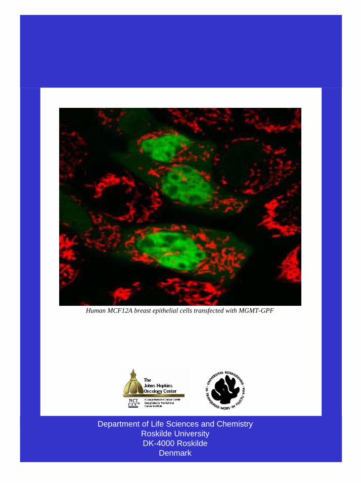
Department of Life Sciences and ChemistryRoskilde UniversityDK-4000 Roskilde
Denmark
Human MCF12A breast epithelial cells transfected with MGMT-GPF
Top Related