


Languages
Pages
Legal

Iatrogenic esophageal
perforation in a preterm
infant
SWISS SOCIETY OF NEONATOLOGY
September 2012
2
Pumberger W, Marwan M, Kargl S, Department of Pediatrics
and Pediatric Surgery (PW, KS), Department of Pediatric
Anesthesia, Children‘s Hospital Linz, Austria
© Swiss Society of Neonatology, Thomas M Berger, Webmaster
3
A premature male newborn was transferred six hours
after birth with a diagnosis of esophageal atresia from
another pediatric institution. The baby had been born
at 31 weeks of gestation to a 29-year-old G2/P1 by
caesarean section due to strong vaginal bleeding and
breech presentation. Pregnancy had been uncomplica-
ted. In particular, several prenatal ultrasound examina-
tions had never demonstrated a polyhydramnios.
The infant had adapted well with Apgar scores of 4, 6,
and 9 at 1, 5, and 10 minutes, respectively. His birth
weight was 1750 g (P50-75), his birth length 46 cm
(P90) and his head circumference 30.5 cm (P50-75).
Within a few minutes of life, the baby developed re-
spiratory distress and an increasing oxygen require-
ment. Oral intubation was only successful after several
attempts, and no oro- or nasogastric tube could be
placed.
Because the pediatricians suspected esophageal atre-
sia, a contrast study of the upper esophageal pouch
was requested. Barium sulfate was used as contrast
medium. When the study failed to demonstrate a con-
tinuous esophagus and the barium projected over the
upper thoracic aperture, esophageal atresia with an
upper pouch and a tracheo-esophageal fistula was su-
spected (type I, according to L. Spitz). With the intent
to stabilize the baby’s respiratory condition, two doses
of surfactant were administered and the infant was
prepared for transfer.
CASE REPORT
4
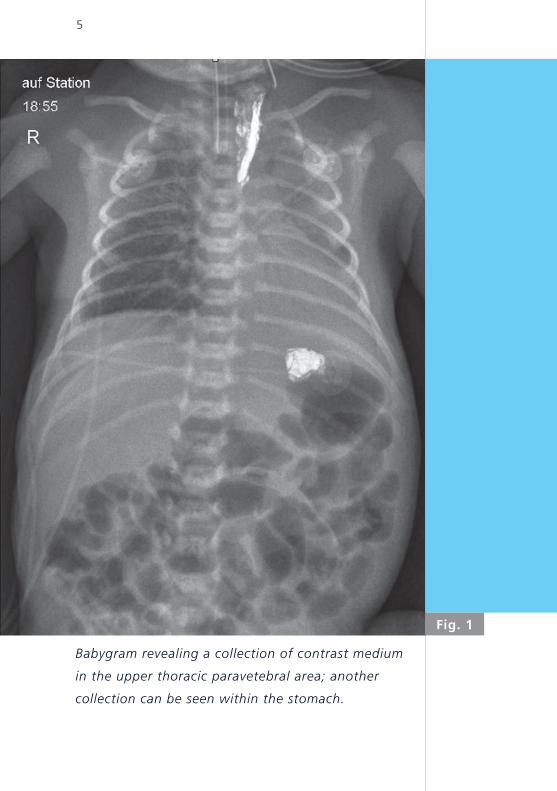
On arrival at our department, the baby’s cardiorespi-
ratory status was stable. A babygram revealed a depot
of contrast medium in the cervical region and another
smaller one within the stomach (Fig. 1). These findings
were inconsistent with the presence of esophageal
atre sia. Therefore another attempt to insert a naso-
gastric tube was made and successfully performed by
an experienced pediatric anesthesiologist (Fig. 2). Thus,
esophageal atresia could definitely be ruled out. Close
inspection of the posterior pharyngeal wall revealed a
fibrin-covered lesion (about 5 mm in diameter) repre-
senting a tear (Fig. 3). A diagnosis of iatrogenic perfo-
ration of the esophagus was made. The child received
broad-spectrum antibiotics, a proton pump inhibitor
and was extubated within two days. Feeding via the
nasogastric tube was started on day three. On day of
life 10, a contrast study using a small amount of wa-
ter-soluble contrast medium showed free passage and
regular peristalsis of the esophagus. As expected, the
barium had stayed in place (Fig. 4). Repeat endoscopy
demonstrated complete healing of the pharyngeal
tear.
The further hospital course of the baby was unevent-
ful. It remains to bee seen whether the barium extra-
vasation will have negative consequences.
5
Babygram revealing a collection of contrast medium
in the upper thoracic paravetebral area; another
collection can be seen within the stomach.
Fig. 1
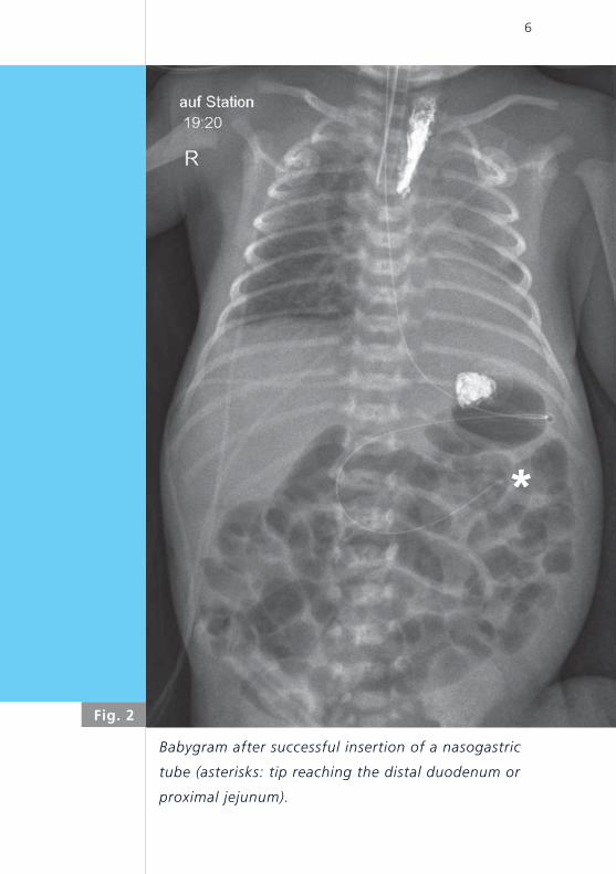
6
Babygram after successful insertion of a nasogastric
tube (asterisks: tip reaching the distal duodenum or
proximal jejunum).
Fig. 2
*
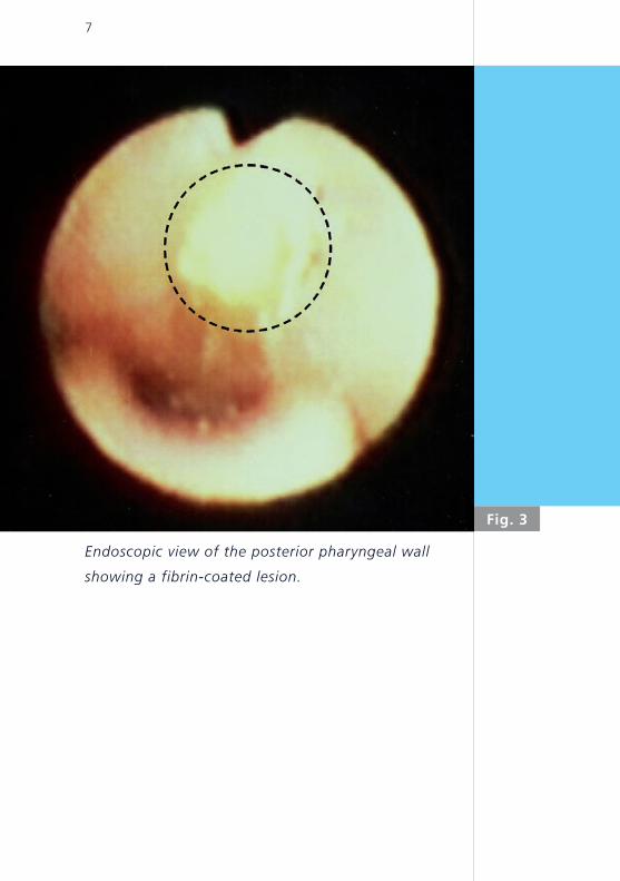
7
Endoscopic view of the posterior pharyngeal wall
showing a fibrin-coated lesion.
Fig. 3
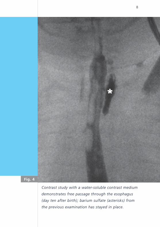
8
Contrast study with a water-soluble contrast medium
demonstrates free passage through the esophagus
(day ten after birth); barium sulfate (asterisks) from
the previous examination has stayed in place.
Fig. 4
*
9
DISCUSSIONIatrogenic esophageal perforation is uncommon in
newborn infants and children. In neonates, particularly
in preterm babies, repetitive placement of oro- and
nasogastric tubes or repetitive pharyngeal suctioning
has been reported to cause esophageal perforation (1-5).
Rigid, hurried or inexperienced manipulation with the
laryngoscope blade or repeat suctioning may cause
spasm of the cricopharyngeal muscle und may result
in closure of the esophageal lumen (3, 6). In addition,
extension of the neck compresses the esophageal wall
against the cervical vertebra and thus facilitates for-
mation of a laceration. Finally, digital manipulation
by the obstetrician during breech delivery (i.e., Veit-
Smellie maneuver) has been reported to result in pha-
ryngeal tears (4, 7). Neonatologists, pediatric anesthe-
siologists and pediatric surgeons should consider this
possibility when confronted with a patient such as the
one presented in this case report.
When a nasogastric tube enters the perforation site
it cannot be passed into the stomach, but will get
stuck at the upper or mid-thoracic level (Fig. 5). This
finding can be misinterpreted as being indicative of
esophageal atresia (3, 8-10). Nearly all patients re-
ported to have iatrogenic esophageal perforation
were referred with the initial diagnosis of esophage-
al atresia. If contrast medium is applied through the
malpositioned nasogastric tube, an irregularly shaped
pouch can be visua lized in the posterior mediastinum,
sometimes extending down to the diaphragm (“dou-
ble-esophageal structure”) (Fig. 6).
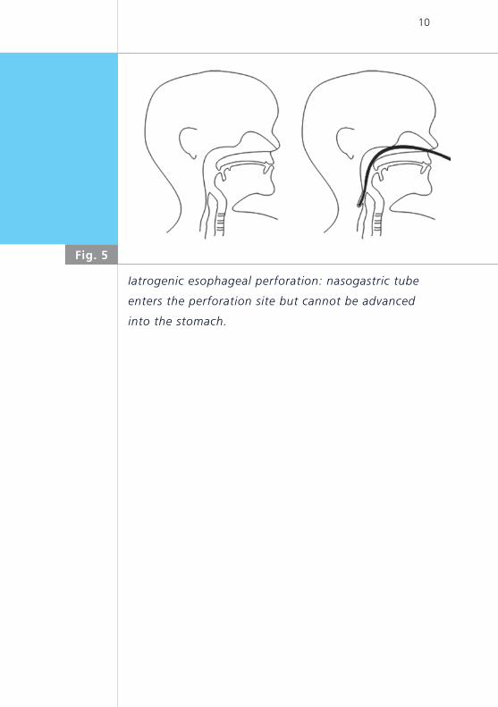
10
Iatrogenic esophageal perforation: nasogastric tube
enters the perforation site but cannot be advanced
into the stomach.
Fig. 5
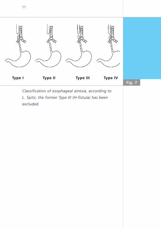
11
Classification of esophageal atresia, according to
L. Spitz; the former Type III (H-fistula) has been
excluded.
Fig. 7Type I Type II Type III Type IV
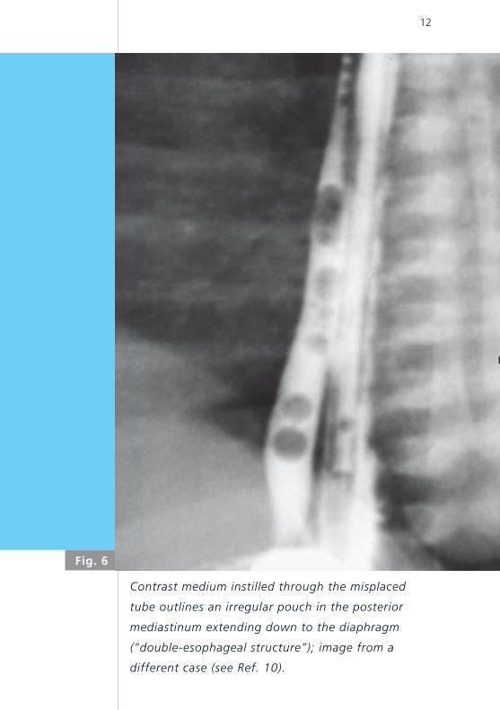
12
Contrast medium instilled through the misplaced
tube outlines an irregular pouch in the posterior
mediastinum extending down to the diaphragm
(“double-esophageal structure”); image from a
different case (see Ref. 10).
Fig. 6
13
Contrast studies are usually unnecessary in suspected
esophageal atresia. A chest x-ray and abdominal plain
film will usually suffice to establish the diagnosis. Vi-
sible air and a coiled tube in the upper pouch, as well
as the presence or absence of air within the bowel
will normally allow proper classification of the type
of esophageal atresia (Fig. 7). Perforation of the up-
per pouch as a complication of continuous suction has
been reported but is probably extremely rare (11).
Mollit et al. described three types of lesions associated
with esophageal perforation: 1) pharyngeal pseudo-
diverticulum created by a local cervical leak, 2) mu-
cosal perforation extending posteriorly in parallel to
the esophagus (“double-esophageal structure”) (Fig.
6), and, 3) free intrapleural perforation with free air
within the mediastinum and pleural space (10, 12).
When perforation has occurred, signs and symptoms
may vary initially; the condition may even be clinically
silent (8-10). However, perforation and dissection may
also cause edema and obstruction of the upper third
of the esophagus. This may result in excessive droo-
ling and difficulties in feeding or vomiting. Coupled
with the inability to advance a nasogastric tube into
the stomach, these signs and symptoms may lead to a
misinterpretation as esophageal atresia (7, 8, 10, 13).
Endoscopy will allow the identification of the perfo-
ration site, possibly reveal a false lumen and show a
patent esophagus; it must be carried out with great
caution to avoid further injury (10, 14).
14
Early diagnosis of esophageal perforation in neonates
may allow nonsurgical management. In contrast, eso-
phageal perforation in adults carries a grave prognosis
(2, 4, 10, 15, 16). Depending on the initial presenta-
tion, most children with iatrogenic esophageal perfo-
ration can be treated conservatively. Broad spectrum
antibiotics and proton pump inhibitors are given for
10 days and a nasogastric tube inserted under fluoro-
scopic control can be used for feeding, whereas oral
feedings are withheld during this time. We suggest
that a repeat esophagography be performed to show
free passage and absence of a leak before oral fee-
dings are started.
Generally, treatment of esophageal perforation in neo-
nates should be successful with a good outcome (2, 4,
10). The consequences of the barium extravasate in
our patient, however, are still unclear. Barium sulfate
in the mediastinum or even within the thoracic cavity
may evoke an inflammatory response with granuloma
formation and ultimately lead to fibrosis (17).
15
REFERENCES1. Eklöf O, Löhr G, Okmian L. Submucosal perforation of the
oesophagus in the neonate. Acta Radiol 1969;8:187-192
2. Gander JW, Berdon WE, Cowles RA. Iatrogenic esophageal
perforation in children. Pediatr Surg Int 2009;25:395-401
3. Clarke TA, Coen RW, Feldmann B, et al. Esophageal perfora-
tions in premature infants and comments on the diagnosis.
Am J Dis Child 1980;134:367-368
4. Panieri E, Millar AJW, Brown RA, et al. Iatrogenic esophageal
perforation in children. Patterns of injury, presentation,
management, and outcome. J Pediatr Surg 1996;31:890-895
5. Krasna IH, Rosenfeld D, Benjamin BG, et al. Esophageal per-
foration in the neonate: an emerging problem in the newborn
nursery. J Pediatr Surg 1987;22:784-790
6. Filippi L, Pezzati M, Poggi C. Use of polyvinyl feeding tubes
and iatrogenic pharyngo-esophageal perforation in very low
birth weight infants. Acta Paediatr 2005;94:1825-1828
7. Duchearme JC, Bertrand R, Debie J. Perforation of the pharynx
in the newborn: a condition mimicking esophageal atresia.
Can Med Assoc J 1971;104:785-787
8. Knight RB, Webb DE, Coppola C. Pharyngeal perforation mas-
querading as esophageal atresia. J Pediatr Surg 2009;44:2216-
2218
9. Seefelder C, Elango S, Rosbe KW, et al. Esophageal perforation
presenting as esophageal atresia in a premature neonate fol-
lowing difficult intubation. Pediatr Anaesthesia 2001;11:112-
118
10. Pumberger W, Bader T, Golej J, et al. Traumatic pharyngo-
esophageal perforation in the newborn: a condition mimicking
esophageal atresia. Paediatr Anaesth 2000;10:201-205
16
11. Wright M, Noblett HR. A complication of continuous up-
per pouch suction in esophageal atresia. J Pediatr Surg
1978;13:369-370
12. Mollitt DL, Schullinger JN, Santulli TV. Selective management
of iatrogenic esophageal perforation in the newborn. J Pediatr
Surg 1981;16:989-993
13. Blair GK, Filler RM, Theodorescu D. Neonatal pharyngo-eso-
phageal perforation mimicking esophageal atresia: clues to
diagnosis. J Pediatr Surg 1987;22:770-774
14. Soong WJ. Endoscopic diagnosis and management of iatro-
genic cervical esophageal perforation in extremely premature
infants. J Chin Med Assoc 2007;70:171-175
15. Baum ED, Elden LM, Handler SD, et al. Management of hy-
popharyngeal and esophageal perforations in children: three
case reports and a review of the literature. Ear Nose Throat J
2008;87:44-47
16. Aoun JB, Gasmi M, Jemal R et al. Iatrogenic esophageal perfo-
ration in the neonate. Tunis Med 2012; 90:72-74.
17. Cohen MD. Choosing contrast media for the evaluation of
the gastrointestinal tract of neonates and infants. Radiology
1987;162:447-456

SUPPORTED BY
CONTACT
Swiss Society of Neonatology
www.neonet.ch
con
cep
t &
des
ign
by
mes
ch.c
h
Top Related