


Languages
Pages
Legal


Clin Liver Dis 8 (2004) 807–838
Hepatic porphyrias: diagnosis and management
Annie T. Chemmanur, MD, Herbert L. Bonkovsky, MD*
Office of Clinical Research, Liver-Biliary-Pancreatic Center, University of Connecticut Health Center,
263 Farmington Avenue, MC 1111, Farmington, CT 06030, USA
bKing George III, who was held in low regard on both sides of the Atlantic as
the stubborn monarch whom the American colony fought for their independence,
was not a well man. . .. the much maligned king suffered spells of a painful and
delirious metabolic disease. . ..Q [1].Porphyrias are a group of metabolic disorders in which there are defects in the
normal pathway for the biosynthesis of heme, the critical prosthetic group for
numerous hemoproteins such as hemoglobin, myoglobin, catalase, and micro-
somal cytochromes b5 and P-450. The term porphyria comes from the Greek
word porphyra, which means purple. The derivation is apt because the
biochemical hallmark of the porphyrias is overproduction and overexcretion of
compounds called porphyrins, which have a deep red or purple color. It is not
certain to this day whether George III truly had variegate porphyria. The first
clinical reports of porphyria appeared in the late nineteenth century, describing
a patient with severe cutaneous photosensitivity and brown pigmentation of
the bones, characteristic of congenital erythropoietic porphyria. Soon thereafter,
the first case of an inducible acute porphyria was described in a drug addict who
passed urine the color of port wine and later died after taking the hypnotic
drug sulfonmethane.
The clinical manifestations of the porphyrias can be highly varied, and pa-
tients may present to general physicians and be referred to a wide variety of
subspecialists because of these manifestations. However, two major clinical forms
are represented by the so-called bacuteQ porphyrias, in which patients suffer
1089-3261/04/$ – see front matter D 2004 Elsevier Inc. All rights reserved.
doi:10.1016/j.cld.2004.07.001
This work was supported by grants and contracts from NIH (to H.L. Bonkovsky): RO1 DK38825;
MO1 RR06192; NO1 DK92326; and UO1 DK065193. A portion of this work was done while A.
Chemmanur was a fellow in Advanced Hepatology, supported by an AASLD-Schering Fellowship.
* Corresponding author.
E-mail address: [email protected] (H.L. Bonkovsky).

A.T. Chemmanur, H.L. Bonkovsky / Clin Liver Dis 8 (2004) 807–838808
recurrent bouts of pain, especially pain in the abdomen, and the bcutaneousQporphyrias, in which patients have painful skin lesions.
Elucidation of the heme biosynthetic pathway provided the basis for exploring
the chemical reasons for the abnormal accumulation of metabolic intermediates in
patients with porphyrias. Characterization of the intermediates involved in heme
biosynthesis and the enzymes responsible for their formation, together with
observed patterns of the excretion of metabolites in patients with porphyria,
suggested which enzyme would be deficient in each type of porphyria (Fig. 1).
Knowledge of the factors chiefly responsible for regulating the rate of synthesis
of heme has helped to explain how drugs and other factors may cause porphyria.
Knowledge of the physical and chemical properties of porphyrins also forms
an important part of the foundation for understanding the clinical manifestations
of these diseases. Thus, the porphyrias can best be understood after reviewing
the chemical properties of porphyrins and heme and the control of their bio-
synthesis [2,3].
Normal physiology and biochemistry of porphyrin and heme metabolism
Structures and properties of porphyrins and heme
Porphyrins are cyclic tetrapyrroles in which the four pyrrole rings are linked
by methene bridges (�CH =), as shown in Fig. 2. All naturally occurring
porphyrins have side chains attached to the carbon atoms of the pyrrole rings that
are not attached to the ring nitrogen. For uroporphyrins, these side chains are
acetate and propionate; for coproporphyrins, they are methyl and propionate; and
for protoporphyrins, they are methyl, propionate, and vinyl groups (see Fig. 2).
Naturally occurring uroporphyrin and coproporphyrin are of two of four possible
isomer types, either the I-isomer, in which the acetate and propionate side chains
alternate, or the III-isomer in which one of the pyrrole rings (the D ring) has been
flipped 180 degrees, so that the two propionate groups on the C and D rings are
vicinal, rather than trans, in their relative orientations (see Fig. 2). In the case of
protoporphyrin, which has three side chains, 15 isomers are possible. Only one
(called protoporphyrin IX, following the numbering and nomenclature originally
proposed by Fischer and Orth) occurs in nature.
Porphyrins are planar, highly stable compounds that absorb light strongly
around 400-nm wavelength (the Soret band). They also are strongly fluorescent
and emit intense red light when excited by light of the Soret band wavelength.
These properties are due to the high degree of resonance of the system of
conjugated double bonds (see Fig. 2). These optical properties facilitate detection
of porphyrins, even at very low concentrations. In contrast, the porphyrinogens,
which are hexahydro- or reduced porphyrins and are the actual intermediates for
most of the steps of heme synthesis (see Fig. 1), lack this resonance structure due
to the fact that the bridges are all reduced to methylene moieties, which lack
double bonds. The porphyrinogens do not absorb light of 400-nm wavelength,

Step Enzyme defect Type of porphyria Urine Stool
—
—COPRO
ISOCOPROISOCOPRO
COPRO(PROTO)
PROTO(COPRO)
PROTO
ALA
ALA, PBGURO,COPRO
ALA, PBGCOPRO
ALA, PBGCOPRO
—
UROURO
Plasma
ALA
ALA, PBGURO
COPRO
PROTO(COPRO)
PROTO
UROURO
RBCs
Zn PROTO
—URO,COPRO
—
—
PROTO
—Zn PROTO
ALA synthase
ALA dehydratase ALA dehydratase deficiency (ADP)
Acute intermittent porphyria (AIP)Congenital erythropoietic porphyria (CEP)
Porphyria cutanea tarda (PCT)Hepatoerythropoietic porphyria (HEP)
(Erythropoietic) Protoporphyria ([E]PP)
Variegate porphyria (VP)
Hereditary coproporphyria (HCP)
PBG deaminaseUroporphyrinogen IIISynthase (cosynthase)
Uroporphyrinogen IIIDecarboxylase
Coproporphyrinogen IIIOxidase
ProtoporphyrinogenOxidase
Ferrochelatase
Glycine + Succinyl-CoA
5-Aminolevulinate (ALA)
Porphobilinogen (PBG)Hydroxymethyl bilane
Uroporphyrinogen III
Protoporphyrinogen IX
Protoporphyrin IX
Fe2+
Heme
Coproporphyrinogen III
Nonenzymatic
UROGEN I
COPROGEN I
Fig. 1. Heme biosynthetic pathway showing the sites of enzyme defects in the porphyrias and the major biochemical abnormalities in biochemically active disease. Only
the major increases in the urine, stool, plasma, and erythrocytes (RBCs) are shown. The dashes (–) represent no abnormalities. For several of the diseases, many patients
are biochemically silent (blatentQ) carriers of the enzymatic defects for most of their lives. COPRO, coproporphyrin; COPROGEN, coproporphyrinogen; ISOCOPRO,
isocoproporphyrin; PROTO, protoporphyrin; URO, uroporphyrin; UROGEN, uroporphyrinogen; Zn, zinc.
A.T.Chem
manur,H.L.Bonkovsky
/Clin
Liver
Dis8(2004)807–838
809

Uroporphyrin I Uroporphyrin III
Protoporphyrin IX
Heme
CH 2
CH
A
N
BN
N
D CN
CH
CH Fe
α
CHγ
δ CH β
CH 3CH
2CH
CH3
CH 2
CH 2
COOH
CH 3
CH2CH
2COOH
CH3
P
NH
N
N
D
HN
CH
CH
CH
CH
A P
A
P
A
P
A
P
NH
N
N
D
HN
CH
CH
CH
CH
A P
A
P
AA
P
V
NH
N
N HN
CH
CH
CH
CH
M V
M
P
MP
M
P
NH
N
N HN
CH
CH
CH
CH
M P
M
P
MP
M
Coproporphyrin III
Fig. 2. Structures of heme and selected porphyrins. All porphyrins have the same basic ring structure
but differ in the side-chains that are attached to the pyrrole rings. Porphyrinogens are reduced
(hexahydro) forms in which the methane bridges linking the pyrroles are replaced by methylene
(�CH2�) groups, and all four nitrogens are linked to hydrogens. A, acetate; M, methyl; P, propionate;
V, vinyl. (Modified from Bloomer JR, Straka JG. Porphyrin metabolism. In: Arias IM, Jakoby
WB, Popper H, Schachter D, Shafritz DA, editors. The Liver: biology and pathobiology. New York:
Raven Press; 1988. p. 451–66; with permission.)
A.T. Chemmanur, H.L. Bonkovsky / Clin Liver Dis 8 (2004) 807–838810

A.T. Chemmanur, H.L. Bonkovsky / Clin Liver Dis 8 (2004) 807–838 811
are colorless, and do not fluoresce. However, they are readily oxidized to the
corresponding porphyrins. Such oxidation may occur within cells in the body, as
well as in excreted urine or stool. Heme differs from protoporphyrin only in that
an iron atom has been inserted into the tetrapyrrole ring. This increases stability
of the structure and also causes it to lose its fluorescent properties. Other
metalloporphyrins, such as zinc protoporphyrin, may also form in intact animals.
Pathway of heme biosynthesis
The enzymes of the pathway are compartmentalized within the cell. The first
and last three steps of the pathway take place in mitochondria, whereas
intermediate steps take place in the soluble fraction of the cell cytoplasm. In the
first step, the enzyme 5-aminolevulinate (ALA) synthase catalyzes the formation
of ALA from glycine and succinyl-CoA in the presence of pyridoxal phosphate.
The ALA formed diffuses or is transported from the mitochondria into the soluble
fraction, where the second enzyme of the pathway, ALA dehydratase or
porphobilinogen (PBG) synthase, condenses two molecules of ALA to form the
monopyrrole PBG. In the next step, the enzyme PBG deaminase (sometimes
called hydroxymethylbilane synthase) carries out the stepwise polymerization of
four molecules of PBG to form the linear tetrapyrrole hydroxymethylbilane. This
latter compound is highly unstable and rapidly undergoes cyclization with ring
closure spontaneously in the absence of further enzymatic activity. Normally,
however, the cytoplasmic fraction contains a large excess of uroporphyrinogen III
cosynthase, the next enzyme of the pathway, which leads to the formation of the
III-, rather than the I-isomer of uroporphyrinogen. The next enzyme of the
pathway, uroporphyrinogen decarboxylase, carries out the stepwise decarbox-
ylation of the four acetate side chains of uroporphyrinogen III, with the eventual
formation of coproporphyrinogen III. This enzyme is also capable of decarbox-
ylating uroporphyrinogen I to coproporphyrinogen I, although it does so at a lower
rate than for the III-isomer. Coproporphyrinogen III is then transported back into
the mitochondria, where the enzyme coproporphyrinogen oxidase catalyzes the
stepwise oxidative decarboxylation of the propionic acid side chains on rings A
and B to form vinyl groups at these positions. This gives rise to the intermediate
protoporphyrinogen IX. Although protoporphyrin readily forms nonenzymatically
from protoporphyrinogen in the presence of oxygen, the enzyme protoporphy-
rinogen oxidase is responsible for this reaction in normal animals. In the final step
of the pathway, ferrous iron is inserted into the protoporphyrin ring by the enzyme
ferrochelatase to form heme, the end product of the pathway.
Regulation of hepatic heme metabolism
Under normal conditions in both developing erythrocytes and other tissues
such as the liver, the first enzyme of the pathway (ALA synthase) is the rate-
controlling enzyme. There are two forms of ALA synthase in higher animals:
ALA synthase 1, a ubiquitous housekeeping form, and ALA synthase 2, the

A.T. Chemmanur, H.L. Bonkovsky / Clin Liver Dis 8 (2004) 807–838812
erythroid form, which is expressed principally in developing erythrocytes. These
two enzymes are regulated quite differently: ALA synthase 1 is downregulated by
heme, whereas ALA synthase 2 is downregulated by lack of sufficient iron,
through a typical iron responsive element located in its promoter. Thus, under
physiologic conditions of iron sufficiency ALA synthase 2 is expressed robustly
in developing erythrocytes. In contrast, under physiologic conditions in liver
cells, ALA synthase 1 is present in relatively low amount, and the maximal rate at
which it is capable of functioning is less than for any of the other enzymes of the
pathway. ALA synthase 1 in the liver has a short life span (approximately 1 hour),
as does the mRNA coding for it (approximately 3 hours). Therefore, agents that
alter the rate of synthesis of the mRNA for ALA synthase 1 or the rate of
translation of the message have a rapid (within minutes) and dramatic effect on
the amount of ALA synthase 1 in the cell and thus on the rate of synthesis of
porphyrins and heme.
Heme synthesis
Mitochondrion
Glycine+
Succinyl-CoA
ALAsynthase
ALA
Uptake intomitochondria
Pre-ALA synthase
Gene forALA synthase
mRNA
DNA
Nucleus
PBG
Heme
Heme degradation
Exogenousheme
Regulatoryheme
Hemoproteins
Apo-proteinsHemeutilization
Hemeoxygenase
Biliverdin + CO + Fe
Bilirubin
mRNAdegradation
+
–
–
Fig. 3. Regulation of the hepatic heme biosynthetic pathway and subcellular localization of the
enzymes of the pathway. Schematic of the synthesis of heme and its regulation. The regulatory heme
pool acts to stimulate (+) or downregulate (�) the indicated steps. Those steps indicated as not within
the nucleus or mitochondrion take place in the cytosol. The dashed line indicates a still controversial
regulatory effect of heme to decrease transcription of the gene for ALA synthase 1. ALA, 5-amino-
levulinate; CO, carbon monoxide; CoA, coenzyme A; PBG, porphobilinogen.

A.T. Chemmanur, H.L. Bonkovsky / Clin Liver Dis 8 (2004) 807–838 813
Chemicals that cause porphyria in rodents (and probably in humans) also
profoundly affect hepatic heme metabolism at sites other than ALA synthase 1. All
are believed to decrease the size of a small but critical regulatory heme pool in
liver cells. Such a depletion is envisioned to lead to a secondary increase in activity
of ALA synthase 1 as the hepatocyte attempts to replenish its regulatory heme
pool. The most efficient classical porphyrogenic chemicals produce rapid and
profound reduction of the heme of cytochromes P-450, leading, in turn, to reduc-
tion of the regulatory heme pool and thus to induction of ALA synthase 1. There is
no doubt that heme in hepatocytes downregulates activity of ALA synthase 1,
although the exact site(s) of action of heme is still somewhat controversial. There is
consensus that heme decreases the stability of the mRNA for ALA synthase 1
and that it also blocks the uptake of ALA synthase into mitochondria.
Some chemicals that are capable of producing experimental porphyria (eg,
hydantoins) do not produce depletion of liver heme but rather induce both ALA
synthase and cytochromes P-450. Such chemicals are generally lipid soluble, and
recent evidence indicates that they upregulate the expression of the genes for Cyp
P-450s and for ALA synthase 1 by interaction with consensus drug regulatory
elements, which are found in the 5V-untranslated regions of these genes [4].
Parenterally administered heme is capable of entering hepatocytes and of
repleting the regulatory heme pool and downregulating the levels of ALA
synthase 1. The overall regulation of the pathway is summarized in Fig. 3.
Nutritional status plays an important role. The activity of hepatic ALA
synthase is increased by fasting or starvation, whereas the feeding of
carbohydrates decreases the basal activity and markedly diminishes the induction
of the enzyme produced by porphyrogenic chemicals. Proteins, probably owing
to their content of gluconeogenic amino acids, can exert similar effects to that of
glucose. The exact mechanism for the glucose effect on ALA synthase is still not
understood, but it is of considerable clinical significance. A mainstay of therapy
of the acute porphyrias is the administration of large amounts of glucose, which
in itself may be sufficient for therapy of mild attacks of the disease.
Overview of the porphyrias
The main clinical manifestations of the porphyrias are cutaneous photo-
sensitivity and neurologic dysfunction, most often presenting as abdominal pain.
The porphyrias are a group of metabolic disorders characterized by the excessive
accumulation and excretion of porphyrins and their precursors. They result from
specific enzyme defects in the heme synthetic pathway, which may be inherited
or acquired. Many patients with the enzyme defects do not have clinical
manifestations. Porphyric attacks can be fatal, so the early diagnosis of carriers
and affected individuals is important to be able to advise the avoidance of
precipitating factors for an acute attack, typically drugs, fasting, or alcohol. If
neurovisceral symptoms suggest an acute porphyric attack, a rapid screening test
for PBG should be performed. If a cutaneous porphyria is suspected, screening

A.T. Chemmanur, H.L. Bonkovsky / Clin Liver Dis 8 (2004) 807–838814
tests for increased erythrocytic porphyrins should be done (if solar urticaria and
acute photosensitivity suggest erythropoietic protoporphyria [EPP]), or screening
tests for urinary porphyrins (if vesiculobullous formation and skin fragility
suggest porphyria cutanea tarda [PCT], hereditary coproporphyria [HCP], or
variegate porphyria [VP]). Positive screening tests should be confirmed by
specific quantitative tests. Enzymic assays and DNA-based tests are useful for
kindred evaluation, genetic diagnosis, and the pinpointing of causative mutations
but are not needed for rapid diagnosis of symptomatic patients. Prevention is a
central component of management of patients with porphyria. Intravenous
hematin, high carbohydrate intake, and pain control are central in the treatment of
acute neurovisceral attacks. Sun avoidance and skin protection are important to
reduce cutaneous manifestations and complications [2,3,5–9].
Classification
The porphyrias are classified based on the principal site of expression of the
enzymic defect, as either hepatic or erythroid (Table 1). They are also classified
based on the dominant clinical presentation, whether with cutaneous manifes-
tations only or with neurovisceral features (with or without cutaneous mani-
festations) (Table 2). The hepatic porphyrias are ADP, AIP, HCP, VP, PCT, and
HEP. The bacuteQ or binducibleQ hepatic porphyrias are the first four of these:
ADP, AIP, HCP, and VP. The erythropoietic porphyrias are congenital eryth-
ropoietic porphynia (CEP) and EPP. HEP may be classified as both hepatic
and erythroid.
For the most part, the porphyrias are inherited as autosomal dominant
disorders (AIP, HCP, VP, EPP, and the familial form of PCT); some are inherited
in a recessive fashion (ADP, HEP, CEP). The majority of PCT patients have an
Table 1
Classification of the porphyrias
Hepatic Hepatic acute inducible Erythropoietic
ADP ADP
AIP AIP
HCP HCP
VP VP
PCT
HEP (HEP)
CEP
EPP
The porphyrias are listed as hepatic, acute inducible hepatic, or erythropoietic porphyrias. HEP may be
considered both a hepatic and erythropoietic porphyria.
Abbreviations: ADP, ALA dehydratase deficiency porphyria; AIP, acute intermittent porphyria;
ALA, 5-aminolevulinate; CEP, congenital erythropoietic porphyria; EPP, erythropoietic protopor-
phyria; HCP, hereditary coproporphyria; HEP, hepatoerythrocytic porphyria; PCT, porphyria cutanea
tarda; VP, variegate porphyria.

Table 2
Summary of major clinical features of the porphyrias
Type of porphyria Neurovisceral
Clinical manifestation
Cutaneous Liver damage
ADP Yes No No
AIP Yes No No
HCP Yes Yes (bullae, fragility) No
VP Yes Yes (bullae, fragility) No
PCT No Yes (bullae, fragility) Yes
HEP +/� Yes (bullae, fragility) Yes
CEP No Yes (bullae, fragility) Occasional
EPP Rarelya Yes (urticaria, erythema) Yes (10%)
The porphyrias are listed with respect to their dominant symptomatology with neurovisceral or
cutaneous manifestations and the presence of potential liver damage. (Modified from: Hahn M,
Bonkovsky HL. Disorders of porphyrin metabolism. In: Wu G, Israel J, editors. Diseases of the
liver and bile ducts: a practical guide to diagnosis and treatment. Totowa, NJ: Humana Press; 1998.
p. 249–72; with permission.)
Abbreviations: ADP, ALA dehydratase deficiency porphyria; AIP, acute intermittent porphyria;
ALA, 5-aminolevulinate; CEP, congenital erythropoietic porphyria; EPP, erythropoietic protopor-
phyria; HCP, hereditary coproporphyria; HEP, hepatoerythrocytic porphyria; PCT, porphyria cutanea
tarda; VP, variegate porphyria.a EPP with end-stage liver disease, especially just after liver transplantation, may rarely be
associated with neurovisceral manifestations.
A.T. Chemmanur, H.L. Bonkovsky / Clin Liver Dis 8 (2004) 807–838 815
acquired disease, albeit perhaps with a genetic predisposition. Because the
principal focus of this article is on the hepatic porphyrias, they are described in
the order listed previously, first the acute hepatic porphyrias (ADP, AIP, HCP,
VP), then the other hepatic porphyrias (PCT, HEP). EPP is also described because
the major complications of EPP affect the liver and biliary tree. A summary
overview of the porphyrias with diagnostic essentials is shown in Fig. 1.
Pathophysiology
Neurovisceral features
Abdominal pain, which is the dominant symptom of most porphyrias
(especially in the acute porphyrias ADP AIP, HCP, and VP), is probably due to
an autonomic neuropathy affecting the gut. The pathogenesis of the neurologic
manifestations likely relates either to excess ALA or PBG, usually precipitated by
an increase in liver ALA synthase 1 activity or to a deficiency of heme within
neurons or other tissues (Fig. 4). It has been proposed that ALA or PBG may be
directly neurotoxic or that ALA may interact with receptors for the inhibitory
neurotransmitter gamma-amino butyric acid or affect motor nerve conduction
velocity, acetylcholine release, Na/K ATPase activity, or other aspects of neuronal
function. Heme deficiency within neurons may compromise mitochondrial
cytochrome levels and impair ATP production. A deficiency of hepatic heme
may lead to increased levels of 5-hydroxytryptophan and tryptamine (5HT,
serotonin) in the nervous system, which may mediate or exacerbate neurovisceral
features of the acute porphyrias.

Glycerine
Succinyl-CoA
↑ALA ↑PBG
↑ALA synthase ↓Heme formation from ALA
?Neurotoxic?Inhibits GABA-mediated transmission
?Neurotoxic ?Neurotoxic
↓Heme
Fig. 4. Pathogenesis of the neurovisceral features of the acute porphyrias. An increase in ALA and
PBG levels, accompanied by reduced heme concentrations in the liver and nervous system, likely
causes the neurovisceral features. ALA, 5-aminolevulinate; PBG, porphobilinogen.
A.T. Chemmanur, H.L. Bonkovsky / Clin Liver Dis 8 (2004) 807–838816
Cutaneous features
Photocutaneous lesions, due to the photosensitizing effects of the excess
porphyrins in the skin or in the dermal blood vessels, may occur in most of the
porphyrias (HCP, VP, PCT, HEP, CEP, EPP) but not in ADP and AIP (as the
enzyme deficiencies in these last two porphyrias precede porphyrin formation).
Skin exposed to light may manifest porphyrin-induced photosensitivity in two
ways: either formation of vesicles or bullae and increased fragility as a result of
mild trauma to light-exposed skin, or acute erythema with burning and itching but
with less chronic injury. When exposed to Soret band radiation the porphyrins
emit two major fluorescence emission peaks at 600 to 610 and 640 to 660 nm.
The Soret band light excites porphyrin electrons into an activated btriplet state.QSome of this energy is then transferred to biological membranes or molecular
oxygen, forming reactive bsingletQ oxygen. Singlet oxygen damages the skin
through several mechanisms, including lipid peroxidation of membranes and
cross-linking of membrane proteins. Release of mediators and enzymes from
mast cells and polymorphonuclear leukocytes contributes to an inflammatory
response modulated by the effects of the porphyrins on the complement and other
pathways. The pathophysiology of the cutaneous features of the porphyrias is
summarized in Fig. 5. The Soret band light that excites porphyrins passes through
ordinary window glass; therefore porphyric patients are not protected from skin
photosensitivity damage by remaining indoors. Typical sunburn reactions and
other photosensitivities result from light of shorter wavelength (290 to 320 nm),
which is absorbed by window glass.
Hepatic features
Some acute porphyrias have mild hepatic abnormalities. Data from Scandi-
navia and France show an increased risk of hepatocellular carcinoma in patients
with acute porphyria. More severe liver damage may occur in PCT and EPP, and
to a lesser degree in HEP. Liver biopsies from patients with PCT show red
fluorescence, hemosiderosis, fatty infiltration, and variable fibrosis and necrosis.
Chronic injury may result in cirrhosis and hepatocellular carcinoma. There is a
high incidence of alcohol excess, HCV infection, iron overload, and hetero-
Cardinal presentingfeatures: symptoms
and signs
Rapid bedsidescreening test
Quantitativeconfirmatory
tests
Diagnosisestablished
Neurovisceralfeatures of acute
porphyric syndromes(ADP, AIP, HCP, VP,
lead poisoning,hereditary
tyrosinemia)
See legendQuantitativeALA and PBG
in urine
Hoesch or Watson-Schwartz test
Acute porphyricattack
See legend See legend
See legend See legend
See legend
Cutaneousfeatures of
acute or chronicporphyrias
(HCP, VP, PCT)
Urine talc oramyl alcohol test
QuantitativeALA, PBG and
porphyrinsin urine
Cutaneousporphyria
Qualitative test forporphyrins or
fluorescence in RBCs
Cutaneousfeatures of
protoporphyria
Quantitativetest for
porphyrins in RBCs, plasma
and stool
Increased Normal Increased Normal Increased Normal
Protoporphyria
+ + +– – –
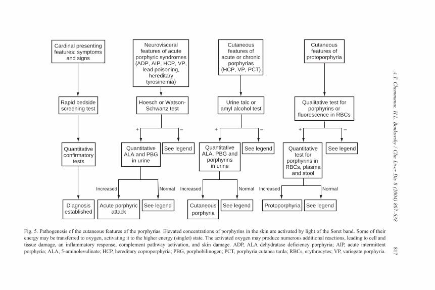
Fig. 5. Pathogenesis of the cutaneous features of the porphyrias. Elevated concentrations of porphyrins in the skin are activated by light of the Soret band. Some of their
energy may be transferred to oxygen, activating it to the higher energy (singlet) state. The activated oxygen may produce numerous additional reactions, leading to cell and
tissue damage, an inflammatory response, complement pathway activation, and skin damage. ADP, ALA dehydratase deficiency porphyria; AIP, acute intermittent
porphyria; ALA, 5-aminolevulinate; HCP, hereditary coproporphyria; PBG, porphobilinogen; PCT, porphyria cutanea tarda; RBCs, erythrocytes; VP, variegate porphyria.
A.T.Chem
manur,H.L.Bonkovsky
/Clin
Liver
Dis8(2004)807–838
817

A.T. Chemmanur, H.L. Bonkovsky / Clin Liver Dis 8 (2004) 807–838818
zygosity for hereditary hemochromatosis in PCT patients. In protoporphyria there
is an increased risk of developing pigment gallstones, with rare cases developing
pigmentary cirrhosis and fatal liver damage.
Enzymatic defects
The enzymatic defects in the heme biosynthetic pathway responsible for the
porphyrias are outlined in Fig. 1. The assay of many of these enzymes is available
only in a research setting. The enzyme abnormality affecting heme biosynthesis
may not by itself be sufficient to cause disease expression. Many patients with a
deficient enzyme activity do not have clinical or biochemical manifestations of
porphyria. Any induction of hepatic ALA synthase 1 may enhance the production
of porphyrin precursors and exacerbate a porphyria. As most of the porphyrias
are genetic disorders, the enzyme defects can be detected in several tissues. For
example, in EPP a defect in the ferrochelatase-catalyzed insertion of iron into
protoporphyrin has been shown in liver, bone marrow cells, peripheral blood
mononuclear cells, and cultured skin fibroblasts. Much progress had been made
in recent years in defining the genetic defects responsible for the porphyrias. A
summary of molecular aspects of the genes and enzymes involved in the por-
phyrias is shown in Table 3.
Traditional methods of diagnosis of the porphyrias, based on urine, serum, and
stool levels of porphyrins or porphyrin precursors or liver and erythrocyte
enzyme assays, are still probably best when a specific mutation is not known.
Mutational screening in such cases is laborious. The diagnostic essentials for each
of the porphyrias are shown in Fig. 1. When a specific mutation is suspected in a
Table 3
Summary of the enzymatic defects causing the porphyrias, showing the sizes and chromosomal
locations of the genes and sizes of the gene products
Disease Enzyme
Size of
gene (kb) #Exons
Chromosomal
location #AAs MW (kDa)
ADP ALA dehydratase 9q34 307 36.3
AIP PBG deaminase 10 15 11q24.1 344/361 37/39
CEP Urogen III cosynthase 10q25 265 28.6
PCT/HEP Urogen decarboxylase 3 10 1p34 365 41
HCP Coprogen oxidase 14 7 3q12 354 41
VP Protogen oxidase 5.5 13 1q23 354 51
EPP Ferrochelatase 45 11 18q 423 47.8
Two numbers are given for the amino acids and molecular weights of PBG deaminase, because there
are two forms of this enzyme, which are products of alternate splicing (see Fig. 6). The gene size and
number of exons of human ALA dehydratase and urogen III cosynthase are not yet known.
Abbreviations: #AAs, number of amino acids; ADP, ALA dehydratase deficiency porphyria; AIP,
acute intermittent porphyria; ALA, 5-aminolevulinate; CEP, congenital erythropoietic porphyria; EPP,
erythropoietic protoporphyria; COPROGEN, coproporphyrinogen; HCP, hereditary coproporphyria;
HEP, hepatoerythrocytic porphyria; kb, kilobases; MW kDa, molecular weight of the enzyme in
kilodaltons; PBG, porphobilinogen; PCT, porphyria cutanea tarda; PROTOGEN, protoporphyrinogen;
UROGEN, uroporphyrinogen; VP, variegate porphyria.

A.T. Chemmanur, H.L. Bonkovsky / Clin Liver Dis 8 (2004) 807–838 819
family or geographic cluster, a variety of molecular methods may simplify
making the diagnosis. However, bclusteringQ of a frequent mutation may not
necessarily imply a familial relationship or implicate a founder effect.
Clinical features
General comments on diagnosis
Diagnosis of the porphyrias is usually made by clinical history in association
with increased amounts of porphyrins or porphyrin precursors in the urine, feces,
and blood [2,3,6,9]. Rapid screening tests are useful in the initial evaluation. The
Watson-Schwartz and Hoesch tests detect PBG in the urine by its reaction with
Ehrlich’s reagent to produce a red color. Urine PBG is increased in most acute
porphyric attacks that manifest neurologic abnormalities. In patients with a
positive screening test, subsequent specific quantitative tests of urinary ALA and
PBG should be done because false positives do occur. If clinical features suggest
a cutaneous porphyria, then for solar urticaria and acute photosensitivity
(suggesting EPP), screening tests for increased erythrocyte porphyrins should
be done; for vesiculobullous formation (suggesting PCT, HCP, or VP), a
screening test for urinary porphyrins should be done. The diagnostic essentials
are shown in Fig. 1; the normal urine and serum levels of the porphyrins and their
precursors are shown in Table 4. The porphyrin precursors ALA and PBG are
excreted in the urine; during acute porphyric attacks these levels are markedly
increased. The porphyrinogens spontaneously, or after the addition of oxidizing
agents, are converted to their corresponding porphyrins, which are then
measured. The water solubility and hence excretion of a porphyrin in urine
depends on the number of its carboxylic acids groups. Thus uroporphyrin with
Table 4
Normal urinary, fecal, and blood levels of porphyrins and precursors
Analyte
UrineFeces mg/gdry wt
Erythrocytes
mg/dL packed RBCs Plasma mg/dLmg/g Cr mg/24 h
ALA b3000 b4000 – – 15–23
PBG b2500 b3500 – – –
URO 10–60 b80 b5 b2 b2
COPRO 50–250 b280 b50 b2 b1
PROTO – – b120 b90 b2
ISOCOPRO – – – – –
Porphyrins (Total) 35–300 50–400 b175 – b2
Typical normal values for adults are tabulated; there is some variability in normal levels, depending on
the laboratory performing the tests and methods used. Normal urine creatinine (Cr) = 0.8–2.0 g/24
hours, Dashes (–) indicate no detectable levels or not routinely tested.
Abbreviations: ALA, 5-aminolevulinate; COPRO, coproporphyrin; ISOCOPRO, isocoproporphyrin;
PBG, porphobilinogen; PROTO, protoporphyrin; URO, uroporphyrin.

A.T. Chemmanur, H.L. Bonkovsky / Clin Liver Dis 8 (2004) 807–838820
8 carboxyl groups is predominantly excreted in the urine; coproporphyrin with
4 carboxyl groups is excreted in both urine and feces; protoporphyrin with only
two carboxyl groups is poorly water soluble and excreted only in feces after its
secretion into bile.
Acute porphyrias
The bacute attacksQ of porphyria, with episodic neurologic dysfunction give
these porphyrias their name rather than any acute hepatic involvement in these
life-long genetic disorders. AIP is the commonest acute hepatic porphyria in the
United States and probably the most common bgeneticQ porphyria; it is usuallyused as the paradigm for all the acute hepatic porphyrias.
Acute intermittent porphyria
Epidemiology. Acute intermittent porphyria (AIP), also known as Swedish
porphyria or pyrroloporphyria, is the most common acute porphyria. It is
autosomal dominant with variable penetrance. The true incidence varies from
region to region. The prevalence of the defective gene is ~5 to 10 per 100,000 in
the United States; it may be about three times higher in hospitalized psychiatric
patients. The highest prevalence of AIP is in Scandinavia, especially among the
Samis in northern Sweden at 1 in 1500. The United Kingdom is another high
incidence region.
Presenting/associated features. Symptomatic AIP occurs more commonly in
women than men. Typically women develop symptoms after puberty in their
twenties; men develop symptoms in their thirties. Attacks often result from a
precipitating factor, which most likely induces ALA synthase 1, such as drugs
(especially barbiturates, sulfonamides and hydantoins), hormonal changes, or
fasting (Box 1). Barbiturates are still used commonly for the rapid induction of
general anesthesia. Sulfonamides may directly inhibit PBG deaminase. The drugs
that can precipitate porphyric attacks in susceptible individuals are listed in
Box 1. Precipitating factors for porphyric attacks
! Porphyrogenic drugs and chemicals! Ethanol! Fasting/ low calorie diet! Steroids (gonadal, endogenous and exogenous)! Infections! Intercurrent illness! Surgery (including dental extraction)
A.T. Chemmanur, H.L. Bonkovsky / Clin Liver Dis 8 (2004) 807–838 821
Table 5. Some women have cyclic menstrual attacks, progesterone increases
heme catabolism, and synthetic estrogens and progesterones induce porphyria.
Sex steroid metabolites induce hepatic ALA synthase 1.
The four most common symptoms of AIP are abdominal pain, extremity pain
or paresthesias, constipation, and vomiting (Table 6). There are no cutaneous
manifestations. The abdominal pain is usually colicky, in the lower abdomen, and
may last for hours to days. While the patient may complain of severe abdominal
pain, and acute crises have been mistaken for conditions requiring surgical
intervention, the abdomen is soft. An autonomic neuropathy may manifest
as tachycardia, systemic arterial hypertension, postural hypotension, vomiting,
constipation, diarrhea, diaphoresis, and abnormal bladder function. The neuro-
pathic features are diverse; virtually any type of neuropathy may occur (Box 2).
In the peripheral nervous system a motor neuropathy is more common than a
sensory one [8]. Back, chest, and extremity pain and paresthesias are common
and can occur in the absence of abdominal pain. Urinary symptoms of retention,
incontinence, dysuria, and frequency may occur. Severely affected patients may
also have CNS symptoms. Symptoms such as depression and anxiety may be
reactive secondary to the illness of porphyria rather than a direct consequence of
it. Seizures, delirium, and coma can occur from the porphyria itself or secondary
to hyponatremia attributed to salt losses or to hypothalamic antidiuretic hormone
(ADH) release. Attacks that prove fatal often result in prolonged respiratory
paralysis and subsequent infectious complications.
The four most frequent presenting signs in patients hospitalized for AIP are
tachycardia, dark urine, confusion, and a peripheral motor deficit (see Table 6).
The white blood cell count may be elevated because of stress or intercurrent
infection. Serum sodium and magnesium concentrations may be decreased. Other
abnormalities noted include increased serum T4, thyroxine binding globulin (but
AIP patients are usually euthyroid, only rarely frankly thyrotoxic), and elevated
cholesterol and low-density lipoprotein (LDL), simulating an exaggerated estro-
gen effect.
A presumed deficiency of hepatic cytochrome P-450 may account for
altered metabolism of some drugs such as salicylamide, antipyrine, and amino-
pyrine. European series of deceased patients with AIP had an increased inci-
dence of hepatocellular carcinoma, but this has not yet been documented in the
United States.
Nature of the metabolic defect in acute intermittent porphyria. AIP is caused by
a deficiency of PBG deaminase resulting in an accumulation of PBG and ALA.
Porphyric attacks are precipitated by inducers of ALA synthase 1 in AIP carriers.
There is one human PBG deaminase gene but two PBG deaminase enzyme
isoforms that differ in size by 2 Kd. These two tissue-specific forms arise by
alternate splicing from two different promoters using overlapping transcription
units but producing two distinct mRNAs (Fig. 6A). The first or upstream
promoter is active in all cells and transcribes exons 1 and 3–15. Exon 2 is split
out and the result is the so-called bubiquitous,Q bhousekeeping,Q or bnonerythroidQ

Table 5
Drugs and chemicals in acute hepatic porphyrias
Drug
Drug action indication
Unsafe Risky Safe
Analgesics
Nonopioids Danazol Phenacetin Acetaminophen
Diclofenac Tramadol Acetylsalicylic acid
Oxyphenbutazone Sulindac
Phenylbutazone Ibuprofen
Piroxicam Naproxen
Indomethacin
Paracetamol
Opioids Dextropropoxyphene Dezocine Codeine
Pentazocine Fentanyl Meperidine
Hydrocodone Methadone
Nalbuphine Morphine
Oxycodone
Anesthetics and
muscle relaxants
Enflurane
Fluroxene
Alcuronium
Halothane
Bupivacaine
Butacaine
Ketamine Isoflurane Cyclopropane
Lidocaine
Mepivicaine
Methoxyflurane
Propofol
(single dose)
Veronal
Ether
Nitrous oxide
Procaine
Propofol
(high doses)
Succinylcholine
Anticholinergics Propantheline Atropine
Benzhexol
Anticonvulsants Barbituratesb
CarbamazepinebDiazepam
(high doses)
Bromides
Gabapentin
Clonazepam
(high doses)
Magnesium sulfate
Vigabatrin
Felbamate
Hydantoinsb
Lamotrigine
Oxcarbazepine
Phenytoinb
Tiagabine
Topiramate
Valproate
Antibiotics/antifungals Chloramphenicol Isoniazid Acyclovir
Dapsone Mefenamic acid Aminoglycosides
Doxycycline Miconazole Amphotericin
Erythromycin Nalidixic acid Cephalosporinsa
Griseofulvin Rifampicin Ethambutol
Ketoconazole Sulfinpyrazone Flucytosine
Metronidazole Sulpiride Norfloxacin
Pyrazinamide Tinidazole Ofloxacin
Sulfonamides Penicillin
Trimethoprim Tetracyclinea
Antihypertensives/
cardiovascular
drugs/diuretics
Furosemide
a-Methyldopa
Amiodarone
Dipyridamole
Pentoxifyline
Ticlopidine
Atropine
Acetazolamide
Amiloride
(continued on next page)
A.T. Chemmanur, H.L. Bonkovsky / Clin Liver Dis 8 (2004) 807–838822

Table 5 (continued)
Drug
Drug action indication
Unsafe Risky Safe
Hydralazine Bethanidine
Enalapril Amlodipine
Lidocaine Captopril
Nifedipine Clonidine
Spironolactone Digoxin
Thiazides Diltiazem
Verapamil Doxazosin
Epinephrine
Ethacrynic acid
Guanethidine
Irbesartan
Lisinopril
Losartan
Nadolol
Nitroglycerine
Norepinephrine
Prazosin
Procainamide
Propranolol
Quinidine
Reserpine
Triamterine
Valsartan
Anti-inflammatory
agents (Nonsteroidal)
Valdecoxib Celecoxib
Rofecoxib
Antidepressants/
sedatives/
tranquilizers
Alprazolam
Chlordiazepoxide
Flurazepam
Glutethimide
Meprobamate
Nefazodone nitrazepam
Thioridazine
Tricyclic antidepressants
Troxidone
Bupropion
Chlorazepate
Diazepam
Loprazolam
Lorazepam
Midazolam
Nitrazepam
Nordazepam
Oxazepam
Phenazone
Prazepam
Tetrazepam
Trazodone
Chloral hydrate
Chlorpromazine
Fluoxetine
Lithium
Lofepremine
Paraldehyde
Paroxetine
Promazine
Temazepam
Antineoplastic agents Azathioprine Cyclophosphamide Chlorambucil
Busulfan
Cyclophosphamide
5-Fluorouracil
Hexamethylmelamine
Procarbazine
Tamoxifin Dacarbazine
Melphalan
Miscellaneous/other Aminophylline Dexfenfluramine Allopurinol
Bemegride Hydroxyzine Benzserazide
Ergotamine Probenecid Chlorpheniramine
Estrogensb,c Ranitidine Cimetidine
(continued on next page)
A.T. Chemmanur, H.L. Bonkovsky / Clin Liver Dis 8 (2004) 807–838 823

Table 5 (continued)
Drug
Drug action indication
Unsafe Risky Safe
Eucalyptol Stanozolol Colchicine
Metoclopramide Corticosteroids
Progestagensb,c Coumarin
Sulfonylureas Heparin
Theophylline Insulin
Laxatives
Leuprolide
Levodopa
Loperamide
Metformin
Probucol
Vitamins A, B,
C, D, E
Warfarin
a These agents are either theoretically risky or reports are controversial.b These chemical are the worst offenders.c The female sex steroids are porphyrogenic but in low doses as oral contraceptives may help to
prevent cyclic monthly attacks of acute porphyria in some women.
Modified from Hahn M, Bonkovsky HL. Disorders of porphyrin metabolism. In: Wu G, Israel J,
editors. Diseases of the liver and bile ducts: a practical guide to diagnosis and treatment. Totowa, NJ:
Humana Press; 1998. p. 249–72.
A.T. Chemmanur, H.L. Bonkovsky / Clin Liver Dis 8 (2004) 807–838824
form of PBG deaminase. Use of the second or downstream promoter preceding
exon 2, active only in erythroid cells, results in the transcription and translation of
exons 3–15 (because exon 2 does not have a start signal) to produce the shorter
erythrocyte specific enzyme (17 amino acids shorter). Activity of this form is
measured in erythrocyte lysates.
Table 6
Symptoms and signs of attacks of acute intermittent porphyria (AIP)
Clinical features of AIP in hospitalized patients
Symptom % Sign %
Abdominal pain 87 Tachycardia 90 (38)
Nausea, vomiting 60 Dark urine 74
Extremity pain/paresthesia 50 Mental confusion 53
Constipation 50 Peripheral motor deficit 47
Back or chest pain 41 Bulbar involvement 46
Diarrhea 7 Hypertension 40
Absent reflexes 29
Peripheral sensory deficit 26
Postural hypotension 21
Palpable dilated bowel loops 21
Seizures 20
Fever 9 (31)
The symptoms and signs refer to patients with AIP based on reported large series of AIP patients.
Numbers in parentheses refer to percentages from individual large series with discrepant frequencies.

Box 2. Neurologic manifestations of the acute porphyries (listed indescending order of frequency)
!Autonomic neuropathy (cardiovascular, bladder, bowel)! Peripheral neuropathy (predominantly motor)! Sensory loss over the trunk!Neuropsychiatric manifestations (anxiety, depression, insom-nia, disorientation, hallucinations, paranoia)
!Cranial neuropathy (mostly lower cranial nerves, VII and X)! Seizures or coma! Rarely, cerebellar, optic nerve, basal ganglion. or pyramidaltract involvement
A.T. Chemmanur, H.L. Bonkovsky / Clin Liver Dis 8 (2004) 807–838 825
AIP is associated with half-normal activity of hepatic PBG deaminase
consistent with a heterozygous state. Most carriers (80%) are asymptomatic
throughout their lives (ie, have latent AIP). The diagnosis of asymptomatic
heterozygotes is crucial for the prevention of potentially life-threatening acute
attacks by the avoidance of known precipitating factors (see Box 1). AIP is the
most virulent of the acute porphyrias because hepatic PBG deaminase activity is
only marginally more active than the rate-limiting step catalyzed by ALA
synthase 1. Therefore a 50% reduction in PBG deaminase activity often becomes
critical, particularly in association with other factors such as a markedly increased
ALA synthase 1 activity, and precipitates an acute porphyric attack. The other
acute porphyrias are less often clinically expressed and are less severe. For
example, the enzyme ALA dehydratase has a much higher usual activity;
therefore the expression of ADP requires a more marked reduction of ALA
dehydratase (N90% reduction) for clinical expression.
The major pathologic findings in AIP are in muscle and nerve. Electro-
myography (EMG) studies may be consistent with muscle denervation. There is a
reduced motor nerve conduction velocity, edema and irregularity of the myelin
sheaths, thinned axons with vacuolization, and degeneration. CNS findings in-
clude vacuolization of neurons and focal demyelination. There are few morpho-
logic abnormalities in the liver even though the underlying defect in classic AIP
is expressed in the liver.
Diagnosis of acute intermittent porphyria. Latent gene carriers of the AIP gene
defects may not have any urinary abnormality in ALA or PBG excretion.
However, all patients with true signs and symptoms of AIP will have increased
urinary ALA and PBG often up to 25 to 100 mg ALA per day, 50 to 200 mg PBG
per day during an attack. This is the sine qua non for the diagnosis of AIP. In AIP
the milligram amount of urinary PBG is greater than that of ALA. If this is not the
case another diagnosis is more likely. In other porphyrias and in lead poisoning or
hereditary tyrosinemia the amount of ALA usually exceeds PBG, even though the

HousekeepingNon-erythroid PBGD H2N
H2N
1 3 15
1 3
1 3 15
15
3 15
COOH
COOH
PH
1 2 3 Exon
PE
5′
5′
3′
3′
Erythroid PBGD
Erythroid PBGD, 37 Kd protein • Reduced in classic AIP (eg, defect in exons 3-15) • Normal in variant AIP (eg, defect in exon 1) E
ryth
rocy
te P
BG
dea
min
ase
activ
ity, %
of m
ean
cont
rolHousekeeping PBGD, 39 Kd protein
• Reduced in classic AIP (eg, defect in exons 3-15) • Normal in variant AIP (eg, defect in exon 1)
mRNA
mRNA
DNA
Classic AIP (85% of patients)
Varient AIP (15% of patients)
Controls
50
100
150
50
100
150
A B
Clinicallyovert
Clinicallylatent
*
A.T.Chem
manur,H.L.Bonkovsky
/Clin
Liver
Dis8(2004)807–838
826
A.T. Chemmanur, H.L. Bonkovsky / Clin Liver Dis 8 (2004) 807–838 827
clinical manifestations may resemble AIP. PBG in urine may be converted
nonenzymically to uroporphyrin; therefore, even though the molecular defect in
AIP is in hepatic PBG deaminase, there may be increased uroporphyrin and
coproporphyrin in the urine of AIP patients. Their urine may turn dark red (due to
porphyrins) or black (due to porphobilin formation) by exposure to air and light
[2,3,6,9].
The diagnosis of AIP can usually be made by determining erythrocyte PBG
levels (~85% to 95% of cases). There is some overlap in erythrocyte PBG
deaminase activities between normal controls and AIP patients, which can result
in ambiguous assay results (the bindeterminate zoneQ) (Fig. 6B). For these
reasons one cannot depend on erythrocyte PBG deaminase assays to diagnose or
exclude AIP. Activity of erythrocyte PBG deaminase is higher in young than in
old cells. Thus, erythrocyte PBG deaminase is increased in hemolytic diseases,
hepatic diseases, and neonates; it is decreased in uremia. The so-called bvariantAIPQ (~5% to 15% of AIP patients) is caused by defects in exon 1 so that the
nonerythroid transcript is defective while the erythroid-specific transcript is
normal and unaffected (Fig. 6A). The diagnosis of type II or variant AIP requires
the demonstration of either PBG deaminase deficiency in nonerythroid cells (eg,
lymphocytes or cultured fibroblasts) or DNA hybridization using oligonucleo-
tides specific for the mutated allele.
A variety of molecular methods has been used to assist in the molecular
diagnosis of the porphyrias. The source of the DNA or RNA is customarily blood
leukocytes, sometimes immortalized, but readily mailed hair roots have been
used as a source of DNA to facilitate family studies. DNA analysis from paraffin-
embedded tissue blocks has also been described. The molecular methods are
beyond the scope of this article but have recently been reviewed [9]. These
molecular methods are not commercially available, and the molecular diagnosis
of AIP is complicated by the large allelic heterogeneity with N100 mutations
reported to date (most in exons 10, 12, and 14) [10]. The molecular methods
are of greatest benefit when the specific mutation within a family or geographic
cluster of patients is already known, for example, the Arg116Tyr mutation in
the Dutch AIP population or the G to A mutation in exon 10 (Tyr198X), the
major AIP mutation in Sweden [11]. In such cases gene carriers can be iden-
tified or excluded with greater accuracy than is possible with conventional bio-
chemical tests.
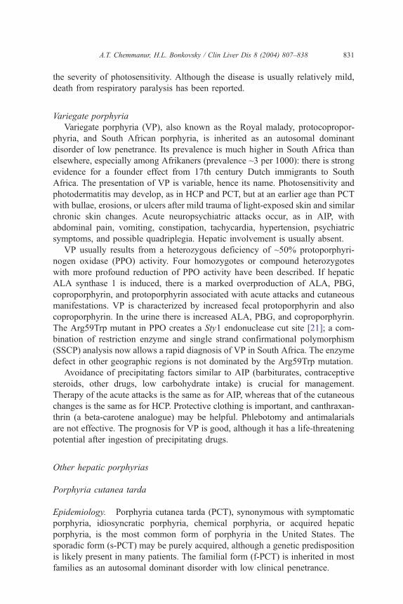
Fig. 6. The enzymatic defects in acute intermittent porphyria (AIP): the two forms of porphobilinogen
deaminase (PBGD) and how the diagnosis of variant AIP is missed by measuring erythrocytic PBGD
activity. The PBGD gene with 15 exons is illustrated. (A) Use of the housekeeping promoter (PH)
results in the 39-kDa nonerythroid PBGD derived from exons 1 and 3–15. Use of the erythroid
promoter (PE) results in a 37-kDa protein derived from exons 3–15. B. When erythroid PBGD is
measured in classic AIP, there is an ~50% reduction compared with controls. An indeterminate zone of
PBGD activity (*) overlaps the lower range of control values and those seen in some AIP patients. In
variant AIP there is no reduction in erythrocytic PBGD levels. The graphs illustrate the mean and
range of measurements anticipated for hypothetical groups of patients with classic and variant AIP.

A.T. Chemmanur, H.L. Bonkovsky / Clin Liver Dis 8 (2004) 807–838828
Management. Prevention is key. Patients should wear a medical alert bracelet.
Carriers of the gene defect should be counseled to avoid situations that may
precipitate acute attacks. AIP patients should be advised to use as few drugs as
possible. Drugs that are used should be bsafeQ based on experimental and clinical
experience (see Table 5) (other drug lists are available on the worldwide web
at www.porphyriafoundation.com; http://www.porphyria-europe.com; www.uct.
ac.za/depts/liver/drugname.htm; http://www.cpf-inc.ca/links.htm; http://www.
porphyrias2003.cz/program.html. Oral carbohydrate intake should always be
adequate, and starvation and fad diets should be avoided. Infections should be
treated promptly. Stresses should be avoided if possible. Relatives who are at risk
for AIP should be screened for the disease, probably best done at centers with
special expertise in the diagnosis and management of porphyria.
The treatment aim is to reduce the activity of hepatic ALA synthase 1. The
essentials of treatment of the acute porphyric attacks are given in Box 3. Mild
attacks may be treated with general supportive measures. Discontinue all known
or potentially harmful drugs that might have precipitated an acute attack. Provide
at least 300 g of carbohydrate per day enterally or parenterally; glucose or another
readily metabolized carbohydrate should be used. Replace fluids, particularly if
patients have poor oral intake or have been vomiting. Monitor patients for
potential hyponatremia or hypomagnesemia and modify administration of
electrolytes and water appropriately. Pain control can be achieved with regular
and frequent doses of morphine or meperidine, although such narcotics may ex-
acerbate urinary retention or constipation. The intermittent nature of the symp-
toms or attacks means that narcotic addiction is unusual. Agitation and anxiety
can be treated with chlorpromazine (50 to 400 mg/day). Sympathetic hyper-
activity may be treated with propranolol (in the absence of contraindications).
The tachycardia and hypertension can be quite labile.
Box 3. Essentials of treatment of acute porphyric attacks
!Discontinue all known or potentially harmful drugs (see Table 5)! 300 g of carbohydrate intake/day! Fluid replacement (fluid restriction if syndrome of inappropriatesecretion of antidiuretic hormone [SIADH])
!Monitor for potential hyponatremia F hypomagnesemia! Pain control: meperidine (400 to 1600 mg/day) or morphine(32 to 128 mg/day)
!Agitation and anxiety: chlorpromazine (50 to 400 mg/day).! Sympathetic hyperactivity: propranolol (40 to 200 mg/day).! IV heme (3 to 5 mg/kg body weight/day).! Prompt treatment of infection! Treatment of intercurrent diseases

A.T. Chemmanur, H.L. Bonkovsky / Clin Liver Dis 8 (2004) 807–838 829
The treatment of choice for any acute attack of porphyria that is severe enough
to warrant hospital admission is prompt therapy with intravenous heme [12–14].
The only preparation currently available for use in the United States is pan-
hematin (Ovation Pharmaceuticals, Deerfield, Illinois, Hematin Hot Line 1-800-
622-2688; 1-800-455-1141 for questions from health care providers). The usual
dose is 3 to 5 mg hematin/kg body weight once daily for 3 to 5 days.
Resuspension of lyophilized hematin powder in human serum albumin will
prolong its usually very limited stability (1 vial of panhematin, 313 mg of heme,
is dissolved in 132 mL of 25% human serum albumin, and administered over
1 hour) [15]. Haem arginate (Normosang, Medica, Helsinki, Finland) is available
in many other countries. In most patients with acute porphyria given IV heme,
there is normalization of the hepatic overproduction and over-excretion of ALA
and PBG within 2–3 days and the porphyric symptoms improve, especially if
they are of recent onset. After IV heme therapy is discontinued, the over-
production of ALA and PBG increases rapidly, but fortunately patients do not
generally redevelop symptoms. Patients with AIP may overexcrete ALA and
PBG for years after an acute attack. There are risks associated with the use of IV
hematin, including a coagulopathy due to adverse effects on the clotting factors
and platelets, vasculitis, hemolysis, and, if large doses are used, transient renal
failure. A phlebitis or thrombophlebitis is frequent. Frequent treatment with
hematin may induce heme oxygenase and reduce its therapeutic benefit. The
duration of effectiveness of heme may be increased significantly if an inhibitor of
heme oxygenase such as tin or zinc meso- or protoporphyrin is given with heme
[7,16–18], but this approach is not yet approved.
Women with cyclical porphyric attacks during the luteal phase of their
menstrual cycles may benefit from oral contraceptives to block their endogenous
cyclic sex hormone production. Leutinizing hormone releasing hormone
(LH-RH) analogues, which block the effects of LH-RH at the pituitary, and the
cyclic secretion of LH and follicle stimulating hormone are also useful.
Leuprolide (Lupron, TAP Pharmaceuticals, Chicago, Illinois) has been used
most widely for this purpose. Alternatively, prophylactic IV heme (given once,
twice, or thrice weekly) may help these women.
The treatment of seizures complicating porphyria is especially difficult be-
cause many of the usual drugs are contraindicated in porphyria (see Table 5).
Clonazepam may benefit some patients and is less likely than hydantoins or
barbiturates to worsen porphyric atttacks. Parenteral magnesium may be useful
but not for chronic therapy. Among newer anticonvulsants, gabapentin and viga-
batrin appear to be safe, whereas felbamate, lamotrigine, and tigabine are not [19].
Prognosis. Attacks may last for a few days to months; in some patients a chronic
porphyria syndrome develops, but most are asymptomatic between attacks. The
prognosis in AIP is generally good, especially if patients who carry a gene defect
are counseled to avoid drugs and other precipitants of an acute attack and if acute
attacks are treated early and vigorously with IV hematin. Patients with a chronic
paresis may be left with a residual deficit, usually a foot drop or wrist drop or

A.T. Chemmanur, H.L. Bonkovsky / Clin Liver Dis 8 (2004) 807–838830
wasting of the intrinsic hand muscles, or they may slowly recover fully. Chronic
renal failure, perhaps partly secondary to sustained arterial hypertension or
analgesic nephropathy, occurs with an increased incidence in AIP.
ALA dehydratase deficiency porphyria
ALA dehydratase deficiency porphyria (ADP) is a rare syndrome with
symptoms similar to AIP. Most ADP patients have had severe repeated porphyric
attacks, with different phenotypes but with no skin photosensitivity. The typical
symptoms are vomiting, extremity pain, and neuropathy including paralysis and
abdominal pain.
ADP results from a severe deficiency of ALA dehydratase (b10% of normal)
with secondary induction of hepatic ALA synthase 1 and overproduction of
ALA. ADP patients excrete large amounts of urinary ALA and coproporphyrin.
For reasons that are not clear, despite markedly decreased activities, ALA
dehydratase levels are ~3% of normal, but erythrocyte coproporphyrin III and
protoporphyrin levels are elevated ~100 fold. The treatment of ADP is suggested
to be the same as for AIP, but not all patients have responded. Some had ex-
acerbations after alcohol, stress, or hunger [20]. The prognosis is guarded at best.
Hereditary coproporphyria
Hereditary coproporphyria (HCP), an inherited autosomal dominant disorder,
is less common than AIP, though latent HCP and HCP carriers are being
increasingly recognized. In Denmark the prevalence of HCP has been estimated
to be 2 per million. The clinical features of HCP may be neurovisceral as in AIP
but milder or cutaneous (in ~30%) with a vesiculobullous eruption, resembling
that in PCT. Attacks have been precipitated by drugs (barbiturates) but also by the
menstrual cycle, contraceptive steroids, and pregnancy. Some patients have had
jaundice and hepatic dysfunction.
HCP results from a deficiency of coproporphyrinogen oxidase. Most patients
have ~50% of normal enzyme activity. The livers of patients with active HCP
have increased coproporphyrin levels, and they fluoresce red when exposed to
light of the Soret band. Many molecular defects in coproporphyrinogen oxidase
(CPO) give rise to HCP, with a variable phenotype. Many HCP patients have a
moderately to markedly increased excretion of coproporphyrin III in the feces
(both during and between attacks) and to some extent in the urine. During acute
porphyric attacks urine ALA and PBG are also increased; typically urinary ALA
PBG excretion (in mg/24 h) exceeds that of PBG but these levels usually
normalize between attacks of HCP, unlike AIP. There are silent carriers, just as
with AIP, who carry mutations in CPO, but in whom the excretion of porphyrins
and their precursors is normal.
The avoidance of precipitating factors is crucial for managing HCP. Acute
attacks of HCP are treated in the same way as for AIP. Opaque sunscreens and the
avoidance of sunlight are recommended in the treatment of cutaneous mani-
festations and their prevention. Beta-carotene may be of some benefit in reducing
A.T. Chemmanur, H.L. Bonkovsky / Clin Liver Dis 8 (2004) 807–838 831
the severity of photosensitivity. Although the disease is usually relatively mild,
death from respiratory paralysis has been reported.
Variegate porphyria
Variegate porphyria (VP), also known as the Royal malady, protocopropor-
phyria, and South African porphyria, is inherited as an autosomal dominant
disorder of low penetrance. Its prevalence is much higher in South Africa than
elsewhere, especially among Afrikaners (prevalence ~3 per 1000): there is strong
evidence for a founder effect from 17th century Dutch immigrants to South
Africa. The presentation of VP is variable, hence its name. Photosensitivity and
photodermatitis may develop, as in HCP and PCT, but at an earlier age than PCT
with bullae, erosions, or ulcers after mild trauma of light-exposed skin and similar
chronic skin changes. Acute neuropsychiatric attacks occur, as in AIP, with
abdominal pain, vomiting, constipation, tachycardia, hypertension, psychiatric
symptoms, and possible quadriplegia. Hepatic involvement is usually absent.
VP usually results from a heterozygous deficiency of ~50% protoporphyri-
nogen oxidase (PPO) activity. Four homozygotes or compound heterozygotes
with more profound reduction of PPO activity have been described. If hepatic
ALA synthase 1 is induced, there is a marked overproduction of ALA, PBG,
coproporphyrin, and protoporphyrin associated with acute attacks and cutaneous
manifestations. VP is characterized by increased fecal protoporphyrin and also
coproporphyrin. In the urine there is increased ALA, PBG, and coproporphyrin.
The Arg59Trp mutant in PPO creates a Sty1 endonuclease cut site [21]; a com-
bination of restriction enzyme and single strand confirmational polymorphism
(SSCP) analysis now allows a rapid diagnosis of VP in South Africa. The enzyme
defect in other geographic regions is not dominated by the Arg59Trp mutation.
Avoidance of precipitating factors similar to AIP (barbiturates, contraceptive
steroids, other drugs, low carbohydrate intake) is crucial for management.
Therapy of the acute attacks is the same as for AIP, whereas that of the cutaneous
changes is the same as for HCP. Protective clothing is important, and canthraxan-
thrin (a beta-carotene analogue) may be helpful. Phlebotomy and antimalarials
are not effective. The prognosis for VP is good, although it has a life-threatening
potential after ingestion of precipitating drugs.
Other hepatic porphyrias
Porphyria cutanea tarda
Epidemiology. Porphyria cutanea tarda (PCT), synonymous with symptomatic
porphyria, idiosyncratic porphyria, chemical porphyria, or acquired hepatic
porphyria, is the most common form of porphyria in the United States. The
sporadic form (s-PCT) may be purely acquired, although a genetic predisposition
is likely present in many patients. The familial form (f-PCT) is inherited in most
families as an autosomal dominant disorder with low clinical penetrance.

A.T. Chemmanur, H.L. Bonkovsky / Clin Liver Dis 8 (2004) 807–838832
Presenting/associated features. Sporadic and familial PCT usually presents in
adults. PCT patients do not present with acute neurologic attacks. Symptoms are
usually limited to the skin. In PCT the photosensitizing skin lesions of increased
skin fragility affect mostly the dorsum of hands and forearms; the hands may
present with bullae, vesicles, blisters, and sores (Fig. 7). These lesions are not
from acute photosensitivity but result from mild trauma in sun-exposed areas.
Milia are 1- to 5-mm pearly white subepidermal inclusions, particularly on the
hands and fingers. Lesions can also be seen on the forehead, ears, neck, and other
sun-exposed areas. The lesions often become infected and tend to heal slowly and
leave residual areas of hypo- or hyperpigmentation or sclerodermatous changes.
Increased facial hair occurs, which is more noticeable in women. Alopecia may
develop at sites of repeated skin damage. The characteristic histopathologic
finding of PCT in the skin is subepidermal bullae with minimal inflammation.
The undulating base of dermal papillae is termed bfestooned.QA typical patient with PCT is a middle-aged man who consumes excess
alcohol and has evidence of hepatic disease with elevated serum amino-
transferases and gamma glutamyl transpeptidase. Alcohol induces hepatic ALA
synthase 1 in patients with PCT and reduces erythrocyte uroporphyrinogen
decarboxylase (UROD) activity. Alcohol also inhibits other enzymes in the heme
pathway, and chronic alcoholism suppresses erythropoiesis and increases dietary
iron absorption. Other patient groups with a relatively high incidence of PCT are
those with diabetes mellitus, young women who use oral contraceptives, men
with prostate cancer who take estrogens, and chronic hemodialysis patients.
These last patients often have iron overload from multiple blood transfusions.
The livers of patients with PCT contain high concentrations of uroporphyrins and
hepta-carboxyl porphyrins, and when exposed to light from a Wood’s lamp, they
show an intense red fluorescence. They also often have fat deposition,
inflammation, and variable necrosis and fibrosis. Some degree of siderosis is
present in 80% of PCT patients on liver biopsy, and most PCT patients have
Fig. 7. Manifestations of porphyria cutanea tarda. Typical cutaneous lesions with bullae, vescicles, and
erosions on the dorsal hands.

A.T. Chemmanur, H.L. Bonkovsky / Clin Liver Dis 8 (2004) 807–838 833
increased ferritin, serum iron, and iron-binding saturation. Iron in the liver plays
an important role in the pathogenesis of PCT. Cirrhosis develops in 30% to 40%
of PCT cases. The incidence of hepatocellular carcinoma in PCT is greater than
normal. There is a high prevalence of HCV antibody markers in PCT patients,
with significant variation between countries (5% in Germany, 12% in the United
Kingdom, 56% in the United States, and 75% to 90% in Italy and Spain) [22].
Nature of the metabolic defect in porphyria cutanea tarda. PCT results from a
defect in UROD. There is an inherited or acquired reduction in hepatic UROD
activity, but a 50% reduction per se is insufficient to cause disease. There may be
formation of an inhibitor of UROD from iron and breakdown products of
uroporphyrinogen. The pathogenesis of PCT is complex. It involves increased
oxidative stress in the liver, which may be mediated by multiple exogenous or
endogenous factors, for example by alcohol, iron, estrogens, porphyrins, chronic
hepatitis C virus infection, polychlorinated biphenyls (PCBs), and polychlo-
rinated cyclic hydrocarbons (the fungicide hexachlorobenzene contaminated
wheat in Turkey and resulted in ~4000 cases of PCT in the 5 or 6 years up to 1961).
With respect to estrogens, their use in therapy of prostate cancer, postmenopausal
replacement, contraception, and their increase in pregnancy have been reported to
precipitate PCT. With respect to iron, it is present both in hepatocytes and in
Kupffer cells. Inheritance of one or more hemochromatosis genes is an important
susceptibility factor for sporadic PCT. Thus, heterozygosity for HLA-linked
hereditary hemochromatosis is frequently present in patients with PCT and
probably is the major factor causing hepatic iron deposition [22,23]. All patients
with PCT should undergo screening for HFE gene mutations and for hepatitis C
infection. All forms of human PCT have reduced liver UROD activity. A sporadic
form (s-PCT or type I PCT) accounts for ~75% to 85% of PCT patients with
decreased UROD activity confined to the liver. The inherited familial autosomal
dominant forms (f-PCT or types II and III PCT) account for the remaining ~15%
to 25% of PCT patients.
Diagnosis. PCT is characterized by a marked increase in urinary uro- (mostly
the I isomer) and heptacarboxy porphyrins. Urinary ALA is often slightly
elevated, but PBG is usually normal. A variety of fecal porphyrins is present in
PCT. Much of the fecal coproporphyrins are isocoproporphyrins (unlike HCP and
VP); therefore elevated stool isocoproporphyrin/coproporphyrin is almost
diagnostic of PCT. Urinary uroporphyrin N coproporphyrin favors PCT; urinary
coproporphyrin N uroporphyrin favors VP or HCP.
Management. Cutaneous symptoms of PCT are treated by stopping precipitating
factors such as the ingestion of alcohol or estrogens. If the urinary uroporphyrin
excretion is very high (N2 mg/day), other measures may be necessary. Patients
should wear protective clothing, avoid strong sunlight, and apply opaque

A.T. Chemmanur, H.L. Bonkovsky / Clin Liver Dis 8 (2004) 807–838834
sunscreens such as zinc oxide paste. Sunscreens that protect against sunburn are
not adequate because they do not screen out the Soret band of light radiation.
Phlebotomy to remove iron from the liver is curative in sporadic PCT and
results in the normalization of activity of hepatic UROD. Initially, 450 mL of
blood is removed one to two times per week, then with increasing intervals. The
aim is to produce a mild degree of iron deficiency (hematocrit b35% and serum
ferritin b10 ng/mL), which usually requires removal of 12 to 16 units of blood
(3 to 4 g of iron removal). Phlebotomy may induce clinical remission, reduce
urinary porphyrins, be associated with regression of the scleroderma-like skin
changes, but it is not proven to improve liver histology. About 10% to 20%
relapse within 1 year but will likely respond again to phlebotomy if there are no
other causative factors. Chloroquine and other antimalarials form water-soluble
complexes with octa- and hepta-carboxyl porphyrins and facilitate their excretion
in the urine. Treatment should be initiated at a low dose (125 mg, two to three
times per week) to reduce the likelihood of acute hepatic injury (fever, right upper
quadrant pain related to massive uroporphyrin removal from the liver) and
retinopathy. Improvement or remission typically takes 6 to 9 months. Patients
with chronic hepatitis C and PCT may experience porphyric remissions after
interferon therapy of the viral hepatitis.
Prognosis. Although the prognosis is good for PCT patients who avoid alcohol,
the general prognosis depends on the nature and severity of the underlying
liver disease.
Hepatoerythropoietic porphyria
Hepatoerythrocytic porphyria (HEP) is a rare form of porphyria. It is caused
by a marked deficiency of UROD due to homozygous or compound
heterozygous defects. The clinical manifestations of HEP, which are similar to
those of congenital erythropoietic porphyria, occur in early childhood, within the
first year of life, as a skin disease with severe photosensitivity, skin fragility, and
subepidermal bullae. There is excess facial hair, and erythrodontia has been
noted. Hepatosplenomegaly has been noted, and hepatic disease develops later.
The liver shows portal inflammation and a red fluorescence; serum amino-
transferases may be mildly elevated. Serum iron is usually normal. Adults with
HEP have a mild normocytic anemia; the erythroid precursors in the bone
marrow fluoresce.
HEP is genetically heterogeneous. As the name implies, excess porphyrins are
synthesized both in the liver and bone marrow. The clinical diagnosis of HEP is
based chiefly on elevated urinary uro- and heptacarboxyl-porphyrins, mostly
type I isomers; additionally zinc protoporphyrin in erythrocytes is elevated.
Isocoproporphyrin, greater or equal to coproporphyrin, is detected in stool (and
urine). Several mutations and deletions have been described. Management of
HEP is the same as for PCT and includes avoidance of the sun. Gene therapy

A.T. Chemmanur, H.L. Bonkovsky / Clin Liver Dis 8 (2004) 807–838 835
might prove effective in the future. The prognosis of HEP is poor due to the
severe defect in UROD activity.
Erythropoietic porphyrias
Erythopoietic protoporphyria
Epidemiology. Protoporphyria, also called erythropoietic protoporphyria or
erythrohepatic protoporphyria (EPP), is the commonest of the erythropoietic
porphyrias and second only to PCT in prevalence of all the porphyrias, 10 to
20/100,000. There is no sex predominance, and it affects all ethnic groups.
Presenting/associated features. The clinical expression is highly variable.
Photosensitivity is the major clinical manifestation of EPP. It is variable among
patients even of the same family. Cutaneous symptoms usually begin in infancy.
Symptoms of EPP are summarized in Table 7. They include burning, itching, or
pain in the skin on exposure to sunlight. This may occur within a few minutes of
exposure. There is subsequent erythema, edema, and occasional urticaria in sun-
exposed areas. Only if the sun exposure is prolonged do vesicles develop (in
contrast to PCT). Shallow waxy depressed scars or thickening with some
wrinkling will develop over the nose, cheeks, or dorsum of hands if there are
repeated episodes of photosensitivity.
EPP patients rarely (b10%) develop severe liver disease with cirrhosis and
acute cholestasis. Some have died of hepatic failure with livers black and nodular
from cirrhosis. Polarization microscopy of such livers shows birefringence due to
crystals of protoporphyrin in hepatocytes and Kupffer cells and bile canaliculi. In
these patients the erythrocyte protoporphyrin levels have exceeded 2000 mg/dL,higher than is usually seen in EPP patients. Only rarely do patients recover after
jaundice supervenes. Usually, the liver failure is not thought to be related to
alcohol or viral or drug-induced hepatitis. Cholelithiasis, due to concretions of
protoporphyrin, occurs with an increased frequency.
Table 7
Cutaneous and other manifestations in erythropoietic protoporphyria (EPP)
Symptoms and signs Incidence (%)
Burning 97
Edema 94
Itching 88
Erythema 69
Scarring 19
Vesicles 3
Anemia 27
Gallstones 4
The manifestations in EPP, mostly cutaneous, from 32 patients are listed.
Adapted from DeLeo VA, Poh-Fitzpatrick M, Mathews-Roth M, Harber LC. Erythropoietic proto-
porphyria. 10 years experience. Am J Med 1976;60:8.

A.T. Chemmanur, H.L. Bonkovsky / Clin Liver Dis 8 (2004) 807–838836
Nature of the metabolic defect. EPP results from a deficiency of ferrochelatase.
The defect is in all heme-forming tissues. Although the inheritance of human EPP
has usually been autosomal dominant, the activity of ferrochelatase in clinically
affected persons is only 15% to 25% of normal, and the parents of many patients
are phenotypically normal and asymptomatic, although about one half of them
demonstrate decreases in activity of the ferrochelatase [24]. Rare compound
heterozygotes with very severe phenotypes, causing liver failure in adolescence,
have also been described [25]. Patients with EPP with more severely affected
phenotypes, particularly those who develop serious porphyric hepatopathy, have
defects in both FC alleles. Usually, there is a missense or other serious mutation
in one allele and a less serious variation in the other. The most common example
of the latter is a variation at position –48 of intron 3, which leads to a lower level
of expression of this allele, albeit with an mRNA and protein that are normal [26].
In EPP excess protoporphyrin is produced mostly in the bone marrow.
Protoporphyrin is excreted unaltered by hepatic clearance into the bile and
subsequently into the feces. The rate-limiting step is canalicular excretion. If the
hepatic load exceeds the excretion capacity, protoporphyrin accumulates in the
liver and causes liver damage, involving a vicious cycle of worsening cholestasis
and worsening accumulation. This may result in pigmentary cirrhosis and
pigmented gallstones associated with hemolysis.
Diagnosis. Erythrocytes, feces, and plasma have increased protoporphyrin, but
urinary porphyrins and precursors are normal. The erythrocytic protoporphyrin is
free and not the zinc chelate (as in lead poisoning and iron deficiency anemia).
Management. Standard sunscreens are not useful. The only topical sunscreens
effective at wavelengths N400 nm have a high sun protection factor (SPF N30)
and are light opaque, containing zinc oxide or titanium dioxide, and may be cos-
metically unacceptable to some patients. Oral beta-carotene (Lumitene, Tishcon
Corporation, Westbury, NY) 30 to 300 mg orally daily (for adults to give a serum
beta-carotene level of 600 to 800 mg/dL after 4–6 weeks of therapy) reduces the
photosensitivity in ~80% of patients, over a 1- to 3-month period after starting
therapy [26]. Beta-carotene causes a faint yellow-orange discoloration in the skin;
topical beta-carotene cream is ineffective. Beta-carotene probably acts by
quenching activated oxygen radicals. The optimal therapy of EPP is unclear.
Several approaches have been proposed and rationalized: (1) RBC transfusion,
(2) IV heme, (3) oral iron to reduce protoporphyrin formation when there is an
iron deficiency, (4) oral chenodeoxycholic acid, (5) oral cholestyramine, (6)
activated charcoal to interrupt the enterohepatic circulation, (7) vitamin E.
Patients should be followed for the development of liver disease; however routine
liver bfunctionQ tests are not reliable indicators of liver damage. Patients with very
high protoporphyrin levels (N1500 mg/dL in erythrocytes or N50 mg/dL in plasma)
or unexplained abnormalities in liver chemistries should be followed closely with
consideration for liver biopsy. Those with necrosis or fibrosis should receive

A.T. Chemmanur, H.L. Bonkovsky / Clin Liver Dis 8 (2004) 807–838 837
therapy as outlined previously. Liver transplant has been used but will not correct
the underlying defect in the bone marrow and other tissues.
Prognosis. Fortunately, the prognosis of EPP is good in the majority of patients.
Acknowledgments
The authors thank J. Clark for help with typing and assembly of the manuscript.
References
[1] Macalpine I, Hunter R. George III and the mad business. New York7 Pantheon; 1969.
[2] Bloomer JR, Bonkovsky HL. The porphyrias. Dis Mon 1989;35:5–54.
[3] Bonkovsky HL. Porphyrin and heme metabolism and the porphyries. In: Zakim D, Boyer T,
editors. Hepatology: a textbook of liver disease. Philadelphia7 Saunders; 1982. p. 351–93.
[4] Fraser DJ, Podvinec M, Kaufmann MR, Meyer UA. Drugs mediate the transcriptional activa-
tion of the 5-aminolevulinic acid synthase (ALAS1) gene via the chicken xenobiotic-sensing
nuclear receptor (CXR). J Biol Chem 2002;277:34717–26.
[5] Bonkovsky HL. The porphyries. In: Rakel RE, editor. Conn’s current therapy. 52nd edition.
Philadelphia7 Saunders; 2000. p. 447–53.
[6] Hahn M, Bonkovsky HL. Disorders of porphyrin metabolism. In: Wu G, Israel J, editors. Dis-
eases of the liver and bile ducts: a practical guide to diagnosis and treatment. Totowa, NJ7
Humana Press; 1998. p. 249–72.
[7] Bonkovsky HL. Advances in understanding and treating bthe little imitator,Q acute porphyria.
Gastroenterology 1993;105:590–4.
[8] Windebank AJ, Bonkovsky HL. Porphyric neuropathy. In: Dyck PJ, Thomas PK, Griffin JW,
Low PA, Poduslo JF, editors. Peripheral neuropathy. 3rd edition. Philadelphia7 Saunders; 1992.
p. 1161–8.
[9] Bonkovsky HL, Barnard GF. Diagnosis of porphyric syndromes: a practical approach in the
era of molecular biology. Semin Liver Dis 1998;18:57–65.
[10] Grandchamp B. Acute intermittent porphyria. Semin Liver Dis 1998;18:17–24.
[11] Andersson C, Thunell S, Floderus Y, Forsell C, Lundin G, Anvret M, et al. Diagnosis of
acute intermittent porphyria in northern Sweden: an evaluation of mutation analysis and
biochemical methods. J Int Med 1995;237:301–8.
[12] Lamon JM, Frykholm BC, Hess RA, Tschudy DP. Hematin therapy for acure porphyria. Medi-
cine 1979;58:252–69.
[13] Pierach CA. Hematin therapy for the porphyric attack. Semin Liv Disease 1992;2:125–31.
[14] Bissell DM. Treatment of acute hepatic porphyria with hematin. J Hepatol 1988;6:1–7.
[15] Bonkovsky HL, Healey JF, Lourie AN, Gerron GG. Intravenous heme-albumin in acute
intermittent porphyria. Evidence for repletion of hepatic hemoproteins and regulatory heme
pools. Am J Gastroenterol 1991;86:1050–6.
[16] Russo SM, Pepe JA, Cable EE, Lambrecht RW, Bonkovsky HL. Synergistic repression of
ALA synthase by heme and zinc-mesoporphyrin in a chick embryo liver cell culture model of
acute porphyria. Eur J Clin Invest 1994;24:406–15.
[17] Dover SB, Moore MR, Fitzsimmons EJ, Graham A, McColl KE. Tin protoporphyrin pro-
longs the biochemical remission produced by heme arginate in acute hepatic porphyria.
Gastroenterology 1993;105:500–6.
[18] Cable EE, Pepe JA, Karamitsios NC, Lambrecht RW, Bonkovsky HL. Differential effects
of metalloporphyrins on mRNA levels of B-aminolevulinate synthase and heme oxygenase:
studies in cultured chick embryo liver cells. J Clin Invest 1994;94:649–54.

A.T. Chemmanur, H.L. Bonkovsky / Clin Liver Dis 8 (2004) 807–838838
[19] Hahn M, Gildemeister OS, Krauss GL, Pepe JA, Lambrecht RW, Donohue S, et al. Effects of
new anticonvulsant medications on porphyrin synthesis in cultured liver cells—potential
implications for patients with acute porphyria. Neurology 1997;49:97–106.
[20] The porphyrias. In: Scriver CR, Beaudet AL, Sly WS, Valle D, editors. The metabolic and
molecular bases of inherited disease, vol. 2. New York7 McGraw-Hill; 1995. p. 2103–61.
[21] Warnich L, Kotze MJ, Groenewald IM, Groenewald JZ, van Brakel MG, van Heerden CJ,
et al. Identification of 3 mutations and associated haplotypes in the protoporphyrinogen oxi-
dase gene in South African families with variegate porphyria. Hum Mol Gen 1996;5:981–4.
[22] Bonkovsky HL, Poh-Fitzpatrick M, Pimstone N, Obando J, Di Bisceglie A, Tattrie C, et al.
Porphyria cutanea tarda, hepatitis C, and HFE gene mutations in North America. Hepatology
1998;27:1661–9.
[23] Roberts AG, Whatley SD, Morgan RR, Worwood M, Elder GH. Increased frequency of
the hemochromatosis C282Y mutation in sporadic porphyria cutanea tarda. Lancet 1997;349:
321–3.
[24] Goerz G, Bunselmeyer S, Bolsen K, Schurer NY. Ferrochelatase activities in patient with
erythropoietic protoporphyria and their families. Br J Dermatol 1996;134:880–5.
[25] Serkany RPE, Cox T. Autosomal recessive erythropoietic protoporphyria: a syndrome of se-
vere photosensitivity and liver failure. Quest J Med 1995;88:541–9.
[26] deRooij FWM, Edixhoven A, Wilson JHP. Porphyria: a diagnostic approach. In: Kadish KM,
Smith KM, Guilard R, editors. The porphyrin handbook. New York7 Elsevier Science; 2003.
p. 211–45.
Top Related