


Languages
Pages
Legal


������������� ���������� ������������������ !"# $"%
Dear Readers,
as members of Special Interest Group (SIG) of Early Pregnancy we would like to present
the guidelines of Association of Early Pregnancy Units, which are obligatory in the United
Kingdom.
We hope the presented material will be useful for the departments specializing in the early
pregnancy. These guidelines have been made accessible to us by dr Roy Farquharson, the co-
ordinator of SIG Early Pregnancy in ESHRE with whom we cooperate.
Prof. dr hab. med. Jana Skrzypczak
Prof. dr hab. Grzegorz H. Bręborowicz
Prof. dr hab. Jan Kotarski
GUIDELINES 2007
of Association of Early Pregnancy Units
Contents
Clinical Guidelines . . . . . . . . . . . . . . . . . . . . . . . . . . . . . . . . . . . . . . . . . . . . . . . . . . . . . . . . . 8
Record Keeping and Data Collection . . . . . . . . . . . . . . . . . . . . . . . . . . . . . . . . . . . . . . . . . . . 9
Guidelines for Ultrasound Scanning . . . . . . . . . . . . . . . . . . . . . . . . . . . . . . . . . . . . . . . . . . 10
Rhesus Anti D Prophylaxis . . . . . . . . . . . . . . . . . . . . . . . . . . . . . . . . . . . . . . . . . . . . . . . . . 13
Guidelines for Viable Intra-Uterine Pregnancy . . . . . . . . . . . . . . . . . . . . . . . . . . . . . . . . . . 13
Management of Non-Viable Pregnancy . . . . . . . . . . . . . . . . . . . . . . . . . . . . . . . . . . . . . . . . 14
Conservative Management of Miscarriage . . . . . . . . . . . . . . . . . . . . . . . . . . . . . . . . . . . . . 14
Medical Management of Miscarriage . . . . . . . . . . . . . . . . . . . . . . . . . . . . . . . . . . . . . . . . . 15
Surgical Evacuation of Non-Viable Pregnancy . . . . . . . . . . . . . . . . . . . . . . . . . . . . . . . . . . . 16
Management of Early Gestational Sac . . . . . . . . . . . . . . . . . . . . . . . . . . . . . . . . . . . . . . . . . 16
Guidelines for hCG . . . . . . . . . . . . . . . . . . . . . . . . . . . . . . . . . . . . . . . . . . . . . . . . . . . . . . . 17
Guidelines on Management of Pregnancies of Unknown Location (PUL) . . . . . . . . . . . . . 18
Surgical Management of Ectopic Pregnancy . . . . . . . . . . . . . . . . . . . . . . . . . . . . . . . . . . . . 23
Management of Ruptured Ectopic with Collapse . . . . . . . . . . . . . . . . . . . . . . . . . . . . . . . . 23
Guidelines on the Management of Women with Recurrent Miscarriage . . . . . . . . . . . . . . 24
Guidelines for Management of Gestational Trophoblastic Disease . . . . . . . . . . . . . . . . . . 26
Introduction
Early pregnancy problems form a major part of all gynae-
cological emergencies. In the past patients were admitted to
the ward and waited for a considerable length of time before
undergoing ultrasound scan and assessment. With the appe-
arance of early pregnancy assessment units (EPU), an in-
creasing number of women are being assessed and managed
as outpatient attenders.
In recent years ultrasound diagnosis and improved under-
standing of problems related to early pregnancy have led to the
introduction of medical and expectant management of mis-
carriage and selected cases of ectopic pregnancy.
It is anticipated that these guidelines will be useful for all
providers of service provision within the EPU. Having under-
gone revision since their first introduction on the website
(earlypregnancy.org.uk) in 2003. These revised guidelines we-
re approved in 2007 and are to be reviewed in 2009.
The development of this updated version is aimed at pro-
viding the best practice guidelines drawn from evidence-based
practice and standardising the care of women with early preg-
nancy problems.
Association of Early Pregnancy Units8
Clinical Guidelines
General Patient Management
• A brief history is taken on the standardised proforma or
locally developed triage protocol (e.g. adapted, audited
and validated Manchester Triage system) (see Appendix)
in accordance with RCOG guidelines including:
1) Previous obstetric history, LMP, urine pregnancy test
in this pregnancy.
2) Pain – description.
3) Bleeding – amount.
4) Passage of Products of conception (POC).
• Clinical examination should be considered if appropriate.
• Transvaginal ultrasound scan (TVS) is performed if less
than 7-8 weeks and also in some circumstances at more
than 8 weeks, which provides the patient with the option
of seeing what is visualised on the screen.
• The procedure and the reasons for the scan should be
explained.
• Patient's wishes should be respected if she strongly.
declines a TVS and where the gender of the professional
is particularly important to the patient.
• A clear explanation should be given by the Gynaecolo-
gist/Sonographer performing the scan as to the possible
or likely diagnosis/diagnoses.
• Appropriate pictures are taken for the patient's records.
Pictures are not usually given to patients in EPAU unless
requested by the patient.
• All items on the proforma should be checked.
• A plan of management should be formulated based on the
guidelines.
• A pregnancy test should be performed if a pregnancy
is not clearly visible.
• Consideration for serum hCG assay should be given if
a pregnancy test is positive.
• Support should be given where the pregnancy is non-vi-
able or the woman is upset.
• A quiet room should be available (core standard).
• Follow up should be arranged before the woman leaves
the clinic.
• Appropriate written advice and telephone numbers for
contact should be given.
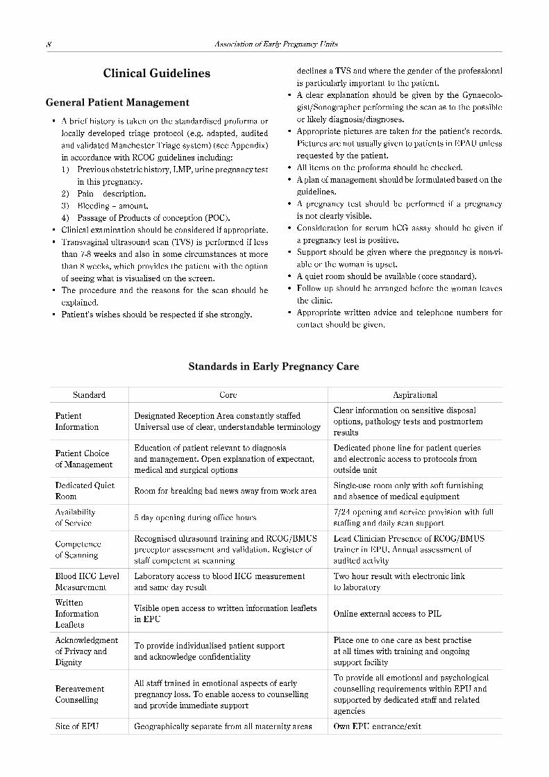
Standards in Early Pregnancy Care
Standard Core Aspirational
Patient
Information
Designated Reception Area constantly staffed
Universal use of clear, understandable terminology
Clear information on sensitive disposal
options, pathology tests and postmortem
results
Patient Choice
of Management
Education of patient relevant to diagnosis
and management. Open explanation of expectant,
medical and surgical options
Dedicated phone line for patient queries
and electronic access to protocols from
outside unit
Dedicated Quiet
RoomRoom for breaking bad news away from work area
Single-use room only with soft furnishing
and absence of medical equipment
Availability
of Service5 day opening during office hours
7/24 opening and service provision with full
staffing and daily scan support
Competence
of Scanning
Recognised ultrasound training and RCOG/BMUS
preceptor assessment and validation. Register of
staff competent at scanning
Lead Clinician Presence of RCOG/BMUS
trainer in EPU. Annual assessment of
audited activity
Blood HCG Level
Measurement
Laboratory access to blood HCG measurement
and same day result
Two hour result with electronic link
to laboratory
Written
Information
Leaflets
Visible open access to written information leaflets
in EPUOnline external access to PIL
Acknowledgment
of Privacy and
Dignity
To provide individualised patient support
and acknowledge confidentiality
Place one to one care as best practise
at all times with training and ongoing
support facility
Bereavement
Counselling
All staff trained in emotional aspects of early
pregnancy loss. To enable access to counselling
and provide immediate support
To provide all emotional and psychological
counselling requirements within EPU and
supported by dedicated staff and related
agencies
Site of EPU Geographically separate from all maternity areas Own EPU entrance/exit

Guidelines 2007 9
Chaperone
Transvaginal ultrasound scanning (TVS) is found to be ex-
tremely well tolerated as a technique by most women [1]. In
the presence of a female chaperon most women feel comfor-
table even if the person doing the scan is male [2]. For most
women the mannerism and expertise of a professional is more
important than the gender. A junior member of the staff
should always be supervised until he/she has attained the re-
quired level of expertise in scanning.
A chaperon can act as advocate for the women, offering re-
assurance and explanation of the procedure or examination.
Women should be given privacy while undressing and dres-
sing.
The woman's age and individual preference should be
taken into account. These can be related to previous experien-
ces which sometimes the women may disclose during consul-
tation or examination. Keep the discussion relevant and avoid
unnecessary personal comments [3].
Whether a female chaperone should always be present du-
ring a transvaginal scan carried out by a male professional de-
pends on the women's choice and the staff situation at the
time of examination.
However the following general principles should be observed:
• Some women may prefer to undergo an examination with-
out the presence of a chaperon. Women's wishes should
be respected and their decision should be documented in
their medical record.
• If a chaperon is not available, examination may be carried
out with a member of the family or friend present.
• If for some reason one can not offer a chaperon, it should
be explained to the patient and, if possible, delay the exa-
mination to a later date. The discussion and its outcome
should be documented.
More details may be obtained from publications of the
Royal colleges of Nursing [4], Radiologists [5] and Obstetri-
cians and Gynaecologists [6].
Guidance on Ultrasound Images
It is not necessary to seek separate permission from the
patient to make the recordings of Ultrasound images. Nor is
consent required to use them for any purpose, provided that,
before use, the recordings are effectively anonymised by the
removal of any identifying marks [7].
References
[1] Basama F.M.S (2001) Audit: Women's perception of transvaginal
sonography in an early pregnancy assessment unit. J. Obstet.
Gynecol. 21: 603-604.
[2] Russel M. (2005) Does patient Ethnicity or Sonographer Gen-
der have any bearing on patient acceptability of transvaginal
ultrasound? Ultrasound 13: 170-172.
[3] Intimate examinations. Guidance for Doctors. General Medical
Council. 2001. www.gmc-uk.org.
[4] Using a chaperon in clinical practice. Standard letter. United
Kingdom Central Council for Nursing, Midwifery and Health Vi-
siting. (1999) London: UKCC.
[5] Intimate examinations, (1998) Royal College of Radiologists 98: 5.
[6] Royal College of Obstetricians and Gynaecologists. Gynaecolo-
gical Examinations: Guidelines for Specialist Practice (2002),
London: RCOG.
[7] Making and Using Visual and Audio Recordings of Patients. Gui-
dance for Doctors (2002) General Medical Council,
www.gmc-uk.org.
Record Keeping and Data Collection
Unless computer based records are available data should
be maintained in hand-written registers.
Accurate record keeping is needed to ensure that preg-
nancy outcome is recorded with sufficient detail and that
feedback is comprehensive. Audit of documentation standards
should be regularly performed in the EPU.
The training of appropriate support staff to maintain high
standards of record-keeping is recommended.
Guidance on Maintaining Registers
The monitoring of the management protocols in terms of
acceptance and outcome can only be achieved through maintai-
ning accurate registers. The following issues are important
toestablish the diagnosis and its management.
1.All first visit scans should be given a diagnosis and
grouped under respective diagnostic groups, such as:
Viable pregnancy/threatened miscarriage – if associated
with bleeding,
Non-viable pregnancy:
a) Complete Miscarriage;
b) Incomplete Miscarriage or
c) Missed Miscarriage.
Ectopic pregnancy.
Hydatidiform mole.
2.Those scans that do not fit into any of the above cate-
gories are grouped under:
a)Pregnancy of Unknown Location (PUL) if an intraute-
rine or extrauterine pregnancy cannot be demonstrated
on scan or
b)Intrauterine Pregnancy (IUP) of Uncertain Viability if
an early small sac is visible (with or without a yolk sac).
With a positive pregnancy test, there could be three rea-
sons for a scan result to be classified as a „Pregnancy of
Unknown Location (PUL)”:
a very early intrauterine pregnancy or
a complete miscarriage or
an early ectopic pregnancy.
At subsequent follow-up visits the diagnosis may become
clear. However, if it is not possible to place a pregnancy into
one of the diagnostic groups in section 1, and symptoms and
signs of pregnancy are resolving (including serum hCG levels),
this can be classified as a „resolving PUL”.

Association of Early Pregnancy Units10
3. A pregnancy in which an embryo measuring <6 mm is
visible, but cardiac activity is not demonstrable on TVS,
is classified as an "IUP of uncertain viability".
4. An intrauterine gestational sac measuring less than 20
mm is also classified as an"IUP of uncertain viability"
until a repeat scan confirms: a viable pregnancy, a demi-
sed embryo or an empty gestational sac. The last is
known as an anembryonic (empty sac) pregnancy (Far-
quharson et al, 2005). Blighted ovum is a term that is
no longer acceptable as embryos seen on earlier scans
frequently get absorbed leaving an empty gestational
sac or some remnants within it. A missed miscarriage
can therefore be simply classified as either fetal or an-
embryonic depending on the presence or absence of
a measurable crown rump length within the gestational
sac.
At a subsequent scan when a diagnosis becomes po-
ssible this will be recorded under the respective groups
as mentioned above under section 1.
5. Scans that are performed after a diagnosis has been
made are grouped under Rescans to avoid repeated
counting of the same patient in a diagnostic category.
6. All non-viable pregnancies – Incomplete/Missed mis-
carriages should be grouped according to the method of
treatment and their outcome recorded.
7. All ectopic pregnancies should be grouped according to
the method of treatment and their outcome recorded.
8. Monthly statistics should be entered on a Data sheet.
The RCOG greentop guideline (Hinshaw, 2006) contains
a simplified assessment algorithm which encourages a simple
classification system of ultrasound appearances into the follow-
ing:
1. Viable IUP;
2. Non-viable IUP (add type eg incomplete, missed etc);
3. IUP of uncertain viability;
4. Ectopic pregnancy;
5. Pregnancy of unknown location (PUL).
References
[1] Farquharson R. G., Jauniaux E., Exalto N. (2005) Updated and
revised nomenclature for description of early pregnancy events.
Human Reproduction, 20: 3008-3011.
[2] Hinshaw K. (2006) The management of early pregnancy loss.
RCOG Greentop Guideline. October RCOG Press, London.
(www.rcog.org.uk/guidelines).
Guidelines for Ultrasound Scanning
RCOG Criteria [1]
If the gestation sac has a mean diameter greater than
20 mm, with no evidence of an embryo or yolk sac, this is
highly suggestive of a missed miscarriage.
If the embryo has a crown rump length greater than 6mm,
with no evidence of heart pulsations, this is highly sugge-
stive of a Missed miscarriage.
When the mean gestation sac is less than 20 mm or the
crown rump length is less than 6 mm a repeat examination
should be performed at least one week later both to assess
growth of the gestation sac and embryo and to establish
whether heart activity exists.
If the gestation sac is smaller than expected for gesta-
tional age the possibility of incorrect dates should always be
considered, especially in the absence of clinical features su-
ggestive of a threatened miscarriage.
In all of the above instances a repeat scan should be un-
dertaken in 7 days. This is necessary to confirm the diagno-
sis.
All scans should be performed by experienced per-
sonnel.
The following individuals are suitably trained to perform
ultrasound:
1.Radiographers/midwives/Nurses with the Diploma in
Medical Ultrasound (DMU)/PGDip or those who have
received training by a recognised Preceptor and Trainer
and who have undertaken assessment and judged to be
competent by the Lead Preceptor of the local EPU.
2.Radiologists with ultrasound training and experience as
recommended by the Royal College of Radiologists.
3.Obstetricians and Radiologists who have completed the
joint obstetric ultrasound training scheme of the Royal
College of Obstetricians and Gynaecologists and The
Royal College of Radiologists, or alternatively who have
appropriate experience and training in obstetric ultra-
sound.
USS - TAS/TVS
‘Pregnancy ofKnown Location’
‘Pregnancy ofUnknown Location’
Intrauterinepregnancy
Ectopicpregnancy
ViableIUP
ResolvedPUL
Non-viableIUP
IUP‘Uncertainviability’
Rescan in7-10 days
Diagnosticalgorithmfor ‘PUL’
KEYUSS - Ultrasound scan
TAS - Transabdominal scanTVS - Transvaginal scan
PUL - Pregnancy of unknown locationIUP - Intrauterine pregnancy
Basic diagnostic algorithm for early pregnancy loss
Guidelines 2007 11
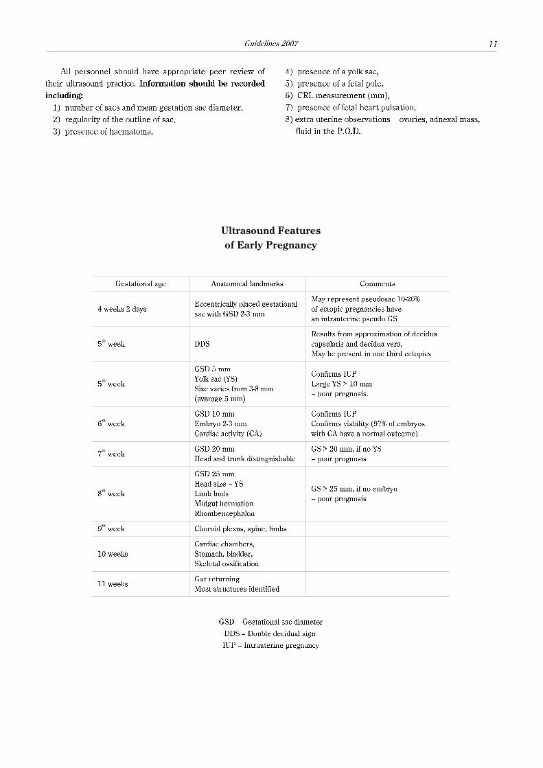
All personnel should have appropriate peer review of
their ultrasound practice. Information should be recorded
including:
1) number of sacs and mean gestation sac diameter,
2) regularity of the outline of sac,
3) presence of haematoma,
4) presence of a yolk sac,
5) presence of a fetal pole,
6) CRL measurement (mm),
7) presence of fetal heart pulsation,
8) extra uterine observations – ovaries, adnexal mass,
fluid in the P.O.D.
Ultrasound Features
of Early Pregnancy
Gestational age Anatomical landmarks Comments
4 weeks 2 daysEccentrically placed gestational
sac with GSD 2-3 mm
May represent pseudosac 10-20%
of ectopic pregnancies have
an intrauterine pseudo GS
5th week DDS
Results from approximation of decidua
capsularis and decidua vera.
May be present in one third ectopics
5th week
GSD 5 mm
Yolk sac (YS)
Size varies from 3-8 mm
(average 5 mm)
Confirms IUP
Large YS > 10 mm
– poor prognosis.
6th week
GSD 10 mm
Embryo 2-3 mm
Cardiac activity (CA)
Confirms IUP
Confirms viability (97% of embryos
with CA have a normal outcome)
7th week
GSD 20 mm
Head and trunk distinguishable
GS > 20 mm, if no YS
– poor prognosis
8th week
GSD 25 mm
Head size = YS
Limb buds
Midgut herniation
Rhombencephalon
GS > 25 mm, if no embryo
– poor prognosis
9th week Choroid plexus, spine, limbs
10 weeks
Cardiac chambers,
Stomach, bladder,
Skeletal ossification
11 weeksGut returning
Most structures identified
GSD – Gestational sac diameter
DDS – Double decidual sign
IUP – Intrauterine pregnancy

Association of Early Pregnancy Units12
A Brief Guide to Management of Early Pregnancy Features
Ultrasound appearance Diagnosis Plan of management
Intrauterine gestational sac (GS),
embryo and cardiac activity (CA)Viable pregnancy Back to GP for referral to ANC
If actively bleeding Admit for reassurance
If a significant haematoma noted Rescan 1 week later
If > 12 weeks Check the need for Anti-D immunoglobulin
GS < 20 mm – no fetal pole Early gestational sac (EGS) Rescan 1 week later
GS > 25 mm – no fetal pole Empty sac
If any doubt Rescan 1 week later If no change
on second scan discuss management (see under
management of nonviable pregnancy
Crown Rump Length (CRL) < 6 mm
CA not demonstratedPregnancy of uncertain viability (PUV) Rescan 1 week later
CRL > 6 mm
CA not demonstratedEarly fetal loss
Rescan 1 week later if in doubt
If no change on second scan discuss
management (see under management of
nonviable pregnancy)
Empty uterus
No adnexal abnormality
Pregnancy of unknown location (PUL)
Serum hCG negative (<5): complete
miscarriage or never pregnant
Serum hCG positive: possible early
pregnancy possible ectopic pregnancy
miscarriage
No follow-up
Repeat serum hCG 48 hours later. Rescan if
necessary (see guidelines for β-hCG)
Warn of the possibility of ectopic pregnancy.
Give possible complete contact numbers to
report if any pain.
Empty uterus
Adnexal mass
Fluid in Pouch of Douglas (POD)
Pain
Ruptured ectopic pregnancyAdmit for assessment: Observation
laparoscopy/laparotomy
Empty uterus
Adnexal mass < 3 cm Unruptured ectopic pregnancy Conservative/medical management
No other findings/symptomsFollow up with serial hCG (see guidelines for
hCG assay)
Endometrium/tissue diameter < 15
mmComplete miscarriage
Advice follow-up 2 weeks later if bleeding
persists
Endometrium/tissue diameter > 15
mmIncomplete miscarriage
Discuss management (see guidelines on
management of incomplete miscarriage
Homogeneous mass within the
uterus
Suspect trophoblastic disease
Serum hCG assay
Surgical evacuation (see guidelines for
trophoblastic disease)
Pregnancy of Unknown Location
(PUL)Diagnosis by exclusion
Follow up with serial hCG (see guidelines for
`Inconclusive scan')
Adequate time should be allowed for women to make
decisions. After giving thorough explanation and answers to
their queries, allow time in privacy. A quiet room would be
more suitable for the woman with her partner/relative/
friend. It is imperative to know that patients will vary in
their response to information at the time. If not receptive
rescan should be arranged for one week. On the other hand
if the patient displays clear understanding and wishes to
know about further management, appropriate choices are to
be given. Women should also be encouraged to go home and
ring later with their decision. They should be reassured that
it will not be harmful to do so if they prefer to discuss this
with their family and contact the unit at a later time.
Women should be informed that the exact cause of
a miscarriage can not be determined and in majority of
cases it is due to a random or one-time genetic abnormality
within the conceptus that leads to a miscarriage [2]. Women
should also be told that miscarriages, in general, are not
linked to parental chromosomal abnormality. A brief expla-
nation of the outcome of the fertilised ova may be helpful in
understanding that not all fertilised ova end up in full term
normal pregnancies.
References
[1] Royal College of Obstetricians and Gynaecologists (1995) Gui-
dance on ultrasound procedures in Early Pregnancy. Standing
joint committee on obstetric ultrasound of RCR/RCOG.
[2] Philipp T., Philipp K., Reiner A. et al. (2003) Embryosopic and
cytogenetic analysis of 233 missed abortions: factors involved
in the pathogenesis of developmental defects of early preg-
nancies. Human Reproduction 18, 1724-32.

Guidelines 2007 13
Rhesus Anti D Prophylaxis
Prophylactic Anti D is not routinely required for rhesus
negative with women bleeding below 12 weeks gestation.
There is minimal evidence that administering Rh immune
globulin for first trimester vaginal bleeding prevents mater-
nal sensitization or development of haemolytic disease of
the newborn [2].
Threatened miscarriage
Anti-D Ig should be given to all non-sensitised RhD ne-
gative women with a threatened miscarriage after 12 weeks
of pregnancy. Where bleeding continues intermittently after
12 weeks' gestation, anti-D Ig should be given at 6-weekly
intervals (send EDTT to check for anti bodies prior to admi-
nistering) (RCOG Grade C recommendation).
However it may be prudent to administer anti-D where
bleeding is heavy or repeated or where there is associated
abdominal pain particularly if these events occur as gesta-
tion approaches 12 weeks (RCOG Grade C recommenda-
tion). The period of gestation should be confirmed by ultra-
sound. Review on an individual basis recommended.
Spontaneous miscarriage
Anti-D Ig should be given to all non-sensitised RhD ne-
gative women who have a spontaneous complete or incomp-
lete miscarriage after 12 weeks of pregnancy (RCOG Grade
B recommendation).
The risk of immunisation by spontaneous miscarriage
before 12 weeks' gestation is negligible when there has
been no instrumentation to evacuate the products of concep-
tion and anti-D Ig is not required in these circumstances
(RCOG Grade C recommendation).
Ectopic pregnancy
Anti-D Ig should be given to all non-sensitised RhD ne-
gative women with a confirmed or suspected ectopic preg-
nancy (RCOG Grade 8 recommendation).
ERPC and Therapeutic termination
of pregnancy
Anti-D Ig should be given to all non-sensitised RhD ne-
gative women having a therapeutic termination of preg-
nancy, whether by surgical or medical methods, regardless
of gestational age (RCOG Grade B recommendation).
Recommended Dose 250-500 i.u. given IM into deltoid
muscle as injections into the gluteal region often only reach
the subcutaneous tissues and absorption may be delayed.
(Dose according to local policy).
References
[1] Use of anti-D immunoglobulin for Rh prophylaxis. (2002) RCOG
'Green-top' Guideline no. 22.
[2] Hannafin B., Lovecchio F., Blackburn P. (2006) Do Rh-negati-
ve women with first trimester spontaneous abortions need Rh
immune globulin? Am. J. Emerg. Med. 24(4): 487-9.
Guidelines for Viable Intra-Uterine
Pregnancy
Definition: A normally sited gestation sac with clearly
identified cardiac activity.
Demonstration of fetal heart activity is generally asso-
ciated with a successful pregnancy rate of 85-97% [1], depen-
ding on the period of gestation.
About 25% of all pregnancies threaten to miscarry.
A threatened miscarriage is one in which:
– the women bleeds a little from the vagina,
– cervical os is closed,
– there is little abdominal pain and,
– pregnancy is still viable.
All women attending EPAU receive a contact number.
Women will go back to GP for referral to ANC via the
usual method.
Follow up appointment may be required in the following
situations:
1. Significant vaginal bleeding and patient refusing to be
admitted.
2. A haematoma [2] is noted.
3. Liquor volume is reduced.
4. Fetal bradycardia.
5. For reassurance at patient's request because
of previous miscarriages.
6. After IUCD removal in the EPAU.
The embryonic heart rate
Theoretically, cardiac activity should always be evident
when the embryo is over 2 mm. However, in around 5-10%
of embryos between 2 and 4 mm, it can not be demonstra-
ted. Perform a follow up scan within one week.
At 6 weeks 60-150 bpm (mean 125 bpm),
6-9 weeks 175 bpm,
Thereafter gradually decreases
14 weeks 160 bpm (approximately).
Bradycardia has been found in pregnancies that subse-
quently miscarried. However, a single observation of slow
heart rate does not necessarily indicate subsequent embryo-
nic death, follow-up is therefore essential.
References
[1] Sotiriadis A. et al. (2004) Threatened Miscarriage: evaluation
and management. BMJ 329: 152-5.
[2] Johns J., Hyett J., Jauniaux E. (2003) Obstetric outcome after
threatened miscarriage with or without a haematoma on ultra-
sound. Obstetrics and Gynaecology 102, 483-7.

Association of Early Pregnancy Units14
Management of Non-Viable Pregnancy
Women feel sensitive about the way we refer to preg-
nancy loss. As their loss is not out of choice, use of language
like termination/abortion can be sometimes offensive to wo-
men at this vulnerable time. Hence documentation for mana-
gement of early pregnancy loss should be worded appro-
priately.
At all times women should be supported in making
informed choices about their care and management. Ade-
quate explanation supplemented with written information
should be given. Ample time should be allowed for making a
decision and if necessary another appointment arranged.
The grief reaction following first trimester miscarriage
can be as profound as after stillbirth.
1. Complete Miscarriage
Ultrasound scan – Endometrial thickness <15 mm
It is the morphology rather than the amount of tissue
that matters. There may be very little pregnancy tissue gi-
ving bright echoes or a large amount of blood showing no
echogenicity at all.
Advise to report if bleeding persists longer than 2/52.
2. Incomplete Miscarriage
Ultrasound scan – Intrauterine tissue diameter 15-50 mm
Conservative method should be offered as an option
provided the bleeding is not heavy and a rescan arranged 2
weeks later or advice may be given to the women to report
if bleeding persisted after 2 weeks.
Alternatively, medical management may be offered if
patient is not willing to wait.
Surgical evacuation is arranged if a patient has a
strong preference it.
Surgical method should be reserved for those who:
1. make a specific request for it,
2. change their mind during the course of conservative
management,
3. have heavy bleeding and/or severe pain,
4. tissue diameter of > 50 mm,
5. have infected tissue.
Conservative management of Incomplete Miscarriage
has excellent success rate and evidence suggests that it is
associated with lower rates of infection than surgical mana-
gement [1].
3. Missed Miscarriage (empty sac/fetal loss)
Since the introduction of TVS, ‘missed miscarriage’
(previously described as ‘anembryonic pregnancy’, absent
fetal echo, ‘blighted ovum’ in the past) are felt to reflect
different aspects or stages of the same clinical process. The
absence of a identifiable fetal pole should be referred to as
an ‘empty sac’.
A previously identified fetal heart action followed by ab-
sence of heart activity should be referred to as a fetal loss.
There is an approximately 5% chance of this happening after
fetal heart action is seen at 7 weeks gestation (Brigham et
al. 1998 [3]) and increases with advanced maternal age.
Following the diagnosis of early pregnancy failure by 2
qualified scanners women should be offered the choices of
conservative, medical or surgical methods of miscarriage
management. Patient preference is important and should be
acknowledged as a determining factor in management deci-
sions.
Conservative management:
·Rescan 2-3 weeks later, if necessary follow up with further
rescans at 2-weekly intervals.
·Give patient a contact number.
Medical management may be offered if patient is not
willing to wait.
Surgical method should be reserved for those:
• who make a specific request for it,
• who change their mind during the course of conserva-
tive management,
• where medical management fails.
The incidence of gynaecological infection after surgical,
expectant, and medical management of first trimester mis-
carriage is low (2-3%). There are small non-significant diffe-
rences in haemorrhage rates (< 3%) and surgical evacuation
carries a !% risk of uterine perforation [2]. The RCOG has
recently updated their guidelines for early pregnancy loss.
References
[1] Trinder J., Brocklehurst P., Porter R. et al. (2006) Manage-
ment of miscarriage: expectant, medical, or surgical? Results
of randomised controlled trial (miscarriage treatment (MIST)
trial). BMJ 332 (7552): 1235-38
[2] Hinshaw K. (2006) The management of early pregnancy loss.
RCOG Greentop Guideline. October. RCOG Press, London.
(www.rcog.org.uk/guidelines).
[3] Brigham S., Conlon C., Farquharson R. G. (1999) A longitudi-
nal study of pregnancy outcome following idiopathic recurring
miscarriage. Human Reproduction 14, 2868-71.
Conservative Management
of Miscarriage
A significant number of women prefer conservative ma-
nagement and it may be continued as long as the patient is
willing, provided there are no signs of infection such as:
– vaginal discharge,
– excessive bleeding,
– pyrexia,
– abdominal pain.
Conservative management requires:
• Motivation and preparation
• A thorough explanation on:
" what to expect: the likely amount of blood loss and
pain,
" what analgesics to be taken,
" what sort of sanitary protection to be used.

Guidelines 2007 15
• Satisfactory answers to their questions and doubts.
• Reassurance that the risk of infection is negligible.
• A contact number should be available 24 hours to ring if
there are any problems such as very heavy loss or
severe pain. An adequately informed and reassured
patient is less likely to contact for any further advice.
• A follow-up appointment for confirmation that the mis-
carriage is complete and to assess if she has any pain or
bleeding.
• An information leaflet to support verbal explanation.
Success rates are higher with prolonged follow-up.
Follow up scans may be arranged at 2 weekly intervals, until
a diagnosis of complete miscarriage is made. However if
patient requests a surgical or medical method at any stage it
should be arranged. At Cardiff, data over a ten year interval
has shown that 90% of women miscarry in three weeks time.
Only a small percentage of women may go up to 6-8 weeks
(AEPU annual meeting lecture, Manchester, 2006).
Medical Management of Miscarriage
The drugs used for medical management of a miscar-
riage include an antiprogesterone, mifepristone (200 mg)
with a prostaglandin such as gemeprost or misoprostol.
The conventional prostaglandin E1 analogue used for
abortion procedures is gemeprost, and is effective in 95% of
cases in combination with mifepristone at <63 days of ame-
norrheoa. The alternative E1 analogue, misoprostol may be
given orally or vaginally and is most effective if administered
vaginally (95% versus 87% respectively) [1]. The main advan-
tages over gemeprost are that it does not require refrigera-
tion, it is cheaper and can be administered orally or vaginally.
The MMM has implications for patients safety as it
avoids the need for an anaesthetic and surgical instrumen-
tation.
The morbidity in those treated medically was lower than
in those requiring surgery (1.7% versus 6.6%) [2] .
Contraindications to Medical management
Absolute: adrenal insufficiency,
long term glucocorticoid therapy,
haemoglobinopathies or anticoagulant therapy,
anaemia (haemoglobin < 10 g/dl),
porphyria,
mitral stenosis,
glaucoma,
non steroidal anti-inflammatory drug ingestion in
previous 48 hours.
Relative: hypertension,
severe asthma.
Varying rates of efficacy have been quoted with medical
management in non-viable pregnancies. The efficacy is grea-
test for those pregnancies of less than 10 weeks or with
a sac diameter of less than 24 mm (92-94%) [3].
Prostaglandin Regimens
Incomplete miscarriage
• Gemeprost 1 mg vaginally or
• Gemeprost 0.5 mg vaginally or
• Misoprostol 800 ug (4 × 200 ug tabs) vaginally.
Missed miscarriage
• Mifepristone 200 mg orally followed 36-48 hours later
by cervagem 1 mg vaginally or
• Mifepristone 200 mg orally followed 36-48 hours later
by misoprostol 800 ug (4 × 200 ug tabs) vaginally.
These regimens are unlicensed.
Since progesterone levels are low in non-viable pregnan-
cy mifepristone may be avoided and prostaglandin only ad-
ministered. Many units use Misoprostol (400 ug PV or PO)
alone for medical management of incomplete miscarriage.
Protocol for Medical Management
of Miscarriage [4]
• Ensure that the patient has read information leaflet.
• Ask if she has any questions.
• Arrange with gynaecology ward for admission to a pri-
vate room with toilet facility.
• Obtain written consent for mifepristone and PG ± sur-
gical evacuation.
• Arrange blood tests
· measurement of haemoglobin concentration and
· determination of AB0 and Rhesus blood groups with
screening for red cell antibodies.
• Anti-D immunoglobulin should be given to all non-sensi-
tised Rh negative women undergoing medical evacua-
tion.
• In the case of a pregnancy occurring with an IUCD in-
situ, this devise should be removed before administra-
tion of mifepristone.
• Prescribe mifepristone 200 mg orally.
• Arrange admission 48 (36-72) hours after mifepristone
administration.
• Inform the patient regarding the length of stay on the
ward. Observe for three – six hours after administration
of prostaglandin and discharge if clinically well.
• Women with gestation:
· < 9 weeks on scan have only one insertion of misopro-
stol 800 micrograms vaginally. Misoprostol tablets
are administered vaginally by the woman or clinician.
If miscarriage has not occurred 4 hours after admini-
stration of misoprostol a further dose of misoprostol
400 micrograms may be administered orally or vagi-
nally.
· > 9 weeks on scan can have a maximum of four fur-
ther doses of misoprostol 400 micrograms at 3-hourly
intervals, vaginally or orally depending on the amount
of bleeding and patient's preference.

Association of Early Pregnancy Units16
• Prescribe PG (Misoprostol 800 ug tabs/Cervagem 1 mg)
vaginally and Metronidazole 1 G rectally on the Drug
chart.
• Prescribe Doxycycline 100 mg bd for 7days with co-dyd-
ramol 2 tabs qds for one week to take home after the
procedure.
• Inform that in case of heavy bleeding ERPC may be
required and therefore she should be prepared to stay
overnight if necessary.
• Women may or may not pass POC while on the ward.
They should be advised of what to expect when they go
home and not referred to EPAU for a scan before their
follow up appointment as most of them would miscarry
at a later stage after discharge from the hospital.
• Any products that are obtained should be sent for histo-
logical examination to exclude a molar pregnancy.
• Give patient information on:
– Admission to gynaecology ward,
– Medical management of non-viable pregnancy,
+ What to expect and the likely amount of blood loss,
+ What analgesics to be taken,
+ What sort of sanitary protection to be used.
C Arrange follow-up in EPAU three weeks later (so as to
avoid further appointments if POC are seen at an early
scan or a surgical intervention).
C Give contact telephone numbers for EPAU/Gynaecolo-
gy ward.
Infection rates after expectant, medical and surgical ma-
nagement are not significantly different and are reassuringly
lows.
References
[1] El-Refaey et al. (1995) Induction of abortion with mifepristone
(RU 486) and oral or vaginal misoprostol. N. Engl. J. Med.
332: 983-7.
[2] Hinshaw H. K. S. (1997) Medical management of miscarriage.
In Grudzinkas T.G., O'Brien PMS, editors. Problems in early
pregnancy: advances in diagnosis and management. London:
RCOG press, 284-95.
[3] De Jonge E. J. M. et al. (1995) Randomised clinical trial of me-
dical evacuation and surgical curettage for incomplete miscar-
riage. BMJ 311: 662.
[4] Royal College of Obstetricians and Gynaecologists (2004) The
care of women requesting induced abortion. Evidence-based
clinical Guideline No. 7. London: RCOG.
[5] Trinder J., Brocklehurst P., Porter R. et al. (2006) Manage-
ment of miscarriage: expectant, medical, or surgical? Results
of randomised controlled trial (miscarriage treatment (MIST)
trial). BMJ 332(7552): 1235-38.
Surgical Evacuation of Non-Viable
Pregnancy
Surgical evacuation should be preferably managed on
a day case basis unless there is heavy bleeding when
the patient should be admitted to Gynaecology ward.
Have a unit protocol for admission system with a pa-
tient pathway clearly described.
Give the patient information on admission procedure in-
cluding appropriate patient information leaflet(s).
Explain the surgical procedure and obtain written con-
sent with Doctor familiar with procedure. Mention rare ana-
esthetic and uncommon surgical risks involved such as ute-
rine perforation (1%), cervical tears, intra-abdominal trauma
(0.1 %), intrauterine adhesions, haemorrhage and infection.
Arrange for measurement of haemoglobin concentration
and determination of AB0 and Rhesus blood groups. Anti-D
immunoglobulin should be given to all non-sensitised Rh ne-
gative women undergoing surgical evacuation.
All at risk women (usually women under the age of 25
years) undergoing surgical evacuation for miscarriage
should be screened for Chlamydia trachomatis.
Alternatively, prescribe prophylactic doxycycline 100
mg orally twice daily for seven days and Metronidazole 1 G
rectally at the time of surgical evacuation as per the local
protocol.
Ensure that products of conception are seen at evacua-
tion.
The RCOG study group [3] recommended that all tissue
obtained at a surgical evacuation for miscarriage should be
sent for histology examination. The reasons are:
1. to diagnose molar pregnancy,
2. to exclude ectopic if chorionic tissue is found on his-
tology.
A follow-up appointment is usually not required after
a surgical evacuation.
Give patient information on "What you may need to
know after a miscarriage". There should be information on
counselling if required in the future.
References
[1] Royal College of Obstetricians and Gynaecologists. (2006)
The Management of Early Pregnancy Loss. Guidelines No.
25. London: RCOG.
[2] Recommendations from the 33th
RCOG Study Group. [In:]
Grudzinkas T. G., O'Brien P. M. S., editors. Problems in early
pregnancy: advances in diagnosis and management. London:
RCOG press, 1997. 327-31.
Management of Early Gestational Sac
At 4+2
weeks blastocyst measuring 1.5-2 mm is recogni-
sable as an early gestational sac. The appearance of an early
gestational sac (EGS) is the earliest reliable sign of preg-
nancy. The ability to demonstrate a true intrauterine gesta-
tional sac practically excludes an ectopic pregnancy since
concurrent intrauterine and extrauterine pregnancies are
rare.
When a gestational sac-like structure is located within
the uterus, its relationship to the endometrial cavity is care
fully studied.

Guidelines 2007 17
Ultrasound features of EGS
• It is seen as an anechoic structure with an echogenic
rim.
• It is eccentrically placed i.e. it remains within a
thickened decidua on one side of the uterine cavity.
• It is typically located in the fundus on the posterior wall.
GSD is a useful indicator of GA before CRL measure-
ment is available.
The gestational sac in such instances should be distin-
guished from the pseudosac that occurs in ectopic pregnan-
cy. The pseudogestational sac may result from an ectopic as
well as fluid or blood collection in the uterine cavity and it
represents the endometrial cavity itself.
Management Protocol
EGSs need to be differentiated from an inconclusive
scan result.
Follow up scan is arranged in a week if confident
or
in 3 days to assess growth of sac if uncertain of the diag-
nosis.
A healthy gestational sac grows by 1.2 mm/day.
A yolk sac will usually be visible at next scan in a normal
pregnancy.
Following up every early gestational sac with serial mea-
surements of hCG leads to increased patient anxiety and
wastage of resources.
There is no risk of missing a complication in this group
as they are all followed up until an embryo with a heart beat
is seen or a miscarriage is diagnosed.
Guidelines for hCG
Understanding hCG measurements
Urine Measurements
The urine test is simple and reliable enough to be used
routinely to establish whether or not a woman is pregnant.
A rapid and simple test should be available in the unit.
Serum Measurements
Measurement of hCG in Serum, permits more accurate
quantification which may be useful in the following [1]:
1. Screening in women at high risk of ectopic pregnancy.
2. Determining the appropriate treatment for women with
suspected ectopic pregnancy.
3. Monitoring during expectant management or medical
management of women with ectopic pregnancy.
4. Evaluation of conservative surgical treatment of ectopic
pregnancy.
Serum hCG levels double approximately every two days
in early (< 8 weeks) normal intrauterine pregnancy; a lesser
increase (< 66% over 48 hours) is associated with ectopic
pregnancy and miscarriage [2].
To find out whether or not a pregnancy is normal or
pathological, the two useful clinical concepts of hCG measu-
rement are the hCG doubling time and the discriminatory
hCG level.
hCG doubling time
It refers to the time taken for the hCG level to double
its original value. A hCG value of <5 IU/l is considered to be
the non pregnant value.
The doubling time is particularly useful in early preg-
nancy i.e. before 5.5 weeks or when the serum hCG level is
< 5000 IU/l. As pregnancy progresses the doubling time
also lengthens.
However 15% of normal pregnancies will have abnormal
doubling time and 13% of ectopic pregnancies will have a nor-
mal doubling time [3].
Caution
1. In multiple pregnancies the level of hCG on D2 would
be a little higher, requiring an extra two or three days
for a sac to become visible.
2. The possibility of a heterotopic pregnancy should be
kept in mind (1 in 3000-4000 of spontaneous concep-
tions and 1%-3% of assisted conceptions).
Discriminatory hCG level
It refers to a defined level of hCG above which the ge-
stational sac of an intrauterine pregnancy should be visible
on ultrasound. In women with an hCG result above the dis-
criminatory level, but absence of an intrauterine gestational
sac on ultrasound, ectopic pregnancy is a distinct possibility.
With the use of high resolution transvaginal ultrasound
the discriminatory level has been reported to be around
1000 IU/l IRP [4]. However the American Fertility Society
suggested that in practice the level ought to be around 2400
IU/l.
The discriminatory level may vary in different units and
depends on three factors:
1) hCG assay,
2) quality of ultrasound,
3) the experience of the person performing the ultra-
sound.
It usually lies between 1000-2400 IU/l.
A diagnosis of ectopic pregnancy is more likely when-
ever intrauterine pregnancy is not detected by ultrasound at
serum hCG concentration above 2400 IU/l.
References
[1] Recommendations for clinical practice arising from 33rd
RCOG
Study Group. (1997) Problems of Early Pregnancy – Advances
in Diagnosis and Management. London: RCOG Press.
[2] Kader et al. (1981) Discriminating hCG zone: Its use in the
Sonographic evaluation for ectopic pregnancy, Obstet. Gyne-
col. 58: 156-61.
[3] Ling and Stovall (1994) Update on the diagnosis and mana-
gement of ectopic pregnancy, Advances in Obstetrics and Gy-
naecology, 1, pp 55-83. Chicago: Mosby Year Book, Inc.
[4] Caeciatore et al. (1990) Diagnosis of Ectopic Pregnancy by
Vaginal Ultrasonography in Combination with a discriminato-
ry serum hCG level of 1000 IU/L (IRP). BJOG 97, 904-8.

Association of Early Pregnancy Units18
Guidelines on Management of
Pregnancies of Unknown Location
(PUL)
Definition
No evidence of an intrauterine or extrauterine pregnan-
cy on transvaginal ultrasound scan (TVS) in women with a
positive pregnancy test.
Initial Assessment
• Clinical history:
– the presence of risk factors for ectopic pregnancy.
• Clinical signs.
• TVS.
• Serum hCG.
Give appropriate information to patient. Explain the
need for further follow up.
Follow up
• Close surveillance with Serum hCG measurements eve-
ry 2-3 days.
• See guidelines for hCG.
• Repeat TVS when serum hCG >1000 IU/l (see Discri-
minatory HCG level on previous page) to look for an
IUP or an ectopic otherwise.
• Provide support.
• Follow up until:
Intrauterine pregnancy identified
or
Ectopic pregnancy diagnosed
or
Levels of serum hCG spontaneously decrease
(failing PUL).
Depending the on the quality of the ultrasound service
provided, anything between 10 and 30% of pregnancies of
unknown locations will subsequently be diagnosed as an ecto-
pic pregnancy.
Management Protocol for Inconclusive Scan Result
after the Initial Visit to EPU using Serum hCG and TVS
hCG IU/l UltrasoundPattern of change of hCG level
after 48 hoursManagement
< 1000
No intrauterine sac No Adnexal
mass No fluid in POD
No symptoms
hCG rise > 66% or doubled
If hCG > 1000 repeat ultrasound
or If hCG < 1000 repeat
hCG
> 1000 No intrauterine sac
1. No adnexal mass No fluid POD
No symptoms
Repeat hCG and repeat
ultrasound 2 days later
A. Falling hCG Serial hCG levels until hCG < 20
B. Rising Or plateauing hCG x 3
Diagnosis: Ectopic or PUL
Laparoscopy
(if symptomatic)
or
Methotrexate (if asymptomatic)
2. Suspicious adnexal mass < 3
cm No fluid POD Asymptomatic
Repeat hCG and repeat
ultrasound 2 days later
A. Falling hCG Serial hCG levels until hCG < 20
B. Rising/plateauing hCG x 3Laparoscopy with D&C or
Methotrexate
3. Ad. Mass > 3 cm or
Fluid POD
or
Symptomatic
Laparoscopy
> 2400No intrauterine sac Adnexal
findings ± Asymptomatic
Fluctuating x 3 Diagnosis:
Ectopic or PULLaparoscopy or Methotrexate
Falling hCG: Diagnosis Early miscarriage or
Pregnancy of Unknown Location – resolving
Likely diagnosis; With bleeding: Miscarriage
Ectopic pregnancy PUL – Resolving
No bleeding: Normal intrauterine pregnancy

Guidelines 2007 19
Transvaginal ultrasound scan
Positive urinary pregnancy test
Pregnancy of unknown location
Haemodynamically unstablePain
Haemodynamically stablePain
Haemodynamically stablePain free
Expectant management Serum hCG Serum hCG
*Serum hCG levels at 0and 48 hours
Consider laparoscopy Consider laparotomy
> 66% increase in serumhCG 0-48 hours < 66% increase or < 15%
decrease in serum hCG 0-48 hours
> 15% decrease in serumhCG 0-48 hours
? Intra-uterine pregnancy ? Ectopic pregnancy
Rescan one week to confirmpregnancy location
? Failing PUL
Repeat serum hCG in oneweek to confirm failing
Consider weekly hCG monitoring until < 15 IU/L
Rescan in two weeks to confirm
viability
Early Intra-uterinepregnancyvisualised
Ectopicpregnancyvisualised
Managementas clinicallyindicated
Repeat hCGnow and 48hours later
PUL
If no pregnancy seen on repeat scan and suboptimal rise in hCG
consider methotrexate
*Consider rescan at 24 hours if PUL and initial serum hCG> 1000 IU/L
Possible Algorithm for Management of Pregnancies of Unknown Location
Algorithm
TVS
Inconclusive result(no evidence of IUP or EP)
Serum hCG measurements every 2-3 days
Falling
Complete miscarriage
Rising (doubling)
Repeat TVS when hCG > 1000 IU/L
No further scans are necessaryFollow up until hCG < 20 IU/L IUP
No further hCG assaysRescan in one week
Suboptimal rise/plateauing/falling slowly after 2-3 measurements
TVS
EPPUL
Non-viable IUPKey:IUP Intrauterine pregnancyEP Ectopic pregnancy

Association of Early Pregnancy Units20
Guidelines for Management
of Ectopic Pregnancy
Incidence: Ectopic pregnancy affects 1 in 80 pregnan-
cies. In the EPAU population the incidence is 3%.
Ectopic pregnancy can be a devastating experience.
Women have to cope with:
the loss of a baby
the possible loss of fertility and
the possible loss of their life.
The psychological impact is not to be overlooked. The
emotional as well as the clinical needs of individual women
should be assessed and sensitively dealt with.
The fallopian tube is the most common site accounting
for nearly 95% of ectopic pregnancies. Other possible sites
of an ectopic pregnancy are, interstitial (2%), cervical (0.1%),
ovarian (0.01%), caesarean section scar or abdominal (rare).
An abdominal ectopic pregnancy may be primary or secon-
dary resulting from a tubal miscarriage.
RISK factors are present only in 25%-50% of pa-
tients with an ectopic pregnancy. They include a history of:
• previous pelvic inflammatory disease,
• tubal surgery,
• previous ectopic pregnancy,
• infertility,
• assisted reproductive technology,
• intrauterine contraceptive device.
Smoking and a maternal age > 40 years are also asso-
ciated with an increased incidence of ectopic pregnancy.
The diagnostic performance based on the combined use
of transvaginal sonography (TVS) and serum hCG measure-
ment reaches sensitivities and specificity range 95%-100%'.
Patients who have had previous ectopic pregnancies or are
at risk of ectopic pregnancy should be advised to present
early, at 6 weeks, in subsequent pregnancies for confirma-
tion of uterine pregnancy [2].
Symptoms
• Amenorrhoea (not universal).
• Vaginal bleeding.
• Lower abdominal pain.
• Faintness/dizziness.
• Shoulder tip pain.
• Gastrointestinal symptoms – diarrhoea or pain on defe-
cation.
Signs
• Lower abdominal tenderness.
• Adnexal tenderness and /or mass.
• Cervical excitation.
• Shock/Collapse.
The clinical presentation and natural course of an ecto-
pic pregnancy are unpredictable.
It is important to have a high index of suspicion for ec-
topic pregnancy, because the patient may not be symptoma-
tic until rupture occurs, or on the other hand the patient
may experience vague abdominal pain and/or vaginal blee-
ding.
Diagnosis
Ultrasound features
Like any pregnancy an ectopic pregnancy too has a na-
tural history of evolution, hence the ultrasound findings de-
pend on the developmental stage at the time of examination.
Almost all ectopic pregnancies occur in the fallopian tu-
be. Ultrasound features suggestive of ectopic pregnancy are
a combination of uterine and adnexal findings:
Uterine
• An empty uterus.
• Variable degree of thickening of endometrium.
• A thin endometrium may exclude the possibility of an
early intrauterine pregnancy as it is not compatible with
an ongoing early implantation.
• An intrauterine pseudosac – mere collection of variable
amount of fluid within uterine cavity, is found in ap-
proximately 5% of all ectopic pregnancies.
Adnexal
• A hyperechogenic tubal ring (‘doughnut’ or ‘bagel’ sign)
is the most common finding on scan, probably due to
early scanning.
• A mixed adnexal mass – either tubal miscarriage or
tubal rupture.
• An ectopic sac with a yolk sac or an embryo with or wit-
hout a heart beat.
• Fluid in the Pouch of Douglas.
The corpus luteum may be present on the ipsilateral si-
de in 85% of cases.
Management
In the absence of any diagnostic features on ultrasound
scan (inconclusive scan result) serial hCG assay are perfor-
med.
If the patient is in significant discomfort she should be
admitted to the ward. If she is clinically stable with no dis-
comfort she may be allowed home to return for follow up.
Direct contact number for the emergency ward should be gi-
ven and the patient asked to attend at any time if her con-
dition deteriorates.
Serum hCG assay
An ectopic pregnancy is more likely when the serum
hCG is more than 1000 IU/l. However in the absence of any
pain, hCG is to be repeated in 48 hours time.
If the hCG is falling it is suggestive of a resolving intra
or extrauterine pregnancy. The rate of fall of hCG tends to
be slower in ectopic pregnancy than with complete miscar-
riages. (see guidelines on serum hCG assay).
A serum hCG level that is increasing or has plateaued
may either show an ectopic pregnancy at subsequent scan
or remain as a PUL.
Transvaginal ultrasound and quantitative assay of serum
hCG not only play a role in the diagnosis of an ectopic preg-

Guidelines 2007 21
nancy but also in determining the management options in a
particular patient.
• Expectant management.
• Medical management.
• Surgical management.
Either laparoscopic or open
a. salpingotomy,
b. salpingectomy (48 hours later, HCG should be
< 35% of preop level).
Both the hCG levels and the patterns of change of
hCG are helpful in constructing a plan for ectopic pregnan-
cy. The clinical picture should always be considered with
hCG measurements.
A Guide to Choosing the appropriate treatment based
on hCG Measurements and the expected Serial hCG
patterns in the follow-up of ectopic pregnancy
hCG level Method of
Treatment
Expected hCG
pattern
Low < 10001U/l Expectant
management
Steady downward
trend
< 1000 IU/l
Fluctuating 1000-
3000 IU/l
Medical
treatment
There may be an
initial rise of hCG
> 3000 IU/l Salpingectomy Repeat after 1 week
Expectant Management of Ectopic
Pregnancy
Not all ectopic pregnancies progress and pose a risk to
the mother. Spontaneous resolution of tubal ectopic pre-
gnancies has been well documented in a number of reports.
Selection Criteria for Expectant Management
1. absence of clinical symptoms,
2. no sign of rupture or intraperitoneal bleeding,
3. absence of haemoperitoneum,
4. a tubal mass of less than 2 cm,
5. no fetal parts,
6. serum hCG concentrations below 1,000 IU/l and decli-
ning progressively [3].
The success rate for a spontaneous resolution was 88%
when the initial hCG level was <2000 IU/l [4] but only 25%
at levels >2000 IU/l [5].
The risk of rupture in a woman with an ectopic exists
until the hCG level has fallen to <10 IU/l. It often involves
frequent hospitalisation and/or follow up. Both the physician
as well as the patient must be well motivated to accept the
long recovery time.
Follow-up: Monitor serum hCG levels every 2-3 days
until less than 201U/l, and rescan when required.
Medical Management of Ectopic Pregnancy
Many agents including prostaglandins, RU-486, potas-
sium chloride and actinomycin-d have all used for the me-
dical management of ectopic pregnancy. However the most
commonly used drug is methotrexate. A single injection of
methotrexate is well tolerated and is effective. Published
studies have shown a success rate varying from 52 to 94%
for single dose methotrexate.
Systemic Methotrexate Treatment
in Ectopic Pregnancy
Methotrexate is a folic acid-antagonist (anti-metabolite)
which prevents the growth of rapidly dividing cells by inter-
fering with DNA synthesis. It can be administered systema-
tically (IV, IM or orally). However, it is most commonly gi-
ven according to a single-dose protocol, which involves a sin-
gle intra-muscular dose of 50 mg/m2. Alternatively it can be
given according a multiple dose regimen with alternate day
administration of intramuscular methotrexate and folinic
acid rescue.
Inclusion Criteria
1. Haemodynamically stable,
2. Indications: Unruptured tubal or other ectopic pregnan-
cy (diagnosed with serial hCG and TVS).
Persistent trophoblast after salpingotomy .
3. An ectopic pregnancy with serum hCG less than 3,000
IU.
4. An ectopic pregnancy with serum hCG value less than
1,000 IU/l should have repeat serum hCG within 48
hours if the patient remains haemodynamically stable.
•The treatment should begin if the levels are plateau-
ing.
•If the levels are rising one must exclude intrauterine
pregnancy before starting treatment.
5. Normal LFT's, U & E's, and FBC.
Exclusion Criteria
1. If there is any evidence of intraperitoneal haemorrhage
i.e. haemoperitoneum on TVS.
2. Any hepatic dysfunction, thrombocytopenia (platelet
count < 100 000), blood dyscrasia (WCC < 2000 cells
cm3).
3. Difficulty or unwillingness of patient for prolonged fol-
low-up (average follow-up 35 days).
4. Ectopic mass >3.5 mm.
5. The presence of cardiac activity in an ectopic pregnancy
6. Women on concurrent corticosteroid therapy.
Treatment Protocol
1. Discuss options for management – expectant/surgical/
medical.
2. Satisfy eligibility and exclusion criteria.
3. Counsel the patient and explain treatment protocol.
Give information leaflet.
4. Take height and weight.
5. Prescribe Methotrexate.
6. Organise base line blood tests, FBC, blood group,
LFTs and U&Es.

Association of Early Pregnancy Units22
7. The prescription with the height and weight documen-
ted on it is sent to Pharmacy to make up the drug.
8. Check blood results, prescribe anti-D immunoglobulin if
Rhesus-negative.
9. Methotrexate is given intramuscularly in buttock or
lateral thigh. The empty syringe or needle should be
placed in a separate Sharp Safe, labelled "Cytotoxic wa-
ste for special incineration".
10. Rest up to one hour. Check for any local reaction. If lo-
cal reaction noted consider anti-histamine or steroid
cream (very rare).
11. Arrange follow-up in EPAU.
Single – Dose Regimen
Day Management
0 Serum hCG, FBC, U&Es, LFTs, G&S
1 Serum hCG Intramuscular methotrexate 50 mg/m2
4 Serum hCG
7 Serum hCG, FBC, LFT
2"d dose of methotrexate if hCG decrease < 15%
day 4-7
If hCG decrease > 15 % repeat hCG weekly
until < 12 U/l
Multiple – Dose Regimen
Days Management
0 Serum hCG, FBC, U&Es, LFTs, G&S
1, 3, 5 Serum hCG
Intramuscular methotrexate 1 mg/kg
2, 4, 6 Serum hCG
Intramuscular folinic acid 0.1 mg/kg
Monitoring
Continue until hCG decreased > 75% in 48
hours or 4 doses of methotrexate given
Information for Clinician
1. Up to 75% of patients may complain of pain on days 3-7
(thought to be due to tubal miscarriage).
2. hCG levels may initially rise days 1-4 (up to 86% of pa-
tients).
3. Mean time to resolution is 35 days.
4. A second dose of Methotrexate may be given at 7 days
if hCG levels fail to fall by more than 15% between day
four and day seven. (3-27% (in published literature)14%
of medically treated women will require more than one
dose of methotrexate).
5. Risk of tubal rupture is 7% and the risk remains while
there is persistent hCG.
6. Folinic acid rescue is not required for the single dose
regime.
7. Avoid vaginal examination. TVS may be undertaken du-
ring first treatment week or subsequently if clinically
indicated.
8. TVS should be used to monitor completeness of resolu-
tion of an ectopic pregnancy after hCG values are nor-
malised [3].
9. Ovarian cysts may be found in the post treatment pha-
se, which undergo spontaneous resolution.
Information for patients
1. Medical treatment for ectopic pregnancy is now well
established, and approximately 90% of patients do not
require further surgery. Methotrexate is used for a va-
riety of clinical conditions, e.g. psoriasis, as well as for
malignancies.
2. Prolonged follow-up is required with blood tests until
serum hCG level is below 20 iu/I.
3. A further dose of methotrexate may be necessary.
4. Three quarters of women experience abdominal pain
following treatment, which is due to the drug acting on
tubal pregnancy. It usually occurs on days 3-7.
5. Pregnancy should be avoided for 3 months after metho-
trexate has been given, because of a possible teratoge-
nic effect – advice should be to use a reliable barrier or
hormonal contraception (RCOG).
6. Side effects of the drug are minimal but may include
nausea, vomiting and stomatitis.
7. Maintain ample fluid intake.
8. Avoid alcohol or folic acid containing vitamins during
treatment.
9. Avoid sexual intercourse until resolution of the ectopic
pregnancy.
10. Avoid exposure to sunlight.
Outcome
• 90% successful treatment with single dose regime.
• Recurrent ectopic pregnancy rate 10-20%.
• Tubal patency approximately 80%.
References
[1] Stovall and Ling (1993) Single dose Methotrexate: An Ex-
panded clinical trial. American Journal of O & G: 168; p 1759-
1765.
[2] Pansky et al. (1989) Local Methotrexate injection. A non-sur-
gical treatment of ectopic pregnancy. AM J O & G 161; 393-
396.
[3] Zullo et al. (1996) Late complications after systemic treat-
ment of ectopic pregnancies: A report of three cases. Eur. J.
Obstet. Gynaecol. Reprod. Biol. 70: 213-4.
[4] Royal College of Obstetricians and Gynaecologists (2004) The
management of tubal pregnancy. Guidelines No. 21 London:
RCOG.
[5] Kirk E., Condous G., Bourne T. (2006) The non-surgical ma-
nagement of ectopic pregnancy. Ultrasound Obstet. Gynecol.
271: 91-100.
[6] Lipscomb G. H., Givens V. A., Meyer N. L., Bran D. (2004)
Previous ectopic pregnancy as a predictor of failure of syste-
mic methotrexate therapy. Fertil. Steril. 81: 1221-4.
[7] Farquhar C. M. (2005) Ectopic pregnancy. Lancet 366: 583-
91.
[8] Condous G., Okaro E., Khalid A., Lu C., Van Huffel S., Tim-
merman D., Bourne T. (2005) The accuracy of transvaginal
ultrasonography for the diagnosis of ectopic pregnancy prior
to surgery. Hum. Reprod. 20: 1404-9.

Guidelines 2007 23
Surgical Management
of Ectopic Pregnancy
Laparoscopy
Advantages – Shorter hospital stay (1-2 days).
– Significantly less blood loss.
– Less adhesions formation.
– Lower analgesic requirements.
– Quicker post operative recovery time.
– Recurrent ectopic pregnancy rate lower.
(5%) than after laparotomy (16.6%).
– Subsequent intrauterine pregnancy (IUP)
rate better (70%) than after laparotomy.
Disadvantages – Increased risk of bowel/vascular damage.
A laparoscopic approach is superior to a laparotomy in
terms of recovery from surgery.
Laparotomy is to be preferred
• in cases with haemorrhagic shock,
• where a surgeon has inadequate experience of operative
laparoscopy ,
• if lack of equipment and instruments,
Do what is safe in the circumstances.
Salpingectomy v Salpinagotomy
In a meta-analysis of nine good quality comparative stu-
dies:
• There was no significant difference in the subsequent
IUP between salpingotomy and salpingectomy groups
(53% v 49.3%).
• The recurrent ectopic pregnancy rate was higher after
salpingotomy (15%) than after salpingectomy (10%).
• Persistence of trophoblast was noted in 4.8% to 11 % of
salpingotomy cases, hence need to monitor hCG post-
operatively.
• In contrast almost no cases of persistence followed sal-
pingectomy. Following salpingectomy, there is no need
to measure hCG in the post-operative period.
In the presence of a healthy contralateral tube there is
no clear evidence that salpingotomy should be used in pre-
ference to salpingectomy.
Laparoscopic salpingotomy should be considered as the
primary treatment when managing tubal pregnancy in the
presence of contralateral tubal disease and the desire for
future fertility [1].
Discuss treatment with the patient and options of
conserving or removing the tube.
Recommendations arising from the 33rd
RCOG Study Group [2]
No. 26 At laparoscopy for ectopic pregnancy, precise docu-
mentation of the state of the pelvis, with particular
emphasis on the affected and contralateral tube and
ovaries, should be undertaken to determine prog-
nosis of future fertility.
No. 27 The definitive procedure undertaken at surgery (re-
moval of the ectopic: salpingotomy; unilateral sal-
pingectomy; bilateral salpingectomy) should be de-
termined by the reproductive aspirations of the pa-
tient, her reproductive history, the state of the pel-
vis and the availability of assisted conception ser-
vices.
No. 28 Fimbrial evacuation (milking) of ectopic pregnancy
from the fallopian tube should not be done as it pre-
disposes to persistence of tubal pregnancy.
Follow-up regime after Salpingotomy
While trophoblast remains in the tube it has a capacity
to rupture.
• Follow-up at weekly intervals until serum hCG level is
<5.
• If hCG level is rising or plateauing consider further
treatment with Methotrexate or surgery if hCG levels
>5 000.
Suturing the salpingotomy lesion provides no benefit.
Outcome after Conservative Surgery
in Women with One Tube
• Recurrent ectopic pregnancy rate 20.5%.
• IUP rate 54%.
Conservative surgery may be appropriate but only if the
patient is aware of the risk involved. Salpingectomy followed
by IVF is an alternative therapy in such cases.
References
[1] Royal College of Obstetricians and Gynaecologists. (2004)
The management of tubal pregnancy. Guidelines No. 21 Lon-
don: RCOG.
[2] Recommendations arising from the 33rd
RCOG Study Group:
Problem in Early Pregnancy (1997) Advances in Diagnosis
and Management. London: RCOG Press.
Management of Ruptured Ectopic
with Collapse
• ABC of resuscitation.
• Get help; call senior SPR on call and anaesthetist.
• Site two IV lines (at least 16 g), commence IV fluids
(crystalloid), give facial oxygen and insert indwelling
catheter.
• Send blood for FBC, Clotting screen and cross-match at
least 4 units of blood.
• Arrange admission and laparotomy.
• Continue fluid resuscitation and ensure intensive moni-
toring of haemodynamic state whilst awaiting transfer to
theatre.
• Do not wait for BP and pulse to normalise prior to tran-
sfer .
• Pfannensteil incision, locate tube directly and clamp.
• Salpingectomy and wash out abdomen.

Association of Early Pregnancy Units24
• Assess bloods consider CVP/HDU discuss with ana-
esthetist.
• Record operative findings including the state of the re-
maining tube.
• Anti-D immunoglobulin to be given to Rhesus negative
women.
Unusual Types of Ectopic Pregnancy
Ultrasound features of non-tubal pregnancies and their
management have been well documented [6-9].
Hetertopic pregnancy.
Interstitial pregnancy.
Cervical pregnancy.
Ovarian pregnancy.
Pregnancy in a caesarean section scar.
Abdominal pregnancy.
Management of these ectopic pregnancies is not
straight forward. Treatment has to be individualised based
on the size of the pregnancy and its viability.
Ectopic pregnancy Ultrasound features Management
Heterotopic – An intrauterine pregnancy and a concurrent ectopic pregnancy
Interstitial
– Bright echogenic trophoblastic tissue or gestational sac (GS) in the
cornual region
– GS located away from the lateral margin of the myometrium
– Thinning of the myometrial mantle
Conservative/Methotrexate
Cervical
– Intracervical GS
– Thick trophoblastic ring
– No distortion of endometrium and cavity
– Closed cervical canal in continuity with the endometrial cavity
– Internal os not funnelled
(Should be differentiated from isthmico-cervical pregnancies that are
implanted low in the uterine cavity, above the cervical canal)
Conservative/Methotrexate
Ovarian– Hyperechogenic mass within the ovary
– Subcapsular bleeding (must be distinguished from a haemorrhagic cyst )Conservative/Methotrexate
Caesarean scar
– Uterine cavity is empty
– GS implanted into the scar
– Negative „sliding sign” [10]
Methotrexate/dilatation and
curettage/cervical packing
Abdominal
– Empty uterus separate from the fetus
– Fetus seen without the surrounding uterine mantle
– Unusual location of the placenta
– Extremely low amount of liquor
Methotrexate/Laparotomy
References
[1] Ling F.W. and Stovall T.G. (1994) Update on the diagnosis
and management of ectopic pregnancy. Advances in obstetrics
and gynaecology, pp 55-83, Chicago. Mosby Year Book, Inc.
[2] Recommendations for clinical practice arising from 33rd
RCOG Study Group (No.23). (1997) Problems of Early Preg-
nan_cy. Advances in Diagnosis and Management. London:
RCOG Press,.
[3] Korhonen et al. (1994) Serum hCG dynamics during sponta-
neous resolution of ectopic pregnancy, Fertil, Steril. 61: 632-
6.
[4] Trio et al. (1995) Prognostic factors for successful expectant
management of ectopic pregnancy. Fert. Ster. 63: 469-72.
[5] Yao M. and Tulandi T. (1997) Current status of surgical and
non surgical management of ectopic pregnancy. Fert. Steril.
67: 421-433.
[6] Condous G., Okaro E., Bourne T. (2003) The conservative ma-
nagement of early pregnancy complications: a review of the
literature. Ultrasound Obstet. Gynecol. 22: 420-430.
[7] Coady A. M. (2005) Ectopic Pregnancy: A Review of Sonogra-
phic Diagnosis. Ultrasound. 13: No. 1. 18-29.
[8] Jurkovic D., Hillaby K., Woelfer B. et al. (2003) First-trimester
diagnosis and management of pregnancies implanted into the
lower uterine segment Caesarean section scar. Ultrasound
Obstet. Gynecol. 21: 220-227.
[9] Gun M., Mavrogiorgis M. (2002) Cervical ectopic pregnancy:
a case report and literature review. Ultrasound Obstet Gy-
necol. 19: 297-301.
[10] Jurkovic D., Hacket E., Campbell S. (1996) Diagnosis and
treatment of early cervical pregnancy: a review and a report
of two cases treated conservatively. Ultrasound Obstet. and
Gynecol. 8: 373-380.
Guidelines on the Management of
Women with Recurrent Miscarriage
Definition: Recurrent miscarriage (RM) is defined as
loss of three or more consecutive pregnancies although
most authorities would accept two consecutive fetal losses.
Prevalence: Based on an incidence of spontaneous
miscarriage of 15 %, the risk of RM should be 0.4% – but it
is double this at 1%. This suggests that for some couples
there is an underlying cause for their RM.

Guidelines 2007 25
Patients who have suffered consecutive pregnancy
losses and have had failed pregnancy despite use of re-
commended treatments are best referred to designa-
ted regional centres (Miscarriage Clinic) for further
assessment.
Causes of Recurrent Miscarriage
1. Unknown or Idiopathic
In 50% of cases of recurrent miscarriage no cause is
found. Most women who have two or three miscarriages
have nothing wrong with them that will cause them to
miscarry every pregnancy. Their miscarriages are caused by
random factors just as women who miscarry only once. This
is the reason why tests and investigations are rarely un-
dertaken before three consecutive miscarriages. Any drug
treatment in this group would be empirical. Even at or abo-
ve the age of forty, there is still a 50% chance of a successful
pregnancy [4]. Couples can obtain a predicted success rate
for future pregnancy by using the following table:
Predicted probability of a successful pregnancy
by age and previous miscarriage history
(95% confidence interval)
Age
(yrs)
Number of Previous Miscarriages
2 3 4 5
20 92 90 88 85
25 89 86 82 79
30 84 80 76 71
35 77 73 68 62
40 69 64 58 52
45 60 54 48 42
2. Genetic and Chromosomal
A high proportion of early miscarriages would be found
to have a chromosomal abnormality.
In less than 3% of cases, either the woman or her part-
ner may possess abnormal chromosomes, which they hap-
pen to repeatedly pass on to the fetus. This can be tested by
taking blood samples from both partners for chromosomal
analysis. It usually takes 4-6 weeks to get the results. If
a chromosomal abnormality is found in a parent, referral to
a clinical geneticist may be necessary. The chances of a su-
ccessful subsequent pregnancy will depend on the type of
chromosomal abnormality. The most common condition cau-
sing recurrent miscarriages is a balanced or reciprocal trans-
location. In this condition the chromosomes although being
of the correct number are arranged differently.
3. Abnormalities of the uterus (womb) or cervix (neck
of the womb)
Abnormalities in the shape of the uterus occur in pro-
bably less than 5% of women with recurrent miscarriages.
Uterine abnormalities such as a bicornuate uterus (double
uterus), unicornuate uterus, septate uterus or fibroid uterus
may be detected on detailed ultrasound scan or hysterosco-
py (telescopic examination through the vagina and neck of
the womb). It is not clear whether there is any benefit in
surgical correction of the abnormalities.
Cervical weakness (formerly known as incompetence)
may be acquired e.g. from previous surgery or following
birth. It causes painless dilatation of the cervix and rupture
of the membranes (breaking of waters) in mid pregnancy. It
may be detected by transvaginal ultrasound scan in the mid-
trimester starting at 14-16 weeks. If the diagnosis is made,
a stitch is usually put in the cervix to prevent opening of the
cervix.
4. Infection
The continuing emergence of Bacterial Vaginosis as a
cause of RM is widely accepted [5]. Rubella (German Meas-
les), Toxoplasmosis, Listeria and Parvovirus are not consi-
dered in the causation of recurrent miscarriage.
5. Hormonal
Imbalance of hormones such as progesterone and hu-
man chorionic gonadotrophin (hCG) has been suggested as
a cause of miscarriage. However there is scientific evidence
of benefit from hCG support but no support for injections of
progesterone.
Some women with recurrent miscarriage have polycy-
stic ovaries (PCO) in which there are multiple small cysts
within the ovary causing an abnormal hormone balance and
this may cause recurrent miscarriage by interfering with su-
ccessful implantation of the fertilised egg. Treatment of
PCO includes, metformin, diathermy to ovaries and indu-
ction of ovulation.
6. Thrombophilia or blood clotting abnormalities
Normally the blood becomes slightly thicker during
pregnancy, but in some women the blood is found to clot
more easily due to the presence of certain antibodies called
Antiphospholipid antibodies. These blood clots in the pla-
cental blood vessels may decrease the blood flow to the baby
resulting in miscarriage. Antiphospholipid antibodies are
present in 15% of women with recurrent miscarriage. The
main types of antiphospholipid antibodies are Lupus Antico-
agulant and Anticardiolipin antibodies (IgG & IgM). The
association between phospholipid antibodies and recurrent
miscarriage is referred to as Antiphospholipid Syndrome
(APS) 6. For a diagnosis of APS to be made one should have
two positive tests at least six weeks apart, one positive re-
sult may be due to viral or other infection. Various treat-
ments are available including low dose aspirin alone (75 mg)
or aspirin plus low molecular weight(LMWH) or unfractiona-
ted(UFH) heparin.
Management
Individual units may have their own protocols for mana-
gement of women with RM. The aim is to make all health
professionals providing early pregnancy care to be aware of
the current approach to this problem.

Association of Early Pregnancy Units26
Preliminary work up
The mainstay of management of these patients is based
upon emotional support supplemented by ultrasound scan in
early pregnancy, which gives "success rates" of between 70-
80% [7, 8].
• Patients should not be subjected to tests without a pro-
per plan of further follow-up and management being out-
lined.
• It is important for both partners to be aware of what is
going to happen, encourage partner's participation.
• Care should be streamlined, and tests should not be me-
rely done to reassure the patient that something is
being done.
• Reassure the couple that all known factors for RM will
be explored. Give explanation of all the tests before ta-
king blood samples.
• Routine testing for inherited clotting disorders is not re-
commended.
• Discus the treatments that are available (it prepares the
couple for their further consultation).
• Discuss lifestyle and preconceptual care.
• Encourage them to talk about their fears and anxieties.
• Arrange for a six-week follow-up appointment for the
couple to see a specialist.
• Advise to contact their GP for referral to EPAU if they
should achieve a pregnancy and arrange an ultrasound
scan at six weeks gestation and thereafter fortnightly
for maternal assurance until seen in the antenatal book-
ing clinic.
Medical consultation
• In all couples with a history of Recurrent miscarriage
cytogenetic analysis of the products of conception
should be performed if the next pregnancy fails.
• Routine screening for thyroid and Diabetes in early
pregnancy loss is uninformative.
• Give results of the tests.
• Discuss possible treatment options
Live birth rate in Antiphospholipid syndrome:
with 75 mg aspirin alone <70%,
with aspirin and low molecular weight heparin >70%.
• Give detailed information about the treatment regimen
that they will need in a future pregnancy:
– aspirin ± heparin is started as soon as pregnancy is
diagnosed,
– both are continued until delivery and thereafter postna-
tally for 6 weeks if obese or Caesarean section or pre-
vious thrombosis history.
• hCG is recommended for women with oligomenorrhoea
(periods more than 35 days apart) from the time of po-
sitive serum hCG until 12 weeks gestation.
General Advice
• Smoking and alcohol intake are thought to be associated
with a higher rate of miscarriage.
• There is no association between the use of computers
and miscarriage.
• Encourage a healthy diet. Low BMI is associated with a
higher rate of miscarriage, while a diet rich in fruit and
vegetables has a reduced risk [11].
• The Department of Health suggests that all women
planning a pregnancy should have 400 pgms of folic acid
before pregnancy until approximately 12 weeks gesta-
tion.
• It is advisable to avoid close contact with sheep, horses
and cattle during lambing, foaling and calving.
• Avoid contact with soiled cat litter – use gloves if neces-
sary.
Further information may be obtained from the Guideli-
nes produced by RCOG & ESHRE [9, 10].
References
[1] Stirrat G. M. (1990) Recurrent miscarriage; definition and
epidemiology. Lancet 348: 1402-6.
[2] Quenby and Farquharson (1993) Predicting recurring miscar-
riage – What is important? Obstet. Gynecol. 82: 132-8.
[3] Stray-Pederson et al. (1984) Etiological factors and subse-
quent performance in 195 couples with a prior history of ha-
bitual abortion. Am. J. Obstet. Gynecol. 148: 140-6.
[4] Brigham S., Conlon C., Farquharson R. G. (1999) A longitu-
dinal study of pregnancy outcome following idiopathic recur-
ring miscarriage. Human Reproduction 14: 2868-71.
[5] Oakeshott P., Hay P., Hay S. et al. (2002) Association bet-
ween bacterial vaginosis or chlamydial infection and miscar-
riage before 16 weeks gestation. BMJ 325: 1334-6.
[6] Branch W. and Khamashta M. (2003) Antiphospholipid Syn-
drome: Obstetric Diagnosis, Management and Controversies,
Obstetrics and Gynecology 101: 1333-44.
[7] Liddel et al. (1991) Recurrent miscarriage: Outcome after
supportive care in early pregnancy. Aus NZ J. Obstet. Gyne-
col. 31 (4): 320-2
[8] Clifford et al. (1997) Future pregnancy outcome in unexplain-
ed recurrent first trimester miscarriage. Human Reproduc-
tion 12: 387-9.
[9] Royal College of Obstetricians and Gynaecologists (2003) Re-
curring Miscarriage. Guidelines No. 17, London: RCOG Octo-
ber.
[10] Jauniaux E., Farquharson R. G., Christiansen O. B., Exalto N.
(2006) Evidence based guidelines for investigation and medi-
cal treatment of recurring miscarriage. Human Reproduction
9: 2216-22.
[11] Maconochie N., Doyle P., Prior S., Simmons R. (2007) Risk
factors for first trimester miscarriage-results from a UK-popu-
lation-based case-control study. BJOG 1, 14: 170-186.
Guidelines for Management
of Gestational Trophoblastic Disease
Incidence
Gestational Trophoblastic Neoplasia (GTN) or Disease
(GTD) which incorporates Hydatidiform mole, invasive mo-
le, choriocarcinoma, and placental site trophoblastic tumour
are rare events with a calculated incidence of 1:714 live
births in the UK. There is evidence of ethnic variation of
GTN in the UK with Asian women having a higher incidence

Guidelines 2007 27
compared with non-Asian women (1/387 v 1/752 live births)
[1].
Hydatidiform moles can be subdivided in complete and
partial moles based on genetic and histopathological featu-
res.
The use of ultrasound in early pregnancy has increased,
however diagnosis of complete and partial molar pregnan-
cies' preevacuation occurs in only in 44% of cases [2]. The-
refore cases that are diagnosed on histological examination
only several weeks after the miscarriage require careful pre-
paration and sensitive discussion to support and inform the
woman.
Ultrasound findings may be described as
Ultrasound features in
trophoblastic diseaseDiagnosis
Uterine cavity filled with
homogeneous central echoes
and no gestational sac.
Complete mole
Complex mass with multiple echo
free spaces in the placenta.
(The ultrasound features of
a complete mole are reliable but
the ultrasound diagnosis of
a partial molar pregnancy is
more complex).
Partial molar
pregnancy
Twin sacs, one viable fetus and the
other complex mass with cystic
spaces.
Twin pregnancy with
a viable fetus and mole
(complete or partial)
Ovaries: Soap bubble or spoke-
wheel appearance of the ovaries
in up to 50% of cases.
Theca lutein cysts'
secondary to the very
high hCG levels
Suspicion of molar pregnancy on ultrasound scan should
be explained and supported with an information leaflet from
the Miscarriage Association on Hydatidiform Mole. The
leaflet provides clear information on the incidence of molar
pregnancies, any future risk of recurrence and clearly de-
scribes the follow up procedure and any subsequent treat-
ments or investigations that may be required.
A twin pregnancy with a partial mole may proceed after
appropriate counselling. However if complications such as
preeclampsia and haemorrhage develop, termination of pre-
gnancy may be indicated. The probability of achieving a viable
baby is 40% [1].
Management of Care
Surgical evacuation of the uterus is the treatment of
choice for suspected complete moles and partial moles with
a CRL > 6 mm with no fetal heart movement seen and con-
firmed by 2 sonographers.
Suction curettage is the method of choice. Medical ter-
mination of complete molar pregnancies and cervical pre-
paration prior to suction evacuation should be avoided
where possible. The routine use of oxyctocic agents remains
a cause of theoretical concern due to the potential to em-
bolise and disseminate trophoblastic tissue through the ve-
nous system. It is recommended that where possible infu-
sions of oxtocic agents are only commenced on completion
of the evacuation [1]. All products of conception obtained
from surgical evacuation of the uterus must be sent for
histopathological examination.
Pre-op investigations:
Full Blood Count.
Group and Save.
Consider baseline measurement of BhCG.
Chest X-Ray if symptomatic.
Thyroid Function Tests only if symptomatic.
Arrange admission for surgical evacuation.
Follow up
Pre-arrange with the woman to provide verbal feedback
of histological findings as soon as they are available with the
understanding that if molar pregnancy confirmed a Gynae
Outpatient appointment will be made for the following week
to answer any queries.
Discuss advice on future pregnancies and contracep-
tion:
• The only safe method of contraception is the sheath
condom or cap. The use of any hormonal preparation in-
cluding "the pill" is not recommended until hCG levels
have returned to normal.
• Await the all clear from the Regional Screening Centre
before trying to conceive. This usually means waiting
until the hCG level has been normal for six months or
follow-up has been completed (whichever is the sooner).
If histology proves positive of molar pregnancy comple-
te the appropriate Regional Screening Centre referral forms
or online registration.
Warn the woman that the information sent by the Regio-
nal Screening Centre will be very informative but on headed
paper from the oncology unit as they co-ordinate the pro-
cess and provide follow up if required. Remind them that
the follow up process is mainly by post and usually does not
involve visiting the Specialist Centre.
In cases where there was no indication prior to diag-
nosis on histology the woman needs to be informed sensiti-
vely with a follow up appointment for a consultation to dis-
cuss and explain the findings.
Persistent Bleeding
If the woman continues to have persistent vaginal blee-
ding advice should be sought from the Regional Screening
Centre before any surgical intervention.
Incidence of Chemotherapy
Women with persistent GTN should be treated by the
Specialist Regional Centre with the appropriate Chemothe-
rapy. The need for chemotherapy following a complete mole
is 15% and 0.5% after a partial mole.

Association of Early Pregnancy Units28
References
[1] The Management of Gestational Throphoblastic Neoplasia
(2004) RCOG Green-Top Guideline No. 38. February.
[2] Fowler D. J., Lindsey I., Seckl M. J., Sebire N. J. (2006) Rou-
tine pre-evacuation ultrasound diagnosis of hydatidiform
mole: experience of more than 1000 cases from a regional re-
ferral centre. Ultrasound in Obstetrics and Gynaecology 27:
56-60.
[3] Dogra V., Paspulati, R. M., Bhatt S. (2006) First Trimester
Bleeding Evaluation. Ultrasound Quarterly 21: 69-85.
Top Related