


Languages
Pages
Legal


Faculty of Veterinary Medicine and Animal Science
Genetic variation in genes associated with
canine brachycephaly
Genetisk variation i gener associerade med brakycefali hos
hund
Elin Johansson
Master´s thesis • 30 credits Uppsala 2019

Genetic variation in genes associated with brachycephaly
Genetisk variation i gener associerade med brakycefali
Elin Johansson
Supervisor: Katja Nilsson, Swedish University of Agricultural Sciences, Department of
Animal Breeding and Genetics
Examiner: Erling Strandberg, Swedish University of Agricultural Sciences,
Department of Animal Breeding and Genetics
Credits: 30 credits
Level: Second cycle, A2E
Course title: Independent project in Animal Science
Course code: EX0872
Course coordinating department: Department of Animal Breeding and Genetics
Place of publication: Uppsala
Year of publication: 2019
Cover picture: Elin Johansson
Online publication: https://stud.epsilon.slu.se
Keywords: Brachycephaly, dog, genetic variation, SMOC2, BMP3,
DVL2
Swedish University of Agricultural Sciences
Faculty of Veterinary Medicine and Animal Science
Department of Animal Breeding and Genetics

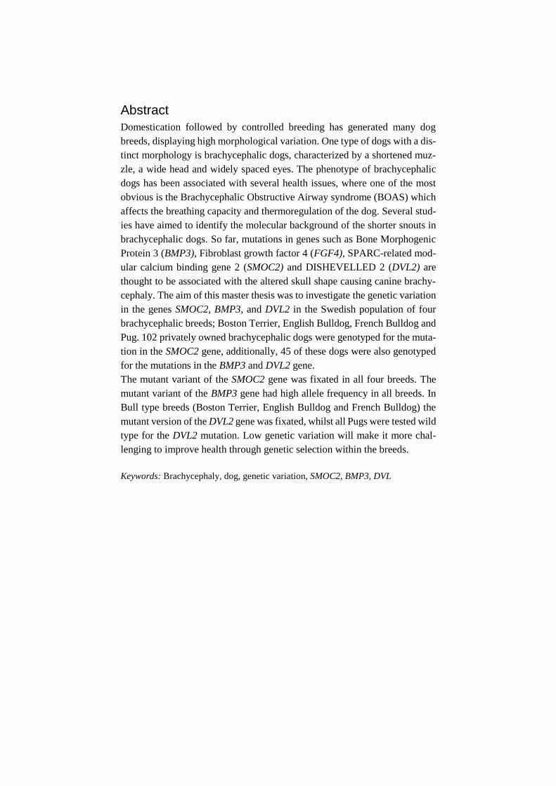
Domestication followed by controlled breeding has generated many dog
breeds, displaying high morphological variation. One type of dogs with a dis-
tinct morphology is brachycephalic dogs, characterized by a shortened muz-
zle, a wide head and widely spaced eyes. The phenotype of brachycephalic
dogs has been associated with several health issues, where one of the most
obvious is the Brachycephalic Obstructive Airway syndrome (BOAS) which
affects the breathing capacity and thermoregulation of the dog. Several stud-
ies have aimed to identify the molecular background of the shorter snouts in
brachycephalic dogs. So far, mutations in genes such as Bone Morphogenic
Protein 3 (BMP3), Fibroblast growth factor 4 (FGF4), SPARC-related mod-
ular calcium binding gene 2 (SMOC2) and DISHEVELLED 2 (DVL2) are
thought to be associated with the altered skull shape causing canine brachy-
cephaly. The aim of this master thesis was to investigate the genetic variation
in the genes SMOC2, BMP3, and DVL2 in the Swedish population of four
brachycephalic breeds; Boston Terrier, English Bulldog, French Bulldog and
Pug. 102 privately owned brachycephalic dogs were genotyped for the muta-
tion in the SMOC2 gene, additionally, 45 of these dogs were also genotyped
for the mutations in the BMP3 and DVL2 gene.
The mutant variant of the SMOC2 gene was fixated in all four breeds. The
mutant variant of the BMP3 gene had high allele frequency in all breeds. In
Bull type breeds (Boston Terrier, English Bulldog and French Bulldog) the
mutant version of the DVL2 gene was fixated, whilst all Pugs were tested wild
type for the DVL2 mutation. Low genetic variation will make it more chal-
lenging to improve health through genetic selection within the breeds.
Keywords: Brachycephaly, dog, genetic variation, SMOC2, BMP3, DVL
Abstract
Domesticering följt av kontrollerad avel har genererat många hundraser med
stora variationer i utseende. En typ av hundar som uppvisar ett distinkt utse-
ende är brakycefala (trubbnosiga) hundar, karaktäriserat av en kort nos, brett
huvud och med stort avstånd mellan ögonen. Fenotypen hos brakycefala hun-
dar har kopplats till hälsoproblem där ett av de mest påtagliga problemen är
Brachycephalic obstructive airway syndrome (BOAS), ett syndrom som på-
verkar hundens andning och förmåga att reglera sin kroppstemperatur. Fler-
talet studier har utförts för att identifiera de molekylära bakomliggande orsa-
kerna till den kortare nosen hos brakycefala hundar. Hittills har mutationer i
gener så som Bone Morphogenic Protein 3 (BMP3), Fibroblast growth factor
4 (FGF4), SPARC-related modular calcium binding gene 2 (SMOC2) och
DISHEVELLED 2 (DVL2) kunnat kopplas till den avvikande huvudformen
hos brakycefala hundar. Syftet för detta examensarbete var att undersöka den
genetiska variationen i generna SMOC2, BMP3 och DVL2 för den svenska
populationen av bostonterrier, engelsk bulldogg, fransk bulldogg och mops.
Totalt genotypades 102 privatägda brakycefala hundar för mutationen i
SMOC2 genen, dessutom genotypades 45 av dessa hundar för mutationerna i
BMP3 och DVL2. Denna studie pekar på att mutationen i SMOC2 var fixerad
i de fyra raserna. I de fyra raserna var allelfrekvensen för den muterade BMP3
allelen hög. I de tre raserna av bull-typ (bostonterrier, engelsk bulldogg och
fransk bulldogg) var mutationen i DVL2 fixerad, medan alla mopsar var ge-
notypade som vildtyp. Låg genetisk variation kommer göra det mer utma-
nande att förbättra hälsan genom genomisk selektion inom raserna.
Nyckelord: Brakycefali, trubbnos, hund, genetisk variation, SMOC2, BMP3, DVL2
Sammanfattning


1 Introduction 9
1.1 The history of the dog 9
1.1.1 Genetic Diversity in dogs 10
1.2 Brachycephaly 13
1.2.1 The Genetics behind Brachycephaly 15
1.3 Aim of study 23
2 Material and Method 24
2.1 Recruitment of dogs 24
2.1.1 Swedish inventory of Brachycephalic dogs 24
2.2 DNA extraction and quantification 26
2.3 PCR amplification 27
2.3.1 Primer design 27
2.3.2 PCR conditions 28
2.4 Sanger Sequencing 28
2.5 TaqMan assays 29
3 Results 31
3.1 GAPDH 31
3.2 Dilution series 31
3.3 SMOC2 31
3.4 Sanger Sequencing 33
3.4.1 BMP3 33
3.4.2 DVL2 34
3.5 BMP3 35
3.6 DVL2 36
4 Discussion and conclusions 38
4.1.1 SMOC2 38
4.1.2 BMP3 39
4.1.3 DVL2 40
4.2 Strengths and weaknesses with this thesis 41
4.3 Relationship in the studied population 42
4.4 Phenotypic variation 43
4.5 Conclusions 44
Table of contents

5 Possible future scenarios for the brachycephalic breeds 45
5.1.1 Enough genetic variation 45
5.1.2 Not enough genetic variation in the breeds 49
5.1.3 Breed standards and breed-specific breeding instructions 51
5.2 Weaknesses in study and future perspectives 52
References 55
Acknowledgements 63
Appendix A Laboratory Protocols 64
Appendix B, Supplementary information and Popular scientific summary in
Swedish 69


8

9
1.1 The history of the dog
Man’s best friend, the dog (Canis lupus familiaris), is one of the most popular pets
today and is, in many cases, considered as a highly beloved family member. The
origin of the dog, being the first animal to be domesticated, is thought to have begun
around 11 000– 30 000 years ago (Thalmann et al. 2013; Freedman et al. 2014).
Both the exact temporal and geographical origin of the dog is however controversial,
and some studies suggests a single domestication event in East Asia while others
suggest several domestication events in different geographic locations (Vila 1997;
Frantz et al. 2016; Wang et al. 2016). While the answer to where and when the
domestication occurred remains indefinite, genetic studies suggest that the dog de-
scends from the gray wolf (Canis lupus) (Wayne 1993; Freedman & Wayne 2017).
The dog has evolved through a mutually beneficial relationship with humans,
sharing food sources and living space. Modern dog breeds are the result of at least
two genetic bottlenecks, where the population size has drastically increased. One
bottleneck being the domestication event and the other through the man ruled inten-
sive selection to create breeds (Lindblad-Toh et al. 2005). Dogs have been selec-
tively bred to support human needs, such as hunting, herding, obedience, guarding,
rescuing and for companionship. This selection has generated a large number of dog
breeds, displaying large variety in behaviour, size, head shape, coat colour and coat
texture (Lindblad-Toh et al. 2005; Schoenebeck & Ostrander 2013). Today, 346 dog
breeds are recognised by the Federation Cynologique Internationale (FCI, 2019),
spanning from the small Chihuahua to the big Great Dane, from the hairless Xo-
loitzcuintle to the long-coated Afghan hound, from the short-nosed Pug to the long-
nosed Collie. No other domesticated species show such variations in phenotype and
behaviour as the ones displayed among dog breeds (Stockard 1941).
1 Introduction

10
The many dog breeds surely display a high morphological variation. For exam-
ple, the skull shape can differ remarkably and might be a breed-defining feature.
Skull shape can be divided into three categories, a short head which is technically
called “brachycephalic” (e.g. Pug and French Bulldog), a long head which is tech-
nically called “dolichocephalic” (e.g. sighthounds and collies) and in between as an
intermediate is the shape called “mesocephalic” or “mesaticephalic” (e.g. Beagle)
(Evans & Miller 2013).
The morphological variation shown in the dog is a result of controlled breeding
and the formation of breed standards. Typically, in the creation of a breed, a small
number of dogs have been used. The small number of founders followed by a small
number of males used for breeding, have reduced the effective population size in
the dogs and created genetic drift, resulting in a reduced genetic diversity within
breeds and a higher variation between breeds (Ostrander & Wayne 2005).
1.1.1 Genetic Diversity in dogs
The genetic diversity can be evaluated in several ways, for example by using gene-
alogical data (i.e. pedigree data), where the average inbreeding coefficient (F) being
one of the most widely accepted tools. At a population level, the inbreeding level
must be evaluated with caution since “inbreeding” can have different meaning, de-
pending on how its computed. When analysing pedigrees, the coefficient of inbreed-
ing is often defined as the probability that two alleles at a given locus are identical
by descent (IBD). Other useful tools for evaluating diversity when using pedigree
data is the effective population size and ancestral constitutions (Leroy et al. 2009;
Leroy 2011).
More recent ways to assess the genetic diversity within-breeds is the use of mo-
lecular data by using genetic markers (Leroy et al. 2009). Genetic markers like au-
tosomal microsatellites, mitochondrial DNA, Y chromosome markers and Single
nucleotide polymorphisms (SNPs) have been used to study genetic polymorphisms,
degree of heterozygosity and phylogeny (Irion et al. 2003).
Irion et al. (2003) assessed genetic diversity by studying 100 microsatellites for
28 breeds recognised by the American kennel club (AKC). The breeds were selected
from each of the seven recognised groups in AKC. An average of 41 dogs per breed
was sampled and screened. By determining the allele frequencies and number of
alleles at specific marker loci, the genetic polymorphism was determined. Total het-
erozygosity (i.e., the probability that two gametes randomly picked from a

11
population carries different alleles), the average heterozygosity among subpopula-
tions and the average breed heterozygosity was calculated. The study showed that
the total heterozygosity was high for all breeds, with an average of 0.618 and with
only three breeds with an average breed heterozygosity below 0.5. It was also found
that a decrease in population size and longer time since the breed recognition, de-
creased both average breed heterozygosity and number of alleles in Hardy-Wein-
berg equilibrium. After breed recognition, forces such as founder effects, bottle-
necks like changes in population size and usage of popular sires will continue to
decrease the genetic diversity in breeds (Irion et al. 2003).
Leroy et al. (2009) performed a study with the aim to assess the genetic diversity
with-in dog breeds using both genealogical and molecular data. In total, 1514 dogs
from 61 different breeds, representing all 10 groups (see supplementary information
for details) recognised by Fédération Cynologique Internationale (FCI) was sampled
and genotyped using 21 autosomal markers. The mean value for non-biased hetero-
zygosity was calculated to 0.62 (Leroy et al. 2009), consistent with the result from
the Irion study (Irion et al. 2003).
Pedersen et al. (2016) performed a study to assess the genetic diversity within
the English Bulldog breed. The control group consisted of 102 bulldogs used for
breeding and additional 37 bulldogs submitted for diagnostic tests was used as a
case group. The study used several techniques to estimate the genetic diversity
within the breed; sequencing the mitochondrial D-loop to decide maternal haplo-
types, using 33 Short tandem repeat (STR) markers, in total seven Dog Leukocyte
Antigen (DLA) class I and class II markers and lastly using six Y-specific markers
for deciding paternal haplotypes. Additionally, SNP assay information from ten
bulldogs and ten Standard poodles from other GWAS studies was used to obtain
information of runs of homozygosity (ROH) in the breed. The observed heterozy-
gosity for the English bulldog was calculated to 0.575 when including both cases
and controls. By analysing the GWAS data, the alleles shared among all individuals,
the runs of homozygosity were larger and involved more chromosomes when com-
paring the English bulldog to the Standard poodle. When analysing the maternal
haplotypes, five haplotypes were identified. Three of the five haplotypes were iden-
tified in 90.9 % of the dogs in the control group, these three haplotypes were also
found in other brachycephalic breeds, including mastiffs. For the paternal haplo-
types, four haplotypes were identified. One of these, named Haplotype 1 was dom-
inant and present in 93.1 % of the 44 males studied. Remaining haplotypes were
observed at each 2.3 %. The dominant haplotype 1 were also found in breeds such
as French Bulldog, Staffordshire Bull terrier, Miniature Bull terrier, Bull terrier,
Beagle, Coton de Tulear and Mastiffs. The shared maternal and paternal haplotypes

12
between mentioned breeds might give an insight to the bulldogs’ ancestry. For the
STR markers, most loci had one or two alleles dominating in frequency, highest
allele number was 11, whereas the lowest allele number was three. Out of 33 loci,
19 loci had a single allele with an allele frequency over 50 %, six loci with one allele
with allele frequency of 70 % or higher and one locus fixed among almost all studied
dogs. However, when analysing the internal relatedness (IR) implying how related
the parents are and the effect on population fitness, the average IR for English bull-
dog was 0.007. As a comparison, a litter born from two sibling parents would have
an IR of 0.25. When adjusting the IR using allele frequencies from village dogs, the
IR increases to 0.34 implying a high degree of inbreeding. Village dogs have been
shown to have genetic links to most modern breeds and since random breeding oc-
curs in these types of populations, the population serves as a reservoir of ancestral
genetic diversity inherited by descent. An increased adjusted IR was also observed
in Standard poodles, but to a lower degree. The study indicates that the English
bulldog has lost genetic diversity through small founder population and intensive
phenotypic selection in breeding, which may make it hard to select for changes in
the breed when it comes to healthier phenotypes (Pedersen et al. 2016).
Domestication followed by the formation of dog breeds with closed stud books,
hard phenotypic selection, inbreeding and usage of popular sires have resulted in
loss of genetic diversity. In some breeds, later bottlenecks where the population has
decreased because of war, isolation or economic depression have also affected the
genetic diversity (Ostrander & Wayne 2005). The high usage of popular males and
a closed gene pool reduces the genetic diversity and increasing the incidence of in-
herited diseases within the breed. In many dog breeds, line breeding (i.e. mating
between related individuals) have been carried out to maintain desired traits in the
breed (Mellersh 2008). Loss of genetic diversity is not always correlated to in-
creased disease incidence and poor health, but has been associated with unhealthy
physiological and morphological traits in many dog breeds (Farrell et al. 2015).
With low genetic variation within in a breed, it can be hard to eliminate deleterious
traits in the population once recognized. A low genetic variation will also limit the
ability to select for new traits or to, for example, improve unhealthy phenotypes
(Pedersen et al. 2016).

13
1.2 Brachycephaly
The morphology of dogs with a short muzzle is highly deviant, both from other dog
breeds but also from their ancestor, the wolf. The brachycephaly phenotype is char-
acterised by a shortened muzzle, a wide head with widely spaced eyes and is often
considered as “flat faced”, due to altered growth of basisphenoid and basioccipital
bones in the skull (Packer et al. 2015). Brachycephalic breeds are for example Pug,
Boxer, English bulldog, French Bulldog, Shih tzu and Japanese Chin. Some brach-
ycephalic breeds, like the Bulldogs and the Boston terrier is thought to share a com-
mon ancestor. The early bulldog, often referred to as Bandogs, was used in bull-
baiting and organized dog fights. These dogs were described as brave and brute dogs
with huge jaws. Baiting with Bandogs were forbidden 1835 in England, and the
number of Bandogs decreased drastically but the breed was rescued and refined,
resulting in the modern bulldog (American Kennel Club).
The pug is, despite its resemblance with bulldogs, thought to be a very old breed
descending from Asia (American kennel club).
Figur 1. A painting by Philip Reinagle from 1790, showing a Bulldog.
Many of the brachycephalic breeds are very popular world-wide as companion
dogs. In Sweden, both the French bulldog and the Pug are popular breeds and were
ranked number 10th and 16th respectively, in 2016 in number of registrations in the
Swedish kennel club (SKK, 2016). Despite the popularity of these breeds, the brach-
ycephalic phenotype is associated with several health issues.
A recent analysis of disease prevalence included more than 1.27 million dogs,
thereof 184 748 brachycephalic dogs, the analysis was performed under a nine-year
14
period. The report identified some conditions more frequently occurring in brachy-
cephalic dogs, compared to non-brachycephalic dogs. For example, the short nose
combined with shallow orbits makes the eyes vulnerable, increasing the risk of eye
injuries such as corneal ulcer, ocular traumas and conjunctivitis. The study showed
that there is a three to four-time higher risk of cornea injuries in brachycephalic
dogs, compared to non-brachycephalic dogs. A shortened skull and compact body
may result in excessive skin and skin folds, increasing the risk for skin problems
such as fungal skin diseases and pyoderma (Feng et al. 2017). However, one of the
most obvious issues related to the brachycephaly conformation is the Brachyce-
phalic Obstructive Airway Syndrome (BOAS), which predominately affects brach-
ycephalic dogs. In a risk factor analysis performed by Njikam et al. (2009) it was
found that brachycephalic dogs were 38 times more likely to have BOAS, compared
to non-brachycephalic dogs (Njikam et al. 2009). The skull of a brachycephalic dog
is significantly shorter, but the soft tissues of the head are not proportionally reduced
as the skull. This is problematic, since the size reduced skull will contain too much
soft tissue, which leaves little space for the passage of air (Harvey 1989). Conse-
quently, BOAS is a respiratory syndrome where soft tissues blocks the dogs airways
during respiration (Packer et al. 2015). BOAS is characterized by anatomical abnor-
malities such as elongated soft palate, hypoplastic trachea, narrowed nostrils and
distortion of the pharyngeal soft tissues which causes resistance to airflow and re-
stricted breathing (Fawcett et al. 2019). Breathing for brachycephalic dogs requires
more laboured breathing to produce a higher negative pressure to cope with the re-
sistance of the airflow (Koch et al. 2003).
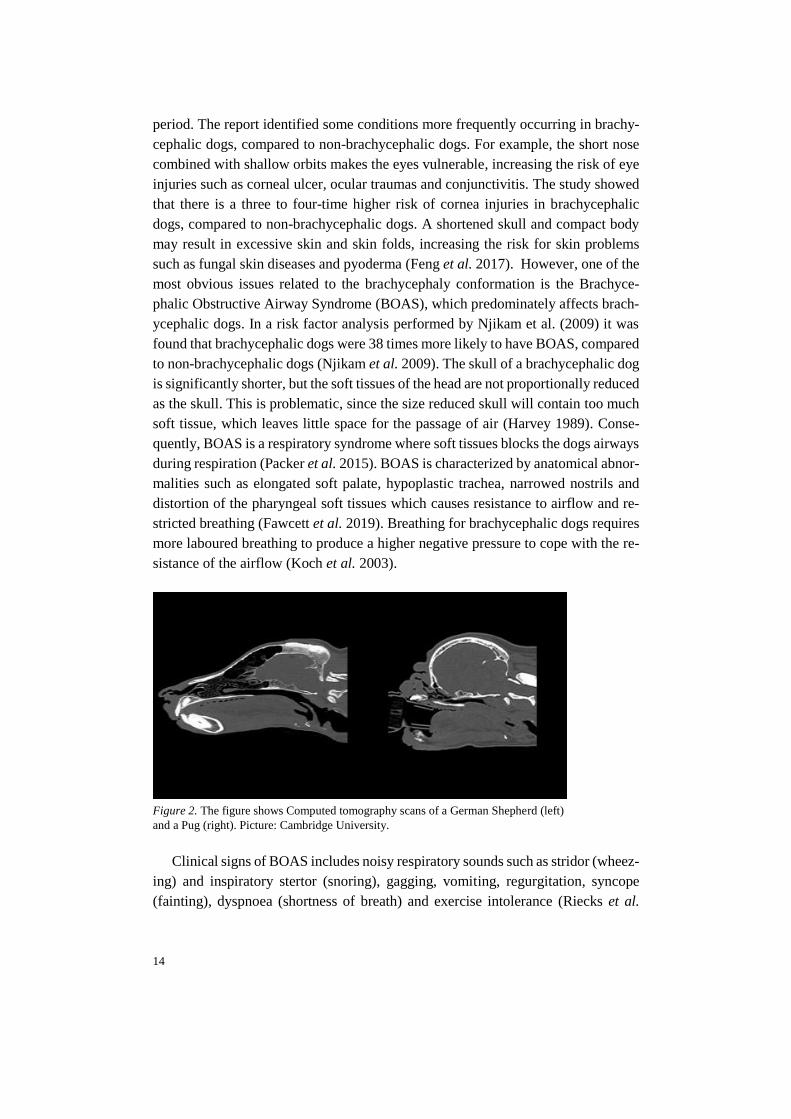
Figure 2. The figure shows Computed tomography scans of a German Shepherd (left)
and a Pug (right). Picture: Cambridge University.
Clinical signs of BOAS includes noisy respiratory sounds such as stridor (wheez-
ing) and inspiratory stertor (snoring), gagging, vomiting, regurgitation, syncope
(fainting), dyspnoea (shortness of breath) and exercise intolerance (Riecks et al.

15
2007). The affected breathing might lead to increased sensibility to heat and im-
paired capability to thermoregulate (Davis et al. 2017), which might lead to a col-
lapse when the dog is excited or hot (Koch et al. 2003).
A study performed by Packer et al. (2015) showed that the BOAS risk increases
as the length of the muzzle shortens. In the 154 studied brachycephalic dogs, BOAS
was only present in dogs with a muzzle length less than half of the cranial length
(length from stop to the occipital protuberance). A thicker neck and obesity was also
factors found to increase the BOAS risk (Packer et al. 2015).
Dogs affected of severe BOAS might, in some cases, need treatment such as
weight loss, housing in cool environment and anti-inflammatory medicine. Some
severe cases of BOAS might also require surgery (Riecks et al. 2007) in order to
widen the nostrils or/and short the soft tissue blocking the airways. A recent study
by Liu et al. (2017) investigated the outcomes and prognostic factors of surgical
treatments in pugs, French and English Bulldogs. The dogs in the study had an im-
proved respiratory function after surgery, although the respiratory function in 68 %
of the dogs remained compromised. Age, the level of laryngeal collapse and body
condition was found to be the main prognostic factors for BOAS surgery (Liu et al.
2017).
1.2.1 The Genetics behind Brachycephaly
Studies have revealed quantitative trait loci (QTL) on canine familiaris chromo-
somes (CFA) 1, 5, 18, 26, 32 and on the X chromosome, associated with the skull
length in dogs (Boyko et al. 2010; Marchant et al. 2017). So far, mutations in genes
such as Bone Morphogenic Protein 3 (BMP3), Fibroblast growth factor 4 (FGF4)
and SPARC-related modular calcium binding gene (SMOC2) are thought to be as-
sociated with the altered skull shape causing canine brachycephaly (Boyko et al.
2010; Schoenebeck et al. 2012; Marchant et al. 2017).
SMOC2
Marchant et al., 2017 found that the QTL on CFA1 was highly significant and as-
sociated with brachycephaly. A 187.7 kb critical interval in CFA1 was common
among 30 out of 37 brachycephalic dogs participating in the study. Five variants
were retained in this interval, one being a Long interspersed nuclear element (LINE-
1) detected within the SMOC2 gene. This LINE-1 insertion was predicted to cause
a premature stop after exon 8 of the canonical 13-exon transcript, causing a fivefold
reduction of the SMOC2 mRNA expression. However, Marchant et al. (2017) claims
that it remains unclear if the isoforms are translated but predicts that the protein

16
products would shear within the thyroglobulin-like domain, resulting in the protein
missing the extracellular calcium-binding-domain. The study suggests that the in-
sertion if the LINE-1 solely explains 36 % of the shortened face displayed in brach-
ycephalic dogs in the study. By studying chick embryos, SMOC2 expression was
observed in the pharyngeal arches and shown to have temporal expression during
the development of the later mandible and viscerocranium (Marchant et al. 2017).
Mansour et al. (2018) genotyped 109 brachycephalic dogs from eleven breeds;
Boston Terrier, Boxer, Brussels Griffon, Bull Mastiff, Bulldog, Cavalier King
Charles Spaniel, French Bulldog, Lhasa Apso, Pekingese, Pug and Shih Tzu for the
SMOC2 insertion. 80 of the 109 genotyped brachycephalic dogs was found to be
homozygous, eight heterozygous (three Boston Terriers, three Bull Mastiffs, one
Lhasa Apso and one Shih Tzu) and 21 dogs was found to be homozygous for the
wild-type variant (one Boxer, six Bull Mastiffs, ten Cavalier King Charles Spaniel,
two Lhasa Apso and two Pekingese).
The SMOC2 gene is one of two homologues, where SMOC1 and SMOC2 are
both members of Secreted protein acidic and rich in cysteines (SPARC) protein
family, also known as BM-40. The members of the SPARC protein family, all con-
tains an extracellular calcium-binding (EC) domain and a follistatin-like (FS) do-
main, (Termine et al. 1981; Maier et al. 2008). SPARC is a matricellular protein
that is secreted into the extracellular matrix, but these proteins are not involved in
primary structures in this location. Matricellular proteins like SPARC modulates
cell function by interacting with hormones, proteases, cytokines and binding to
growth factors and cell-surface receptors. SPARC proteins are expressed in high
levels during embryonic development, in adult tissue both SPARC proteins are pre-
sent in tissues undergoing repair or remodelling due to processes like wound healing
or disease. (Bornstein 2009). SMOC2 mRNA has been found to be expressed in
many different tissues, such as spleen, skeletal muscle, ovary, testis, brain, thymus,
lung, liver and in the heart (Vannahme et al. 2003). SMOC2 has also been demon-
strated to stimulate endothelial cell proliferation and migration, but also to be in-
volved in formation of new capillaries (Rocnik et al. 2006).
Liu et al. (2008) investigated SMOC2 expression in mouse embryos and found
that SMOC2 was distributed in the pharyngeal arches (i.e. the developing face), de-
veloping limbs and somites (Liu et al. 2008). Somites are epithelial cell clusters
positioned bilaterally of the neural tube, which gives rise to cartilage, tendons, skel-
etal muscle, the vertebrae and ribs and the dermis of the back (Gilbert & Barresi
2016). Feng et al. (2009) examined gene expression during the development of the
mouse facial prominences. Gene expression in the developing facial prominences

17
in mouse embryos were investigated between embryonic day 10.5 and 12.5 and it
was found that several genes are expressed during the development, one gene being
SMOC2, expressed in varying levels in samples from the maxillary, mandibular and
frontonasal area(Feng et al. 2009).
In a case with dental defects in humans, Bloch-Zupan et al. (2011) found two
cousins each homozygous for a mutation in the canonical-splice donor site in the
first intron of the SMOC2 gene. A zebra fish model was used to further investigate
effects of mutations in SMOC2 and the effect on dental development. Two types of
morpholino knockdowns were created, one to target the initiation codon and one to
target the boundary between exon 2 and intron 2 (Bloch-Zupan et al. 2011). Mor-
pholino oligonucleotides (MO) is a tool to inhibit the translation of RNA transcripts.
MOs is typically a oligomer of 25 morpholino bases that by complementary base
pairing binds to the target RNA (Bill et al. 2009). When studying embryos carrying
MO, the teeth were smaller with different shape and some dermal bones in the jaws
and the fifth ceratobrancial bone were missing calcification, indicating that the skull
is affected in morphants. Zebrafishes with the MOs also displayed a smaller head.
Though, it should be noted that the development of teeth in zebra fish resembles the
development in vertebrates, but the anatomy differs compared with for example hu-
mans. The knockdown of smoc2 showed to effect expression of genes such as dlx2b,
bmp2a and pitx2, all involved in tooth development (Bloch-Zupan et al. 2011).
Melvin et al. (2013) identified several genes that alter craniofacial conformation
in zebrafish. Eight genes were fully analysed and grouped depending on what struc-
ture the alterations would affect; genes essential for formation of the ventral viscer-
ocranium, genes essential for development of anterior neurocranium and genes crit-
ical for morphogenesis of all neural crest derived structures (including viscerocra-
nium and neurocranium). Smoc2 was analysed as one of the genes essential for the
formation of viscerocranium. Two MOs were used to knockdown smoc2, one trans-
lation blocking MO to the 5´untranslated region (UTR) and one splice-blocking MO
targeting a splice donor site for exon 11. Both MOs reduced the size of the head and
eye abnormalities, 10 % in UTR MOs and 50-60 % of the splice site morphants were
affected. The ceratohyals (hyoid horn) were flattened and inverted in morphants,
compared to controls. The splice site donor MO altered the processing of smoc2
RNA significantly, suggesting the observed affected craniofacial phenotype in mor-
phants are due to loss of wild-type smoc2 transcripts (Melvin et al. 2013).
The craniofacial defects investigated in these studies suggests that Smoc genes
encode proteins that have a functional role in the development of the facial skeleton.
18
BMP3
By investigating the QTL at chromosome 32, a critical interval of 85 kb spanning
the bone morphogenic protein 3 (BMP3) gene was found (Schoenebeck et al. 2012).
After filtering, a Single nucleotide polymorphism (SNP) encoding for a missense
mutation was found, nearly fixated among small brachycephalic dogs in the study.
The mutation, called BMP3F452L, is caused by a C to A transversion at position CFA
32:5231894, where C being the ancestral allele and A being the derived allele. The
missense mutation causes a nonsynonymous substitution, changing the amino acid
phenylalanine to leucine (Schoenebeck et al. 2012).
By using a zebra-fish model, bmp3 mRNA expression was found as highly dy-
namic, first appearing during mid-somitogenesis (i.e. formation of somites) and later
expressed throughout the head, brain, ventricles and posterior somites. 48 hours af-
ter fertilization, the bmp3 expression is found in pectoral fins, the pharyngeal arch
region, in the jaw structures and in the heart. The early expression of bmp3 in the
cranial structure suggested a role for Bmp3 in craniofacial development. By knock-
ing down endogenous Bmp3 activity using translation-blocking antisense MO, it
was found that MO treated zebrafish embryos demonstrated severe deficiency in the
jaw development. Multiple cartilage elements that forms the viscerocranium and
neurocranium where lost or underdeveloped in knocked downed embryos. The re-
sults of the study indicates that Bmp3 is required for craniofacial development in
zebra fishes and that the identified BMP3F452L gene variant influences skull shape
in brachycephalic dogs. (Schoenebeck et al. 2012).
Bone morphogenic proteins (BMPs), being multi-functional growth factors, be-
longs to the transforming growth factor β superfamily (TGFβ). The TGFβ pathway
plays a central role in signalling networks controlling bone development, tissue re-
pair, cell growth, cell differentiation and cell proliferation in metazoans. The TGFβ
family can be divided into several subfamilies, including protein families such as
TGF-β, BMPs, activin and growth differentiation factors (GDF). TGFβ initiate sig-
nalling through specific interactions with receptors on the surface of target cells
(Massagué et al. 2000).
BMPs are involved in embryonic development and in multiple different cellular
function in adult animals. BMP signalling is involved in heart, neural and cartilage
development. It also plays an important role in the postnatal bone formation (re-
viewed in Chen et al., 2004). BMP3, also known as osteogenin, works as an antag-
onist for other BMPs. BMP3 antagonises the ability of BMP2 to induce differentia-
tion and cell commitment of osteogenic cells. Bmp3 is suggested to negatively affect
bone density, where Bmp3 null mice (Bmp3 -/-) showed to have twice as much

19
trabecular bone (spongy bone), compared to non-mutated littermates. Though, no
differences in the size of the femur were observed and there were no difference in
bone density between wild-type and heterozygous littermates (Daluiski et al. 2001).
By studying Xenopus embryos, Gamer et al. (2005) found that BMP3 inhibits
activin and BMP-4 signalling. The activin type II receptor (ActRII) is a common
receptor that binds to multiple ligands, such as activin and several BMPs. Binding
between ActRII, activin or BMPs is required for patterning during the development
of germ layers in the embryo. The binding between activins and BMPs and the re-
ceptor phosphorylates receptor regulated Smad proteins (R-Smad), R-Smad to-
gether with other transcriptional regulatory proteins regulates transcription of target
genes. It was showed that if BMP-3 binds to its receptor (ActRII), BMP-3 works as
an antagonist against activin and BMP-4. BMP-3 reduces the phosphorylation of R-
Smad, and thereby inhibits the mesoderm-inducing activities of activin and BMP-4.
By injecting BMP-3 in Xenopus embryos, overexpression of BMP-3 presents a phe-
notype with deviant tail formation, a shortened and curved body, reduced eyes and
head structures and enlarged cement glands. However, the overexpression of BMP-
3 could be rescued by co-injections of a truncated type II activin receptor
(xActRIIB), which rescued the body axis and head structure in a dose dependent
manner (Gamer et al. 2005).
FGF4
In the study by Marchant et al.(2017), a retrogene for the Fibroblast growth factor 4
(FGF4) located on chromosome CFA 18 was associated with the size of the neu-
rocranium centroid size in brachycephalic dogs (Marchant et al. 2017). Earlier stud-
ies on FGF4 by Parker et al. (2009) identified the FGF4 retrogene on chromosome
18 that causes short legs (canine chondrodysplasia), present in dog breeds such as
Dachshunds and Corgis (Parker et al. 2009).
The fibroblast growth factor family, composed of several secreted proteins, func-
tions in the earliest stages of the embryonic development, organogenesis and in adult
tissue where they act as homeostatic factors important for maintenance of the tissue,
tissue repair and regeneration and metabolism. FGFs signals to receptor tyrosine
kinases and intracellular non-signalling proteins (i.e. intracellular FGFs) that serves
as cofactors. The FGF family is conserved and can be found in both invertebrates
and vertebrates and is expressed in almost all tissues (reviewed in Ornitz & Itoh,
2015).
By investigating RNA expression in mice, Niswander and Martin (1992) found
that Fgf-4 expression is important during early embryonic development. Fgf-4
20
mRNA could be detected at late blastocyst stage in cells giving rise to embryonic
lineages. Later, in early gastrulation, Fgf-4 was expressed in the primitive streak
where definitive endoderm and mesoderm are formed. After the establishment of
the three germ layers, Fgf-4 mRNA was detected in branchial arch units, somite
myotome and in the apical ectodermal ridge where the limb buds develop. Expres-
sion was also found in tooth bud, suggesting that Fgf-4 has multiple roles during the
development of the embryo.
Boulet et al., (2004) studied Fgf8 and Fgf4 in mice embryos and found that mice
lacking Fgf4 in apical ectodermal ridge had normal limbs. In Fgf8 mutants, the limb
development was severely affected, with some skeletal elements of the limb miss-
ing. When creating mutants lacking both Fgf4 and Fgf8 in the forelimb apical ecto-
derm ridge, the forelimb does not develop, due to that the limb mesenchyme fails to
survive in absent of both FGFs. When inactivating FGf4 and Fgf8 in both forelimb
and hindlimb, all limbs fails to develop. It was suggested that Fgf8 is necessary for
maintenance or initiation of Sonic hedgehog (Shh) and that Fgf4 partially can com-
pensate for the loss of Fgf8 in development of the distal limb (Boulet et al. 2004).
FGFs is also thought to be involved in the development of the hair follicle.
Salmon Hillbertz et al. (2007) found that ridged dog breeds, such as Rhodesian
Ridgeback and Thai Ridgeback, are either heterozygous or homozygous for a large
duplication. The duplication that is thought to be the causative mutation for the
ridge, included genes such as FGF3, FGF4, FGF19, ORAOV1 and the 3’ end of
CCND1, encoding for cyclin D1. The ridge also predisposes dermoid sinus, a con-
genital developmental disorder (Salmon Hillbertz et al. 2007), caused by an incom-
plete separation of the neural tube and the skin .
DVL2
A recent study by Mansour et al. (2018) identified a frameshift mutation in the Wnt
pathway gene Dishevelled Segment Polarity Protein 2 (DVL2) located on canine
chromosome CFA 5, associated with the screw tail that is characteristic for English
bulldogs, French bulldogs and Boston terriers. The frameshift mutation, caused by
a deletion of a C (g.32195043_32195044del), preserves the majority of the DVL2
protein, though the last 48 amino acids at the C terminus of the protein is replaced
by novel sequence of 26 amino acid residues. The results indicated that the Wnt-
dependent phosphorylation of the mutant variant DVL2 protein was reduced, com-
pared to the wild type variant. Out of 177 dogs from screw tailed breeds, 171 dogs
were found homozygous for the mutation in DVL. The exception was six Boston
Terriers, where four individuals were heterozygous, and two individuals were ho-
mozygous wild type. The pugs tested was found to be of wild types, as expected,
21
since the tail of a pug is of full length without malformations of the caudal vertebras
(Mansour et al. 2018). Mutations in Dishevelled genes in humans is characterised
by craniofacial, limb and vertebral malformations, causing a syndrome named Robi-
now syndrome. Affected individuals often displays distinctive facial features such
as widely spaced eyes, a wide forehead, short nose and a broad mouth (Genetics
Home Reference 2019). Due to the resemblance between the phenotype of humans
affected by the Robinow syndrome and the phenotype displayed in brachycephalic
dogs, it is possible that the DVL2 mutation in screw tail dogs also have an effect on
the brachycephalic phenotype (Mansour et al. 2018).
The first dishevelled mutation was identified in Drosophila, where a viable allele
was associated with a phenotype with disoriented hairs on the body and wings. It
was later shown that Dishevelled being a signalling molecule, works as a key com-
ponent in the Wnt signalling pathway (Boutros & Mlodzik 1999).
Wnt proteins are one of the major families of developmentally important signal-
ling molecules, involved in processes such as embryonic induction, generation of
cell polarity and cell fate specification during embryonic development and tissue
homeostasis. It is also suggested that Wnt signalling is involved in cancer genesis
in humans (Cadigan & Nusse 1997). One of the best understood Wnt gene is the
wingless (wg) gene, essential for segmentation and patterning events in the fruit fly
Drosophila melanogaster. Wg is required in several events during embryo develop-
ment, such as in patterning of the segmented ectoderm in the trunk, the development
of the head, the central nervous system and the legs. Wg is also required for growth
of the wing blade in the development of the wing disc, hence the name wingless. In
the adult fly, Wg is required for patterning of bristles and body hair, in antennae,
eyes and genitalia (reviewed in Klingensmith & Nusse, 1994).
Wnt genes have been identified in invertebrates and vertebrates, with ortholog
relationship between species such as Wnts in Drosophila and in humans. Between
vertebrates, the Wnt genes are highly conserved, sharing sequence identity and gene
structure. It has been showed that signaling by members of the Wnt family have
diverse functions during embryogenesis, and has been suggested that Wnt genes
play roles in the development of different structures of the brain, development of
dorsal-neural tube derivatives, development of kidneys, hair growth, limb polarity
and the development of the placenta. Mutations in Wnt genes has also been associ-
ated with shortened anterior-posterior axis and female infertility due to defects dur-
ing development of female reproductive organs (rewieved in Miller, 2001). In
mouse, mutations in Wnt genes have been associated with defects in the tail, the
tailbud and in caudal somites (Cadigan & Nusse 1997).
22
The Wnt pathway is activated when the Wnt ligand binds to the two membrane
receptors, Frizzled (Fz) and low-density lipoprotein receptor related protein 6
(LRP6) or LRP5. The complex of ligand and receptors, together with the recruitment
of Dishevelled results in phosphorylation of LRP6/LRP5 and the recruitment of the
axin complex to the receptors. Next, the axin complex is inhibited, which allows β-
catenin located in the cytoplasm to enter the nucleus and form complexes with
DNA-bound T cell factor/lymphoid enhancer factor (TCF/LEF), where β-catenin
serves as a co-activator which activates Wnt target gene expression. In absence of
Wnt, β-catenin in the cytoplasm is constantly degraded by the axin complex and the
Wnt target genes are repressed (MacDonald et al. 2009). Proteins encoded by the
dishevelled genes works as important transducers in Wnt pathways, that conducts
the Wnt signal to downstream cellular machinery (Na et al. 2007).
The Dishevelled proteins have three known structural domains, DIX (Dishev-
elled and Axin) domain at the N-terminus, PDZ (Post-synaptic density protein-95)
and DEP (Dishevelled, Egl-10 and Pleckstrin). By investigating DVL2, Gammons
et al. (2016) found that the DEP domain, binding to Frizzle receptors is essential for
the Wnt-dependent signaling to β-catenin. The PDZ domain was dispensable for the
β-catenin response (Gammons et al. 2016).
Tadjuidje et al. (2011) showed that both Dvl2 and Dvl3 plays a part in early
embryo development in the Xenopus. It was found that both Dvl2 and Dvl3 are ma-
ternally encoded for. Before the zygotic gene activation (ZGA), where the embryo
starts to produce its own transcripts and proteins, development is dependent of
stored maternal factors in the oocyte. Genes encoding for such factors are referred
to as maternal effect genes, are important for the oogenesis, enables ZGA and the
progression of the early embryo development (Kim & Lee 2014). By studying
Xenopus oocytes, it was found that Dvl2 and Dvl3 mRNA are expressed early in the
development and decreases during the developmental process. Dvl2 was later found
to increase after the ZGA when the zygotic transcription starts, but Dvl3 remained
low. Dvl1 was not detected until later in the development. By using different anti-
sense oligos to block translation of Dvl2 and Dvl3, it was found that maternal de-
pletion of Dvl2 or Dvl3 results in similar phenotypes. Depletion of Dvl resulted in
shortened body axis, blastopores not closing and open neural folds. The heads were
also found to be smaller in Dvl depleted embryos. Since maternal depletion of either
Dvl2 or Dvl3 affects early development, it was suggested that Dvl2 and Dvl3 are
not redundant to each other and that the proteins might interact with each other.
Dvl2 and Dvl3 was found to be required for activation in the c-jun N-terminal kinase
23
(JNK) pathway which is important for the convergence extension (Tadjuidje et al.
2011), where cell movements results in elongation of the body axis.
Xing et al. (2018) studied the effects of mutations in zygotic Dvl, maternal Dvl
and the effects when combining mutations in both zygotic and maternal Dvl in
zebrafishes. By generating mutated lines for the five known dvl genes in zebrafish,
it was found that only dvl2 and dvl3 were maternally expressed. Maternal and zy-
gotic mutants for four out of five of the dvl genes developed normally and survived
to adult zebrafish. Dvl2 mutants were however found to have defects. Zygotic dvl2
(Zdvl2) mutants displayed a slightly reduced anterior-posterior axis, compared to
wild type embryos. Half of the Zdvl2 mutants survived to adult, one third of the
surviving females could not spawn and all male mutants were not able to produce
offspring due to absent courtship behaviour. Embryos with mutations in both ma-
ternal and zygotic dvl2 (MZdvl2) developed craniofacial defects, fused eyes and the
pharyngeal cartilages protruded outwards. MZdvl3 embryos displayed just a weak
axis extension defect. When generating double heterozygote, (dvl2+/-; dvl3+/-), these
embryos showed a slight reduction of the axis extension. However, when mating
two double heterozygous fishes, embryos displayed a shortened anterior-posterior
axis, a compressed head and reduced swim bladder. Absence of zygotic Dvl2 and
Dvl3 resulted in moderate convergence extension defects, while absence of both
zygotic and maternal Dvl3 and Dvl3 generated severe convergence extension de-
fects. Progressive reduction of Dvl dosage was found to gradually induce anterior-
posterior patterning defects, ranging from posterior deficiency to complete lack of
the trunk and tail. In conclusion, Xing et al. (2018) found that Dvl3, and in particular
Dvl2, is important in the convergence extension movement and in anterior-posterior
patterning during embryonic development (Xing et al. 2018).
1.3 Aim of study
The aim of this thesis was to investigate the genetic variation in three of the genes
associated with canine brachycephaly by looking into the Swedish population of
four brachycephalic dog breeds; English bulldog, French bulldog, Pug and Boston
terrier.

24
2.1 Recruitment of dogs
In total, data from 102 privately owned brachycephalic dogs (44 males and 58 fe-
males) were used in this thesis. The dogs were phenotypically described at six gath-
erings, held between 17th of September 2018 and 20th of January 2019. Three of the
gatherings were organised by the Swedish kennel club (SKK), one gathering was
organised by SKK and the English Bulldog club and two were organised by SKK
and the French Bulldog club. The gatherings held by SKK and SKK/English Bull-
dog club were held at SKKs facility, while gatherings organised by the French Bull-
dog club were held in dog training facilities at two different locations.
Information from 18 English Bulldogs (9 males and 9 females), 48 French Bull-
dogs (23 males, 24 females and one with unknown sex), 21 Pugs (7 males and 14
females) and 15 Boston terriers (5 males and 10 females) were used for this thesis.
The dogs in the inventory was between 9 months and 13 years old. In addition, DNA
from one Labrador Retriever, one Golden Retriever, two Pugs and two Chinese
Crested Dogs were used.
2.1.1 Swedish inventory of Brachycephalic dogs
The data used in this project was collected as part of a bigger Nordic research study
with the aims to investigate the phenotypic and genotypic variation in the four
breeds; English Bulldog, French Bulldog, Pug and Boston terrier, and if it’s possible
to by selection achieve changes in anatomy to reduce the predisposition of BOAS.
The ambitions in the Swedish project was to recruit 50 – 100 dogs of each breed,
approximately 25 dogs of each breed should later be selected and genotyped. The
inventory is a part of the SKK’s ambition to reduce extreme conformations in
2 Material and Method

25
Swedish dogs. At the gatherings, the procedure consisted of four stations. Station
one, where the dog owner filled in information of the dog, such as birth date, sex,
registration number, medical information and gave consent to participation in the
study. At the second station, a veterinary inspection was performed to record meas-
urements of the dog and parameters such as respiration and circulation. Some of the
measurements taken is illustrated in figure 3 below.
Figure 3. Illustration of some of the measurements taken during the inventories (Photo and illustration,
Elin Johansson.
The skull length was measured from the stop (2) to the external occipital protuber-
ance at the back of the skull (3). The snout length was measured from the stop (2)
to the nose tip (1). Other measurements taken were the neck girth (NG), neck length
(3 to 4), chest girth (CG), back length (4 to 5), body length (6 to 8), height at withers
(4 to 9), elbow height (10 to 9) and chest length measured between the top of the
breast bone (6) and the end of the sternum (7). By dividing the snout length with the
skull length, the craniofacial ration was obtained. The degree of nostril stenosis was
also graded according to a four graded scale from open nostrils to severe stenosis,
described by (Liu et al. 2017). The dogs were also weighed, and the body condition

26
score was assessed, were a scale from one (undernourished) to nine (obese) was
used. The dog’s skin and presence of excessive skin folds were also examined.
At a third station, DNA was collected, and the dog was photographed from dif-
ferent angles (profile, front, back and from above). At the last station, additional
conformation traits were recorded, such as height at shoulders, height at elbow,
back length, body length, neck girth, chest girth and chest length. The placement
and appearance of eyes, nose and ears and front and rear angles, appearance of paws,
top line, wrinkles on body and nose folds and the length of tail and its appearance
were also assessed. The dog’s movement was scored when letting the dog trot back
and forth on the floor.
Each dog was given a specific identification number at the gathering, partly to
easy have access to an overview of the number of dogs in the inventory and partly
to handle the information more objectively by just using the inventory number
and/or breed information.
A second master thesis was done within the same Swedish project, with the aim
to investigate the phenotypic variance correlated to BOAS in the Swedish popula-
tion of four brachycephalic breeds; English Bulldog, French Bulldog, Pug and Bos-
ton Terrier and discuss their welfare implications (Bertilsson, 2019).
2.2 DNA extraction and quantification
Buccal cells were collected from the participating dog’s cheeks by two or three bris-
tle cytology brushes. The cytology brushes were left to air dry for a couple of
minutes and later placed in a marked paper envelope. The envelopes with the cytol-
ogy brushes was then stored in room temperature until DNA extraction. The enve-
lopes were stored between one day and four months, depending on when the sample
was collected and when the extraction was performed.
Genomic DNA was isolated using the QIAsymphony® SP automated system
with the QIAsymphony DSP DNA Midi Kit (QiaGen, ID 937236, Hilden, Ger-
many) (protocol in appendix I).
To determine the nucleic acid concentration in the samples, spectrophotometry
was used by loading 2 µl of the samples on a NanoDrop™ 8000 Spectrophotometer
(Thermo Fisher Scientific), this was performed on eight of the samples. Due to the
large variation in nucleic acid concentration and the uncertainty of how much of the
27
nucleic acid is derived from dog’s own buccal cells and how much is from bacteria
or feed residues in the mouth, it was decided not to use the spectrophotometer on
the other samples.
To ensure that the samples contained sufficient amount of canine genomic DNA,
a polymerase chain reaction (PCR) of the canine housekeeping gene Glyceralde-
hyde 3-phosphate dehydrogenase (GAPDH) was set up, using the HotStarTaq® kit
(QiaGen ID: 203601, Hilden, Germany) (protocol 1, Appendix A). In total, the PCR
was set up for nine samples, including eight brachycephalic dogs and one Labrador
Retriever. A gel electrophoresis was performed to segregate the PCR products for
validation.
A dilution series was performed to investigate which concentrations that could
work for a PCR. Three samples were picked out, based on the nucleic acid concen-
tration measured with NanoDrop™, one sample with high, intermediate and low
concentration was chosen. The samples were diluted to concentrations 1:10 and
1:100 and a second PCR for the GAPDH gene was set up (Appendix A, protocol 1).
This procedure was mainly performed to assess the least possible amount of ge-
nomic DNA to use from each sample due to the expected limited amount of genomic
DNA template obtained from the cheek swab samples available in this study.
In total, DNA from five additional dogs were used in the study as positive con-
trols. DNA from one Labrador Retriever, one Golden Retriever, two Pugs and two
Chinese Crested Dogs. For the Retrievers and the additional Pug, the DNA extrac-
tion was made from whole blood. For the two Chinese Crested Dogs, cytology
brushes were used as described for the brachycephalic dogs above.
2.3 PCR amplification
2.3.1 Primer design
In order to detect the LINE-1 insertion in the SMOC2 gene, PCR primers for geno-
typing described in Marchant et al. (2017) was used. The primers were checked,
using the Primer3 software. Primers were obtained from TAG Copenhagen A/S
(Denmark).

28
In total, three primers were used (table 1). All three primers were used in the same
reaction. The melting temperatures of the primers were between 55.3°C and 60°C,
with a GC content between 47.8% and 70 % (Appendix A, protocol 2).
Table 1. Primers used for amplification of SMOC2.
Primer Primer sequence Size
SMOC2 F GGC AGG GGA TGG GGA AGC CT 531 bp
SMOC2 R (wild type) ACT GTG TGC TTT GCC CAA ACT CA
SMOC2 F GGC AGG GGA TGG GGA AGC CT 698 bp
SMOC2 R (mutated) TGC CCA TAA AGT TCA GGG TCC ACT
Heterozygous will show as two visible bands on an agarose gel.
2.3.2 PCR conditions
For 102 dogs, the DNA sequences containing the SMOC2 gene was amplified using
the kit HotStarTaq®. The total reaction volume of the PCR was 10 µL, composed
of; 1x PCR Buffer (15 mM MgCl2, ph 8.7) + dNTP [0.25 mM] + primers [0.3
µM/primer] + 0.125 µL Taq Polymerase [0.025 U/µL] + 1 µL DNA and addition of
RNase-free water until the completion of the 10 µL reaction volume. One negative
control per master mix was included, were the DNA was substituted with 1 µL of
water (Appendix A, protocol 3)
PCR reactions were performed in a thermal cycler (ProFlex PCR system, Ap-
plied Biosystems by Life Technologies), using a touchdown PCR program, accord-
ing to following protocol. Initiation step: 95 °C for 2 minutes. 3-step cycle (20 cy-
cles) Denaturation step: 98 °C for 10 seconds. Annealing 72 - 52 °C (reduced with
1 °C/cycle) for 30 seconds. Extension: 72 °C for 45 seconds. Followed by another
3-step cycle; 98 °C for 10 seconds, 55 °C for 30 seconds, 72 °C for 45 seconds.
Finishing synthesis step: 72 °C for 7 minutes. The lid heat was put on 98 °C. There-
after, infinite hold in 4 °C until the products were quantified on an 2 % agarose gel,
using a 100 bp precision size marker. The gel electrophoresis was run in 110V for
approximately two to three hours, depending on the size of the gel.
The PCR protocol was kindly provided through personal communication with
Marchant, T (March 2019).
2.4 Sanger Sequencing
Since no dog in this study had a known genotype for the BMP3 and DVL2, Sanger
Sequencing with capillary electrophoresis was performed to obtain the genotype for
29
three dogs, one Golden Retriever, one English Bulldog and one Pug, and use these
dogs as positive controls in later TaqMan assays. Two sets of M13 tailed primers
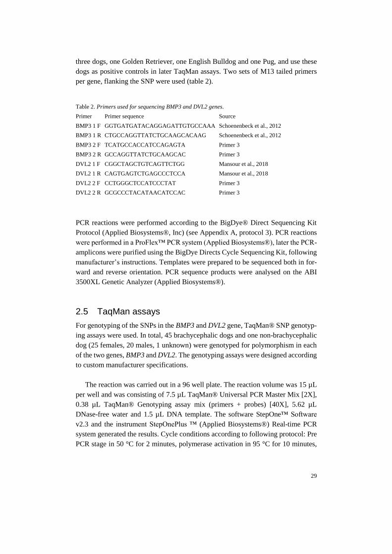
per gene, flanking the SNP were used (table 2).
Table 2. Primers used for sequencing BMP3 and DVL2 genes.
Primer Primer sequence Source
BMP3 1 F GGTGATGATACAGGAGATTGTGCCAAA Schoenenbeck et al., 2012
BMP3 1 R CTGCCAGGTTATCTGCAAGCACAAG Schoenenbeck et al., 2012
BMP3 2 F TCATGCCACCATCCAGAGTA Primer 3
BMP3 2 R GCCAGGTTATCTGCAAGCAC Primer 3
DVL2 1 F CGGCTAGCTGTCAGTTCTGG Mansour et al., 2018
DVL2 1 R CAGTGAGTCTGAGCCCTCCA Mansour et al., 2018
DVL2 2 F CCTGGGCTCCATCCCTAT Primer 3
DVL2 2 R GCGCCCTACATAACATCCAC Primer 3
PCR reactions were performed according to the BigDye® Direct Sequencing Kit
Protocol (Applied Biosystems®, Inc) (see Appendix A, protocol 3). PCR reactions
were performed in a ProFlex™ PCR system (Applied Biosystems®), later the PCR-
amplicons were purified using the BigDye Directs Cycle Sequencing Kit, following
manufacturer’s instructions. Templates were prepared to be sequenced both in for-
ward and reverse orientation. PCR sequence products were analysed on the ABI
3500XL Genetic Analyzer (Applied Biosystems®).
2.5 TaqMan assays
For genotyping of the SNPs in the BMP3 and DVL2 gene, TaqMan® SNP genotyp-
ing assays were used. In total, 45 brachycephalic dogs and one non-brachycephalic
dog (25 females, 20 males, 1 unknown) were genotyped for polymorphism in each
of the two genes, BMP3 and DVL2. The genotyping assays were designed according
to custom manufacturer specifications.
The reaction was carried out in a 96 well plate. The reaction volume was 15 µL
per well and was consisting of 7.5 µL TaqMan® Universal PCR Master Mix [2X],
0.38 µL TaqMan® Genotyping assay mix (primers + probes) [40X], 5.62 µL
DNase-free water and 1.5 µL DNA template. The software StepOne™ Software
v2.3 and the instrument StepOnePlus ™ (Applied Biosystems®) Real-time PCR
system generated the results. Cycle conditions according to following protocol: Pre
PCR stage in 50 °C for 2 minutes, polymerase activation in 95 °C for 10 minutes,

30
followed by a touch-down series starting with denaturation in 95 °C for 15 seconds
followed by cycling stage of 50 cycles with annealing/extension in 60 °C/55°C for
BMP3 and DVL2 respectively, followed by 95°C for 15 seconds and an anneal-
ing/extension step in 55°C for 1 minute. Finally, a hold stage of 60 °C for 30 sec-
onds.
The TaqMan assay relies on two sequence specific primers (forward and reverse)
for PCR amplification. The assay also contains two allele specific TaqMan probes,
each probe can detect one of the two alleles. The probes carry a dye label at the
5’end, a so called “reporter”. Commonly, VIC® reporter is used for allele one and
FAM™ reporter for allele two. At the 3’ end, the probe carries a non-fluorescent
quencher bound to a minor groove binder (MGB). Each probe anneals specifically
to the complementary sequence, depending on which allele is present. At denatura-
tion, the primer pair and the corresponding probe anneals to the sequence. At
polymerisation, probes hybridized to the target gets cleaved. The cleavage separates
the reporter dye from the quencher and increases the fluorescence of the reporter,
which can be detected by the device. The fluorescence generated during PCR am-
plification indicates which of the alleles that are present in the sample (Ther-
moFisher Scientific).
For genotyping of BMP3, VIC® reporter was used to detect the wild-type allele
C (allele 1) and FAM™ reporter was used to detect the derived A allele (allele 2).
For genotyping of DVL2, VIC® reporter was used to detect C allele (allele 1) for
wild-type and FAM™ reporter was used for the derived A allele (allele 2).
The dogs with known genotyped was used as positive controls, other marked as
unknown. After sequencing, the results were analysed using StepOne™ software
and the ThermoFisher Cloud.
Figure 4. Schematic figure of the TaqMan assay for BMP3. Primers are marked out as arrows.
Figure 5. Schematic figure of the TaqMan assay for DVL2. Primers are marked out as arrows.
31
3.1 GAPDH
For eight samples, a PCR was setup against the housekeeping gene GAPDH to en-
sure that the samples contained enough canine DNA for later PCR of the SMOC2
gene. All eight samples generated a band of correct size of 108 bp on the agarose
gel. It was concluded that the canine DNA concentration in the samples should be
sufficient for further testing.
3.2 Dilution series
Since there was limited amount of DNA from each dog in the study, a dilution series
was performed to check if the samples could be diluted so that less DNA could be
used for each testing. Three of the eight samples described above was diluted to
1:10 and 1:100 and tested for the GAPDH gene. However, for one sample, the DNA
concentration of DNA after diluting was probably too low to get amplified, resulting
in no visible bands after gel electrophoresis. Consequently, it was decided not to
dilute DNA samples from the buccal swabs for further testing.
3.3 SMOC2
By setting up a PCR, the genotypes for the SMOC2 gene was obtained. Amplifica-
tion products were visualised on an agarose gel (figure 6), where the wild type allele
is expected to generate a fragment of 531bp, and the mutated allele is expected to
generate a fragment of 698 bp.
3 Results
32
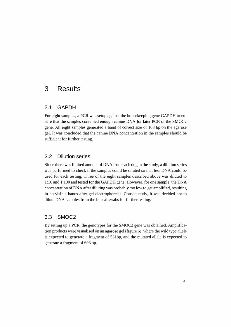
Figure 6. Agarose gel electrophoresis. Agarose gel electrophoresis showing SMOC2 PCR amplicons
from different individuals. One band at about 700 bp indicates that the individual is homozygous for
the mutation. Sample 1002, displaying two bands, is heterozygous for the mutation, carrying both the
mutated LINE-1 insertion and the wild-type haplotype.
Table 3. SMOC2 LINE-1 insertion.
Breed Total genotyped dogs WT
SMOC2+/+
HET
SMOC2+/-
MUT
SMOC2-/-
Unknown
English Bulldog 18 0 0 15 3
French Bulldog 48 0 0 46 2
Boston Terrier 15 0 0 14 1
Pug 23* 0 0 23 0
Labrador Retriever 1 1 0 0 0
Golden Retriever 1 1 0 0 0
Chinese Crested Dog 2 1 1 0 0
Total 108 3 1 98 6
*Including DNA extracted from blood from two additional pugs, not participating in the inventory.
Out of 108 dogs genotyped for the SMOC2 mutation, 102 dogs (98 brachycephalic
dogs and four non brachycephalic dogs) were successfully genotyped. All 98 suc-
cessfully genotyped brachycephalic dogs were found to be homozygous for the mu-
tation. As a control group, four non brachycephalic dogs were genotyped, consisting
of one Labrador and one Golden Retriever and two Chinese crested dogs. One dog,
a Chinese crested dog was found to be heterozygous for the SMOC2 mutation, the
33
other three dogs of the control group was genotyped as homozygous for the wild-
type variant.
The allele frequency for the mutated allele when calculated for all brachyce-
phalic dogs is 1.
3.4 Sanger Sequencing
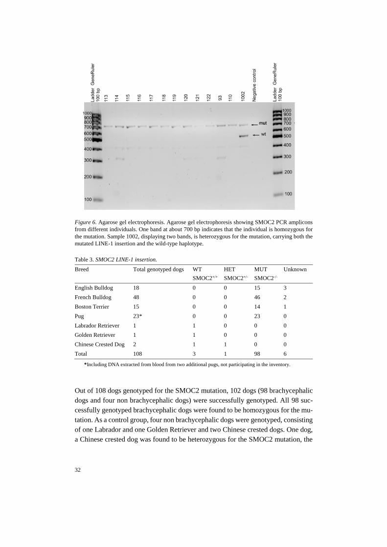
By using Sanger sequencing, the genotypes for BMP3 and DVL2 for three dogs
were obtained.
3.4.1 BMP3
The Golden Retriever was found homozygous for the ancestral C allele. Both the
Pug and the English Bulldog was found homozygous for the derived A allele (Figure
7).
Figure 7. The figure shows the output for all three sampled dogs, Golden Retriever forward and
reverse strand on top, Pug forward and reverse in the middle and English Bulldog forward and
reverse bottom. The red box indicates the SNP position.

34
3.4.2 DVL2
The Golden Retriever and the Pug was found homozygous for the ancestral C allele.
The English Bulldog was found homozygous for the deletion, causing the mutation.
Figure 8. Visualisation of the SNP using Sanger Sequencing. The figure shows the output for all
three sampled dogs. Golden Retriever front and reverse strand on top, Pug forward and reverse
strand in the middle and English Bulldog forward and reverse strand bottom. The red box indi-
cates the SNP position.
In summary, the English Bulldog was found homozygous for the mutations in both
BMP3 and DVL2. The Pug was found homozygous for mutated variant of BMP3
and homozygous for the ancestral variant of DVL2. The non-brachycephalic Golden
Retriever was homozygous for the ancestral variant in both studied genes (Table 4).
Table 4. Genotypes for BMP3 and DVL2 of three dogs.
Breed BMP3 DVL2
English Bulldog A/A Del/del
Pug A/A C/C
Golden Retriever C/C C/C
35
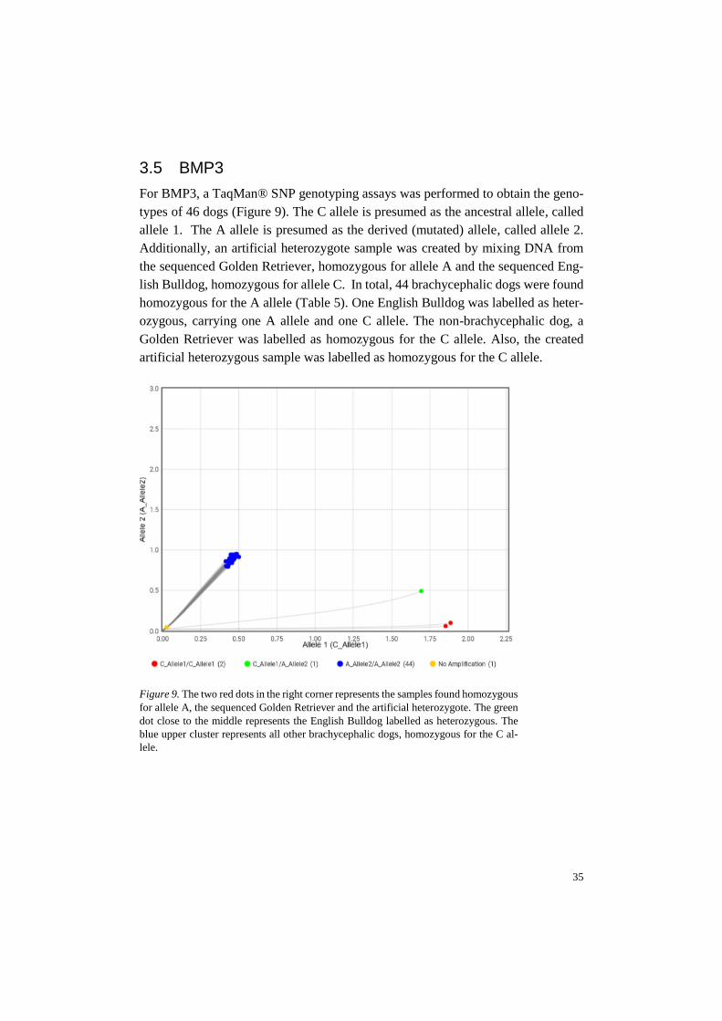
3.5 BMP3
For BMP3, a TaqMan® SNP genotyping assays was performed to obtain the geno-
types of 46 dogs (Figure 9). The C allele is presumed as the ancestral allele, called
allele 1. The A allele is presumed as the derived (mutated) allele, called allele 2.
Additionally, an artificial heterozygote sample was created by mixing DNA from
the sequenced Golden Retriever, homozygous for allele A and the sequenced Eng-
lish Bulldog, homozygous for allele C. In total, 44 brachycephalic dogs were found
homozygous for the A allele (Table 5). One English Bulldog was labelled as heter-
ozygous, carrying one A allele and one C allele. The non-brachycephalic dog, a
Golden Retriever was labelled as homozygous for the C allele. Also, the created
artificial heterozygous sample was labelled as homozygous for the C allele.
Figure 9. The two red dots in the right corner represents the samples found homozygous
for allele A, the sequenced Golden Retriever and the artificial heterozygote. The green
dot close to the middle represents the English Bulldog labelled as heterozygous. The
blue upper cluster represents all other brachycephalic dogs, homozygous for the C al-
lele.
36
Table 5. BMP3 status variant across breeds
Breed Total genotyped dogs WT
C/C
HET
C/A
MUT
A/A
English Bulldog 13 0 1 12
French Bulldog 9 0 0 9
Boston Terrier 13 0 0 13
Pug 10 0 0 10
Golden Retriever 1 1 0 0
Total 46 1 1 44
The allele frequency for the mutated allele when calculated for all four breeds to-
gether is 0.988.
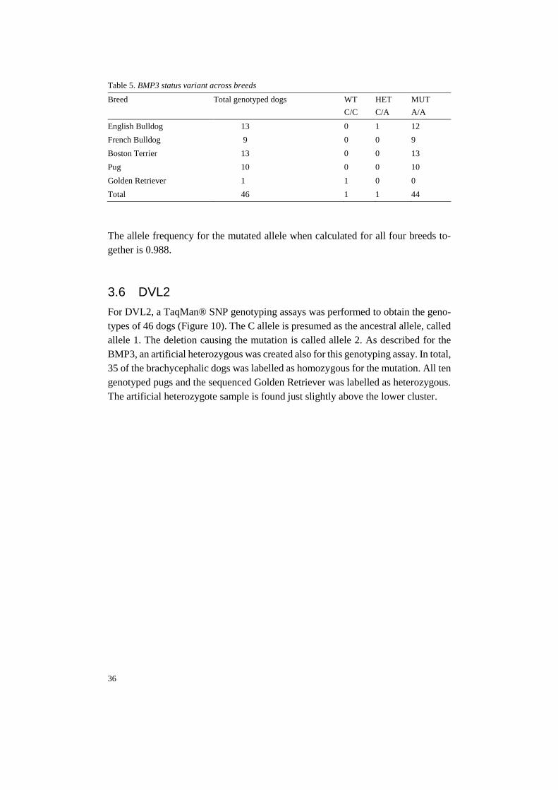
3.6 DVL2
For DVL2, a TaqMan® SNP genotyping assays was performed to obtain the geno-
types of 46 dogs (Figure 10). The C allele is presumed as the ancestral allele, called
allele 1. The deletion causing the mutation is called allele 2. As described for the
BMP3, an artificial heterozygous was created also for this genotyping assay. In total,
35 of the brachycephalic dogs was labelled as homozygous for the mutation. All ten
genotyped pugs and the sequenced Golden Retriever was labelled as heterozygous.
The artificial heterozygote sample is found just slightly above the lower cluster.
37
Figure 10. The two red dots in the lower green cluster in the plot represents the se-
quenced homozygous Golden Retriever and the sequenced homozygous Pug. All
other green dots in the lower cluster is Pugs. The green dot slightly above the
lower cluster represents athe artificial heterozygous. The upper blue cluster repre-
sents all English Bulldogs, French Bulldogs and Boston Terriers.
Table 6. DVL2 status (DVL2c.2044delC) variant across breeds
Breed Total genotyped dogs WT
C/C
HET
C/del
MUT
Del/del
English Bulldog 13 0 0 13
French Bulldog 9 0 0 9
Boston Terrier 13 0 0 13
Pug 10 10 0 0
Golden Retriever 1 1 0 0
Total 46 11 0 35
Allele frequency for the mutated allele when calculated for all four breeds together
is 0.75. When calculating the allele frequency for just the bull type breeds (Boston
Terrier, English Bulldog and French Bulldog), the allele frequency for the mutated
allele is 1.
38
The aim of this thesis was to investigate the genetic variation in known genes asso-
ciated with brachycephaly in dogs. Three genes were studied, SMOC2, BMP3 and
DVL2. In total, 102 brachycephalic dogs were genotyped for the mutation in the
SMOC2 gene and 46 of these dogs were also genotyped for the mutations in the
BMP3 gene and DVL2 gene respectively.
4.1.1 SMOC2
The SMOC2 LINE-insertion is thought to highly influence the brachycephalic phe-
notype. Marchant et al. (2017) suggest that the size effects of the associated SMOC2
haplotype accounts for 36 % of the facial length variation in the dogs in their study.
In this thesis, all 102 successfully genotyped brachycephalic dogs were found to be
homozygous for the SMOC2 LINE-1 insertion. Consequently, the allele frequency
for the derived allele was calculated to 1 in all studied breeds, suggesting that the
mutation is or is close to fixation.
In a communication from Bannasch (2019), the allele frequency for the LINE-1
insertion was calculated to 0.97 for dogs of Bull type (Boston Terrier, Bulldog and
French Bulldog) and 0.75 for Asian type breeds (Pug, Pekingese and Shih Tzu). The
high allele frequency in the bull type breeds is similar when compared to the result
in this thesis. For the Pugs, representing the Asia type breeds, the allele frequency
in this study was higher since all individuals were found homozygous for the muta-
tion.
In contrast to the study by Mansour et al. (2018), were three of the studied Boston
Terriers was found heterozygous, all brachycephalic dogs were found homozygous
in this thesis.
4 Discussion and conclusions

39
One interesting finding when genotyping dogs for the SMOC2 mutation was that
one of the Chinese crested dogs (non-brachycephalic breed) used as a positive con-
trol was found heterozygous, carrying one wild type allele and one mutated allele.
The other Chinese crested, related to the first individual, was found homozygous for
the wild type allele. Due to this, it would be interesting to sequence the heterozygous
Chinese crested to confirm the genotype and later to genotype even more non-brach-
ycephalic dogs, and especially Chinese crested dogs to better understand the muta-
tion and investigate if heterozygous individuals occur in other non-brachycephalic
breeds.
4.1.2 BMP3
BMP3 is thought to be required for craniofacial development in zebrafishes and the
identified mutation in brachycephalic dogs is thought to be associated with skull
shape.
DNA from three dogs in this thesis was sequenced, using the Sanger Sequence
method, to determine the nucleotide sequence of the DNA covering the mutation.
The sequenced Golden Retriever was found to be homozygous for the wild type
allele and the two brachycephalic dogs (one English Bulldog and one Pug) was
found homozygous for the mutated allele. The sequenced dogs were used as positive
controls in the TaqMan genotyping assay, which was used to genotype 43 additional
brachycephalic dogs. As a fourth positive control, there was an attempt of using an
artificial heterozygous by mixing DNA from a homozygous wild type and homozy-
gous mutated. However, the difference in DNA concentration between the two sam-
ples were not accounted for, which most likely lead to an overload of wild type
DNA in the sample. Consequently, the artificial heterozygous was not recognized
as heterozygous and positioned just slightly above the two homozygous wild types
in the plot (figure 9).
Most of the brachycephalic dogs genotyped using the TaqMan assay clustered
with the sequenced and confirmed homozygous mutated dogs. Therefore, it’s a high
possibility that also these dogs are homozygous mutated. The allele frequency for
the mutated allele was 0.98, calculated for all dogs together. Communication from
Bannasch (2019) suggests an allele frequency for the mutated allele at 0.99 in dogs
of Bull type (Boston Terrier, Bulldog and French Bulldog) 0.99 and 0.89 in Asian
type (Pug, Pekingese, Shih Tzu). The allele frequencies found in this study is con-
sistent with these results, suggesting that the mutation in BMP3 is or is close to
fixation in brachycephalic dogs. However, one English Bulldog, was labelled as
heterozygous for the BMP3 mutation. It may be hard to draw any conclusions for

40
that sample. To determine if it truly is heterozygous or if errors or contamination
have occurred would probably need to be confirmed by sequencing. Previous gen-
otyped English Bulldogs in the literature have been found to be homozygous,
thereof more work need to be done to correctly genotype the stated heterozygous
Bulldog.
4.1.3 DVL2
The DVL2 gene has been found to be a key component in the Wnt signalling path-
way, important for several steps in the embryonic development. A rare syndrome in
humans associated with craniofacial malformations, called Robinow syndrome,
caused by mutations in the human DVL1 and DVL3 genes. The phenotype of people
with Robinow syndrome have similar appearance to that of brachycephalic dogs,
for example widely spaced eyes, wide forehead and a short nose. This has raised
suspicions that the mutation in the canine DVL2 gene causing the screw tail in brach-
ycephalic breeds might also be involved in the shortened skull in such breeds
(Mansour et al. 2018).
As mentioned above for the BMP3 gene, the same three dogs were also se-
quenced using the Sanger sequencing method to determine the nucleotide sequence,
covering the deletion in the DVL2 gene. Two of the dogs, the Golden Retriever and
the Pug was found homozygous for the wild type, while the English Bulldog was
found to be homozygous for the mutation. The sequenced dogs were used as positive
controls in the TaqMan genotyping assay, which was used to obtain the genotypes
of additionally 43 brachycephalic dogs. For this genotyping assay there was also an
attempt of using an artificial heterozygous. Like described in the section above, it is
most likely that the higher concentration influenced the genotyping, resulting in that
the artificial heterozygous was positioned just slightly in the lower cluster seen in
plot (Figure 10).
In total, 46 dogs were genotyped. Dogs of bull type (e.g. Boston Terrier, English
Bulldog and French Bulldog) was clustered as homozygous mutated, in the same
cluster as the English Bulldog confirmed as homozygous mutated after sequencing.
All genotyped Asian type dogs (e.g. Pugs) are visible in a second lower cluster in
the allelic discrimination plot (green lower cluster in figure 10). The algorithm la-
bels samples in this cluster as heterozygous, though, the two dogs confirmed as ho-
mozygous wild type (the Golden Retriever and the Pug) is also positioned in this
cluster. Therefore, it is more likely that samples in this cluster carries two wild type
alleles, which is not recognised in the software for unknown reasons. The study by
Mansour et al. (2018) also supports that Pugs, as a breed lacking screw tails, are

41
homozygous for the wild type allele. It is possible that the assay or the protocol
needs further optimisation to correctly label wild type samples.
The allele frequency for the mutated allele is calculated to 0.75 when including
all four breeds. When calculating the allele frequency only including Bull type, the
allele frequency for the mutated allele was found to be 0.94. Since all Pugs was
found homozygous wild type, it may be more correct to exclude Pugs when calcu-
lating allele frequency. Bannasch (2019) suggest an allele frequency of 0.98 in dogs
of Bull type and an allele frequency of 0.01 in dogs of Asia type, consistent with the
results in this thesis. This suggests that also the mutation in the DVL2 gene is or is
close to fixation in the studied population of Boston Terrier, English Bulldog and
French Bulldog.
Studied Pugs were found to be homozygous for the ancestral type allele of DVL2,
still, Pugs are very much brachycephalic. The mutation has been associated with the
screw tail, characteristic for breeds such as Boston Terrier, English Bulldog and
French Bulldog. (Mansour et al. 2018). However, if and in that case how the DVL2
gene is involved in the craniofacial development and if the found mutation can be
associated with the shortened skull in brachycephalic breeds needs to be studied
further.
4.2 Strengths and weaknesses with this thesis
One strength with this study is that even though it can be considered as a rather
small study, 102 dogs were successfully genotyped for the SMOC2 LINE-1 inser-
tion, which is a rather high number of dogs in relation to the size of the project.
In previous studies of the SMOC2 mutation, 374 (37 brachycephalic) and 152 (109
brachycephalic) dogs were genotyped, respectively (Marchant et al. 2017; Mansour
et al. 2018).
However, one weakness with this study was that only four dogs of non-brachy-
cephalic breeds was used as positive controls when studying the three genes. Previ-
ous studies of described genes have studied and genotyped dogs from several
breeds, both brachycephalic and non-brachycephalic, which can be considered as a
strength. For future research, more dogs of different breeds should be included.
Another strength with this thesis is that the found allele frequencies for the three
genes are all consistent with the frequencies in previous performed studies.
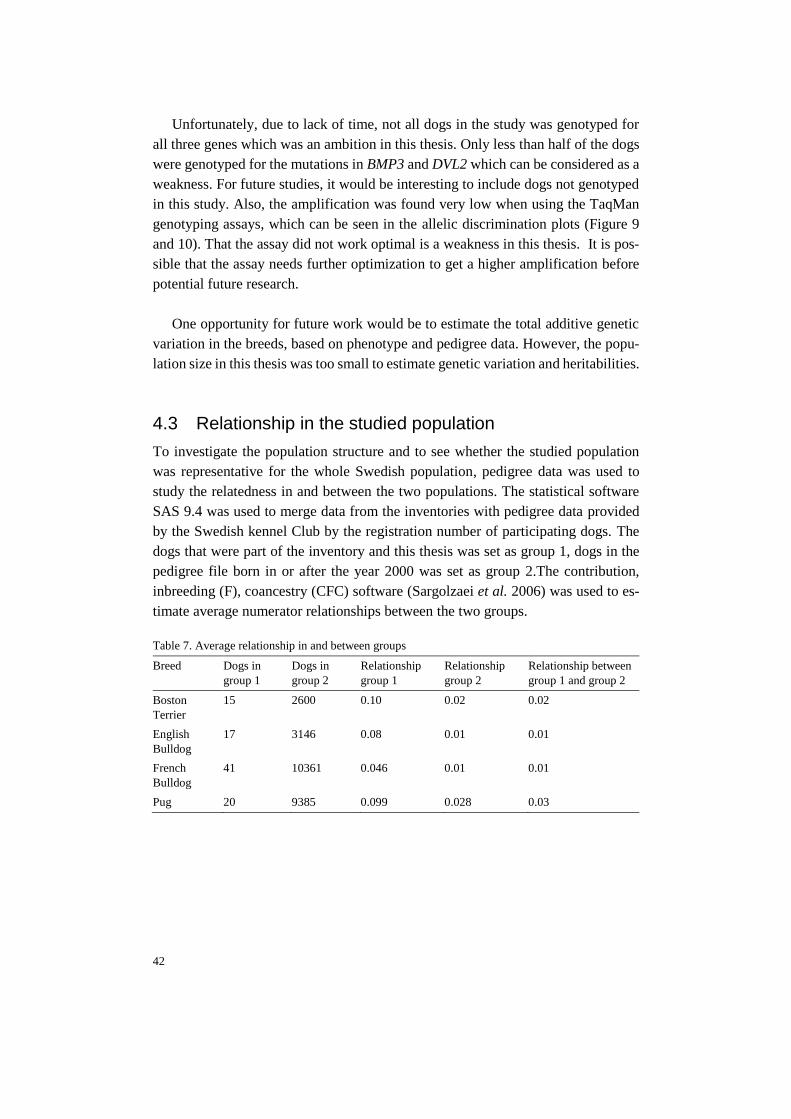
42
Unfortunately, due to lack of time, not all dogs in the study was genotyped for
all three genes which was an ambition in this thesis. Only less than half of the dogs
were genotyped for the mutations in BMP3 and DVL2 which can be considered as a
weakness. For future studies, it would be interesting to include dogs not genotyped
in this study. Also, the amplification was found very low when using the TaqMan
genotyping assays, which can be seen in the allelic discrimination plots (Figure 9
and 10). That the assay did not work optimal is a weakness in this thesis. It is pos-
sible that the assay needs further optimization to get a higher amplification before
potential future research.
One opportunity for future work would be to estimate the total additive genetic
variation in the breeds, based on phenotype and pedigree data. However, the popu-
lation size in this thesis was too small to estimate genetic variation and heritabilities.
4.3 Relationship in the studied population
To investigate the population structure and to see whether the studied population
was representative for the whole Swedish population, pedigree data was used to
study the relatedness in and between the two populations. The statistical software
SAS 9.4 was used to merge data from the inventories with pedigree data provided
by the Swedish kennel Club by the registration number of participating dogs. The
dogs that were part of the inventory and this thesis was set as group 1, dogs in the
pedigree file born in or after the year 2000 was set as group 2.The contribution,
inbreeding (F), coancestry (CFC) software (Sargolzaei et al. 2006) was used to es-
timate average numerator relationships between the two groups.
Table 7. Average relationship in and between groups
Breed Dogs in
group 1
Dogs in
group 2
Relationship
group 1
Relationship
group 2
Relationship between
group 1 and group 2
Boston
Terrier
15 2600 0.10 0.02 0.02
English
Bulldog
17 3146 0.08 0.01 0.01
French
Bulldog
41 10361 0.046 0.01 0.01
Pug 20 9385 0.099 0.028 0.03

43
For all four breeds, the dogs in group 1 was estimated to be more related to each
other than to the dogs in group 2. For future studies, it might however be of interest
to study dogs less related that would better represent the bigger population.
4.4 Phenotypic variation
Bertilsson (2019) investigated the phenotypic variance correlated to BOAS in 119
brachycephalic dogs collected during the inventories described in this thesis. Some
phenotypic variation was found in the four breeds; Boston Terrier, English Bulldog,
French Bulldog and Pug (Table 8). However, only English Bulldog and French Bull-
dog was suggested to have enough phenotypic variation to improve the BOAS prob-
lem. The Boston Terrier population was to small and the BOAS problem was not
severe enough to get any results. For Pugs, the phenotypic variance was found to be
very small and a need of a more drastic approach to handle the BOAS problems was
suggested.
Table 8. Phenotypic variance and mean value for measurements and ratios
Boston Terrier English Bulldog French Bulldog Pug
Snout length (mm) Variance
Mean
20-30
23.5
15-46
32.8
19-38
29.3
11-25
17
Cranial length (mm) Variance
Mean
80-125
105.7
125-185
150.1
95-170
121.3
90-150
111.5
CFR Variance
Mean
0.16-0.31
0.23
0.12
0.22
0.15-0.39
0.24
0.09-0.22
0.16
Adapted from Bertilsson (2019), with permission.
As can be seen in Table 8 and the master’s thesis by Bertilsson, there is some phe-
notypic variation in the four breeds. Dogs not affected by BOAS were also shown
to have significantly longer snouts, longer sternum and higher CRF, compared to
BOAS affected dogs. Non affected dogs also had a significantly lager variance for
snout length, chest girth, sternum length, back length, CFR an NGR, compared to
affected dogs (Bertilsson, 2019).
The thesis by Bertilsson suggests that there is some phenotypic variation that can
be observed in the studied dogs. On the contrary, the genetic variation in the studied
genes associated with brachycephaly is suggested as low. Marchant et al. (2017)
suggests that homozygous-derived alleles of the SMOC2 LINE-1 solely explains 36
% of the viscerocranium variation while BMP3 is thought to explain 12 % of the
variation. It should be noted that the variances are not additive but implies the
44
maximum potential proportion of each genotype. It is likely that the brachycephalic
phenotype is complex trait, affected by many genes, both known and probably yet
unknown genes. Most likely, more dogs need to be studied and genotyped to get a
better picture of genes associated with brachycephaly and to further investigate the
genetic variation in the populations. For the Swedish project, about 50-100 dogs per
breed will be genotyped using Illumina CanineHD Whole-Genome Genotyping
BeadChip (170K) to identify genomic regions associated with increased risk for
BOAS. For future genotyping, it would be of interest to base the selection on pedi-
gree data to include animals not related to each other.
4.5 Conclusions
In this master thesis, 102 privately owned brachycephalic dogs was genotyped for
the mutation in the SMOC2 gene, additionally, 45 of these dogs were also genotyped
for the mutations in the BMP3 and DVL2 gene. The results in this thesis indicates
that there is no or very little variation in the three genes and that the mutations in
previous mentioned genes is or is close to fixation in the studied population. The
mutant variant of the SMOC2 gene was found to be fixated in the studied four
breeds. The mutant variant of the BMP3 gene had high allele frequency in all breeds.
In Bull type breeds (Boston Terrier, English Bulldog and French Bulldog) the mu-
tant version of the DVL2 gene was fixated, whilst all Pugs were tested wild type for
the DVL2 mutation. However, the studied population is too small to draw any con-
clusion and further research is needed to thoroughly investigate the genetic variation
in the four breeds.

45
As suggested, more research is needed to genotype more dogs in order to get a better
picture of the genetic variation in Swedish brachycephalic dogs. The following part
of the will therefore discuss two possible scenarios after further future research.
5.1.1 Enough genetic variation
Future studies of the brachycephalic dog populations may conclude that the genetic
variation in brachycephalic breeds is not lower than the genetic variation observed
in other dog breeds. If it is deemed possible to genetically select dogs within breed
for a conformation less associated with welfare problems, there might be a need to
implement assessment tools in the breeding plans to reduce the health issues ob-
served in brachycephalic dogs.
Today, the Swedish kennel club have several ongoing projects to decrease health
issues in brachycephalic breeds and how to come to terms with extreme confor-
mations causing respiratory problems. One of these projects is a new puppy exami-
nation protocol that was introduced in 2017, after an uprising where hundreds of
Swedish veterinarians was demanding the Swedish kennel club and Swedish board
of Agriculture to come to terms with breeding and import of dogs with respiratory
problems. The examination protocol is to be used for all registered puppies up to six
months of age and can also be used for unregistered puppies. The purpose of the
examination protocol is to early catch dogs with respiratory problems (Svenska ken-
nelklubben 2017).
5 Possible future scenarios for the brachycephalic breeds
46
An examination before breeding of brachycephalic dogs has also been tried out
and accepted. During the examination, the dog is examined during rest and after
exercise, were the dog is supposed to trot 1 000 meters in less than 12 minutes. The
examination is intended to work as a tool to assess symptoms of respiratory prob-
lems and difficulties to thermoregulate during rest and exercise. The conformation
and the possible contributions to health issues associated with brachycephaly of the
dog and its offspring is also assessed. In February 2019, the examination was ac-
cepted as a health program on level 1, which means it not mandatory but the results
from the examination will be registered at the Swedish kennel club and displayed at
the SKK breeding data online. In the future, the health program will be evaluated
and might be transformed to a health program on level 2 (requirements on performed
examination) or at level 3 (requirements on performed examination with demands
of certain result). The examination can be performed at two years of age and the dog
can redo the examination after six months (Svenska kennelklubben 2019).
The Swedish kennel club has also suggested that surgical procedures on dogs of
brachycephalic type and/or death or euthanasia due to problems associated to brach-
ycephaly should be registered at the kennel club. Veterinary clinics should handle
the reporting to the kennel club and dogs that have gone through surgery should not
be used for breeding (Svenska kennelklubben).
Additionally, the Swedish kennel club is stating that healthy dogs with less ex-
treme conformation should be prioritized and placed at dog shows. Breed specific
instructions (BSI) have therefore been used since 2009 in Sweden and since 2014
there is a Nordic collaboration against extreme conformation with the help of a com-
mon BSI, which should be used as a complement to the breed standards. The breed
specific instructions identify areas of risk and aims at preventing possible future
problems. BSI includes both instructions concerning all dogs, and instructions only
concerning specific “breed types”. The BSI states that “All dogs should be able to
breathe normally, also when moving” (The Nordic Kennel Club, 2018, p 10 ). The state-
ment above regards all dogs, in addition, brachycephalic breed type also has instruc-
tions implying that exaggerations in typical features such as skull, muzzle, jaw, eyes,
ribcage and skin might lead to serious health problems and not exclusively problems
in breathing and thermoregulation. The breathing distress should be assessed in the
show ring. Nonsignificant affected breathing should be noted but not necessarily
affecting the quality grading of the dog. Milder respiratory problems for example
pinched nostrils and too short nose should influence the quality grading. Obvious
signs of respiratory problems should motivate for disqualification (The Nordic Ken-
nel Club 2018). From 2020 to 2024 only Nordic show judges will be allowed to
judge brachycephalic breeds. Further, the Swedish kennel club is actively working

47
to educate and inform dog show judges about health problems associated with ex-
treme conformations.
As stated above, the Swedish kennel club has taken many actions to reduce
health problems in brachycephalic breeds by several selection criterions. The prob-
lems with brachycephalic dogs will need to be handled by a united mass of both
breeders, dog owners, show judges, veterinarians and the kennel club. This can of
course be problematic, but the work led by the kennel club is a step in the right
direction. The current suggested actions to obtain brachycephalic dogs with less ex-
treme phenotypes and better health would need to be evaluated before taking dis-
cussing further actions.
The health of brachycephalic dogs is a hot topic, not only in Sweden. In Febru-
ary 2019, the Dutch animal welfare legislation published a report with changes the
animal welfare legislation, mainly regarding breeding of brachycephalic breeds. The
workgroup suggests a so-called “traffic light model” to be used as a tool to easier
select for dogs with less extreme conformation and a better health. Each criterium
in the suggestion has a traffic light coding; green, orange or red. In total, 28 criteri-
ons are listed in the report, divided into categories such as traits associated with
BOAS and Brachycephalic Ocular syndrome (BOS). Examples from the traffic light
model is described in table 9. It’s suggested that dogs that are categorised as green
can be used for breeding, dogs with one orange “light” can be used for breeding if
all other criteria are green. Dogs categorised as red are not allowed to be used for
breeding. The suggestion is to allow dogs categorised as orange for a shorter period,
for example for two generations and later only allow “green” breeding animal. The
stepwise motion of the suggestion is to not reduce the breeding population drasti-
cally. Breeders have been given a time to breed toward a healthier type, if this don’t
occur, a mandatory breeding program will be discussed.
Table 9. Examples of criterium listed in Dutch animal welfare legislation
Criterium Green (ideal) Orange Red Consequence if
not ideal
Nostrils Open nostrils Mild stenosis Severe stenosis Orange may be
used if no other
criteria is ex-
ceeded
CFR ≥0.5 0.3-0.5 ≤0.3 Orange may be
used if no other
criteria is ex-
ceeded

48
Criterium Green (ideal) Orange Red Consequence if
not ideal
Abnormal
respiratory
noise
Absent - Strong sniffing, snor-
ing or noisy breath-
ing at rest
Exceeding stand-
ards, regardless
of other criteria
6 minutes
test
>500 m, restored
heart frequency and
body temp within 5
min
>500 m, restored
heart frequency and
body temp within 10
min
<500 m, heart fre-
quency and body
temp not restored
within 10 min
Orange may be
used if no other
criteria is ex-
ceeded
1000 m test Maximum 12 min
Table adapted from van Hagen (2019).
The Dutch report highlights many criterions associated with brachycephaly and
BOAS. There are similarities between the work led by The Swedish kennel club and
the Dutch animal welfare legislations, for example that breeding animals should
show good respiration and good thermoregulation, both in rest and during/after ex-
ercise. The Dutch legislation can however be considered as more drastic when in-
volving criterions regarding measures and ratios. The report refers to many scien-
tific articles concerning brachycephaly and/or BOAS but the exact reasoning for
using the stated measures and ratios stated is not clear.
By looking at the studied Swedish population of brachycephalic dogs, it would
be hard or perhaps not even possible to incorporate such criterions on measures and
ratios to the breeding programs. The Dutch legislation states that a dog with a CFR
under 0.3 should not be used for breeding. A dog with a CFR between 0.3 and 0.5
should only be used if all other criterions are fulfilled. For the studied population,
the mean values for CFR is 0.22, 0.21, 0.24 and 0.15 for Boston Terrier, English
Bulldog, French Bulldog and Pug respectively. For the three bull-type breeds, there
would only be a small number of dogs fulfilling the CFR criterion, provided that the
dog fulfils all other criteria. No pug in this studied population would fulfil the CFR
criterion, according to the Dutch legislation. Implying such criteria on the Swedish
brachycephalic dog population would probably drastically decrease the effective
population size. The health program, the new puppy examination protocol and the
focus on awarding healthy and less “extreme” dogs at dog shows are selection cri-
terions provided by the Swedish kennel club today and the consequences of using
these tools should be evaluated before taking more drastic solutions under consid-
eration.
Breeders and keepers should carefully evaluate future breeding animals to ensure
that dogs with a healthy conformation is used for breeding.
49
If the phenotypic variation and heritabilities is shown to be high enough for traits
associated with BOAS, there might be possible to implement traditional estimated
breeding values (EBV). Most conformation traits have been found to have quite high
heritabilities. For example, in horses where body length has shown to have an esti-
mated heritability of 0.72. The head length had an estimated heritability of 0.55
(Mcmanus et al. 2002).
EBVs have been used for hip and elbow dysplasia, where abnormal formation of
the joint can cause pain, are complex traits that have used estimated breeding values
to come to terms with the problems. Breeding values for hip- and elbow dysplasia
is estimated by using so called Best Linear Unbiased Prediction (BLUP), which uses
both information from the animal, its offsprings and from relatives, using pedigree
data to estimate breeding values. BLUP is widely used for several animal categories,
including horses. However, BLUP requires reliable pedigrees and systematically
recording of phenotypes. To be able to use BLUP for brachycephalic dogs, meas-
urements of conformations traits would need to be recorded for a high number of
dogs. One suggestion could be to collect such phenotype data at the same time as
earlier mentioned evaluation for breeding.
Recently, there has also been several research projects to investigate the possi-
bilities to use genomic selection for hip and elbow dysplasia. Sánchez-Melano et al.
(2015) found that the accuracy was highest when combining pedigree and genomic
data, compared to traditional breeding evaluation (Sánchez-Molano et al. 2015).
For the future, it would perhaps be a possibility to use genomic selection to come to
term with health problems associated with canine brachycephaly.
5.1.2 Not enough genetic variation in the breeds
With low genetic variation, it will be difficult to select for traits to decrease extreme
conformations and health problems in the brachycephalic breeds. To improve the
overall health of brachycephalic dogs, outcrossing may be a future option. For a
small number of breeds in Sweden outcrossing projects have been implemented in
the breeding strategies.
In 2001 the Clumber Spaniel club applied for outcrossing to increase the genetic
variation in the breed. For the outcrossing, a Cocker Spaniel of hunting type was
selected since the Cocker Spaniel is closely related to the Clumber, is a healthy and
sound breed without extreme conformation and without diseases not present in the
Clumber breed. In 2003 the first outcrossed litter was born. From that first litter, two
50
females were backcrossed with Clumber Spaniel males. Two females from the back-
cross litter was later accepted and registered as purebred Clumber Spaniels. The
health of the dogs in the outcrossing project is similar to the health of the whole
Clumber Spaniel population, except occurrence of elbow dysplasia that was slightly
higher for the first outcross generation. The mentality of the latest generation in the
outcrossing project is not deviating from the rest of the Clumber population (Clum-
ber Spaniel Klubben 2014).
Another breed with an ongoing outcrossing project is the Norwegian Lundehund.
The Lundehund has been close to extinction multiple times, for example in the 60’s
where the breed consisted of just six known individuals. After the multiple genetic
bottle necks, the genetic variation in the breed was found low. The Norwegian Lun-
dehund Club therefor applied for an outcrossing project, which was accepted by the
Norwegian kennel club. The outcrossing projects aim is to increase the genetic var-
iation in the breed and by that hopefully increase fertility and decrease the occur-
rence the disease Intestinal lymphangiectasias currently present in the breed. Re-
lated breeds such as the Norwegian Buhund, Norrbotten-Spitz and the Icelandic
Sheepdog has been selected for the outcrossing project to form a side population
registered in a so-called X-register. Females of these three breeds can be mated with
a Lundehund male to generate the first generation of crosses (F1), females from the
F1 generation can later be backcrossed with Lundehund to generate a F2 generation.
Health, exterior and mentality will be tested for later generations before crosses in
the X-register can be allowed in to the Lundehund breed (Fossen 2019).
If an outbreeding project would be in question for brachycephalic breeds, there
could be some problematic parts that would have to be handled:
• What breed or breeds could be suitable for cross breeding?
• What would be the criteria for such possible breeds?
• Are breeders and dog owners willing to participate in such projects?
• Which criteria would need to be fulfilled for including crossbreds in the
purebred population?
• Will future crossbred individuals be accepted, and would they be used for
breeding?
If it will come to a point where an outbreeding project would be the solution, joined
efforts from breeders, the kennel club and dog owners will be needed.

51
5.1.3 Breed standards and breed-specific breeding instructions
A suggestion is to investigate if there is a need to clarify and improve breed stand-
ards for the four studied breeds. All four breed standards state the muzzle should be
short, relatively short or very short (FCI-standard N° 149, 2011; FCI-standard
N°253, 2010; FCI-standard N°101, 2015). The standard for the Boston Terrier states
that the length of the muzzle should not be longer than one third of the length of the
head (FCI-standard N° 140, 2014). Breed standards, breeders and dog show judges
have possibilities to together improve the health of brachycephalic dogs.
All breeds have breed-specific breeding instructions (RAS) that describes prob-
lems and strong aspects within the breed and breeding recommendations. All breed-
ing recommendations mentions that dogs used for breeding should be able to breathe
normally. The breeding recommendations for Boston Terrier recommends not to
breed on dogs with respiratory problems. The recommendation also states that the
breed today do not have any conformation problems (Svenska Bostonterrierklubben
2011).
The recommendations for the French Bulldog recommend that dogs with ex-
treme conformation should not be used for breeding. Further, dogs with stenotic
nostrils, elongated soft palate, narrowing of or problems in the respiratory tract
should not be used for breeding. (Fransk Bulldogg klubb 2010)
The English Bulldog recommendations states that normal breathing function
should be of highest priority when selecting breeding animals. Sufficient length of
muzzle and other structures affecting breathing should be a selection criterion (Pro-
jektgruppen för engelsk bulldogg 2015).
For the Pug, dogs with normal breathing function and less nose wrinkled should
be prioritized. Dogs with stenotic nostrils, elongated soft palate or narrowing of or
problems in the larynx or throat should not be used for breeding (Mopsorden 2015).
The mentioned recommendations are a positive step in the right direction. It may
however be necessary to investigate how well the recommendations are applied in
the breeding work.

52
5.2 Weaknesses in study and future perspectives
This part of the thesis will concern steps that did not work as good as predicted.
Maybe, it will be helpful for other students or scientist struggling.
Buccal cells were sampled using 2-3 cytology brushes per dog, simply by strok-
ing the inside of the dog’s cheek with the brush. This method is very simple, not
invasive for the animal and can be performed by anyone by simple instructions. The
cytology brushes were airdried before putting them in a marked paper envelope and
sealed. The envelopes were later stored in room temperature before performing the
DNA extraction. It’s hard to say if this was the best way of storing the brushes, or
if it would been better to perform the extraction closer to the sampling. Most swab
kits require that the swab is put in a stabilizing buffer to prevent fragmentation of
the DNA. However, if this would improve the amplification is hard to say. Also,
since many bacteria’s can be present in a dog’s mouth, the measurements of nucleic
acid got problematic since there was no way to tell how much of the nucleic acid
that was canine and how much that was bacterial. The variation in DNA concentra-
tion in the samples were problematic when performing a dilution series, therefor,
samples from swabs was not diluted.
For the SMOC2 PCR, the standard protocol for the HotStar® polymerase using
suitable annealing temperature did not work optimal, resulting in many unspecific
bands could visible after gel electrophoresis. Also, optimising by performing a se-
ries of different annealing temperatures did just slightly improve the specificity. Af-
ter communication with Marchant, T., the corresponding author of the SMOC2 ar-
ticle, a working and optimized protocol was received. By using this protocol, frag-
ments of expect size was amplified, but still there was some unspecific amplicons
visualised after gel electrophoresis. Attempts to optimize the PCR conditions by
optimizing the Primer concentrations, dNTP concentration and the magnesium con-
centration did increase the primer specificity, after gel electrophoresis there were
still unspecific bands showing. It should be mentioned that the amplification worked
well enough to determine the genotypes of the dogs, but to get rid of unspecific
bands further optimization is probably needed. One suggestion on optimization is to
use the same polymerase as described my Marchant, T (personal communication,
2019) and to add SDS when performing gel electrophoresis to increase the specific-
ity.
53
Another problem for this PCR was that not all samples was amplified correctly,
leaving the genotype undetermined. This is most likely due to low amount of DNA
in the samples or possibly due to fragmentation or inhibition of the DNA in the swab
samples.
For the TaqMan genotyping assays, the first qPCR was run without any positive
controls since no dog had a known genotype from start. Consequently, all samples
were labelled as undetermined by the StepOne™ software. The amplification was
also, as mentioned earlier, quite low. Due to this result, it was decided to sequence
three samples using Sanger sequencing to obtained dogs with a confirmed genotype
and to use these as positive controls. The sequencing step was not planned for and
took extra time, resulting in not having enough time to genotype all samples using
the TaqMan assay. As mentioned for the SMOC2 PCR the different DNA concen-
trations in the samples might have contributed to the problems with the TaqMan
assays. The assays are sensitive and requires a certain amount of DNA to generate
correct assignment. For both assays, the amplification remained low, even after add-
ing the positive controls and optimizing the annealing temperature for each primer.
For DVL2, dogs confirmed as homozygous wildtype was labelled as heterozygous.
Due to lack of time, this result could not be investigated further but would probably
need to be sorted out before continuing with future research. Possibilities for these
kinds of errors could be the lack of standardisation of DNA concentration or addi-
tional SNPs in the target area, affecting the efficiency of the amplification. Since
there are difficulties determining the concentration of canine DNA when sampled
from swabs, one alternative could perhaps be to use DNA sampled from blood to
easier measure the DNA concentration and use correct DNA amount for the assays.
Blood samples may however be more stressful for the dog and would require trained
personnel, which should be considered before deciding on changing sample method.
For future perspectives, it would be of high importance and interest to investigate
more dogs and in particular, more genes found associated with the brachycephalic
phenotype. There is ongoing research in the field and success in such research
should be observed and if suitable applied on the Swedish population. Only three of
known genes associated with brachycephaly is covered in this thesis, recent research
has for example studied genes such as FGF, P2RX7, STC2 and a QTL on chromo-
some 30. A recently published article discovered missense mutation in the gene
ADAMTS3 on chromosome 13. The mutation was associated with a respiratory dis-
ease syndrome in the Norwich Terrier, same as can be seen in the Bulldog breed.
The study screened a large number of dogs for the mutation and suggests that the
found variant may influence BOAS in French and English Bulldogs. It would be of
high interest to also screen the Swedish population of brachycephalic dogs for the

54
ADAMTS3 mutation for further investigation and explore the possibilities to use
such mutations as a diagnostic marker for disease.
55
American Kennel Club Bulldog history (Origin of the English bulldog). Available
at: http://www.bulldoginformation.com/english-bulldog-history.html
[2019-02-25]
American kennel club Pug Dog Breed Information. American Kennel Club. Avail-
able at: https://www.akc.org/dog-breeds/pug/ [2019-03-18]
Berilsson, I. (2019) Phenotypic variation of BOAS within four brachycephalic dog
breeds - Can good welfare be obtained? Unpublished manuscript. The Swedish
University of Agricultural Sciences. Uppsala.
Bill, B.R., Petzold, A.M., Clark, K.J., Schimmenti, L.A. & Ekker, S.C. (2009). A
Primer for Morpholino Use in Zebrafish. Zebrafish, vol. 6 (1), pp. 69–77
Bloch-Zupan, A., Jamet, X., Etard, C., Laugel, V., Muller, J., Geoffroy, V.,
Strauss, J.-P., Pelletier, V., Marion, V., Poch, O., Strahle, U., Stoetzel, C.
& Dollfus, H. (2011). Homozygosity Mapping and Candidate Prioritiza-
tion Identify Mutations, Missed by Whole-Exome Sequencing, in
SMOC2, Causing Major Dental Developmental Defects. The American
Journal of Human Genetics, vol. 89 (6), pp. 773–781
Bornstein, P. (2009). Matricellular proteins: an overview. Journal of Cell Commu-
nication and Signaling, vol. 3 (3), p. 163
Boulet, A.M., Moon, A.M., Arenkiel, B.R. & Capecchi, M.R. (2004). The roles of
Fgf4 and Fgf8 in limb bud initiation and outgrowth. Developmental Biol-
ogy, vol. 273 (2), pp. 361–372
Boutros, M. & Mlodzik, M. (1999). Dishevelled: at the crossroads of divergent in-
tracellular signaling pathways. Mechanisms of Development, vol. 83 (1–
2), pp. 27–37
Boyko, A.R., Quignon, P., Li, L., Schoenebeck, J.J., Degenhardt, J.D.,
Lohmueller, K.E., Zhao, K., Brisbin, A., Parker, H.G., vonHoldt, B.M.,
Cargill, M., Auton, A., Reynolds, A., Elkahloun, A.G., Castelhano, M.,
Mosher, D.S., Sutter, N.B., Johnson, G.S., Novembre, J., Hubisz, M.J.,
Siepel, A., Wayne, R.K., Bustamante, C.D. & Ostrander, E.A. (2010). A
Simple Genetic Architecture Underlies Morphological Variation in Dogs.
PLOS Biology, vol. 8 (8), p. e1000451
Cadigan, K.M. & Nusse, R. (1997). Wnt signaling: a common theme in animal de-
velopment. Genes & Development, vol. 11 (24), pp. 3286–3305
References

56
Chen, D., Zhao, M. & Mundy, G.R. (2004). Bone Morphogenetic Proteins.
Growth Factors, vol. 22 (4), pp. 233–241
Clumber Spaniel Klubben (2014). Rasspecifik Avelsstrategi för Clumber Spaniel.
p. 20
Daluiski, A., Engstrand, T., Bahamonde, M.E., Gamer, L.W., Agius, E., Steven-
son, S.L., Cox, K., Rosen, V. & Lyons, K.M. (2001). Bone morphogenetic
protein-3 is a negative regulator of bone density. Nature Genetics, vol. 27
(1), pp. 84–88
Davis, M.S., Cummings, S.L. & Payton, M.E. (2017). Effect of brachycephaly and
body condition score on respiratory thermoregulation of healthy dogs.
Journal of the American Veterinary Medical Association, vol. 251 (10),
pp. 1160–1165
Evans, H.E. & Miller, M.E. (2013). Miller’s anatomy of the dog. Fourth edition.
St. Louis, Missouri: Elsevier.
Farrell, L.L., Schoenebeck, J.J., Wiener, P., Clements, D.N. & Summers, K.M.
(2015). The challenges of pedigree dog health: approaches to combating
inherited disease. Canine Genetics and Epidemiology, vol. 2 (1), p. 3
Fawcett, A., Barrs, V., Awad, M., Child, G., Brunel, L., Mooney, E., Martinez-
Taboada, F., McDonald, B. & McGreevy, P. (2019). Consequences and
Management of Canine Brachycephaly in Veterinary Practice: Perspec-
tives from Australian Veterinarians and Veterinary Specialists. Animals,
vol. 9 (3). DOI: https://doi.org/10.3390/ani9010003
Feng, T., McConnell, C., O’Hara, K., Chai, J. & Spadafori, G. (2017). Brachyce-
phalic Breed Disease Prevalence Study. Nationwide. Available at:
http://nationwidedvm.com/wp-content/uploads/2017/03/NWBrachycel-
phalicStudy0317.pdf [2019-03-05]
Feng, W., Leach, S.M., Tipney, H., Phang, T., Geraci, M., Spritz, R.A., Hunter,
L.E. & Williams, T. (2009). Spatial and Temporal Analysis of Gene Ex-
pression during Growth and Fusion of the Mouse Facial Prominences.
(Bridger, J. M., ed.) PLoS ONE, vol. 4 (12), p. e8066
Fossen, R. (2019-04-09). Prosjektets bakgrunn. Norsk Lundehund Klubb. Availa-
ble at: http://lundehund.no/index.php/krysningsprosjektet1/artikler-om-
krysningsprosjektet/896-prosjektets-bakgrunn [2019-05-15]
Fransk Bulldogg klubb (2010). Avelspolicy och Avelsstrategi för Fransk Bull-
dogg. Svenska Kennelklubben. Available at: https://www.skk.se/globalas-
sets/dokument/utstallning/breed-special-specific-instructions---bsi-a8.pdf
[2019-05-16]
Frantz, L.A.F., Mullin, V.E., Pionnier-Capitan, M., Lebrasseur, O., Ollivier, M.,
Perri, A., Linderholm, A., Mattiangeli, V., Teasdale, M.D., Dimopoulos,
E.A., Tresset, A., Duffraisse, M., McCormick, F., Bartosiewicz, L., Gál,
E., Nyerges, É.A., Sablin, M.V., Bréhard, S., Mashkour, M., Bălăşescu,
A., Gillet, B., Hughes, S., Chassaing, O., Hitte, C., Vigne, J.-D., Dobney,
K., Hänni, C., Bradley, D.G. & Larson, G. (2016). Genomic and archaeo-
logical evidence suggest a dual origin of domestic dogs. Science, vol. 352
(6290), pp. 1228–1231
Freedman, A.H., Gronau, I., Schweizer, R.M., Vecchyo, D.O.-D., Han, E., Silva,
P.M., Galaverni, M., Fan, Z., Marx, P., Lorente-Galdos, B., Beale, H.,

57
Ramirez, O., Hormozdiari, F., Alkan, C., Vilà, C., Squire, K., Geffen, E.,
Kusak, J., Boyko, A.R., Parker, H.G., Lee, C., Tadigotla, V., Siepel, A.,
Bustamante, C.D., Harkins, T.T., Nelson, S.F., Ostrander, E.A., Marques-
Bonet, T., Wayne, R.K. & Novembre, J. (2014). Genome Sequencing
Highlights the Dynamic Early History of Dogs. PLOS Genetics, vol. 10
(1), p. e1004016
Freedman, A.H. & Wayne, R.K. (2017). Deciphering the Origin of Dogs: From
Fossils to Genomes. Annual Review of Animal Biosciences, vol. 5 (1), pp.
281–307
Gamer, L.W., Nove, J., Levin, M. & Rosen, V. (2005). BMP-3 is a novel inhibitor
of both activin and BMP-4 signaling in Xenopus embryos. Developmental
Biology, vol. 285 (1), pp. 156–168
Gammons, M.V., Rutherford, T.J., Steinhart, Z., Angers, S. & Bienz, M. (2016).
Essential role of the Dishevelled DEP domain in a Wnt-dependent human-
cell-based complementation assay. Journal of Cell Science, vol. 129 (20),
pp. 3892–3902
Genetics Home Reference (2019). Robinow syndrome. Genetics Home Reference.
Available at: https://ghr.nlm.nih.gov/condition/robinow-syndrome [2019-
02-25]
Gilbert, S.F. & Barresi, M.J.F. (2016). Developmental biology. Eleventh edition.
Sunderland, Massachusetts: Sinauer Associates, Inc.
van Hagen, M.A. (2019). FOKKEN MET KORTSNUITIGE HONDEN Criteria ter
handhaving van art. 3.4. Besluit Houders van dieren Fokken met Ge-
zelschapsdieren. Departement Dier in Wetenschap en Maatschappij en het
Expertisecentrum Genetica Gezelschapsdieren.
Harvey, C.E. (1989). Inherited and congenital airway conditions. Journal of Small
Animal Practice, vol. 30 (3), pp. 184–187
Irion, D.N., Schaffer, A.L., Famula, T.R., Eggleston, M.L., Hughes, S.S. & Peder-
sen, N.C. (2003). Analysis of Genetic Variation in 28 Dog Breed Popula-
tions With 100 Microsatellite Markers. Journal of Heredity, vol. 94 (1),
pp. 81–87
Kim, K.-H. & Lee, K.-A. (2014). Maternal effect genes: Findings and effects on
mouse embryo development. Clinical and Experimental Reproductive
Medicine, vol. 41 (2), pp. 47–61
Klingensmith, J. & Nusse, R. (1994). Signaling by wingless in Drosophila. Devel-
opmental Biology, vol. 166 (2), pp. 396–414
Koch, D.A., Arnold, S., Hubler, M. & Montavon, P.M. (2003). Brachycephalic
Syndrome in Dogs. p. 7
Leroy, G. (2011). Genetic diversity, inbreeding and breeding practices in dogs:
Results from pedigree analyses. The Veterinary Journal, vol. 189 (2), pp.
177–182 (Special Issue: Canine Genetics)
Leroy, G., Verrier, E., Meriaux, J.C. & Rognon, X. (2009). Genetic diversity of
dog breeds: within-breed diversity comparing genealogical and molecular
data. Animal Genetics, vol. 40 (3), pp. 323–332
Lindblad-Toh, K., Wade, C.M., Mikkelsen, T.S., Karlsson, E.K., Jaffe, D.B.,
Kamal, M., Clamp, M., Chang, J.L., Kulbokas Iii, E.J., Zody, M.C.,
Mauceli, E., Xie, X., Breen, M., Wayne, R.K., Ostrander, E.A., Ponting,
58
C.P., Galibert, F., Smith, D.R., deJong, P.J., Kirkness, E., Alvarez, P.,
Biagi, T., Brockman, W., Butler, J., Chin, C.-W., Cook, A., Cuff, J., Daly,
M.J., DeCaprio, D., Gnerre, S., Grabherr, M., Kellis, M., Kleber, M.,
Bardeleben, C., Goodstadt, L., Heger, A., Hitte, C., Kim, L., Koepfli, K.-
P., Parker, H.G., Pollinger, J.P., Searle, S.M.J., Sutter, N.B., Thomas, R.,
Webber, C., Broad Sequencing Platform Members, Baldwin, J., Abebe,
A., Abouelleil, A., Aftuck, L., Ait-zahra, M., Aldredge, T., Allen, N., An,
P., Anderson, S., Antoine, C., Arachchi, H., Aslam, A., Ayotte, L.,
Bachantsang, P., Barry, A., Bayul, T., Benamara, M., Berlin, A., Bessette,
D., Blitshteyn, B., Bloom, T., Blye, J., Boguslavskiy, L., Bonnet, C., Bou-
khgalter, B., Brown, A., Cahill, P., Calixte, N., Camarata, J., Cheshatsang,
Y., Chu, J., Citroen, M., Collymore, A., Cooke, P., Dawoe, T., Daza, R.,
Decktor, K., DeGray, S., Dhargay, N., Dooley, K., Dooley, K., Dorje, P.,
Dorjee, K., Dorris, L., Duffey, N., Dupes, A., Egbiremolen, O., Elong, R.,
Falk, J., Farina, A., Faro, S., Ferguson, D., Ferreira, P., Fisher, S., FitzGer-
ald, M., Foley, K., Foley, C., Franke, A., Friedrich, D., Gage, D., Garber,
M., Gearin, G., Giannoukos, G., Goode, T., Goyette, A., Graham, J.,
Grandbois, E., Gyaltsen, K., Hafez, N., Hagopian, D., Hagos, B., Hall, J.,
Healy, C., Hegarty, R., Honan, T., Horn, A., Houde, N., Hughes, L., Hun-
nicutt, L., Husby, M., Jester, B., Jones, C., Kamat, A., Kanga, B., Kells,
C., Khazanovich, D., Kieu, A.C., Kisner, P., Kumar, M., Lance, K.,
Landers, T., Lara, M., Lee, W., Leger, J.-P., Lennon, N., Leuper, L., LeV-
ine, S., Liu, J., Liu, X., Lokyitsang, Y., Lokyitsang, T., Lui, A., Macdon-
ald, J., Major, J., Marabella, R., Maru, K., Matthews, C., McDonough, S.,
Mehta, T., Meldrim, J., Melnikov, A., Meneus, L., Mihalev, A., Mihova,
T., Miller, K., Mittelman, R., Mlenga, V., Mulrain, L., Munson, G.,
Navidi, A., Naylor, J., Nguyen, T., Nguyen, N., Nguyen, C., Nguyen, T.,
Nicol, R., Norbu, N., Norbu, C., Novod, N., Nyima, T., Olandt, P.,
O’Neill, B., O’Neill, K., Osman, S., Oyono, L., Patti, C., Perrin, D.,
Phunkhang, P., Pierre, F., Priest, M., Rachupka, A., Raghuraman, S., Ra-
meau, R., Ray, V., Raymond, C., Rege, F., Rise, C., Rogers, J., Rogov, P.,
Sahalie, J., Settipalli, S., Sharpe, T., Shea, T., Sheehan, M., Sherpa, N.,
Shi, J., Shih, D., Sloan, J., Smith, C., Sparrow, T., Stalker, J., Stange-
Thomann, N., Stavropoulos, S., Stone, C., Stone, S., Sykes, S., Tchuinga,
P., Tenzing, P., Tesfaye, S., Thoulutsang, D., Thoulutsang, Y., Topham,
K., Topping, I., Tsamla, T., Vassiliev, H., Venkataraman, V., Vo, A.,
Wangchuk, T., Wangdi, T., Weiand, M., Wilkinson, J., Wilson, A.,
Yadav, S., Yang, S., Yang, X., Young, G., Yu, Q., Zainoun, J., Zembek,
L., Zimmer, A. & Lander, E.S. (2005). Genome sequence, comparative
analysis and haplotype structure of the domestic dog. Nature, vol. 438
(7069), pp. 803–819
Liu, N.-C., Oechtering, G.U., Adams, V.J., Kalmar, L., Sargan, D.R. & Ladlow,
J.F. (2017). Outcomes and prognostic factors of surgical treatments for
brachycephalic obstructive airway syndrome in 3 breeds: LIU et al. Veteri-
nary Surgery, vol. 46 (2), pp. 271–280
Liu, P., Lu, J., Cardoso, W.V. & Vaziri, C. (2008). The SPARC-related Factor
SMOC-2 Promotes Growth Factor-induced Cyclin D1 Expression and
59
DNA Synthesis via Integrin-linked Kinase. Molecular Biology of the Cell,
vol. 19 (1), pp. 248–261
MacDonald, B.T., Tamai, K. & He, X. (2009). Wnt/β-catenin signaling: compo-
nents, mechanisms, and diseases. Developmental cell, vol. 17 (1), pp. 9–26
Maier, S., Paulsson, M. & Hartmann, U. (2008). The widely expressed extracellu-
lar matrix protein SMOC-2 promotes keratinocyte attachment and migra-
tion. Experimental Cell Research, vol. 314 (13), pp. 2477–2487
Mansour, T.A., Lucot, K., Konopelski, S.E., Dickinson, P.J., Sturges, B.K., Ver-
nau, K.L., Choi, S., Stern, J.A., Thomasy, S.M., Döring, S., Verstraete,
F.J.M., Johnson, E.G., York, D., Rebhun, R.B., Ho, H.-Y.H., Brown, C.T.
& Bannasch, D.L. (2018). Whole genome variant association across 100
dogs identifies a frame shift mutation in DISHEVELLED 2 which contrib-
utes to Robinow-like syndrome in Bulldogs and related screw tail dog
breeds. PLoS Genetics, vol. 14 (12). DOI: https://doi.org/10.1371/jour-
nal.pgen.1007850
Marchant, T.W., Johnson, E.J., McTeir, L., Johnson, C.I., Gow, A., Liuti, T., Ku-
ehn, D., Svenson, K., Bermingham, M.L., Drögemüller, M., Nussbaumer,
M., Davey, M.G., Argyle, D.J., Powell, R.M., Guilherme, S., Lang, J., Ter
Haar, G., Leeb, T., Schwarz, T., Mellanby, R.J., Clements, D.N. &
Schoenebeck, J.J. (2017). Canine Brachycephaly Is Associated with a Re-
trotransposon-Mediated Missplicing of SMOC2. Current Biology, vol. 27
(11), pp. 1573-1584.e6
Massagué, J., Blain, S.W. & Lo, R.S. (2000). TGFβ Signaling in Growth Control,
Cancer, and Heritable Disorders. Cell, vol. 103 (2), pp. 295–309
Mcmanus, C., A. Silva, J., S. Mariante, A., Sereno, J.R.B., Santos, S., Abreu, U.,
Miserani, M. & C. Mazza, M. (2002). Heritability estimates for biometric
measures of the Pantaneiro horse. Archivos de zootecnia, ISSN 0004-0592,
Vol. 51, No 193-194, 2002,
Mellersh, C. (2008). Give a dog a genome. The Veterinary Journal, vol. 178 (1),
pp. 46–52
Melvin, V.S., Feng, W., Hernandez-Lagunas, L., Artinger, K.B. & Williams, T.
(2013). A Morpholino-based screen to identify novel genes involved in
craniofacial morphogenesis. Developmental dynamics : an official publi-
cation of the American Association of Anatomists, vol. 242 (7), pp. 817–
831
Miller, J.R. (2001). The Wnts. Genome Biology, vol. 3 (1), p. reviews3001.1
Mopsorden (2015). Rasspecific Avelsstrategi för mops. Svenska Kennelklubben.
Available at: https://www.skk.se/globalassets/dokument/rasdokument/ras-
mops.pdf [2019-06-05]
Na, J., Lykke-Andersen, K., Torres Padilla, M.E. & Zernicka-Goetz, M. (2007).
Dishevelled proteins regulate cell adhesion in mouse blastocyst and serve
to monitor changes in Wnt signaling. Developmental Biology, vol. 302 (1),
pp. 40–49
Njikam, I.N., Huault, M., Pirson, V. & Detilleux, J. (2009). The Influence of Phy-
logenic Origin on the Occurrence of Brachycephalic Air- way Obstruction
Syndrome in a Large Retrospective Study. The International Journal of
Applied research in Veterinary Medicine, vol. 7 (3), pp. 138–143
60
Ornitz, D.M. & Itoh, N. (2015). The Fibroblast Growth Factor signaling pathway.
Wiley Interdisciplinary Reviews. Developmental Biology, vol. 4 (3), pp.
215–266
Ostrander, E.A. & Wayne, R.K. (2005). The canine genome. Genome Research,
vol. 15 (12), pp. 1706–1716
Packer, R.M.A., Hendricks, A., Tivers, M.S. & Burn, C.C. (2015). Impact of Fa-
cial Conformation on Canine Health: Brachycephalic Obstructive Airway
Syndrome. PLOS ONE, vol. 10 (10), p. e0137496
Parker, H.G., VonHoldt, B.M., Quignon, P., Margulies, E.H., Shao, S., Mosher,
D.S., Spady, T.C., Elkahloun, A., Cargill, M., Jones, P.G., Maslen, C.L.,
Acland, G.M., Sutter, N.B., Kuroki, K., Bustamante, C.D., Wayne, R.K. &
Ostrander, E.A. (2009). An Expressed Fgf4 Retrogene Is Associated with
Breed-Defining Chondrodysplasia in Domestic Dogs. Science (New York,
N.Y.), vol. 325 (5943), pp. 995–998
Pedersen, N.C., Pooch, A.S. & Liu, H. (2016). A genetic assessment of the English
bulldog. Canine Genetics and Epidemiology, vol. 3 (1). DOI:
https://doi.org/10.1186/s40575-016-0036-y
Projektgruppen för engelsk bulldogg (2015). Rasspecifik avelsstrategi RAS för
Engelsk bulldogg. Svenska kennelklubben. Available at:
https://www.skk.se/globalassets/dokument/rasdokument/ras-engelsk-bull-
dogg.pdf [2019-06-07]
Riecks, T.W., Birchard, S.J. & Stephens, J.A. (2007). Surgical correction of brach-
ycephalic syndrome in dogs: 62 cases (1991–2004). Journal of the Ameri-
can Veterinary Medical Association, vol. 230 (9), pp. 1324–1328
Rocnik, E.F., Liu, P., Sato, K., Walsh, K. & Vaziri, C. (2006). The Novel SPARC
Family Member SMOC-2 Potentiates Angiogenic Growth Factor Activity.
Journal of Biological Chemistry, vol. 281 (32), pp. 22855–22864
Salmon Hillbertz, N.H.C., Isaksson, M., Karlsson, E.K., Hellmén, E., Pielberg,
G.R., Savolainen, P., Wade, C.M., von Euler, H., Gustafson, U., Hed-
hammar, Å., Nilsson, M., Lindblad-Toh, K., Andersson, L. & Andersson,
G. (2007). Duplication of FGF3, FGF4, FGF19 and ORAOV1 causes hair
ridge and predisposition to dermoid sinus in Ridgeback dogs. Nature Ge-
netics, vol. 39 (11), pp. 1318–1320
Sánchez-Molano, E., Pong-Wong, R., Clements, D.N., Blott, S.C., Wiener, P. &
Woolliams, J.A. (2015). Genomic prediction of traits related to canine hip
dysplasia. Frontiers in Genetics, vol. 6. DOI:
https://doi.org/10.3389/fgene.2015.00097
Sargolzaei, M., Iwaisaki, H. & Colleau, J. (2006). CFC: A tool for Monitoring Ge-
netic Diveristy. Proceedings of 8th World Congress on Genetics Applied
to Livestock Production (WCGALP), Brazil, August 13 2006. Brazil: Belo
Horizonte
Schoenebeck, J.J., Hutchinson, S.A., Byers, A., Beale, H.C., Carrington, B.,
Faden, D.L., Rimbault, M., Decker, B., Kidd, J.M., Sood, R., Boyko,
A.R., Iii, J.W.F., Wayne, R.K., Bustamante, C.D., Ciruna, B. & Ostrander,
E.A. (2012). Variation of BMP3 Contributes to Dog Breed Skull Diver-
sity. PLOS Genetics, vol. 8 (8), p. e1002849

61
Schoenebeck, J.J. & Ostrander, E.A. (2013). The Genetics of Canine Skull Shape
Variation. Genetics, vol. 193 (2), pp. 317–325
Stockard, C., R. (1941). The Genetic and endocrinic basis for differenced in form
and behaviour. Philadelphia: The Wistar Institute of Anatomy and Biol-
ogy.
Svenska Bostonterrierklubben (2011). Rasspecifik avelsstrategi för bostonterrier.
Svenska Kennelklubben. Available at: https://www.skk.se/globalas-
sets/dokument/rasdokument/ras-bostonterrier.pdf [2019-06-05]
Svenska kennelklubben (2017). Besiktningsintyg för valpar | För kliniker. Availa-
ble at: https://www.skk.se/sv/SKK-klinikwebb/Halsointyg/Valpbesik-
tningsintyg/ [2019-05-14]
Svenska kennelklubben (2019). Protokoll fört vid sammanträde med Svenska Ken-
nelklubbens avelskomitté. (1-2019). Rotebro. Available at:
https://www.skk.se/globalassets/dokument/protokoll/avelskommit-
ten/avelskommitten-1-2019.pdf?_t_id=1B2M2Y8AsgTp-
gAmY7PhCfg%3d%3d&_t_q=brakycefali&_t_tags=andquery-
match%2clanguage%3asv%7clanguage%3a7D2DA0A9FC754533B091F
A6886A51C0D%2csiteid%3a821afd82-44fd-4b1f-8345-
260bc2345d00%7csiteid%3a84BFAF5C52A349A0BC61A9FFB6983A66
%2clanguage%3asv&_t_ip=192.168.20.142&_t_hit.id=Skk_Skk_se_Busi
ness_Models_Media_GenericMedia/_88420198-243f-4781-ab20-
ab8ae65f1bdf&_t_hit.pos=5 [2019-05-14]
Svenska kennelklubben Registrering av operativa ingrepp | Svenska Kennelklub-
ben. Available at: https://www.skk.se/sv/uppfodning/SKKs-arbete-for-
sund-avel/trubbnosiga-hundar/registrering-av-operativa-ingrepp/ [2019-
05-14]
Tadjuidje, E., Cha, S.-W., Louza, M., Wylie, C. & Heasman, J. (2011). The func-
tions of maternal Dishevelled 2 and 3 in the Early Xenopus embryo. De-
velopmental Dynamics, vol. 240 (7), pp. 1727–1736
Termine, J.D., Kleinman, H.K., Whitson, S.W., Conn, K.M., McGarvey, M.L. &
Martin, G.R. (1981). Osteonectin, a bone-specific protein linking mineral
to collagen. Cell, vol. 26 (1 Pt 1), pp. 99–105
Thalmann, O., Shapiro, B., Cui, P., Schuenemann, V.J., Sawyer, S.K., Greenfield,
D.L., Germonpré, M.B., Sablin, M.V., López-Giráldez, F., Domingo-
Roura, X., Napierala, H., Uerpmann, H.-P., Loponte, D.M., Acosta, A.A.,
Giemsch, L., Schmitz, R.W., Worthington, B., Buikstra, J.E., Druzhkova,
A., Graphodatsky, A.S., Ovodov, N.D., Wahlberg, N., Freedman, A.H.,
Schweizer, R.M., Koepfli, K.-P., Leonard, J.A., Meyer, M., Krause, J.,
Pääbo, S., Green, R.E. & Wayne, R.K. (2013). Complete Mitochondrial
Genomes of Ancient Canids Suggest a European Origin of Domestic
Dogs. Science, vol. 342 (6160), pp. 871–874
The Nordic Kennel Club (2018). Breed Specific Instructions (BSI) regarding exag-
gerations in pedigree dogs. (second edition) Available at:
https://www.skk.se/globalassets/dokument/utstallning/breed-special-spe-
cific-instructions---bsi-a8.pdf
ThermoFisher Scientific How TaqMan Assays Work - SE. Available at:
https://www.thermofisher.com/uk/en/home/life-science/pcr/real-time-
62
pcr/real-time-pcr-learning-center/real-time-pcr-basics/how-taqman-assays-
work.html [2019-05-13]
Vannahme, C., Gösling, S., Paulsson, M., Maurer, P. & Hartmann, U. (2003).
Characterization of SMOC-2, a modular extracellular calcium-binding
protein. Biochemical Journal, vol. 373 (3), pp. 805–814
Vila, C. (1997). Multiple and Ancient Origins of the Domestic Dog. Science, vol.
276 (5319), pp. 1687–1689
Wang, G.-D., Zhai, W., Yang, H.-C., Wang, L., Zhong, L., Liu, Y.-H., Fan, R.-X.,
Yin, T.-T., Zhu, C.-L., Poyarkov, A.D., Irwin, D.M., Hytönen, M.K.,
Lohi, H., Wu, C.-I., Savolainen, P. & Zhang, Y.-P. (2016). Out of south-
ern East Asia: the natural history of domestic dogs across the world. Cell
Research, vol. 26 (1), pp. 21–33
Wayne, R.K. (1993). Molecular evolution of the dog family. Trends in Genetics,
vol. 9 (6), pp. 218–224
Xing, Y.-Y., Cheng, X.-N., Li, Y.-L., Zhang, C., Saquet, A., Liu, Y.-Y., Shao, M.
& Shi, D.-L. (2018). Mutational analysis of dishevelled genes in zebrafish
reveals distinct functions in embryonic patterning and gastrulation cell
movements. PLoS Genetics, vol. 14 (8). DOI: https://doi.org/10.1371/jour-
nal.pgen.1007551

63
First, I would like to thank my main supervisor Katja Nilsson, for her endless posi-
tivism, help and support to accomplish this master thesis.
I would also like to thank Göran Andersson and Tomas Bergström for all your help,
expertise and guidance during this project.
In addition, thank you to to Suvi Mäkeläinen and Sofia Mikko for your input, time
and help.
Finally, a big thank you to my family and friends for all the love and support
throughout this process.
Sincerely
Elin
Acknowledgements

64
Protocol 1. PCR protocol for GAPDH
Component Volume/reaction (µL) Final concentration
HotStar 10X buffer 1 1X
dNTP (25mM) 0.1 250 µM
Primer F
(10 µM)
0.5 0.5 µM
Primer R
(10 µM)
0.5 0.5 µM
HotStarTaq 0.125 -
RNase-free water 6.775 -
DNA 1 variable
Total reaction volume
PCR protocol
10 -
Stage Temp Time
Hold 94°C 10min
Cycle (30 cycles) 94°C
60°C
72°C
30sec
45sec
1 min
Hold 72°C 10min
Hold 4 °C ∞
Protocol 2. Protocol for preparing samples for DNA extraction, using QIAsym-
phony® DSP DNA Mini Kit
Preparing the samples:
1. Place the cytobrush in a 2 ml Sarsted Screw skirt tube.
2. Add 220 µL of ATL buffer over the brush to rinse it. Tilt the tube and swirl
the brush to rinse it further.
3. Add 20 µL of Proteinase K (20mg/ml) to the tube. Tilt the tube and swirl
the brush for mixing.
4. Cut the end of the brush so that it fit into the tube, screw on the lid and
vortex the tube. Centrifuge the tube if to much foam.
5. Place the tube in a ThermoShaker and incubate at 56 °C and 900 rpm shake
for 1 hour.
6. Centrifuge the tubes and press the brush to the tube wall to squeeze out the
fluid. Remove the brush, the sample is now ready for extraction.
7. Place the sample in the QIAsymphony SP automated system.
Appendix A Laboratory Protocols

65
Procedure DNA extraction using DNA QIAsymphony SP automated system
(QIAsymphony® DNA Handbook, pages 18-20).
Procedure
1. Close all drawers and the hood.
2. Switch on the QIAsymphony SP and wait until the “Sample Preparation”
screen appears, and the initialization procedure has finished.
3. Log on to the instrument.
4. Ensure the “Waste” drawer is loaded properly and perform an inventory scan
of the “Waste” drawer, including the tip chute and liquid waste. Replace the tip
disposal bag if necessary.
5. Load the required elution rack into the “Eluate” drawer.
6. Load the required reagent cartridge(s) and consumables into the “Reagents
and Consumables” drawer.
7. Perform an inventory scan of the “Reagents and Consumables” drawer.
8. Place the samples into the appropriate sample carrier and load them into
the “Sample” drawer.
9. For Virus Blood applications: The tube(s) containing the internal control–
Buffer ATE mixture should be placed in slot A of the “Sample” drawer.
10. Using the touchscreen, enter the required information for each batch of
samples to be processed.
11. Press the “Run” button to start the purification procedure. All processing
steps are fully automated. At the end of the protocol run, the status of the
batch changes from “RUNNING” to “COMPLETED”
12. Retrieve the elution rack containing the purified nucleic acids from the
“Eluate” drawer. The DNA is ready to use or can be stored at 2–8°C, –20°C,
or –80°C.
Protocol 3. PCR reaction set up for SMOC2 Table a. Reaction setup using HotStarTaq® (Qiagen)
Component Volume/reaction
(µL)
Final concentration
HotStar 10X buffer 1 1X
dNTP 0.1 300 µM
Primer F
(10 µM)
0.3 0.3 µM
Primer RA
(10 µM)
0.3 0.3 µM
Primer RD
(10 µM)
0.3 0.3 µM
HotStarTaq 0.125 -
RNase-free water 6.875 -
DNA 1 variable
Total reaction vol-
ume
10 -
66
Table b. Oligonucleotides used for SMOC2
Primer Gene Primer sequence,
5’ – 3’
GC content
(%)
Tm
(°C)
SMOC2
Forward
SPARC-related
modular calcium
binding gene
GGC AGG GGA TGG
GGA AGC CT
70 60
SMOC2
Reverse
(Derived)
SPARC-related
modular calcium
binding gene
TGC CCT AAA GTT
CAG GGT CCA CT
50 57.4
SMOC2
Reverse
(Ancestral)
SPARC-related
modular calcium
binding gene
ACT GTG TGC TTG
CCC AAA CTC A
47.8 55.3
Primers were purchased desalted from TAGC Copenhagen.
Table c. 2 % Agarose gel:
Component Small gel Medium gel Big gel
1 x TBE
50 µL 100 µL 200 µL
Agarose 1 gram 2 grams 4 grams
GelRed® nucleic acid gel stain 3 µL 6 µL 12 µL
Add appropriate amount of 1xTBE and agarose to a container (i.g. an Erlenmeyer
Flask) with at least double the volume. Heat in the microwave until the liquid is
clear. Cool the liquid to about 60°C and add the GelRed®. Pour the agarose in a
tray, place combs. Cool until solid.
Protocol 4. BigDye® Direct Cycle Sequencing Kit (Applied Biosystems®,
Carlsbad, USA) 02/2011 (Rev. C)
Prepare and run PCR reactions:
1. For each forward or reverse reaction, add the components to an appropriate
reaction plate:
Component Volume
Genomic DNA (4ng/ µL) 1.0 µL
M13-tailed PCR primer mix (0.8 µM each primer) 1.5 µL
BigDye® Direct PCR Master Mix 5.0 µL
Deionized water 2.5 µL
Total volume for each reaction 10.0 µL
2. Pipet up and down to mix well, seal the plate with adhesive film or caps,
then spin the plate briefly.
3. Run the reactions in a thermal cycler
67
Stage Temp Time
Hold 95°C 10min
Cycle (35 cycles) 96°C
62°C
68°C
3sec
15sec
30sec
Hold 72°C 2min
Hold 4 °C ∞
4. Optional stopping point. The amplified DNA can be stored at 4°C overnight
or at -15°C or -25°C for long-term storage.
Perform cycle sequencing:
1. Prepare a forward or reverse sequencing reaction mix in a tube on ice:
Component Volume
BigDye® Direct Sequencing Master Mix 2.0 µL
BigDye® Direct M13 forward primer
Or
BigDye® Direct M13 reverse primer
1.0 µL
Total volume for each reaction 3.0 µL
2. For each sequencing reaction, add 3 μL of the sequencing reaction mix to the
appropriate well in the respective forward or reverse reaction plate.
3. Seal the reaction plate with adhesive film or caps, then spin the plate briefly.
4. Run the reactions in a thermal cycler:
Stage Temp Time
Hold 37°C 15min
Hold 80°C 2min
Hold 96°C 1min
Cycle (25 cycles) 96°C
50°C
60°C
10sec
5sec
75sec
Hold 4 °C ∞
5. After the cycle sequencing reactions are complete, spin the plate briefly.
6. Optional stopping point. The amplified DNA can be stored at 4°C overnight or
at -15°C or -25°C for long-term storage.

68
Purify the sequencing products using the BigDye® Xterminator® Purification
Kit:
1. Spin the reaction plate at 100 x g for 1 minute, then remove the seal.
2. Prepare a premix with SAM™ Solution and XTerminator® Solution in an
appropriately sized tube:
Component Volume for 1 well Volume for 96 wells
SAM™ Solution 45 µL 4752 µL
XTerminator® Solution 10 µL 1056 µL
Total volume 55 µL 5808 µL
a. Add the SAM™ Solution to the tube using a conventional pipette tip.
Note: Make sure there are no particulates in the SAM Solution before
pipetting. If there are particulates, heat the SAM Solution to 37°C and mix to
resuspend. Cool to room temperature before using.
b. Vortex the XTerminator® Solution bulk container at maximum speed for
at
least 10 seconds, until the solution is homogeneous.
c. Using a wide-bore pipette tip, aspirate the XTerminator® Solution.
IMPORTANT! Avoid pipetting from the top of the liquid.
d. Mix the reagents until homogeneous.
3. Add 55 μL of SAM™ Solution/XTerminator® Solution premix to each well.
4. Seal the plate using Adhesive Films, heat seal or a Septa mat.
5. Vortex the reaction plate for 20 minutes, using the following conditions:
Vortexer Speed
Digital Vortex-Genie® 2 1800 rpm
IKA MS3 Digital 2000 rpm
IKA Vortex 3 Setting 5
Taitec MicroMixer E-36 Maximum
Union Scientific Vertical Shaker Setting 100
6. In a swinging-bucket centrifuge, spin the plate at 1000 × g for 2 minutes.
7.Optional Stopping point. If you plan to store the plate before proceeding with
capillaryelectrophoresis, store the sample plates sealed with heat seal film or adhe-
sive film for up to 48 hours at room temperature (20 to 25°C) or up to 10 days at
4°C or −20°C.

69
Breed groups based on Federation Cynologique Internationale Nomenclature
Group 1: Sheepdogs and Cattledogs
Group 2: Pinscher and Schnauser – Molossoid and Swiss Mountain and Cattledogs
Group 3: Terriers
Group 4: Dachshunds
Group 5: Spitz and primitive types
Group 6: Scent hounds and related breeds
Group 7: Pointing Dogs
Group 8: Retrievers -Flushing Dogs – Water dogs
Group 9: Companion and Toy Dogs
Group 10: Sighthounds
Appendix B, Supplementary information and Popular scientific summary in Swedish

Kortnosiga hundar – mer lika än olika Hälsoproblemen hos kortskalliga och plattnosade (brakycefala) hundar, där
andningssvårigheter och problem med värmereglering är vanligt, har fått stor
uppmärksamhet senaste åren. Forskning har utförts för att kartlägga vilka gener
som bidrar till kort nos hos hund. Genom att undersöka tre av de idag kända
generna kopplade till brakycefali, visades att variationen var låg - vilket kan göra
det svårt att komma till rätta med hälsoproblemen i dessa raser.
Inventering av kortnosiga hundar
För att förbättra hälsoläget för kortnosiga
hundar har en inventering av bostonterrier,
engelsk och fransk bulldogg samt mops gjorts i
ett projekt lett av Svenska Kennelklubben.
Projektet, som är en del av ett nordiskt
samarbete och kommer att undersöka
genetisk och exteriör variation inom varje ras.
Som en del av detta projekt har den genetiska
variationen inom 3 kända gener kopplade till
brakycefali undersökts närmare.
Gener kopplade till brakycefali
Hittills har ett antal gener kopplade till
brakycefali kartlagts. Många av dessa gener är
viktiga för normal fosterutveckling, framförallt
för normal utveckling av huvudet. Mutationer
i dessa gener, det vill säga förändringar i
cellernas genetiska material (DNA) tros ligga
bakom den korta skallen hos kortnosiga
hundar. Generna SMOC2, BMP3 och DVL2
undersöktes närmre i den aktuella studien.
Låg genetisk variation
Totalt ingick 102 svenska kortnosiga hundar i
studien där DNA från varje hund samlades in
och studerades närmare. Alla franska och
engelska bulldoggar samt bostonterrier i
studien visade sig bära på mutationer i alla 3
undersökta gener. Mutationen i genen DVL2,
som även bidrar till skruvsvans, återfanns hos
samtliga bulldoggar och bostonterriers men ej
hos mops. Den genetiska variationen i dessa
gener bedöms vara låg. Genetisk variation, det
vill säga förekomsten av genetiskt olika
individer inom en population är en
förutsättning för avel och naturligt urval.
Genetisk variation ökar av till exempel
mutationer och minskar genom avel i stängda
populationer och avel mellan relaterade
individer. Vid låg genetisk variation kan
sjukdomar och ohälsosamma egenskaper bli
vanligare. I en population med låg genetisk
variation, där alla individer är genetiskt lika kan
det därför vara svårt att bli av med
hälsoproblem eller att selektera för nya
egenskaper. Låg variation kan även göra det
svårare att kunna selektera för att minska
problem kopplade till utseende, som till
exempel kort nos.
Vad är brakycefali?
Brakycefali hos hund kännetecknas av en kort
nos, ett brett huvud och runda ögon som ofta
sitter brett isär. Raser som boxer, engelsk och
fransk bulldogg, bostonterrier och mops
räknas ofta som brakycefala men ibland även
shih-tzu, japanese chin, pekingese och
griffoner. Hos kortnosiga hundar är skallen
kraftigt förkortad, men rymmer samma
strukturer som i en normal skalle, vilket lämnar
lite plats för luft att passera. Förutom den
korta nosen förekommer knipta näsborrar och
andra missbildningar i luftvägarna som också
kan påverka hundens andning. Nedsatt
andningsförmåga påverkar även hundens
förmåga att reglera sin kroppstemperatur.
Top Related