


Languages
Pages
Legal

General Microbiology Course
Lecture 2
(Bacterial Structure and Classification )
Dr. Mohammad Odaibat
Department of Microbiology and Pathology
Faculty of Medicine, Mutah University

Objectives
To study:
• Shapes of Bacteria.
• Structure external to cell wall.
• Structure internal to cell wall.
• The history of Gram stain.
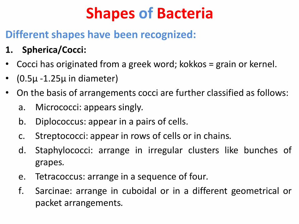
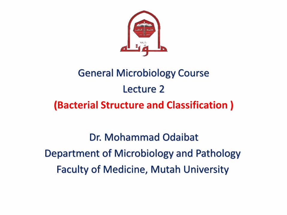
Shapes of BacteriaDifferent shapes have been recognized:
1. Spherica/Cocci:
• Cocci has originated from a greek word; kokkos = grain or kernel.
• (0.5µ -1.25µ in diameter)
• On the basis of arrangements cocci are further classified as follows:
a. Micrococci: appears singly.
b. Diplococcus: appear in a pairs of cells.
c. Streptococci: appear in rows of cells or in chains.
d. Staphylococci: arrange in irregular clusters like bunches ofgrapes.
e. Tetracoccus: arrange in a sequence of four.
f. Sarcinae: arrange in cuboidal or in a different geometrical orpacket arrangements.
Shapes of Bacteria

2. Rod Shaped Bacteria or Bacillus:
• From greek word, bacilli means rod orstick.
• Their ends are rounded flat or pointed.
• 0.5-1.2µ in diameter and 3-7µ in length.
• Flagellated or non-flagellated.
• Types:
Monobacillus: arrange singly.
Diplobacillus: present in a group of two.
Streptobacillus : in chains.
Palisade: Very rarely the bacillusarrange in a palisade arrangement.
Shapes of Bacteria
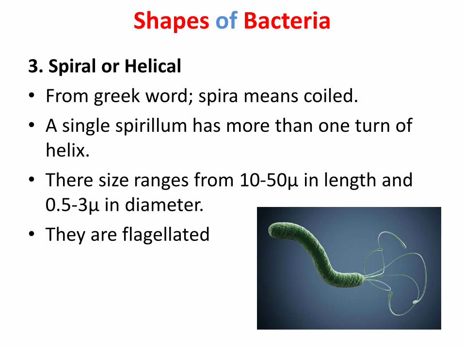
3. Spiral or Helical
• From greek word; spira means coiled.
• A single spirillum has more than one turn of helix.
• There size ranges from 10-50µ in length and 0.5-3µ in diameter.
• They are flagellated
Shapes of Bacteria

4. Vibrio or Coma:
• They bear flagella at their end.
• Their size ranges from 1.5-1.7µ in diameter and upto 10µ in length
• e.g. Vibrio cholarae.
Shapes of Bacteria

5. Spirochaeta:
• These bacteria appears like a cork screw and atrichous.
• Their length is more as compared to their diameter.
• Their body is more flexible.
Shapes of Bacteria
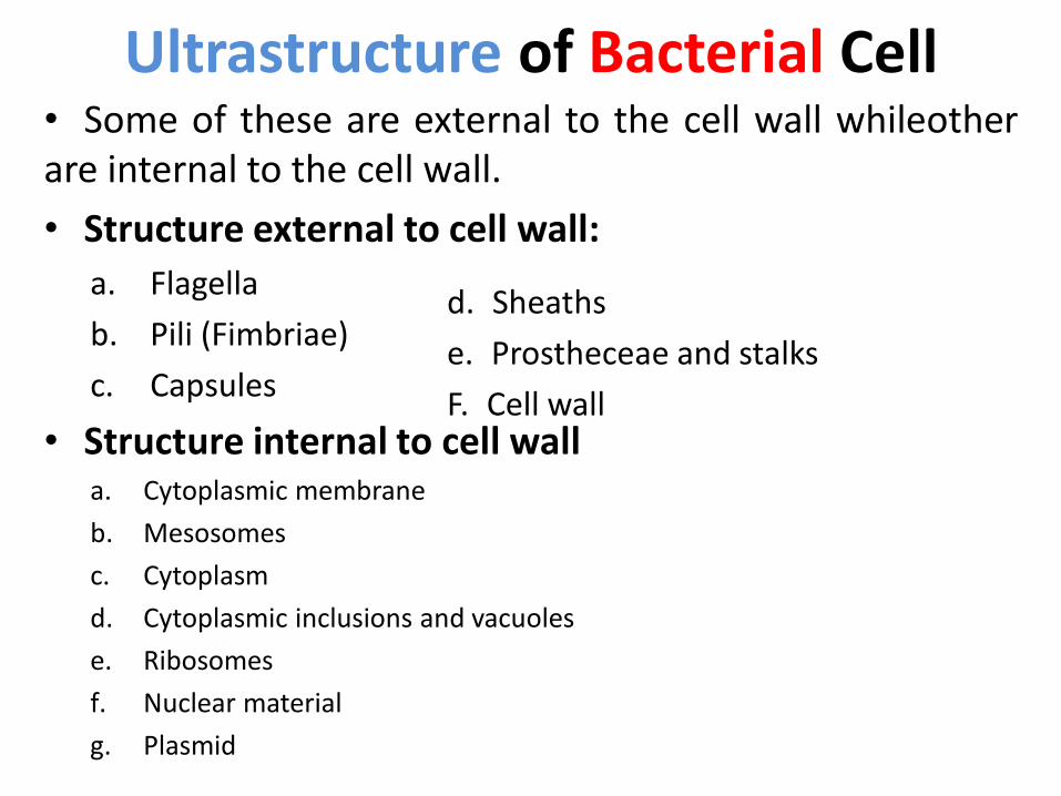
Ultrastructure of Bacterial Cell• Some of these are external to the cell wall whileotherare internal to the cell wall.
• Structure external to cell wall:
a. Flagella
b. Pili (Fimbriae)
c. Capsules
• Structure internal to cell walla. Cytoplasmic membrane
b. Mesosomes
c. Cytoplasm
d. Cytoplasmic inclusions and vacuoles
e. Ribosomes
f. Nuclear material
g. Plasmid
d. Sheaths
e. Prostheceae and stalks
F. Cell wall

Ultrastructure of Bacterial Cell
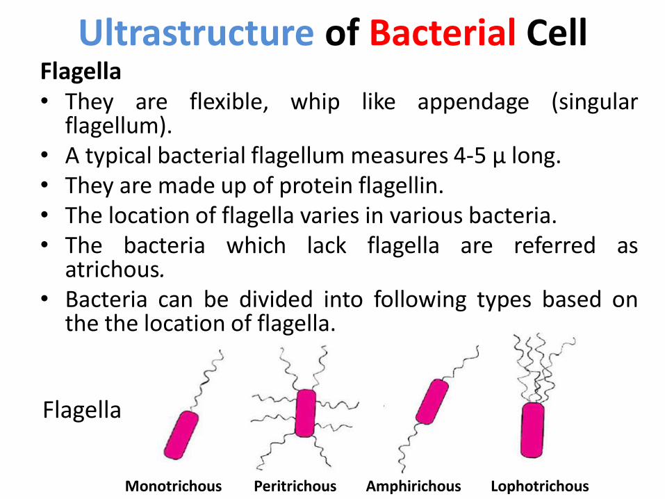
Flagella• They are flexible, whip like appendage (singular
flagellum).• A typical bacterial flagellum measures 4-5 µ long.• They are made up of protein flagellin.• The location of flagella varies in various bacteria.• The bacteria which lack flagella are referred as
atrichous.• Bacteria can be divided into following types based on
the the location of flagella.
Monotrichous LophotrichousPeritrichous Amphirichous
Flagella
Ultrastructure of Bacterial Cell
Pili• These are hair like appendages present on the
surface of most of the gram negative bacteria.
• They are smaller than flagella, have no role inthe motility of bacteria.
• A single bacterial cells bears about 100-500 piliwhich are arranged peritrichously.
• They are composed of protein named pilin.
• Two types: Somatic pili and sex pili or conjugatepili
Somatic pili:• Each bacterial cell bears about 100 somatic pili.
• Function: is to help the bacterium for attachment to a substratum.
Sex Pili or Conjugate Pili :
• known as F pili.
• They are comparatively long (20 µ) and broad in width.
• There number ranges from 1-10 in male or donor bacterium.
• Male donor (+ factors) or female receptor/ receiver (- factor).
• The sex pili of male donor recognize the receptor protein on thesurface of female or recipient.
Pili
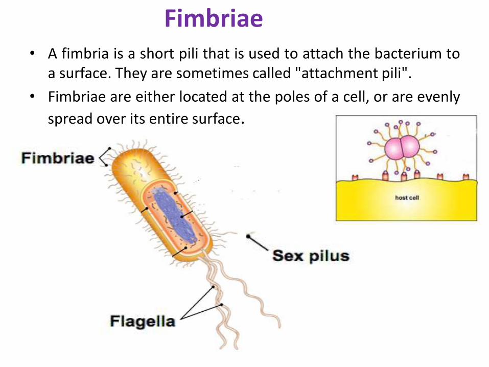
• A fimbria is a short pili that is used to attach the bacterium toa surface. They are sometimes called "attachment pili".
• Fimbriae are either located at the poles of a cell, or are evenly
spread over its entire surface.
Fimbriae
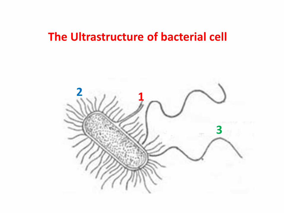
The Ultrastructure of bacterial cell
12
3

Is a network of di- or polysaccharide or polypeptidesforming a covering layer around the bacterial cell wall.
• Functions They provide protection against temporary drying by
binding water molecules.
They may be antiphagocytic i.e. they inhibit theengulfment of pathogenic bacteria by white blood cells
Capsule
capsule Bacterial cell

History• The Gram stain was first used in 1884 by the Danish
scientist Hans Christian Gram (Gram,1884).
• Gram was searching for a method that would allowvisualization of bacteria in tissue sections of lungs ofthose who had died of pneumonia.
• He did this with both Streptococcus pneumoniae andKlebsiella pneumoniae bacteria, observing thatStreptococcus pneumoniae retained the stain afterwashing with alcohol whereas Klebsiella pneumoniaedid not.
The Cell Wall

German pathologist Carl
Weigert (1845- 1904)
History of Gram Staining
Danish scientist Hans
Christian Gram (1853–1938)
S. pneumoniae K. pneumoniae
S. pneumoniae K. pneumoniae
The Cell Wall1. Located below the external structures and above to the
cytoplasmic membrane is the cell wall.
2. Function:
a) Very rigid structure and provide definite shape to the cell.
b) Prevent the cell from expanding and bursting due to thehypotonic environment that the bacteria live in.
3. It may account for as such 10-40% of the dry weight ofbacterial cell.
4. Generally the cell wall is made up of large number of layers.
5. The thickness of these different layers varies both in gram+ve and gram -ve bacteria.

The bacterial cell wall
Gram-Negative Versus Gram-Positive Cell Walls
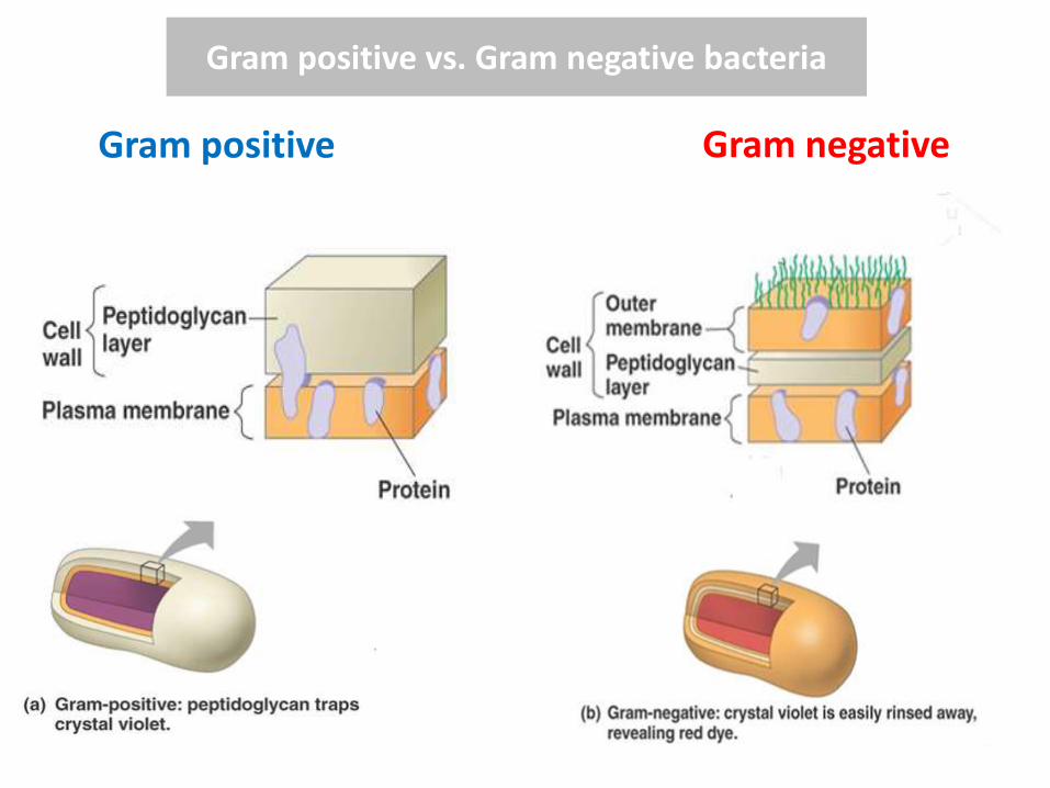
Gram positive vs. Gram negative bacteria
Gram positive Gram negative
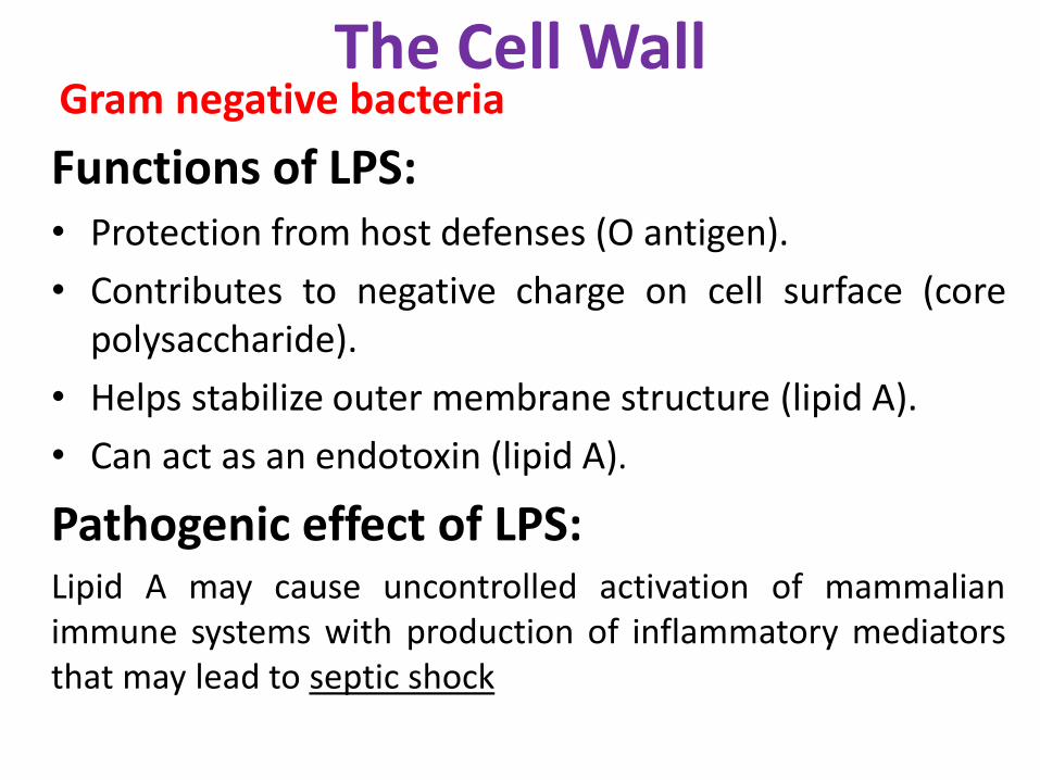
Functions of LPS:• Protection from host defenses (O antigen).
• Contributes to negative charge on cell surface (corepolysaccharide).
• Helps stabilize outer membrane structure (lipid A).
• Can act as an endotoxin (lipid A).
Pathogenic effect of LPS:Lipid A may cause uncontrolled activation of mammalianimmune systems with production of inflammatory mediatorsthat may lead to septic shock
The Cell WallGram negative bacteria
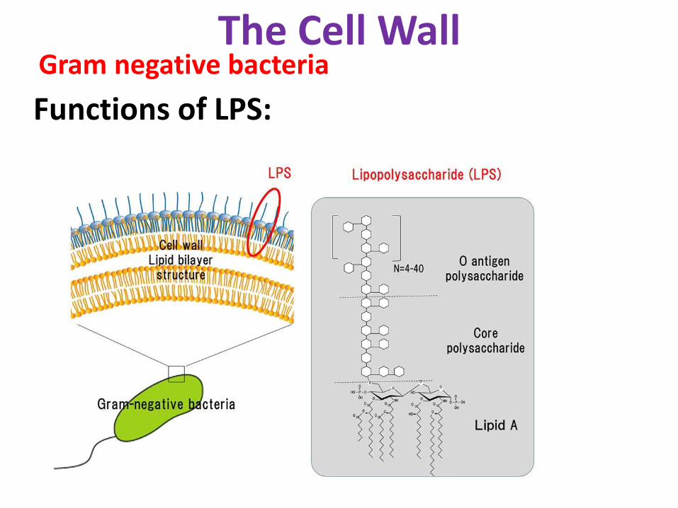
Functions of LPS:
The Cell WallGram negative bacteria


Structure Internal to Cell Wall

Ultrastructure of Bacterial Cell• Some of these are external to the cell wall whileotherare internal to the cell wall.
• Structure external to cell wall:
a. Flagella
b. Pili (Fimbriae)
c. Capsules
• Structure internal to cell walla. Cytoplasmic membrane
b. Mesosomes
c. Cytoplasm
d. Cytoplasmic inclusions and vacuoles
e. Ribosomes
f. Nuclear material
g. Plasmid
d. Sheaths
e. Prostheceae and stalks
F. Cell wall

Inclusion Bodies:
• Granules of organic or inorganic material thatare stocked by the cell for future use.
• Some are enclosed by a singlelayeredmembrane – membranes vary in composition –some made of proteins; others contain lipids
Structure Internal to Cell Wall

Inclusion Bodies
Inclusion Composition Function
Glycogen poly-glucose Reserve carbon andenergy source
Poly-betahydroxybutyricacid (PHB)
lipid Reserve carbon andenergy source
Poly-phosphates polymers of PO4 Reserve phosphate,possibly high-energy PO4
Sulfur globules elemental S Reserve energy and orelectrons
Magnetosomes magnetite (iron oxide) Provide orientation inmagnetic field
Gas vesicles protein shells inflatedwith gases
Provide buoyancy inaquatic environments
Parasporal crystals protein Produced by endospore-forming Bacilli - toxic toinsects
Structure Internal to Cell Wall
1. Hierarchical classification
2. Shapes and Forms of Bacteria
3. Physiology
4. Molecular techniques: DNA , RNA, and protein analysis
Classification of Bacteria
Different methods are used to Classify bacteria:
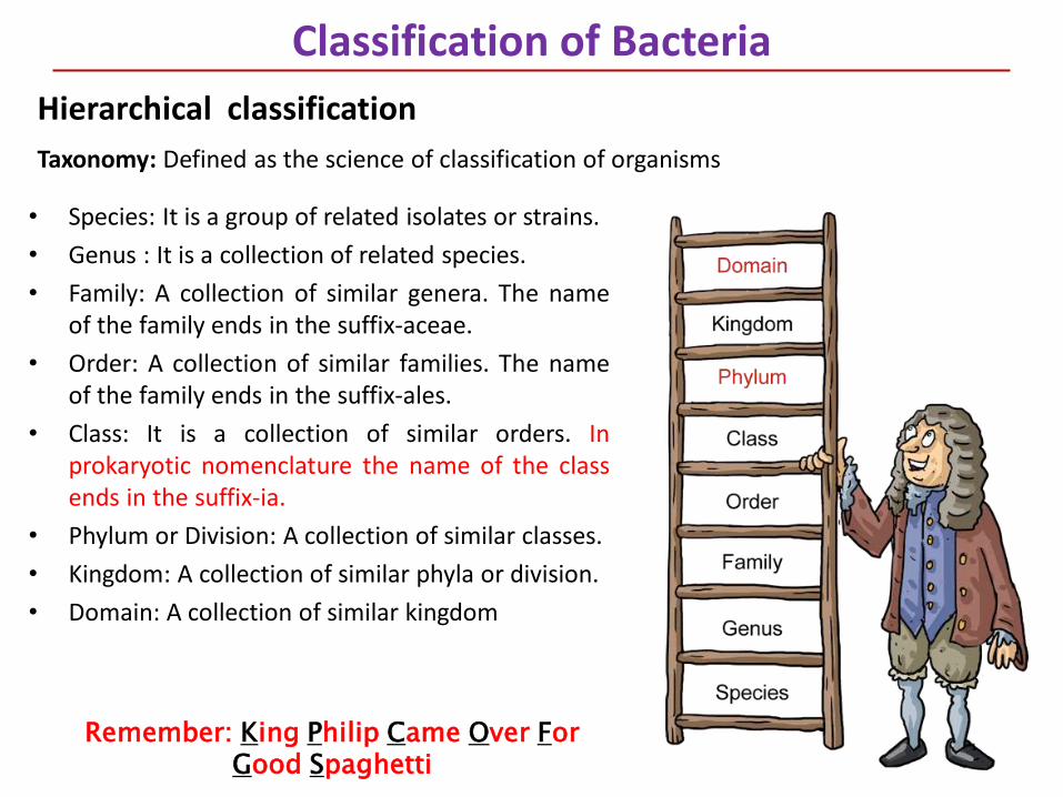
• Species: It is a group of related isolates or strains.
• Genus : It is a collection of related species.
• Family: A collection of similar genera. The nameof the family ends in the suffix-aceae.
• Order: A collection of similar families. The nameof the family ends in the suffix-ales.
• Class: It is a collection of similar orders. Inprokaryotic nomenclature the name of the classends in the suffix-ia.
• Phylum or Division: A collection of similar classes.
• Kingdom: A collection of similar phyla or division.
• Domain: A collection of similar kingdom
Classification of Bacteria
Hierarchical classification
Taxonomy: Defined as the science of classification of organisms
Remember: King Philip Came Over For Good Spaghetti
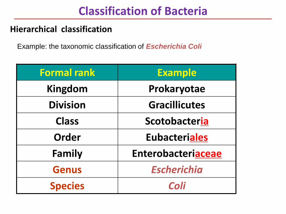
Formal rank Example
Kingdom Prokaryotae
Division Gracillicutes
Class Scotobacteria
Order Eubacteriales
Family Enterobacteriaceae
Genus Escherichia
Species Coli
Classification of Bacteria
Hierarchical classification
Example: the taxonomic classification of Escherichia Coli
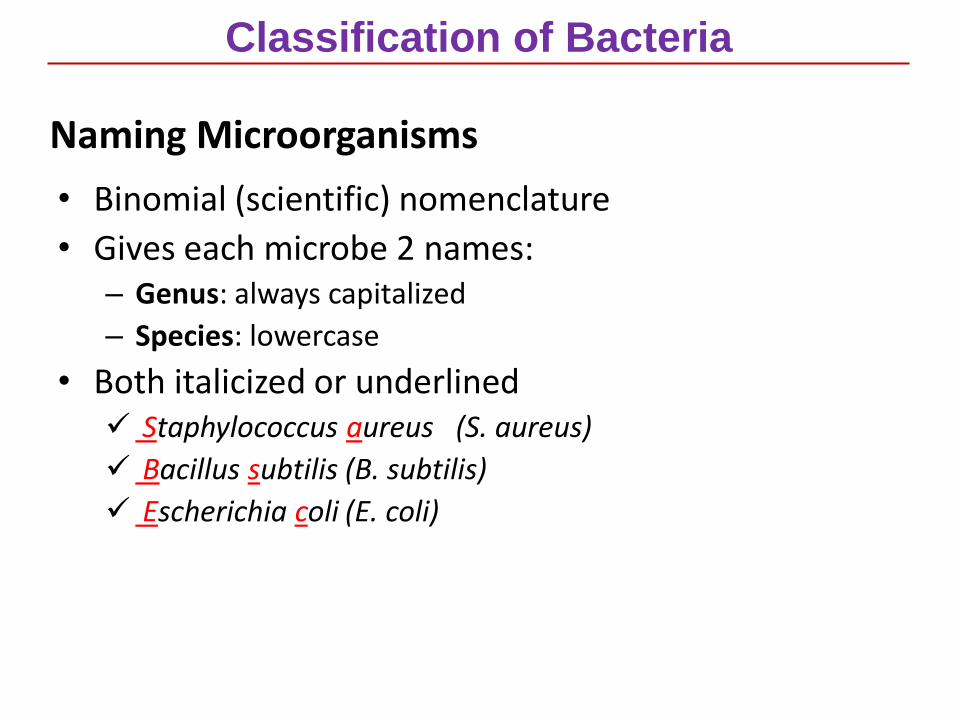
Naming Microorganisms
• Binomial (scientific) nomenclature
• Gives each microbe 2 names:– Genus: always capitalized
– Species: lowercase
• Both italicized or underlined Staphylococcus aureus (S. aureus)
Bacillus subtilis (B. subtilis)
Escherichia coli (E. coli)
Classification of Bacteria
Top Related