


Languages
Pages
Legal

ADDIS ABABA UNIVERSITY
COLLEGE OF HEALTH SCIENCES
SCHOOL OF ALLIED HEALTH SCIENCE
DEPARTMENT OF MEDICAL LABORATORY SCIENCES
EVALUATIONS OF SUB-CHRONIC TOXICITY OF HYDRO-ETHANOLIC SEED
EXTRACTS OF ALBIZIA GUMMIFERA AND MILLETTIA FERRUGINEA ON BLOOD,
HEART AND SMALL INTESTINE OF ALBINO WISTAR RATS
By:
Molla Getu, BSc
Advisors: Aster Tsegaye, MSc, PhD
Co-advisors: Melaku Tamene, MSc, PhD candidate
Bekure Tsegaye, MD, Pathologist
Asfaw Debella, MSc, PhD
Mekbeb Afework, MSc, PhD
A thesis submitted to the Department of Medical Laboratory Sciences, School of Allied Health Science, College of Health Science, Addis Ababa University, in partial fulfillment of Master of Science Degree in Clinical Laboratory Sciences (Hematology and Immunohematology)
Oct, 2016
Addis Ababa, Ethiopia

i
Addis Ababa University
School of Graduate Studies
This is to certify that the thesis prepared by Molla Getu, entitled:
Evaluations of the sub-chronic toxicity of hydro-ethanolic seed extracts of Albizia gummifera
and Millettia ferruginea on blood, heart and small intestine of albino wistar rats submitted in
partial fulfilment of the requirements for Master of Science Degree in Clinical Laboratory
Sciences (Hematology and Immunohematology) complies with the regulations of the University
and meets the accepted standards with respect to originality and quality.
Signed by the Examining Committee:
Examiner__________________________ Signature ___________Date ____________
Examiner__________________________ Signature ___________Date ____________
Advisor___________________________ Signature ___________Date ____________
Advisor___________________________ Signature ___________Date ____________
Advisor___________________________ Signature ___________Date ____________
Chairman of the Department or Graduate Program Coordinator
ii
Acknowledgements
First, I would like to praise Almighty God for all the strength He has given me throughout
my life. I would like to express my sincere thanks to my principal advisor Dr. Aster Tsegaye for
her continuous follow-up, guidance, suggestion and constructive comments given
throughout the work.
My gratitude goes to my advisor Mr. Melaku Tamene who has spent his precious time in
advising and correcting on my draft proposal and thesis.
Genuine gratitude goes to my advisors Dr. Asfaw Debella, Directorate of Traditional and
Modern Medicine Department, EPHI for insightful guidance during collection, extraction
and administration, and also for their constructive comments on the thesis paper.
I am very much grateful to my advisor Dr. Bekure Tsegaye, Pathologist, and Dr. Tufa
Gemechu, pathologist for thoroughly analyzing the slides and giving me their comments
on the histopathological changes of the tissue sections. I am also indebted to Dr. Mekbeb
Afework, Histologist head Department of Anatomy, for assisting in the investigation of
histological slides and Mr. Jemal Alemu and Mr. Kassu Desta, Ass. Professor, for
encouraging me throughout the study period.
I wish to extend my thanks to AAU for providing financial assistance that was useful for
the successful accomplishment of this study. I would like to acknowledge EPHI, because
this work was supported by a grant from EPHI. I would like to thank Addis Ababa
University College of Health Science School of Medicinefor sponsoring me to pursue
postgraduate study.
My heartfelt thanks to Ato Negaro Gemmeda, Ato Bekasho Geleta, Ato Abiy Abebe, W/t
Keristena Haile, Ato Yewalshet Belete, Ato Birhanu Tesfaye, Ato Birhanu Asaye, Ato
Kissi Mude, W/t Yewubidar Haile of EPHI, Ato Endris Aragaw, Kidanu Fikadu and
Mohammed Aragaw of AAU, department of anatomy, for facilitating a good working
environment throughout my study period.Credit must also be given to my beloved
families, friends and colleagues.
The last but not least, my sincere and deepest gratitude forwards to the School of Medical
Laboratory Science and the whole staffs.
iii
Table of contents pages
Ackowledgment……………………………………………………………………………………ii
Contents…………………………………………………………………………………….……..iii
List of tables…………………………………………………………………………………….....vi
List of figures………………………………………………………..………………...……..…..viii
List of abbreviations………………………………………………………………………..…..….ix
Abstract…………………………………………………………………………………………….x
1. INTRODUCTION………………………………………………………………………….…..1
1.1. Background………………………………………………………………..………………1
1.1.1. Bioactive Ingredients of A. gummifera and M. ferruginea…………...……………3
1.1.2. Blood composition and function …………………..………………………………4
1.1.3. Heart structure and function ……….…………………………................................6
1.1.4. Small intestine structure and function…………………...…………………………6
1.2. Statement of the problem……………………………………………………...........……..7
1.3. Significance of the study…………………………………………………………………..7
2. LITRATURE REVIEW………………………………………………………………....……..8
2.1. Blood and biochemical markers…………………………………………………………...8
2.2. Histopathology of the heart……………………………………………………………......9
2.3. Histopathology of the small intestine………………………………………………….......9
3. OBJECTIVES……………………………………………………………………..………….10
3.1. General objective………………………………………………………………….....…..10
3.2. Specific objectives………………………………………………………………….....…10
4. MATERIALS AND METHODS……………………………………………………………..11
4.1. Study place……………………………………………………………………………….11
4.2. Study design and period…………………………………………………………….……11
4.3. Study population…………………………………………………………………....……11
iv
4.4. Sample size and sampling technique…………………………...…………………..…….11
4.4.1. Sample size………………………………………………………………………..11
4.4.2. Sampling technique……………………………………………………………….11
4.5. Data collection……………………………………………………………………......….11
4.5.1. Plant collection and processing………………………………………………...…11
4.5.2. Plant material extraction…………………………………………………………..12
4.5.3. Experimental animals……………………………………………………………..12
4.6. Acute toxicity test of A. gummifera and M. ferruginea seed extract in rats……...……...12
4.7. Sub-chronic toxicity test of A. gummifera and M. ferruginea seed extract in rats………13
4.7.1. Specimen collection……………………………………………………………....14
4.7.2. Hematological and biochemical analysis…………………………………………14
4.7.3. Histopathological studies……………………………………………………...….15
4.7.3.1. Tissue processing……………………………………………...……………..15
4.7.3.2. Tissue staining…………………………………………...………….………..16
4.7.3.3. Microscopy and photomicrography……………………………………...…...16
4.8. Statistical analysis………………………………………………………………….…….16
4.9. Ethical consideration……………………………………………………………………..17
4.10. Data quality assurance……………………………………………………………....…..17
5. RESULTS…………………………………………………………………………...………...18
5.1. Acute toxicity study…………………………………………………………………..….18
5.2. Sub-chronic toxicity study……………………………………………………………….18
5.2.1. Effect of the extract of A. gummifera and M. ferruginea on health and body
weight......................................................................................................................18
5.2.2. Effect of the extract of A. gummifera and M. ferruginea hydroalcohol seed extract
on hematological parameters………………………....……………………….…..24
5.2.3. Effect of the extract of A. gummifera and M. ferruginea hydroalcohol seed extract
on hematological biochemical parameters……………………………….……….28
5.2.4. Macroscopic observations and organ weight………….………………………….31
5.2.5. Microscopic observations……………………………………………………...….33
v
6. DISCUSSION……………………………………………………………………...…………39
7. CONCLUSSION AND RECOMMENDATIONS………………………………………...….45
7.1. Conclusion ……………………………………………………………………………….45
7.2. Recommendations …………………………………………………………………...…..46
8. STRENGTH AND LIMITTATIONS OF THE STUDY……..……………………..…..........46
8.1. Strength …………………………………………………………………………….……46
8.2. Limitations ………………………………………………………………………………46
9. REFERENCES………………………………………………………………………………..47
10. APPENDIX…………………………………………………………………………………...54
10.1. Appendix I: Preparations of working solutions……………...………………………….54
10.2. Appendix II: Tissue processing schedules for paraffin blocks for manual technique…..55
10.3. Appendix III: Routine hematoxylin and eosin (H and E) staining schedule for tissue
sections………………………………………………………………………………….56
10.4. Appendix IV: Routine procedures of CELL-DYn 3700 hematology analyzer………....56

vi
List of Tables page
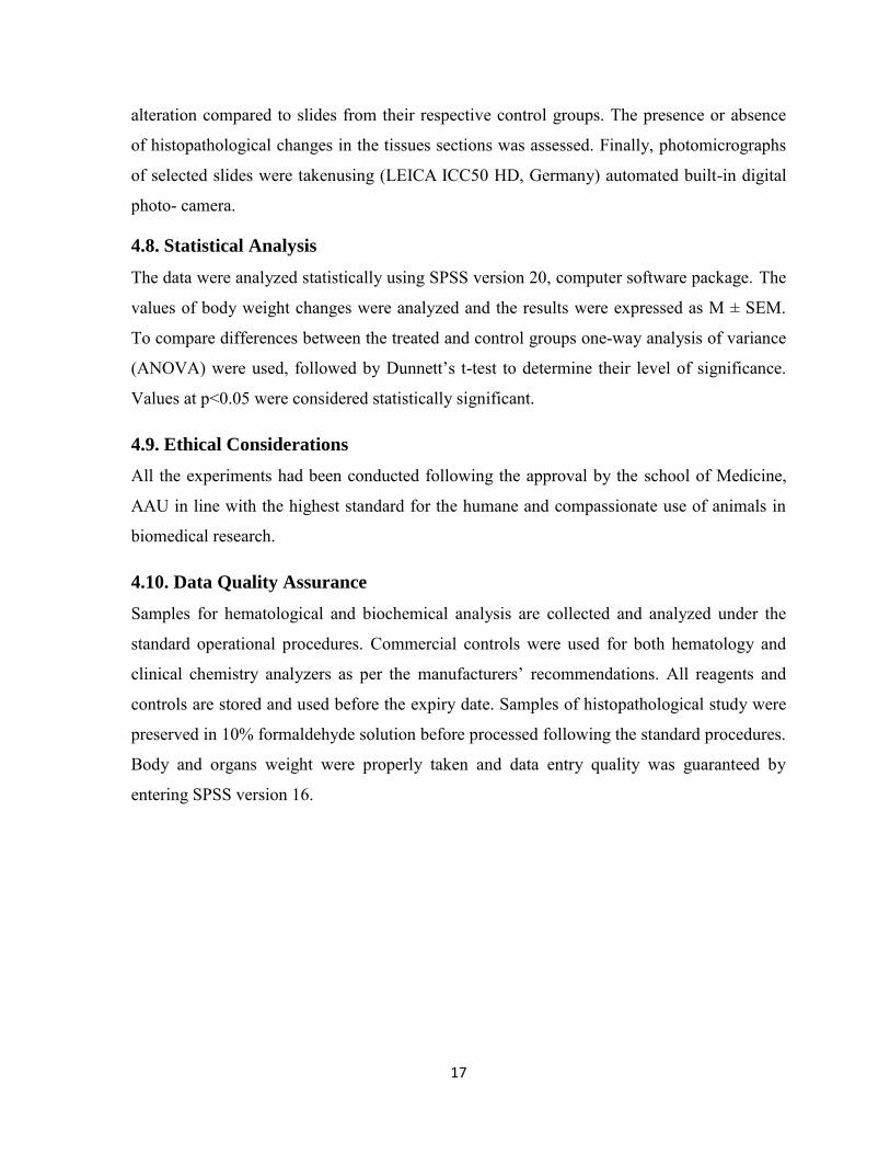
Table 1: Effect of the sub-chronic adminstration of A. gummifera seed extract on the body weight
of male and female rats……………………………………………………………............21
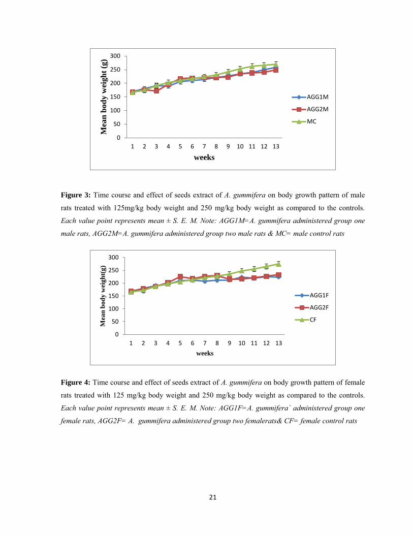
Table 2: Effect of seed extract of M. ferruginea on the body weight of male and female during
the 13th week of administration…………………………………………..………………..……22
Table 3: Hematological parameters of male rats treated with 125 and 250 mg/kg body weight of
seed extract of A. gummifera for 13 weeks……………………………….………………………25
Table 4: Hematological parameters of female rats treated with 125 and 250 mg/kg body weight of
seed extract of A. gummifera for 13 weeks………………………………….………………….26
Table 5: Hematological parameters of male rats treated with 125 and 250 mg/kg body weight of
seed extract of M. ferruginea for 13 weeks……………………………………………………….27
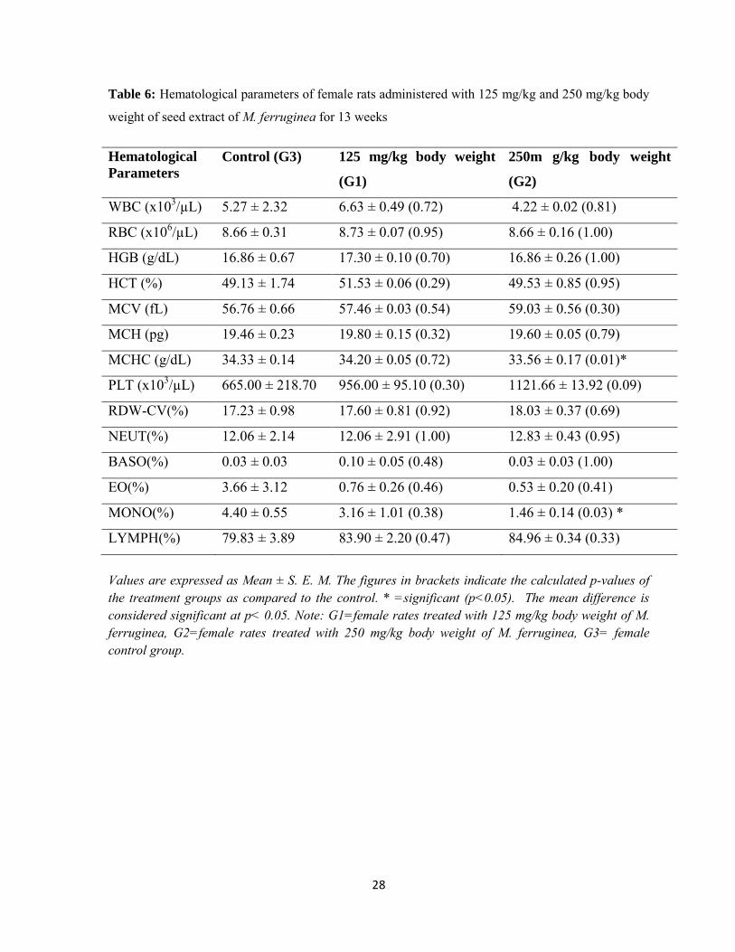
Table 6: Hematological parameters of female rats treated with 125 and 250 mg/kg body weight of
seed extract of M. ferruginea for 13 weeks……………………………………………………….28
Table 7: serum biochemical parameters of male rats treated with 125 and 250 mg/kg body weight
of seed extract of A. gummifera for 13 weeks……………………...…………….………………29
Table 8: Serum biochemical parameters of female rats treated with 125 and 250 mg/kg body
weight of seed extract of A. gummifera for 13 weeks……………………………...…………….30
Table 9: Serum biochemical parameters of male rats treated with 125 and 250 mg/kg body weight
of seed extract of M. ferruginea for 13 weeks……………………………………….………....30
Table 10: Serum biochemical parameters of female rats treated with 125 and 250 mg/kg body
weight of seed extract of M.ferruginea for 13 weeks………………………………………...…...31
Table 11: Organ weights of male rats treated with 125 &250 mg/kg body weight doses of the
seed extracts of A.gummifera………………………………………………………..…..………..32
vii
Table 12: Organ weights of female rats treated with 125 &250 mg/kg body weight doses of the
seed extracts of A. gummifera…………………………………………………………………….32
Table 13: Organ weights of male rats treated with 125 & 250 mg/kg body weight doses of the
seed extracts of M. ferruginea…………………………………………………………………….32
Table 14: Organ weights of female rats treated with 125 & 250 mg/kg body weight doses of the
seed extracts of M. ferruginea ………………………………….…………………………33
viii
List of Figure Page
Figure 1: Photograph of A. gummifera showing the leaves and its pods……………….....………2
Figure 2: Photograph of seeds of M. ferruginea……………………………...….………………..3
Figure 3:Time course and dose effect of seed extract of A. gummifera on body growth pattern of male rats……………………………………………………………………………………….….21
Figure 4: Time course and dose effect of seed extract of A. gummifera on body growth pattern of female rats…………………………………………………………………………………..….21
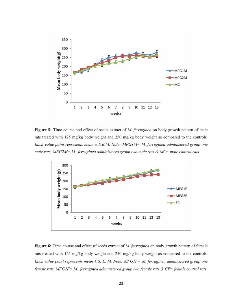
Figure 5: Time course and dose effect of seed extract of M. ferruginea on body growth pattern of male rats…………………………………………………………………….………………….23
Figure 6: Time course and dose effect of seed extract of M. ferruginea on body growth pattern of female rats……………………......…………………………………………………………….23
Figure 7: Photo graph of H and E stained Heart sections from rats administered with hydroethanolic seed extracts of A. gummifera………………...…………………………………34
Figure 8: Photograph of H and E stained Heart sections from rats administered with hydroethanolic seed extracts of M. ferruginea………………..…………………………………35
Figure 9: Photograph of H and E stained small intestine sections from rats administered with hydroethanolic seed extracts of A. gummifera…………………………………...………………37
Figure 10: Photograph of H and E stained small intestine sections from rats administered with hydroethanolic seed extracts of M. ferruginea……………………………………………..……38
ix
List of abbreviations
ALP ------------------------------------Alkaline phosphatase
ALT--------------------------------------Alanine aminotransferase
AST--------------------------------------Aspartate aminotransferase
BASO ----------------------------------Basophils
CCK--------------------------------------Cholecystokinin
CK----------------------------------------Creatin kinase
EOS -----------------------------------Eosinophils
EPHI-------------------------------------Ethiopian Public Health Institute
FC----------------------------------------Female rats control group
HCT--------------------------------------Hematocrit
HGB -------------------------------------Hemoglobin
LD50--------------------------------------Lethal dose for 50 percent of the population
LYMPH ---------------------------------Lymphocytes
MC----------------------------------------Male rats control group
MCH -------------------------------------Mean corpuscular hemoglobin
MCHC -----------------------------------Mean corpuscular hemoglobin concentration
MCV--------------------------------------Mean cell volume
PCV----------------------------------------Packed cell volume
PLT ----------------------------------------Platelets
RBC----------------------------------------Red blood cells
RDW-CV --------------------------------Red cell distribution width
SEM ---------------------------------------Standard error of the mean
SPSS --------------------------------------Statistical package for social sciences

x
ABSTRACT
Background: In Ethiopia there are large numbers of plants having medicinal values; among them are
Albizia gummifera and Millettia ferruginia. The effect of seeds extracts of these plants on blood
parameters and organs such as heart and small intestine is not fully investigated.
Objective: This study aimed to evaluate the sub-chronic toxic effects of hydro-ethanolic (70%) seed
extracts of A. gummifera and M. ferruginea on the heart, small intestine and on some blood parameters in
albino wistar rats.
Method: Seeds were collected from different areas of Ethiopia. They were dried and crushed to powder
and macerated with hydro-alcohol and placed in orbital shaker. The extract was then filtered, evaporated to
dryness by Rota vapor and further concentrated by water bath at 40oc. The gummy residue extract was
weighed and packed in air tight brown glass bottles with proper label and kept at 4Oc. The sample size for
the study was 158 rats.To determine the LD50 of the extract, 108 female rats were treated with different
doses of the extracts. For the sub-chronic toxicity study, 50 rats (25 male and 25 female) were
administered at doses of 125 mg/kg/day and 250 mg/kg/day for 90 days. Hematological analysis was
performed usingautomatic hematology analyzer, cell-DYN-3700 (Abbott Diagnostic Division, USA) and
clinical chemistry analyzer, Human star 80 (Human GmbH, Germany) was used to determine biochemical
parametrs. Data was cleaned, entered and analyzed using SPSS 16.Finally, the animals were quickly
dissected. Heart and small intestine were carefully removed. Portions of these organs were fixed in 10%
formalin for histopathological examination.
Result: The LD50 of A. gummifera and M. ferrugina were found to be 4000 mg/kg and 3500 mg/kg,
respectively. The seed extract of A. gummifera decreased the MCH (P<0.05) in the male rats at 125 mg/kg
and 250 mg/kg doses; MCHC at both doses in the female rats; and MCH in the female rats at higher dose
but increased RDW-CV in the male rats at all doses. Furthermore, it increased neutrophil at the highest
dose in both female and male. The seed extract of M. ferruginea decreased the MCHC and monocyte (p <
0.05) of female rats at the highest doses. But CK, ALP, ALT and Urea were significantly elevated in
female rats administered with 250 mg/kg of M. ferruginea seed extract. Whereas, seed extract of A.
gummifera increased CK and urea in male rats at 250 mg/kg. Some histopathological changes in heart and
small intestine were also observed for both plants extracts.
Conclusion:This study suggests that 70% hydro-ethanolic seed extracts of A. gummifera and M.
ferruginea may slightly induce anemia and inflammation, maybe phytotoxic to the heart, liver and kidneys
that resulted in a rise in serumCK, ALP, ALT and Urea at high dose and cardiac and small intestinal tissue
damage at all doses. Further studies would be needed to isolate the active ingredients which cause the
toxicities.
Key words: A. gummifera, M. ferruginea, Seed, hydroethanolic extract, Toxicity, Wistar Albino Rats.

xi

1
1. INTRODUCTION
1.1. Background
Before the beginning of scientific medicine, with all the limitations like being toxic to blood
cells and other organs, traditional cultures around the world employed the use of medicinal
plants as disease remedies for centuries [1].
Global traditional medicine usage is widespread and growing in various parts of the
developing world such as Africa, China, India, Japan and Latin America in which about 80%,
40%, 65%, 60‐70% and 40%-71% respectively of their population use traditional medicine to
meet their primary health care needs [2] since they are often more available and affordable
than western medicine. To this effect, there are researches on toxicological effects of
different medicinal plant extract on blood and different organs of administered animals so as
to be cautious not to use them as a medicine unless their safety is proved by appropriate
scientific evidences [3-6].
In Africa, more than 2,000 plants have been identified and used as remedies to treat several
ailments, but very few of these plants have been screened for their safety [7].
Ethiopia has a long history of using plants as traditional medicine, but knowledge about the
extent and characteristics of traditional healing practices is limited and it has even been
ignored in the national health care system [8]. The folk knowledge and traditions of Ethiopia
utilize the herbal resources available in nature. This knowledge is transferred from generation
to generation orally as guarded secrets [9]. Albizia gummifera and Millettia ferruginea are
among the many plants used for traditional medicine in Ethiopia.
One of the plants used as traditional medicine in Ethiopia is A. gummifera (peacock flower-
in English, Sessa- in Amharic, Ambabesamuka- in Oromifa). It belongs to the family
Leguminosae, and a sub family Mimosaideae. A. gummifera is a large deciduous tree with
flattened canopy, growing up to 35m high and trunk up to 75cm in diameter. It is found in
east Africa, the Democratic Republic of Congo, Madagascar, and West Africa, ranging from
dry or wet lowlands to upland forest edges, and in riverine forest, at an altitude of 2400m
above sea level. It is indigenous in few countries namely; Angola, Cameroon, Democratic

2
Republic of Congo, Ethiopia, Kenya, Madagascar, Nigeria, Tanzania, Uganda, and Zambia.
It is, however, exotic in Brazil [10].The picture of the plant is shown in figure1.
Figure 1: Photograph of A. gummifera showing the
leaves and its pods
Various parts of A. gummifera are used in traditional medicine. Its bark is used to treat
malaria, head ache, scabies, and psoriasis. It also hastens partrusion. Its root is used to treat
pain, skin diseases, diarrhea, eye diseases and sleeping sickness. Their leaf is used in the
treatment of diarrhea, eye troubles, asthma, sores, and fractures. An extract of crushed pods
is drunk to treat stomach-ache [11].
A. gummifera has shown effects against different bacteria at different gradient of dilution
[12]. It has also shown potential molluscidal activities against Biomphalaria pfeifferi, Bulinus
sp. and physaacuta [13]. The seed extract of A. gummifera shows larvicidal activities against
Aedes agypti, Aedes africanus, and Culex quinque fasciatus [14]. Furthermore, crude hydro-
alcoholic (20-80 %) extracts of A. gummifera was effective against reference strain of N.
gonorrhoeae [15].
M. ferruginea (birbira- in Amharic, sotallo- in Oromiffa, sari- in OromiffaArsi, Yego- in
OromifaHarar, Enghediksho- in Sidama, Zaghia- in Wallayita), belongs to the family
Leguminosae and a sub family papilionoideae.It is an indigenous plant species found only in
Ethiopia. There are two sub-species known to be found in this country. These are: M.
ferruginea which is confined to the northern part of the country and M. darasana which is
found in southern provinces, particularly Sidama region. The tree is umbrella-shaped or

3
flattened at top, and grows up to a height of 25-35m [16, 17]. The picture of the plant seeds is
shown in figure 2.
Figure 2: Photograph of seeds of M. ferruginea
The seed extract of M. ferruginea showed promising larvicidal activities against Aedes
agypti, Aedes africanus and Culexquinque fasciatus [14]. Traditionally, bark and mature fruit
and seeds of M. ferruginea are used as fishing poison [18]. The fruits, leaves, seeds and stem
decoction of M. ferruginea are used for the treatments of pain, earache & bacterial infection
of nails, insecticidal properties and toothaches respectively [18]. Furthermore,
Millettiaferruginea leaf has anti bacteria effect [19]. Acute toxicity studies of these plants on
mice showed medium lethal dose values ranging from 150 mg/kg- 450 mg/kg when the
aqueous extracts were administered intraperitonealy [13, 14]. Several toxicity studies on
blood and organs using animal models are being carried out to strengthen the body of
knowledge and ensure their safe use [3-6].
1.1.1 Bioactive Ingredients of A. gummifera and M. ferruginea
The constituents of a given plant species determines its therapeutic effects.
Phytochemicalstudies have shown the presence of various bioactive ingredients in some
traditionally used plant extracts, which are responsible for their medicinal uses. Aqueous
seed extracts of A. gummifera contain chemical constituents such as alkaloids, polyphenols,
unsaturated sterol/or triterpens, saponins, glycosides and carbohydrates [13]. Pytochemical
screening showed that aqueous seed extracts of M. ferruginea contain chemical constituents
such as polyphenols, tannins, unsaturated sterol/or triterpens, glycosides and carbohydrates
[13].

4
1.1. 2. Blood Composition and Functions
The two components which are formed elements and plasma make the blood. As liquid
connective tissue, blood transports many substances through the body and helps to maintain
homeostasis of nutrients, wastes and gases. In humans the relative volume of blood cells and
plasma in whole blood is approximately 45% and 55%, respectively. Total blood volume in
the average adult person is about 5 to 6 L or 7% to 8% of the total body weight [20].
Whereas, the mean blood volume for male rats was found to be 55.6µl/gm and the
corresponding value for female rats is 53.1µl/gm [21].
The erythrocytes make up about 45% of blood volume. An estimate of the volume of packed
erythrocytes per unit volume of blood (hematocrit) has various clinical applications [20, 22].
Erythrocytes transport oxygen in the blood through the red pigment hemoglobin.
Hemoglobin contains iron and proteins which increase the oxygen carrying capacity of
erythrocytes [23, 24].
The WBCs, also known as leukocytes, make up a very small percentage of the total number
of cells in the blood stream, but have important functions in body’s immune system. Based
on the presence of chemical filled vesicles in their cytoplasm that give them their function
leukocytes can be classified in to granulocytes(neutrophils, eosinophils, and basophils) and
agranulocytes (monocytes and lymphocytes). Neutrophils contain digestive enzymes that
neutralize bacteria during inflammation. Eosinophils contain digestive enzymes specialized
for digesting viruses that have been bound to by antibodies in the blood. Basophils release
histamine to intensify allergic reactions and help protect the body from parasites. During
inflammation, granular leukocytes leave the blood stream by migrating between the
endothelial cells of the venules and capillaries by a process known as diapedesis, and enter
the connective tissue spaces to perform their function [22, 23].
Lymphocytes include T cells and natural killer cells that fight off infection caused by viruses
and intracellular pathogens and B cells that produce antibodies against infections by
pathogens. Whereas, circulating monocytes undergo further maturation up on leaving the
vasculature and migrating into the various tissues and body cavities [25]. Inside body tissues,
monocytes are differentiated into macrophages [25].
5
Macrophages play role in immune system of the body. They engulf and ingest parasites,
microbes and the dead cells from wound and infections. They regulate lymphocyte activation
and proliferation and they are essential in the activation process of T- and B- lymphocytes by
antigens and allogenic cells.The mean age of the granulocyte is 8.7 to 9.4 days [26]. The
majority of the granulocytes enter the blood stream at an age of about 6 days. Less than 5 %
of the granulocytes in the blood stream are less than 5 days old, and a negligible percentage
is older than 3 weeks. That of lymphocytes form two groups; the Younger have a mean age
of about 3 to 4 days while the others have a mean age of about 100 to 200 days [27].
Megakayrocytes inside the red bone marrow periodically rupture and release thousands of
pieces of membrane or small cell fragments (Platelets or thrombocytes) responsible for the
clotting of blood and the formation of scabs. Platelets do not contain nucleus and only
survive in the body for up to a week before macrophage capture and digest them. Their
transport towards the vessel wall is influenced by the hematocrit, red blood cell (RBC) size,
and shape. They are the primary cells responsible for the control of bleeding and under
normal circumstances their activation in response to bleeding triggers the clotting process
[23, 28]. A study on the analysis of normal rat blood using KX-21 Symex hematological
analyzer showed a platelet value of 943.14 -946.96 ×10³/µL [29].
Plasma which is the non-cellular or liquid portion of the blood makes up 55% of the bloods
volume. Components of plasma include water, proteins, and dissolved substances. Water
makes 90% of plasma. The proteins within plasma include antibodies, albumin and other
plasma proteins. Antibodies which are part of the immune system bind to antigens on the
surface of pathogens that infect the body. Albumin helps maintain the body’s osmotic
balance by providing isotonic solution for cells of the body. Many different substances can
be found dissolved in the plasma, including glucose, oxygen, carbon dioxide, electrolytes,
nutrients, and cellular waste products. The plasma functions as transportation medium for
these substances as they move throughout the body [20].
6
1.1. 3 Heart: structure and Functions
The anatomical organization of human and rat hearts are almost similar. The heart of the rat
is a four chambered organ like other mammals. There is no normal communication between
the left and the right chambers. The heart is enclosed within a thin, transparent pericardium
that is attached to the major arteries and veins at the base of the heart [30].
The topographic features of the heart are similar to those of other mammals, with a clearly
defined atrio-ventricular groove separating the atria from the ventricles. Anterior and
posterior papillary muscles in the left ventricle projecting into the ventricular lumen anchor
the chordae tendineae of the mitral valve. The anterior papillary muscle of the rat is located
more laterally than that in the man. Papillary muscles in the ventricle are slender, elongated
structures varying from 2 to 5 or more in number [30].
The cardiac valves of rat are similar to those in other species. The aortic and pulmonary
valves have 3 leaflets; the mitral valve, anterior and posterior leaflets; and the tricuspid, a
posterior septal leaflet and 2 lateral leaflets on the free wall portion of the right ventricular
wall. There are multiple thin chordae tendineae connecting the mitral and tricuspid leaflets to
the papillary muscle [30].
1.1. 4 Small intestine: Structure and Functions
The small intestine of rats is made up of the duodenum, the jejunum and the ileum. The
duodenum, the first part of the small intestine occupies the right dorso-lateral part of the
abdominal cavity and had the largest diameter. The jejunum is located distal to the duodenum
and makes the longest segment of the small intestine. It can be differentiated from the
duodenum by its folding nature and is made up of many folds held together by mesentery.
The jejunum was related craniomedially to the caecum. The ileum extends distal to the
jejunum with the same diameter as the jejunum and only differed from the jejunum by the
absence of folds seen on the jejunum [31].
These three segments have many common histological features like the villi and some minor
structural differences. The duodenum has intestinal villi which were seen in the mucosa.
Intestinal glands (Brunner’s) are seen in the submucosa and make the major distinguishing
features observed in the duodenum. The jejunum has long leaflike villi, which are mucosa
7
projections with numerous intestinal glands (crypts of Lieberkuhn) that opened into pits
between the bases of the villi and penetrated the mucosa as far as the muscularis. The ileum
also contains villi and the intestinal glands around the mucosa. Mucosal associated lymphoid
tissues (Payers patches) are localized in the submucosa, while the tunica muscularis has
longitudinal muscles [32].
1.2 Statement of the problem
Despite the presence of solid scientific evidence with regard to the biological activities of
most of the natural products used in folk remedy, there is little information or evidence
available concerning the possible toxicity that medicinal plants may cause to the consumers
[11]. Nowadays, there is great concern by health authorities, pharmaceutical industries, and
consumers in relation to drug discovery and development because the need for safe and
effective drugs is increased by the general public. Because plants having medicinal value are
used by the societies for long period, consumers might assume that these plants have little or
no side effect. But there are studies which showed the adverse effect of medicinal plants
applied in traditional medicine [33]. Therefore, evaluating the toxicological effects of any
medicinal plant extract intended to be used in animals or humans is a crucial part of its
assessment for potential toxic effects.
1.3 Significance of the study
A. gummifera and M. ferruginea are widely used in the treatment of various health problems
but the issues of appropriate dosage and harmful side effects of these plants have not been
adequately researched. So far, no literature is available on histopathologic implications in
sub-chronic oral administration of the seeds extract of these two plants.
This study, therefore, will be an input for researchers, health authorities, pharmaceutical
industries, and consumers as it intends to investigate the histopathologic effect of the extract
on the blood, heart and small intestine.
8
2. Literature review
Traditional medicine is widely used to treat several ailments, and is often more available and
affordable than western medicine. But these traditional medicines are not without limitations;
several studies are conducted to assess the safety of these herbal medicines. Most of the
studies are able to demonstrate the toxicological effect of the herbal remedies on organs and
tissues of administered animals. This shows that herbal medicines should not be used as
medicine unless their safety is proved by appropriate scientific evidences [5, 6].
2.1 Blood and Biochemical markers
Blood offers important profile to study the toxicological impact on animal tissues. Different blood
parameters are often subjected to change depending upon stress condition and various other
environmental factors. Decrease or increase in certain blood parameters can be associated with the
nature of species and the toxicants in different studies. A study on the leaf extracts of A.
schimperiana against trypanosome congolense infection in mice showed that the extract
significantly prevent the hemolytic effect of the parasite by increasing the packed cell
volume (PCV) [34]. Another study done on the effect of ethanol leaf extracts of M.
aboensison hematological parameters of treated Wistar albino rats at the dose 3000-5000
mg/kg body weight showed significant increase in packed cell volume (PCV), hemoglobin
concentration of the cell, total white blood cell counts and platelets count compared to
control. Results from this study suggest that the ethanolic leaf extract of M. aboensis altered
the activities of the hemopoietic system [35].
Rats treated with aqueous leaf extract of Ocimum gratissimum also showed reduction of
platelet and lymphocyte counts and elevation of neutrophil and total white blood cell counts.
The result of this study provides caution not to consume quantities of O. gratissimum [29].
Measurement of blood biomedical parameters can be used as important diagnostic tool for
the detection of abnormalities in various body tissues and organs. Following tissue injury
biochemicals stored inside the cytoplasm of injured cells are released into the extracellular
spaces where they join the circulation. So the type and amount of biochemicals present in the
blood shows the type and extent of tissue injury. Presence of large amount of creatine kinase
in the blood for example shows extensive damage to the myocardium. But an increase in
serum amylase level showed pancreatic damage [36]. A report on the effect of oral
administration of A. lebbeck aqueous steam bark extract on some liver function indices in rats

9
indicated a significant increase in the activities of Aspartate aminotransferase (AST), Alanine
aminotransferase (ALT) and Alkaline phosphatase (ALP) in the test groups compared with that
of the control group which is an indicative of hepatotoxicity of the extract [37].
A study on antifatigue effect of M. speciosae Champ (Leguminosae) extract in Mice
demonstrated a delayed accumulation of bilirubin (BUN) and decreased level of serum
creatin kinase (CK) which indicates that the extract might have curative effect on
myocardium [38].
2.2 Histopathology of rat heart
Histopathological features observed following toxic insult to the heart may include
enlargement of the connective tissue, vacuolation and deposit of serum in the endomysial
capillary, vasodilation, area of hemorrhage, diffused and degenerated cardiac muscle fibers,
and loss of cellular components and nuclei [39]. A study on the effect of methanolic seed
extract of Albizia species on the heart of rat demonstrated area of hemorrhage and
vasodilation inside the myocardium which showed the toxic nature of the extract [40].
Histopathological study done on cardio toxicity of doxorubicin drug treated rat showed
severe necrotic changes along with inflammatory cells, marked fragmentation of muscle
fibers, congestion of myocardial vessels, lack of cross striations in most of the cardiac
myocytes, increased cytoplasmic eosinophilia, and pyknosis of nuclei of cardiac myocytes.
Whereas administration of doxorubicin sensitized rats with grape seeds extract along with
vitamin prevents the DOX induced myocardial toxicity by boosting the endogenous
antioxidant activity [41]. So plant extract can even treat organs or tissue insulted by other
plant extract.
2.3 Histopathology of rat small intestine
Histopathological lesions observed in the wall of the small intestine following toxic insults
by plant extract include mucosal atrophy, hemorrhage, lymphocytic infiltration, villi atrophy
and desquamation of the mucosa [32].
The effect of sub-chronic doses of chloroform extract of Artemisia maciverae on the
histology of the small intestine of male albino rats was studied and the extract caused
significant mucosal necrosis in the small intestine of treatment groups at 50, 100 and 200
10
mg/kg compared with the control group. Number of epithelial cells which are responsible for
the synthesis of digestive enzymes was markedly reduced as a result of mucosal necrosis. So
the impaired digestive process ultimately caused the observed weight loss in treated groups
[42].
Similarly seeds extract of A. fabaceae resulted in vasodilation and vascular membrane hyper
permeability in the mucosa and submucosa of small intestine of the treated rats. So the
observed inflammatory infiltrate of polymorphonuclear neutrophils in histological section of
the small intestine are caused by an increase in vascular permeability and vasodilation [40].
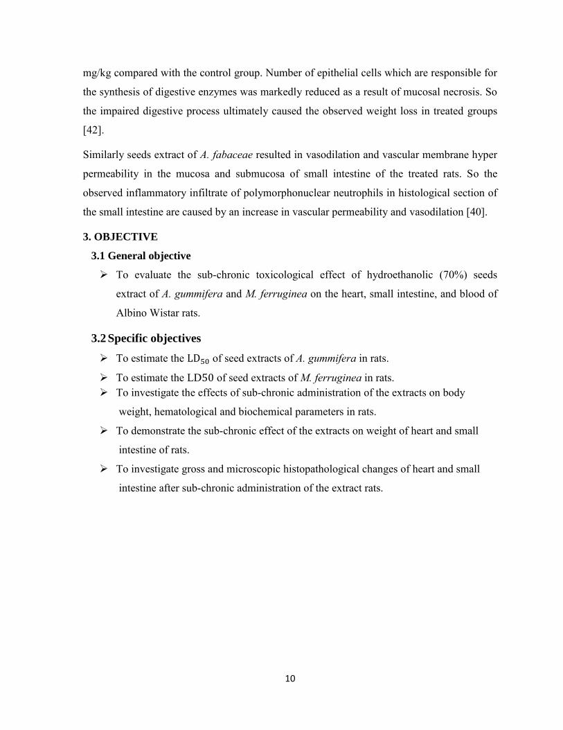
3. OBJECTIVE
3.1 General objective
To evaluate the sub-chronic toxicological effect of hydroethanolic (70%) seeds
extract of A. gummifera and M. ferruginea on the heart, small intestine, and blood of
Albino Wistar rats.
3.2 Specific objectives
To estimate the of seed extracts of A. gummifera in rats.
To estimate the LD of seed extracts of M. ferruginea in rats. To investigate the effects of sub-chronic administration of the extracts on body
weight, hematological and biochemical parameters in rats.
To demonstrate the sub-chronic effect of the extracts on weight of heart and small
intestine of rats.
To investigate gross and microscopic histopathological changes of heart and small
intestine after sub-chronic administration of the extract rats.
11
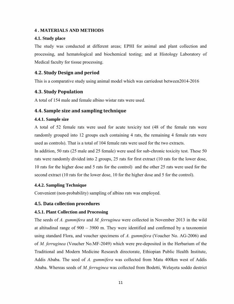
4 . MATERIALS AND METHODS
4.1. Study place
The study was conducted at different areas; EPHI for animal and plant collection and
processing, and hematological and biochemical testing; and at Histology Laboratory of
Medical faculty for tissue processing.
4.2. Study Design and period
This is a comparative study using animal model which was carriedout between2014-2016
4.3. Study Population
A total of 154 male and female albino wistar rats were used.
4.4. Sample size and sampling technique
4.4.1. Sample size
A total of 52 female rats were used for acute toxicity test (48 of the female rats were
randomly grouped into 12 groups each containing 4 rats, the remaining 4 female rats were
used as controls). That is a total of 104 female rats were used for the two extracts.
In addition, 50 rats (25 male and 25 female) were used for sub-chronic toxicity test. These 50
rats were randomly divided into 2 groups, 25 rats for first extract (10 rats for the lower dose,
10 rats for the higher dose and 5 rats for the control) and the other 25 rats were used for the
second extract (10 rats for the lower dose, 10 for the higher dose and 5 for the control).
4.4.2. Sampling Technique
Convenient (non-probability) sampling of albino rats was employed.
4.5. Data collection procedures
4.5.1. Plant Collection and Processing
The seeds of A. gummifera and M. ferruginea were collected in November 2013 in the wild
at altitudinal range of 900 – 3900 m. They were identified and confirmed by a taxonomist
using standard Flora, and voucher specimens of A. gummifera (Voucher No. AG-2006) and
of M. ferruginea (Voucher No.MF-2049) which were pre-deposited in the Herbarium of the
Traditional and Modern Medicine Research directorate, Ethiopian Public Health Institute,
Addis Ababa. The seed of A. gummifera was collected from Matu 400km west of Addis
Ababa. Whereas seeds of M. ferruginea was collected from Bodetti, Welayeta soddo destrict

12
300km south of Addis Ababa.The seeds of the plant were dried and crushed to powder at the
Traditional and Modern Medicine Research Directorate of the Ethiopian Public Health
Institute (EPHI).
4.5.2. Plant Material Extraction
Hydroalcoholic extract of the seed powder was used for these studies. To this effect, 1250
gm of the powdered material of A. gummifera and 1230 gm of the powdered seeds of M.
ferruginea using wooden-made pestle and mortarwere macerated separately with
hydroalcohol (70% ethanolic) in 1:4 solute to solvent ratio. The preparation was placed in
orbital shaker at room temperature for 72 hrs. This step was repeated three times to extract
exhaustively until the extract gave faint or no coloration. The extract was then filtered
through Whatman filter paper No.1 and the filtrate was evaporated to dryness under reduced
pressure by Rota vapor and further concentrated by water bath at 40 oc.
Then, the gummy residue extract was weighed and packed in air tight brown glass bottles
with proper label and kept in a refrigerator at 4oc. until used for the preparation of stock
solutions required in the subsequent experimental tests. For preparation of tested doses,
appropriate amount of the crude extract was weighed and dissolved in 2-5ml distilled water
immediately before administration.
4.5.3. Experimental Animals
For acute toxicity study, 108 female rats were used but for the sub-chronic toxicity study 50
rats (25 female and 25 male) were used. All the experimental animals used in this study were
bred at EPHI animal rearing unit. The male and female rats were kept in separate cagesand
were maintained on a 12hrs light/dark cycle, at room temperature and with free access to
water and food, except for overnight fasting prior to drug administration for the acute toxicity
study. They were all acclimatized prior to drug administration. The leftover food and water
were changed daily and the cages were cleaned with the husk changed every three days. All
the animals were apparently seen healthy.

13
4.6. Acute Oral Toxicity Test of A. gummifera and M. ferruginea Seed
Extracts in Rats
A total of 108 female rats were used for the study. Forty eight female rats were divided into
12 groups (each group contains 4 rats) were treated with seed extracts of the two plants at
different doses for acute toxicity study. After overnight fasting, rats in group I- XII were
given the extract at single oral doses of 50, 100, 150, 250, 500, 1000, 1500, 2000, 2500,
3000, 3500, and 4000 mg/kg body weight, respectively for both plants separately.
After treatment, post treatment observation were conducted to look for any sign of acute
toxicity related to behavioral and general health alteration such as hyperactivity, ataxia,
altered sleep, altered feeding, vomiting and diarrhea. And the other main objective of this
acute toxicity study was to determine the of the extracts of the plants. A control group
of four rats was given only the vehicle. At the end of two weeks, one animal from each group
was randomly sacrificed by cervical dislocation and post-mortem gross observation was
carried out on the internal organs (heart and small intestine).
4.7. Sub-chronic Oral Toxicity Test of A. gummifera and M. ferruginea
Seed Extracts in Rats
This study was conducted on 25 male and 25 female rats to investigate the effect of sub
chronic treatment with seed extracts of the two plants on general body weight and weight of
the organs (Heart and Small intestine); and on blood parameters as well as histopathology of
heart and small intestine tissues. The rats were randomly assigned to two groups of ten
animals, five males and five females each for one of the plant extract. Similarly, the animals
were randomly assigned to two groups of ten animals, five males and five females each for
the other plant extract. One group containing ten animals (five male and five females) was
assigned as control group. Throughout the experimental period, the female and male rats
were housed in separate cages. Animals in one group received single daily dose of 125 mg/kg
body weight and the second group was administered with a single daily dose of 250 mg/kg
body weight for 90 days of the seeds extract of A. gummifera. Similarly, the seeds extract of
M. ferruginea were given for one group a single daily dose of 125 mg/kg body weight and
for the other, 250 mg/kg body weight for a period of 90 days. The control group received
only vehicle daily throughout the period of study. The actual dose of the plant extract
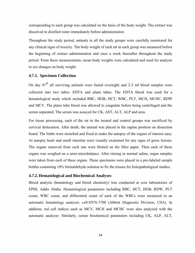
14
corresponding to each group was calculated on the basis of the body weight. The extract was
dissolved in distilled water immediately before administration.
Throughout the study period, animals in all the study groups were carefully monitored for
any clinical signs of toxicity. The body weight of each rat in each group was measured before
the beginning of extract administration and once a week thereafter throughout the study
period. From these measurements, mean body weights were calculated and used for analysis
to see changes on body weight.
4.7.1. Specimen Collection
On day 91th all surviving animals were fasted overnight and 2-3 ml blood samples were
collected into two tubes: EDTA and plane tubes. The EDTA blood was used for a
hematological study which included RBC, HGB, HCT, WBC, PLT, MCH, MCHC, RDW
and MCV. The plane tube blood was allowed to coagulate before being centrifuged and the
serum separated. The serum was assayed for CK, AST, ALT, ALP and urea.
For tissue processing, each of the rat in the treated and control groups was sacrificed by
cervical dislocation. After death, the animal was placed in the supine position on dissection
board. The limbs were stretched and fixed to make the autopsy of the organs of interest easy.
At autopsy heart and small intestine were visually examined for any signs of gross lesions.
The organs removed from each rats were blotted on the filter paper. Then each of these
organs was weighed on a semi-microbalance. After rinsing in normal saline, organ samples
were taken from each of these organs. These specimens were placed in a pre-labeled sample
bottles containing 10% formaldehyde solution to fix the tissues for histopathological studies.
4.7.2. Hematological and Biochemical Analyses
Blood analysis (hematology and blood chemistry) was conducted at core laboratories of
EPHI, Addis Ababa. Hematological parameters including RBC, HCT, HGB, RDW, PLT
count, WBC count, and differential count of each of the WBCs were measured in an
automatic hematology analyzer, cell-DYN-3700 (Abbott Diagnostic Division, USA). In
addition, red cell indices such as MCV, MCH and MCHC were also analyzed with the
automatic analyzer. Similarly, serum biochemical parameters including CK, ALP, ALT,

15
AST, and urea were determined usingclinical chemistry analyzer, Human star 80 (Human
GmbH, Germany)
Principle of automated hematological analysis using CELL-Dina 3700
CELL-DYN 3700 hematology analyzer performs simultaneous impedance and laser
measurements on white blood cells. For impedance method cell counting and sizing is based
on the detection and measurement of changes in electrical impedance (resistance) produced
by a particle as it passes through a small aperture. Particles such as blood cells are non-
conductive but are suspended in electrically conductive diluents. As a dilute suspension of
cells is drawn through the aperture, the passage of each individual cell momentarily increases
the impedance of the electrical path between two electrodes that are located on each side of
the aperture.
Whereas, in the optical method, laser light is used and a diluted blood specimen passes in a
steady stream through which a beam of laser light is focused. As each cell passes through the
sensing zone of the flow cell, it scatters the focused light. Then, scattered light is detected by
a photodetector and converted to an electrical impulse. The number of impulses generated is
directly proportional to the number of cells passing through the sensing zone [43].
Principle of Human star 80 clinical chemistry analyzer
Serum samples are loaded on tray. Then, a pipette aspirates a precisely measured aliquot of
sample and discharges it into the reaction vessel; a measured volume of diluents rinses the
pipette. Reagents are dispensed into the reaction vessel. After the solution is mixed, it is
either passed through a colorimeter, which measures its absorbance while it is still in the
reaction vessel, or aspirated into a flow cell, where its absorbance is measured by a flow-
through colorimeter. The analyzed then calculates the analyte′s chemical concentration [44].
4.7.3. Histopathological studies
4.7.3.1. Tissue Processing
Tissue samples taken at autopsy were processed for histopathological study at Histology
laboratory, Department of Anatomy, College of Health Sciences, AAU. Each tissue sample
taken from heart and small intestine of all extract treated and control animals were fixed
separately in 10% neutral buffered formalin (Appendix I).
16
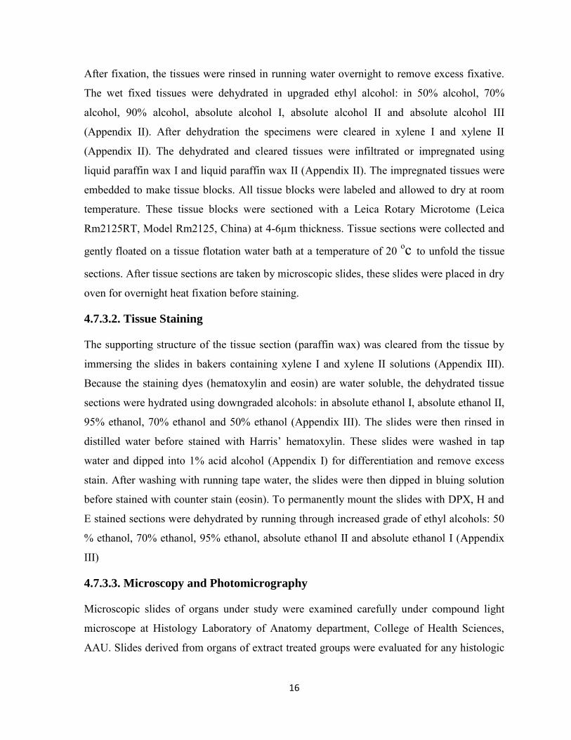
After fixation, the tissues were rinsed in running water overnight to remove excess fixative.
The wet fixed tissues were dehydrated in upgraded ethyl alcohol: in 50% alcohol, 70%
alcohol, 90% alcohol, absolute alcohol I, absolute alcohol II and absolute alcohol III
(Appendix II). After dehydration the specimens were cleared in xylene I and xylene II
(Appendix II). The dehydrated and cleared tissues were infiltrated or impregnated using
liquid paraffin wax I and liquid paraffin wax II (Appendix II). The impregnated tissues were
embedded to make tissue blocks. All tissue blocks were labeled and allowed to dry at room
temperature. These tissue blocks were sectioned with a Leica Rotary Microtome (Leica
Rm2125RT, Model Rm2125, China) at 4-6µm thickness. Tissue sections were collected and
gently floated on a tissue flotation water bath at a temperature of 20 oc to unfold the tissue
sections. After tissue sections are taken by microscopic slides, these slides were placed in dry
oven for overnight heat fixation before staining.
4.7.3.2. Tissue Staining
The supporting structure of the tissue section (paraffin wax) was cleared from the tissue by
immersing the slides in bakers containing xylene I and xylene II solutions (Appendix III).
Because the staining dyes (hematoxylin and eosin) are water soluble, the dehydrated tissue
sections were hydrated using downgraded alcohols: in absolute ethanol I, absolute ethanol II,
95% ethanol, 70% ethanol and 50% ethanol (Appendix III). The slides were then rinsed in
distilled water before stained with Harris’ hematoxylin. These slides were washed in tap
water and dipped into 1% acid alcohol (Appendix I) for differentiation and remove excess
stain. After washing with running tape water, the slides were then dipped in bluing solution
before stained with counter stain (eosin). To permanently mount the slides with DPX, H and
E stained sections were dehydrated by running through increased grade of ethyl alcohols: 50
% ethanol, 70% ethanol, 95% ethanol, absolute ethanol II and absolute ethanol I (Appendix
III)
4.7.3.3. Microscopy and Photomicrography
Microscopic slides of organs under study were examined carefully under compound light
microscope at Histology Laboratory of Anatomy department, College of Health Sciences,
AAU. Slides derived from organs of extract treated groups were evaluated for any histologic
17
alteration compared to slides from their respective control groups. The presence or absence
of histopathological changes in the tissues sections was assessed. Finally, photomicrographs
of selected slides were takenusing (LEICA ICC50 HD, Germany) automated built-in digital
photo- camera.
4.8. Statistical Analysis
The data were analyzed statistically using SPSS version 20, computer software package. The
values of body weight changes were analyzed and the results were expressed as M ± SEM.
To compare differences between the treated and control groups one-way analysis of variance
(ANOVA) were used, followed by Dunnett’s t-test to determine their level of significance.
Values at p<0.05 were considered statistically significant.
4.9. Ethical Considerations
All the experiments had been conducted following the approval by the school of Medicine,
AAU in line with the highest standard for the humane and compassionate use of animals in
biomedical research.
4.10. Data Quality Assurance
Samples for hematological and biochemical analysis are collected and analyzed under the
standard operational procedures. Commercial controls were used for both hematology and
clinical chemistry analyzers as per the manufacturers’ recommendations. All reagents and
controls are stored and used before the expiry date. Samples of histopathological study were
preserved in 10% formaldehyde solution before processed following the standard procedures.
Body and organs weight were properly taken and data entry quality was guaranteed by
entering SPSS version 16.

18
5. RESULTS
5.1. Acute Toxicity Study
A single dose toxicity test were done to estimate lethal dose of the 70% ethanolic seeds
extract of A. gummifera and M. ferruginea in rats’ model. These extracts at 50, 100, 150,
250, 500, 1000, 1500, 2000 and 2500mg/kg body weight were administered orally to rat
models and did not cause death or mortality. One and two rats died among the 3000 and
3500 mg/kg M. ferruginea seed extract administered groups, respectively. While two rats
died among the 4000 mg/kg A. gummifera seed extract treated group. Acute toxicity study
showedthat the LD50 of A. gummifera and M. ferrugina were 4000 mg/kg and 3500 mg/kg
respectively. No pathological lesions of heart and small intestine were observed.
5.2. Sub-chronic Toxicity Study
5.2.1. Effect of the extract of both plants on general health and body weight
For sub-chronic toxicity evaluation, all the male and female rats were orally administered
with the repeated doses of both 125 mg/kg and 250 mg/kg body weight for 90 days. Through
the period of administration, there were no extract related noticeable changes in their health
and general behavior of the treated rats as compared to the control group for both plant
extracts. No abnormal findings on gross observation of the heart and small intestine were
observed in this rat. But throughout the period of administration, there were no toxicity
related deaths.
The effect of the seeds extracts of A. gummifera and M. ferruginea on the body weight of
male and female rats during the 13 weeks of sub-chronic treatment are summarized in
(Tables 1 and 2), respectively. Body weight of both the treated and control groups increased
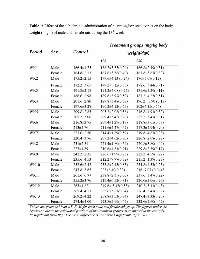
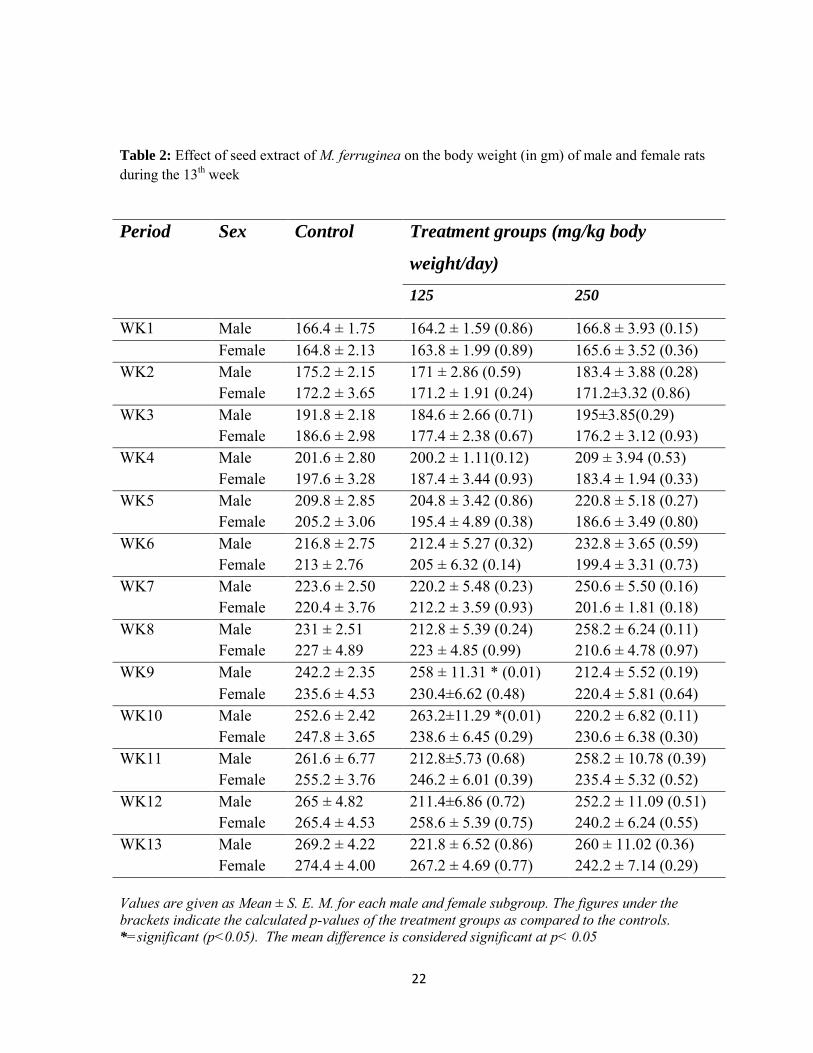
with increasing duration. As it can be seen from Figures 1 to 4, the body weight increase
patterns of the male and female rats during the sub-chronic treatment with both of the plant
extracts seem to be normal. In the seeds extract of A. gummifera administered groups, no
significant difference was observed in the mean values of the body weights of male and
female rats treated with 125 mg/kg body weight and males treated with 250 mg/kg body
weight as compared with their respective controls. However, significant difference was
observed in the mean body weight of female rats treated with 250mg/kg body weight as it
decreased at the 10th week by 12.8% as compared with the control group. Similarly, in seed

19
extract of M. ferruginea treated groups, the mean body weight difference between the
administered and control groups were not significant, except, in the males’ rats treated with
125 mg/kg body weight during the 9th and 10th weeks of administration period, where the
mean body weight increased by 6.5% in the 9th week and further by 4.2% during the 10th
week.
20
Table 1: Effect of the sub-chronic administration of A. gummifera seed extract on the body
weight (in gm) of male and female rats during the 13th week
Period
Sex
Control Treatment groups (mg/kg body
weight/day) 125 250
WK1 Male 166.4±1.75 168.2±3.35(0.24) 166.8±2.49(0.51) Female 164.8±2.13 167.6±3.36(0.40) 167.8±3.67(0.32)
WK2 Male 175.2±2.15 179.6±4.13 (0.24) 176±5.09(0.12)
Female 172.2±3.65 179.2±5.12(0.53) 178.6±3.44(0.91) WK3 Male 191.8±2.18 191.2±4.08 (0.25) 171.6±5.29(0.11)
Female 186.6±2.98 189.6±3.97(0.59) 187.2±4.25(0.51) WK4 Male 201.6±2.80 189.8±3.48(0.68) 196.2± 5.90 (0.18)
Female 197.6±3.28 196.2±4.12(0.67) 202±4.15(0.66) WK5 Male 209.8±2.85 205.2±2.08(0.56) 216.8±4.91(0.32)
Female 205.2±3.06 209.4±5.42(0.29) 225.2±3.47(0.81) WK6 Male 216.8±2.75 209.4±1.29(0.17) 218.8±3.65(0.59)
Female 213±2.76 211.6±4.27(0.42) 217.2±2.94(0.90) WK7 Male 223.6±2.50 214.4±1.89(0.59) 219.8±4.83(0.23)
Female 220.4±3.76 207.2±4.62(0.70) 226.8±2.08(0.28) WK8 Male 231±2.51 221.4±1.80(0.54) 220.8±3.80(0.44)
Female 227±4.89 210.6±4.61(0.91) 229.8±2.39(0.19) WK9 Male 242.2±2.35 226.6±1.99(0.75) 222.2±4.59(0.22)
Female 235.6±4.53 212.2±7.77(0.32) 215.2±1.39(0.23) WK10 Male 252.6±2.42 233.8±2.15(0.83) 234.8±4.53(0.25)
Female 247.8±3.65 223±6.40(0.32) 216±7.07 (0.04) * WK11 Male 261.6±6.77 238.8±2.35(0.06) 237.6±3.47(0.22)
Female 255.2±3.76 219.4±6.52(0.31) 220.6±2.06(0.27) WK12 Male 265±4.82 249.6± 3.43(0.53) 240.2±5.11(0.43)
Female 265.4±4.53 225.6±5.81(0.64) 226.4±3.47(0.62) WK13 Male 269.2±4.22 258.8±3.53(0.74) 248.4±3.33(0.20)
Female 274.4±4.00 223.8±5.99(0.45) 232.6±2.60(0.42) Values are given as Mean ± S. E. M. for each male and female subgroup. The figures under the brackets indicate the calculated p-values of the treatment groups as compared to the controls. *=significant (p˂0.05). The mean difference is considered significant at p˂ 0.05
21
Figure 3: Time course and effect of seeds extract of A. gummifera on body growth pattern of male
rats treated with 125mg/kg body weight and 250 mg/kg body weight as compared to the controls.
Each value point represents mean ± S. E. M. Note: AGG1M=A. gummifera administered group one
male rats, AGG2M=A. gummifera administered group two male rats & MC= male control rats
Figure 4: Time course and effect of seeds extract of A. gummifera on body growth pattern of female
rats treated with 125 mg/kg body weight and 250 mg/kg body weight as compared to the controls.
Each value point represents mean ± S. E. M. Note: AGG1F=A. gummifera` administered group one
female rats, AGG2F= A. gummifera administered group two femalerats& CF= female control rats
0
50
100
150
200
250
300
1 2 3 4 5 6 7 8 9 10 11 12 13
Mea
n b
od
y w
eigh
t (g
)
weeks
AGG1M
AGG2M
MC
0
50
100
150
200
250
300
1 2 3 4 5 6 7 8 9 10 11 12 13
Mea
n b
od
y w
eig
ht(
g)
weeks
AGG1F
AGG2F
CF
22
Table 2: Effect of seed extract of M. ferruginea on the body weight (in gm) of male and female rats during the 13th week
Period Sex Control Treatment groups (mg/kg body
weight/day)
125 250
WK1 Male 166.4 ± 1.75 164.2 ± 1.59 (0.86) 166.8 ± 3.93 (0.15) Female 164.8 ± 2.13 163.8 ± 1.99 (0.89) 165.6 ± 3.52 (0.36) WK2 Male 175.2 ± 2.15 171 ± 2.86 (0.59) 183.4 ± 3.88 (0.28)
Female 172.2 ± 3.65 171.2 ± 1.91 (0.24) 171.2±3.32 (0.86) WK3 Male 191.8 ± 2.18 184.6 ± 2.66 (0.71) 195±3.85(0.29)
Female 186.6 ± 2.98 177.4 ± 2.38 (0.67) 176.2 ± 3.12 (0.93) WK4 Male 201.6 ± 2.80 200.2 ± 1.11(0.12) 209 ± 3.94 (0.53)
Female 197.6 ± 3.28 187.4 ± 3.44 (0.93) 183.4 ± 1.94 (0.33) WK5 Male 209.8 ± 2.85 204.8 ± 3.42 (0.86) 220.8 ± 5.18 (0.27)
Female 205.2 ± 3.06 195.4 ± 4.89 (0.38) 186.6 ± 3.49 (0.80) WK6 Male 216.8 ± 2.75 212.4 ± 5.27 (0.32) 232.8 ± 3.65 (0.59)
Female 213 ± 2.76 205 ± 6.32 (0.14) 199.4 ± 3.31 (0.73) WK7 Male 223.6 ± 2.50 220.2 ± 5.48 (0.23) 250.6 ± 5.50 (0.16)
Female 220.4 ± 3.76 212.2 ± 3.59 (0.93) 201.6 ± 1.81 (0.18) WK8 Male 231 ± 2.51 212.8 ± 5.39 (0.24) 258.2 ± 6.24 (0.11)
Female 227 ± 4.89 223 ± 4.85 (0.99) 210.6 ± 4.78 (0.97) WK9 Male 242.2 ± 2.35 258 ± 11.31 * (0.01) 212.4 ± 5.52 (0.19)
Female 235.6 ± 4.53 230.4±6.62 (0.48) 220.4 ± 5.81 (0.64) WK10 Male 252.6 ± 2.42 263.2±11.29 *(0.01) 220.2 ± 6.82 (0.11)
Female 247.8 ± 3.65 238.6 ± 6.45 (0.29) 230.6 ± 6.38 (0.30) WK11 Male 261.6 ± 6.77 212.8±5.73 (0.68) 258.2 ± 10.78 (0.39)
Female 255.2 ± 3.76 246.2 ± 6.01 (0.39) 235.4 ± 5.32 (0.52) WK12 Male 265 ± 4.82 211.4±6.86 (0.72) 252.2 ± 11.09 (0.51)
Female 265.4 ± 4.53 258.6 ± 5.39 (0.75) 240.2 ± 6.24 (0.55) WK13 Male 269.2 ± 4.22 221.8 ± 6.52 (0.86) 260 ± 11.02 (0.36)
Female 274.4 ± 4.00 267.2 ± 4.69 (0.77) 242.2 ± 7.14 (0.29) Values are given as Mean ± S. E. M. for each male and female subgroup. The figures under the brackets indicate the calculated p-values of the treatment groups as compared to the controls. *=significant (p˂0.05). The mean difference is considered significant at p˂ 0.05
23
Figure 5: Time course and effect of seeds extract of M. ferruginea on body growth pattern of male
rats treated with 125 mg/kg body weight and 250 mg/kg body weight as compared to the controls.
Each value point represents mean ± S.E.M. Note: MFG1M= M. ferruginea administered group one
male rats, MFG2M= M. ferruginea administered group two male rats & MC= male control rats
Figure 6: Time course and effect of seeds extract of M. ferruginea on body growth pattern of female
rats treated with 125 mg/kg body weight and 250 mg/kg body weight as compared to the controls.
Each value point represents mean ± S. E. M. Note: MFG1F= M. ferruginea administered group one
female rats, MFG2F= M. ferruginea administered group two female rats & CF= female control rats
0
50
100
150
200
250
300
350
1 2 3 4 5 6 7 8 9 10 11 12 13
Mea
n b
od
y w
eig
ht(
g)
weeks
MFG1M
MFG2M
MC
0
50
100
150
200
250
300
1 2 3 4 5 6 7 8 9 10 11 12 13
Mea
n b
od
y w
eigh
t (g
)
weeks
MFG1F
MFG2F
FC
24
5.2.2. Effect of A. gummifera and M. ferruginea Hydro alcohol Seed
Extract on Hematological Parameters
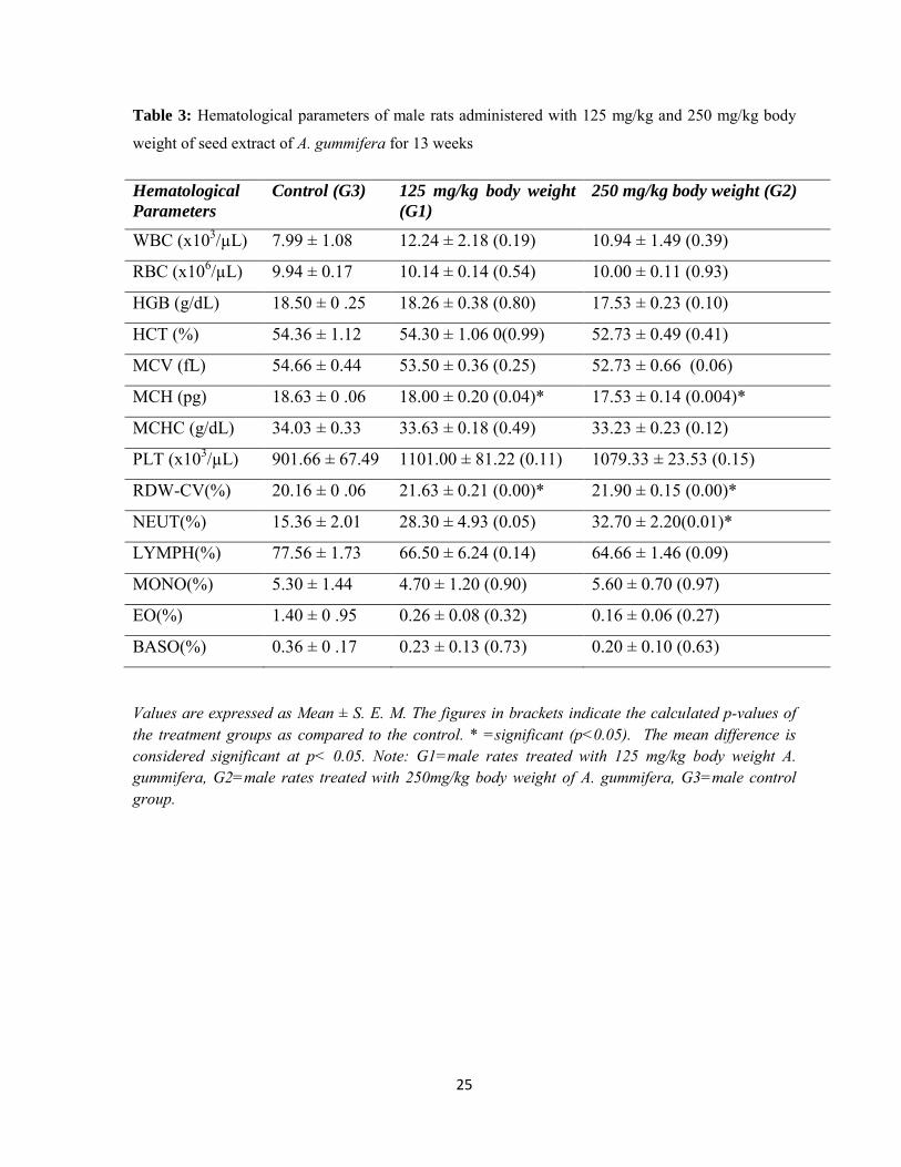
The effect of A. gummifera seed extract on hematological parameters of male rats as
compared to the controls after 13 weeks of sub-chronic administration is illustrated in Table
3. Male rats administered at the dose of 125 mg/kg and 250 mg/kg body weight seed extract
of A. gummifera did not show significant change on their hematological parameters. But few
hematological parameters (MCH, RDW-CV) showed significant change (p˂0.05). MCH was
significantly decreased by 3.4% and 5.9% for male rats administered at 125 mg/kg and 250
mg/kg respectively. And RDW-CV significantly increased by 7.3% and 8.6% for male rats
administered at125 mg/kg and 250 mg/kg doses respectively. Neutrophil also showed
significant increase from 15.36±2.01 to 32.70±2.20 for male rat administered at 250 mg/kg.
The effect of A. gummifera seed extract on hematological parameters of female rats treated
for 13 weeks is shown in Table 4. MCH was significantly decreased by 5.6% for female rat
treated at 250 gm/kg dose. Whereas MCHC was significantly decreased by 2.0% and 2.7%
for female rats treated at 125mg/kg and 250mg/kg doses, respectively. Besides, neutrophil
was significantly increased from 12.06±2.14 to 35.00±1.80 for female rats treated at 250
gm/kg dose.
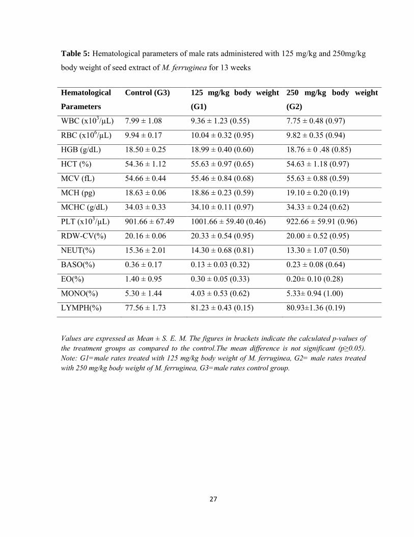
The effect of seed extract of M. ferruginea on hematological parameters of male and female
rats following sub-chronic administration is illustrated in (Tables 5 and 6), respectively.
Hematological parameters in both sexes were not significantly affected by sub chronic
treatment of these rats at 125 mg/kg and 250 mg/kg doses. But significant (p˂0.05) decrease
in MCHC and monocyte by 2.2% and 66.8% respectively was observed for female rats
administered at 250 mg/kg.
25
Table 3: Hematological parameters of male rats administered with 125 mg/kg and 250 mg/kg body
weight of seed extract of A. gummifera for 13 weeks
Hematological
Parameters Control (G3) 125 mg/kg body weight
(G1) 250 mg/kg body weight (G2)
WBC (x103/µL) 7.99 ± 1.08 12.24 ± 2.18 (0.19) 10.94 ± 1.49 (0.39)
RBC (x106/µL) 9.94 ± 0.17 10.14 ± 0.14 (0.54) 10.00 ± 0.11 (0.93)
HGB (g/dL) 18.50 ± 0 .25 18.26 ± 0.38 (0.80) 17.53 ± 0.23 (0.10)
HCT (%) 54.36 ± 1.12 54.30 ± 1.06 0(0.99) 52.73 ± 0.49 (0.41)
MCV (fL) 54.66 ± 0.44 53.50 ± 0.36 (0.25) 52.73 ± 0.66 (0.06)
MCH (pg) 18.63 ± 0 .06 18.00 ± 0.20 (0.04)* 17.53 ± 0.14 (0.004)*
MCHC (g/dL) 34.03 ± 0.33 33.63 ± 0.18 (0.49) 33.23 ± 0.23 (0.12)
PLT (x103/µL) 901.66 ± 67.49 1101.00 ± 81.22 (0.11) 1079.33 ± 23.53 (0.15)
RDW-CV(%) 20.16 ± 0 .06 21.63 ± 0.21 (0.00)* 21.90 ± 0.15 (0.00)*
NEUT(%) 15.36 ± 2.01 28.30 ± 4.93 (0.05) 32.70 ± 2.20(0.01)*
LYMPH(%) 77.56 ± 1.73 66.50 ± 6.24 (0.14) 64.66 ± 1.46 (0.09)
MONO(%) 5.30 ± 1.44 4.70 ± 1.20 (0.90) 5.60 ± 0.70 (0.97)
EO(%) 1.40 ± 0 .95 0.26 ± 0.08 (0.32) 0.16 ± 0.06 (0.27)
BASO(%) 0.36 ± 0 .17 0.23 ± 0.13 (0.73) 0.20 ± 0.10 (0.63)
Values are expressed as Mean ± S. E. M. The figures in brackets indicate the calculated p-values of the treatment groups as compared to the control. * =significant (p˂0.05). The mean difference is considered significant at p˂ 0.05. Note: G1=male rates treated with 125 mg/kg body weight A. gummifera, G2=male rates treated with 250mg/kg body weight of A. gummifera, G3=male control group.

26
Table 4: Hematological parameters of female rats administered with125 mg/kg and 250
mg/kg body weight of seed extract of A. gummifera for 13 weeks
Hematological
Parameters Control (G3) 125 mg/kg body weight
(G1) 250 mg/kg body weight
(G2)
WBC (x103/µL) 5.27 ± 2.32 9.55 ± 1.71 (0.42) 10.48± 3.17 (0.30)
RBC (x106/µL) 8.66 ± 0.31 8.99 ± 0.18 (0.51) 9.32± 0.14 (0.14)
HGB (g/dL) 16.86 ± 0 .67 17.16 ± 0.55 (0.88) 17.13 ± 0.06 (0.90)
HCT (%) 49.13 ± 1.74 51.36 ± 1.40 (0.42) 50.93 ± 0.34 (0.55)
MCV (fL) 56.76 ± 0.66 57.10 ± 0.40 (0.88) 54.63 ± 0.61 (0.06)
MCH (pg) 19.46 ± 0.23 19.06 ± 0.23 (0.42) 18.36 ± 0.23 (0.02)*
MCHC (g/dL) 34.33 ± 0 .14 33.63 ± 0.12 (0.03)* 33.40 ± 0.20 (0.01) *
PLT (x103/µL) 665.00 ± 218.70 1029.00 ± 12.34 (0.18) 1222.33 ± 92.09 (0.05)
RDW-CV(%) 17.23 ± 0 .98 18.60 ± 0.20 (0.26) 19.43 ± 0.27 (0.07)
NEUT(%) 12.06 ± 2.14 20.90 ± 6.96 (0.32) 35.00 ± 1.80 (0.02)*
BASO(%) 0.03 ± 0 .03 0.10 ± 0.05 (0.48) 0.06 ± 0.03 (0.81)
EO(%) 3.66 ± 3.12 0.50 ±0.05 (0.40) 0.26 ± 0.12 (0.36)
MONO(%) 4.40 ± 0.55 5.13 ± 1.88 (0.94) 6.53 ± 2.51 (0.64)
LYMPH(%) 79.83 ± 3.89 73.36 ± 8.51 (0.67) 58.13 ± 4.16 (0.07)
Values are expressed as Mean ± S. E. M. The figures in brackets indicate the calculated p-values of the treatment groups as compared to the control. * =significant (p˂0.05). The mean difference is considered significant at p˂ 0.05. Note: G1=female rates treated with 125 mg/kg body weight of A. gummifera, G2=female rates treated with 250 mg/kg body weight of A. gummifera, G3=female control group.
27
Table 5: Hematological parameters of male rats administered with 125 mg/kg and 250mg/kg
body weight of seed extract of M. ferruginea for 13 weeks
Hematological
Parameters
Control (G3) 125 mg/kg body weight
(G1)
250 mg/kg body weight
(G2)
WBC (x103/µL) 7.99 ± 1.08 9.36 ± 1.23 (0.55) 7.75 ± 0.48 (0.97)
RBC (x106/µL) 9.94 ± 0.17 10.04 ± 0.32 (0.95) 9.82 ± 0.35 (0.94)
HGB (g/dL) 18.50 ± 0.25 18.99 ± 0.40 (0.60) 18.76 ± 0 .48 (0.85)
HCT (%) 54.36 ± 1.12 55.63 ± 0.97 (0.65) 54.63 ± 1.18 (0.97)
MCV (fL) 54.66 ± 0.44 55.46 ± 0.84 (0.68) 55.63 ± 0.88 (0.59)
MCH (pg) 18.63 ± 0.06 18.86 ± 0.23 (0.59) 19.10 ± 0.20 (0.19)
MCHC (g/dL) 34.03 ± 0.33 34.10 ± 0.11 (0.97) 34.33 ± 0.24 (0.62)
PLT (x103/µL) 901.66 ± 67.49 1001.66 ± 59.40 (0.46) 922.66 ± 59.91 (0.96)
RDW-CV(%) 20.16 ± 0.06 20.33 ± 0.54 (0.95) 20.00 ± 0.52 (0.95)
NEUT(%) 15.36 ± 2.01 14.30 ± 0.68 (0.81) 13.30 ± 1.07 (0.50)
BASO(%) 0.36 ± 0.17 0.13 ± 0.03 (0.32) 0.23 ± 0.08 (0.64)
EO(%) 1.40 ± 0.95 0.30 ± 0.05 (0.33) 0.20± 0.10 (0.28)
MONO(%) 5.30 ± 1.44 4.03 ± 0.53 (0.62) 5.33± 0.94 (1.00)
LYMPH(%) 77.56 ± 1.73 81.23 ± 0.43 (0.15) 80.93±1.36 (0.19)
Values are expressed as Mean ± S. E. M. The figures in brackets indicate the calculated p-values of the treatment groups as compared to the control.The mean difference is not significant (p≥0.05). Note: G1=male rates treated with 125 mg/kg body weight of M. ferruginea, G2= male rates treated with 250 mg/kg body weight of M. ferruginea, G3=male rates control group.
28
Table 6: Hematological parameters of female rats administered with 125 mg/kg and 250 mg/kg body
weight of seed extract of M. ferruginea for 13 weeks
Hematological
Parameters Control (G3) 125 mg/kg body weight
(G1)
250m g/kg body weight
(G2)
WBC (x103/µL) 5.27 ± 2.32 6.63 ± 0.49 (0.72) 4.22 ± 0.02 (0.81)
RBC (x106/µL) 8.66 ± 0.31 8.73 ± 0.07 (0.95) 8.66 ± 0.16 (1.00)
HGB (g/dL) 16.86 ± 0.67 17.30 ± 0.10 (0.70) 16.86 ± 0.26 (1.00)
HCT (%) 49.13 ± 1.74 51.53 ± 0.06 (0.29) 49.53 ± 0.85 (0.95)
MCV (fL) 56.76 ± 0.66 57.46 ± 0.03 (0.54) 59.03 ± 0.56 (0.30)
MCH (pg) 19.46 ± 0.23 19.80 ± 0.15 (0.32) 19.60 ± 0.05 (0.79)
MCHC (g/dL) 34.33 ± 0.14 34.20 ± 0.05 (0.72) 33.56 ± 0.17 (0.01)*
PLT (x103/µL) 665.00 ± 218.70 956.00 ± 95.10 (0.30) 1121.66 ± 13.92 (0.09)
RDW-CV(%) 17.23 ± 0.98 17.60 ± 0.81 (0.92) 18.03 ± 0.37 (0.69)
NEUT(%) 12.06 ± 2.14 12.06 ± 2.91 (1.00) 12.83 ± 0.43 (0.95)
BASO(%) 0.03 ± 0.03 0.10 ± 0.05 (0.48) 0.03 ± 0.03 (1.00)
EO(%) 3.66 ± 3.12 0.76 ± 0.26 (0.46) 0.53 ± 0.20 (0.41)
MONO(%) 4.40 ± 0.55 3.16 ± 1.01 (0.38) 1.46 ± 0.14 (0.03) *
LYMPH(%) 79.83 ± 3.89 83.90 ± 2.20 (0.47) 84.96 ± 0.34 (0.33)
Values are expressed as Mean ± S. E. M. The figures in brackets indicate the calculated p-values of the treatment groups as compared to the control. * =significant (p˂0.05). The mean difference is considered significant at p˂ 0.05. Note: G1=female rates treated with 125 mg/kg body weight of M. ferruginea, G2=female rates treated with 250 mg/kg body weight of M. ferruginea, G3= female control group.

29
5.2. 3. Effect of A. gummifera and M. ferruginea Hydro alcohol Seed
Extract on Serum Biochemicalparameters
Effects of sub-chronic treatment with hydro-alcoholic seed extract of A. gummifera on serum
biochemical parameters of male and female rats are shown in (Tables7 and 8), respectively.
Urea and CK were significantly increased by 28.94% and 12% respectively for male rats
administered at 250 mg/kg body weight. CK was also increased significantly by14.59% for
female rats administered at 250 gm/kg. But the other parameters measured were not
significantly different between the control and extract administered groups at both doses. The
effects of sub-chronic treatment with hydro-alcoholic seed extract of M. ferruginea on serum
biochemical parameters of male and female are shown in (Tables 9 and 10), respectively.
Except CK which increased significantly by 9% for male rats treated at 250 mg/kg, all the
other parameters measured in male rats which received any of the doses were not
significantly different from those of the controls. Urea, ALP, ALT and CK, however, were
found to be significantly different in the female rats at 250 mg/kg body weight as increased
by 8.84%, 59.40%, 27.45% and 19.56%, respectively. Table 7: Serum biochemical parameters of male rats administered with 125 mg/kg and 250 mg/kg
body weight of seed extract of A. gummifera for 13 weeks
Biochemical
Parameters
Control (G3) 125 mg/kg dose
(G1)
250 mg/kg dose
(G2)
Ck(U/L) 239.00 ± 1.95 243.00 ± 1.95(0.323) 267.80 ± 2.29(0.00)*
Albumin(g/dl) 4.70 ± 0.22 4.79 ± 0.26(0.94) 4.45 ± 0.19(0.65)
ALP (U/L 135.75 ± 11.69 168 ± 12.79 (0.44) 160.75 ± 29.26(0.59)
ALT(U/L) 79.75 ± 2.59 94.25 ± 5.15(0.11) 79.75 ± 6.21 (1.00)
AST(U/L) 179.75 ± 5.20 251.5 ± 43.65(0.34) 253.75± 47.42 (0.32)
Urea (mg/dL) 28.50 ± 0.96 33 ± 0.41 (0.09) 36.75± 2.21 (0.005)*
Values are expressed as Mean ± S. E. M. The figures in brackets indicate the calculated p-values of the treatment groups as compared to the control. * =significant (p˂0.05). The mean difference is considered significant at p˂ 0.05. Note: G1=male rates treated with 125 mg/kg body weight of A. gummifera, G2=male rates treated with 250 mg/kg body weight of A. gummifera, G3= male rates control groups.

30
Table 8: Serum biochemical parameters of female rats administered with 125 mg/kg and 250 mg/kg
body weight of seed extract of A. gummifera for 13 weeks
Biochemical
Parameters
Control (G3) 125 mg/kg dose
(G1)
250 mg/kg dose
(G2)
CK(U/L) 209.80 ±1.36 219.00 ± 2.00(0.149) 240.40±5.53(0.00)*
Albumin(g/dl) 5.16 ± 0.19 5.01 ± 0.16(0.79) 4.87 ± 0.16 (0.42)
ALP (U/L 75.75 ± 12.49 88.75 ± 8.41 (0.79) 134.75 ± 22.87(0.05)
ALT(U/L) 63.75 ± 5.66 74.75 ± 4.00(0.26) 61.50 ± 5.33 (0.93)
AST(U/L) 194 ± 16.92 237.25 ± 62.26(0.65) 192.25 ± 13.88 (0.99)
Urea (mg/dL) 34.50 ± 2.33 38.75 ± 2.49 (0.29) 37.00 ± 1.08 (0.62)
Values are expressed as Mean ± S. E. M. The figures in brackets indicate the calculated p-values of the treatment groups as compared to the control. The mean difference is considered significant at p˂ 0.05. Note: G1= female rates treated with 125 mg/kg body weight of A. gummifera, G2=female rates treated with 250 mg/kg body weight of A. gummifera, G3=female rates control group.
Table 9: Serum biochemical parameters of male rats administered with 125 mg/kg and 250 mg/kg
body weight of seed extract of M. ferruginea for 13 weeks
Biochemical
Parameters
Control (G3)
125 mg/kg dose (G1)
250 mg/kg dose (G2)
CK(U/L) 254.00 ± 2.70 261.00 ± 2.61(0.12) 277.00±3.77(0.00)*
Albumin(g/dl) 4.70 ± 0.22 5.03 ± 0.20(0.39) 4.85 ± 0.10(0.82)
ALP (U/L) 135.75 ± 11.69 158.00 ± 17.56 (0.48) 161.25 ± 13.74(0.39)
ALT(U/L) 79.75 ± 2.59 85.75±4.27(0.39) 80.50± 3.28 (0.98)
AST(U/L) 179.75 ± 5.20 210.25 ± 21.44(0.22) 181.25 ± 5.07 (0.99)
Urea (mg/dL) 28.50 ± 0.96 32.25 ± 1.49 (0.17) 30.00 ± 1.73(0.69)
Values are expressed as Mean ± S. E. M. The figures in brackets indicate the calculated p-values of the treatment groups as compared to the control. The mean difference was significant (p< 0.05). Note: G1=male rates treated with 125 mg/kg body weight of M. ferruginea, G2= male rates treated with 250 mg/kg body weight of M. ferruginea, G3=male rates control group.

31
Table 10: Serum biochemical parameters of female rats administered with 125 mg/kg and 250 mg/kg
body weight of seed extract of M. ferruginea for 13 weeks
Biochemical
Parameters Control (G3) 125 mg/kg dose (G1) 250 mg/kg dose (G2)
CK(U/L) 262.40 ± 2.94 271.80±5.44(.19) 285.60±2.84(0.02)*
Albumin(g/dl) 5.16 ± 0.19 5.34 ± 0.15 (0.62) 5.15 ± 0.10(1.00)
ALP (U/L) 75.75 ± 12.49 87.00 ± 6.01 (0.54) 120.75±2.05 (0.006)*
ALT(U/L) 63.75 ± 5.66 74.25± 4.34 (0.20) 81.25±2.28(0.03)*
AST(U/L) 194 ± 16.92 206.00 ± 20.33 (0.95) 264.00 ± 46.59 (0.24)
Urea (mg/dL) 34.50 ± 2.33 34.25 ± 0.75 (0.99) 41.25 ± 1.38 (0.03)*
Values are expressed as Mean ± S. E. M. The figures in brackets indicate the calculated p-values of the treatment groups as compared to the control. * =significant (p˂0.05). The mean difference is considered significant at p˂ 0.05. Note: G1= female rates treated with 125 mg/kg body weight of M. ferruginea, G2=female rates treated with 250 mg/kg body weight of M. ferruginea, G3= female rates control group.
5.2. 4. Macroscopic Observations and Organ Weights
After treatment period is completed, both treated and control groups were sacrificed. During
dissection, post mortem examination revealed no gross abnormal findings in the heart and
small intestine. The mean organ weight of heart and intestine of the seed extract of A.
gummifera administered groups and control group are shown in (Tables 11 and 12) for the
male and female rats, respectively. The result showed no significant difference in the organ
weights between extract treated and control rats of both sex except for the weight of heart of
female rats treated at higher dose which was decreased by 28%. The mean organ weight of
heart and small intestine of the seed extract of M. ferruginea administered groups and control
group are shown in Tables 13 and 14 for the male and female rats, respectively. No
significant difference was noted in the organ weights between extract treated and control rats
of either sex except for the weight of heart of female rats which was decreased by 22.5%.
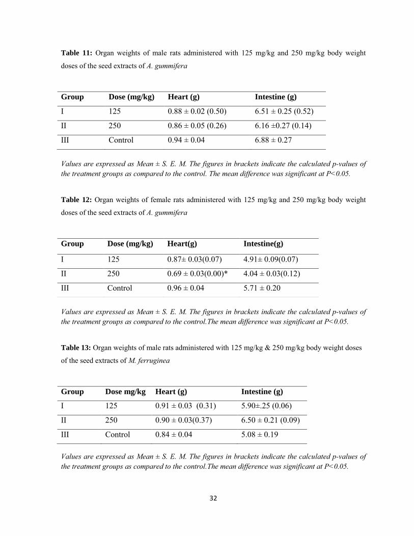
32
Table 11: Organ weights of male rats administered with 125 mg/kg and 250 mg/kg body weight
doses of the seed extracts of A. gummifera
Group Dose (mg/kg) Heart (g) Intestine (g)
I 125 0.88 ± 0.02 (0.50) 6.51 ± 0.25 (0.52)
II 250 0.86 ± 0.05 (0.26) 6.16 ±0.27 (0.14)
III Control 0.94 ± 0.04 6.88 ± 0.27
Values are expressed as Mean ± S. E. M. The figures in brackets indicate the calculated p-values of the treatment groups as compared to the control. The mean difference was significant at P<0.05.
Table 12: Organ weights of female rats administered with 125 mg/kg and 250 mg/kg body weight
doses of the seed extracts of A. gummifera
Values are expressed as Mean ± S. E. M. The figures in brackets indicate the calculated p-values of the treatment groups as compared to the control.The mean difference was significant at P<0.05.
Table 13: Organ weights of male rats administered with 125 mg/kg & 250 mg/kg body weight doses
of the seed extracts of M. ferruginea
Group Dose mg/kg Heart (g) Intestine (g)
I 125 0.91 ± 0.03 (0.31) 5.90±.25 (0.06)
II 250 0.90 ± 0.03(0.37) 6.50 ± 0.21 (0.09)
III Control 0.84 ± 0.04 5.08 ± 0.19
Values are expressed as Mean ± S. E. M. The figures in brackets indicate the calculated p-values of the treatment groups as compared to the control.The mean difference was significant at P<0.05.
Group Dose (mg/kg) Heart(g) Intestine(g)
I 125 0.87± 0.03(0.07) 4.91± 0.09(0.07)
II 250 0.69 ± 0.03(0.00)* 4.04 ± 0.03(0.12)
III Control 0.96 ± 0.04 5.71 ± 0.20

33
Table 14: Organ weights of female rats administered with 125 mg/kg & 250 mg/kg body weight
doses of the seed extracts of M. ferruginea
Group Dose (mg/kg) Heart (g) Intestine (g)
I 125 0.80 ± 0.01 (0.07) 4.32 ± 0.20 (0.65)
II 250 0.69 ± 0.01 (0.00)* 5.00 ± 0.12 (0.07)
III Control 0.89±.02 4.28 ± 0.18
Values are expressed as Mean ± S. E. M. The figures in brackets indicate the calculated p-values of the treatment groups as compared to the control.The mean difference was significant at P<0.05.
5.2. 5 Microscopic observations
Effect of hydroalcoholic seed extracts A. gummifera and M. ferruginea on
histopathology of the heart
Hematoxylin and eosin stained heart tissue sections were examined to assess the effect of the
90 days sub-chronic oral administration with 70% ethanolic seed extracts of A. gummifera
and M. ferruginea on this tissue. Light microscopic examination of the heart sections of
125mg/kg of both male and female rats for seed extract of A. gummifera administered group
showed blood congestion and area of necrosis (Figure 5). The 250 mg/kg administered group
also showed congestions of blood in the myocardium and focal cellular necrosis and
pykinosis (Figure 5).
In addition, light microscopic examination of the heart sections of 125 mg/kg of both male
and female rats for seed extract of M. ferruginea administered group showed congestions of
blood in vessels, and necrosis (Figure 6). At the 250 mg/kg, besides congestion it has shown
major cellular necrosis and inflammation (Figure 6).
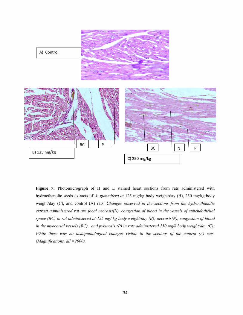
34
Figure 7: Photomicrograph of H and E stained heart sections from rats administered with
hydroethanolic seeds extracts of A. gummifera at 125 mg/kg body weight/day (B), 250 mg/kg body
weight/day (C), and control (A) rats. Changes observed in the sections from the hydroethanolic
extract administered rat are focal necrosis(N), congestion of blood in the vessels of subendothelial
space (BC) in rat administered at 125 mg/ kg body weight/day (B); necrosis(N), congestion of blood
in the myocarial vessels (BC), and pykinosis (P) in rats administered 250 mg/k body weight/day (C);
While there was no histopathological changes visible in the sections of the control (A) rats.
(Magnifications, all ×2000).
BC N P BC P
C) 250 mg/kg
B) 125 mg/kg
A) Control
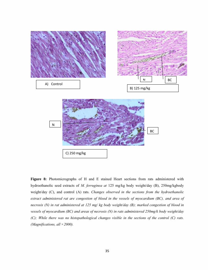
35
Figure 8: Photomicrographs of H and E stained Heart sections from rats administered with
hydroethanolic seed extracts of M. ferruginea at 125 mg/kg body weight/day (B), 250mg/kgbody
weight/day (C), and control (A) rats. Changes observed in the sections from the hydroethanolic
extract administered rat are congestion of blood in the vessels of myocardium (BC), and area of
necrosis (N) in rat administered at 125 mg/ kg body weight/day (B); marked congestion of blood in
vessels of myocardium (BC) and areas of necrosis (N) in rats administered 250mg/k body weight/day
(C); While there was no histopathological changes visible in the sections of the control (C) rats.
(Magnifications, all ×2000).
BC N A) Control
B) 125 mg/kg
BC
N
C) 250 mg/kg

36
Effect of hydroalcoholic seed extract of both plants on histopathology of the small
Intestine
Histological examinations of sections of the intestine from rats administered with the seeds
extract of A. gummifera at 125mg/kg in both male and female rats have shown inflammation
(I) and desquamation of the mucosa (D) (Figure 7). Small Intestine histology of both sex
administered with 250 mg/kg body weight have also shown desquamation and inflammation
of the mucosa, and submucosal atrophy (Figure 7).
Similarly, histological examinations of sections of the intestine from rats administered with
the seed extract of M. ferruginea at 125mg/kg for both male and female rat showed
desquamation and infiltration of the mucosa and submucosal atrophy (Figure 8). Intestine
histology of both sexes administered at 250 mg/kg also expressed desquamation and
infiltration of the mucosa and marked blood congestions in the submucosa (Figure 8).
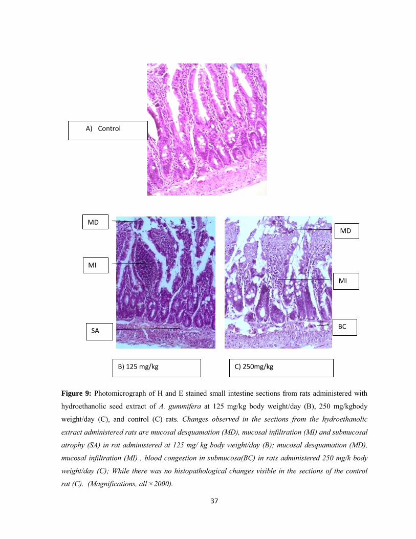
37
Figure 9: Photomicrograph of H and E stained small intestine sections from rats administered with
hydroethanolic seed extract of A. gummifera at 125 mg/kg body weight/day (B), 250 mg/kgbody
weight/day (C), and control (C) rats. Changes observed in the sections from the hydroethanolic
extract administered rats are mucosal desquamation (MD), mucosal infiltration (MI) and submucosal
atrophy (SA) in rat administered at 125 mg/ kg body weight/day (B); mucosal desquamation (MD),
mucosal infiltration (MI) , blood congestion in submucosa(BC) in rats administered 250 mg/k body
weight/day (C); While there was no histopathological changes visible in the sections of the control
rat (C). (Magnifications, all ×2000).
A) Control
MI
MD
d
SA
s
MD
MI
BC
B) 125 mg/kg C) 250mg/kg

38
Figure 10: Photomicrograph of H and E stained small intestine sections from rats administered with
hydroethanolic seed extract of M. ferruginea at 125 mg/kg body weight/day (B), 250 mg/kgbody
weight/day (C), and control (A) rats. Changes observed are: Desquamation of the epithelial layer of
the mucosa (MD), mucosal infiltration (MI), submucosal atrophy (SA) in rat administerd at 125 mg/
kg body weight/day (B); atrophy, desquamation and infiltration of the mucosa (MD,MA,MI) and
marked blood congestion in the submucosa(BCS) in rats administered 250 mg/kg body weight/day
(B); While there was no histopathological changes visible in the sections of the control rat (A).
(Magnifications, all ×2000).
A) Control
MD
MI
SA
B) 125 mg/kg
MI
BC
MD
C) 250 mg/kg
39
6. DISCUSSION
Albino rats were treated with different doses of seed extracts of A. gummifera and M.
ferruginea to study acute toxicity test. The result showed no sign of toxicity and death after
the aqueous extracts of both plants were orally administered to rats to the level of
3000mg/kg. The present study showed that the LD50 of M. ferruginea and A. gummifera were
found to be 3500 mg/kg and 4000 mg/kg body weight respectively as 50 % of the animals
died with 3500 mg/kg and 4000 mg/kg for M. ferruginea as well as A. gummifera seeds
extract treated groups, respectively. In other studies the aqueous leaf extract of A. chevalieri
also showed LD50 of greater than 3000 mg/kg body weight which is almost similar with the
hydoethanoic seed extract of A. gummifera [45]. Another study conducted on mice also
showed the LD50 of A. gummifera and M. ferruginea to be 2300 and 2500 mg/kg body weight
respectively [14].
The sub-chronic toxicity was studied by administering seeds extract of A. gummifera and M.
ferruginea to experimental animals (rats) for 90 days and showed no death and abnormal
signs throughout the study period. But during the 3rd week of administration of M.
ferruginea, one male rat was found dead with unknown reason because inspection of the
necropsy of the heart and small intestine showed no gross abnormality. Besides, the seeds
extracts of both plants had no harmful effect on body growth patterns of test groups. Except
for female rats whose body weight insignificantly decreased during the 9th week of
administration with seed extract of A. gummifera in a dose dependent manner. The body
weight of both the test and control groups of both sexes were increased as the duration of
treatment increased. Moreover, female rats during the 10th week of treatment with 250 mg/kg
body weight of A. gummifera seed extract showed statistically significant decrease in body
weight. The reason for this significant weight loses might be due to components of the
extract which might interfere with the digestive activity. Previous toxicity studies which were
done on administration of aqueous extract of Vernonia amygdalina [46] and administration
of aqueous extract of Clerodendrum myricoides on experimental animals also showed similar
result [47].
When there is underlying disease condition, it is common to see altered blood and
biochemical parameters [48]. So toxicities can be demonstrated by the presence of changes in
hematological as well as biochemical parameters [49]. The different hematological
40
parameters (RBC count, HCT (PCV), HGB, MCV, MCH or MCHC, and RDW-CV) are used
to assess types of anemia [27, 50]. RDW-CV, which is an automated parameter providing
information on the degree of variation of individual red cell size, has been used with the
traditional red cell indices in order to narrow down the possible causes of anemia in an
individual patient [51]. Anemia is mainly due to a decrease in RBC count and HGB content.
These alterations may be secondary to defective hematopoiesis, inefficient erytheropoiesis or
extensive destruction of red blood cells [52, 53].
A. gummifera seed extract administered at 125 and 250gm/kg body weight/ day to male rats
for 90 days did not decrease RBC count, PLT, WBC, LYMPH, MONO, EO, BASO, HCT
and HGB as compared with the male control group. This finding is in line with the result of
other study conducted on related species in which oral administration of saponins isolated
from Albizia lebbeck bark extract did not decrease hematological parameters. But neutrophil
is significantly increased in male rats which are treated with 250 mg/kg A. gummifera seed
extract as compared to the control. RDW–CV is also significantly increased at both doses as
compared to the male control. Even though the extract of this plant did not decrease MCV
and MCHC it significantly decreased the level of MCH at both doses as compared to the
male control. Based on alterations observed on these hematological parameters, A.
gummifera may slightly induce anemia in male rats. At both doses, the seed extract of A.
gummifera did not decrease total RBC count, PLT, WBC, LYMPH, MONO, EO, BASO,
HCT, HGB and RDW-CV in the female rats. But it significantly increased the neutrophil at
250 mg/kg as compared to the control. Seed extract of A. gummifera did not decrease the
level of MCH but, decreased MCV at 250 mg/kg administered group and MCHC at both
doses (125 and 250 mg/kg body weight) in the female rats as compared to the female control.
These findings also indicate the plant seeds extract might induce anemia in female rats.
Further researches on the seed extract of A. gummifera are required to validate the present
findings [54].
Sub-chronic (90 days) oral administration of M. ferruginea seed extract at 125 and 250
mg/kg body weight/day to male rats did not significantly affect total RBC count, WBC, PLT,
LYMPH, MONO, BASO, EO, HCT and HGB as compared to the male control group.
Besides, the levels of red blood cell indices MCV, MCHC, MCH were not affected and the
RDW-CV was not increased following administration of seeds extract of this plant to male
41
rats at both doses as compared to the male control. Sub-chronic (90 days) oral administration
of M. ferruginea seed extract at 125 and 250 mg/kg body weight/day to female rats did not
alter the blood parameters. Previous study done on the effect of ethanol leaf extract of
Millettia aboensis on hematological parameters of wister albino rats at dose lower than 2000
mg/kg body weight of the extract also showed similar results. But, at 250 mg/kg body weight
of oral administration of seed extract of M. ferruginea, MCHC and monocytes have
decreased as compared to female control. These may show the presence of a sub-chronic
inflammatory process [35].
When cells are exposed to toxic substance persistently they become irreversibly injured and
released their components into the surrounding blood vessels. These released cellular
components are known as biomarkers. By measuring and quantifying the different serum
biochemical profiles one can estimate the extent of tissue injury and know the type of tissue
which is injured [55]. The determination of creatine kinase is utilized in the diagnosis and
monitoring of myocardial injury. Following injury to the myocardium creatine kinase is
released from the damaged myocardial cells [56].
The present study showed that following the sub-chronic oral administration of seed extract
of A. gummifera at 250 mg/kg to male and female rats, the level of CK is increased by 12%
and 14.59%, respectively when compared to the male and female control groups. On the
other hand, seed extract of M. ferruginea with 125 mg/kg and 250 mg/kg increased serum
level of CK by 6.7% and 9.0% respectively in male rats when compared with the male
control group. But in female rats, this extract increased CK by 8.8% only at 250mg/kg when
compared to female control group. These findings are in agreement with the previous study
done on toxicological evaluation of aqueous leaf extract of Chromolaena odorata in male
Wister albino rats which showed significant increase in CK level at 538.5mg/kg when
compared to the control group [57]. The increase in serum level of CK in this study may
indicate that seed extracts of both plants have toxic effect on the hearts of male and female
rats at higher dose (250 mg/kg). In addition seed extract of M. ferruginea at lower dose (125
mg/kg) could also have deleterious effect on the heart of male rats.
ALT which is one of the serum biomarkers is localized in liver tissue, and trace amount is
found in skeletal muscle and heart tissue. ALT is localized in cytoplasm and mitochondria of

42
hepatocytes, skeletal myocytes and cardiomyocytes. If these tissues are injured because of
various reasons, ALT could leak out from damaged tissues and appears in the blood [58, 59].
AST is also a biomarker found in liver, heart, muscle, brain and kidney tissues. It is also
localized in the cytoplasm and mitochondria of various body cells. Leakage of AST to the
blood stream is mainly due to hepatocellular necrosis [58, 60]. ALP is localized in intestine,
liver, bile duct, bone, placenta, and kidney tissues. It is manly localized in the cell membrane
of various body cells. If there is injury to above mentioned organs there will be over
production AST and released in to the surrounding blood vessels [61, 62]. The levels of some
cellular products like Albumin are reduced in the blood following damage to their cells of
origin [63].
The study showed that sub-chronic (90 days) oral administration of seed extract of A.
gummifera at 125 and 250 mg/kg body weight to male and female rats did not significantly
affect serum biochemical parameters (Albumin, ALP, ALT, and AST) as compared to the
control groups. Previous study done on oral administration of aqueous leaf extract of Albizia
chevalieri and saponins (from Albizia lebbeck bark) to experimental rats also showed no
significant effect on serum biochemical parameters [45, 54]. The serum urea which was seen
increased at higher dose in male rats may show seed extract of A. gummifera might induce
renal toxicity. Serum urea was increased at 250 mg/kg body weight as compared to the male
control. This indicates that the seed extract of A. gummifera may induce renal toxicity in
male rats at higher doses as supported by previous study in which the serum levels of urea
and creatinine in different drug-induced nephrotoxicity were higher in males than females
[64-66].
The sub-chronic (90 days) oral administration of seed extract of M. ferruginea at 125 and 250
mg/kg body weight/day to male rats showed no significant alteration on serum biochemical
levels except for CK which is statistically elevated at both doses. Similar finding is also
observed in a study on cardiotoxicicty by oral administration of Rhododendri Mollis Flos
extract in rats [67]. An increase in the level of CK might show cardiotoxicity of the extract.
This is also evidenced by previous study that showed increased serum CK level in
Doxorubicin treated mice [68].

43
Whereas, in female rats, M. ferruginea seed extract significantly increases the level of CK,
ALP, ALT and Urea at the higher dose. But albumin and AST levels are not affected.
Biochemical parameters (albumin and AST) analyzed for female rats as compared to the
control at both doses was also normal. On the other hand, ALP, ALT and Urea were
statistically found significant at 250 mg/kg body weight compared to the female control. This
shows the seed extract of M. ferruginea may induce cardiac, hepatic and renal toxicity in
female rats at higher dose. Similar results were also obtained by other researchers with
aqueous extract of Tithonia diversifolia in rats [69], and Clerodendrum myricoides in mice
[47].
To assess the effect of these extracts on the weight of the heart and small intestine, the weight
of these 2 organs were measured and analyzed. The present study shows statically significant
decrease in the weight of the heart of treated rats as compared with the control. This finding
is in agreement with previous study done on Doxorubicin treated mice which showed
significant decrease in the weight of the heart of treated mice as compared with the control
[68]. But the extracts at both doses did not affect the weight of the small intestine of both
male and female rats.
To make toxicological study more soundable, besides hematological, biochemical and gross
morphological analysis, histopathology of the heart and small intestine was done.
Histopathology of the heart and small intestine assesses and investigates cellular injury to
these organs. Some of the possible histopathological alterations observed on heart tissue after
exposure to toxic agents include: blood congestion, myofibre degeneration, clumped nuclei,
eunucleated muscle fibers and pycnotic nuclei [70]. Likewise, histopathological alterations
evident in toxic small intestine may include, necrosis, degeneration like dilation of villi,
separated basal membrane, degeneration in epithelium and infiltration [71].
In the present study, after treatment of rats with 125 and 250 mg/kg of A. gummifera and M.
ferruginea sees extracts is completed, post mortum inspection and evaluation of organs of
rats were made and showed no abnormal gross finding on the heart and small intestine. But
light microscopic examination of the heart sections of both male and female rats which are
sub-chronically administered with 125 mg/kg of seed extract of A. gummifera showed blood
congestion and small area of necrosis. Whereas, the 250 mg/kg administered group showed
44
congestions of blood in the myocardium and focal cellular necrosis and pykinosis. In
addition, light microscopic examination of the heart sections of both male and female rats
which are sub-chronically administered with125 mg/kg of seed extract of M. ferruginea
showed congestions of blood in vessels, and small area of necrosis. At the 250 mg/kg,
besides congestion has shown major cellular necrosis and inflammation. Histopathological
alterations of the heart observed in this study are in agreement with other study done on
effect of Acacia nilotica pod extract on some biochemical parameters and histopathological
features in albino rats which showed marked hemorrhage and myocardial necrosis [72].
Histological examinations of sections of the intestine from rats administered with the seed
extract of A. gummifera at 125mg/kg in both male and female rats have shown inflammation
and desquamation of the mucosa. But at higher dose (250 mg/kg) besides mucosal
desquamation and inflammation, tissue sections of small intestine of male and female rats
have shown submucosal atrophy. Similarly, histological examinations of sections of the
small intestine from rats administered with the seed extract of M. ferruginea at 125mg/kg for
both male and female rat showed desquamation and infiltration of the mucosa and
submucosal atrophy. Intestinal histology of both sex administered at 250 mg/kg also
expressed desquamation and infiltration of the mucosa and marked blood congestions in the
submucosa. Histopathological alterations of small intestine of rats observed in this study are
in line with previous study done on histopathological and biochemical studies after
administration of aqueous extract of raw Aloe vera leaves to rats which showed
desquamation of the mucosa, mononuclear cellular infiltration and goblet cells hyperplasia
[48].
45
7. CONCLUSION AND rRECOMENDATION
7.1. Conclusion
Analysis of blood parameters for male rats treated with hydro-alcoholic seed extract
of A. gummifera at 125 and 250 mg/kg body weight showed decreased MCH and
increased RDW-CV, MCH, and increased RDW-CV.
Decreased MCHC at both doses and MCV at higher dose in female rats which may
indicate anemia can be developed in both male and female rats as a result of the
extract.
An increased neutrophil number at 250 mg/kg in male and female rats which may
show inflammatory process during the course of the treatment.
All serum chemistry analyzed in this study for the seeds extract of A. gummiffera
were found normal in both male and female rats, except for serum CK which is
elevated in male and female rats at 250mg/kg body weight.
An increase in serum urea level in male rats administered at 250 mg/kg body weight
of A. gummiffera seed extract.
The histopathological alterations observed in the heart and small intestine tissues of
male and female rats are indicatives of the toxic effect of seed extract of A.
gummiffera on these organs especially at higher doses.
Hematological parameters analyzed for the hydro-alcoholic seed extract of M.
ferruginea administered at 125gm/kg and 250gm/kg body weight to male and female
rats were found within the normal ranges.
A decrease in monocyte count in female rats is observed which may be due to
infiltration of monocytes to the injured tissue.
MCHC was also found decreased in female rats at the higher dose which is an
indicative of anemia.
An increased serum CK, ALT, ALP and urea at 250 mg/kg body weight which may
have toxic effect on multiple organs at its higher dose.
46
The sub-chronic (90 days) administration of seeds extract of M. ferruginea at 125 and
250 mg/kg body weight to male and female rats demonstrated histopathological
alterations on the heart and small intestines such as congestion of blood in the vessels
of myocardium,and area of necrosis at both doses;desquamation of the epithelial layer
of the mucosa, mucosal infiltration, submucosal atrophy at the lower dose and
atrophy, desquamation and infiltration of the mucosa and marked blood congestion in
the submucosa at the higher dose.
Those hematological, biochemical and histopathological alterations observed on the
studied organs and tissues may be resulted from toxic ingredients in the seeds extract
of both plants so further studies would be required to isolate the specific components
of the plants responsible for the toxicity in order to standardize the plants preparation
for maximum therapeutic benefit.
7.2. Recommendation
Further studies are recommended o to isolate the active ingredients responsible for the observedtoxicities o to describe the mechanism of action of the extract forthe toxic effects. o to examine the toxic effects of these plants on other organs using similar
animal model. o to assess the toxic effects of these plants on bloodparameters and
histopathology of internal organson other animal models.
8. STRENGTH AND LIMITATION OF THE STUDY
8.1. Strength
Effects of the seed extracts were investigated using multiple parameters (Hematological, Biochemical, Histopathological, Acute toxicity)
8.2. Limitation
Troponin which is one of cardiac injury markers is not included because of lack of reagents.

47
9. REFERENCES
1. WHO. National policy on traditional medicine and regulation of herbal medicines.
Report of a WHO global survey. Geneva. 2005; 5-8.
2. WHO. Traditional medicine strategy 2002-2005. WHO Geneva.2002;
WHO/EDM/TRM/2002.
Also available at: http://whqlibdoc.who.int/hq/2002/WHO_EDM_TRM_2002.1.pdf
3. Afolayan AJ and Yakubu MT. Effect of Bulbine natelensis baker stem extract on the
functional indices and histology of the liver and kidney of male wistar rats. Journal of
Medicinal Food.2009; 12(4): 814-820.
4. Ajibade VA and Famurawa D. Histological and toxicological effects of crude saponin
extract from phyllanthus niruri on organs in animal studies. Global Journal of
Medicinal Research. 2012; 12(1): 31-38.
5. Builders MI, Isichie CO and Aguiyi JC. Toxicity studies of the extracts of Parkia
biglobosa stem bark in rats. British Journal of pharmaceutical Research. 2012; 2(1):
1-16.
6. Ogunka-Nnoka CU, Uwakwe AA and Nwachoko NC. Serum enzyme and
histological studies of albino rat treated with ethanol/potash extract of Sorghum
bicolor leaf sheath. Indian Journal of Drugs and Diseases. 2012; 1(3): 74-78.
7. Fennell CW, Lindsey KL, McGaw LJ, Sparg SG, Stafford GI, Elgorashi EE, et al.
Assessing African medicinal plants for efficacy and safety: pharmacological
screening and toxicology. Journal of Ethnopharmacology. 2004; 94(3): 205-217.
8. Deribe K, Amberbir A, Getachew B, and Mussema Y.A historical overview of
traditional medicine practices and policy in Ethiopia. Ethiopian Journal of Health
Development. 2006; 20(2): 127-134.
9. Yadav R. Medicinal plants in folk medicine system of Ethiopia. Journal of Poisonous
and Medicinal Plants Research. 2013; 1(1): 001-00.
10. Schmidt L and Mwaura L. Albizia gummifera (J.F.Gmel.) C.A.sm. Seed leaflet. 2010;
No.141
11. Dias FDL and Takahashi CS. “Cytogenetic evaluation of aqueous extracts of the
medicinal plants Alpinia mutans rose (Zingerberaceae) and Pogostemon hyena nusm
48
benth (Labitae) on Wistar rats and Allium cepa (Liliaceae) root tip cells.”Brazilian
Journal of Genetics.1994; 17(2): 175–180.
12. Geyid A, Abebe D, Debella A, Makonnen Z, Aberra F, Teka F, et al. Screening of
some plantsof Ethiopia for their anti-microbial properties and chemical profiles.
Journal of Ethnopharmacology. 2005; 97: 421-427.
13. Woldemicheal T, Debella A, Abebe D, Tassew G, Biru T, Mekonnen Y, et al.
Screening of some medicinal plants of Ethiopia for their molluscidal activities and
phytochemical constituents. Pharmacology online. 2006; 3: 245-258.
14. Debella A, Taye A, Abebe D, Mudi K, Melaku D and Taye G. Screening of some
Ethiopian medicinal plants for Mosquito Larvicidal effects and phytochemical
constituents. Pharmacologyonline. 2007; 3: 231-243.
15. Tefera M, Geyid A and Debella A. In vitro anti-Neisseria gonorrhoeae activity of
Albizia gummifera and Croton macrostachyus. Revista CENIC, Ciencias Biológicas.
2010; 41: 1-11
16. Karunamoorthi K, Bishaw D and Mulat T. Toxic effects of traditional Ethiopian fish
poisoning plant Millettia ferruginea (Hochst) seed extract on aquatic macro
invertebrates. European Review for Medical and pharmacological sciences. 2009; 13:
179-185.
17. Kothai S and Befirdu G. Ethno botany and antimicrobial activities of medicinal plants
used for skin infections in Jimma, Ethiopia. Discovery Pharmacy. 2012; 2(4): 5-11.
18. Banzouzi JJ, Prost A, Rajemiarimiraho M and Ongoka P. Traditional uses of the
African Millettia species (Fabaceae). International Journal of Botany. 2008; 4(4):
406-420.
19. Taye B, Giday M, Animut A, Seid J. Antibacterial activities of selected medicinal
plants in traditional treatment of human wounds in Ethiopia. Asian Pacific Journal of
Tropical Biomedicine. 2011;10: 370- 375.
20. Ross H and Pawlina W. Histology: A Text and Atlas: with cell correlation and
molecular biology. 6th ed, Lippincott Williams and Wilkins. 2011; pp: 268-299; 628-
643; 699-720.
49
21. Everett N, Simmons B and Lasher E. Distribution of blood (Fe59) and plasma (I131)
volumes of rats determined by liquid nitrogen freezing. Circulation Research. 1956;
4: 419-424
22. Junqueira LC and Carneiro J. Basic Histology, 11th edition. The McGraw-Hill
Companies, Inc. 2005; pp: 223-237, 323-324.
23. Guyton AC and Hall JE. Text book of Medical Physiology, 11th edition. Elsevier
Saunders. 2006; pp 307-309, 859-862.
24. Tortora GJ and Derrickson B. Principles of Anatomy and Physiology, 12th edition.
John Wiley & Sons, Inc. 2009; pp: 114-132, 234-260.
25. Kreutz M, Krause S, Hennemann B, Rehm A and Andreesen R. Macrophage
heterogeneity and differentiation : defined serum-free culture conditions induce
different types of macrophages in vitro. Research Immunology.1992; 143: 107-115.
26. OTTESEN J. On the age of human white cells in peripheral blood. Zoophysiological
laboratory, department of biological isotope research, and the institute for theoretical
Physics. University of Copenhagen.1954; 76-93.
27. Hume R, Dagg J, Goldenberg A. Refractory anemia with dysproteinemia: Long term
therapy with low-dose corticosteroids. Blood.1973; 41(1): 27-35.
28. Aarts P, Banga J, Houwelingen H, Heethaar R and Sixma J. Increased red blood cell
deformability due to isoxsuprine administration decreases platelet adherence in a
perfusion chamber: a double-blind cross-over study in patients with intermittent
claudication. Blood.1986; 67(5): 1474-1481.B
29. Okon UA, Ita SO, Ekpenyong CE, Davies K. G and Inyang OI. Reduction of platelet
and lymphocyte counts and elevation of neutrophil counts in rats treated with aqueous
leaf extract of Ocimum gratissimum. African Journal of Biochemistry Research.
2011; 5(9): 303-306.
30. Grimm AF, Katele KV, Klein SA and Lin HL. Growth of rat heart Left ventricular
morphology and sarcomere length growth. Pubmed. 1973;37: 189-206.
31. Nzalak JO, Wanmi N, Imam J, Ali MN and Umosen AD. Anatomical and
Histological Studies of the Small Intestine of the African Giant Rat. Annals of
Experimental Biology. 2015; 3 (4):20-26.

50
32. Vigueras RM, Rojas-Castaneda J , Hernandez R, Reye G & Alvarez C. Histological
characteristics of the intestinal mucosa of the rat during the first year of life .Ltd.
Laboratory Animals.1999; 33: 393-400.
33. Koduru S, Grierson DS, and Afolayan AJ. “Antimicrobial activity of
Solanumaculeastrum.”Pharmaceutical Biology. 2006; 44(4): 283–286.
34. Tesfaye A, Tefera G, Giday M and Shibeshi W. Invivo Anti-Trypanosomal Activity
of the leaf Extracts of Albizia schimperiana against trypanosome congolense infection
in mice. Journal of Clinical and Experimental Pharmacology. 2015; 5:(2): 3-8.
35. Blessing M, Onyegeme O, Eugene N, Onyeike F, and OkeE.Effect of ethanol leaf
extract of Millettia aboensis on selected haematological indices of Wistar albino rats.
Global Advanced Research Journal of Medicinal Plants (GARJMP). 2013; 2(1): 004-
011.
36. Srivasta T and Chosdol K. Clinical enzymology and its application. Ansari Nagar
New Delhi-110029, 2007.
37. Ibrahim L. Effect of oral Administration of Albezia Lebbeck Aqueous Stem Bark
Extract on Some Liver Function Indices In Rats. The American Journal of Innovative
Research and Applied Sciences. 2014; Available at:www.american-jiras.com
accessed on April, 05, 2015
38. Zhao XN, Wang XF, Liao JB, Guo HZ, Yu XD, Liang JL, et al. Antifatigue Effect of
Millettiae speciosae Champ (Leguminosae) Extract in Mice. Tropical Journal of
Pharmaceutical Research March. 2015; 14 (3): 479-485.
39. Ajibade AJ, Fakunle PB, Adewusi MO and Oyewo OO. Some morphological
findings on the heart of adult Wistar rats following experimental artesunate
administration. Current research in Cardiovascular Pharmacology.2012;1:1-9.
40. Randrianarivo HR, Razafindrakoto AR, Ratsimanohatra HC, Randriamampianina
1LJ, Rajemiarimoelisoa CF, Ramamonjisoa L, et al. Toxic Effects of Seed
Methanolic Extracts of Endemic Albizia Species (Fabaceae) from Madagascar on
Animals. Journal of Life Sciences, ISSN 1934-7391, USA. 2014; 8(8): 676-689.
41. Sowayan NSA and Mahmoud NH. The Protective Effect of Grape Seed Extract on
Cardiotoxicity Induced by Doxorubicin Drug in Male Rats. Advances in Bioscience
and Biotechnology. 2014; 5: 1078-1089.
51
42. Ene AC, Atawodi SE, Fatihu MY and Adamu S. Pathological changes in some
organs of rats administered sub-chronic doses of chloroform extract of Artemisia
maciverae Linn. Adv. Agric. Biol. 2014;2 (2): 61-65.
43. Bystrykik IK. Automated Hematologic Analyzer Chair and Department of Laboratory
diagnostic. Medical University of Lublin. 2011; 24(3): 7.
44. http://www.who.int/medical_devices/en/index.html.accessed on July,12, 2015.
45. Saidu Y, Bilbis LS, Lawal M, Isezuo SA, Hassan SW, Abbas AY. Acute and sub-
chronic toxicity studies of crude aqueous extract of Albizzia chevalieri Harms
(Leguminosae). Asian J Biochem. 2007; 2(4): 224-36.
46. Amole O, lzegbu M, Onakoya J and Dada M. Toxicity studies of the aqueous extract
of Vernonia amygdalina. Biomedical Research. 2006; 17: 39-40.
47. Kebede H, Afework M, Makonnen E, Ergete W and Urga K. The effect of
Clerodendrum myricoides aqueous extract on blood, liver and kidney tissues of
mice.MEJS. 2011; 3 (2): 48-63.
48. Taiwo VO, Olukunle OA ,Ozoro IC and Oyejobi, AT. Consumption of Raw Aloe
Vera Leaves: Histopathological and Biochemical studies in Rat. Africa Journal of
Biomedical Research. 2005; 8: 169-178.
49. Bin-Jaliah I, Dallak M, Al-Hashem F, Nwoye L, Sakr H, Jamil A, et al. Derangement
of hemopoiesis and hematological indices in Khat (Catha edulis) - treated rats.
African Journal of Biotechnology. 2014; 13(2): 349-355.
50. Sagone AL, Lawrence JR, Balcerzak SP. Effect of smoking on tissue oxygen supply.
Blood. 1973; 4(6): 845-852.
51. Sysmex. Educational enhancement and development. Seed Hematology. 2012;
Available at: www.sysmex-europe.com accessed on May,05,2015.
52. Selmanoglu G, Barlas N and Songur S. Carbendazim-induced haematological,
biochemical and histopathological changes to the liver and kidney of male rats.
Journal of Human and Experimental Toxicology. 2001; 20: 625-630.
53. Choudhari C and Deshmukh P. Acute and sub-chronic toxicity study of Semecarpus
anacardium on hemoglobin percent and RBC count of male albino rat. Journal of
Herbal Medicine and Toxicology. 2007; 1: 43-45.
52
54. Gupta RS, Chaudhary R, Yadav RK, Verma SK, Dobhal MP. Effect of saponins of
Albizia lebbeck (L.) Benth bark on the reproductive system of male albino rats.
Journal of Ethnopharmacology. 2005; 96 (1-2): 31-36.
55. Hathway DE. Toxic action/toxicity. Biol Rev CambPhilos Soc. 2000; 75:95.
56. Kemp M, Donovani J, Higham H and Hooper J; Biochemical markers of myocardial
injury; British journal of anesthesia. 2004; 93(1): 63-73.
57. Asomugha RN, Okafor PN, Ijeha II, Orisakwe OE, Asomugha AL and Ndefo JC.
Toxicological Evaluation of Aqueous Leaf Extract of Chromolaena Odorata in Male
Wistar Albino Rats. Journal of Applied pharmaceutical Science. 2013;3 (13): 089-
092.
58. Dufour R, Lott J, Nolte F, Gretch D, Koff R, and Seeff L. Diagnosis and monitoring
of hepatic injury II. Recommendations for use of laboratory tests in screening,
diagnosis, and monitoring. National Academy of Clinical Biochemistry. 2000; 46(12):
2050–2068.
59. Amacher DE. A toxicologist’s guide to biomarkers of hepatic response. Human &
Experimental Toxicology. 2002; 21: 253- 262.
60. Ozer J, Ratner M, Shawc M, Bailey W and Schomaker S. The current state of serum
biomarkers of hepatotoxicity. Toxicology. 2008; 245: 194–205.
61. Saukkonen J, Cohn D, Jasmer R, Schenker S, Jereb J, Nolan C, et al. An official ATS
statement: hepatotoxicity of antituberculosis therapy. Am J Respir Crit Care Med.
2006; 174: 935–952.
62. Ramaiah S. A toxicologist guide to the diagnostic interpretation of hepatic
biochemical parameters. Food and Chemical Toxicology.2007; 45: 1551–1557.
63. Thapa B and Walia A. Liver function tests and their interpretation. Indian J Pediatr.
2007; 74 (7): 663-671.
64. Bennett WM, Parker RA, Elliott WC, Gilbert DN and Houghton DC. Sex-related
differences in the susceptibility of rats to gentamicin nephrotoxicity. Journal of
Infect Dis.1982; 145(3): 370-3.
65. Goodrich JA and Hottendorf GH. Tobramycin gender-related nephrotoxicity in
fischer but not sprague-dawley rats. ToxicolLett. 1995; 75(1-3): 127-31.
53
66. Laborin LR and Vargas CMN. Amphotericin B: side effects and toxicity. Rev
Iberoam Micol. 2009; 26(4): 223-7.
67. Dong Lc, Zhang XH, Ma J, Luo N, Song W and Lip HJ. The integrated
pharmacokinetics of major rhodojaponins correlates with the cardiotoxicity after oral
administration of Rhododendri Mollis Flos Extract in rats. Journal of
Ethnopharmacol. 2014; 157: 69-78.
68. Chen CT, Wang ZH, Hsu CC, Lin HH and Chen JH. In vivo protective effects of
Diosgenin against Doxorubincin-Induced Cardiotoxicity: Nutrients. 2015; 7: 4938-
4954.
69. Nerurkar P, Dragull K and Tang C. In vitro toxicity of kava alkaloid,
pipermethystine, in hep G2 Cells compared to kavalactones. Journal of Toxicological
Science. 2004; 79: 106-111.
70. Sharma S and Thakur A. Histological alterations induced in mice heart after Lead
acetate treatment. Int. Journal of PharmBioSci. 2015; 6(1): 984-990.
71. Adiguzel C andKalender Y. Lead Nitrate induced toxic effects on Small Intestine
Tissue in diabetic and non-diabetic rats. Gazl University Journal of Science. 2015;
28(4): 541-544.
72. Umaru B. some Biochemical parameters and Hitophathological features following
prolonged administration of aqueous pod extract of Acacia nilotica in albino rats.
Journal of medicinal plants Research. 2012;6(8): 1422-1428.
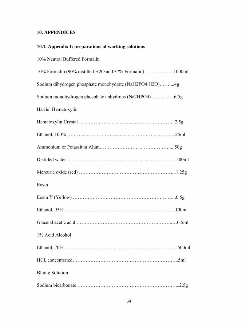
54
10. APPENDICES
10.1. Appendix I: preparations of working solutions
10% Neutral Buffered Formalin
10% Formalin (90% distilled H2O and 37% Formalin) ……………...1000ml
Sodium dihydrogen phosphate monohydrate (NaH2PO4.H2O)……....4g
Sodium monohydrogen phosphate anhydrous (Na2HPO4) …………..6.5g
Harris’ Hematoxylin
Hematoxylin Crystal …………………………………………………..2.5g
Ethanol, 100% …………………………………………………………25ml
Ammonium or Potassium Alum…………………………………….....50g
Distilled water ………………………………………………………….500ml
Mercuric oxide (red) …………………………………………………...1.25g
Eosin
Eosin Y (Yellow) ……………………………………………………....0.5g
Ethanol, 95% ……………………………………………………….….100ml
Glaceial acetic acid …………………………………………………..…0.5ml
1% Acid Alcohol
Ethanol, 70% ……………………………………………………………500ml
HCl, concentrated………………………………………………………..5ml
Bluing Solution
Sodium bicarbonate ……………………………………………………...2.5g

55
Ethanol……………………………………………………………………1000ml
Distilled water ……………………………………………………………500ml
10.2. Appendix II: Tissue processing schedules to form paraffin blocks for manual
technique
Fixation
Buffered formalin, 10% ……………………………………………..24 hrs
Washing
Running tape water………………………………………………......24 hrs
Dehydration
Alcohol, 70% …………………………………………………………1 hr
Alcohol, 80% …………………………………………………………1 hr
Alcohol, 95% …………………………………………………………1 hr
Absolute alcohol I …………………………………………………….1 hr
Absolute alcohol II…………………………………………………….1 hr
Clearing
Xylene I ……………………………………………………………….1 hr
Xylene II ………………………………………………………………1 hr
Infiltration (in paraffin oven)
Paraffin wax I 56 oC (52-64oc)…………………………………………1½ hrs
Paraffin wax II 56 oC(52-64oc)………………………………………..1½ hrs
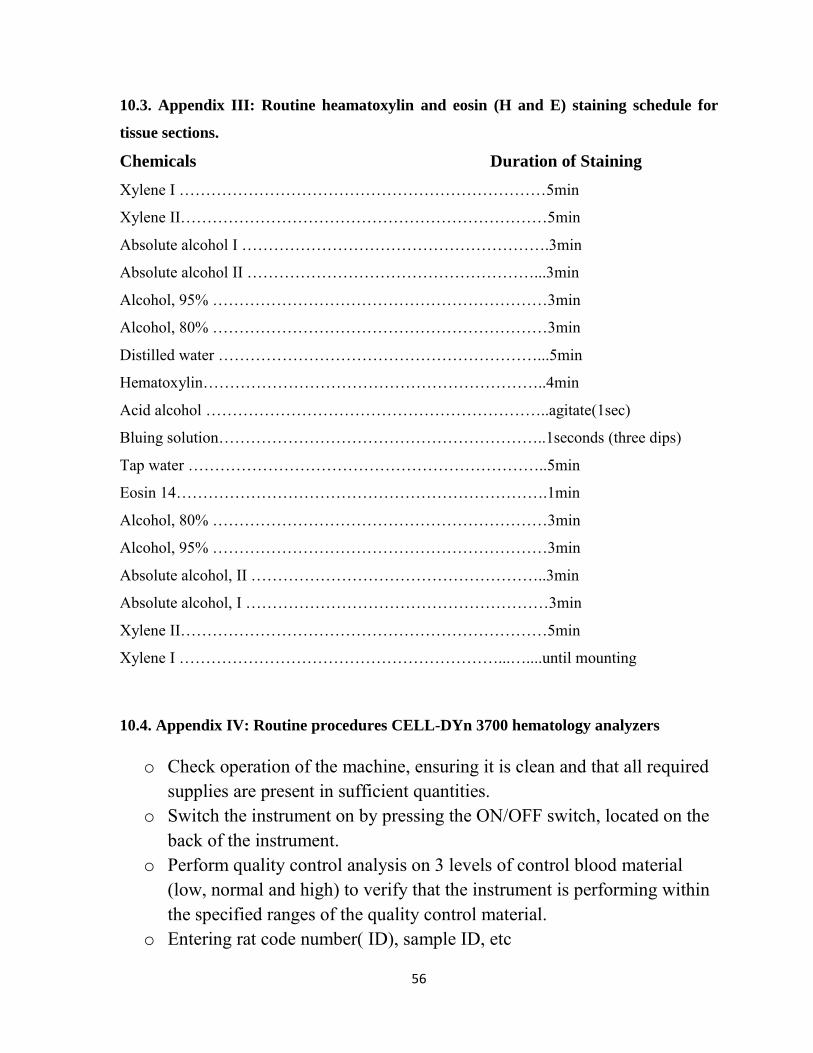
56
10.3. Appendix III: Routine heamatoxylin and eosin (H and E) staining schedule for
tissue sections.
Chemicals Duration of Staining
Xylene I ……………………………………………………………5min
Xylene II……………………………………………………………5min
Absolute alcohol I ………………………………………………….3min
Absolute alcohol II ………………………………………………...3min
Alcohol, 95% ………………………………………………………3min
Alcohol, 80% ………………………………………………………3min
Distilled water ……………………………………………………...5min
Hematoxylin………………………………………………………..4min
Acid alcohol ………………………………………………………..agitate(1sec)
Bluing solution……………………………………………………..1seconds (three dips)
Tap water …………………………………………………………..5min
Eosin 14…………………………………………………………….1min
Alcohol, 80% ………………………………………………………3min
Alcohol, 95% ………………………………………………………3min
Absolute alcohol, II ………………………………………………..3min
Absolute alcohol, I …………………………………………………3min
Xylene II……………………………………………………………5min
Xylene I ……………………………………………………...…....until mounting
10.4. Appendix IV: Routine procedures CELL-DYn 3700 hematology analyzers
o Check operation of the machine, ensuring it is clean and that all required
supplies are present in sufficient quantities. o Switch the instrument on by pressing the ON/OFF switch, located on the
back of the instrument. o Perform quality control analysis on 3 levels of control blood material
(low, normal and high) to verify that the instrument is performing within the specified ranges of the quality control material.
o Entering rat code number( ID), sample ID, etc
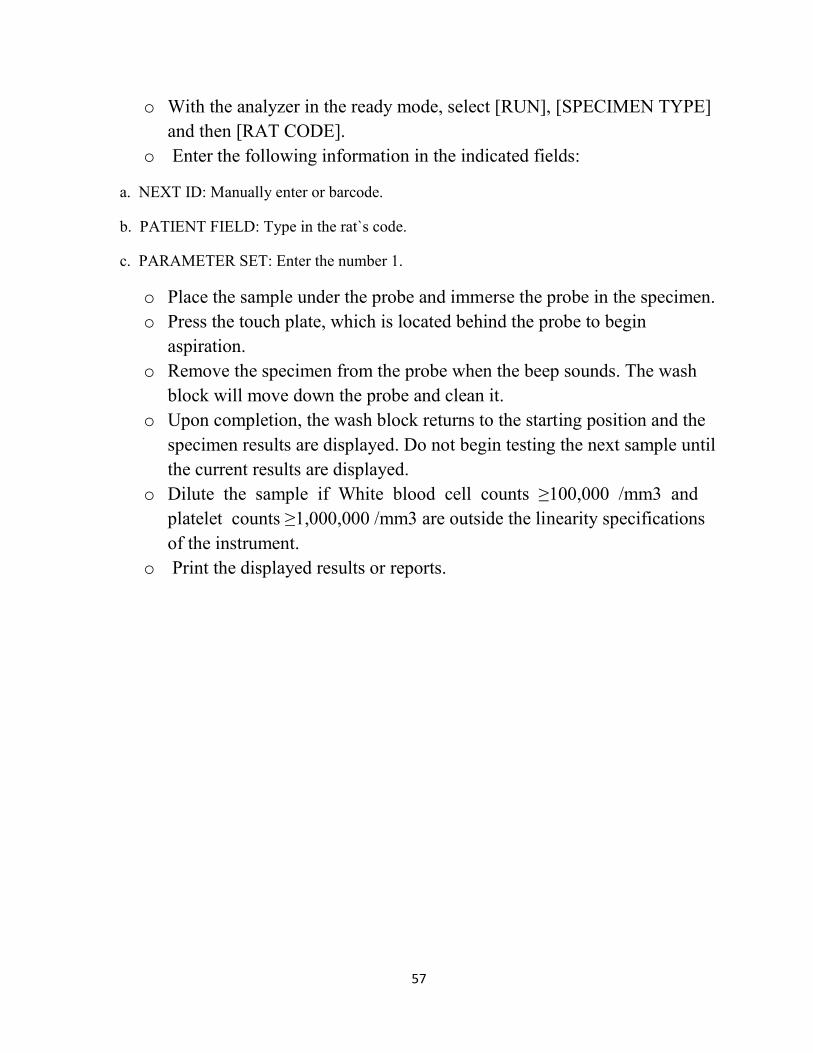
57
o With the analyzer in the ready mode, select [RUN], [SPECIMEN TYPE] and then [RAT CODE].
o Enter the following information in the indicated fields:
a. NEXT ID: Manually enter or barcode.
b. PATIENT FIELD: Type in the rat`s code.
c. PARAMETER SET: Enter the number 1.
o Place the sample under the probe and immerse the probe in the specimen. o Press the touch plate, which is located behind the probe to begin
aspiration. o Remove the specimen from the probe when the beep sounds. The wash
block will move down the probe and clean it. o Upon completion, the wash block returns to the starting position and the
specimen results are displayed. Do not begin testing the next sample until the current results are displayed.
o Dilute the sample if White blood cell counts ≥100,000 /mm3 and platelet counts ≥1,000,000 /mm3 are outside the linearity specifications of the instrument.
o Print the displayed results or reports.
58
Declaration
I, the undersigned, declare that this M.Sc. thesis is my original work, has not been presented
for a degree in this or any other university and that all sources of materials used for the thesis
have been duly acknowledged.
M.Sc. candidate: MollaGetu (B.Sc.)
Signature: __________________
Date of submission: __________________
This thesis has been submitted with our approval as advisors.
Advisor: Aster Tsegaye (MSc, PhD)
Signature: ____________________
Date: ____________________
Place: Addis Ababa, Ethiopia.
Advisor: Melaku Tamene (MSc, PhD candidate)
Signature: ___________________
Date: ___________________
Place: Addis Ababa, Ethiopia.
Top Related