


Languages
Pages
Legal

Int. J. Mol. Sci. 2013, 14, 13626-13644; doi:10.3390/ijms140713626
International Journal of
Molecular Sciences ISSN 1422-0067
www.mdpi.com/journal/ijms
Article
Early Phenylpropanoid Biosynthetic Steps in Cannabis sativa: Link between Genes and Metabolites
Teresa Docimo 1, Roberto Consonni 2, Immacolata Coraggio 1 and Monica Mattana 1,*
1 Institute of Agricultural Biology and Biotechnology, CNR, Via Bassini 15, Milan 20133, Italy;
E-Mails: [email protected] (T.D.); [email protected] (I.C.) 2 Institute for Macromolecular Studies, NMR Department, CNR, Via Bassini 15, Milan 20133, Italy;
E-Mail: [email protected]
* Author to whom correspondence should be addressed; E-Mail: [email protected];
Tel.: +39-02-2369-9677; Fax: +39-02-2369-9411.
Received: 13 May 2013; in revised form: 13 June 2013 / Accepted: 14 June 2013 /
Published: 28 June 2013
Abstract: Phenylalanine ammonia-lyase (PAL), Cinnamic acid 4-hydroxylase (C4H) and
4-Coumarate: CoA ligase (4CL) catalyze the first three steps of the general phenylpropanoid
pathway whereas chalcone synthase (CHS) catalyzes the first specific step towards flavonoids
production. This class of specialized metabolites has a wide range of biological functions
in plant development and defence and a broad spectrum of therapeutic activities for human
health. In this study, we report the isolation of hemp PAL and 4CL cDNA and genomic
clones. Through in silico analysis of their deduced amino acid sequences, more than an
80% identity with homologues genes of other plants was shown and phylogenetic
relationships were highlighted. Quantitative expression analysis of the four above
mentioned genes, PAL and 4CL enzymatic activities, lignin content and NMR metabolite
fingerprinting in different Cannabis sativa tissues were evaluated. Furthermore, the use of
different substrates to assay PAL and 4CL enzymatic activities indicated that different
isoforms were active in different tissues. The diversity in secondary metabolites content
observed in leaves (mainly flavonoids) and roots (mainly lignin) was discussed in relation
to gene expression and enzymatic activities data.
Keywords: Cannabis sativa; phenylalanine ammonia lyase; 4-Coumarate: CoA ligase;
cinnamic acid 4-hydroxylase; chalcone synthase; phenylpropanoid; secondary metabolism;
expression analysis; NMR metabolic profiling
OPEN ACCESS

Int. J. Mol. Sci. 2013, 14 13627
1. Introduction
The Cannabaceae family, order Rosales, includes the two economic important genera, Humulus
and Cannabis, which evolution, due to long time cultivation, has been strongly influenced by man
pressure. Humulus is mainly used in the brewery industry whereas the annual plant hemp
(Cannabis sativa), that is present as monoecious and dioecious plants, has multiple applications such
as production of fibre, oil and narcotic resins [1]. Its cultivation as fibre crop remains one of the oldest
in the word. After a decline in cultivation during the 19th century, in the last decades there has been an
emerging interest toward non-food crops and increasing attention on the use of natural fibres [2].
Low tetrahydrocannabinol (THC) cannabis plants show a wide range of applications either in
human consumption or industrial uses. In particular, such plants are mostly cultivated for fibre
production and vegetable oil used for food [3–5]. Nevertheless, the main interest for cannabis is linked
to the pharmacological activity of cannabinoid compounds. Therefore, studies on secondary
metabolism in Cannabis sativa have been focalized on a cannabinoid biosynthetic pathway that has
been partially elucidated [1,6,7]. However, hemp plants, beside the cannabinoids, produce a number of
other specialized metabolites directly or indirectly derived from phenylpropanoid pathway.
Figure 1. Phenylpropanoid pathway in Cannabis sativa. PAL, phenylalanine ammonia
lyase; TAL, tyrosine ammonia lyase; C4H, cinnamic acid 4-hydroxylase; 4CL, 4-coumaric
acid: CoA ligase; CHS, chalcone synthase; CHI, chalcone isomerase; FS flavonol
synthase, F3’H flavonol 3’ hydroxylase; COMT, caffeic acid O-methyltransferase;
CCR, cinnamoyl-CoA reductase; CAD, cinnamyl alcohol dehydrogenase.
Because of the commercial interest for their application in pharmacological and other industrial
fields, phenylpropanoids biosynthesis and functions have been intensively studied in many species. In
the general biosynthetic scheme (Figure 1), phenylalanine, derived from the shikimate pathway, is
converted by phenylalanine ammonia-lyase (PAL, EC 4.3.1.5) into cinnamic acid, which after
hydroxylation by cinnamate-4-hydroxylase (C4H, EC 1.14.13.11) to p-coumaric acid, is converted in
p-coumaroyl CoA by addiction of a CoA thioester by a 4-Coumarate: CoA ligase enzyme (4CL,

Int. J. Mol. Sci. 2013, 14 13628
E.C 6.2.1.12). This high energy intermediate is funnelled into one of the branched pathways leading to
several classes of compounds involved in many functions such as cell wall constituents (lignins),
pigments (flavonoids, antocians), UV protectant (coumarins, flavonoids), plant defence (isoflavonoids,
furano-coumarins) [8,9].
PAL, the first enzyme involved in phenylpropanoid derivative metabolism, is one of the
most extensively studied for its crucial function as a branch point between primary and
secondary metabolism [10].
Since its discovery in Hordeum vulgare, PAL has been identified in plants including certain algae,
fungi, yeast and prokaryotes, whereas, to our knowledge, there have been no reports in animals [11–17].
PAL genes, in plants, occur in multigene families usually of 2–6 members reaching a dozen or more in
few species such as tomato and potato [18–22]. In most cases the PAL genes contain one intron at a
conserved insertion site [23]. The distinct members of PAL gene family encode for specific isoforms
that are expressed differently during plant development, in different tissues and in response to
stress stimuli [10].
The slow irreversible reaction catalyzed by C4H is located at a branching strategic point as its
product (p-coumaric acid) that can be diverted to flavonoids or lignin biosynthesis through the action
of 4CL and COMT, respectively. C4H cDNA and genomic clones have been isolated in many plant
species, i.e., Arabidopsis, bean, Populus, rice, citrus [24–29]. C4H encoded protein, belongs to the
cytochrome P450 superfamily [30] and mRNA level and enzyme activity are both regulated by a
plethora of stimuli, such as wounding, pathogen attack and light [31].
The third enzyme of the general phenylpropanoid pathway is the 4CL, which is involved in the
formation of Co-A-esters of cinnamic acids. In both angiosperm and gymnosperm the 4CL genes are
present as a family with multiple members differentially regulated and possibly involved in specific
biological processes [32–35]. This hypothesis is strongly supported by the finding that in several
species the members of 4CL family vary for their expression pattern and for their ability to utilize
different substrates [32,35]. For instance, the expression pattern of the five 4CLs rice genes differs
with respect to the tissues, developmental stage and stress response, and the five corresponding
enzymes show distinct kinetic properties in function of the used substrate. On the basis of the obtained
results, the authors conclude that only the Os4CL2 is associated with flavonoid biosynthesis whereas
the others are involved in lignin synthesis [34].
Another key enzyme involved in the flavonoid biosynthesis pathway is chalcone synthase (CHS),
which catalyzes iterative decarboxylative condensations of malonyl unit with a CoA-linked starter
molecule. This protein belongs to the superfamily of plant type III polyketide synthase (PKSs) [36,37].
In most angiosperm, including Cannabis sativa, CHS constitutes a multigene family and its expression
is induced in response to a wide range of stimuli such as UV light, pathogens, elicitors and
wounding [38–40].
Although secondary metabolism in hemp has been deeply investigated, few studies have been
focalized on phenylpropanoids. To our knowledge, the only report deeply looking into genes involved
in hemp phenylpropanoid pathway focuses on lignine rather than secondary metabolite biosynthesis.
Among the sequences found as differentially expressed between core and bast hemp fibers, the authors
identified four: PAL-, one C4H- and two 4CL- ESTs [40].
Int. J. Mol. Sci. 2013, 14 13629
Taking advantage of the EST sequences present in GenBank, we isolated, from Cannabis sativa
var. Futura, the full size cDNAs (KC970300 and KC970301) and the relative genomic sequences
corresponding to one PAL (KC970302) and one 4CL (KC970303) gene. Furthermore, we compared, in
different plant tissues, the expression pattern of PAL, C4H, 4CL and CHS, the enzymatic activities of
PAL and 4CL and the aromatic compounds content through the use of the wide spectrum chemical
analysis technique 1H NMR. Finally, we discussed the expression and enzymatic data with regard to
the accumulation of phenolic compounds, including lignin.
2. Results and Discussion
2.1. Isolation and Characterization of Cannabis Sativa Full-Length PAL and 4CL cDNAs
Based on the PAL hemp EST sequence (EC855392), specific primers were designed for 5' and 3'
end RACE-PCR. A nucleotide fragment of 2270 bp was cloned and, after sequencing, an open reading
frame (ORF) of 2124 bp was confirmed. The ORF, present in the assumed PAL cDNA, encodes for a
putative protein of 708 aa with predicted molecular mass of 77.09 kD and isolectric point at pH 6.24.
Full length cDNA sequence was used to search homologous sequences via BLAST algorithm [41]
in the National Center for Biotechnology Information (NCBI). The similarity search analysis unequivocally
indicated this cDNA as a PAL encoding sequence (CsPAL, Acc. Num. KC970300). Indeed, it showed a
high degree of homology with PAL of other species: namely, CsPAL amino acid sequence shared an 89%
identity with Ricinus communis and Vitis vinifera being also very similar to Morus alba, Jatropa curcas,
Trifolium pratense, Manihot esculentia and Populus trichocarpa (87%–88%, identity, Figure S1).
All these sequences contained, as revealed by Prosite Scan Available online: (http://www.expasy.ch/
tools/scanprosite/) [42], the conserved active site (GTITASGDLVPLSYIA aa 210–235) including the
invariable active site motif, the Ala-Ser-Gly triad, which may be converted into a 3,5-dihydro-5-
methylidine-4h-imidazol-4-one (MIO) prosthetic group [43,44]. Furthermore, the Tyr 363 residue
involved in the proton release, the Gly 506 residue in the active site pocket and the post transcriptional
phosphorilation site Thr 561, involved in the modulation of PAL activities, were also conserved
(Figure S1) [45], thus suggesting that CsPAL protein could be enzymatically active.
To better understand the phylogenetic relationship between PAL and related genes from other plant
species and fungi, CsPAL amino acid sequence was aligned with sequences with higher score of
similarity and with all members of PAL families belonging to several species (Arabidopsis thaliana,
Oryza sativa, Vitis vinifera, Trifolium pratense, Populus trichocarpa, etc.). Namely, 73 PAL
sequences were included to generate a maximum likelihood phylogenetic tree with MEGA 5 program.
As shown in Figure 2b, three main groups are evident: dicots, monocots and fungi. However, some
single sequences of both mono and dicots did not belong to such clusters, suggesting that they derive
from a very ancient duplication (i.e., Arabidopsis, rice). In the large dicots cluster, the family members
of some species (i.e., Vitis vinifera, Trifolium pratense) grouped together suggesting a recent
duplication following the species formation. Conversely, in other species, such as Coffea canephora,
the single members belonged to distinct sub-groups indicating that their duplication precedes
speciation and evoking different specialized functions. This clustering is in agreement with that
reported by Lepelley [22]. Interestingly, specific functions, although partially overlapped, characterize

Int. J. Mol. Sci. 2013, 14 13630
PAL family members even in species where they clusterize strictly. A clear example derives from the
deep investigation on the seven PAL genes of Cucumis sativus where a pattern of gene expression
(tissue and stress responsive) specific for the different members has been highlighted [20].
Figure 2. Gene structure and phylogenetic analysis of hemp PAL. (a) Representation of
CsPAL genomic sequence (KC970302) and cDNA (KC970300). Black and white boxes
indicates CsPAL coding sequence (white box indicates region not covered by PAL genomic
clone), grey box indicates the intronic region, solid line represents the fragment used as
probe for Southern-blot; (b) The PAL proteins identified from other species were aligned
using Clustal X, and the PAL phylogeny was constructed using the neighbor-joining
method with the MEGA 5.1 program. The branch lengths are indicated above the branch
lines. The clades indicate monophyletic groups of dicots, monocots and fungi. CsPAL is
highlighted by a red diamond. * Indicates similarities of C. sativa ESTs (EC 5006722/ EC
55372 and EC JK497725) to M. alba and C. roseus PAL, respectively. Accession numbers
for protein sequences used to build the PAL tree are reported in Table S2.
Int. J. Mol. Sci. 2013, 14 13631
CsPAL was located in the large dicots group, specifically in the subgroup of A. thaliana PAL1 and
PAL2. These two Arabidopsis genes are involved in lignifications of the vascular system and in
phenylpropanoids synthesis in response to stress and pathogens [46–48]. A blastn analyses of the four
hemp PAL ESTs isolated by van den Broeck et al. [40] vs. nt database indicated that they correspond
with at least two different genes. Their putative positions, based on the hit homology as indicated in
Figure 2b, suggest, for at least one of them, a very early duplication.
As above described for CsPAL, also Cs4CL (KC970301) was isolated taking advantage of the EST
present in the database (EC855340). Specific oligos designed at the 5' and 3' ends of 4CL EST
sequence allowed the isolation by RACE-PCR of the full size cDNA. The sequence analysis of the
cloned fragment identified a 1653 bp ORF, encoding for a putative protein of 553 aa, with 60.72 kD
molecular mass and isoelectric point at 5.7 pH.
Cs4CL contains the strictly conserved Box I (228–230 aa, AMP binding domain, PYSSGTTGLPKG),
Box II motif (425–433 aa, GEICIRG) and the hydroxycinnamate pocket (276–384 aa) responsible for
the substrate binding (Figure S2) [49,50]. Blastp analysis pointed out high similarity of the putative
Cs4CL with 4Cl from Humulus lupulus (91% identity) and with Sorbus aucuparia, Pyrus pyrifolla,
Betula platyphylla, Medicago tranculata, Ruta graveolans (81%, 81% 78%, 75%, 74%, respectively).
The high degree of homology with other 4CL proteins and the presence of conserved functional
features indicates that Cs4CL encodes for an enzymatically active protein.
In order to analyze the relatedness between Cs4CL and other 4CLs from plants, mosses and fungi,
44 protein sequences were subjected to a neighbor joining analysis. Within plant sequences, we included
members of 4CL families of selected species, (Arabidopsis thaliana, Oryza sativa, Glycine max,
Populus trichocarpa, etc.) and the 4CL protein sequences with higher score of similarity to Cs4CL. As
displayed in Figure 3b, a phylogenetic tree was constructed based on the NJ analysis results.
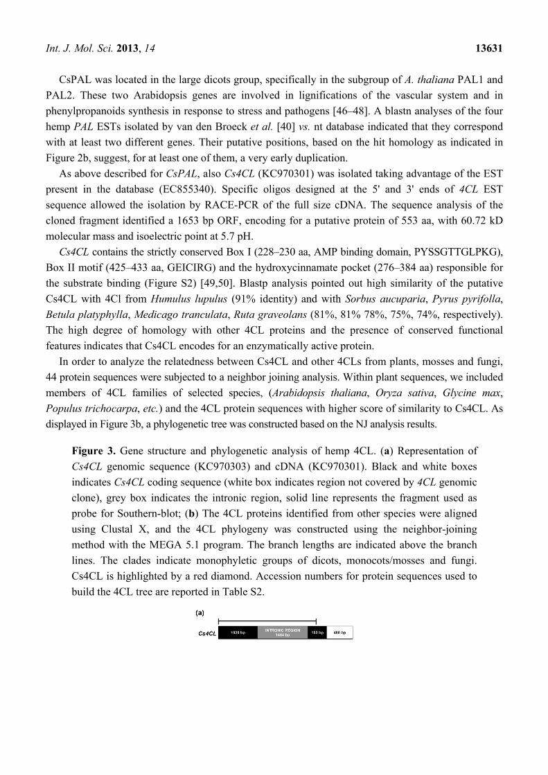
Figure 3. Gene structure and phylogenetic analysis of hemp 4CL. (a) Representation of
Cs4CL genomic sequence (KC970303) and cDNA (KC970301). Black and white boxes
indicates Cs4CL coding sequence (white box indicates region not covered by 4CL genomic
clone), grey box indicates the intronic region, solid line represents the fragment used as
probe for Southern-blot; (b) The 4CL proteins identified from other species were aligned
using Clustal X, and the 4CL phylogeny was constructed using the neighbor-joining
method with the MEGA 5.1 program. The branch lengths are indicated above the branch
lines. The clades indicate monophyletic groups of dicots, monocots/mosses and fungi.
Cs4CL is highlighted by a red diamond. Accession numbers for protein sequences used to
build the 4CL tree are reported in Table S2.
Int. J. Mol. Sci. 2013, 14 13632
Figure 3. Cont.
In agreement with previous reported data, the phylogenetic analysis revealed distinct fungi,
monocots, mosses and dicot clades; interestingly, an additional cluster containing both mono and dicot
genes emerged, indicating a very ancient duplication [34,35,51]. The Cs4CL grouped within the more
closely related dicot specific clade, being very similar to Humulus lupulus 4CL (Figure 3b). As
described for PAL, the members of the 4CL family of several species also clusterised in different
clades. Moreover, the differences in the expression profiles and substrates affinity suggest specific
functions for the 4CL isoforms, as reported, for instance, in soybean and rice [35,49,51].
2.2. CsPal and Cs4CL Genomic Sequences
After PCR amplification of genomic DNA, with primers designed on specific regions of the cDNA
sequences (CsPAL1, CsPAL2 and Cs4CL1, Cs4CL2, respectively), CsPAL (KC970302) and Cs4CL
(KC970303) genomic fragments of 2847 and 2627 bp were isolated. The complete sequences of three
independent recombinants of each gene highlighted the identity of the exon regions with the cDNAs.
In both genes the coding sequences were interrupted by the presence of at least one intron. Most PAL
genes consist of two exons and one intron, with variable size, in the highly conserved position at an
arginine encoding codon [19,23,52,53]. Accordingly, in the CsPAL here described, the single phase 2
intron of 1253 bp starts at nucleotide 377 within the conserved arginine codon.
In regard to 4CL genes, most genomic sequences of Angiosperm contain several introns that are
neither conserved in position nor in length [35,54]. In the Cs4CL one intron of 1454 bp was found
starting at nucleotide 1021 within a glutamine codon.
To estimate the PAL and 4CL copy number in the hemp genome, a southern blot analysis was
performed. Genomic DNA of hemp leaves was digested with the EcoRI restriction enzyme, which
does not cut in both CsPAL and Cs4CL genes and hybridized with 32P labelled probes corresponding to
almost the entire coding sequences (1821 and 1143 bp for PAL and 4CL, respectively (Figures 2a and 3a).
Two PAL and five 4CL genes seemed to be present in C. sativa (Figure S3). Our findings agree
with the presence of PAL and 4CL small gene families as extensively reported in literature for other
species [18–22,32–35,55].
Int. J. Mol. Sci. 2013, 14 13633
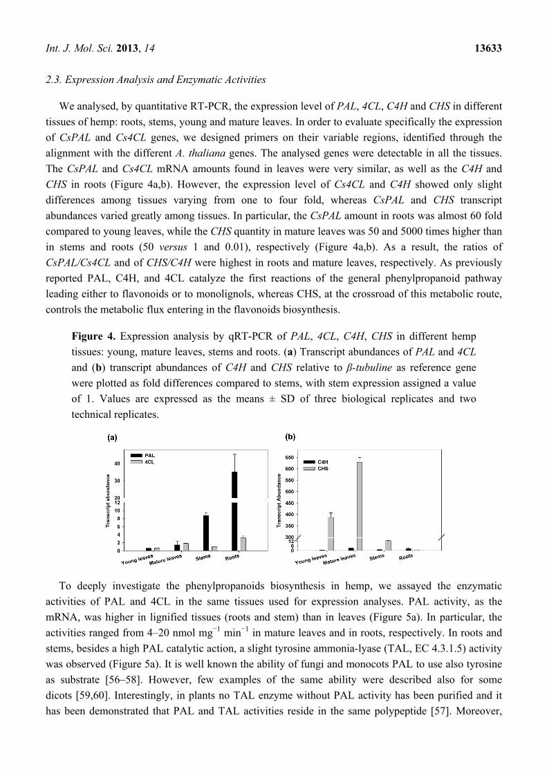
2.3. Expression Analysis and Enzymatic Activities
We analysed, by quantitative RT-PCR, the expression level of PAL, 4CL, C4H and CHS in different
tissues of hemp: roots, stems, young and mature leaves. In order to evaluate specifically the expression
of CsPAL and Cs4CL genes, we designed primers on their variable regions, identified through the
alignment with the different A. thaliana genes. The analysed genes were detectable in all the tissues.
The CsPAL and Cs4CL mRNA amounts found in leaves were very similar, as well as the C4H and
CHS in roots (Figure 4a,b). However, the expression level of Cs4CL and C4H showed only slight
differences among tissues varying from one to four fold, whereas CsPAL and CHS transcript
abundances varied greatly among tissues. In particular, the CsPAL amount in roots was almost 60 fold
compared to young leaves, while the CHS quantity in mature leaves was 50 and 5000 times higher than
in stems and roots (50 versus 1 and 0.01), respectively (Figure 4a,b). As a result, the ratios of
CsPAL/Cs4CL and of CHS/C4H were highest in roots and mature leaves, respectively. As previously
reported PAL, C4H, and 4CL catalyze the first reactions of the general phenylpropanoid pathway
leading either to flavonoids or to monolignols, whereas CHS, at the crossroad of this metabolic route,
controls the metabolic flux entering in the flavonoids biosynthesis.
Figure 4. Expression analysis by qRT-PCR of PAL, 4CL, C4H, CHS in different hemp
tissues: young, mature leaves, stems and roots. (a) Transcript abundances of PAL and 4CL
and (b) transcript abundances of C4H and CHS relative to β-tubuline as reference gene
were plotted as fold differences compared to stems, with stem expression assigned a value
of 1. Values are expressed as the means ± SD of three biological replicates and two
technical replicates.
To deeply investigate the phenylpropanoids biosynthesis in hemp, we assayed the enzymatic
activities of PAL and 4CL in the same tissues used for expression analyses. PAL activity, as the
mRNA, was higher in lignified tissues (roots and stem) than in leaves (Figure 5a). In particular, the
activities ranged from 4–20 nmol mg−1 min−1 in mature leaves and in roots, respectively. In roots and
stems, besides a high PAL catalytic action, a slight tyrosine ammonia-lyase (TAL, EC 4.3.1.5) activity
was observed (Figure 5a). It is well known the ability of fungi and monocots PAL to use also tyrosine
as substrate [56–58]. However, few examples of the same ability were described also for some
dicots [59,60]. Interestingly, in plants no TAL enzyme without PAL activity has been purified and it
has been demonstrated that PAL and TAL activities reside in the same polypeptide [57]. Moreover,

Int. J. Mol. Sci. 2013, 14 13634
different PAL isoforms belonging to the same species have different substrate specificity as reported
for Bambusa oldhamii where a slight, a clear and no TAL activity were measured for BoPAL2,
BoPAL4 and BoPAL1, respectively [58]. Our results on TAL activities in lignified tissues indicated
the action of an additional PAL isoform in such tissues.
Figure 5. Specific activities of PAL and 4CL in different hemp tissues: young, mature
leaves, stems and roots. (a) PAL and TAL activities were measured toward phenylalanine
and tyrosine substrates, respectively. (b) 4CL activities were measured toward p-coumaric,
cinnamic, caffeic, ferulic, sinapic acids substrates. Specific activities are expressed as the
mean values ± SD of three biological replicates.
The range of substrates used by 4CLs varies within and between plant species [34,35,51].
Therefore, the 4CL activity using the five known substrates (coumaric, sinapic, cinnamic, caffeic and
ferulic acids) was compared in roots, stems and leaves. As shown in Figure 5b, in all tissues the lowest
activity was displayed toward sinapic acid whereas with coumaric acid the maximum value was
reached. However, each tissue presented a specific pattern of substrate preference: leaves, beside
cumarate, exhibited an almost marginal activity with the other substrates; in roots significant activity
was observed, also toward caffeate and ferulate; and in stem, the activity toward coumarate and
caffeate was comparable. These data suggested that different 4CL genes were active in the analysed
tissues. Indeed, it has been reported that the 4CL genes have characteristic expression profiles with
respect to tissues and environmental stimuli [34,35,51,61]. Therefore, the differences in gene
expressions and enzymatic activities observed in cannabis tissues suggested that distinct branches of
phenylpropanoids pathway may be preferentially followed in green and lignified organs ([36–38] and
references therein).
2.4. Aromatic Compounds
To investigate whether the differences found in gene expression and enzymatic activity of the
analysed hemp tissues reflected specific metabolic profiles, monodimensional 1H NMR technique was
employed. Conventionally, 1H NMR spectrum is divided into three regions: the aliphatic region
(between 0.8 and 4 ppm) contains peaks corresponding to amino and organic acids, the anomeric
region (between 4 and 5.5 ppm) includes peaks belonging to the anomeric protons of saccharides and
the aromatic or phenolic region (between 5.5 and 8.5 ppm) comprising aromatic compounds.

Int. J. Mol. Sci. 2013, 14 13635
In order to verify a possible connection in the phenylpropanoid pathway between gene expression
and metabolites accumulation, we analysed the aromatic region of the 1H NMR spectra of roots (RT),
stems (ST) mature and young leaves (ML and YL), as reported in Figure 6a–d.
Figure 6. Aromatic region of 1H-NMR spectra from different hemp tissues:
(a) roots, (b) stems, (c) young leaves, (d) mature leaves. 1: trigonellin, 2: formiate,
3: apigenin-7-O-glucoside, 4: cytidine, 5: unknown, 6: luteolin-7-O-glucoside, 7: fumarate,
8: unknown, 9: unknown.
Resonance assignment was established by the aid of TOCSY and HSQC spectra and by comparison
with spectra of standard molecules. The metabolic content was in agreement with previous
findings [1,62].
As shown in Figure 6, the most relevant signals were due to flavonoidic structures. The signals at
7.95 and 7.98 ppm coupled to signals centered at 7.01 and 7.03 ppm, and signals at 6.89, 6.75, 6.59 are
typical moieties of flavonoids, confirmed as apigenin-7-O-glucoside and its derivatives on the basis of
the observed correlations in the TOCSY and HSQC spectra. The signals at 7.50, 7.02, 6.75 and
6.59 ppm identified luteolin-7-O-glucoside. Other signals due to cytidine (5.87, 5.91 and 7.96 ppm),
fumarate (6.55 ppm) formiate (8.20 ppm) and trigonelline (9.17, 8.89 and 8.13 ppm) were detected in a
smaller amount, whereas signal at 7.87 ppm and the one centered at 6.16 ppm corresponded to
unidentified compound.
The metabolic profile of the aromatic region was almost identical in young and mature leaves,
showing a higher compounds concentration in the latter (Figure 6c,d). Moreover, the same metabolites
were detected in stems at a very low amount (Figure 6b). Through the integral values of flavonoidic
signals in the different tissues, we evaluated the relative amount of apigenin and luteolin using ML
content as reference. Namely, apigenin content was 68.5% and 11.5% in YL and ST, respectively,
whereas luteolin was 98.4% and 31.8%. According to previously reported data [1], roots spectra
showed the complete absence of flavonoids; moreover, a specific accumulation of another unrelated
compound still under investigation was highlighted (Figure 6a).
Int. J. Mol. Sci. 2013, 14 13636
Although in roots no phenylpropanoids were present, PAL and 4CL expression and enzymatic
activities were higher than in the other analyzed tissues; therefore, the level of total lignin was
evaluated. As expected, the amount of total lignin varied among the tissues, being about three times
higher in roots than in young leaves. In detail, the content of lignin found in young and mature leaves
was almost comparable 10.8, 15.5 mg g−1 Dry Matter (DM) respectively, whereas in stems and roots a
higher concentration was detected (22.5 and 35.5 mg g−1 DM).
Differences in the aromatic compound profiles may depend on the activities of specific PAL and
4CL isoforms found in the analysed tissues. Indeed, the relative high activity of Cs4CL toward caffeate
and ferulate substrates occurring in more lignified tissues, such as stems and roots, was in agreement
with literature data on the requirement of coumaric acid for the synthesis of flavonoids and
caffeic/ferulic acid for the specific biosynthesis of lignins (Figure 1), [10,63]. Moreover, this
hypothesis was further supported by the higher CHS expression level found in leaves (Figure 4b),
since this enzyme is involved in phenylpropanoids, but not in lignin biosynthesis [10].
3. Experimental Section
3.1. Plant Materials, Vector and Strain
Plants of Cannabis sativa (var. Futura) were grown in soil in a growth chamber at 24 °C under long
day condition (16 h light and 8 h dark) at light intensity of 200 µmol s−1 m−2. After 2 months, roots,
stems, young and mature leaves were harvested for DNA, RNA, protein and metabolites extraction.
Samples were collected in triplicate and stored at −80 °C until used.
Vector plasmid pGEMT (Promega, Madison, WI, USA) was used for CsPAL and Cs4CL cDNAs
and genomic DNA cloning and E. coli strains JM109 was used for plasmid amplification.
3.2. Extraction of Plant Material and NMR Measurements
Roots, stems, mature and young leaves were extracted following the method of Kim et al. [62] with
slight modifications. Briefly, the plant tissues were ground in a mortar under liquid nitrogen and then
lyophilized. The samples were weighted and suspended in CH3OH-d4 and KH2PO4 buffer in D2O
(pH 6.0). After centrifugation at room temperature for 10 min at 15,000× g, the clear supernatant was
used for NMR analysis. 1H monodimensional, 1H–13C heteronuclear (HSQC) and 1H–1H homonuclear (TOCSY)
bidimensional spectra were acquired on a Bruker Avance spectrometer operating at 16.1 T, equipped
with z-gradient coils with a proton resonance frequency at 600.13 MHz. All spectra were recorded at
298 K, with 8000 Hz of spectral width and referenced to isotopic residual methanol signal. Solvent
suppression was achieved by applying a presaturation scheme with low power radiofrequency
irradiation. NMR spectra were acquired and processed with Topspin Bruker software (v. 1.3) [64]
(Bruker BioSpin GmbH, Rheistetten, Germany). Resolution enhancement functions were applied to
both mono- and bidimensional spectra by applying exponential multiplication and shifted square sine
bell, respectively, prior to Fourier transformation. The optimized heteronuclear coupling constant for
HSQC was set to 145 Hz, while the spin lock for TOCSY spectra was set to 80 ms.
Int. J. Mol. Sci. 2013, 14 13637
Spectra assignment was performed on the basis of reported chemical shifts values and by
comparison with spectra of standard compounds. Standard molecules for NMR analysis were
purchased from Sigma Aldrich (Sigma-Aldrich, St. Louis, MO, USA).
3.3. Molecular Cloning of PAL and 4CL cDNAs
Total RNA was isolated from Cannabis sativa leaves var. Futura using the TRIzol® reagent
(Invitrogen, Carlsbad, CA, USA). RNA quality was assessed on an Agilent Bioanalyzer 2100 using a
RNA 6000 Nano Kit (Agilent Technologies, Palo Alto, CA, USA). RNA quantification was carried out
using a NanoDrop 2000c (NanoDrop Technologies, Wilmington, DE, USA). The RNA solution was
digested with RNase-free DNase I (Invitrogen, Carlsbad, CA, USA) to remove any contaminating
genomic DNA before the reverse transcription reaction. First strand cDNA, was synthesized from 1 µg
of total RNA using Superscript II reverse transcriptase according to the manufacturer’s instructions
(Invitrogen, Carlsbad, CA, USA). Partial cDNA fragments of the putative PAL and 4CL homologues
were isolated by PCR with specific primers designed on PAL and 4CL EST’s sequences available from
the Hemp Uni-Zap XR cDNA library clone 33D7 (EC855392) and 13A2 (EC855340) for PAL and for
4CL, respectively [40]. The purified 155 bp and 156 bp PCR-amplified gene specific fragments of
putative PAL and 4CL were sequenced and their identity as PAL and 4CL were confirmed. To isolate
the putative full-length cDNA clones, 5' RACE and 3' RACE were carried out using the SMART
RACE Amplification kit, according to the manufacturer’s instructions (Clontech, Mountain View, CA,
USA) by means of gene-specific primers (PAL 5'GSP1/2 and 3'GSP1/2 and 4CL 5'GSP1/2 and
3'GSP1/2, reported in Table S1). The single fragments obtained from each RACE reaction were
amplified and sequenced on both strands. The full length cDNA sequences were obtained by the
combination of fragment sequences with Vector NTI Contig Express program. Full length sequences
of CsPAL and Cs4CL were amplified with primers designed on the ATG and the stop codon
CsPAL1- CsPAL2 and Cs4CL1-Cs4CL2, respectively. The full length amplicons were ligated into
pGEMT vector and transferred in E. coli JM109. Isolated plasmids were used for sequence
determination. Genomic fragments of the two genes were produced by using CsPAL1- CsPAL2 and
Cs4CL1-Cs4CL2 primers for the amplification of partial genomic sequences. CsPAL and Cs4CL
genomic sequences, as reported above for cDNA sequences, were ligated in pGEMT vector and
transferred in E. coli JM109. Isolated plasmids were used for DNA sequences determination by using
subsequent primers designed on intronic regions (Table S1). All sequences were produced by the
Primm Available online: http://www.primmbiotech.com [65].
3.4. Sequence and Phylogenetic Analysis
The ORF finder program of Vector NTII was used to search for open reading frames in the putative
Cannabis sativa PAL and 4CL cDNA nucleotide sequences. Full lengths CsPAL and Cs4CL cDNA
sequences were used to search homologous via blastx Available online: http://blast.ncbi.nlm.nih.gov/ [66].
Multiple alignments were performed using CLUSTALW version 2.1 released 17 November 2010,
University College Dublin Available online: http://align.genome.jp/ [67] and visualized by BioEdit
Sequence alignment Editor program [68,69]. Phylogenetic analyses were performed with Mega 5.1
program [70] using the Maximum likelihood algorithm with distances computed using 1000 bootstrap
Int. J. Mol. Sci. 2013, 14 13638
replicates tree (Figures 2b and 3b). Accession numbers of sequences used for alignments and
phylogenetic trees are reported in Table S2.
3.5. Southern Blot and Genomic Sequences
Genomic DNA was extracted from hemp leaves, utilizing the DNeasy plant mini kit (Qiagen,
Valencia, CA, USA) following manufacturer’s instructions. Quantitative and qualitative DNA
evaluations were performed on a NanoDrop 2000c (NanoDrop Technologies, Wilmington, DE, USA).
For Southern-Blot analysis, 2.5 μg of genomic DNA were cut with the EcorI restriction enzyme
following manufacturer’s instructions and load on a 0.8% agarose gel using the 1 kb marker as
molecular weight indicator. DNA was blotted on nitrocellulose filter (Amersham, GE Healthcare,
Buckinghamshire, UK) and hybridized with fragments corresponding to almost the entire coding
regions. Probes for PAL and 4CL genes were obtained by PCR amplification with genes specific
primers, namely CsPal1 and qPCRPALrev and Cs4CL1 and qPCR4CLrev, respectively. Amplicons of
1821 bp and 1143 bp were labelled with α-[32P]-dCTP using a random primer DNA labelling kit,
according to manufacturer’s instructions (Fermentas Thermo Fisher Scientific, MA, USA). Filters
were washed at high stringency (20% SSC, 0.1% SDS, 68 °C) and exposed seven days at −80 °C.
3.6. qRT-PCR of PAL, 4CL, CH4 and CHS
Reverse transcription reactions were performed using one microgram of RNA as above reported.
Each PCR reaction contained 10 µL of SYBR Green PCR Master Mix (Applied Biosystems, Foster
City, CA, USA), 0.3 µM primers and 5 µL cDNA (diluted 1:25). The cycling parameters used were
95 °C for 5 min followed by 40 cycles of 95 °C for 10 s, 60 °C for 30 s, and a standard dissociation
protocol (95 °C 15 s, 60 °C for 1 min, 60–95 °C in 0.3 °C increments for 15 s). All reactions were
performed on a 7300 Real-Time PCR System (Applied Biosystems, Foster City, CA, USA) in three
biological and two technical replicates. Gene-specific primers to produce amplicons of 150–250 bp
were designed on PAL (KC970300), 4CL (KC970301), and C4H (EC855348.1), CHS (AY082343) and
β-tubuline clone P31nr078 (EW701637) available from the Hemp Uni-Zap XR cDNA library. Oligos
were designed with Primer3 (v. 0.4.0) [71], available online: http://primer3.wi.mit.edu [72]
and they were in silico validated with Beacon Designer program, available online:
http://www.premierbiosoft.com [73]. Primer sequences and expected amplicon sizes are reported in
Table S1. β-tubuline was used as internal reference gene since its expression was found stable in all the
analysed tissues. The efficiencies for all the primer pairs were 90%–110% as calculated by the
standard curve method. CT values were calculated using ABI 7300 system software (Applied
Biosystem, Foster City, CA, USA). The ∆∆CT method was used for relative gene expression
analysis [74]. The relative expression of each gene, in the different tissues, was calculated by using the
stem tissue as calibrator and therefore its expression was set equal to 1.
3.7. Protein Extraction and Enzymatic Assays
All protein extractions were performed at 4 °C following the method described by Weitzel
et al. [75] with slight modifications. Plant tissues were ground with 0.1 M K-potassium phosphate

Int. J. Mol. Sci. 2013, 14 13639
buffer pH 7.5 containing 1 mM DTT, 0.1 mM EDTA, 5 mM ascorbic acid, 1 mM PMSF, 0.15% w/v
PVP. Then the homogenate was centrifuged at 12,000× g for 20 min at 4 °C and the supernatant was
used as a source of crude enzymes for assaying PAL and 4CL activities. Protein concentration was
evaluated by the method of Bradford [76].
PAL activity was determined spectrophotometrically. The reaction mixture contained 50 mM
Tris-HCl buffer pH 8.9, 3.6 mM NaCl, 10 mM phenyalanine and 20 µL protein extract. The reaction
was incubated at 37 °C for 1 h and stopped by adding 150 µL 6 M HCl. The tubes were centrifuged for
10 min at 12,000× g to pellet the denaturated protein. The absorbance was read at 290 nm using as
control a reaction without phenylalanine. The rate of appearance of cinnamic acid was taken as a measure
of enzyme activity using an increase of 0.01 A290 equal to 3.09 nmol of cinnamic acid formed [77].
Also, 4CL enzyme activity was measured spectrophotometrically. The reaction mixture contained
0.1 M potassium phosphate buffer, pH 7.5, 2.5 mM ATP, 2.5 mM MgCl2, 1 mM DTT, 20 µL protein
preparation and 0.5 mM of substrate. The reaction was started by the addition of 0.3 mM CoA and
incubated 1 h at 40 °C. The formation of the respective CoA thioesters was measured at different path
length depending on the used substrate: 311 nm (cinnamic acid), 333 nm (p-coumaric acid), 346 nm
(caffeic acid), 345 nm (ferulic acid) and 352 nm (sinapic acid). The extinction coefficient of these
esters was used to calculate enzyme activity [78,79].
3.8. Determination of Total Lignin Content
The amount of total soluble lignin was determined by derivatization with thioglycolic acid as
described by Brinkmann et al. [80]. The plant tissues were ground in liquid nitrogen and lyophilized.
Dry plant powder (20 mg) was suspended in 2 mL of washing buffer (100 mM K2HPO4/KH2PO4
pH 7.8, 0.5% Triton X-100), gently stirred for 30 min at room temperature and then centrifuged. The
pellet was washed three times in 100% MeOH and the resulting pellet was dried at 80 °C for 12 h.
Aliquots of 2 mg of the dried pellet were mixed with 1.5 mL 2 N HCl and 0.3 mL thioglycolic acid.
After incubation at 95 °C for 4 h, the samples were centrifuged (10 min at 15,000× g), the obtained
pellets were washed three times in distilled water and then incubated with 1 mL of 0.5 N NaOH for 18
h at room temperature. The samples were centrifuged and the resulting supernatant mixed with 0.3 mL
of 37% w/w HCl. Samples were incubated at 4 °C for 4 h and after centrifugation the pellet was
solubilised in 1 mL of 0.5 N NaOH and the absorbance was read at 280 nm. Calibration curve was
prepared with commercial lignin.
4. Conclusions
In this paper, the full size cDNAs and the relative genomic sequences corresponding to one PAL
and one 4CL from Cannabis sativa var. Futura were isolated. Transcript abundances of these two
genes together with C4H and CHS, PAL and 4CL enzymatic activities, metabolic profile and lignin
content were evaluated in different tissues (young and mature leaves, stems and roots). Our data
highlighted an accumulation of different phenylpropanoids in green and lignified tissues. In stems and
roots both PAL and 4CL expression and activities were higher than in leaves where CHS expression
was more abundant. Moreover, the ability of PAL and 4CL enzymes to use different substrates,
suggested that various isoforms of these two enzymes are active in the distinct analyzed tissues.
Int. J. Mol. Sci. 2013, 14 13640
Interestingly, the observed flavonoids accumulation in leaves and lignin in roots may depend on the
different substrate affinity of Cs4CL leading to specialised products in these two tissues [34,35,51,61].
The high CHS expression level found in leaves, but not in lignified tissues, further supports this
hypothesis since this enzyme catalyzes the first specific step towards flavonoids [36–38]. The observed
accumulation of lignin in stems and roots compared to flavonoids in leaves agrees with their different
biological roles, since lignins have a structural/mechanical function, whereas flavonoids are involved
mostly in the interaction between the plant and the environment.
Acknowledgments
We thank Incoronata Galasso (IBBA-CNR, Milan, IT) for helping with Southern-Blot analysis,
Michael Reichelt (MPI, Jena, DE) and Piccinelli Anna Lisa (UniSA, Fisciano, IT) for assistance with
metabolites assignments. This research was supported by the Project “VeLiCa—From ancient crops
materials and products for the Future” financed by Regione Lombardia/CNR—Accordo Istituzionale
n.14840/RCC 2011–2013.
Conflict of Interest
The authors declare no conflict of interest
References
1. Flores-Sanchez, I.J.; Verpoorte, R. Secondary metabolism in cannabis. Phytochem. Rev. 2008, 7,
615–639.
2. Li, X.; Wang, S.; Du, G.; Wu, Z.; Meng, Y. Variation in physical and mechanical properties of
hemp stalk fibers along height of stem. Ind. Crops Prod. 2013, 42, 344–348.
3. Ware, M.A.; Tawfik, V.L. Safety issues concerning the medical use of cannabis and cannabinoids.
Pain Res. Manag. 2005, 10, 31A–37A.
4. Jiang, H.E.; Li, X.; Zhao, Y.X.; Ferguson, D.K.; Hueber, F.; Bera, S.; Wang, Y.F.; Zhao, L.C.;
Liu, C.J.; Li, C.S. A new insight into Cannabis sativa (Cannabaceae) utilization from 2500-year-old
Yanghai Tombs, Xinjiang, China. J. Ethnopharmacol. 2006, 108, 4014–4422.
5. Kostic, M.; Pejic, B.; Skundric, P. Quality of chemically modified hemp fibres. Biorsource
Technol. 2008, 99, 94–99.
6. Marks, M.D.; Tian, L.; Wenger, J.P.; Omburo, S.N.; Soto-Fuentes, W.; He, J.; Gang, D.R.;
Weiblen, G.D.; Dixon, R.A. Identification of candidate genes affecting ∆9-tetrahydrocannabinol
biosynthesis in Cannabis sativa. J. Exp. Bot. 2009, 60, 3715–3726.
7. Stout, J.M.; Boubakir, Z.; Ambrose, S.J.; Purves, R.W.; Page, J.E. The hexanoyl-CoA precursor
for cannabinoid biosynthesis is formed by an acyl-activating enzyme in Cannabis sativa
trichomes. Plant J. 2012, 71, 353–365.
8. Treutter, D. Significance of flavonoids in plant reistance: A review. Env. Chem. Lett. 2006, 4, 147–157.
9. Vogt, T. Phenylpropanoid biosynthesis. Mol. Plant 2010, 3, 2–20.
10. Hahlbrock, K.; Scheel, D. Physiology and molecular biology of phenylpropanoid metabolism.
Annu. Rev. Plant Physiol. Plant Mol. Biol. 1989, 40, 347–369.

Int. J. Mol. Sci. 2013, 14 13641
11. Koukol, J.; Conn, E.E. The metabolism of aromatic compounds in higher plants. IV. Purification
and properties of the phenylalanine deaminase of Hordeum vulgare. J. Biol. Chem. 1961, 236,
2692–2698.
12. Czichi, U.; Kindi, H. A model of closely assembled consecutive enzymes on membranes:
Formation of hydroxycinnamic acids from L-phenylalanine on thylakoids of Dunaliella marina.
Hoppe Seylers. Z Physiol. Chem. 1975, 356, 475–485.
13. Camm, E.L.; Towers, G.H.N. Phenylalanine ammonia lyase. Phytochemistry 1973, 12, 961–973.
14. Young, M.R.; Towers, G.H.; Neish, A.C. Taxonomic distribution of ammonia lyases for
L-phenylalanine and L-tyrosine in relation to lignification. Can. J. Bot. 1966, 44, 341–349.
15. Bandoni, R.J.; Moore, K.; Subba Rao, P.V.; Towers, G.H. Phenylalanine and tyrosine ammonia
lyase activity in some Basidiomycetes. Phytochemistry 1968, 7, 205–207.
16. Hodgens, D.S. Yeast phenylalanine ammonia lyase: Purification, properties, and the identification
of catalytically essential dehydroalanine. J. Biol. Chem. 1971, 246, 2977–2985.
17. Emes, A.V.; Vining, L.C. Partial purification and properties of L-phenylalanine ammonia lyase
from Streptomyces verticillatus. Can. J. Biochem. 1970, 48, 613–622.
18. Chang, A.; Lim, M.H.; Lee, S.W.; Robb, E.J.; Nazar, R.N. Tomato phenylalanine ammonia lyase
gene family, highly redundant but strongly underutilized. J. Biol.Chem. 2008, 283, 33591–33601.
19. Joos, H.-J.; Halhbrock, K. Phenylalanine ammonia lyase in potato (Solanum tuberosum L.).
Eur. J. Biochem. 1992, 204, 621–629.
20. Shang, Q.-M.; Li, L.; Dong, C.-J. Multiple tandem duplication of the phenylalanine ammonia
lyase genes in Cucumis. sativus L. Planta 2012, 236, 1093–1105.
21. Olsen, K.M.; Lea, U.S.; Slimestad, R.; Verheul, M.; Lillo, C. Differential expression of
four Arabidopsis PAL genes; PAL1 and PAL2 have functional specialization in abiotic
environmental-triggered flavonoid synthesis. J. Plant Physiol. 2008, 165, 1491–1499.
22. Lepelley, M.; Mahesh, V.; McCarthy, J.; Rigoreau, M.; Crouzillat, D.; Chabrillange, N.;
de Kochko, A.; Campa, C. Characterization, high-resolution mapping and differential expression of
three homologous PAL genes in Coffea canephora Pierre (Rubiaceae). Planta 2012, 236, 313–326.
23. Cramer, C.L.; Edwards, K.; Dron, M.; Liang, X.; Dildine, S.L.; Bolwell, G.P.; Dixon, R.A.;
Lamb, C.Y.; Schuch, W. Phenylalanine ammonia lyase gene organization and structure.
Plant Mol. Biol. 1989, 12, 367–383.
24. Bell-Lelong, D.A.; Cusumano, J.C.; Meyer, K.; Chapple, C. Cinnamate-4-hydroxylase expression
in Arabidopsis—Rregulation in response to development and the environment. Plant Physiol.
1997, 13, 729–738.
25. Mizutani, M.; Ward, E.; Dimao, J.; Otha, D.; Ryals, J.; Sato, R. Molecular cloning and sequencing
of a cDNA encoding mung bean cytochrome-P450 (P450 C4H) possessing cinnamate-4-hydroxylase
activity. Biochem. Biophys. Res. Comm. 1993, 190, 875–880.
26. Mizutani, M.; Otha, D.; Sato, R. Isolation of a cDNA and genomic clone encoding
cinnamate-4-hydroxylase from Arabidopsis and its expression manner in planta. Plant Physiol.
1997, 113, 755–763.
27. Kawai, S.; Mori, A.; Shiokawa, T.; Kajita, S.; Katayama, Y.; Morohoshi, N. Isolation and analysis
of cinnamic acid 4-hydroxylase homologous genes from a hybrid aspen, Populus kitatamiensis.
Biosci. Biotechnol. Biochem. 1996, 60, 1586–1597.
Int. J. Mol. Sci. 2013, 14 13642
28. Yang, D.H.; Chung, B.Y.; Kim, J.S.; Kim, J.H.; Yun, P.Y.; Lee, Y.K.; Lim, Y.P.; Lee, M.C. cDNA
cloning and sequence analysis of the rice cinnamate-4-hydroxylase gene, a cytochrome P450-dependent
monooxygenase involved in the general phenylpropanoid pathway. J. Plant Biol. 2005, 48, 311–318.
29. Betz, C.; McCollum, T.G.; Mayer, R.T. Differential expression of two cinnamate 4-hydroxylases
in “Valencia” orange (Citrus sinensis Osbeck). Plant Mol. Biol. 2001, 46, 741–748.
30. Ehlting, J.; Hamberger, B.; Million-Rousseau, R.; Werck-Reichhart, D. Cytochrome P450 in
phenolic metabolism. Phytochem. Rev. 2006, 5, 239–270.
31. Chapple, C. Molecular genetic analysis of plant cytochrome P450-dependent monookygenases.
Annu. Rev. Plant Physiol. Plant mol. Biol. 1998, 46, 311–343.
32. Soltani, B.M.; Ehlting, J.; Hamberger, B.; Dougla, C.J. Multiple cis-regulatory elements
regulate distinct and complex patterns of developmental and wound-induced expression of
Arabidopsis thaliana 4CL gene family members. Planta 2006, 224, 1226–1238.
33. Silber, M.V.; Meimberg, H.; Ebel, J. Identification of a 4-coumarate:CoA ligase gene family in
the moss, Physcomitrella patens. Phytochemistry 2008, 69, 2449–2456.
34. Sun, H.; Li, Y.; Feng, S.; Zou, W.; Guo, K.; Fan, C.; Si, S.; Peng, L. Analysis of five rice
4-coumarate:coenzyme A ligase enzyme activity and stress response for potential roles in lignin
and flavonoid biosynthesis in rice. Biochem. Biophys. Res. Commun. 2013, 430, 1151–1156.
35. Lindermayr, C.; Möllers, B.; Fliegmann, J.; Uhlmann, A.; Lottspeich, F.; Meimberg, H.; Ebel, J.
Divergent members of a soybean (Glycine. max L.) 4-coumarate:coenzyme A ligase gene family.
Eur. J. Biochem. 2002, 269, 1304–1315.
36. Abe, I.; Morita, H. Structure and function of the chalcone synthase superfamily of plant type III
polyketide synthases. Nat. Prod. Rep. 2010, 27, 809–838.
37. Dao, T.T.H.; Linthorst, H.J.M.; Verpoorte, R. Chalcone synthase and its functions in plant
resistance. Phytochem. Rev. 2011, 10, 397–412.
38. Flores-Sanchez, I.J.; Verpoorte, R. PKS activities and biosynthesis of cannabinoids and
flavonoids in Cannabis sativa L. plants. Plant Cell Physiol. 2008, 49, 1767–1782.
39. Koes, R.E.; Quattrocchio, F.; Mol, J.N.M. The flavonoid biosynthetic pathway in plants: Function
and evolution. BioEssays 1994, 16, 123–132.
40. Van den Broeck, H.C.; Maliepaard, C.; Ebskamp, M.J.M.; Toonen, M.A.J.; Koops, A.J.
Differential expression of genes involved in C1 metabolism and lignin biosynthesis in wooden
core and bast tissues of fibre hemp (Cannabis sativa L.). Plant Sci. 2008, 174, 205–220.
41. Altschul, S.F.; Lipman, D.J. Protein database searches for multiple alignments. Proc. Natl. Acad.
Sci. USA 1990, 87, 5509–5513.
42. SIB Swiss institute of Bioinformatic Home page. Available online: http://www.expasy.ch/
tools/scanprosite (accessed on 12 February 2013).
43. Rétey, J. Discovery and role of methylidene imidazolone, a highly electrophylic prosthetic group.
Biochem. Biophys. Acta 2003, 424, 233–242.
44. MacDonald, M.J.; D’Cunha, G.B. A modern view of phenylalanine ammonia-lyase. Biochem.
Cell Biol. 2007, 85, 273–282.
45. Allwood, E.G.; Davies, D.R.; Gerrish, C.; Ellis, B.E.; Bolwell, G.P. Phosphorylation of
phenylalanine ammonia-lyase: Evidence for a novel protein kinase and identification of the
phosphorylated residue. FEBS Lett. 1999, 457, 47–52.
Int. J. Mol. Sci. 2013, 14 13643
46. Rohde, A.; Morreel, K.; Ralph, J.; Goeminne, G.; Hostyn, V.; de Rycke, R.; Kushnir, S.;
van Doorsselaere, J.; Joseleau, J.P.; Vuylsteke, M.; et al. Molecular phenotyping of the pal1 and
pal2 mutants of Arabidopsis thaliana reveals far-reaching consequences on phenylpropanoid,
amino Acid, and carbohydrate metabolism. Plant Cell 2004, 16, 2749–2771.
47. Huang, J.L.; Gu, M.; Lai, Z.B.; Fan, B.F.; Shi, K.; Zhou, Y.H.; Yu, J.Q.; Chen, Z.X. Functional
analysis of the arabidopsis PAL gene family in plant growth, development, and response to
environmental stress. Plant Physiol. 2010, 153, 1526–1538.
48. Wong, J.H.; Namasivayam, P.; Abdullah, M.P. The PAL2 promoter activities in relation to
structural development and adaptation in Arabidopsis thaliana. Planta 2012, 235, 267–277.
49. Schneider, K.; Hövel, K.; Witzel, K.; Hamberger, B.; Schomburg, D.; Kombrink, E.; Stuible, H.-P.
The substrate specificity-determining amino acid code of 4-coumarate:CoA ligase. Proc. Natl.
Acad. Sci. USA 2003, 100, 8601–8606.
50. Stuible, H.P.; Kombrink, E. Identification of the substrate specificity-conferring amino acid
residues of 4-coumarate:coenzymeA ligase allows the rational design of mutant enzymes with
new catalytic properties. J. Biol. Chem. 2001, 276, 26893–26897.
51. Bannai, H.; Tamada, Y.; Maruyama, O.; Nakai, K.; Miyano, S. Extensive feature detection of
N-terminal protein sorting signals. Bioinformatics 2002, 18, 298–305.
52. Gui, J.; Shen, J.; Li, L. Functional characterization of evolutionarily divergent
4-coumarate:coenzyme A ligases in rice. Plant Physiol. 2011, 157, 574–586.
53. Wanner, L.A.; Li, G.; Ware, D.; Somssich, I.E.; Davis, K.R. The phenylalanine ammonia-lyase
gene family in Arabidopsis thaliana. Plant Mol. Biol. 1995, 27, 327–338.
54. Lee, S.W.; Robb, J.; Nazar, R.N. Truncated phenylalanine ammonia-lyase expression in tomato
(Lycopersicon esculentum). J. Biol. Chem. 1992, 267, 11824–11830.
55. Ehlting, J.; Buttner, D.; Wang, Q.; Douglas, C.J.; Somssich, I.E.; Kombrink, E. Three 4-coumarate:
Coenzyme A ligases in Arabidopsis thaliana represent two evolutionarily divergent classes in
angiosperms. Plant J. 1999, 19, 9–20.
56. Wei, X.X.; Wang, X.-Q. Evolution of 4-coumarate: Cenzyme A ligase (4CL) gene and divergence
of Larix. (Pinaceae). Mol. Physiogen. Evol. 2004, 31, 542–553.
57. Parkhurst, J.R.; Hodgins, B.S. Phenylalanine and tyrosine ammonia-lyase activity in
Sporobolomyces pararoseus. Phytochemistry 1971, 10, 2997–3000.
58. Rösler, J.; Krekel, F.; Amrhein, N.; Schmid, J. Maize phenylalanine ammonia-lyase has tyrosine
ammonia-lyase activity. Plant Physiol. 1997, 113, 175–179.
59. Hsieh, L.-S.; Ma, G.-J.; Yang, C.-C.; Lee, P.-D. Cloning, expression, site-directed mutagenesis
and immunolocalization of phenylalanine ammonia-lyase in Bambusa oldhamii. Phytochemistry
2010, 71, 1999–2009.
60. Scott, D.A.; Hammond, P.M.; Brearley, G.M.; Price, C.P. Identification by high-performance
liquid chromatography of tyrosine ammonia-lyase activity in purified fractions of Phaseulus
vulgaris phenylalanine ammonia-lyase. J. Chromatogr. 1992, 573, 309–312.
61. Hyun, M.W.; Yun, Y.H.; Kim, J.Y.; Kim, S.H. Fungal and plant phenylalanine ammonia-lyase.
Mycobiology 2011, 39, 257–265.
62. Endler, A.; Martens, S.; Wellmann, F.; Matern, U. Unusually divergent 4-coumarate:CoA-ligases
from Ruta graveolens L. Plant Mol. Biol. 2008, 67, 335–346.
Int. J. Mol. Sci. 2013, 14 13644
63. Kim, K.H.; Choi, Y.H.; Verpoorte, R. NMR-based metabolomic analysis of plants. Nat. Prot.
2010, 5, 536–549.
64. Topsin Bruker, version 1.3, software for NMR data acquisition and processing. Bruker BioSpin
GmbH: Rheistetten, Germany, 2003.
65. PrimmBiotech, Inc. Home page. Available online: http://www.primmbiotech.com (accessed on 25
January 2013).
66. Blastx basic local alignment search tool Home page. Available online: http://blast.ncbi.
nlm.nih.gov (accessed on 10 February 2013).
67. CLUSTALW. Available online: http://align.genome.jp/ (accessed on 4 March 2013).
68. Dixon, R.A.; Paiva, N.L. Stress-induced phenylpropanoid metabolism. Plant Cell 1995, 7, 1085–1097.
69. Saitou, N.; Nei, M. The neighbor-joining method—A new method for reconstructing phylogenetic
trees. Mol. Biol. Evol. 1987, 4, 406–425.
70. Hall, T.A. BioEdit: A user-friendly biological sequence alignment editor and analysis program for
Windows 95/98/NT. Nucleic Acids Symp. Ser. 1999, 41, 95–98.
71. Primer3, version 0.4.0, software for primers design. Whitehead Institute for Biomedical Research:
Cambridge, MA, USA, 2007.
72. Primer 3 Home page. Available online: http://primer3.wi.mit.edu (accessed on 13 November 2012).
73. Premier biosoft Home page. Available online: http://www. premierbiosoft.com/molecular_beacons/
index.html (accessed on 20 February 2013).
74. Tamura, K.; Peterson, D.; Peterson, N.; Stecher, G.; Nei, M.; Kumar, S. MEGA5: Molecular
evolutionary genetics analysis using maximum likelihood, evolutionary distance, and maximum
parsimony methods. Mol. Biol. Evol. 2011, 28, 2731–2739.
75. Pfaffl, M.W. A new mathematical model for relative quantification in real-time PCR. Nucl. Acids
Res. 2001, 29, 2002–2007.
76. Weitzel, C.; Petersen, M. Enzyme of phenylpropanoid metabolism in the important medicinal
plant Melissa officinalis L. Planta 2010, 232, 731–742.
77. Bradford, M.M. A rapid and sensitive method for the quantitation of microgram quantities of
protein utilizing the principle of protein dye binding. Anal. Biochem. 1976, 72, 248–254.
78. Saunders, J.A.; McClure, J.W. Phytochrome controlled phenylalanine ammonia lyase activity in
Hordeum vulgare plastids. Phytochemistry 1975, 14, 1285–1289.
79. Chen, J.-Y.; Wen, P.-F; Kong, W.-F.; Pan, Q.-H.; Wan, S.-B.; Huang, W.-D. Changes and
subcellular localizations of the enzymes involved in phenylpropanoid metabolism during grape
berry development. J. Plant Physiol. 2006, 163, 115–127.
80. Brinkmann, K.; Blaschke, L.; Polle, A. Comparison of different methods for lignin determination
as a basis for calibration of near-infrared reflectance spectroscopy and implications of
lignoproteins. J. Chem. Ecol. 2002, 28, 2483–2501.
© 2013 by the authors; licensee MDPI, Basel, Switzerland. This article is an open access article
distributed under the terms and conditions of the Creative Commons Attribution license
(http://creativecommons.org/licenses/by/3.0/).
Top Related