


Languages
Pages
Legal


TOYC L OL GA YG N SE O
B C T IES TE Y
W
Prof Subir Kumar Dutta Memorial CME
thXIV Annual State Cytology Conference, 2021
and
Organized by:
2021October 2 and 3
nd rd
Published by
Conference Secretariatt h14 Annual State Cytology Conference
Of West Bengal Cytology Society 2021
West Bengal Cytology Society
VIRTUAL CONFERENCE
Theme of CME- “Let’s Swallow the Salivary Gland”
E- Souvenier

Resume of Prof. Dr. Subir Kumar Du�aProf. Dr. Subir Kumar Du�a, an eminent pathologist of India, a teacher of teachers was born on 24th November, 1935 in a doctor's family. Dr. Subir Kumar Du�a passed matricula�on from pres�gious Hindu School in 1951, MBBS from Medical College, Calcu�a, DCP from School of Tropical Medicine and MD (Path & Bact) from University College of Medicine, Calcu�a.
Ini�ally he joined government service as medical teacher and served from 1963 to 1989. Later he was professor of pathology, University College of Medicine where he guided hundreds of postgraduate students.
In truest sense, he was a great mo�vator, friend, philosopher and guide. He was Dean, faculty of medicine, Calcu�a University for 3 terms. He had more than 75 publica�ons in Na�onal & Interna�onal journals.
Prac�cally it is impossible to depict here all the posts, chairs he hadheld in academics, socio medical ac�vi�es, administra�on and differentassocia�ons.
Just to highlight a few of them—He was past Na�onal President (2004) of Indian Associa�on ofPathologist and Microbiologists.Vice-President of West Bengal Medical Council.Medical Secretary of Asia�c Society.Member of the Senate & Syndicate, University of Calcu�a.Examiner & Paper se�er for MD, PhD, DNB, DCP, MBBS, DMLT, BMLT Courses.Chairman Finance Commi�ee University of Health Sciences.Member Dental Council of India.
He had received
“Eminent Teacher” award in 2003 from Calcu�a University.Government of West Bengal awarded him “Bishista Chikista Samman” in 2019.Life�me achievement award from Medical College Kolkata & IMA Bengal Chapter.Life�me achievement award from Sambad Pra�din 2019
TOYC L OL GA YG N SE O
B C T IES TE Y
W

A�er gradua�ng from Patna University, Dr. Roy went to U.K. for higher studies and obtained a number of Degrees and Diplomas like FRCS (Path.) DCP & D. Path from various U.K. Universi�es.
A�er coming back to India, he joined Army Medical Services where he worked for 30 years in various capaci�es. During the tenure, he was a teaching staff AFMC Pune for 20 Years from where he re�red as Prof. & HOD in Pathology. He was also a Senior Advisor in Pathology in Military Hospital, Jallandhar, Central & North Command Hospitals, Lucknow & Udhampur. Subsequently, he was engaged as a Histopathologists in VIMS �ll his death in 2002.He was the Na�onal President of IAPM during 1987 and was a Founder Member of Indian Academy of Cytologists. He delivered Dr. B.K. Aikat Memorial Ora�on on Sarcoidosis Osmania Medical College, Hyderabad in 1987 as Na�onal President of IAPM.As a human being, he was very charming and music lover as well as very humorous and broad minded. As a teacher, he was very dedicated with full passion for teaching and can be termed a teacher of teachers. He developed many good teachers in Pathology and Cytology including upgrading these subjects.
Brief Resume of Brigadier Prof. Manoj Mohan Roy
TOYC L OL GA YG N SE O
B C T IES TE Y
W
th14 Annual State Cytology ConferenceWest Bengal Cytology Society 2021
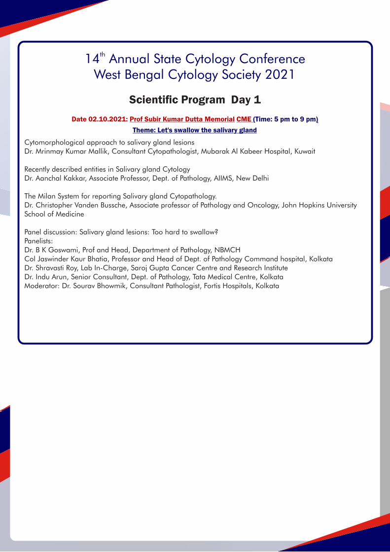
Cytomorphological approach to salivary gland lesionsDr. Mrinmay Kumar Mallik, Consultant Cytopathologist, Mubarak Al Kabeer Hospital, Kuwait
Recently described entities in Salivary gland CytologyDr. Aanchal Kakkar, Associate Professor, Dept. of Pathology, AIIMS, New Delhi
The Milan System for reporting Salivary gland Cytopathology.Dr. Christopher Vanden Bussche, Associate professor of Pathology and Oncology, John Hopkins University School of Medicine
Panel discussion: Salivary gland lesions: Too hard to swallow?Panelists: Dr. B K Goswami, Prof and Head, Department of Pathology, NBMCHCol Jaswinder Kaur Bhatia, Professor and Head of Dept. of Pathology Command hospital, KolkataDr. Shravasti Roy, Lab In-Charge, Saroj Gupta Cancer Centre and Research InstituteDr. Indu Arun, Senior Consultant, Dept. of Pathology, Tata Medical Centre, KolkataModerator: Dr. Sourav Bhowmik, Consultant Pathologist, Fortis Hospitals, Kolkata
Scientific Program Day 1
Date 02.10.2021: Prof Subir Kumar Dutta Memorial CME (Time: 5 pm to 9 pm)
Theme: Let's swallow the salivary gland

09:00 am to 9:30 am: Inauguration of conferenceIntroduction: Dr. Indranil Chakrabarti, Secretary, WBCSWelcome note: Dr. Mamata Guha Mallick Sinha, President, WBCS
Inauguration of Conference
09:30 am to 11:30 am: Award paper presentationsChairperson: Dr. Sayed. M. Nadeem, President Elect, WBCS11:30 am to 12:30pm: Brig M M Roy Memorial OrationTopic - Aggressive GCT of Bone - My Experience of more than Three Decades'By Dr. Asitava Mondal, IAC president 2006, Founder President, WBCSChairpersons:Dr. Manoj Choudhuri, ex-President WBCSDr. Swapan Kumar Sinha, ex-President WBCS
12:30 pm to 01:00 pm: Break
01:00 pm to 03:00 pm: Parallel Sessions in Hall A and Hall BHall A: E-poster presentationsChairperson: Dr. Palash Kumar Mandal, Vice President, WBCS Hall B: Free paper presentationsChairperson: Dr. Anjali Banerjee, Joint Secretary, WBCS
03:00 pm to 04:30 pm: Potpourri of interesting casesChairpersons: Dr Santosh Kumar Mondal, AIIMS, KalyaniDr. Anup Kumar Boler, Burdwan Medical College & Hospital, BurdwanSpeakers:Dr Sanhita Chatterjee, Medica Superspeciality Hospital, KolkataDr Anjan Kumar Das, CGMCH, Cooch BeharDr Sukanta Chakraborty, Theism Diagnostics, KolkataDr Aniket Haldar, IPGMER and SSKM Hospital, Kolkata4: 30 pm to 5 pm: Valedictory Session and Prize announcements
thDate 03.10.2021: XIV Annual State Cytology Conference, 2021
Day 2Scientific Program

West Bengal Cytology Society(Indian Academy of Cytologists, West Bengal Chapter)
th14 Annual State Cytology Conference of West Bengal Cytology SocietyOrganized by West Bengal Cytology Society
TOYC L OL GA YG N SE OB C
T IES TE YW
Mission Supremo- Prof. (Dr.) Debasis Bhattacharya, DME Government of West Bengal.
Patrons
Prof. (Dr.) Sabitri Sanyal
Prof. (Dr.) Lakshmi Majumder
Prof. (Dr.) Anjali Dasgupta
Prof. (Dr.) Swapan Kumar Sinha
Prof. (Dr.) Manoj Kumar Chaudhuri.
Advisors
Dr. Asitava Mondal
Prof. (Dr.) Anup Roy
Prof. (Dr.) Tamal kanti Ghosh
Dr. Rajat Mukherjee
Dr. Mala Banerjee
Prof. (Dr.) Sarbani Chattopadhyay
Organizing Committee
Organizing Chairperson: Dr Mamata Guha Mallick Sinha
Organizing Co-chairperson: Dr Sayed. M Nadeem
Organizing Secretary: Dr Indranil Chakrabarti
Organizing Jt. Secretary: Dr Anjali Banerjee
Treasurer: Dr Suchandra Ray
Joint Treasurer: Dr Madhumita Mondal
Scientic chairperson: Dr Palash Kumar Mandal
Registration: Dr Sourav Bhowmik, Dr Madhumita Mondal
Moderation and event management: Dr Senjuti Das Gupta
Scientific CommitteeDr Palash Kumar Mandal – Scientic ChairpersonDr Sanhita ChatterjeeDr Santosh Kumar MondalDr Anup Kumar BolerDr Anadi Roy ChowdhuryDr Senjuti Das GuptaDr Madhumita Mondal
Cytologist of West Bengal Felicitated by WBCS till date
1. Dr. Padma Lahiri 2. Dr. Lakshmi Mazumdar3. Dr. Jayashri Roy Choudhury4. Dr. Subir Kumar Dutta5. Dr. Sabitri Sanyal6. Dr. Anjali Das Gupta7. Dr. Swapan Kumar Sinha8. Dr. Manimala Roy (2016)9. Dr. Aparna Bhattacharya (2016)10. Dr. Manoj Kumar Chaudhuri11. Dr. Asitava Mondal (2018)12. Prof Keya Basu and Dr Rajat Mukherjee (2019)
West Bengal Cytology Society(Indian Academy of Cytologists, West Bengal Chapter)
th14 Annual State Cytology Conference of West Bengal Cytology SocietyOrganized by West Bengal Cytology Society
TOYC L OL GA YG N SE OB C
T IES TE YW

Governing Body of WBCS – 2021Advisory Body1. Dr Sabitri Sanyal2. Dr Aparna Bhattacharya4. Dr Subodh Kumar Bhattacharya3. Dr Amita Giri4. Dr Ranu Sarkar5. Dr Mala Banerjee
Ex-Officio Members ( Past Office Bearers)1. Dr Asitava Mandal2. Dr Swapan Kr Sinha3. Dr Manoj Choudhury4. Dr Anjali Dasgupta5. Dr Bidyut Krishna Goswami6. Dr Rajat Mukherjee7. Dr Dipanwita Nag8. Dr Anup Roy9. Dr Sarbani Chattopadhyay
Office Bearers1. President. Dr Mamata Guha Mallick Sinha2. President Elect. Dr Sayed Mahmood Nadeem3. Vice President . Dr Palash Kumar Mandal4. Secretary. Dr Indranil Chakrabarti5. Joint Secretary. Dr Anjali Banerjee6. Treasurer. Dr Suchandra Ray
Executive body members1. Dr Shravasti Roy.2. Dr Anup Boler3. Dr Ranjana Bandhopadhyay4. Dr Madumita Mondal5. Dr Mallika Pal6. Dr Ranu Roy Biswas7. Dr Santosh K Mondal8. Dr Sanhita Chatterjee9. Dr Senjuti Das Gupta10. Dr Sourav Bhowmick
E-newsletter editorial board1. Dr Asitava Mondal2. Dr Mala Banerjee3. Dr Rajat Mukherjee4. Dr Anup Roy
West Bengal Cytology Society(Indian Academy of Cytologists, West Bengal Chapter)
th14 Annual State Cytology Conference of West Bengal Cytology Society
Organized by West Bengal Cytology Society
TOYC L OL GA YG N SE OB C
T IES TE YW

Brig M M Roy Memorial Ora�ons conducted by IAC WB Chapter
SI NO: Year Venue Orators1 2008 BM Birla Heart Research Centre Kolkata Inaugural Oration by President of IAC, Dr. R J W Pinto (Goa Medical College), Goa
2 2008 CMRI, Kolkata Dr. Suresh Bhambani (ICMR)- New Delhi
3 2009 RKM Seva Pratishthan, Kolkata Dr. Dilip Das (Kuwait)
4 2010 Medical College, Kolkata Dr. V. K. Iyer (AIIMS) – New Delhi
5 2011 Medical College, Kolkata Dr. M.K.Rai (RIMS) Ranchi
Dr. Pranab Dey (PGIMER), Chandigarh
Dr. Kalpalata Tripathy (SCB Medical College), Cuttack
7 2013 Medical College, Kolkata Dr. Amitava Chatterjee (CNCI)- Kolkata
8 2014 CMRI, Kolkata Dr. Radhika Srinivasan, (PGIMER), Chandigarh
9 2015 IPGME&R, Kolkata Dr. Tanuja Shet, TMH, Mumbai
10 2016 IPGME&R, Kolkata Dr. Meherbanu Kamal (GMC) Nagpur
11 2017 NBMC Sushrutanagar Dr. Aruna K Prayaga (NIMS) Hyderabad
12 2018 IPGME&R, Kolkata Col U. S. Dinesh Dharwad
13 2019 NBMC Sushrutanagar Dr. Naresh N Rai (Govt. Medical College) Kolkata
2012 Medical College, Kolkata6

Message from DME

It is wonderful to hear that the West Bengal Cytology Society is organizing the Prof Subir Kumar Dutta
Memorial CME and XIVth Annual state Cytology Conference 2021 on 2nd and 3rd of October under the
stewardship of Dr Mamata Guha Mallick Sinha, President, and Dr Indranil Chakrabarti, Secretary, WBCS. Going through the scientic programme I note that the meeting addresses very contemporary topics in cytopathology. The ofine meeting restrictions imposed by the Covid pandemic I am sure will not come in the way of spread of knowledge. In fact, the online platform being adopted will have a pan India reach rather than being restricted to the boundaries of West Bengal. Heraclitus, a Greek philosopher, has been quoted as saying “change is the only constant in life.” One's ability to adapt to those changes will determine one's success in life. Well, this applies equally to the eld of cytopathology. Such conferences and CMEs provide the platform for the practicing cytopathologists and the residents to update their knowledge and keep abreast with the ever-evolving practice of cytology.I hope that the online CME and Conference will witness enthusiastic participation by postgraduate students and practicing cytopathologists. Best wishes for the successful organisation of the event.
Jai Hind
Brig (Dr.) Vijay Shrawan NijhawanPresident, Indian Academy of Cytologists
Message from IAC President
I am very happy to learn that the West Bengal Cytology Society is organizing the Prof. Subir Kumar Dutta Memorial CME and the XIV Annual State Cytology Conference, on Oct2-3, 2021. The conference is being organized in a fully virtual mode in view of the ongoing COVID pandemic. Indeed, I look ta this development as a positive development as online formats have the potential for greater outreach and minimizes the carbon footprint and wastage of resources that ofine conferences entail. The Scientic program looks very appetizing with salivary gland as the theme of the CME and following the CME, I am sure that this topic will be more easily digestible! The Scientic program of the conference has excellent speakers including Dr.Mondal, former President of IAC. State conferences also give the students of that state the opportunity to present posters and papers. My best wishes for the success of the CME and conference and warm regards to all the Ofce bearers, members of the society and the conference delegates. I do hope that following the wonderful appetizer by WBCS, all of you will join the main Feast of Golden Jubilee conference of the Indian Academy of Cytologists scheduled for November 20-21 with Pre-conference CME, post-conference workshops, award sessions, lectures and symposia.
Warm regards,Radhika Srinivasan,Secretary, IAC
Message from IAC Secretary
Message From Organizing Secretary
Dear friends,
It is my proud privilege to welcome you all to the Prof Subir Kumar Du�a Memorial CME and XIVth Annual State
Cytology Conference, 2021 organized by the West Bengal Cytology Society, the West Bengal Chapter of Indian
Academy of Cytologists (IAC) on 2nd and 3rd of October,2021.
Due to the COVID-19 pandemic, that has gripped the whole world, an offline meet is best avoided and hence, we
are again forced to organize this year's academic feast on virtual mode.
The pre-conference CME on 2nd October 2021 has been organized as a tribute to the legendary teacher,
pathologist and laboratory consultant Late Prof (Dr) Subir Kumar Du�a. The theme of this year's CME is Salivary
Gland pathology and will give the delegates an opportunity to gain insights from the vast experience of our
na�onal and interna�onal speakers.
The XIVth Annual State Cytology Conference, 2021 on 3rd October will highlight the pres�gious Brigadier M M Roy
Memorial Ora�on, potpourri of interes�ng cytology cases and a vibrant collec�on of award and proffered papers
as well as e-posters.
We assure you a grand academic fiesta and look forward to your whole-hearted support, co-opera�on, and
ac�ve par�cipa�on.
With best regards,
Dr Indranil Chakrabar�
Organizing Secretary Honorary Secretary, West Bengal Cytology Society
Message From the Desk of Organizing Chairperson
Dear friendsI feel privileged to welcome you all to 14th Annual State Cytology Conference and Preconference CME in the name Prof Dr Subir Du�a on 2nd & 3rd of October,2021. Every year before this pandemic we used to hold Conference and CME in different Ins�tutes of our state. North Bengal Medical College hosted the last State conference in the year 2019 with great success. Each year we felicitate renowned dedicated pathologists. As we have a great desire to carry on with our academics even during the Covid pandemic, North Bengal Medical College again hosted a virtual CME last year. This year we are organizing a 3D Virtual Conference and CME. Many interna�onal & na�onal speakers will grace our program. Postgraduate trainees will present award papers and posters. There will be case based panel discussion and interes�ng case presenta�ons by renowned pathologists of West Bengal. Young enthusias�c pathologists of our state are working hard to make our conference highly successful. Hope for the bestLong live WBCS.
Thank you,
Prof. Dr. Mamata Guha Mallick Sinha President WBCS.

CURRICULUM VITAE : BRIEF NAME: Dr. Mrinmay Kumar Mallik
CURRENT POSITION: Consultant Cytopathology Department of Laboratory Medicine Cytopathology Unit Mubarak Al Kabeer Hospital KuwaitPROFESSIONAL QUALIFICATION: MBBS: R G Kar Medical. Calcu�a University 1991 MD (Pathology): PGIMER Chandigarh India 1996WORK EXPERIENCE: 1. 2011 – Current Date: Consultant cytopathology: Cytopathology Unit Mubarak al Kabeer Hospital 2. 2008-2011: Senior specialist cytopathology: Cytopathology Unit Mubarak al Kabeer Hospital3. 2002-2008: Specialist cytopathology: Cytopathology Unit Mubarak al Kabeer Hospital4. 1998-2002: Senior registrar: Cytopathology Unit Mubarak al Kabeer Hospital5. 1996-1998: Senior resident Pathology: PGIMER Chandigarh India6. 1993-1996: Junior resident: Pathology: PGIMER Chandigarh; India
AREAS OF INTEREST: Cytopathology of pancreas, GIT, Thyroid, so� �ssues and the role of human papilloma viruses in cervical cancer.
RELEVANT PUBLICATIONS1. Mallik MK, Alramadhan B, Dash� H, Al-Shaheen A, Al Juwaiser A, Das DK, George SS, Kapila K. Human papillomaviruses other than 16,18 And 45 are the major high risk HPV genotypes amongst women with abnormal cervical smear cytology residing in Kuwait: Implica�ons for future vaccina�on strategies. Diagn Cytopathol 2018. 46:1036-1039.
2. Mallik MK, Qadan LR, Al Naseer A, AlAli A, Al Ansari T, Naquib SAI, Das DK, Kapila K. The applicability of Papanicolaou Society of Cytopathology system on repor�ng endoscopic ultrasound-guided fine needle aspira�on cytology specimens of pancrea�c lesions in situa�ons with limited availability of ancillary tests. Experience at a single laboratory. Cytopathology. 2020; 00:1-8.3. Mallik MK, Kapila K, Mohanty A, Inamdar SA, Al Ali A, Al-Nasser A. Endoscopic ultrasound guided fine needle aspira�on cytology of pancrea�c adenocarcinoma revisited. A detailed cytological analysis. J Cytology. 2021; 38:31-37
Cytomorphological approach to salivary gland lesions
In recent �mes medical science professionals have been increasingly using the concept of “algorithms” to refine their approaches towards solving complex diagnos�c problems. The one which I intend to present does so by peeling out cellular en��es which we encounter while evalua�ng fine needle aspirates of salivary gland lesions. However before placing the first slide on the stage, it is essen�al to have a clear idea about the epidemiological data, the salivary gland from where the material has been aspirated and the imaging findings. Salivary glands as we know are located at a very busy traffic intersec�on jammed by many non-salivary structures. My approach involves grouping lesions based upon the conspicuousness of inflammatory cell elements, the solid/ cys�c nature of the lesion, the conspicuousness of myoepithelial derived stroma and the nucleo-cytoplasmic ra�o of the lesional cells. Of course there are lots of overlaps, promiscui�es and other oddi�es which occasionally throw such algorithm out of the window. So while it is important to have a plan, it is pragma�c to be flexible. Once in a while ancillary methods like immunohistochemistry, flowcytometry and molecular pathology can extract us out of s�cky situa�ons. It is also incredibly essen�al to accomodate the ever-growing list newly described en��es and to be aware of the management implica�ons of our reports. Thankfully the Standardized repor�ng protocols, like the Milan system integrates the challenges faced by the morphologist with the brass tacks of “Risk of malignancy”, associated with our sign outs.
Brief Resume

Name – Dr. Anchal Kakkar, MD, Pathology
Fine needle aspiration cytology (FNAC) of salivary gland masses has high sensitivity and specicity, with
the added benets of safety and cost- effectiveness. It reliably distinguishes between inammatory and
neoplastic, and benign and malignant lesions, thus aiding in clinical decision making.
In recent years, the high accessibility to molecular genetic analysis accompanied by expanded
immunohistochemical panels has led to a renement in nomenclature of salivary gland neoplasms, as
well as led to the recognition of new tumor entities. Due to their rarity of these recently described entities,
and the marked heterogeneity in cytomorphology inherent to most salivary gland neoplasms, their
cytological diagnosis often presents signicant challenges. However, knowledge of histological
characteristics and identication of their cytological correlates, supported by ancillary techniques, can
assist in arriving at an accurate diagnosis.
Recently Described Entities in Salivary Gland Cytology
Brief Resume
Present Designation
Associate Professor, Department of Pathology, All India Institute of Medical Sciences , New
Delhi (2016 - present)
Areas of interest Head and Neck pathology
Pediatric tumor pathology
Publications: 135 publications in International & National journals
12 book chapters
Awards and Recognition: Dr. N. C. Nayak Award for Talented Young Pathologist by IAPM Delhi Chapter
Invited to contribute to chapters in 5th editions of WHO Classification “Blue books”
Contributed a chapter entitled “Head and Neck” to South Asia content in Robbins and
Cotran Pathologic Basis of Disease

CURRICULUM VITAEThe Johns Hopkins University School of Medicine
Christopher J. VandenBussche
DEMOGRAPHIC AND PERSONAL INFORMATION
Current Appointments 1/19-present Associate Professor of Pathology and Oncology7/14-present Associate Director, Cytopathology Laboratory; Johns Hopkins University School of Medicine, Bal�more, MD7/14-present Staff Pathologist; Johns Hopkins Hospital, Bal�more, MD 7/17-present Cytopathology Fellowship Program Director; Johns Hopkins University School of Medicine, Bal�more, MD
Personal Data
Business Address:Pathology 406Department of PathologyThe Johns Hopkins Hospital600 N. Wolfe StreetBal�more, MD 21287
Phone: (410) 955-1180Fax: (410) 614-9556Email: [email protected]
Educa�on and Training8/97-5/01 BS (Chemistry, Biology); The College of William and Mary, Williamsburg, VA8/01-5/09 MD-PhD (Tumor Biology); Lombardi Comprehensive Cancer Center, Washington, DC;Georgetown University School of Medicine, Washington, DC 7/09-6/13 Resident, Anatomic and Clinical Pathology; Johns Hopkins University School of Medicine, Bal�more, MD7/13-6/14 Clinical Fellow, Cytopathology; Johns Hopkins University School of Medicine
Professional Experience7/14-1/19 Assistant Professor of Pathology; Johns Hopkins University School of Medicine, Bal�more, MD7/14-present Associate Director, Cytopathology Division; Johns Hopkins University School of Medicine, 7/14-present Staff Pathologist, Johns Hopkins Hospital, Bal�more, MD7/17-present Cytopathology Fellowship Program Director; Johns Hopkins University School of Medicine 1/19-present Associate Professor of Pathology and Oncology; Johns Hopkins University School of Medicine
Publica�ons – 81 Original Research 4 Case Reports 15 Review Ar�cles

The ne needle aspiration of salivary gland is challenging due to the diverse and numerous neoplasms that can arise
within the gland. While some of these entities can be denitely diagnosed on FNA cytomorphology, many entities
require complete histologic examination for denitive characterization. Thus, it is important for the pathologist to utilize
a reliable framework while assessing these specimens. While experienced pathologists may have developed their own
frameworks to assess these lesions, a standardized nomenclature and approach were not available until the release of
The Milan System (TMS) for Reporting Salivary Gland Cytopathology in 2018. TMS uses a pattern-based approach that
makes the classication of salivary gland lesions seen on FNA much more accessible. TMS provides examples of entities
that can be denitively diagnosed on FNA cytopathology, but, perhaps most importantly, introduces an approach that
can minimize the many pitfalls in salivary gland cytopathology. This session introduces TMS and highlights its important
points to the audience.
The Milan System for Reporting Salivary Gland Cytopathology
Chris VandenBussche was born in Rochester, Michigan and grew up in Traverse City, Michigan. He majored in chemistry
at the College of William and Mary in Williamsburg, Virginia and then completed a combined M.D.-Ph.D. degree at the
Georgetown University School of Medicine and Lombard Comprehensive Cancer Center. After residency and
fellowship (cytopathology) training at the Johns Hopkins University School of Medicine, he joined the faculty and is
currently an Associate Professor of Pathology and Oncology, the Associate Director of Cytopathology, and the Program
Director of the Cytopathology Fellowship.
Dr. VandenBussche is a member of the College of American Pathologist's Cytopathology Committee, led the Pelvic
Washings chapter of The International System for Reporting Serous Fluid Cytopathology, and leads two chapters for
forthcoming updated version of The Paris System for Reporting Urinary Cytology (“TPS 2.0”). He served on the American
Society of Cytopathology's Scientic Program Committee for four years and is currently chair of the Clinical Practice
Committee.
Dr. VandenBussche predominantly focuses on the study of genitourinary cytopathology and has published 8 books and
over 100 peer-reviewed scientic papers in the eld of cytopathology. He serves on numerous editorial boards and is an
Associate Editor for Diagnostic Cytopathology, an Assistant Editor for the American Journal Clinical Pathology, and a
Section Editor for the Journal of the American Society of Cytopathology. He is frequently invited as a national and
international speaker on cytopathology topics.

Brief Resume
Dr. Asitava Mondal Clinical Cytologist and Oncopathologist, Kolkata.
He is (MBBS, Medical College Kolkata), MD (PGIMER, Chandigarh), President IAC (2006), Vice President IAPM (2013), Founder President of West Bengal Cytology Society-WBCS (2007-2011) Founder President of Association of Cytologists and Histopathologists--ACH [2019-2023] , National scholar,Formerly attached to various cancer hospitals in Kolkata, Started FNAC in West Bengal in 1984. He is recipient of 19 gold medals and 10 silver medals, published 182 articles in National and International journals, delivered 34 orations in various States of India and abroad, invited as Guest speakers in 171 National and International Conferences. He was felicitated as Life Time Achievement awards from Jharkhand, Rajasthan and Chhattisgarh Chapters of IAPM and Mayor of Kolkata for contributions in Cytology. He organized National IAC Conference in Kolkata in 1996 and started West Bengal Chapter of IAC in 2007. He is in the Editorial Board of 5 National and International Journals of Pathology, Cytology and Oncology.
Brigadier M M Roy Memorial Oration 2021
My Experience of more than Three decadesAggressive GCT of Bone -Topic -
Aggressive GCT of Bone- My experience of more than three decades Dr. Asitava Mondal MD ( PGIMER, Chandigarh) Clinical Cytologist and Oncopatholgist, Kolkata. Correct diagnosis of any bone tumour depends on a team approach of orthopaedic surgeon, radiologist and cytologist/ histopathologist. Aggressive GCT of bone cannot be diagnosed confidently without proper imaging modality like CT scan or MRI. According to widely accepted grading and staging system by Lichtenstein, Campanacci and Enneking, GCT is classified into Grade/ Stage 1 ( Latent/ Quiescent), 2 ( Active Intraosseous ) and 3 ( Aggressive Extraosseous). These grading and staging were done according to radiological findings. No detail cytological or histopathological findings were described according to the above grades of GCT of bone on which treatment and prognosis depend. Correct diagnosis of GCT in active intraosseous stage without breakdown of bony cortex is most crucial where treatment can be conservative surgery. Once the tumour breaks the outer cortex and becomes extraosseous and aggressive, treatment becomes extremely difficult and may need en bloc resection. The cytomorphology is very characteristics in all these three grades/stages of GCT. Equal importance is given to the stromal cells and osteoclastic giant cells to assess the activity of this bone tumour. Attention was given only to the nature of stromal cells. But detailed study highlighted the importance of osteoclastic giant cells in assessing the aggressiveness of the tumour. The treatment of GCT has revolutionized in recent times. Previous treatments were only curretage with bone graft and enbloc resection. At present multifaceted treatments are curretage with PMMC cementation, Zoledronic acid, Bisphosphonate therapy, Neoadjuvant therapy with Denosumab with or without embolization, Simvastatin and recent advancement with other targets including mutant protein H3.3-G34W. Whatever may be the advancement of therapy, the best result is obtained when diagnosis is made in Active Intraosseous stage without destruction of bony cortex . Once the tumour becomes Aggressive Extraosseous, en bloc resection becomes almost mandatory due to repeated recurrences. Radiation therapy should be avoided as far as possible. Secondary malignant GCT with sarcomatous changes occur after radiation therapy and repeated recurrences. Primary malignant GCT is extremely rare. In conclusion the basic pathophysiology of GCT lies in the recent nomenclature of this bone tumour as " Osteoclastogenic Stromal Cell Tumour of Mesenchymal Origin".
Abstract ID: 11 for WBCS CONFERENCE 2021 (Auto-Generated October 1, 2021 8:43 pm)
Copyright 2021 WBCS CONFERENCE 2021 powered by WPAbstracts Pro
An evaluation of FNAC of breast lesions with
histopathological correlation among patients presenting in a
tertiary care hospital with special reference to Modified
Masood’s scoring indexby DR. ANINDYA ADDHYA | DR. SUCHANDRA RAY | DR. MOUMITA SENGUPTA | PROF. DR. MAMATA
GUHA MALLICK SINHA | POST GRADUATE TRAINEE | ASSOCIATE PROFESSOR | ASSOCIATE
PROFESSOR | PROFESSOR
Abstract ID: 11
Submitted: September 14, 2021
Event: WBCS CONFERENCE 2021
Category: Award Papers
An evaluation of FNAC of breast lesions with histopathological correlation among
patients presenting in a tertiary care hospital with special reference to Modified
Masood’s scoring index
ABSTRACT
CONTEXT & BACKGROUND
Cytological grading of breast lesion, which is a simple, cost-effective, and reproducible
method, can be used as a tool for the selection of treatment modality. The proposed
Modified Masood’s scoring index establishes one guideline for reporting of breast cytology
and thus helps in individualized treatment and follow-up.
AIMS & OBJECTIVES
The aims and objectives of this study were to (1) establish the validity and reliability of the
Modified Masood’s scoring index in breast lesions and (2) to calculate the malignancy risk.
MATERIALS & METHODS
This prospective & observational study was designed in clinically diagnosed breast lesions
at the Department of Pathology of a tertiary care referral hospital. Fine-needle aspiration
(FNA) was done, and stained smears were examined under light microscope and cytological
findings were noted according to the Modified Masood’s scoring index. Tissue for the
histopathological study was obtained in 80 cases. The previous cytological findings were
compared to subsequent histopathology report.
RESULTS
Among 80 FNAs, 46% were Non proliferative breast disease and 7% were Proliferative
breast disease without atypia. About 3% was grouped in the Proliferative breast disease
with atypia, 44% of cases were categorized as Carcinoma in situ/ Invasive carcinoma.
CONCLUSION
The four-tier diagnostic categories of the Modified Masood Scoring System help in
segregating patients with breast lesions into the management categories of follow-up,
conservative surgery, and radical surgery with/without chemotherapy.
Award Paper Presentation

Award Paper Presentation
Abstract ID: 12 for WBCS CONFERENCE 2021 (Auto-Generated October 1, 2021 8:43 pm)
Comparative Study Between Conventional and Liquid Based
Cytology in Urinary Cytology Specimens Classified According
to The Paris System.by Dr. Soumya Dey | Dr.Kavita Jain | Dr. SM sarfaraj | Prof. Dilip Kumar Pal | Prof.Chhanda Datta |
Prof.Mamata Guha Mallick Sinha | Prof. Uttara Chatterjee | IPGMER, Pathology | IPGMER, Pathology |
IPGMER, Pathology | IPGMER, Urology | IPGMER, Pathology | IPGMER, Pathology | IPGMER, Pathology
Abstract ID: 12
Submitted: September 15, 2021
Event: WBCS CONFERENCE 2021
Category: Award Papers
Introduction:
Cystoscopy, though remained as gold-standard for diagnosis and evaluation of urothelial
neoplasms, invasiveness and poor follow up potential of the procedure led to widespread
usage of urinary cytology as a screening tool. Conventional cytology (CC) is a proven
method for detection of urothelial neoplasms, but newer liquid based cytology (LBC) is
gaining popularity because of clear background and monolayering of cells. The present
study compared the LBC (SurePath) and CC in the diagnosis of urothelial neoplasms,
applying The Paris System (TPS) for reporting.
Materials and Methods:
A total of 150 patients with bladder tumour (June 2017-December 2018) were enrolled. Pre-
operative CC and LBC were processed from freshly voided urine samples. The individual
categories of TPS were compared for CC and LBC samples and correlated with histology.
Results:
No significant differences were observed in sensitivity and specificity between the 2
methods in cases with positive cytology for carcinoma. LBC and CC (specificity 96.7% vs
91.8%) were highly specific for detection of bladder carcinoma, but lacked sensitivity (LBC
56.6% vs CC 57.8%). According to TPS, suspicious for HGUC cases of both CC and LBC
smears correlated with positive findings of carcinoma in most cases (CC positive predictive
value [PPV] of 94.7% vs LBC PPV 95.6%).
Conclusion:
The present study concludes that LBC does not offer a better detection of malignant cells in
the urine than CC, but improved cell yield, cell-preservation and reduced artifacts in LBC
aid in better understanding of cellular morphology.
Key Words:
Urinary cytology, LBC, The Paris System

Award Paper Presentation
Abstract ID: 18 for WBCS CONFERENCE 2021 (Auto-Generated October 1, 2021 8:42 pm)
FINE-NEEDLE ASPIRATION OF RENAL TUMORS :
ANALYSIS OF RESULTSby Dr. Binata Bandyopadhyay | Prof (Dr.) Madhumita Mukhopadhyay | Prof (Dr.) Mamata Guha Mallick
Sinha | 1st Year Post Graduate Trainee in MD Pathology, IPGME&R and SSKM Hospital. | Professor,
Department of Pathology, IPGME&R and SSKM Hospital | Professor and Head of the Department,
Department of Pathology, IPGME&R and SSKM Hospital
Abstract ID: 18
Submitted: September 15, 2021
Event: WBCS CONFERENCE 2021
Category: Award Papers
Abstract
Fine-needle aspiration(FNA) of the kidney has a traditionally well-defined role in the
diagnosis and treatment of renal lesions. Recent improvements in renal imaging techniques
have also brought renal FNA to the forefront, since Small and asymptomatic renal masses
are increasingly being detected. Before the physician institutes a treatment plan, such
lesions usually require a definitive diagnosis that is best provided by FNA. The cytological
features are given priority compared to the histological growth forms for classification of
renal cell tumors. However, the latter are not to be neglected in the overall evaluation of a
tumor.
Objective - To know the profile of Kidney tumor by Needle aspiration Cytology.
Materials & Methods - All renal FNAs performed during a 2 year interval were retrieved.
Indication for the procedure was determined from the clinical notes and radiology reports.
Results - Out of 10 adult patients, 9 were cases of Renal cell carcinoma(RCC) and one was
transitional Cell Carcinoma(TCC). Among 9 cases of RCC, 4 were of Clear Cell RCC
(CCRCC),3 were of Papillary RCC(PapRCC) and 2 were of Chromophobe RCC(ChRCC).
Among 10 Patients, 7 were măle and 3 were female. Out of 10 paediatric patients, there
were 9 cases of Wilms’ tumor and 1 case of Neuroblastoma. Among 9 cases of Wilms’ tumor,
7 were male and 2 were female and the patient of Neuroblastoma was male.
Conclusion - Fine needle aspiration cytology is a safe and useful diagnostic tool in case of
Renal tumors.
Keywords - Fine needle aspiration Cytology, imaging techniques, radiology, renal,
Indication.

Award Paper Presentation
Abstract ID: 7 for WBCS CONFERENCE 2021 (Auto-Generated October 1, 2021 8:43 pm)
Reproducibility of “International Academy of Cytology
Yokohama System for Reporting Breast Cytology”-A
Retrospective analysis of 70 casesby 1. Ankita Chakraborty | 2. Anup Kumar Boler | 3. Shreosee Roy, | 4. Arghya Bandyopadhyay | 1st year
Post Graduate Trainee,Department of Pathology, Burdwan Medical College, West Bengal University of
Health sciences, West Bengal, India) | MD(Pathology), Department of Pathology, Burdwan Medical
College, West Bengal University of Health Sciences, West Bengal, India) | MD(Pathology), Department of
Pathology, Burdwan Medical College, West Bengal University of Health Sciences, West Bengal, India |
MD(Pathology), Department of Pathology, Burdwan Medical College, West Bengal University of Health
Sciences, West Bengal, India)
Abstract ID: 7
Submitted: September 10, 2021
Event: WBCS CONFERENCE 2021
Category: Award PapersAbstract
Introduction : Fine-needle aspiration cytology is most practiced initial method for
evaluation of breast lesions. The International Academy of Cytology Yokohama System for
Reporting Breast fine-Needle Aspiration Biopsy Cytopathology (IAC YSRB) is developed to
standardize the reporting system. Aim of this study is to establish the inter-observer
reproducibility of this system in diagnosing breast lesions. Method: Over a period of 1 year,
seventy consecutive specimens obtained from fine-needle aspiration cytology (FNAC) from
breast lesions, were independently evaluated by 3 experienced cytopathologists, who
allotted 1 to 5 IAC YSRB categories for each case. Percent overall agreement was calculated
by using Fleiss’ Kappa. Result: Percent overall agreement between observers was 70.48%
and free marginal kappa was 0.63. After combining category 4(Suspicious) and category 5
(Malignant) overall agreements was 80.95% and free marginal kappa was 0.75.
Conclusions: Inter-observer agreement of 3 cytopathologists was good (70.48%).
Agreement can improve if we combine certain categories, especially “Suspicious” and
“Malignant”. Limitation of sampling technique plays a significant role in diagnosing
“Atypical’ and “Suspicious” cases. However, inter-observer variability can be improved if we
make it a practice to follow the IAC YSRB criteria meticulously in day to day practice.needle aspiration, breast cytology.
Award Paper Presentation
Abstract ID: 17 for WBCS CONFERENCE 2021 (Auto-Generated October 1, 2021 8:43 pm)
Utility of intraoperative imprint cytology in diagnosis and
grading of Glioma: A study in a tertiary care Centre from
Eastern India.by Dr.shivam chakraborty. | Dr. Chhanda dutta | Dr. Mamata Guha Mallick Sinha. | PGT | Assistant
Professor. | Professor.
Abstract ID: 17
Submitted: September 15, 2021
Event: WBCS CONFERENCE 2021
Category: Award Papers
Abstract .
Background : Gliomas are most common primary CNS neoplasm in adult population.
Gliomas are predominantly arise from brain parenchyma, invasion of adjacent normal
parenchyma is a prominent feature.
Aims and objectives : The study was undertaken to study the epidemiological incidence of
glial tumors and the viability and accuracy of intraoperative cytology in diagnosis and
grading glial tumors in a tertiary care center.
Materials and methods : A prospective study was done on 30 patients who underwent
excision surgery at department of neurosurgery. Intraoperative imprint touch cytology of
the specimens were done. Part of the tissue were kept for FFPE and subsequent
histopathological examination were done. Results of intraoperative imprint cytology were
compared with final histopathology report and grading.
Result: Most of the patients presented with frontal lobe lesion. Out of 30 cases 29 were
histologically confirmed to be of glial origin. These 29 cases were diagnosed as different
glial neoplasm on intraoperative imprint cytology and was confirmed by histology.
According to histological subtype 52% were glioblastoma, 34% were diffuse astrocytoma,
10%were pilocytic astrocytoma, 4% was ependymoma. Intraoperative impression cytology
diagnosis was compared with confirmatory histological diagnosis. Sensitivity and septicity
was found to be 93% and50% respectively, PPV 96%, NPV33%, diagnostic accuracy 86%
and P<0.5 was statistically significant.
Conclusion: Here in this study, we see a good correlation between imprint cytology and
confirmatory histopathology report. Hence, we conclude intraoperative imprint cytology is a
fairly accurate, rapid and inexpensive method of diagnosis and grading of gliomas.
Glioma, Intraoperative, cytology.Keywords:

Award Paper Presentation
Abstract ID: 15 for WBCS CONFERENCE 2021 (Auto-Generated October 1, 2021 8:43 pm)
VISCERAL MALIGNANCIES PRESENTING AS CUTANEOUS
DEPOSITS -A CYTOPATHOLOGIST’S PERSPECTIVE.by Dr Anannya Ghatak | Dr Chhanda Das | Prof Dr Mamata Guha Mallick Sinha | Junior Resident |
Assistant Professor | Professor and head of the department
Abstract ID: 15
Submitted: September 15, 2021
Event: WBCS CONFERENCE 2021
Category: Award Papers
Enter description here.
ABSTRACT
Introduction: Cutaneous metastases can occur in a wide variety of internal malignancies
and may be the first indication of an underlying malignancy or recurrence in a patient with
a known primary and in rare instances can arise from a second primary. AIM : This study is
designed to analyze cases of cutaneous metastasis from a known or unknown primary and
evaluate usefulness of fine needle aspiration cytology as a diagnostic modality. Materials
and methods: This is a prospective study conducted in a tertiary care hospital. We studied
the cases according to their age, sex, the clinical presentations, site, the treatment provided
and the cytopathological findings. Results : This study comprises of eight cases of cutaneous
metastatic deposits from various primary visceral malignancies at different sites-scalp,
anterior abdominal wall, chest with varied presentations. The patients have undergone
treatment and were referred to surgery and radiotherapy department for further
management. Conclusion : Cutaneous metastasis is a manifestation of the disease due to
either hematogenous or lymphatic spread. This study also reiterates that fine needle
aspiration biopsy is a very helpful and cost-effective modality in determining the nature of
such lesions. So, the study of their clinical profile and cytopathological findings paves way
for early diagnosis of primary lesion or recurrence and better management.
Keywords: cutaneous, cytology, metastasis, recurrence, underlying
E - Poster Presentation
Abstract ID: 21 for WBCS CONFERENCE 2021 (Auto-Generated October 1, 2021 8:43 pm)
Copyright 2021 WBCS CONFERENCE 2021 powered by WPAbstracts Pro
A study of correlation between Bronchoalveolar lavage and
Bronchial brush cytology in evaluation of suspected cases of
lung carcinoma.by Lt Col (Dr) Vishal Raut | Col (Dr) Jasvinder Kaur Bhatia | Command hospital , Kolkata | Command
hospital , Kolkata
Abstract ID: 21
Submitted: September 16, 2021
Event: WBCS CONFERENCE 2021
Category: E Poster
Title : A study of correlation between Bronchoalveolar lavage and Bronchial brush cytology
in evaluation of suspected cases of lung carcinoma.
Authors : Lt Col (Dr) Vishal Raut, Col (Dr) Jasvinder Kaur Bhatia
1: 3 rd year post graduate trainee, 2 : Prof and HOD, Dept of Pathology
Institution : Command Hospital(Eastern Command), Kolkata
Type of submission : E-Poster presentation
A study of correlation between Bronchoalveolar lavage and Bronchial brush
cytology in evaluation of suspected cases of lung carcinoma.
Introduction : Lung carcinoma has a wide incidence and prevalence in both genders,
including primary and secondary carcinoma due to metastasis. It is one of the most common
cause of mortality and morbidity throughout the world. Cytopathology being fast and less
invasive plays an important role in early evaluation and subsequent patient management,
especially when lesions are peripheral, small or non visualized on bronchoscopy and hence
biopsy for histopathology cannot be done.
Methods and Result : Broncholveolar lavage(BAL) collected as normal saline suspension in
sterile container is centrifuged and smears prepared. Same patients Brush cytology dried
and wet fixed smears of two to three brushings are taken. Both specimens are stained by
Papanicaloau stain and Leishman’s stain. The slides are evaluated and results analyzed to
evaluate the correlation between the said parameters including age and gender using
appropriate statistical software.
Conclusion : There is a positive correlation between BAL, Brush cytology combined all
together so as to early effective evaluation of most lung lesions especially which are
difficult to visualize on bronchoscopy due to small size, vision blockade and peripheral
lesions.

E - Poster PresentationAbstract ID: 20 for WBCS CONFERENCE 2021 (Auto-Generated October 1, 2021 8:43 pm)
Copyright 2021 WBCS CONFERENCE 2021 powered by WPAbstracts Pro
CASE REPORT : FNAC Of Supraclavicular Mass – An Unusual
Finding Of ‘Metastasis Of A Rhabdoid Tumor’ In An Adult
Maleby Dr Ankita Kumari | CHEC, Kolkata
Abstract ID: 20
Submitted: September 15, 2021
Event: WBCS CONFERENCE 2021
Category: E Poster
Author affiliations : Department of Pathology, CHEC Kolkata
Corresponding Author : Dr. Ankita Kumari ( Postgraduate student )
Guided by : Col Jasvinder Kaur Bhatia
HOD and Senior Advisor
Abstract :
Introduction : Rhabdoid tumor of kidney is a rare ‘aggressive malignancy’ and most
malignant renal neoplasm of childhood, most of which occur before the age of 2 years. The
death rate is over 75% and patients usually die within 12 months of diagnosis. High tumor
stage and male sex are unfavourable prognostic signs. We report a rare case of rhabdoid
tumor metastases in a supraclavicular mass in a 53 year male and discuss the FNAC
findings.
Case report: 53-year male presented with a supraclavicular mass (2x1.5 cm) which was
firm, mobile, non-tender.
The patient was a known case of ‘Rhabdoid tumor of Left kidney’(Post nephrectomy)
FNAC aspirate stained smears showed presence of loose clusters of atypical cells with
rhabdoid morphology. The cells were highly pleomorphic with round to oval eccentric
nucleus, nucleomegaly, high NC ratio, vesicular chromatin and eosinophilic cytoplasm. Few
large, bizzare cells and occasional binucleate cells were also noted. Occasional mitosis was
seen. No lymphoid elements or lymphoglandular bodies noted.
Cytomorphological features were suggestive of ‘Metastasis of a carcinoma (In a known case
of Rhabdoid tumor of Left Kidney).
CONCLUSION : Rhabdoid tumor is a widely metastasizing tumor with grave prognosis. The
case has been reported due to unusual clinical presentation and characteristic cytological
features.
ETHICS : This case report has been completely anonymized not to cause harm or prejudice
to the patient or patient’s family
E - Poster Presentation
Abstract ID: 16 for WBCS CONFERENCE 2021 (Auto-Generated October 1, 2021 8:43 pm)
Cytodiagnosis of chordoma corroborating with radiological
diagnosis and histological confirmationby Dr Debarghya Mukherjee | MD PGT DEPT. OF PATHOLOGY,SSKM HOSPITAL
Abstract ID: 16
Submitted: September 15, 2021
Event: WBCS CONFERENCE 2021
Category: E Poster
Chordoma is a rare slow growing malignant tumor which arises from primitive notochordal
remnants.A sixty five year old female patient attended the neurology OPD with complains of
swelling in the lower back gradually increasing over 3 years period with inability to
walk,bladder and bowel incontinence.FNAC revealed characterestic physaliferous cells seen
in chordoma.FNAC served as first line tool for early diagnosis and treatment.
Abstract ID: 22 for WBCS CONFERENCE 2021 (Auto-Generated October 1, 2021 8:43 pm)
CYTODIAGNOSIS OF MEDIASTINAL MASS AND
CONFIRMATION BY IHC FROM CELL BLOCKby Dr. Manchamani Ghatak, Dr. Sriranjan Mukherjee, Dr. Srishti Butta ,Prof.Dr.M.G.M.Sinha | Affiliated
Abstract ID: 22
Submitted: September 18, 2021
Event: WBCS CONFERENCE 2021
Category: E Poster
We can usually diagnose the lymphoproliferative disorder and other malignancy of lymnodes
by FNAC but there are some fuzzy areas when excision biopsy is needed. To overcome the
situation we may go for cell block which helps us to make a final diagnosis as well as IHC
confirmation.
E - Poster Presentation
Abstract ID: 23 for WBCS CONFERENCE 2021 (Auto-Generated October 1, 2021 8:43 pm)
CYTODIAGNOSIS OF METASTATIC GERM CELL TUMOR IN
LIVER –IS IT HELPFUL FOR EXPECTATIONAL OUTCOME?by Dr. Sona Das | IPGME&R
Abstract ID: 23
Submitted: September 18, 2021
Event: WBCS CONFERENCE 2021
Category: E Poster
Testicular germ cell tumor is one of the common tumor of testis which may have
better prognosis when diagnosed early. Here we report a case of a 28 years old
adult with painless small scrotal mass with distant metastasis. The patient
presented with Low back pain for one year followed by painless scrotal mass,
paraparesis and neurological complains. Rapid clinical progression to coma was
noted during the staging work up. A diagnosis of testicular mixed germ cell tumor
with multiorgan metastasis (lymph node, lung, liver and brain) was made on
cytology. The patients physical condition didn’t let him bear chemotherapy and
expired.
Abstract ID: 6 for WBCS CONFERENCE 2021 (Auto-Generated October 1, 2021 8:44 pm)
CYTODIAGNOSIS OF SCALP METASTASIS FROM THYROID
FOLLICULAR CARCINOMA- report of a rare case.by Dr. Akanksha Kaushik | PGT, IPGME&R, KOLKATA
Abstract ID: 6
Submitted: September 10, 2021
Event: WBCS CONFERENCE 2021
Category: E Poster
A case report of a slow growing , frontal scalp swelling diagnosed as metastasis from
thyroid follicular carcinoma, after FNAC smears showed smears showed cells resembling
those of thyroid gland and subsequent investigations led to a discovery of occult thyroid
follicular carcinoma.

E - Poster PresentationAbstract ID: 5 for WBCS CONFERENCE 2021 (Auto-Generated October 1, 2021 8:44 pm)
Eosinophilic pleural effusion - A rare case having
unsuspected malignancy.by 1.Dr Sourav Biswas | Dr Rabindra Nath Chatterjee | Dr Amitava Sinha | Dr Keya Basu | MD PGT
,Department of Pathology, KPC Medical College | Assistant professor, Department of Pathology, KPC
Medical College | Professor, Department of Pathology, KPC Medical College | Professor &
HOD,Department of Pathology, KPC Medical College
Abstract ID: 5
Submitted: September 10, 2021
Event: WBCS CONFERENCE 2021
Category: E Poster
Introduction
Eosinophilic pleural effusion (EPE) is stated when 10 percent or more eosinophils with in a
differential count of white cells is present in pleural fluid .The incidence of EPE is 5 percent
to 16 percent of all pleural effusion. The causes of EPEs include malignancy, tuberculosis,
parapneumonic effusion, parasite infection, connective tissue diseases,post trauma and
idiopathic. But the diagnostic significance of EPE remains unclear.
The aim of the study was to find out whether EPE is reliable predictor of malignancy.
Case Presentation
A 34 year old male patient presented with right sided chest pain since one month .There
was past history of pulmonary tuberculosis and completed ATD course 10 years back.Right
sided pleural effusion was found on chest x ray. Hematological and investigations for
infection including tuberculosis were done.The pleural fluid analysis showed exudative in
nature and cytological examination revealed Eosinophilic pleural effusion (EPE).The case
was evaluated by clinical ,radiological and cytological aspect.
Conclusion
Though EPE is uncommon, malignancy could be narrowed upon as a cause based on EPE
findings in our study .However, further studies may provide additional and alternative
perspective.
Keywords:Eosinophils, pleural effusion, malignancy, cytology, pleural fluid, Eosinophilic.

E - Poster Presentation
Abstract ID: 4 for WBCS CONFERENCE 2021 (Auto-Generated October 1, 2021 8:44 pm)
HOW TO APPLY BETHESDA FOR THYROID CYTOLOGY: A
STUDY OF THREE CASESby Dr. Poulomi Biswas | Junior Resident
Abstract ID: 4
Submitted: September 9, 2021
Event: WBCS CONFERENCE 2021
Category: E Poster
Type of Presentation- Poster
Authors : Dr. Poulomi Biswas ¹, Dr. Anup Kumar Roy², Dr. Ranu Sarkar³, Dr. Rajib Kumar
Mondal⁴, Dr. Rama Das⁵, Dr. Ananya Pal⁵, Dr. Anway Sen⁶, Dr. Sohini Roy⁶, Dr. Sarbari Kar
Rakshit⁶, Dr. Ambalika Mondal⁶, Dr. Surya Kumar Bera⁶
Affiliations :1- Junior Resident, 2- Professor, 3- Professor and HOD, 4- Associate Professor,
5- Assistant Professor, 6- Demonstrator.
Department of Pathology, Nil Ratan Sircar Medical College & Hospital.
ABSTRACT
3 cases of thyroid swelling are presenting here. FNAC done and interpretation of smear
following Bethesda criteria are presented here. Bethesda category 3,4,5 and their
overlapping criteria has been shown in these cases.
Case 1: 47 years old female presented with ill defined nodule in right lobe of thyroid.
Impression on FNAC: Bethesda category 3.
Case 2: 39 years old female presented with well defined nodule in left lobe of thyroid.
Impression on FNAC: Bethesda category 4.
Case 3: 49 years old female presented with ill defined thyroid nodule in left lobe of thyroid.
Impression on FNAC: Bethesda category 5.
Conclusion: Even after application of Bethesda criteria category 3,4,5 may produce some
confusions and inter examiner variation due to some overlapping criteria. Therefore repeat
aspiration and/or biopsy is often helpful for correct interpretation.
Keywords: Thyroid Nodule, Bethesda, Cytology, FNAC
HOD Letter
https://www.wbcsconference2021.com/wp-content/uploads/2021/09/Cytology-Sponsor.pdf

E - Poster Presentation
Abstract ID: 10 for WBCS CONFERENCE 2021 (Auto-Generated October 1, 2021 8:44 pm)
INTERESTING CASE OF AN INTRAPERITONEAL LYMPH
NODE SWELLING: A case reportby Dr. Sudeshna Nandi | Post Graduate Trainee, Pathology, IPGME&R
Abstract ID: 10
Submitted: September 13, 2021
Event: WBCS CONFERENCE 2021
Category: E Poster
Respected Sir and Madam,
We presented a case of 40 years old male who came to SSKM hospital OPD with an
abdominal discomfort and swelling for last 2 months on 02/03/2021. On USG, enlarged
retroperitoneal lymph nodes were identified. USG guided FNAC was done and the stained
smears showed cell rich aspirate with plenty of clusters of atypical cells with high N:C ratio
floating in pools of mucin. Few signet ring shaped cells identified in the smear. Overall
cytological features are suggestive of Metastatic Lesion (Mucinous primary).
An exploratory laparotomy was suggested and subsequently a mass was identified in the
ascending colon. The microscopic section shows a tumor mass composed of neoplastic cells
floating in pools of mucin and few signet ring cells with large cytoplasmic vacuolation. The
overall features were suggestive of MUCINOUS CARCINOMA of ascending colon.
Immunohistochemistry was done and HER2/neu shows negative expression. Surgery was
done in the patient stage was pT3N2Mx. Chemotherapy was given followed by radiotherapy.
Keywords: mucinous, retroperitoneal, metastatic, colorectal carcinoma
E - Poster Presentation
Abstract ID: 9 for WBCS CONFERENCE 2021 (Auto-Generated October 1, 2021 8:44 pm)
Pilomatrixoma – incidental diagnosis by fine needle
aspiration cytologyby DR DEBADITYA SAMANTA | PGT
Abstract ID: 9
Submitted: September 11, 2021
Event: WBCS CONFERENCE 2021
Category: E Poster
Authors
Debaditya Samanta1, Asim Kumar Manna
2, Mamata Guha Mallick Sinha
3
Post Graduate Trainee, 2. Professor, 3. Professor and Head, Department of Pathology,1.
Institute of Post Graduate Medical Education & Research, Kolkata.
Abstract
Pilomatrixoma or calcified epithelioma of Malherbe, is a tumour with differentiation toward
hair cells. The face and the upper extremities are the most common sites with an exception
at cubital fossa region. Here we present a case of a middle-aged man came with complaint
of a swelling at left arm for 2-3 years. On fine needle aspiration cytology, it showed clump of
anucleate keratinized squamous cells, small basaloid cells and inflammatory cells. We made
a provisional diagnosis of pilomatrixoma which was later confirmed histopathologically.

E - Poster Presentation
Abstract ID: 14 for WBCS CONFERENCE 2021 (Auto-Generated October 1, 2021 8:44 pm)
Primary spindle cell sarcoma of Breast-A case report
by Dr.Ruth Lalmuanpuii | (Prof) Dr.Dipanwita Nag | Dr.Srishtidhar Mangal | Dr.Aparajita Samaddar |
Dr.Nandini Das | Dr.Ipsita Saha | PGT,Dept.of Pathology,Medical College Kolkata | Professor,Dept.of
Pathology,Medical College Kolkata | Associate Professor,Dept.of Pathology,Medical College Kolkata |
Assistant Professor,Dept.of Pathology,Medical College kolkata | Demonstrator,Dept.of Pathology,Medical
College Kolkata | Demonstrator,Dept.of Pathology,Medical College Kolkata
Abstract ID: 14
Submitted: September 15, 2021
Event: WBCS CONFERENCE 2021
Category: E Poster
Most invasive breast neoplasms are epithelial tumours,and mesenchymal tumours are rarely
seen. Primary pure breast sarcoma constitutes 0.2-1% of all mammary malignancies.
Here, we present a rare case of primary spindle cell sarcoma of the breast in a 35 years old
lady without any history of radiation exposure or family history of breast cancers.
Keywords: Breast tumour, Spindle cell sarcoma, Primary breast sarcoma.

E - Poster Presentation
Abstract ID: 8 for WBCS CONFERENCE 2021 (Auto-Generated October 1, 2021 8:44 pm)
REPORT OF A RARE CASE OF INVASIVE PAPILLARY
CARCINOMA OF MALE BREASTby Dr. Smritiparna Das | Post graduate trainee
Abstract ID: 8
Submitted: September 11, 2021
Event: WBCS CONFERENCE 2021
Category: E Poster
Respected Sir/Madam,
I have registered for WBCS CONFERENCE 2021 and would like to submit the
e-poster. So I have herewith attached the abstract and e poster entitled ''REPORT OF A
RARE CASE OF INVASIVE PAPILLARY CARCINOMA OF MALE BREAST'' for the same.
Thanks and regards,
Smritiparna Das,
IPGMER & SSKM,
Post graduate trainee.

Abstract ID: 19 for WBCS CONFERENCE 2021 (Auto-Generated October 1, 2021 8:43 pm)
VALIDATION OF CERVICAL SMEARS SCREENING BY
ARTIFICIAL INTELLEGENCEby Neha Mala Krishna | Doctor
Abstract ID: 19
Submitted: September 15, 2021
Event: WBCS CONFERENCE 2021
Category: E Poster
Title: Validation of Artificial Intelligence Software for cervical screening
Introduction: Cervical cancer is the fourth most common cancer in women worldwide and
second most common in developing nations like India. Seeing the population strata, it
affects approximately 12,000 women per year. The examination of Papanicolaou (Pap)
smears is an efficient technique for diagnosing cervical pre-malignant and malignant
diseases. Deep learning methods are being devised with softwares being developed for
screening for cervical cancer. We aimed to test the accuracy of this software with the
manual interpretation of pap smears.
Methods: The study included 100 women between the ages of 25-65 years reporting to the
gynae OPD and whose pap smear test was performed for various conditions. The results
showed predominantly inflammatory lesion without intraepithelial malignant lesion followed
by bacterial vaginosis, atrophic smear, atypical squamous cell of undetermined significance
and low grade squamous intraepithelial lesion. The results of the liquid-based cytology and
pap smear slides were compared with the artificial intelligence software.
Conclusion: The software can be used to screen the pap smears and may reduce the time
taken for screening of pap smears.
AUTHOR: DR NEHA MALA KRISHNA 1 COL DR JK BHATIA2
2nd
year PG RESIDENT 2. SENIOR ADVISOR AND HOD PATHOLOGY1.
INSTITUTION: COMMAND HOSPITAL EASTERN COMMAND KOLKATA
E - Poster Presentation

Abstract ID: 13 for WBCS CONFERENCE 2021 (Auto-Generated October 1, 2021 8:44 pm)
Role of Pleural Fluid Cytology in COVID -19
by Maj Arnab Sen Gupta 1, Col (Dr ) Jasvinder Kaur Bhatia 2, Lt Col Manisha Agarwal | Command
Hospital Kolkata
Abstract ID: 13
Submitted: September 15, 2021
Event: WBCS CONFERENCE 2021
Category: E Poster
Abstract
Introduction: SARS- CoV2 or COVID 19 as we have come to know it commonly is affecting
thousands of people in our country and the world since early 2020. The changes that occur
in Covid-19 involve single or multiple organs. The primary target of the disease remains the
lung. COVID 19 pneumonia is a highly infectious disease and is life threatening to patients
with comorbidities. Pleural effusion is not a very common complication in COVID -19
pneumonia. There have been only a few documented studies as to the characteristic findings
of pleural effusion in COVID-19 Pneumonia.
Method and Results : In this present study we studied the pleural fluid cytology findings
of 15 cases of COVID-19 pneumonia presenting with pleural effusion since July 2020 to Aug
2021. These cases on most occasions had radiological findings related to COVID-19
pneumonia. Subsequently the patients developed pleural effusion as a complication. We
studied the pleural fluid cytology and found a predominantly transudative effusion with
predominance of lymphocytes and lympho-histiocytic proliferation with hemophagocytosis.
Severe Covid-19 pneumonia complicated by bacterial infection showed features of exudative
effusion. These findings were compared with the hospital stay and final outcome in patients
and found to have a positive correlation.
Conclusion : To conclude it can be stated that pleural fluid cytology findings in Covid 19
need to be considered to detect complications and prognosis of Covid-19. Thus, it may be
stated that even though not a very common complication pleural fluid cytology has a role in
patient care in the ongoing scenario.
Free Paper Presentation

Free Paper Presentation
Abstract ID: 3 for WBCS CONFERENCE 2021 (Auto-Generated October 1, 2021 8:44 pm)
Retrospective study of Fine needle Aspiration cytology of
thyroid lesion according to The Bethesda System for
reporting Thyroid Cytopathology (TBSSRTC)by Dr. Chandni nakum | Dr. Bhaskar thakkar | GMERS medical college, Gandhinagar | GMERS medical
college, Gandhinagar
Abstract ID: 3
Submitted: September 9, 2021
Event: WBCS CONFERENCE 2021
Category: Free Papers
ABSTRACT
Background: The Bethesda system for reporting thyroid Cytopathology (TBSRTC)
established a standardized reporting system with a limited number of diagnostic categories
for thyroid fine-needle aspiration specimens. Using TBSRTC, cytopathologist can
communicate their diagnosis to the referring clinician in terms that are clinically useful.
Aims: Objective of this study was to classify thyroid FNAC according to TBSRTC .
Material and Methods: A 3 years retrospective study of thyroid lesion cases were
classified in to six categories of TBSRTC and their distribution in each category was
calculated.
Results: A total 147 cases of thyroid lesion were taken for study. Thyroid lesions were
classified according to TBSRTC as Non diagnostic (ND), benign, atypical follicular lesion of
undetermined significance (AFLUS), follicular neoplasm (FN), suspicious of malignancy
(SM) and malignancy and result will be discussed later.
Conclusion: Use of TBSRTC for thyroid cytopathology reporting helps to improve
comunication between cytopathologist and clinicians along with interlaboratory agreement
for results leading to most effective manangment.
*I want to encourage use of Bethesda system for reporting thyroid lesion because of less
likely to be used at most of institute inspite of its simplicity and usefulness.

List of Members of West Bengal Cytology Society
Sl No Name IAC No Mobile
1 Dr Sabitri Sanyal LS-65 9830717161
2 Dr Padma Lahiri 9831028159
3 Dr Lakshmi Majumdar LM-009 9836762531
4 Dr Jayashree Roy Choudhury
5 Dr Anjali Das Gupta LA-65 9830155182
6 Dr Swapan Kr. Sinha LS-70 9903941722, 9830040329
7 Dr Manoj Choudhuri LC-16 9434015932
8 Dr Asitava Mondal LM-17 9830909655
9 Dr Anup Kr. Roy LR-36 9830519420
10 Dr Rajat Mukherjee LM-74 9830048005
11 Dr Tapan Kumar Ghosh LG-085 9831109964
12 Dr Bidyut Krishna Goswami LG-33 9434350570
13 Dr Mamata Guha Mallick (Sinha) LM-44 9433256628
14 Dr Nandita Basu L-27 9433090229
15 Dr Sarbani Chatterjee LS-40 9830127616
16 Dr Dipanwita Nag LN-48 9433162800
17 Dr Santosh Kr. Mondal LM-106 9433894629
18 Dr Anjali Bandyopadhyay LB-103 9830120612, 9674666246
19 Dr Madhumita Mukherjee LM-42 9433273156
20 Dr Prof Keya Basu LB-26 9830402552
21 Dr Mala Banerjee LB-92 9830228291
22 Dr Palash Kr. Mandal LM-106 9883917838
23 Dr Indranil Chakrabarti LC-67 9433187448
24 Dr Shilaj Chakraborty LS-54 9830263597
25 Dr Shravasti Roy LR-69 9830202645
26 Dr Amita Majumdar (Giri) LG-98 9434001105
27 Dr Sugat Sanyal LN-213 9836943476
28 Dr Anup Kumar Boler LB-100 9433316454
29 Dr Soumitra Biswas L-99 9831181794
30 Dr Debashish Chakraborty LC-064 9830351269
31 Dr Aparna Bhattacharya LB-25 9830369806
32 Dr Sumit Kr. Ghosh LG-086 9830198371
33 Dr Sudipta Roy LR-047 9830556711
34 Dr Sayed Mahmood Nadeem LN-10 9830026389, 7980957712
35 Dr Sanjay Sen Gupta LS-205 9433243411
36 Dr Koli Kundu LK-137 9831527111
37 Dr Anadi Roy Chowdhury LR-068 9433135378
38 Dr Ranjana Bandyopadhyay LB-096 9830862433
39 Dr Ranu Roy Biswas LR-066 9432455838
40 Dr Karabi Konar 9433225129

Sl No Name IAC No Mobile
41 Dr Pulakesh Pramanik (Maldah) 9474654831
42 Dr Shamalendu Mondal 9433055993
43 Dr Suchandra Ray 7278821967
44 Dr Sudipta Bhattacharya LS-252/15 9831149482
45 Dr Bhawna Bhutoria Jain LJ-81/15 9433089319
46 Dr Mallika Pal LP-117/16 7980482924
47 Dr Debashish Guha LG-212 9433106176, 9831590176
48 Dr Kalyan Khan LK-172/16 9733347243
49 Dr Ranu Sarkar LS-207 9830139179
50 Dr Divya Mridha 9903176246
51 Dr Gopinath Barui 9831250951
52 Dr Gayatri Ghosh Gupta 9830038172
53 Dr Indranil Das LD-84/16 7595946472, 9836359795
54 Dr Sourav Bhowmik LB-152/21 9163549943
55 Dr Rabeya Basari LB-150/21 9002545786
56 Col Dr Tanushri Mukherjee 1212 8697980702
57 Dr Rajib Kumar Mondal LM-123 9434654628
58 Dr Subodh Kumar Bhattacharya LK-47/97 9434386686, 7384711947
59 Dr Swarnendu Pal LP-132/21 9903739228, 8918734402
60 Dr Maitrayee Saha LS-308/21 8902550606
61 Dr Sanhita Chatterjee LC-039 9830854963
62 Dr Dilip Barman LB-147/21 9832523535
63 Dr Biswajit Haldar LH-26/21 9932625302
64 Dr Senjuti Das Gupta LD-95/19 9830510051
65 Dr Subhashis Mitra LM-135/13 8697648408
66 Dr Sukanta Chakraborty LC-84/19 9836622744
67 Dr Mimi Gangopadhyay 8016244148
68 Dr Rupsha Pal LD-99/21 9474877900
69 Dr Subrata Bhattacharjee LB-151/21 8906086010
70 Dr Pranati Bera 9832446318
71 Dr Tarak Banik LB- 153/21 7797900230
72 Dr Vaswati Das 9474092747
73 Dr Mona Tirkey LT-54/21 7439136859
74 Dr Madhumita Barua LB-148/21 9434503385
75 Dr Nabanita Banerjee LAM-109/18 9830827823
76 Dr Sutapa Chaudhuri LC-87/21 9477027638
77 Dr Col Jasvinder Kaur Bhatia LB - 094 8552825142
78 Dr Madhumita Mondal LM-159/18 9475817691
79 Dr Tirthankar Dhar LD-102/21 9831084025, 8777604570
80 Dr Iftekhar Jalil Baig LB-128/15 8902643520


Warm Wishes by OSB Life Science
Top Related