


Languages
Pages
Legal


Draft
Protective effect of rimonabant, a canabinoid receptor 1
antagonist on nonalcoholic fatty liver disease in a rat model by modulating the hepatic expression of activin A and
follistatin .
Journal: Canadian Journal of Physiology and Pharmacology
Manuscript ID cjpp-2017-0070.R1
Manuscript Type: Article
Date Submitted by the Author: 12-Mar-2017
Complete List of Authors: Hussien, Noha; faculty of medicine, physiology El-kerdasy, Hanan; Benha University Faculty of Medicine, Anatomy Ibrahim, Mohamed; Benha University Faculty of Medicine, internal medicine
Is the invited manuscript for consideration in a Special
Issue?: IACS Sherbrooke 2016 special issue Part 1
Keyword: Nonalcoholic fatty liver disease, canabinoid receptor 1, activin A, follistatin,
oxidative stress
https://mc06.manuscriptcentral.com/cjpp-pubs
Canadian Journal of Physiology and Pharmacology

Draft
1
Protective effect of rimonabant , a canabinoid receptor 1 antagonist on
nonalcoholic fatty liver disease in a rat model by modulating the hepatic
expression of activin A and follistatin .
By
Noha I. Hussien1 , Hanan I. El-kerdasy
2, Mohammad El-tantawy Ibrahim
3
1Department of physiology, Faculty of Medicine, Benha University, Egypt.
2Department of anatomy, Faculty of Medicine, Benha University, Egypt.
3Department of internal medicine, Faculty of Medicine, Benha University, Egypt
Noha I. Hussien1 [email protected]
Hanan I. El-kerdasy2 [email protected]
Mohammad El-tantawy Ibrahim 3 [email protected]
Corresponding author:
Noha I. Hussien, MD, assistant professor.
Department of physiology, Faculty of Medicine, Benha University, Egypt.
Tel: 01289845991.
e-mail: [email protected]
Page 1 of 31
https://mc06.manuscriptcentral.com/cjpp-pubs
Canadian Journal of Physiology and Pharmacology

Draft
2
ABSTRACT
Non-alcoholic fatty liver disease (NAFLD) is a major cause of liver morbidity and mortality
with no proven effective therapy yet. Endocannabinoid system plays an important role in
various liver diseases. Activin A is a member of the transforming growth factor beta (TGF-
β) superfamily and inhibits hepatocyte growth. Follistatin antagonizes the biological actions
of activin A. The present study was designed to investigate the effect of rimonabant (a
potent cannabinoid receptor1 (CB1) antagonist) on NAFLD induced by choline deficient
(CD) diet in rats, as well as to detect whether it can modulate the hepatic expression of
activin A and follistatin or not. Forty rats were divided into 4 groups; control group,
rimonabant group (normal rats received rimonabant), CD diet group (NAFLD induced by
CD diet), CD diet+ rimonabant group (NAFLD treated with rimonabant).It was found that
CD diet caused significant increase in liver index, serum levels of liver enzymes,
malondialdehyde (MDA) ,TGF-β1, activin A and CB1 expression in liver tissue with
significant decrease in glutathione peroxidase (GSH-Px) and follistatin mRNA expression in
liver tissues . Administration of rimonabant caused significant improvement in all studied
parameters compared to CD diet group. The histopathological examination supported these
results. We concluded that rimonabant significantly counteracted NAFLD induced by CD
diet by decreasing oxidative stress , hepatic expression of TGF-β1 and modulating hepatic
expression of activin A and follistatin .
Keywords: Nonalcoholic fatty liver disease, canabinoid receptor 1, TGF-β , activin A ,
follistatin, oxidative stress.
Page 2 of 31
https://mc06.manuscriptcentral.com/cjpp-pubs
Canadian Journal of Physiology and Pharmacology

Draft
3
Introduction
NAFLD is a worldwide disease considered as the most common cause of
abnormal liver function tests and chronic liver disease in both developing and developed
countries (Oh et al. 2008). NAFLD is a group of chronic diseases including fatty liver,
steatosis, as well as more sever lesions including lobular necroinflammation, steatohepatitis
with fibrosis, or cirrhosis. NAFLD-related cirrhosis can lead to end-stage liver disease and
hepatocellular carcinoma ( Bieghs and Trautwein 2014).
The exact reasons and mechanisms by which NAFLD advances from one stage to
the next are not known. NAFLD has been considered a condition with a “two-hit” process of
pathogenesis. Basically, the first hit is the development of hepatic steatosis via accumulation
of triglycerides in hepatocytes, which increases the vulnerability of the liver to various
possible “second hits” that in turn lead to the inflammation, fibrosis and cellular death
characteristics of steatohepatitis. Hormonal imbalances, oxidative stress, and mitochondrial
abnormalities are possible causes for this “second hit” phenomenon (Tziomalos et al. 2012).
Fibrosis is the main process in the development of NAFLD from the start to the
end. An animal model of "fibrosing steatohepatitis" that resembles the histologic features of
human non-alcoholic steatohepatitis (NASH) explains the series of steatosis, inflammatory
cell injury and fibrogenesis, mediated by hepatic stellate cells via up-regulation of TGF-β1
(George et al.2003).
Activins are members of the TGF-β superfamily that structurally formed of
bioactive dimeric proteins composed of two beta subunits. Activin A, a homodimer
composed of two beta A subunits, is involved in the pathogenesis of several liver disorders,
including NAFLD and liver fibrosis (Yndestad et al. 2011). Activin A inhibits the
replication of hepatocytes and inducing apoptosis, so it is considered to be a negative
regulator of liver growth. Follistatin, a glycoprotein that binds activin A with high affinity
and blocks Activin A signaling, counteracts the biological actions of activin (Ooe et al.
2012). Both Activin-A and follistatin are expressed on the hepatic cells and have been
considered as main regulators of liver biology, liver pathology and liver regeneration
(Rodgarkia-Dara et al.2006).
Page 3 of 31
https://mc06.manuscriptcentral.com/cjpp-pubs
Canadian Journal of Physiology and Pharmacology

Draft
4
Endocannabinoids (ECs) are endogenous arachidonic acid-derived mediators
synthesized on demand from membrane phospholipids. They are released from cells
immediately after production and activate CB1 to elicit a biological response, after which
they are inactivated through reuptake (Romero-Zerbo and Bermudez-Silva 2014). The
upregulation of CB1 in NAFLD, alcoholic liver disease, autoimmune and viral hepatitis,
ischemia/reperfusion and cirrhosis were demonstrated. So ECs are involved in numerous
pathophysiological processes in chronic liver diseases (Pisanti et al. 2015).
The liver is identified as a primary site for endocannabinoid-mediated modulation
of lipogenesis. Actually, the activation of the CB1 receptor increases the expression of
lipogenic genes in the liver, which is the major source of de novo fatty acid synthesis in the
body. It is suggested that hepatic CB1 receptors are involved in the progress of fatty liver in
diet-induced hepatic steatosis (Li et al.2011; Pagotto et al.2006; Schwabe, 2005). We
hypothesized that rimonabant ,a potent CB1 antagonist could have a hepatoprotective effect
against CD diet induced NAFLD.
Based on this background, this study was designed to investigate the possible
protective effect of rimonabant on NAFLD induced by CD diet ,as well as to detect its
effect on oxidative stress markers and profibrotic cytokine(TGF-β1).Besides to elucidate
whether activin A and follistatin may participate in its molecular mechanism or not.
MATERIALS AND METHODS
Chemicals and reagents
Rimonabant was provided by Akros Pharma (Sigma-Chemical .CO, St. Louis, MO,
USA). Tween 80 was supplied by Calbiochem, Merck 27 Millipore (Billerica, MA, USA).
Beta-A activin , CB 1 receptors, follistatin mRNA and GAPDH antibodies were provided
from Applied Biosystems (Foster City, CA). SYBR Green PCR Master Mix was from
(Applied Biosystems Inc., USA).AST, ALP, ALT, Albumin and bilirubin estimation kits
were supplied by Egyptian company of biotechnology (Egypt). MDA and GSH-Px
estimation kits were purchased from Biodiagnostic (Egypt). TGF- β was provided from
ABCAM Company (United States).
Composition of the diet used:
Rat Diet was formulated according to NRC (1995) as shown in (table 1) and (table 2)
Page 4 of 31
https://mc06.manuscriptcentral.com/cjpp-pubs
Canadian Journal of Physiology and Pharmacology

Draft
5
Animals:
Forty male Sprague–Dawley rats (body weight 200 ± 20 g) were used. They were
obtained from the Experimental Animal Unit of Moshtohor faculty of agriculture, Benha
University. The animals were acclimatized to the laboratory conditions for 10 days prior to
the initiation of the experiment. They were housed in the animal room at controlled
temperatures in a 12:12 h light/dark cycle and had free access to water and diet. This study
was carried out in strict accordance with the recommendations in the Guide for the Care and
Use of Laboratory Animals of the National Institutes of Health (NIH publication No. 85–
23, revised 1996). The protocol was approved by the Committee on the Ethics of Animal
Experiments of the Faculty of Medicine, Benha University.
Experimental design
The animals used were randomly divided into 4 groups as follow : group I(control
group): Consisted of 10 rats, served as control group and received standard diet for 12 weeks
then were given 0.1% Tween 80 in distilled water by oral gavage for two weeks before
scarification , group II (rimonabant group):Consisted of 10 rats, received standard diet for
12 weeks then were treated with daily dose of rimonabant (10 mg/kg) by oral gavage for two
weeks before scarification (Bojan et al. 2015) , group III (CD diet group):Consisted of 10
rats, received only CD diet for 12 weeks, group IV(CD diet+ rimonabant group) Consisted
of 10 rats, received CD diet for 12 weeks then were treated with daily dose of rimonabant
(10 mg/kg) by oral gavage for two weeks before scarification (Bojan et al. 2015).
Rimonabant was dissolved into 0.1% Tween 80 in distilled water and for 20 s sonificated on
ice using a digital Branson sonificator before administration. Retro-orbital blood samples
were obtained from retro-orbital venous plexus to measure liver enzymes before receiving
the CD diet and then at the end of the tenth week. The rats had non-significant difference in
liver enzymes were excluded from the study, while those had significant difference included
in the study and treated with rimonabant.
At the end of the 12th week after an overnight fasting, the animals were
anesthetized with ketamine (100 mg/kg intraperitoneally /i.p). The animals were fixed on
operating table and the blood samples and liver biopsies were taken as follow: A cranio
caudal incision of about 2 cm is made for blood sample collection, parallel and with slightly
Page 5 of 31
https://mc06.manuscriptcentral.com/cjpp-pubs
Canadian Journal of Physiology and Pharmacology

Draft
6
to the left of the sternum through the skin and pectoral muscles to expose the ribs. A blunt
curved forceps is then binged between the 5th and 6th ribs, through the intercostals muscles.
The gap is widened so that the rapidly beating heart becomes visible, then the blood samples
were taken from the right ventricle. The previous incision was continued through the animal's
anterior abdominal wall, the abdominal cavity was entered by cutting the muscles and
peritoneum .The liver was exposed then freed from the surrounding tissues and is pulled out
of the incision gently (Corbin and Minuk 2003). Then it was immediately isolated, washed
with ice-cold saline and weighted. Then the liver was divided into two halves the first one was
rapidly frozen and stored at liquid nitrogen −70 °C for measurement of oxidative stress
markers and real-time PCR study. The second one was kept in formaldehyde to be prepared
for histopathological examination with Hematoxylin and Eosin (H&E) for detection of the
histopathological signs of NAFLD and immunohistochemical examination for assessment of
TGF-β1 expression.
Liver weight index calculation:
Liver weight index was calculated according to Iwo et al.(2017) as follow: (liver
weight/body weight x 100).
Assessment of hepatic function
Serum levels of aspartate transferase (AST), alanine transferase (ALT) ,alkaline
phosphatase (ALP), total bilirubin and albumin were measured using commercial assay kits
according to the manufacturer’s instructions.
Assessment of Oxidative Stress in hepatic tissue
Frozen liver samples were cut and homogenized using a Mixer Mill MM400
(Retsch, Germany) to measure GSH-Px according to Tappel (1978) and the results were
expressed as units/mg. Also lipid Peroxidation contents (LPO) in the form of MDA level were
measured by a modified method of Ohkawaet al.(1979) and the results were expressed as
nmoles /gm liver tissue.
RNA extraction and quantitative real-time PCR
Frozen liver samples were cut and homogenized using a Mixer Mill MM400
(Retsch, Germany) to isolate the mRNA. Total RNA was isolated from 40 mg tissue using
total RNA purification kit Jena Bioscience Germany. The concentration and purity of the
RNA were determined by measuring the absorbance at 260 nm and 280 nm. The amount of
Page 6 of 31
https://mc06.manuscriptcentral.com/cjpp-pubs
Canadian Journal of Physiology and Pharmacology

Draft
7
beta-A activin and follistatin mRNA was determined with ABI Prism 7900HT quantitative
real-time PCR (Applied Biosystems, Foster City, CA). The primer sequences used were: beta-
A activin (forward primer: 5′-ATGGACCTAACTCTCAGCCAGA-3′; reverse primer: 5′-
CTCTCCCCCTTCAAGCCCAT-3′); follistatin (forward primer: 5′-
GGCGTACTGCTTGAAGTGAA-3′; reverse primer: 5′-GGGAAGCTGTAGTCCTGGTC-
3′); Canabinoid-1 receptor (forward primer: 5’ACCTACCTGATGTTCTGGATTGGG3’;
reverse primer: 5’CGTGTGGATGATGATGCTCTTCTG3’) GAPDH (forward primer: 5′-
GATGCTGGTGCTGAGTATGTCG-3′; reverse primer: 5′-
GTGGTGCAGGATGCATTGCTGA-3′); For real-time PCR, 20 ng cDNA and 0.4 µM of
each primer were used in a 25 µL reaction volume containing SYBR Green PCR Master Mix
(Applied Biosystems Inc., USA). The temperature program was as follow:
inactivation of reverse transcriptase at 95°C for 5min, followed by 45-cycles of 95°C for 30 s,
60°C for 1min, and 72°C for 30s. The specificity of the PCR results was confirmed by
dissociation curve analysis. According to the RQ manager program ABI SDS software (ABI
7900), the data are produced as sigmoid shaped amplification plots in which the number of
cycle is plotted against fluorescence (when using linear scale). The Threshold Cycle (CT)
serves as a tool for calculation of the starting template amount in each sample. Because the
samples of control group and also samples of treated group are used as calibrators, the
expression levels are set to 1. Because the relative quantities of the beta-A activin and
follistatin gene are normalized against the relative quantities of the endogenous control
glyceraldehyde-3-phosphate dehydrogenase (GAPDH) gene fold expression changes are
calculated using the equation 2-∆∆ct (Alhusseini et al. 2010).
Morphometric analysis
Livers were fixed in 10% neutral buffered formalin and embedded in paraffin.
This was followed by the dehydration of fixed tissue in various grades of alcohol (100%,
90%, 80%, 70% v/v) and then cleared in benzene. To evaluate liver injury 5 µm thick sections
were cut using a microtome from the paraffin blocks for Hematoxylin & eosin (H&E). Liver
biopsies were blindly evaluated using the NASH Clinical Research Network Histological
Scoring System (Kleiner 2006). NAFLD activity score is a sum of three histological scores,
including steatosis (0-3), lobular inflammation (0-2), hepatocellular ballooning (0-2). 0=
absent; 1= mild; 2= moderate; 3= severe. The mean area% of TGF-β immunoreaction in
hepatocyte was quantified in 10 images of high-power magnification ×400 for each Group
using Image-Pro Plus program version 6.0 (Media Cybernetics Inc., Bethesda, Maryland,
USA) in the Pathology Department, Faculty of Medicine, Benha University.
Page 7 of 31
https://mc06.manuscriptcentral.com/cjpp-pubs
Canadian Journal of Physiology and Pharmacology

Draft
8
Statistical analysis
All the data are presented as mean ± standard deviation (SD). Evaluation of
differences between groups was performed using one-way ANOVA with post hoc test (LSD)
between groups with SPSS 19.0 software. The correlation between CB-1 receptor gene
expression, Activin β A mRNA and Follistatin mRNA levels were analyzed using Pearson’s
correlation coefficient (r) 2 tailed test. A P-value of less than 0.05 was considered statistically
significant.
Results:
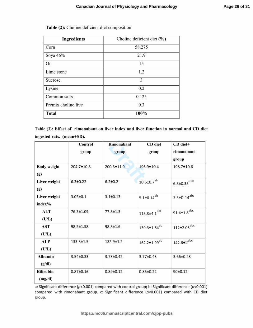
Effect of rimonabant on liver index and liver function in all experimental groups (table 3)
Serum ALT, AST, ALP, liver weight and liver index were significantly higher (P<0.001) in
CD diet group compared to control group and rimonabant group. There was no statistically
significant difference in body weight, albumin and bilirubin between all experimental groups.
Treatment with rimonabant induced a significant decrease (P<0.001) in serum ALT, AST,
ALP, liver weight and liver index in CD diet+ rimonabant group compared to CD diet group.
In contrast, there was no statistically significant difference in ALT, AST, ALP, liver weight,
body weight and liver index between rimonabant group and control group.
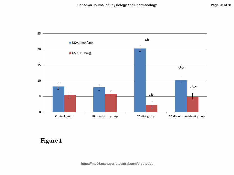
Effect of rimonabant on oxidative stress in all experimental groups (Fig.1)
MDA concentration was significantly increased (p<0.001) and GSH-Px activity was
significantly decreased (p<0.001) in CD diet group when compared to control group. On the
other hand, rimonabant treatment induced a significant decrease in MDA concentration and
significant increase in GSH-Px activity (p<0.001) in CD diet+ rimonabant group when
compared to CD diet group. Non-significant effect was also observed in rimonabant group
compared to control group.
Effect of Rimonabant on Activin β A mRNA expression, Follistatin mRNA expression, Activin
β A/ Follistatin mRNA ratio and CB-1 receptor gene expression in rat liver (table 4).
Activin β A mRNA expression, Activin β A/Follistatin mRNA ratio and CB -1 receptor gene
expression were significantly higher (P<0.001) in CD diet group compared to control group
Page 8 of 31
https://mc06.manuscriptcentral.com/cjpp-pubs
Canadian Journal of Physiology and Pharmacology

Draft
9
and rimonabant group. On the other hand, Follistatin mRNA expression was significantly
decreased in CD diet group compared to control group and rimonabant group. Treatment with
rimonabant induced a significant decrease (P<0.001) in Activin β A mRNA expression,
Activin β A/Follistatin mRNA ratio and CB -1 receptor gene expression with a parallel
significant increase (P<0.05) in Follistatin mRNA expression compared to CD diet group.
Moreover Pearson’s correlation analysis revealed a positive correlation of CB-1 receptor
gene expression with Activin β A mRNA level (r = 0.989; p < 0.01) . A negative correlation
of CB-1 receptor gene expression with Follistatin mRNA level (r = -0.992; p < 0.01) was also
observed Fig.2(A,B) .
Effect of Rimonabant on NAFLD score by H & E and the area% of TGF-β immunoreaction
of hepatocytes (Table 5) , Fig.3(A,B,C,D) and Fig.4(A,B,C,D)
CD diet group showed typical features of NAFLD, such as macrovesicular Steatosis and foci
of lobular inflammation and ballooning degeneration Fig.3(C). The NAFLD scores was
significantly higher (P<0.001) in CD diet group compared to control Fig.3(A) and rimonabant
Fig. 3 (B) groups. Treatment with rimonabant in (CD diet + rimonabant group) as in Fig.3 (D)
induced a significant decrease (P<0.001) in NAFLD scores compared to CD diet group.
Similarly to NAFLD score, the area% of TGF-β immunoreaction of hepatocytes was
significantly higher (P<0.001) in CD diet group Fig. 4(C) compared to control Fig. 4(A) and
rimonabant Fig. 4(B) groups. Additionally treatment with rimonabant in (CD diet +
rimonabant group) as in Fig. 4(D) induced a significant decrease (P<0.001) in area% of TGF-
β immunoreaction compared to CD diet group.
Discussion:
Despite the high prevalence of NAFLD and its potential for serious sequelae, the
underlying etiologic factors that determine disease progression remain poorly understood;
therefore, effective therapeutic strategies need to be further explored. The mechanisms that
underling hepatic fat accumulation and triggering of hepatocyte injury and hepatic fibrosis in
NASH are still largely unknown. In particular, little is known about the mediators that could
trigger the extensive hepatic fibrogenic response in certain individuals with NAFLD, leading
to advanced NASH (Claudia et al. 2015).
In the current study we chose a CD diet as it induces a comprehensive histological and
dysmetabolic phenotype resembling human NASH (Li et al.2011). This phenotype is
Page 9 of 31
https://mc06.manuscriptcentral.com/cjpp-pubs
Canadian Journal of Physiology and Pharmacology

Draft
10
characterized by fatty liver, inflammation, fibrosis, cirrhosis, and even hepatocellular
carcinoma (Pagotto et al.2006). We preferred using CD diet rather than high fat diet as it
manipulates liver fat content without affecting adipose fat stores (Peter et al.2015). Moreover
CD diet reaches the 2nd
hit of NAFLD model in shorter duration (10-12 weeks) than that of
high fat diet (minimum 24 weeks) (Masahiko et al.2013).
This study has shown that CD diet administration for 12 weeks resulted in
development of NAFLD, manifested by significant increase in serum liver biomarkers ALT,
ALP and AST when compared to control group. The mechanism by which CD diet induces
fatty liver was explained by Noga and Vance (2003) who revealed that choline is an important
element for formation of phosphatidylcholine, which is a critical component of very-low-
density lipoproteins (VLDL), which are responsible for transporting triglycerides out of the
liver.
Also our results were in agreement with Han et al. (2015) and Cheung and Sanyal,
(2010)who reported that CD diet induces NAFLD by causing mitochondrial dysfunction
which is a central mechanism in the pathogenesis of NAFLD . Mitochondria play an
important role in hepatocyte metabolism, being the primary site for oxidation of fatty acids
and oxidative phosphorylation. The mitochondrial dysfunction decreases the capacity to
oxidize fatty acids, increases the delivery and transport of free fatty acids into the liver, and
augments hepatic fatty acid synthesis which is likely to play a significant role in the
pathogenesis of NAFLD (Pessayre and Fromenty 2005). Another mechanism explained by
Malhi and Kaufman (2011) who showed that CD diet caused NAFLD through endoplasmic
reticulum stress, that inhibits the activity of insulin sensitizing kinase which mediates insulin
signaling in hepatocytes leading to insulin resistance which can promote hepatic steatosis
(Ron and Walter 2007).
Regarding the serum level of albumin and bilirubin, there was non-significant effect of
CD diet in comparison to control group .These results were in agreement with Arora and
Sharma (2012) and Smith and Adams (2011) who stated that total bilirubin and albumin are
usually normal in NASH and NAFLD and only increase in cases associated with cirrhosis that
developed in the end stage of the disease.
The results of our study also indicated that there was a significant increase in liver
weight and liver index with a non significant effect on the body weight in CD diet group when
compared to control group. These results were in agreement with Kitson et al. (2016) and Han
et al. (2015) who stated that there was no body weight gain in the CD diet-induced NAFLD
Page 10 of 31
https://mc06.manuscriptcentral.com/cjpp-pubs
Canadian Journal of Physiology and Pharmacology

Draft
11
models, also Raubenheimer et al. (2006) who reported that low choline leads to reduced
secretion of liver triglyceride as VLDL, resulting in accumulation of liver triglycerides
without affection of enzymes involved in denovo lipogenesis, So the liver weight increases
without change in body weight.
In the current study particular attention is paid to oxidative stress and its role in the
development and progression of NAFLD and its sequelae such as fibrosis. As our results
demonstrated that CD diet enhanced oxidative stress state in liver tissue evidenced by
reduction in antioxidant enzyme as GSH-Px and increase in marker of lipid peroxidation
(MDA).In agreement with these finding Juliana et al.(2015)and Rolo et al.(2012)
demonstrated that lipid accumulation in liver causes alteration in electron transport chain and
causes an increase of reactive oxygen species (ROS) production. Moreover, activation of
Kupffer cells and other inflammatory cells also generates ROS through nicotinamide adenine
dinucleotide phosphate oxidase (Gornicka et al.2011). These species can oxidize
polyunsaturated fatty acids present in cell and organelle membranes producing lipid
peroxidation metabolites like MDA (Rolo et al.2012).
We have also considered the consequences of CD diet on activin- β A, Follistatin
mRNA and Activin β A/Follistatin mRNA ratio and the results showed significant increase in
activin- β A, Activin β A/Follistatin mRNA ratio and significant decrease in follistatin mRNA
expression in liver tissues of CD diet group when compared to control group. These results
were parallel to that of Yndestad et al. (2009) who demonstrated that serum levels of activin
A and activin A/follistatin mRNA ratio in liver are increased in patients with NAFLD,
potentially reflecting increased activin A bioactivity. Follistatin antagonizes the biological
actions of activin A and blocks its signaling (Refaat et al. 2015).
The author's attention was also focused on immunohistochemical examination of
TGFβ1 that showed a strong positive expression of TGFβ1 in CD diet group when compared
to that of control group .These results were in agreement with Yang et al. ( 2014) and Vincent
et al. (2012) who reported that TGF-β1 plays a pivotal role in hepatic fibrosis by mediating
the activation of stellate cells and their production of extracellular matrix proteins. Indeed,
Kupffer and stellate cells produce TGF-β1that increases synthesis and deposition of type I
collagen ending by transformation of resting stellate cells to myofibroblasts. Moreover, this
finding parallel to that of Tarantino et al. (2008) who revealed that enhanced serum TGF-β1
Page 11 of 31
https://mc06.manuscriptcentral.com/cjpp-pubs
Canadian Journal of Physiology and Pharmacology

Draft
12
concentrations could represent a marker of early activation of mesenchymal hepatic stellate
cells and ultimately leading to liver damage.
Our results were supported with histopathological examination of the liver
tissues and the results showed fatty liver changes in the form of severe steatosis of
hepatocytes and inflammatory cells infiltration in CD diet group. These results were in
agreement with Han et al. ( 2015) who stated that CD diet causes macrovesicular steatosis ,
ballooning degeneration and foci of lobular inflammation. Moreover Juliana et al. (2015)
explained the morphofunctional alterations in rats having NAFLD by lipid peroxidation of
polyunsaturated fatty acids in mitochondrial membrane which is associated with
apolipoprotein B proteolysis, and this reduces VLDL secretion, promoting triacylglycerol
accumulation in liver.
Regarding hepatic CB1 receptor expression, we showed significant increase of
hepatic CB1 receptor gene expression in CD diet group when compared to control group. This
finding agree with a clinical study on human, which showed that CB1 mRNA expression was
significantly high in NASH (Teresa et al.2011).Also van der et al.(2010) observed that hepatic
CB1 expression correlated with the extent of steatosis and was significantly up-regulated in
those with increased steatosis grade, suggesting CB1 receptor activation and signaling.
Additionally we analyzed that it was positively correlated with activin-A and negatively
correlated with follistatin hepatic expression suggesting a deleterious role of CB1 in NAFLD.
On studying the effect of rimonabant on rats receiving normal standard diet, there was
non significant effect on all parameters when compared to control group .This can be
explained by Jeong et al.(2008) who stated that in normal liver, the expression of CB1
receptors is modest, which probably explains why the focus of research on the role of CB1
receptors in the liver pathophysiology has come only recently. However, during liver
pathology, endocanabinoid system is activated, and CB1 receptors undergo marked up-
regulation.
We have also considered that rimonabant caused significant decrease in liver
enzymes, liver weight and liver index and exhibited minimal activation of kupffer cells with
very minimal hydrobic degeneration of some hepatocytes in CD diet+ rimonabant group when
compared to CD diet group. These results coincide with Chanda et al. (2011); Mallat et al.
(2011) and Tam et al. (2012) who explained that the major pathway is CB1 activation of
lipoprotein lipase in adipose tissue, resulting in increased fatty-acid release and transfer to
Page 12 of 31
https://mc06.manuscriptcentral.com/cjpp-pubs
Canadian Journal of Physiology and Pharmacology
Draft
13
liver . Additional mechanisms mediated via hepatic CB1 receptors include increased
denovo hepatic lipogenesis, decreased fatty acid oxidation and decreased secretion of
triglyceride-rich VLDL by increasing expression of the lipogenic transcription factor sterol
regulatory element-binding protein and its target enzymes: Acetyl coenzyme-A carboxylase-1
and fatty acid synthase (Joseph et al.2011).
Another mechanism explained by Osei-Hyiaman et al.(2008) who revealed that the activity of
hepatic carnitine palmitoyltransferase 1, the rate-limiting enzyme in mitochondrial fatty acid
β-oxidation, is suppressed by treatment with a CB1 agonist, and is prevented by rimonabant.
Additionally Migrenne et al.(2009) reported that CB1 blockade increases plasma adiponectin,
which is a key stimulator of fatty acid β-oxidation.
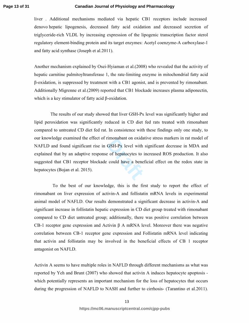
The results of our study showed that liver GSH-Px level was significantly higher and
lipid peroxidation was significantly reduced in CD diet fed rats treated with rimonabant
compared to untreated CD diet fed rat. In consistence with these findings only one study, to
our knowledge examined the effect of rimonabant on oxidative stress markers in rat model of
NAFLD and found significant rise in GSH-Px level with significant decrease in MDA and
explained that by an adaptive response of hepatocytes to increased ROS production. It also
suggested that CB1 receptor blockade could have a beneficial effect on the redox state in
hepatocytes (Bojan et al. 2015).
To the best of our knowledge, this is the first study to report the effect of
rimonabant on liver expression of activin-A and follistatin mRNA levels in experimental
animal model of NAFLD. Our results demonstrated a significant decrease in activin-A and
significant increase in follistatin hepatic expression in CD diet group treated with rimonabant
compared to CD diet untreated group; additionally, there was positive correlation between
CB-1 receptor gene expression and Activin β A mRNA level. Moreover there was negative
correlation between CB-1 receptor gene expression and Follistatin mRNA level indicating
that activin and follistatin may be involved in the beneficial effects of CB 1 receptor
antagonist on NAFLD.
Activin A seems to have multiple roles in NAFLD through different mechanisms as what was
reported by Yeh and Brunt (2007) who showed that activin A induces hepatocyte apoptosis -
which potentially represents an important mechanism for the loss of hepatocytes that occurs
during the progression of NAFLD to NASH and further to cirrhosis- (Tarantino et al.2011).
Page 13 of 31
https://mc06.manuscriptcentral.com/cjpp-pubs
Canadian Journal of Physiology and Pharmacology

Draft
14
Furthermore Patella et al. (2006) reported that administration of the activin-binding protein
follistatin in an animal model of liver fibrosis has been shown to reduce fibrosis development,
at least partly due to inhibited hepatocyte apoptosis.
Another mechanism explained by Yndestad et al.(2011) who reported a role for activin A in
the promotion of fibrogenesis in NAFLD by increasing the release of TGF- β from
hepatocytes and by activating hepatic stellate cells. Also in vitro study on primary rat
hepatocytes done by Gressner et al. (2008) showed that activin A may induce expression of
connective tissue growth factor which would promote fibrogenesis.
Furthermore another mechanism reported by Yndestad et al.(2011) who showed that activin
A has a role in increasing inflammation in NAFLD, as activin A has been shown to potently
stimulate the production of proinflammatory cytokines and opposes the anti-inflammatory
cytokines.
On the other hand, the immunohistochemical results of the current study showed that
the immunostaining intensity of the TGF-β1 decreased in the CD diet + rimonabant treated
group when compared to CD diet untreated group. These results runs parallel with DeLeve et
al. (2008) who showed that administration of rimonabant to wild-type mice or genetic
inactivation of CB1 receptors were both associated with a significant reduction in fibrosis
progression. Rimonabant-treated or CB1 knock-out mice also displayed reduced hepatic
expression of the TGF-β1, and a decrease in the number of fibrogenic cells. Antifibrogenic
properties of the CB1-selective antagonist were ascribed to antiproliferative property of
rimonabant in hepatic myofibroblasts (Domenicali et al. 2009).
Conclusions
In light of the results of this study it can be concluded that cannabinoid receptor1 (CB1)
antagonist, rimonabant has a potential therapeutic effect on NAFLD induced by CD diet. This
might be due to suppression of oxidative stress and hepatic expression of TGF-β1. The study
also demonstrated for the first time that rimonabant has hepatoprotective effect on NAFLD by
modulating hepatic expression of activin A and follistatin.
Acknowledgements
Experimental Animal Unit of Moshtohor faculty of agriculture is acknowledged for
providing us with animals and formulated diet. We also acknowledge department of
Page 14 of 31
https://mc06.manuscriptcentral.com/cjpp-pubs
Canadian Journal of Physiology and Pharmacology

Draft
15
pathology Faculty of Medicine, Benha University for helping in performing the
histopathological examination.
Conflict of interest:
The authors declare that they have no conflict of interest.
References:
Alhusseini, N.F., Odaa ,M.M., Mohamed, M.A., Abd El Wahab ,W.B., and Hasan, A.A.,
2010.Expression of adiponectin receptors in human placenta and its possible implication in
gestationaldiabetes. Am. J. Biochem. Biotechnol. 6: 136-140. doi:
10.3844/ajbbsp.2010.136.140.
Arora, A., and Sharma, P., 2012. Non-invasive Diagnosis of Fibrosis in Non-alcoholic
Fatty Liver Disease. Journal of Clinical and experimental hepatology, 2(2) :145–155.
https://doi.org/10.1016/S0973-6883(12)60103-0.
Bieghs, V. and Trautwein, C., 2014. Innate immune signaling and gut-liver interactions in
non-alcoholic fatty liver disease. Hepatobiliary Surg. Nutr.3: 377-385. doi:
10.3978/j.issn.2304-3881.2014.12.04.
Bojan, J., Dušan, M., Milica, N., Milena, V., Vesna, D., Aleksandar, V.,Danijela, V., et al.
2015. Rimonabant Improves Oxidative/Nitrosative Stress in Mice with Nonalcoholic Fatty
Liver Disease. Oxidative Medicine and Cellular Longevity,Volume 2015, Article ID
842108, 11 pages. http://dx.doi.org/10.1155/2015/842108.
Chanda, D., Kim, DK., Li, .T, Kim, YH., Koo, SH., Lee, CH., et al. 2011.Cannabinoid
receptor type 1 (CB1R) signaling regulates hepatic gluconeogenesis via induction of
Page 15 of 31
https://mc06.manuscriptcentral.com/cjpp-pubs
Canadian Journal of Physiology and Pharmacology

Draft
16
endoplasmic reticulum-bound transcription factor cAMP-responsive element-binding
protein H (CREBH) in primary hepatocytes. J. Biol. Chem. 12; 286(32):27971-9. doi:
10.1074/jbc.M111.224352.
Cheung, O., and Sanyal, A.J., 2010. Recent advances in nonalcoholic fatty liver disease.
Curr. Opin.Gastroenterol.26: 202–208.http://dx.doi.org/10.1097/MOG.0b013e328337b0c4.
Claudia, F., Barry, J.G., Richard, N., Williams, and Arun, S.,2015. Non-Alcoholic
Steatohepatitis: Limited Available Treatment Options but Promising Drugs in
Development and Recent Progress Towards a Regulatory Approval PathwayDrugs. 75(12):
1373–1392. doi: 10.1007/s40265-015-0437-3.
Corbin, IR., and Minuk, GY., 2003. Serial percutaneous liver biopsies in laboratory rats.
Dig. Dis. Sci. Oct;48(10):1939-43.doi: 10.1023/A:1026228018643.
DeLeve, LD., Wang, X., Kanel, GC., Atkinson, RD. and McCuskey, RS.,2008.Prevention
of hepatic fibrosis in a murine model of metabolic syndrome with nonalcoholic
steatohepatitis Am. J. Pathol. 173(4):993-1001. doi: 10.2353/ajpath.2008.070720.
Domenicali, M., Caraceni, P., Giannone, F., Pertosa, AM., Principe, A., Zambruni, A., et
al.2009. Cannabinoid type 1 receptor antagonism delays ascites formation in rats with
cirrhosis. Gastroenterology, 137(1):341-9. doi:10.1053/j.gastro.2009.01.004.
Iwo, M.I., Sjahlim S.L.,and Rahmawati, S.F.,2017. Effect of Vernonia amygdalina Del.
Leaf Ethanolic Extract on Intoxicated Male Wistar Rats Liver. Sci. Pharm. 85, 16.
doi:10.3390/scipharm85020016.
Page 16 of 31
https://mc06.manuscriptcentral.com/cjpp-pubs
Canadian Journal of Physiology and Pharmacology

Draft
17
George, J., Pera, N., Phung, N., Leclercq, I., Yun Hou, J., and Farrell, G.,2003. Lipid
peroxidation, stellate cell activation and hepatic fibrogenesis in a rat model of chronic
steatohepatitis. J. Hepatol. 39:756–64. doi: http://dx.doi.org/10.1016/S0168-
8278(03)00376-3.
Gornicka, A., Morris-Stiff, G., Thapaliya, S., Papouchado, B. G., Berk, M., and Feldstein,
A. E., 2011 “Transcriptional profile of genes involved in oxidative stress and antioxidant
defense in a dietary murine model of steatohepatitis,” Antioxidants and Redox Signaling,
15. 2: 437–445. doi: 10.1089/ars.2010.3815.
Gressner, O. A., Lahme, B., Siluschek, M., Rehbein, K., Weiskirchen, R., and Gressner, A.
M., 2008. Intracrine signalling of activin A in hepatocytes upregulates connective tissue
growth factor (CTGF/CCN2) expression. Liver Int. 28(9):1207–1216. doi: 10.1111/j.1478-
3231.2008.01729.x.
Han, H., Cui, M., You, X., Chen, M., Piao, X., et al. 2015 A role of 1,25(OH)2D3
supplementation in rats with non alcoholic steatohepatitis induced by choline deficient diet
. Nutrition, Metabolism & Cardiovasclar diseases ,25:556-561. doi:
http://dx.doi.org/10.1016/j.numecd.2015.02.011.
Jeong ,WI., Osei-Hyiaman, D., Park, O., Liu, J., Bátkai, S., Mukhopadhyay, P., et al.2008.
Paracrine activation of hepatic CB1 receptors by stellate cell-derived endocannabinoids
mediates alcoholic fatty liver. Cell Metab. 7:227–35. doi:
http://dx.doi.org/10.1016/j.cmet.2007.12.007.
Joseph, T., Jie, L., Bani, M., Resat, C., Grzegorz, G., and George, K., 2011.
Endocannabinoids in Liver Disease. Hepatology, 53:346-355. doi: 10.1002/hep.24077.
Page 17 of 31
https://mc06.manuscriptcentral.com/cjpp-pubs
Canadian Journal of Physiology and Pharmacology

Draft
18
Juliana, C. F. , Orlando, R. P. , Iara, B. , Kívia, Q., Fabiana, A. M., Salete, S., et al..2015
Choline and Cystine Deficient Diets in Animal Models with Hepatocellular Injury:
Evaluation of Oxidative Stress and Expression of RAGE, TNF-α, and IL-1β. Oxidative
Medicine and Cellular Longevity, Volume 2015 , Article ID 121925, 11 pages.
http://dx.doi.org/10.1155/2015/121925.
Kitson, MT. , Pham, A. ,Gordon, A. , Kemp, W. , Roberts, SK. 2016.High dose vitamin D
supplementation and liver histology in NASH. Gut, 65(4):71-78. Available from
http://dx.doi.org/10.1136/gutjnl-2015-310417.
Kleiner, BM. 2006.Macroergonomics: analysis and design of work systems. Appl. Ergon.
37: 81-89. doi:10.1016/j.apergo.2005.07.006.
Li, C.,Jones, P. M., Persaud, S. J.,2011 “Role of the endocannabinoid system in food
intake, energy homeostasis and regulation of the endocrine pancreas,” Pharmacol. Ther.
129, 3: 307–320. doi:10.1016/j.pharmthera.2010.10.006.
Malhi, H. and Kaufman, R.J., 2011. Endoplasmic reticulum stress in liver disease. J
Hepatol; 54:795–809. doi: http://dx.doi.org/10.1016/j.jhep.2010.11.005.
Mallat, A., Teixeira-Clerc, F., Deveaux, V., Manin, S.and Lotersztajn, S., 2011. The
endocannabinoid system as a key mediator during liver diseases: new insights and
therapeutic openings .Br. J. Pharmacol., 163 , 1432–1440. doi: 10.1111/j.1476-
5381.2011.01397.x.
Page 18 of 31
https://mc06.manuscriptcentral.com/cjpp-pubs
Canadian Journal of Physiology and Pharmacology

Draft
19
Masahiko, M., Natsuko, H., Yoshiyuki, S., Akiko, U., Toshihiko, S., Chiaki, T., et al.2013.
An improved mouse model that rapidly develops fibrosis in non-alcoholic steatohepatitis.
Int. J. Exp. Pathol. 94(2): 93–103. doi: 10.1111/iep.12008.
Migrenne, S., Lacombe, A., Lefevre, A.L., Pruniaux, M.P., Guillot, E., Galzin, A.M. et
al.2009. Adiponectin is required to mediate rimonabant-induced improvement of insulin
sensitivity but not body weight loss in diet-induced obese mice. Am. J. Physiol. Regul.
Integr. Comp. Physiol.296: 929-935.doi: 10.1152/ajpregu.90824.2008.
Noga, A.A., and Vance, D.E.,2003. A gender-specific role for phosphatidylethanolamine
Nmethyltransferase - derived phosphatidylcholine in the regulation of plasma high density
and very low density lipoproteins in mice. J. Biol. Chem. 278:21851–21859.
doi:10.1074/jbc.M301982200.
NRC. 1995. Nutrient Requirements of Laboratory Animals. 4th ed.: Subcommittee on
Laboratory Animal Nutrition, Committee on Animal Nutrition, Board on Agriculture,
National Research Council, p.13. doi: https://doi.org/10.17226/4758.
Oh, M.K., Winn, J. and Poordad, F., 2008. Review article: diagnosis and treatment of
nonalcoholic fatty liver disease. Aliment. Pharmacol. Ther. 28: 503–22.
doi:10.1111/j.1365-2036.2008.03752.x.
Ohkawa, H., Ohishi, N. , Yagi, K.,1979. Assay for lipid peroxides in animal tissues by
thiobarbituric acid reaction. Anal Biochem. 95: 351–358. Available from
https://doi.org/10.1016/0003-2697(79)90738-3.
Page 19 of 31
https://mc06.manuscriptcentral.com/cjpp-pubs
Canadian Journal of Physiology and Pharmacology

Draft
20
Ooe, H., Chen, Q., Kon. J., Sasaki, K., Miyoshi, H. and Ichinohe, N.,2012. Proliferation of
rat small hepatocytes requires follistatin expression. J. Cell Physiol . 227: 2363-2370. doi:
10.1002/jcp.22971.
Osei-Hyiaman, D., Liu, J., Zhou, L., Godlewski, G., Harvey-White, J., Jeong, W.I. et al.
2008.Hepatic CB(1) receptor is required for development of diet-induced steatosis,
dyslipidemia, and insulin and leptin resistance in mice. J. Clin. Invest .118:3160-3169.
doi:10.1172/JCI34827.
Pagotto,U. , Marsicano G., Cota, D. Lutz, B.,and Pasquali, R., 2006“The emerging role of
the endocannabinoid system in endocrine regulation and energy balance,” Endocrine
Reviews, 27.1: 73–100. doi: 10.1210/er.2005-0009. PMID:16306385
Patella, S., Phillips, D. J., Tchongue, J., de Kretser, D. M., and Sievert, W., 2006.
Follistatin attenuates early liver fibrosis: Effects on hepatic stellate cell activation and
hepatocyte apoptosis. Am. J. Physiol. Gastrointest. Liver Physiol. 290.1: 137–144. doi:
10.1152/ajpgi.00080.2005. PMID:16123203
Pessayre, D. ,and Fromenty, B., 2005.NASH: a mitochondrial disease. J. Hepatol. 42:928–
940 doi: 10.1016/j.jhep.2005.03.004. PMID:15885365.
.
Peter J. R, Moffat J. N.,and Brian R. W.,2006. A Choline-Deficient Diet Exacerbates Fatty
Liver but Attenuates Insulin Resistance and Glucose Intolerance in Mice Fed a High-Fat
Diet .Diabetes , 55(7): 2015-2020. Available from https://doi.org/10.2337/db06-0097.
Pisanti, S., Picardi, P., Pallottini, V., Martini, C., Petrosino, S. and Proto, MC.2015.
Anandamide drives cell cycle progression through CB1 receptors in a rat model of
Page 20 of 31
https://mc06.manuscriptcentral.com/cjpp-pubs
Canadian Journal of Physiology and Pharmacology

Draft
21
synchronized liver regeneration. J. Cell Physiol. 230:2905-2914.doi: 10.1002/jcp.24959.
PMID: 25684344
Raubenheimer, P.J. , Nyirenda, M.J.,and Walker, B.R., 2006. A choline
deficient diet exacerbates fatty liver but attenuates insulin resistance and glucoseintolerance
in mice fed a high-fat diet. Diabetes. 55(7):2015-20. doi:10.2337/db06-0097.
PMID:16804070.
Refaat B., El-Shemi, A.G., and Ashshi, A.M.,2015. The effects of pegylated interferon -α
and ribavirin on liver and serum concentrations of activin-A and follistatin in normal
Wistar rat: a preliminary report. BMC Res. Notes,8:265:1-8. doi 10.1186/s13104-015-
1253-2.
Rodgarkia-Dara, C., Vejda, S., Erlach, N., Losert, A., Bursch, W., and Berger, W., 2006.
The activin axis in liver biology and disease. Mutat. Res. 613:123–137. doi:
10.1016/j.mrrev.2006.07.002. PMID: 16997617.
Rolo, A. P, Teodoro, J. S., and Palmeira, C. M.,2012 “Role of oxidative stress in the
pathogenesis of nonalcoholic steatohepatitis,” Free Radical Biology and Medicine. 52. 1:
59–69. doi: 10.1016/j.freeradbiomed.2011.10.003.
Romero-Zerbo, SY. and Bermudez-Silva, EJ., 2014.“Cannabinoids, eating behavior, and
energy homeostasis,” Drug Testing and Analysis, 6, 1-2, 52–58. Available from
https://www.ncbi.nlm.nih.gov/pubmed/20347862. PMID: 20347862.
Ron, D., Walter, P. 2007.Signal integration in the endoplasmic reticulum unfolded protein
response. Nat. Rev. Mol. Cell Biol. 8:519–529. doi:10.1038/nrm2199. PMID:17565364.
Page 21 of 31
https://mc06.manuscriptcentral.com/cjpp-pubs
Canadian Journal of Physiology and Pharmacology

Draft
22
Schwabe, R. F., 2005“Endocannabinoids promote hepatic lipogenesis and steatosis through
CB1 receptors,” Hepatology, 42. 4:959–961. doi: 10.1002/hep.20900
Smith, B. W., and Adams, L. A., 2011. “Nonalcoholic fatty liver disease and diabetes
mellitus: pathogenesis and treatment,” Nature Reviews Endocrinology,7. 8: 456–465.
doi:10.1038/nrendo.2011.72.
Tam, J., Cinar, R., Liu, J., Godlewski, G., Wesley, D., Jourdan, T., et al. 2012. Peripheral
cannabinoid-1 receptor inverse agonism reduces obesity by reversing leptin resistance. Cell
Metab. 8. 16(2):167-79. doi: 10.1016/j.cmet.2012.07.002.
Tappel, A. L.1978.Glutathione peroxidase and hydroperoxides. Methods Enzymol.52:506–
513.
Tarantino, G., Colao, A., Capone, D., Conca, P., Tarantino, M., Grimaldi, E.,et al. 2011.
Circulating levels of cytochrome C, gamma-glutamyl transferase, triglycerides and
unconjugated bilirubin in overweight/obese patients with non-alcoholic fatty liver disease.
J. Biol. Regul. Homeost. Agents, 25(1):47-56. PMID:21382273.
Tarantino, G., Conca, P., Riccio, A., Tarantino, M., Matteo, N., Minno, Di., et al. .2008
Enhanced serum concentrations of transforming growth -beta1 in simple fatty liver: is it
really benign? J. Transl. Med. 6: 72. doi:10.1186/1479-5876-6-72.
Teresa, A., Alba, B., Esther, G-J., Ximena, T., Salomé, M., et al.2014. Endocannabinoid
Receptors Gene Expression in Morbidly Obese Women with Nonalcoholic Fatty Liver
Page 22 of 31
https://mc06.manuscriptcentral.com/cjpp-pubs
Canadian Journal of Physiology and Pharmacology
Draft
23
Disease. Biomed. Res. Int. 2014: Article ID:502542. 7 pages. Available from
http://dx.doi.org/10.1155/2014/502542.
Tziomalos, K., Athyros, VG. and Karagiannis, A., 2012. Non-alcoholic fatty liver disease
in type 2 diabetes: pathogenesis and treatment options. Curr. Vasc. Pharmacol . 10: 162-
172. PMID: 22239625.
van der, P D., Shahidi, M., Tay, E., Sesha, J., Tran, K., McLeod, D., et al. 2010.Hepatitis C
virus induces the cannabinoid receptor 1. PLoS One, 5:e12841. Available from
https://doi.org/10.1371/journal.pone.0012841.
Vincent, B., Giorgio, L. V., François, M. and Fabrizio, M.,.2012.
Role of cytokines and chemokines in non-alcoholic fatty liver disease World J.
Gastroenterol. 28. 18(8): 727–735. doi: 10.3748/wjg.v18.i8.727.
Yang, L., Roh, Y.S., Song, J., Zhang, B., Liu, C., Loomba, R., et al. 2014.
Transforming growth factor beta signaling in hepatocytes participates in steatohepatitis
through regulationof cell death and lipid metabolism in mice.Hepatology, 59(2):483-
95. doi: 10.1002/hep.26698.
Yndestad, A., Haukeland, JW., Dahl, TB., Bjoro, K., Gladhaug, IP. and Berge, C. 2009. A
complex role of activin A in non-alcoholic fatty liver disease. Am. J. Gastroenterol . 104:
2196-2205. doi: 10.1038/ajg.2009.318.
Yndestad, A., Haukeland, JW., Dahl, TB., Halvorsen, B.,and Aukrust, P.,2011. Activin A
in nonalcoholic fatty liver disease. Vitam. Horm . 85: 323-342. doi: 10.1016/B978-0-12-
385961-7.00015-9.
Page 23 of 31
https://mc06.manuscriptcentral.com/cjpp-pubs
Canadian Journal of Physiology and Pharmacology

Draft
24
Figure Caption:
Figure 1:
Effect of rimonabant on oxidative stress in all experimental groups
a: Significant difference (p<0.001) compared with control group; b: Significant difference
(p<0.001) compared with rimonabant group. c: Significant difference (p<0.001) compared
with CD diet group.
Figure 2 (A & B):
(A) :Correlation of CB-1 receptor gene expression with follistatin mRNA level.
(B): Correlation of CB-1 receptor gene expression with Activin β A mRNA level.
Figure 3 (A , B,C,D):
Histological changes of rat liver stained with H&E. A.control group showing normal
histological structure of hepatic lobule .B. Rimonabant group showing normal histological
structure of hepatic lobule. C. CD diet group showing Steatosis (S) of hepatocytes and focal
inflammatory cells infiltration(IC) D. CD diet + Rimonabant group showing slight hydropic
degeneration of hepatocytes and minimal activation of kupffer cells (H & E X 400).
Figure 4 (A , B,C,D):
Immunostaining reaction for TGF-β.Photomicrographs of TGF-β immune-stain reaction
stained liver sections. A. Immunohistochemical staining of TGF-β in liver of rat from
control group showing no expression of TGF-β (negative immunohistochemical reaction).B.
Immunohistochemical staining of TGF-β in liver of rat from Rimonabant group showing no
expression of TGF-β (negative immunohistochemical reaction). C. Immunohistochemical
staining of TGF-β in liver of rat from CD diet group showing strong positive expression of
TGF-β (dark brown colour). C. Immunohistochemical staining of TGF-β in liver of rat from
CD diet + Rimonabant group showing weak positive expression of TGF-β (X 400).
Page 24 of 31
https://mc06.manuscriptcentral.com/cjpp-pubs
Canadian Journal of Physiology and Pharmacology

Draft
Table (1): Ingredients % of the control diet
Feed ingredients Control diet (%)
Sunflower oil 15.00
Concentrate mixture (45%)1 10.00
Yellow corn 49.00
Soybean meal (44%) 11.00
Wheat bran 10.00
Molasses 03.00
Common salt 00.50
Ground limestone 00.20
Dicalcium phosphate 00.10
Lysine 00.20
DL-Methionine 00.70
Mineral-vitamin premix 2 00.30
Total 100%
1Concentrate mixture composed of corn gluten 60%, dicalcium phosphate, soybean meal 48%,
fish meal (65%), limestone, broiler premix, L-lysine HCl, DL-methionine, and common salt.
2Each 3 kg contains: vit. A 12000000 IU, vit. D3 2000000 IU, vit. E 10000 mg, vit. K3 1000 mg, vit.
B1 1000 mg, vit. B2 5000 mg, vit. B6 1500 mg, vit. B12 10 mg, biotin 50 mg, pantothenic acid 10000
mg, nicotinic acid 30000 mg, folic acid 1000 mg, manganese 60000 mg, zinc 50000 mg, iron 30000
mg, copper 4000 mg, iodine 300 mg, selenium 100 mg, cobalt 100 mg, carrier (CaCo3) up to 3 kg.
Page 25 of 31
https://mc06.manuscriptcentral.com/cjpp-pubs
Canadian Journal of Physiology and Pharmacology
Draft
Table (2): Choline deficient diet composition
Ingredients Choline deficient diet (%)
Corn 58.275
Soya 46% 21.9
Oil 15
Lime stone 1.2
Sucrose 3
Lysine 0.2
Common salts 0.125
Premix choline free 0.3
Total 100%
Table (3): Effect of rimonabant on liver index and liver function in normal and CD diet
ingested rats. (mean+SD).
Control
group
Rimonabant
group
CD diet
group
CD diet+
rimonabant
group
Body weight
(g)
204.7±10.8 200.3±11.9 196.9±10.4 198.7±10.6
Liver weight
(g)
6.3±0.22 6.2±0.2 10.6±0.7ab
6.8±0.33abc
Liver weight
index%
3.05±0.1 3.1±0.13 5.1±0.14ab
3.5±0.14abc
ALT
(U/L)
76.3±1.09 77.8±1.3 115.8±4.1
ab 91.4±1.8
abc
AST
(U/L)
98.5±1.58 98.8±1.6 139.3±1.64ab
112±2.05abc
ALP
(U/L)
133.3±1.5 132.9±1.2 162.2±1.99ab
142.6±2abc
Albumin
(g/dl)
3.54±0.33 3.73±0.42 3.77±0.43 3.66±0.23
Bilirubin
(mg/dl)
0.87±0.16 0.89±0.12 0.85±0.22 90±0.12
a: Significant difference (p<0.001) compared with control group; b: Significant difference (p<0.001)
compared with rimonabant group. c: Significant difference (p<0.001) compared with CD diet
group.
Page 26 of 31
https://mc06.manuscriptcentral.com/cjpp-pubs
Canadian Journal of Physiology and Pharmacology

Draft
Table (4): Effect of rimonabant on Activin β A , Follistatin mRNA expression, Activin β A
/Follistatin mRNA ratio and CB-1 receptor gene expression in normal and CD diet ingested rats. The
mRNA expressed by Log10 relative units of relative quantitation (RQ) (mean+SD).
Control
group
Rimonabant
group
CD diet
group
CD diet+
rimonabant
group
Activin β A
mRNA level
1.24±0.18 1.2±0.19 6.27±0.22ab
3.28±0.23abc
Follistatin
mRNA level
1.47±0.14 1.39±0.12 0.60±0.05ab
0.96±0.04abc
Activin β A/
Follistatin
mRNA
0.84±0.12 0.86±0.2 10.45±0.4 ab
3.4±0.6 abc
CB-1 receptor
gene expression
0.84±0.14 0.83±0.12 7.2±0.19 ab
4.3±0.25 abc
a: Significant difference (p<0.001) compared with control group; b: Significant difference (p<0.001)
compared with rimonabant group. c: Significant difference (p<0.001) compared with CD diet
group.
Table 5: Effect of rimonabant on NAFLD score by H & E and the area% of TGF-β
immunoreaction of hepatocytes in normal and CD diet ingested rats (mean+SD).
steatosis Lobular
inflammation
hepatocellular
ballooning
The mean area
% of TGF-β
immunoreaction
Control
group
0.00±0.00 0.00±0.00 0.00±0.00 0.00±0.00
Rimonabant
group
0.00±0.00 0.00±0.00 0.00±0.00 0.00±0.00
CD diet
group
3.00±0.00ab 2.5±0.2ab 2.7±0.3ab 6.6±0.17ab
CD diet+
rimonabant
group
1.9±0.42abc 1.3±0.3abc 1.2±0.5abc 1.2±0.13abc
a: Significant difference (p<0.001) compared with control group; b: Significant difference (p<0.001)
compared with rimonabant group. c: Significant difference (p<0.001) compared with CD diet
group.
Page 27 of 31
https://mc06.manuscriptcentral.com/cjpp-pubs
Canadian Journal of Physiology and Pharmacology
Draft0
5
10
15
20
25
Control group Rimonabant group CD diet group CD diet+ rimonabant group
MDA(nmol/gm)
GSH-Px(U/mg)
a,b
a,b
a,b,c
a,b,c
Page 28 of 31
https://mc06.manuscriptcentral.com/cjpp-pubs
Canadian Journal of Physiology and Pharmacology

Draft
Figure 2(A,B)
A B
A B
Page 29 of 31
https://mc06.manuscriptcentral.com/cjpp-pubs
Canadian Journal of Physiology and Pharmacology

DraftB
C D
S
IC
Figure 3(A,B,C,D)
A B
D C
Page 30 of 31
https://mc06.manuscriptcentral.com/cjpp-pubs
Canadian Journal of Physiology and Pharmacology

Draft
Figure 4(A,B,C,D)
D
B A
C
Page 31 of 31
https://mc06.manuscriptcentral.com/cjpp-pubs
Canadian Journal of Physiology and Pharmacology
Top Related