


Languages
Pages
Legal


0
Scuola Dottorale in Biologia
Sezione “Biologia Applicata alla Salute dell’Uomo”
Ciclo XXIV
Differentiation and modulation of innate immunity response
in human cord blood cells
Differenziazione e modulazione della risposta dell’immunità
innata nel cordone ombelicale
DOTTORANDA
Alessandra Ciucci
Docente guida/Supervisore interno: Prof. Paolo Visca
Supervisore esterno: Dott. Giorgio Mancino

1
Università degli studi di Roma “Roma Tre”
Differentiation and modulation of innate immunity response in human
cord blood cells
Differenziazione e modulazione della risposta dell’immunità innata
nel cordone ombelicale
Alessandra Ciucci
Dottorato di Ricerca in Biologia
Sezione “Biologia Applicata alla Salute dell’Uomo”
XXIV Ciclo
Docente guida/Supervisore interno: Prof. Paolo Visca
Supervisore esterno: Dott. Giorgio Mancino

2
To my family
3
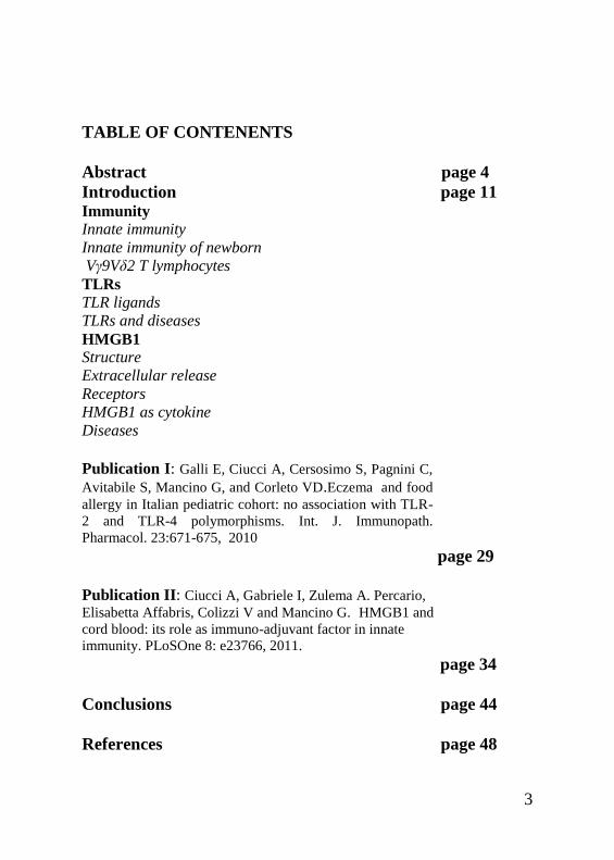
TABLE OF CONTENENTS
Abstract page 4
Introduction page 11 Immunity
Innate immunity
Innate immunity of newborn
Vγ9Vδ2 T lymphocytes
TLRs
TLR ligands
TLRs and diseases
HMGB1 Structure
Extracellular release
Receptors
HMGB1 as cytokine
Diseases
Publication I: Galli E, Ciucci A, Cersosimo S, Pagnini C,
Avitabile S, Mancino G, and Corleto VD.Eczema and food
allergy in Italian pediatric cohort: no association with TLR-
2 and TLR-4 polymorphisms. Int. J. Immunopath.
Pharmacol. 23:671-675, 2010
page 29
Publication II: Ciucci A, Gabriele I, Zulema A. Percario,
Elisabetta Affabris, Colizzi V and Mancino G. HMGB1 and
cord blood: its role as immuno-adjuvant factor in innate
immunity. PLoSOne 8: e23766, 2011.
page 34
Conclusions page 44
References page 48

4
ABSTRACT
Introduzione Lo sviluppo delle barriere difensive si è rivelato un evento essenziale per
l’evoluzione, in quanto ha consentito di separare l’ambiente esterno dal
sempre più complesso ambiente interno. Siamo costantemente esposti ad
agenti infettivi e tuttavia, nella maggior parte dei casi, siamo in grado di
contrastarli, grazie al nostro sistema immunitario che agisce mediante due tipi
di risposta:
- la risposta non specifica o innata, che costituisce la prima linea di difesa ed
opera in modo non selettivo verso antigeni estranei;
- la risposta specifica o adattativa, diretta verso antigeni specifici.
Il tipo di risposta è dettato dalla natura dell’antigene. Una risposta
immunitaria completa richiede la partecipazione coordinata di entrambe.
La risposta immunitaria innata è mediata da proteine recettoriali appartenenti
alla classe dei “pattern recognition receptors” (PPR), che riconoscono motivi
strutturali conservati presenti sugli antigeni, come componenti espresse
esclusivamente da microorganismi, definite “pathogen associated molecular
patterns” (PAMPs), o molecole rilasciate in seguito ad un danno cellulare
definite “damage-associated molecular patterns” (DAMPs). In particolare, un
tipo di PPR sono i “toll-like receptors” (TLR), una famiglia di proteine
transmembrana espresse soprattutto sulla superficie delle cellule
immunocompetenti, ossia monociti, macrofagi e cellule dendritiche, ma
anche sulla superficie delle cellule epiteliali. L’espressione dei TLRs ha
assunto un’importanza considerevole per la salute neonatale con la recente
dimostrazione che i TLRs partecipano al riconoscimento di patogeni rilevanti
per il neonato, tra cui Streptococco di gruppo B, Listeria monocytogenes,
Mycoplasma hominis, Candida. albicans e Citomegalovirus riconosciuti dal
TLR-2 o Enterobacteriaceae, C. albicans e il Virus Respiratorio Sinciziale
dal TLR-4. Anche se l'espressione basale dei TLRs sui monociti del sangue
del neonato è simile a quella degli adulti, le conseguenze funzionali della loro
attivazione sono molto diverse con implicazioni in una vasta gamma di
malattie, quali infezioni, immunodeficienza, sepsi, malattie autoimmuni e
allergie.
Il sistema immunitario neonatale è generalmente considerato immaturo e
meno funzionale rispetto alla controparte adulta e funzionalmente carente nel
contrastare l’attacco di patogeni, aumentando nel neonato la suscettibilità a
contrarre infezioni con conseguente incremento della mortalità. L’immaturità
5
del sistema immunitario neonatale deriva da effetti combinati di una serie di
fattori quali l’immaturità dei suoi componenti cellulari, la mancanza di
esposizione agli antigeni, l’esposizione intrauterina ad unico ambiente, che
può favorire uno sviluppo di una risposta linfocitaria di tipo Th2, la bassa
capacità di proliferazione dei linfociti T e l’alterata produzione di citochine
Th1.
La proteina “High mobility group box 1” (HMGB1) è una molecola DAMP
che lega il TLR-4 e il recettore RAGE (“receptor for advanced glycation
end-products”). In principio HMGB1 è stato studiato come co-fattore
nucleare coinvolto nella regolazione della trascrizione genica, ma
successivamente è stato dimostrato che HMGB1 viene anche rilasciato
attivamente o passivamente dalle cellule per poi agire come un citochina pro-
infiammatoria. Molti studi dimostrano come HMGB1 sia implicato nella
patogenesi di diverse patologie, quali artrite, cancro, epatite, malaria,
ischemia del miocardio, sepsi.
Obbiettivi della ricerca L’obbiettivo principale del lavoro è stato quello di comprendere meglio i
meccanismi che sono alla base delle risposte dell’immunità innata nel
neonato e nella prima infanzia per aprire la strada a possibili approcci di
immunomodulazione a scopo terapeutico. In considerazione dell’importanza
dei recettori TLRs nella risposta innata e della loro anomala attivazione in
associazione ad alcune malattie a base allergica, è stata valutata
l’associazione tra polimorfismi a singolo nucleotide (SNP) nel TRL-2 o TRL-
4 e allergie atopiche in una coorte di bambini italiani allergici.
Un’altra parte dello studio è stata invece rivolta all’analisi dell’espressione di
HMGB1 nel sangue del cordone ombelicale. L'espressione di HMGB1 e il
suo ruolo nella risposta immunitaria sono stati studiati quasi esclusivamente
nel sangue periferico (PB) di individui adulti e solo recentemente, è stato
osservato che HMGB1, insieme con il recettore solubile (sRAGE), possono
essere importanti mediatori del danno cellulare nel feto e fattore cruciale
nella nascita pre-termine. Data l’importanza di comprendere il profilo
immunitario immaturo del neonato, è stata comparata l’espressione di
HMGB1 nelle cellule isolate dal sangue di cordone ombelicale (CB) rispetto
al PB caratterizzando sia l'espressione di HMGB1 e la sua distribuzione nelle
diverse popolazioni presenti nel sangue che valutando la sua possibile
modulazione ad opera di diversi stimoli in termini di presenza nella cellula e
secrezione extracellulare.

6
Risultati Per studiare una possibile associazione tra polimorfismi del TLR ed allergie
in pazienti in età pediatrica, SNPs R753Q nel TLR-2 o D299G nel TLR-4
sono stati individuati mediante PCR Real-Time da DNA isolato da sangue
periferico. Nel gruppo di controllo, composto da 147 individui sani, il
polimorfismo R753Q aveva una prevalenza del 2,5% mentre la frequenza
della mutazione D299G del 12%. Nessuno dei 159 pazienti allergici ha
mostrato il SNP R753Q. Nel TLR-4, invece, 7/57 pazienti affetti da allergia
alimentare (12%) e 6/102 pazienti con eczema (6%) presentavano la
mutazione in D299G. I dati mostrano assenza di correlazione tra i
polimorfismi studiati nel TLR e allergie atopiche (eczema e allergia
alimentare), suggerendo che non costituiscono marcatori per le malattie
atopiche nei bambini in Italia. Recentemente, nelle malattie asmatiche, è stato
riscontrato un altro fattore che sembrerebbe svolgere un ruolo importante:
HMGB1. In considerazione del fatto che HMGB1, come citochina pro-
infiammatoria, è stata caratterizzata esclusivamente nel sangue periferico di
individui adulti, abbiamo deciso di analizzare l'espressione di HMGB1 in
cellule mononucleate isolate dal CB mediante analisi in citofluorimetria
(FACS). Come atteso, la totalità delle cellule permeabilizzate del CB o PB
esprimevano HMGB1, in quanto HMGB1 è un cofattore nucleare ubiquitario.
In assenza di permeabilizzazione, è stato possibile rilevare la presenza di
HMGB1 anche sulla superficie delle cellule del CB con una percentuale del
13(±4)% (n=8), che era esattamente paragonabile a quella riscontrata sulle
cellule HeLa (una linea stabilizzata da un carcinoma umano della cervice
uterina che è noto esprimere HMGB1). Nel PB la proteina era presente nel
6.5(±1.8)% delle cellule (n = 8). È interessante notare che le cellule del CB
presentavano un’espressione di HMGB1 più alta rispetto alle cellule del PB e
tale differenza era statisticamente significativa (P=0.02).
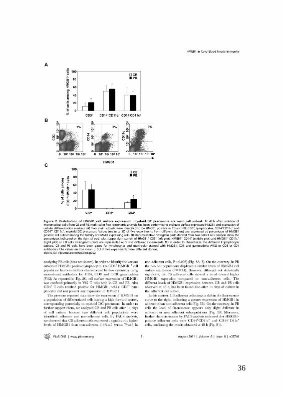
Al fine di caratterizzare l’espressione di HMGB1 sulle differenti popolazioni
cellulari presenti nel sangue, è stata effettuata una analisi citofluorimetrica
utilizzando più anticorpi contemporaneamente. Nelle cellule del cordone,
così come nel periferico, circa il 90% di HMGB1 era espresso su precursori
delle cellule dendritiche (DCs) di tipo mieloide identificati mediante l’analisi
dell’espressione di due marcatori fenotipici di membrana: CD14 +CD11c
+
(monociti) e CD14 -CD11c
+ (DCs). Solo una piccola percentuale di linfociti
CD3+
esprimeva HMGB1 [11(±8)%]. Al fine di identificare le varie
sottopopolazioni linfocitarie HMGB1+ positive, le cellule CD3
+ sono state
ulteriormente caratterizzate mediante FACS utilizzando anticorpi
monoclonali diretti verso il CD4, CD8 e TCR gammadelta (V2). Nel CB e

7
PB, HMGB1 era espresso principalmente sulle cellule T e CD4+ mentre i
linfociti CD8+
sono risultati negativi. Questi esperimenti sono stati eseguiti a
48 ore dalla purificazione delle cellule mononucleate da sangue intero. Dopo
14 giorni di coltura, invece, sono state analizzate due differenti popolazioni
cellulari: cellule aderenti e cellule in sospensione. Mediante analisi al FACS,
è stato osservato che le cellule aderenti del CB esprimevano livelli
significativamente più alti di HMGB1 rispetto alle non aderenti [14( ±5)%
contro il 7( ± 3)% delle non-aderenti; P = 0,003]. Al contrario, nel PB le due
popolazioni cellulari mostravano un livello simile di espressione. Inoltre,
cellule aderenti del CB presentavano livelli di espressione di HMGB1 più
elevate rispetto alla controparte del PB, in accordo con i risultati ottenuti a 48
ore, indicando che l’espressione di HMGB1 è confinata principalmente a
cellule maggiormente differenziate che anche in questo caso sono state
identificate al FACS come monociti e DCs.
Nel sangue periferico, la proteina HMGB1 si comporta come una citochina
rilasciata dalle cellule immunitarie attivate, capace di mediare risposte a
infezioni, lesioni e infiammazioni. Abbiamo così indagato se segnali diversi
di attivazione, quali stimoli pro-infiammatori (TNF-, IL-2 o IL-15) o che
mimano infezioni, quali il trattamento con lipopolisaccaride batterico (LPS) o
l’enterotossina B di Staphylococcus aureus (SEB) o il “Phorbol 12-Myristate
13-Acetate” (PMA), influenzino l'espressione di HMGB1 e la sua secrezione.
L’analisi citofluorimetrica ha mostrato che tutti gli stimoli erano in grado di
incrementare l'espressione di HMGB1 sulle cellule del CB e PB. Inoltre
l'aumenta espressione della proteina sulla membrana cellulare era associata
ad un aumento significativo dei livelli di HMGB1 rilasciati nel terreno di
coltura, analizzati tramite Western Blot.
Per determinare se la modulazione dell'espressione di HMGB1 nelle cellule
CB fosse associata a una sua diversa localizzazione intracellulare, cellule
trattate con LPS sono state analizzate in microscopia confocale.
L’immunofluorescenza ha mostrato che, in cellule del CB non trattate,
HMGB1 presentava un’espressione eterogenea, localizzata principalmente
nel nucleo e citoplasma. Dopo 48 ore di stimolazione con LPS, l’espressione
di HMGB1 appariva principalmente sul perimetro esterno delle cellule, come
indicato dalla sua co-localizzazione con la membrana plasmatica. Il
coinvolgimento della membrana cellulare nella secrezione di HMGB1,
osservato al FACS e in microscopia confocale, è stato ulteriormente
confermato studiando l'effetto del gliburide, un inibitore del trasporto
proteico di tipo non-classico. I risultati hanno mostrato che il gliburide
induceva una riduzione di circa il 50% dell’espressione costitutiva di
HMGB1 sulla superficie cellulare sia in CB che in PB. Inoltre, l’inibitore era
8
in grado di bloccare il rilascio della proteina procurato dal trattamento con
LPS, ripristinando l'espressione di superficie di HMGB1 che era stata ridotta
come conseguenza della sua secrezione.
Gli aminobisfosfonati (ABs) (Pamidronato o PAM, e Zoledronato o ZOL),
farmaci utilizzati nell’osteoporosi e nella terapia antitumorale, sono potenti
attivatori dei linfociti T . Dal momento che è stato provato che HMGB1 è
espresso sui linfociti T PAM e ZOL sono stati utilizzati per verificare le
loro possibili capacità di modulare l’espressione di HMGB1. E’ stato
dimostrato che PAM e ZOL inducevano nelle cellule del CB e PB: i)
l’espressione di HMGB1 sulla superficie cellulare; ii) la proliferazione di
linfociti T HMGB1+ e iii) la secrezione di HMGB1. Per dimostrare che la
secrezione di HMGB1 indotta da ABs non fosse determinata da morte
cellulare, a 14 giorni di trattamento sono state valutate sia l'apoptosi che la
necrosi mediante l'analisi al FACS. Non è stato osservato alcun cambiamento
significativo del numero di cellule annessina V+ e ioduro di propidio positive
nel trattamento con ABs rispetto al controllo.
Avendo riscontrato che HMGB1 viene rilasciato dalle cellule a seguito di
differenti stimoli, terreni pre-condizionati, generati dalla coltura di cellule del
CB o PB trattate con IL-2, sono stati utilizzati per valutarne l’effetto sulla
migrazione cellulare. E’ stato osservato che i terreni pre-condizionati erano in
grado di indurre la migrazione delle cellule monocitiche del CB e PB. Inoltre,
la presenza del frammento N-terminale di HMGB1, Box A, antagonista di
HMGB1 stesso, o dell’anticorpo neutralizzante anti-RAGE inibivano la
migrazione di circa il 50%. Questi risultati dimostrano chiaramente che
HMGB1, rilasciato dalle cellule del cordone o del sangue periferico, è
funzionalmente attivo e manifesta attività chemiotattica.
Conclusioni I TLRs hanno un ruolo fondamentale nella risposta dell’immunità innata
neonatale. La perdita della loro corretta funzionalità aumenta la suscettibilità
o la predisposizione di un individuo a sviluppare immunodeficienze o
malattie autoimmuni. Una migliore comprensione dei meccanismi che sono
alla base delle risposte dell’immunità innata neonatale potrebbe portare allo
sviluppo di nuove terapie per patologie quali infezioni, cancro e allergie.
Agonisti dei TLRs, per esempio, potrebbero rappresentare strumenti idonei
per migliorare la difesa dell’individuo nei confronti di agenti patogeni o per
ridurre potenziali allergie, modulando risposte immunitarie di tipo Th2. In
realtà, diversi studi hanno indicato che alcuni polimorfismi del TLR-4 e
TLR-2 sono stati associati ad allergie, quali asma o eczema atopico. In
accordo con i recenti dati di letteratura, abbiamo dimostrato che, al contrario,
9
alcuni polimorfismi del TLR-2 e TLR-4 non sono associati con eczema e
allergie alimentari nei bambini italiani allergici, a indicare che la correlazione
tra la malattia e il polimorfismo del TLR potrebbe essere influenzata
positivamente o negativamente da fattori diversi quali il corredo genetico di
ogni singolo individuo, la natura degli antigeni associati o l’ambiente in cui
vive.
Recentemente, nel fluido di lavaggio bronco-alveolare di pazienti con
malattia polmonare ostruttiva cronica sono stati osservati elevati livelli di
HMGB1 che è stato considerato, insieme al suo recettore solubile sRAGE, un
nuovo biomarker nell'asma grave. Questi dati suggeriscono che HMGB1
potrebbe avere un ruolo nelle malattie asmatiche. Considerando che l'asma e
le allergie atopiche sono considerate malattie infiammatorie, inibire il rilascio
extracellulare di HMGB1 potrebbe rappresentare un trattamento terapeutico
idoneo per il trattamento di queste patologie.
E’ anche stata messa in luce l'importanza di HMGB1 come mediatore di
infiammazione, nel sistema neonatale oltre che nell’adulto. Infatti, il lavoro
svolto nel corso del dottorato ha messo in evidenza, per la prima volta, che
cellule mononucleate isolate dal sangue umano del cordone ombelicale
esprimono e rilasciano HMGB1. HMGB1 è presente principalmente su una
popolazione di cellule differenziate, quali DCs e monociti, e in misura minore
su i linfociti T CD4 e . Questi risultati sono completamente in accordo con
il ruolo che HMGB1 ha nella risposta immunitaria innata, che vede
macrofagi attivati, monociti, DCs e linfociti T come principali attori.
Inoltre, è stato rilevato che stimoli diversi, quali stimoli pro-infiammatori o
antigeni dei linfociti T , come Pamidronato e Zoledronato, modulano
l'espressione di HMGB1 e la sua secrezione secondo la via di secrezione non
classica. Questo studio fornisce la prima dimostrazione che gli
aminobifosfonati sono in grado di modulare l'espressione di HMGB1 in
cellule del CB e del PB, coinvolgendo linfociti T direttamente o attraverso
le cellule APC (antigen presenting cells). HMGB1, rilasciato nell’ambiente
extracellulare, può funzionare come citochina ed esprimere capacità
chemottatiche verso i monociti. L'identificazione di molecole capaci di
inibire l’attività di HMGB1 sta assumendo notevole interesse clinico. Alcuni
studi hanno dimostrato la fattibilità dello sviluppo di modulatori di HMGB1
per nuove terapie, sistemiche e locali, che hanno come bersaglio patologie
infiammatorie. La recente identificazione della funzione inibitrice della
glicirrizina ha portato sul mercato italiano alla commercializzazione di uno
spray nasale per la terapia della rinite e della poliposi.
Inibire il rilascio extracellulare di HMGB1 potrebbe rappresentare una
strategia terapeutica idonea per il trattamento dell’infiammazione, mentre

10
indurne il rilascio potrebbe permettere lo sviluppo di una risposta
immunitaria cellulo-mediata di tipo Th1 essenziale per un’ottima
immunizzazione a seguito di vaccinazione. Per immunoterapia biologica si
intende un trattamento basato sull’uso e/o la modulazione di componenti del
sistema immunitario per promuovere una risposta immunitaria efficace
contro le malattie. E’ stato dimostrato che HMGB1 extracellulare funziona
come immuno-adiuvante ad esempio aumentando l'immunogenicità di cellule
di linfoma oppure ottimizzando la risposta anticorpale alla proteina solubile
ovalbumina. Inoltre, Hp91, un corto frammento peptidico della proteina
HMGB1, induce attivazione di DCs, aumentando la secrezione di citochine
pro-infiammatorie di tipo Th1 e di chemochine. Pertanto, HMGB1 viene
proposto come nuovo adiuvante per vaccini. In questo scenario, gli ABs, che
sono in grado di stimolare l'immunità innata, tramite l’attivazione di linfociti
e la secrezione di HMGB1, potrebbero essere presi in considerazione
come immuno-modulanti per patologie neonatali. Considerando che ZOL o
PAM sono utilizzati nella terapia antitumorale, gli ABs potrebbero interferire
nella complessa interazione tra tumore e sistema immunitario dell’ospite
tramite il rilascio di mediatori infiammatori, come HMGB1, che mediano la
presentazione di antigeni tumorali e l’induzione di una risposta antigene
tumorale-specifica dei linfociti T citotossici. La capacità immuno-adiuvante
di HMGB1 rende questa proteina un candidato promettente anche
nell’immunoterapia dei tumori.

11
INTRODUCTION
IMMUNITY
We are constantly being exposed to infectious agents and yet, in
most cases, we are able to resist them. It is our immune system that enables
us to resist infections. The immune system is composed of two major
subdivisions, the innate or non-specific immune system and the adaptive or
specific immune system. The innate immune system is our first line of
defense against invading organisms while the adaptive immune system acts
as a second line of defense and also affords protection against re-exposure to
the same pathogen. Each of the major subdivisions of the immune system has
both cellular and humoral components by which they carry out their
protective function. In addition, the innate immune system also has
anatomical features that function as barriers to infection. Although these two
arms of the immune system have distinct functions, there is interplay between
these systems (i.e., components of the innate immune system influence the
adaptive immune system and vice versa).
Although the innate and adaptive immune systems function to protect against
invading organisms, they differ in a number of ways. The adaptive immune
system requires some time to react to an invading organism, whereas the
innate immune system includes defenses that, for the most part, are
constitutively present and ready to be mobilized upon infection. Second, the
adaptive immune system is antigen specific and reacts only with the
organism that induced the response. In contrast, the innate system is not
antigen specific and reacts equally well to a variety of organisms. Finally, the
adaptive immune system demonstrates immunological memory. It
“remembers” that it has encountered an invading organism and reacts more
rapidly on subsequent exposure to the same organism. In contrast, the innate
immune system does not demonstrate immunological memory.
The main function of the immune system is self/non-self discrimination. This
ability to distinguish between self and non-self is necessary to protect the
organism from invading pathogens and to eliminate modified or altered cells
(e.g. malignant cells). Since pathogens may replicate intracellularly (viruses
and some bacteria and parasites) or extracellularly (most bacteria, fungi and
parasites), different components of the immune system have evolved to
protect against these different types of pathogens. It is important to remember
that infection with an organism does not necessarily mean diseases, since the
immune system in most cases will be able to eliminate the infection before
12
disease occurs. Disease occurs only when the bolus of infection is high, when
the virulence of the invading organism is great or when immunity is
compromised or immature, as in umbilical cord blood. Although the immune
system, for the most part, has beneficial effects, there can be detrimental
effects as well. During inflammation, which is the response to an invading
organism, there may be local discomfort and collateral damage to healthy
tissue as a result of the toxic products produced by the immune response. In
addition, in some cases the immune response can be directed toward self
tissues resulting in autoimmune disease.
Innate immunity Broadly defined, the innate immune system includes all aspects of
the host’s immune defense mechanisms that are encoded in their mature
functional forms by the germ-line genes of the host. These include physical
barriers, such as epithelial cell layers that express tight cell-cell contacts
(tight junctions, cadherin-mediated cell interactions, and others) or the
secreted mucus layer that overlays the epithelium in the respiratory,
gastrointestinal and genitourinary tracts. To protect the body from attack
extraneous agents and to mount an immune response, cellular components
are essential: i) with phagocytic activity (neutrophilis and
monocytes/macrophages); ii) with cytotoxic activity Natural (Natural Killer
or NK); iii) dendritic cells and γδ T cells.
Soluble proteins and bioactive small molecules that are either constitutively
present in biological fluids (such as the complement proteins, defensins, and
ficolins1–3) or that are released from cells as they are activated (including
cytokines that regulate the function of other cells, chemokines that attract
inflammatory leukocytes, lipid mediators of inflammation, reactive free
radical species, and bioactive amines and enzymes that also contribute to
tissue inflammation) are also involved in innate response.
Several different receptors, called pattern-recognition receptors (PRRs) [1]
are used by the innate immune system to recognize and signal presence of
pathogens. This recognition can lead to different events, such as stimulation
of phagocytosis when the macrophage mannose receptor is engaged. Signal
through the evolutionary conserved Toll-like receptor (TLRs) can upregulate
co-stimulatory molecules on macrophages and dendritic cells, enabling these
to initiate an adaptive immune response. Thus TLRs are an important bridge
between innate and adaptive immunity. Unlike adaptive immunity, innate
immunity is programmed to recognize series of molecular patterns present at
the infected lesion: i) the patterns that are presented by microorganisms
[pathogen-associated molecular patterns (PAMPs)], and (ii) the patterns of

13
host intracellular molecules secreted by dying host cells into the extracellular
spaces upon microorganism-induced damage [damage-associated molecular
patterns (DAMPs)] [2-5]. Thus, the emergence of PAMPs and DAMPs
together signals of invasion by pathogenic microorganisms are closely
associated to tissue damage. Thus, the emergence of PAMPs and DAMPs
together signals of invasion by pathogenic microorganisms are closely
associated to tissue damage. TLRs represent a key molecular link between
tissue injury, infection, and inflammation.
Innate immunity of newborn All organ systems of the body undergo a dramatic transition at birth,
from a sheltered intra-uterine existence to the radically distinct environment
of the outside world. This acute transition is then followed by a gradual, age-
dependent maturation. The fetal and neonatal immune systems are associated
with physiological demands that are: protection against infection, including
viral and bacterial pathogens at the maternal–fetal interface [6, 7]; avoidance
of potentially harmful pro-inflammatory/T helper 1 (Th1)-cell polarizing
responses that can induce alloimmune reactions between mother and fetus
[8], and mediation of the transition between the normally sterile intra-uterine
environment to the foreign antigen-rich environment of the outside world,
including primary colonization of the skin and intestinal tract by
microorganisms. Given the limited exposure to antigens in utero and the
well-described defects in neonatal adaptive immunity [9], newborns must
rely on their innate immune systems for protection to a significant extent [10,
11]. The neonatal immune system is generally considered to be immature and
less functional compared to adult counterpart [6]. In fact the impairment of
the newborn immune system may result from the combined effects of a
number of factors as: immaturity of its cellular components; lack of previous
exposure to antigens; intra-uterine exposure to unique hormonal and cytokine
environment which may favor Th2 subset development; low proliferation
capacity of T lymphocytes and its impaired Th1 cytokine production. This
immaturity is thought to account for the failure of the newborn to mount
robust and protective response against several pathogens, resulting thus in
increased mortality [12, 13]. However there is still controversy about the
factors underlying this hypo-responsiveness.
The potential relevance of TLR expression in neonatal health and disease
includes the recent demonstration that TLRs participate in the recognition of
microbial pathogens that are relevant to neonates, including pathogens
recognized by TLR2 (group B Streptococcus, Listeria monocytogenes,
Mycoplasma hominis, C. albicans hyphae and cytomegalovirus) or TLR4

14
(Enterobactericeae, C. albicans blastoconidia1and respiratory syncitial virus).
Although basal TLR expression of full-term neonatal blood monocytes is
similar to that of adults, the functional consequences of neonatal TLR
activation are very different. It has been appreciated for some time that,
despite the presence of higher concentrations of monocytes at birth, the
addition of LPS to whole cord blood from human newborns results in
diminished production of TNF compared with adult peripheral blood [14].
In view of importance to comprehend immature and innate immune profiles
of newborns, numerous studied are performed in umbilical cord blood (or
cord blood, CB) in comparison to adult counterpart (PB). Several authors
have reported that CB is characterized by a phenotypically and functionally
immature immune system [15]. Therefore, in comparison to mononuclear
cells of PB, CB shows a reduced production of cytokines such as IL-2, IL-3,
IL-4, the factor stimulating colony formation of granulocyte (G-CSFs), IL-
13, the factor stimulating the formation of colonies of macrophages (M-CSF),
transforming growth factor β1 (TGF-β1), IL-12, IL-15, IL-18, TNF and IFNγ
and granule-specific molecules, such as lipase or perforin [16, 17]. Moreover,
T lymphocytes and NK cells of CB possess a lower reactivity than adult cells,
indicating a lower proliferative capacity in response to alloantigens [18, 19].
Anyway, the functional role of these cells into the neonatal immune response
is not clear.
γδ T lymphocytes γδ T cells (gamma delta T cells) represent a small subset of T cells
that possess a distinct T cell receptor (TCR) on their surface. A majority of T
cells have a TCR composed of two glycoprotein chains called α- and β- TCR
chains. In contrast, in γδ T cells, the TCR is made up of one γ-chain and one
δ-chain. This group of T cells is usually much less common than αβ T cells,
but are found at their highest abundance in the gut mucosa, within a
population of lymphocytes known as intraepithelial lymphocytes (IELs).
The antigenic molecules that activate γδ T cells are still largely unknown and
current concepts of γδ T cells as 'first line of defense', 'regulatory cells', or
'bridge between innate and adaptive responses' [20].
γδ T cells, specifically the Vγ9/Vδ2 subset, are unique to humans and
primates and represent a minor and unconventional constituent of the
leukocyte population in PB (5-10%) and CB (1-3%). They are assumed to
play an early and essential role in sensing 'danger' by invading pathogens as
they expand dramatically in many acute infections and may exceed all other
lymphocytes within a few days, e.g. in tuberculosis, salmonellosis,
ehrlichiosis, brucellosis, tularemia, listeriosis, toxoplasmosis and malaria.

15
γδ T cells are peculiar in that they do not seem to require antigen processing
and MHC presentation of peptide epitopes and recognize in a TCR-dependent
fashion a restricted set of phosphorylated compounds referred to as
“phospho-antigens” (PhAgs), which are produced through the isoprenoid
biosynthetic pathway [21-23]. The discover and identification of γδ T cell
specific antigens started with the observation that Vγ9Vδ2 T cells are
reactive against extract from Mycobacterium tuberculosis (Mtb) [24-26]. The
initial antigens from Mtb were shown to be small, soluble, non-peptidic,
phosphorylated compounds [25, 27]. A number of T cell antigens have
been identified, mainly anionic molecules that invariably contain a phosphate
moiety. Aminobisphosphonate (ABs) are synthetic compounds [28], known
as potent inhibitors of osteoclast-mediated bone resorption used for the
treatment of osteoporosis, bone metastasis and cancer [29-32]. It has been
shown that bisphosphonates exert a stimulatory effect on adult PB T cells,
in vitro and in vivo, by inhibiting the mevalonate pathway [21, 22, 33]
Considering that CB Vγ9Vδ2 T cells are considered to be immature because
they have naïve phenotypes and display poor proliferative [34] or cytokine
responses [35], recently, we have reported that the treatment with ABs
induces proliferative responses in cord blood V2 T cells accompanied by
modifications their naïve phenotype towards a regulatory subset, indicating
that they are not inherently unresponsive [36, 37].
It’s already known that Vγ9Vδ2 T cell activation play a wide immunological
role in the orchestration of the immune response. They are able to directly
inhibit viral replication both through cytolitic and non-cytolitic mechanisms
and, on the other hand, Vγ9Vδ2 T cells induce the activation or
differentiation of other immune cells. Specifically, they can drive Th1
polarization, DCs differentiation and B cell activation [38, 39]. Extensive
studies were performed on DC-Vγ9Vδ2 T cells interaction. PhAgs-activated
γδ T cells induce the maturation of DCs by inducing the expression of
costimulatory markers, MHC molecules and chemokine receptors for homing
in the lymphoid organs, suggesting that Vγ9Vδ2 T cell activation cooperate
in the induction of adaptive response. On the other hand, DCs promote γδ T
cell activation resulting in the expression of high levels of CD69 and
production of pro-inflammatory cytokines such as TNF-α and IFN-γ,
suggesting a reciprocal interaction and a positive feedback [40]. Most studies
are focused on understanding of immunology of Vγ9Vδ2 T cell population
isolated from PB, while still little is known about the γδ T cells isolated from
human umbilical cord blood.

16
TLRs TLRs are the most widely studied PRRs and are considered to be the
primary sensors of pathogens. The field of TLR immunobiology expanded
rapidly after the discovery of toll proteins in flies [41]. In humans, 10 TLR
family members have been identified (there are 12 in mice). TLRs are type I
membrane glycoproteins and consist of extracellular leucine rich repeats
(LRRs) that are required for ligand recognition, and a cytoplasmic
Toll/interleukin-1 receptor (TIR) domain, required for downstream signaling.
The crystal structure of the extracellular recognition domain of several TLRs
bound to their agonist or antagonist PAMPs has been characterized. TLRs
have a unique horseshoe, or “m” shaped architecture [42, 43]. The
intracellular domain is required for the interaction and recruitment of various
adaptor molecules to activate the downstream signaling pathway [44, 45].
TLRs are expressed on various immune cells, including DCs, macrophages,
and B cells, but its expression can vary depending on the activation status or
the cell subset. TLR expression has been even identified on non-immune
cells, such as fibroblasts and epithelial cells [46, 47]. To date, 11 TLRs in
humans and 13 TLRs in mice have been identified, with each receptor
recognizing distinct PAMPs derived from various pathogens, including
bacteria, viruses, protozoa, and fungi. TLRs are expressed in distinct cellular
compartments: TLR1, TLR2, TLR4, TLR5, TLR6, and TLR11 (only found
in mice) are expressed on the cell surface, whereas TLR3, TLR7, TLR8, and
TLR9 are located in the endosome.
TLR ligands TLRs can be classified into several groups based on the types of
PAMPs - also known as TLR ligands - they recognize (Table I).
Table I PRRs and Their Ligands
PRRs Localization Ligand Origin of the
Ligand
TLR1 Plasma
membrane
Triacyl
lipoprotein Bacteria
TLR2 Plasma Lipoprotein Bacteria, viruses,

17
PRRs Localization Ligand Origin of the
Ligand
membrane parasites, self
TLR3 Endolysosome dsRNA Virus
TLR4 Plasma
membrane LPS
Bacteria, viruses,
self
TLR5 Plasma
membrane Flagellin Bacteria
TLR6 Plasma
membrane
Diacyl
lipoprotein Bacteria, viruses
TLR7 (human
TLR8) Endolysosome ssRNA Virus, bacteria, self
TLR9 Endolysosome CpG-DNA Virus, bacteria,
protozoa, self
TLR10 Endolysosome Unknown Unknown
TLR11 Plasma
membrane
Profilin-like
molecule Protozoa
TLRs sense mainly components of the bacterial cell wall and nucleic acids
expressed by microbes. TLR1, 2, 4, 5 and 6 are primarily expressed on the
cell surface and recognize PAMPs derived from bacteria, fungi and protozoa,
whereas TLR3, 7, 8 and 9 are exclusively expressed within endocytic
compartments and primarily recognize nucleic acid PAMPs derived from
various viruses and bacteria [48, 49]. Upon ligation, TLRs dimerize to homo-
or heterodimers. TLR4, together with its coreceptors MD-2 and CD14,
recognizes lipopolysaccharide (LPS) from gram-negative bacteria [50, 51].
TLR2 forms heterodimers with TLR1, TLR6, and non-TLRs such as CD36 to
discriminate a wide variety of TLR ligands, including peptidoglycan,
lipopeptides, and lipoproteins of gram-positive bacteria, mycoplasma
18
lipopeptides and fungal zymosan. In particular, TLR1/2 and TLR2/6 are able
to discriminate triacyl- and diacyl-lipopeptide, respectively [52]. Flagellin
from flagellated bacteria is recognized by TLR5 [53], whereas mouse TLR11
senses yet unknown structures of uropathogenic bacteria [54] and profilin-
like protein of the protozoan parasite Toxoplasma gondii [55].
Intracellular TLRs, expressed in the endosome, are involved in the
recognition of bacterial and viral-derived nucleic acids. TLR3 recognizes
double-stranded RNA (dsRNA), which is generated during replication of
many viruses. PolyI:C is a synthtetic ligand of TLR3 [56]. TLR7 senses
synthetic imidazoquinoline-like molecules, guanosine analogues such as
loxoribine, single-stranded RNA (ssRNA), and small interfering RNA
(siRNA) [57]. Human TLR8, with highest homology to TLR7, participates in
the detection of imidazoquinolines and ssRNA, whereas in mice the function
and ligands of TLR8 remain elusive. TLR9 is responsible for the recognition
of CpG-DNA motifs present in bacterial and viral genomes [58].
TLRs as PRRs are critically involved in the discrimination between “self”
and “non-self”. In the last decade a number of endogenous molecules
specifically generated upon tissue injury, DAMPs, activate TLRs, especially
TLR4, TLR7/8, and TLR9. Some are intracellular molecules normally
inaccessible to the immune system that are released into the extracellular
milieu as a result of cell necrosis or activation following injury, including
high mobility group box 1 (HMGB1), heat shock proteins, interleukin-1
(IL-1), defensins, annexins, and S100 [59-63] (Fig. 1). Others are
extracellular matrix (ECM) molecule fragments that are released upon tissue
damage or ECM molecules that are specifically upregulated in response to
tissue injury [64]. DAMPs are key danger signals that alert the organism to
tissue damage and initiate the process of tissue repair.
According to the theory of the “danger” model postulated by Polly Matzinger
[4, 65], the immune system does not solely tend to discriminate between
“self” and “foreign”, but is rather activated by “danger” signals derived from
damaged and stressed tissue. Thus, stimulation of TLRs by endogenous
ligands may contribute to the pathogenesis of many inflammatory and
autoimmune diseases.

19
FIG 1. DAMPs. Endogenous molecules released by dying cells, such as HMGB1,
heat-shock proteins (Hsp) and ECM components, are recognized by TLR2, TLR4 or
TLR2-TLR4. Amyloid-β and oxidized LDL (Ox-LDL) are both sensed by TLR4-
TLR6 along with the coreceptor CD36. Oxidized (Ox-) phospholipids generated after
infection and the antimicrobial peptide β-defensin 2 are recognized by TLR4.
Recognition of these endogenous molecules by cell surface TLRs leads to
inflammation as well as repair responses. Self DNA and RNA in complex with LL37
are internalized into early endosomes and are recognized by TLR9 and TLR7,
respectively. The HMGB1–self DNA complex is internalized via RAGE and is
recognized by TLR9. Immune complexes containing self nucleic acids are
internalized via Fc receptors, such as FcγRIIa, and stimulate TLR7 and TLR9. Self
DNA incompletely digested during apoptosis is probably sensed by an intracellular
DNA sensor that activates TBK1. The recognition of self nucleic acids by TLR7,
TLR9 and an as-yet-undefined DNA sensor leads to the induction of type I interferon
and promotes autoimmune and/or inflammatory diseases [5].

20
TLRs and diseases Dysfunction of TLRs is implicated in a wide range of human
diseases, especially in infection, immunodeficiency, sepsis, autoimmune
disorders and allergy. TLRs recognize a wide variety of putative host-derived
agonists that have emerged as key mediators of innate immunity. TLR
signaling also plays an important role in the activation of the adaptive
immune system by inducing pro-inflammatory cytokines and up-regulating
costimulatory molecules of antigen presenting cells (APCs). Inappropriate
activation of TLRs by endogenous ligands released by damaged tissues may
result in sterile inflammation. Sepsis, rheumatoid arthritis (RA), systemic
lupus erythematosus (SLE), inflammatory bowel disease (IBD), type I
diabetes, and multiple sclerosis (MS) are characterized by aberrant TLR
activation. Aberrant TLR activation is also thought to contribute to cancer
and atherosclerosis (reviewed in [66- 70]).
In spite of the protective effects of TLRs upon infection, faulty TLR
signaling and polymorphisms in the TLR genes are increasingly implicated in
the pathogenesis of allergic diseases. One explanation has been related to the
so called hygiene hypothesis. This hypothesis states that a lack of early
childhood exposure to infectious agents, symbiotic microorganisms (e.g. gut
flora) and parasites increases the susceptibility to allergic diseases by
modulating the development of the immune system. During normal
circumstances, infectious stimuli (via TLRs) lead to Th1-mediated responses.
A reduction in TLR activation reduces the Th1 responses, resulting in
unrestrained Th2-mediated immunity that is associated with atopy. This
might explain the increase in allergic diseases seen in the western world
during the last decades. Actually, several studies have indicated that
polymorphisms or the impaired signalings of TLRs were correlated with a
increased risk for allergy in adults or children. TLR-4 D299G and TLR-2
R753Q polymorphisms have been associated with asthma or atopic eczema
[71-73] and its defective signaling led to allergic sensitization to food protein
in mice [74, 75]. High risk newborns for allergy have also been noted to have
altered generation of putative regulatory T-cell populations after LPS
stimulation, presumably through TLR-4 pathways [72]. Also TLR-2 mutation
has been associated with a higher risk for asthma in European children [73]
and with atopic dermatitis having severe phenotype [76, 77]
Hence, in recent years TLRs and associated signalling molecules have
become attractive targets for the development of new drugs, as adjuvants for
existing and new vaccines.
21
HMGB1
High mobility group box 1 (HMGB1) was described over three
decades ago as nuclear protein. The protein was given its name because of its
ability to migrate rapidly in agarose gels during electrophoresis [78].
HMGB1 is an abundant protein and is distributed in all mammalian nucleated
cells. More than one million molecules per nucleus can be found in the
thymus [79]. Intracellulary, HMGB1 is more concentrated in the cytoplasm
of cells in the cytoplasm of cells in the liver and brain and is concentrated in
the nuclei of most other tissue [80].
Over the years HMGB1 has been studied and additional properties besides its
originally described nuclear functions have been revealed. Extracellular
HMGB1 induces migration, recruits stem cells, possesses antibacterial
functions and complexed HMGB1 induces cytokine production. In this
section the different properties of HMGB1 will be discussed.
Structure HMGB1 is highly conserved between species with a sequence
omology of 99% between the rodent and human forms, and is present in all
mammalian tissues. HMGB is a family of three nuclear proteins including
HMGB1 (previously named amphoterin or HMG1), HMGB2 (previously
named HMG2), and HMGB3 (previously named HMG4 or HMG2b) [81].
HMGB1 is 215 amino acids in length with two domains composed of 80
amino acids referred to as “ HMG boxes A and B”. The “A” and “B” box of
the protein interact with DNA and lead to distortion and bending of the
double helix [82] (Fig. 2). It facilitates the binding of several regulatory
protein complexes to DNA, facilitates the integration of transposons, such as
the Sleeping Beauty transposon [83] and enhances the interaction of other
proteins with DNA, as p53, NF-κB, homeobox-containing proteins, steroid
hormone receptors and recombination activating gene 1/2 (RAG1/2) proteins
which are needed for VDJ recombination in T and B lymphocytes [84-86] .

22
FIG. 2. Representation of HMGB1. HMGB1 is a 30 kD nuclear protein of 215 amino
acids. It comprises two DNA-binding domains (red): the A box and the B box, and a negatively
charged C-terminal tail (green). The blue boxes do not encode functional domains. Truncation of
HMGB1 demonstrates that the recombinant A box (1–89) acts as a specific antagonist, whereas the cytokine activity of HMGB1 is produced by the recombinant B box (90–176). The first 20
amino acids of the recombinant B box represent the minimal peptide that maintains cytokine
activity. The region involved in the interaction of HMGB1 with the receptor for advanced glycation end-products (RAGE) is located between residues 150 and 183 [87].
Extracellular release HMGB1 can be secreted from cells in two ways, either passively or
actively (Fig. 3). Necrotic cells release their HMGB1 passively whereas,
according to the previous observations, cells undergoing apoptotic cell death
would not release HMGB1 due to sequestration of HMGB1 to the condensed
chromatin [88, 89]. However, it has recently been demonstrated that certain
apoptotic cells undergoing secondary necrosis and any passively leak
HMGB1, indicating that the originally described dichotomy between necrosis
and apoptosis may be not actually be so distinct [90].
In response to pro-inflammatory stimuli such as LPS, IFN-, INF-/, and
nitric oxide, HMGB1 can actively be released from a number of different cell
types including macrophages, pituicytes, mature dendritic cells, NK cells and
fibroblasts [91, 92] and protein synthesis is not required [93]. HMGB1
associates and dissociates rapidly from the chromatin in living cells and
continually traffics between the nucleus and cytosol [94]. Following exposure
to inflammatory stimuli, HMGB1 relocates from the nucleus to the cytosol,
where acetylation of its lysine residues blocks its import into the nucleus.
HMGB1 lacks a secretory signal sequence and is not routed through the
endoplasmic reticulum and the Golgi apparatus [93, 95]. It is instead

23
packaged into secretory lysosomes, a specific population of lysosomes
present in haematopoietic cells [96] before being released extracellularly.
The mechanism underlying the active secretion is incompletely understood.
Studies demonstrate that HMGB1 release is mediated via a non-classical
pathway by Ca2+ regulated endose-like organelles termed secretory
lysosomes [93]. Secretory lysosomes are expressed in hemopoietic cells
which do play an important role in immune and inflammatory events. The
role of transporters in non-classical protein secretion has been widely studied.
One suggested transporter for HMGB1 is the ATP-binding cassette
transporter [93]. Inhibition of this transporter inhibits HMGB1 release from
monocytes /macrophages [97].
FIG. 3 Release of HMGB1 within the extracellular space. In most cells, HMGB1
(green) is a mobile nuclear protein that constantly shuttles between the nucleus and the cytosol. There are two situations in which HMGB1 reaches the extracellular environment: (a) passive
release from necrotic cells or (b) active secretion from cells of the innate immune system.
Necrotic cells have leaky plasma membranes and HMGB1 diffuses into the surrounding milieu. By contrast, apoptotic cells retain HMGB1 bound to chromatin. Active secretion of HMGB1
from monocytes (Mo) or macrophages (MΦ) occurs following activation by microbial and
proinflammatory stimuli. This leads to acetylation of specific lysine residues, which blocks the import of HMGB1 into the nucleus. Cytosolic HMGB1 is then packaged in secretory vesicles
before being released into the environment. The effects induced by extracellular HMGB1 vary with the target cells and are mediated by binding to membrane receptors [87].

24
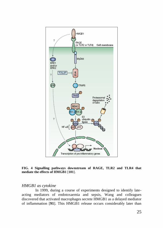
Receptors The first described receptor for HMGB1 was the receptor for
advanced glycation end products (RAGE) [98]. However, since RAGE-
deficient cells were shown to still be able to respond to HMGB1 stimulation
and anti-rage antibodies only partially suppressed the activity of HMGB1,
RAGE is not believed to be the only receptor for HMGB1 [99]. Recently,
HMGB1 has been described to be a ligand of toll-like receptors (TLR) 2 and
4 [100].
RAGE belongs to the immunoglobulin (Ig) superfamiles and comprises of
three extracellular Ig domains, a single transmembrane segment and short
cytoplasmic tail. It interacts with other structurally unrelated ligands
including several members of the S100 family, amyloid-beta peptide,
transthyretin and beta2 integrin Mac-1.
RAGE signals through pathways that involve ERK1 (extracellular-signal-
regulated kinase) and/or ERK2, and the mitogen-activated protein kinase p38,
and it promotes the activation of nuclear factor-κB (NF-κB). In a MyD88
(myeloid differentiation primary-response protein 88)-dependent manner,
high-mobility group box 1 protein (HMGB1)-mediated signalling leads to
activation of the IKK complex (inhibitor of NF-κB (IκB) kinase complex),
consisting of IKK-α,IKK-β and NF-κB essential modulator (NEMO), which
phosphorylates IκBα and thereby releases NF-κB for translocation to the
nucleus and allows the transcription of pro-inflammatory genes (such as
interleukin-1 (IL-1), IL-6 and tumour-necrosis factor).
Interaction of HMGB1 with TLR2 and TLR4 might therefore enable
HMGB1 to promote inflammatory responses that are similar to those of
lipopolysaccharide. The common signalling pathway for RAGE, TLR2 and
TLR4 involves a MyD88-dependent pathway that ultimately leads to NF-κB
activation. Little is known about the precise means by which phospho-ERK
and p38 are activated by HMGB1, but these molecules, together with stress-
activated protein kinase (SAPK) and JUN aminoterminal kinase (JNK), are
activated within several minutes, even in the absence of expression of RAGE
or the IL-1 receptor α-chain137 (Fig. 4).
25
FIG. 4 Signalling pathways downstream of RAGE, TLR2 and TLR4 that
mediate the effects of HMGB1 [101].
HMGB1 as cytokine In 1999, during a course of experiments designed to identify late-
acting mediators of endotoxaemia and sepsis, Wang and colleagues
discovered that activated macrophages secrete HMGB1 as a delayed mediator
of inflammation [91]. This HMGB1 release occurs considerably later than

26
secretion of the classical early pro-inflammatory mediators TNF and IL-1. In
a standardized model in which mice are administered lipopolysaccharide
(LPS) to generate endotoxaemia, serum HMGB1 levels begin to increase 12–
18 hours after peak levels of TNF, which occur at 2 hours, and of IL-1, which
occur at 4–6 hours [102, 103]. Administration of HMGB1-specific antibodies
confers significant protection against the lethal effects of endotoxin, even
when antibody dosing is delayed until after the peak levels of TNF and IL-1.
HMGB1 is now recognized as a cytokine because it mediates systemic
inflammatory responses, is secreted by activated immune cells, activates
prototypical inflammatory responses in immune cells and endothelial cells.
HMGB1 stimulation of PBMC led to release of TNF, IL-1a, IL-1b, IL-6, IL-8
macrophage inflammatory protein (MIP)-1a and (MIP)-1b. Moreover, a dose-
dependent increase in ICAM, VCAM and RAGE expression on endothelial
cells following HMGB1 stimulation has also been observed [104, 105].
HMGB1 is one of several DAMPs (including heat-shock proteins, uric acid,
ATP and S100 molecules) that facilitate the recruitment and activation of
macrophages, plasmacytoid DCs (pDCs) and myeloid DCs, thereby
promoting inflammation and/or tissue repair. Activated NK cells (which
accumulate in response to HMGB1 and other pro-inflammatory signals)
provide an additional source of HMGB1, which is released into the
immunological synapse between NK cells and immature DCs and promotes
the maturation of DCs and the induction of T-helper-1-cell responses [90,
106, 107]. More speculative is the ability of mature DCs to produce HMGB1
and therefore stimulate mature T cells. HMGB1 might also have a role in
inhibiting the IFN- response of pDCs to CpG-containing DNA. In the
lymphnode, mature DCs not only provide both antigen in the form of
peptide–MHC complexes and co-stimulatory molecules, but also are a source
of HMGB1, which matures additional DCs that are recruited across high
endothelial venules (HEVs), enabling further interaction with, and
stimulation of, naive CD4+ and CD8+ T cells.
However recent studies indicate that highly purified HMGB1 may not by
itself be active as a pro-inflammatory mediator. Several recent reports
indicate that HMGB1 needs to form a complex with pro-inflammatory
ligands, as LPS, CpG-DNA or IL-1, to exert its synergistic influence [108,
109].

27
Table II Biological function of HMGB1 and its target cells
Biological activity Target cells
Secretion of pro-inflammatory
factors
Promotes transendothelial
migrations of monocites
Monocytes and macrophages, DCs
Increased expression of genes for
pro-inflammatory factors Neutrophils
Increased immunogenicity of
soluble or corpusculate antigens DCs
Maturation of DCs and Th1
polarization DCs
Upregulation of adhesion
molecules Endothelial cells
Chemotaxis
Promotes differentiation Stem cells
Cytoskeleton reorganization and
transendothelial migration
Monocytes, vascular smooth muscle
cells, vessel-associated stem cells
(mesangioblasts)
Proliferation Vessel-associated stem cells
(mesangioblasts)
Enhances Invasiveness Tumor cells
Diseases Several studies implicate HMGB1 in the pathogenesis of various
inflammatory conditions and diseases, as arthritis, cancer, hepatitis, malaria,
myocardial ischemia, sepsis.

28
The first evidence that links HMGB1 to sepsis was obtained more than ten
years ago when, in a pioneering study, HMGB1 was identified as a late
mediator of lethal systemic inflammation and as being involved in the
delayed lethality of endotoxin and systemic inflammation [91].
Since then, HMGB1 has been an increasingly attractive target for drug
development because considerable data has been generated on its role in both
acute and chronic inflammatory diseases. Preclinical studies have validated
the possibility of targeting HMGB1 as a therapeutic agent, by using
independent approaches [110], including anti-HMGB1 antibodies and the A
box fragment of HMGB1, which has antagonistic actions. Recently,
encouraging results have been obtained, including the blocking of RAGE-
HMGB1 signaling [111]. The identification of HMGB1 polymorphisms as
significant factors associated with early and late mortality systemic
inflammatory response syndrome and sepsis hints at a possible role for
HMGB1 genetics in predictive medicine [112, 113]. HMGB1 has also been
linked also to tumor formation, progression, and metastasis and to the
responses to chemotherapeutics. Its expression is elevated in several solid
tumors, and HMGB1 serum levels are often associated with worse prognosis
[110, 114]. On the other hand, HMGB1 plays a role in the immune responses
against tumors elicited by conventional therapies. HMGB1 is released from
irradiated and doxorubicin-treated tumor cells, and through TLR4, HMGB1
is efficient in activating DCs to cross-present tumor antigens, suggesting a
dual role for the molecule [115, 116]. The redox state of HMGB1 is
important in this context. Reduced HMGB1 binds to RAGE, but not to
TLR4, promoting tumor resistance to chemotherapeutic agents such as
melphalan, paclitaxel, UV, and oxaliplatin. Oxidized HMGB1, in contrast,
apparently increases the cytotoxicity of the agents, with the eventual death of
tumor cells [117].
.

29

30

31
32

33

34

35
36

37

38

39

40

41

42

43

44
CONCLUSIONS
The present studies demonstrate important roles of TLRs and HMGB1 in
neonatal and child’s immune system.
The identification and functional characterization of TLRs in Drosophila and
mammals have brought our understanding of the innate immune system to a
new level. The role of the TLRs in host defense is fundamental, it is likely
that their function affects most aspects of the mammalian immune system.
Loss-of-function mutations in TLRs result in immunodeficiencies, whereas
gain-of-function mutations might predispose an individual to inflammatory or
autoimmune disorders. The importance of the TLRs in the control of adaptive
immune responses also makes them crucial targets for immune intervention.
Therefore, complete understanding of the mechanisms of innate immunity
will be helpful for the future development of innovative therapies for
manipulation of infectious diseases, cancer and allergies. Owing to the
important role of innate immunity in neonatal health and disease, the intense
biopharmaceutical development of molecules that are derived from or that
modulate the innate immune system, including antimicrobial proteins,
peptides and TLR agonists, could have clinical relevance to neonatal
medicine. Therefore, TLR agonists might represent tools to enhance the
defense against microorganisms [118, 119] or to shift innate immune
responses of neonatal APCs away from the production of TH2-cell-polarizing
cytokines, thereby potentially reducing allergy [120, 121].
Actually, several studies have indicated that polymorphisms of TLR-4 and
TLR-2 are associated with allergy, asthma or atopic eczema [71-73] and its
defective signaling led to allergic sensitization to food protein in mice [74,
75]. High risk newborns for allergy have also been noted to have altered
generation of putative regulatory T-cell populations after LPS stimulation,
presumably through TLR-4 pathways [72]. Also TLR-2 mutation has been
associated with a higher risk for asthma in European children [73] and with
atopic dermatitis having severe phenotype [76, 77]. Conversely and In
agreement with some recent papers [122, 123], we have observed that
specific polymorphisms in TLR-2 and TLR-4 are not associated with eczema
and food allergy in Italian allergic children, indicating that correlation
between disease and TLR polymorphism might influence allergic responses
positively or negatively as a function of the individual genetic background
and the nature of the associated antigens.
Recently, Ferhani et al. [124] reported that levels of HMGB1, that is a
DAMP molecule and TLR-4 ligand, were also elevated in the fluid from
45
bronchoalveolar lavage of patients with chronic obstructive pulmonary
disease. Straub et al. [125] reported that HMGB1 inhibitors significantly
diminished the ovalbumin-induced increase in response to methacholine in a
mouse asthmatic model sensitized and challenged with ovalbumin. Moreover,
it was suggested that measurement of HMGB1 and soluble RAGE (sRAGE)
might be novel biomarkers in asthma with severe airflow limitation [126].
These reports suggest HMGB1 as an important mediator of asthma. Asthma
and atopic allergies are considered inflammatory disorders, therefore a
blockade of extracellular HMGB1 might represent a suitable therapeutic
intervention for the treatment of these pathologies.
The importance of HMGB1 as inflammatory mediator examined firstly in
adult immune system has been also discovered in fetuses and newborns in
which sRAGE and HMGB1 are active participants of the tissue injury
process [127]. The important role of HMGB1 in fetal immune system has
been corroborated by data demonstrating that in response to stimuli HMGB1
secreted by human umbilical vein endothelial cells (HUVEC) triggers
inflammatory responses through up-regulation of adhesion molecules and
release of soluble proinflammatory mediators from endothelial cells [15,
104]. The expression of HMGB1 and its role in immune response has been
demonstrated successfully in adult PB and only recently, Buhimashi CS et al
[128] proposed that HMGB1, together with soluble receptor for advanced
sRAGE and S100, are important mediators of cellular injury in fetuses and
crucial factor in preterm birth induced inflammation. Here we have shown for
the first time that HMGB1 is expressed and secreted from mononuclear cells
isolated from human cord blood. We demonstrated that constitutive HMGB1
expression is confined to a population of differentiated cells, likely myeloid
DC precursors in CB and PB. Conversely, lymphocytes, as CD4 or T cells,
present a small cell fraction positive for HMGB1. These findings are
completely in agreement with the role of HMGB1 in regulating innate
immune response, in which activated macrophages/monocytes, DC and T
cells are the main components. Moreover, we demonstrated that stimuli that
mimic infection or pro-inflammatory mediators induce the cell surface
expression of HMGB1 and its secretion via non-classical secretion pathway.
HGMB1 is compartmentalized within cytoplasmic vesicles during active
release and is predominantly localized toward the apical ends, areas of cell-
cell contact, as mediator of cell-to-cell or cell-to-matrix interaction to
facilitate cell recruitment by binding RAGE at endothelial cells.
Interesting results were obtained by cell treatment with ABs, that we have
recently demonstrated to be inducers of proliferative responses in cord blood
V2 T-cells [36, 37]. Pamidronate and Zoledronate are able to trigger cell
46
surface expression of HMGB1 in CB and PB cells with an evident increase of
HMGB1 positive T cells. Furthermore, ABs treatment leads to remarkable
secretion of HMGB1 in extracellular environments. This study provide the
first demonstration that ABs treatment modulate the expression of HMGB1
in CB cells, involving V2 T cells directly or throught their presentation by
APC cells (monocyte lineage) which modulate surface molecules or release
cytokines needed for optimal Vδ2 T-cell activation, as reported by Miyagawa
F et al [129].
Results presented herein provide a new insight into the role of HMGB1 in
CB innate immune response. We demonstrate that stimuli, such as LPS or
cytokines or synthetic compounds, can initiate a cascade of events that lead to
the activation of immune cells and secretion of mediators, as HMGB1. Once
secreted into extracellular milieu, HMGB1 can function as a
cytokine/chemokine recruiting CB monocytes via RAGE. Human umbilical
vein endothelial cells release HMGB1 and express RAGE. Therefore,
HMGB1, which is involved in a paracrine interaction, might play a crucial
role in transendothelial migration and consequently in inflammatory immune
response in CB.
To date a number of HMGB blocking therapies have been tested in several
experimental models of different diseases. Recent structure-based drug
discovery efforts have been aimed at increasing the number of small
molecule- and biologics-based prototype therapeutics targeting HMGB1.
Small molecule drugs that may provide therapeutic benefit through HMGB1-
directed mechanisms involve HMGB1 inhibitory ligands, Toll-like receptor
antagonists, RAGE antagonists, alpha7 nicotinic acetylcholine receptor
agonists, G2A antagonists, serine protease inhibitors, and alpha-dicarbonyl-
based soft electrophiles. Using some of these agents, pharmacological
modulation of HMGB1-associated cutaneous pathology has been achieved
with an acceptable toxicity profile, and preclinical proof-of-concept
experimentation has demonstrated feasibility of developing HMGB1-
modulators into novel systemic and topical therapeutics that target
inflammatory dysregulation. The identification of HMGB1 inhibitor,
glycyrrhizin, is therefore of significant experimental and clinical interest.
Glycyrrhizin binds to high-mobility group box 1 protein and inhibits its
cytokine activities. Mollica et al [130] show that, a natural anti-inflammatory
and antiviral triterpene in clinical use, inhibits HMGB1 chemoattractant and
mitogenic activities, and has a weak inhibitory effect on its intranuclear
DNA-binding function. Recently, a new derivative of glycyrrhizin is used as
HMGB1 scavenger in clinical therapy of rhinitis via nasal spray.
47
Whereas a blockade of extracellular HMGB1 might represent a suitable
therapeutic target for the treatment of inflammation, the development of the
appropriate cell-mediated immunity, associated with a Th1 type immune
response, is essential for successful immunization practice. Extracellular
HMGB1 has been shown to act as immune adjuvant by enhancing
immunogenicity of apoptotic lymphoma cells and eliciting antibody
responses to soluble ovalbumin protein [89]. Moreover, a short peptide,
named Hp91, identified within the B box domain of HMGB1, induced
activation of human and mouse DCs, increasing secretion of pro-
inflammatory cytokines and chemokines, including the Th1 cytokine, IL-12
[131]. Therefore, HMGB1 might be considered a probable candidate as
adjuvant for vaccine. In this scenario, aminobishoshponates that are able to
stimulate innate immunity, as T-cells in CB [37], and to induce HMGB1
secretion may be an immuno-modulating tool to approach neonatal
pathologies. Moreover, in adult counterpart, considering that ZOL or PAM
exert also anti-cancer activity by inducing apoptosis, ABs might interfere in
the complex interaction between tumor and host immune system by the
release of inflammatory mediators, such as HMGB1, which mediate cross-
presentation of tumor antigens via binding on TLR4 and the promotion of
tumor specific cytotoxic T-cell responses [132]. The ability of immuno-
adjuvant to induce antigen-specific cell mediated, Th1 immune response,
may make it suitable as an adjuvant in cancer immunotherapies as well as in
vaccines against infectious diseases caused by intracellular bacteria or
viruses.
Thus HMGB1 appear to be a double-edged sword. While being vital for
tissue repair or immuno-adjuvant because it exerts stimulatory effect on
immune system, it also plays a role in the pathogenesis of many
inflammatory and autoimmune diseases that feature aberrant TLR activation.
HMGB1–TLR–RAGE constitutes a tripod. Blockade or downregulation of
HMGB1, or control of the inflammatory tripod, represent a promising
therapeutic approach for the treatment of adult and neonatal pathologies.

48
REFERENCES
1. Areschoug T, Gordon S (2008) Pattern recognition receptors and their
role in innate immunity: focus on microbial protein ligands. Contrib
Microbiol. 15:45-60.
2. Kono H, Rock KL (2008) How dying cells alert the immune system to
danger. Nat Rev Immunol 8:279–289.
3. Matzinger P (2002) An innate sense of danger. Ann N Y Acad Sci
961:341–342.
4. Matzinger P (2002) The danger model: a renewed sense of self. Science
296:301–305.
5. Kawai T and Akira S (2010) The role of pattern-recognition receptors in
innate immunity: update on Toll-like receptors. Nat Immunol 11:373-384
6. TKlein J & Remington J (2001) Infectious Diseases of the Fetus and
Newborn Infant (eds Remington, J. & Klein, J.) 1–23 (W. B. Saunders
Company, Philadelphia.
7. McDonagh, S Maidji E, Ma W, Chang HT, Fisher S, Pereira L (2004)
Viral and bacterial pathogens at the maternal–fetal interface. J Infect Dis
190: 826–834.
8. Makhseed MR, Raghupathy R, Azizieh F, Omu A, Al-Shamali E and
Ashkanani L (2001) Th1 and Th2 cytokine profiles in recurrent aborters
with successful pregnancy and with subsequent abortions. Hum Reprod
16: 2219–2226.
9. Adkins B, Leclerc C & Marshall-Clarke S (2004) Neonatal adaptive
immunity comes of age. Nat Rev Immunol 4: 553–564.
10. Krishnan S, Craven M, Welliver R.C Ahmad N & Halonen M (2003)
Differences in participation of innate and adaptive immunity to
respiratory syncytial virus in adults and neonates. J Infect Disease 188:
433–439.
11. Firth MA, Shewen PE & Hodgins DC (2005) Passive and active
components of neonatal innate immune defenses. Anim. Health Res. Rev.
6: 143–158.
12. Kovarik J, Siegrist CA (1998) Immunity in early life. Immunol Today
19:150-152.
13. Wilson CB (1986) Immunologic basis for increased susceptibility of the
neonate to infection. J Pediatr 108:1-12.
14. Cohen L, Haziot A, Shen DR, Lin XY, Sia C, et al (1995) CD14-
independent responses to LPS require a serum factor that is absent from
neonates. J Immunol 155: 5337–5342.

49
15. Christensen RD (1989) Hematopoiesis in the fetus and neonate. Pediatr
Res 26:531-535.
16. Quian JX, Lee SM, Suen Y, Knoppel E, van de Ven C, Cairo MS. (1997)
Decreased interleukin-15 from activated cord versus adult peripheral
blood mononuclear cells and the effect of interleukin-15 in upregulating
antitumor immune activity and cytokine production in cord blood. Blood
90: 3106-3117.
17. Berthou C, Legros-Maida S, Soulie A, Wargnier A, Guillet J, et al. (1995)
Cord blood T lymphocytes lack constitutive perforin expression in
contrast to adult peripheral blood T lymphocytes. Blood 85:1540-1546
18. Harris DT, Schumacher MJ, Locascio J, Besencon F J, Olson G B et al.
(1992) Phenotypic and functional immaturity of human umbilical cord
blood T lymphocytes. Proc Natl Acad Sci U S A 89: 10006-10010.
19. Sato K, Nagayama H, Takahashi TA (1999). Aberrant CD3- and CD28-
mediated signalling events in cord blood T cells are associated with
dysfunctional regulation of fas ligand-mediated cytotoxicity. J Immunol
162: 4464-4471.
20. Holtmeier W, Kabelitz, D (2005) Gammadelta T cells link innate and
adaptive immune responses. Chem Immunol and allergy 86: 151–183.
21. Bonneville M, Fournie JJ (2005) Sensing cell stress and transformation
through Vγ9Vδ2 T cell-mediated recognition of the isoprenoid pathway
metabolites. Microbes Infect 7:503-509 .
22. Poupot M, Fournie JJ (2004) Non-peptide antigens activating human
Vγ9/Vδ2 T lymphocytes. Immunol Lett 95:129-138.
23. MoritaCT, Jin C, Sarikonda G, and Wang H. (2007) Nonpeptide antigens,
presentation mechanisms, and immunological memory of human
Vgamma2Vdelta2 T cells: discriminating friend from foe through the
recognition of prenyl pyrophosphate antigens. Immunol Rev 215:59-76.
24. Kabelitz D, Bender A, Schondelmaier S, Schoel B, and Kaufmann SH.
(1990) A large fraction of human peripheral blood gamma/delta + T cells
is activated by Mycobacterium tuberculosis but not by its 65-kD heat
shock protein. J Exp Med 171:667-679. 25. Pfeffer K, Schoel B, Gulle H, Kaufmann SH and Wagner H. (1990)
Primary responses of human T cells to mycobacteria: a frequent set of
gamma/delta T cells are stimulated by protease-resistant ligands. Eur J
Immunol 20:1175-1179.
26. Pfeffer K, Schoel B, Gulle H, Kaufmann SH and Wagner H. (1991)
Human gamma/delta T cells responding to mycobacteria. Behring Inst
Mitt 88: 36-42
50
27. Constant P, Davodeau F, Peyrat MA, Poquet Y, Puzo G, et al. (1994)
Stimulation of human gamma delta T cells by nonpeptidic mycobacterial
ligands. Science 264:267-270. 28. Tanaka Y, Sano S, Nieves E, De Libero G, Rosa D et al (1994)
Nonpeptide ligands for human γδ T cells. Proc Natl Acad Sci U S A
91:8175-8179.
29. Berenson JR. (2005) Recommendations for zoledronic acid treatment of
patients with bone metastases. Oncologist. 10:52-62.
30. Kunzmann V, Bauer E, Feurle J, Weissinger F, Tony HP (2000)
Stimulation of gammadelta T cells by aminobisphosphonates and
induction of antiplasma cell activity in multiple myeloma. Blood 96:384–
392.
31. Dieli F Gebbia N, Poccia F, Caccamo N, Montesano C, et al (2003)
Induction of gammadelta T-lymphocyte effector functions by
bysphosphonate zoledronic acid in cancer patients in vivo. Blood
102:2310–2311.
32. Ferlazzo V, Sferrazza C, Caccamo N, Di Fede G, Di Lorenzo G, et al.
(2006) In vitro effects of aminobisphosphonates on Vgamma9Vdelta2 T
cell activation and differentiation. Int J Immunopathol Pharmacol.
19:309-317.
33. Gober HJ, Kistowska M, Angman I, Jenö P, Mori L, De Libero G (2003)
Human T cell receptor γδ cells recognize endogenous mevalonate
metabolites in tumor cells. J Exp Med 197:163-168.
34. Montesano C, Gioia C, Martini F, Agrati C, Cairo C, et al (2001)
Antiviral activity and anergy of gammadelta T lymphocytes in cord blood
and immunocompromised host. J. Biol. Regul Homeost Agents 15:257-
264.
35. Engelmann I, Moeller U, Santamaria A, Kremsne PG, and Luty AJF
(2006) Differing activation status and immune effector molecule
expression profiles of neonatal and maternal lymphocytes in an African
population. Immunol 119: 515–521.
36. Cairo C, Propp N, Auricchio G, Armstrong CL, Abimiku A et al (2008)
V2 T-Lymphocyte response in cord blood samples from Italy and Côte
d’Ivoire. Mol Immunol 45: 3190-3197.
37. Placido R, Auricchio G, Gabriele I, Galli E, Brunetti E, et al (2011)
Characterization of cord-blood derived t cells immune response after
stimulation with aminobisphosphonate. Int J Immunopathol Pharmacol
24: 101-110.

51
38. Poccia F, Gougeon ML, Agrati C, Montesano C, Martini F, et al. (2002)
Innate T-cell immunity in HIV infection: the role of Vgamma9Vdelta2 T
lymphocytes. Curr Mol Med 2:769-781.
39. Brandes M, Willimann K, Lang AB, Nam KH, Jin C, et al. (2003)
Flexible migration program regulates gamma delta T-cell involvement in
humoral immunity. Blood 102: 3693-3701.
40. Conti L, Casetti R, Cardone M, Varano B, Martino A, et al. (2005)
Reciprocal activating interaction between dendritic cells and
pamidronate-stimulated gammadelta T cells: role of CD86 and
inflammatory cytokines. J Immunol 174:252–260.
41. Lemaitre B, Nicolas E, Michaut L. (1996) The dorsoventral regulatory
gene cassette spatzle/toll/cactus controls the potent antifungal response in
drosophila adults. Cell 86:973–983
42. Jin MS, Lee JO. (2008) Structures of the Toll-like receptor family and its
ligand complexes. Immunity 29:182–191.
43. Carpenter S, O'Neill LA. (2009) Recent insights into the structure of Toll-
like receptors and post-translational modifications of their associated
signalling proteins. Biochem J 422:1–10.
44. Kawai T and Akira S (2007) TLR signaling. Semin Immunol 19: 24-30
45. Kumar H, Kawai T and Akira S. (2009). Pathogen recognition in the
innate immune response. Biochem J 420: 1-16.
46. Akira, S., Uematsu, S., and Takeuchi, O. (2006). Pathogen recognition
and innate immunity. Cell 124: 783-801.
47. Iwasaki A and Medzhitov R. (2004). Toll-like receptor control of the
adaptive immuneresponses. Nat Immunol 5: 987-995.
48. Kawai T, Akira S. (2010) The role of pattern-recognition receptors in
innate immunity: Update on Toll-like receptors. Nat Immunol 11: 373–
384.
49. Takeuchi O and Akira S (2009) Pattern recognition receptors and
inflammation. Cell 2010;140:805–820. Kumar H, Kawai T, Akira S.
Pathogen recognition in the innate immune response. Biochem J 420:1–
16.
50. Lien, E, Means, TK, Heine H, Yoshimura A, Kusumoto S, et al. (2000).
Toll-like receptor 4 imparts ligand-specific recognition of bacterial
lipopolysaccharide. J Clin Invest 105: 497-504.
51. Poltorak A, He X, Smirnova I, Liu MY, Van Huffel C, et al. (1998).
Defective LPS signaling in C3H/HeJ and C57BL/10ScCr mice: mutations
in Tlr4 gene. Science 282: 2085-2088.
52. Akira S, Uematsu S and Takeuchi O (2006). Pathogen recognition and
innate immunity. Cell 124: 783-801.

52
53. Hayashi F, Smith KD, Ozinsky A, Hawn TR, Yi EC, Goodlett DR, Eng
JK, Akira S, Underhill DM and Aderem A. (2001). The innate immune
response to bacterial flagellin is mediated by Toll-like receptor 5. Nature
410: 1099-1103.
54. Zhang D, Zhang G, Hayden MS, Greenblatt MB, Bussey C, Flavell RA,
and Ghosh S (2004). A toll-like receptor that prevents infection by
uropathogenic bacteria. Science 303: 1522-1526.
55. Yarovinsky F, Zhang D, Andersen JF, Bannenberg GL, Serhan CN, et al
(2005). TLR11 activation of dendritic cells by a protozoan profilin-like
protein. Science 308: 1626-1629.
56. Alexopoulou L, Holt AC, Medzhitov R, and Flavell RA (2001).
Recognition of doublestranded RNA and activation of NF-kappaB by
Toll-like receptor 3. Nature 413: 732-738.
57. Diebold SS, Kaisho T, Hemmi H, Akira S and Reis e Sousa C (2004).
Innate antiviral responses by means of TLR7-mediated recognition of
single-stranded RNA. Science 303: 1529-1531. ]. 58. Hemmi H, Takeuchi O, Kawai T, Kaisho T, Sato S, et al. (2000). A Toll-
like receptor recognizes bacterial DNA. Nature 408: 740-745.
59. Bianchi ME, Manfredi AA (2007) High-mobility group box 1 (HMGB1)
protein at the crossroads between innate and adaptive immunity. Immunol
Rev 220: 35–46.
60. Bianchi ME (2007) DAMPs, PAMPs and alarmins: all we need to know
about danger. J Leukoc Biol 81:1–5.
61. Lotze MT, Zeh H J, Rubarteli A, Sparvero LJ, Amoscato AA et al.
(2007) The grateful dead: damage-associated molecular pattern molecules
and reduction/oxidation regulate immunity. Immunol Rev 220: 60–81.
62. Rubartelli A, Lotze MT (2007) Inside, outside, upside down: damage-
associated molecular-pattern molecules (DAMPs) and redox. Trends
Immunol 28:429–436.
63. Bustin M (1999) Regulation of DNA-Dependent activities by the
functional motifs of the High-Mobility-Group Chromosomal proteins.
Mol Cell Biol 19: 5237-5246.
64. Kono H and Rock KL (2008) How dying cells alert the immune system to
danger. Nature Reviews Immunology 8:279–289.
65. Matzinger P. (1994). Tolerance, danger, and the extended family. Annu
Rev Immunol 12: 991-1045.
66. Chen K, Huang J, Gong W, Iribarren P, Dunlop NM, Wang JM. (2007)
Toll-like receptors in inflammation, infection and cancer. Int
Immunopharmacol 7: 1271–1285.
53
67. Liew FY, Xu D, Brint EK, O’Neill LAJ. (2005) Negative regulation of
Toll-like receptor-mediated immune responses. Nature Rev Immunol 5:
446–458.
68. Lotze MT, Zeh HJ, Rubartelli A, Sparvero LJ, Amoscato AA, et al.
(2007) The grateful dead: damage-associated molecular pattern molecules
and reduction/oxidation regulate immunity. Immunol Rev 220:60–81.
69. Mantovani A. (2009) Cancer: inflaming metastasis. Nature 457:36–37.
70. Midwood KS, Piccinini AM, Sacre S. (2009) Targeting Toll-like
receptors in autoimmunity. Curr Drug Targets 10:1139–1155.
71. Yang IA, Fong KM, Holgate ST, Holloway JW. (2006). The role of Toll-
like receptors and related receptors of the innate immune system in
asthma. Curr Opin Allergy Clin Immunol. 6:23-28.
72. Haddeland U, Karstensen AB, Farkas L, Bo KO, Pirhonen J, et al. (2005)
Putative regulatory T cells are impaired in cord blood from neonates with
hereditary allergy risk. Pediatr Allergy Immunol 16: 104–112.
73. Eder W, Klimecki W, Yu L, von Mutius E, Riedler J, et al. (2004) Toll-
like receptor 2 as a major gene for asthma in children of European
farmers. J Allergy Clin Immunol 113: 482–488. 4.
74. Bashir ME, Louie S, Shi HN, Nagler-Anderson C. (2004) Toll-like
receptor 4 signaling by intestinal microbes influences susceptibility to
food allergy. J Immunol 172: 6978-6987.
75. Berin MC, Zheng Y, Domaradzki M, Li XM, Sampson HA. (2006) Role
of TLR4 in allergic sensitization to food proteins in mice. Allergy 61: 64-
71. 76. Ahmad-Nejad P, Mrabet-Dahbi S, Breuer K, Klotz M, Werfel T, et al.
(2004) The toll-like receptor 2 R753Q polymorphism defines a subgroup
of patients with atopic dermatitis having severe phenotype. J Allergy Clin
Immunol 113: 565–67.
77. Niebuhr M, Langnickel J, Draing C,Renz H, Kapp A and Werfel T.
(2008) Dysregulation of toll-like receptor-2 (TLR-2)-induced effects in
monocytes from patients with atopic dermatitis : impact of the TLR-2
R753Q polymorphism. Allergy 63: 728-734
78. Goodwin GH, Sanders C and Johns EW. (1973) A new group of
chromatin-associated proteins with a high content of acidic and basic
amino acids. Eur J Biochem 38: 14–19.
79. Müller S, Ronfani L, Bianchi ME (2004) Regulated expression and
subcellular localization of HMGB1, a chromatin protein with a cytokine
function. J Intern Med 225:332-343
80. Moscvilsky Ml, Novitskaya VA, Iogannsen MG Mark A. Zabezhinsky
(1989) Tissue specificity of nucleo-cytoplasmic distribution of HMG1

54
and HMG2 proteins and their probable functions. Eur J Biochem
185:303-310.
81. Bustin M (1999) Regulation of DNA-dependent activities by the
functional motifs of the high mobility group chromosomal proteins. Mol
Cell Biol 19:5237–5246
82. Read CM ,Cary PD, Crane-Robinson C, Driscoll PC and Norman PG
(1993), Solution structure of a DNA-binding domain from HMG1,
Nucleic Acids Res 21:3427–3436.
83. Zayed, H., Izsvak, Z., Khare, D., Heinemann, U. & Ivics, Z. (2003) The
DNA-bending protein HMGB1 is a cellular cofactor of Sleeping Beauty
transposition. Nucleic Acids Res. 31: 2313–2322.
84. Brickman MJ, Adam M, and Ptashn M (1999) Interactions between an
HMG-1 protein and members of the Rel family Proc. Natl Acad Sci USA
96:10679-10683.
85. Knapp, S. Müller S, Digilio G, Bonaldi T, Bianchi ME, Musco G. (2004)
The long acidic tail of high mobility group box 1 (HMGB1) protein forms
an extended and flexible structure that interacts with specific residues
within and between the HMG boxes. Biochemistry 43: 11992–11997.
86. Bianchi ME (2004) Significant (re)location: how to use chromatin and/or
abundant proteins as messages of life and death. Trends Cell Biol. 14:
287–293.
87. Dumitriu IE, Baruah P, Manfredi AA, Bianchi ME and Rovere-Querini P.
(2005) HMGB1: guiding immunity from within. Trends Immunol, 26:
381-387.
88. Scaffidi P, Misteli T, Bianchi ME (2002) Release of chromatin protein
HMGB1 by necrotic cells triggers inflammation Nature 418:191–195.
89. Rovere-Querini P, Capobianco A , Scaffidi P, Valentinis B, Catalanotti F
(2004) HMGB1 is an endogenous immune adjuvant released by necrotic
cells. EMBO Rep 5: 825–830.
90. Bell, CW, W. Jiang, CF Reich, DS Pisetsky (2006) The extracellular
release of HMGB1 during apoptotic cell death. Am. J. Physiol. 291:
C1318-C1325
91. Wang H, Bloom O, Zhang M, Vishnubhakat J M, Ombrellino M, et al
(1999) HMG-1 as a late mediator of endotoxin lethality in mice Science
285: 248-251.
92. Wang H, Liao M, Ochani M, Justiniani X Lin (2004) Cholinergic
agonists inhibit HMGB1 release and improve survival in experimental
sepsis. Nat Med 10:1216-1221.

55
93. Gardella S, Andrei C, Ferrera D, Lotti LV, Torrisi MR et al (2002) The
nuclear protein HMGB1 is secreted by monocytes via a non-classical,
vesicle-mediated secretory pathway. EMBO Rep 3: 995-1001.
94. Bonaldi T, Talamo F, Scaffidi P, Ferrera D, Porto A, et al. (2003)
Monocytic cells hyperacetylate chromatin protein HMGB1 to redirect it
towards secretion. EMBO J 22: 5551–5560.
95. Rendon-Mitchell B, Ochani M, Li J, Han J, Wang H, et al., (2003) IFN-γ
induces high mobility group box 1 protein release partly through a TNF-
dependent mechanism, J. Immunol 170: 3890–3897.
96. Blott EJ and Griffiths GM. (2002) Secretory lysosomes. Nat Rev Mol
Cell Biol 3: 122–131.
97. Rouhiainen A, Kuja-Panula J, Wilkman E, Pakkanen J, Stenfors J, et al
(2004) Regulation of monocytes migration by amphoterin (HMGB1)
Blood 104: 1174-1182.
98. Parkkinen J, Raulo E, Merenmies J, Nolo R, Kajander EO et al. (1993)
Amphoterin, the 30-kDa protein in a family of HMG1-type polypeptides.
Enhanced expression in transformed cells, leading edge localization, and
interactions with plasminogen activation. J Biol Chem 268:19726-19738.
99. Li J, Kokkola R, Tabibzadeh S, Yang R, Ochani M et al (2003).
Structural basis for the proinflammatory cytokine activity of high
mobility group box 1. Mol Med 9: 37-45.
100. Park JS, Gamboni-Robertson F, He Q, Svetkauskaite D, Kim JY et al
(2006) High mobility group box 1 protein interacts with multiple Toll-like
receptors. Am J Physiol Cell Physiol 290:C917-924.
101. Lotze MT, Tracey KJ (2005) High-mobility group box 1 protein
(HMGB1): nuclear weapon in the immune arsenal. Nat Rev Immunol 5:
331-342.
102. Yang H. Ochani M, Li J, Qiang X, Tanovic M et al. (2004) Reversing
established sepsis with antagonists of endogenous high-mobility group
box 1. Proc. Natl Acad Sci USA 101: 296–301.
103. Wang, H., Czura, C. J. & Tracey, K. J. (2004) Lipid unites disparate
syndromes of sepsis. Nature Med 10: 124–125.
104. Mullins GE, Sunden-Cullberg J, Johansson AS, Rouhiainen A,
Erlandsson-Harris H et al (2004) Activation of human umbilical vein
endothelial cells leads to relocation and release of high-mobility group
box chromosomal protein1. Immunol 60:566-573.
105. Luan ZG, Zhang H, Yang PT, Ma XC, Zhang C et al (2010) HMGB1
activates nuclear factor-κB signaling by RAGE and increases the
production of TNF-α in human umbilical vein endothelial cells.
Immunobiol 215:956-962.

56
106. Dumitriu IE, Bianchi ME, Bacci M, Manfredi AA, Rovere-Querini P
(2007) The secretion of HMGB1 is required for the migration of maturing
dendritic cells. J Leukoc Biol 81:84–91
107. Messmer D, Yang H, Telusma G, Knoll F, Li J, et al (2004) High
mobility group box protein 1: an endogenous signal for dendritic cell
maturation and Th1 polarization. J Immunol 173:307–313
108. Sha Y, Zmijewski J, Xu Z, Abraham E. (2008) HMGB1 develops
enhanced proinflammatory activity by binding to cytokines. J Immunol
180:2531-2537.
109. Hreggvidsdottir HS, Ostberg T, Wähämaa H, Schierbeck H, Aveberger
AC et al. (2009) The alarmin HMGB1 acts in synergy with endogenous
and exogenous danger signals to promote inflammation. J Leukoc Biol
86:655-662.
110. Sims GP, Rowe DC, Rietdijk ST, Herbst R, Coyle AJ (2010)
HMGB1and RAGE in inflammation and cancer. Annu Rev Immunol
28:367–388.
111. Susa Y, Masuda Y, Imaizumi H, Namiki A (2009) Neutralization of
receptor for advanced glycation end-products and high mobility group
box-1 attenuates septic diaphragm dysfunction in rats with eritonitis. Crit
Care Med 37:2619–2624.
112. Kornblit B, Munthe-Fog L, Madsen HO, Strøm J, Vindeløv L, Garred P
(2008) Association of HMGB1 polymorphisms with outcome in patients
with systemic inflammatory response syndrome. Crit Care 12:R83
113. Kornblit B, Masmas T, Petersen SL, Madsen HO, Heilmann C et al
(2010) Association of HMGB1 polymorphisms with outcome after
allogeneic hematopoietic cell transplantation. Biol Blood Marrow
Transplant 16: 239–252.
114. Sparvero LJ, Asafu-Adjei D, Kang R, Tang D, Amin N, et al (2009)
RAGE (receptor for advanced glycation endproducts), RAGE ligands,
and their role in cancer and inflammation. J Transl Med 7:17-39.
115. Apetoh L, Ghiringhelli F, Tesniere A, Obeid M, Ortiz C, et al (2007a)
Toll-like receptor 4-dependent contribution of the immune system to
anticancer chemotherapy and radiotherapy. Nat Med 13:1050-1059.
116. Apetoh L, Ghiringhelli F, Tesniere A, Criollo A, Ortiz C, et al (2007b)
The interaction between HMGB1 and TLR4 dictates the outcome of
anticancer chemotherapy and radiotherapy. Immunol Rev 220:47–59.
117. Tang D, Kang R, Cheh CW, Livesey KM, Liang X, et al (2010)
HMGB1 release and redox regulates autophagy and apoptosis in cancer
cells. Oncogene 29: 5299-5310.

57
118. Ito S, Ishii K J, Gursel M, Shirotra H, Ihata A and Klinman1 DM
(2005). CpG oligodeoxynucleotides enhance neonatal resistance to
Listeria infection. J Immunol 174: 777–782.
119. Barrier M, Lacroix-Lamande S, Mancassola R, Auray G, Bernardet N,
et al. (2006). Oral and intraperitoneal administration of phosphorothioate
oligodeoxynucleotides leads to control of cryptosporidium parvum
infection in neonatal ice. J Infect Dis 193: 1400–1407.
120. Fasciano S & Li L (2006). Intervention of Toll-like receptor mediated
human innate immunity and inflammation by synthetic compounds and
naturally occurring products. Curr Med Chem 13: 1389–1395.
121. Lambert P H (1997) Vaccines for the world: major challenges for the
future. Southeast Asian J. Trop. ed. Public Health 28: 122–126.
122. Hsieh YY, Wan L, Chang CC, Tsai CH, Tsai FJ. (2009) STAT2*C
related genotypes and allele but not TLR4 and CD40 gene
polymorphisms are associated with higher susceptibility for asthma. Int J
Biol Sci 5: 74-81.
123. Weidinger S, Novak N, Klopp N, Baurecht H, Wagenpfeil S, et al.
(2006) Lack of association between Toll-like receptor 2 and Toll-like
receptor 4 polymorphisms and atopic eczema. J Allergy Clin Immunol
118: 277-79.
124. Ferhani N, Letuve S, Kozhich A, Thibaudeau O, Grandsaigne M, et al
(2010) Expression of high-mobility group box 1 and of receptor for
advanced glycation end products in chronic obstructive pulmonary
disease. Am J Respir Crit Care Med 181:917–927.
125. Straub C, Midoro-Horiuti T, Goldblum R, Pazdrak K, Kurosky A,
(2010). Elucidating the role of high mobility group box 1 (HMGB1)
cytokine in a murine model of allergic asthma. J Allergy Clin Immunol
125:AB108
126. Watanabe T, Asai K, Fujimoto H, Tanaka H, Kanazawa H, Hirata K.
(2011) Increased levels of HMGB-1 and endogenous secretory RAGE in
induced sputum from asthmatic patients. Respir Med 105:519-25.
127. Okazaki K, Kondo M, Kato M, Kakinuma R, Nishida A (2008)
Elevation of High-Mobility Group Box 1 Concentration in Asphyxiated
Neonates. Neonatology 94: 105-109.
128. Buhimschi CS, Baumbusch MA, Dulay AT, Oliver EA, Lee S et al
(2009) Characterization of RAGE, HMGB1, and S100beta in
inflammation-induced preterm birth and fetal tissue injury. Am J Pathol
175:958-975.
129. Miyagawa F Tanaka Y, Yamashita S, Minato N. (2001) Essential
requirement of antigen presentation by monocyte lineage cells for the

58
activation of primary human gamma delta T cells by
aminobisphosphonate antigen. J Immunol 166:5508–5514.
130. Mollica L, De Marchis F, Spitaleri A, Dallacosta C, Pennacchini D, et
al. (2007) Glycyrrhizin binds to high-mobility group box 1 protein and
inhibits its cytokine activities. Chem Biol 14:431-441.
131. Telusma G, Datta S, Mihajlov I, Ma W, Li J, et al. (2006) Dendritic cell
activating peptides induce distinct cytokine profiles. Int Immunol 18:
1563–1573.
132. Gallucci S, Lolkema M, Matzinger P (1999) Natural adjuvants:
endogenous activators of dendritic cells. Nat Med 5: 1249-1255.
Top Related