


Languages
Pages
Legal


Diagnostic Applications of Simultaneously Acquired Dual-Isotope Single-Photon Emission CT Scans
Dana Mathews, 1 Brandy S. Walker, 1 Beth C. Allen,2 Hunt Batjer,2 and Phillip D. Purd / ·3
PURPOSE: To report the development and validation of a technique of dual tracer single-photon
emission CT brain imaging using technetium-99m hexamethyl-propyleneamine oxime and iodine-
123 iodoamphetamine agents and the application of this technique in patients with a variety of
diagnoses. METHODS: Contamination between the two isotopes' energy windows was calculated
by opening both energy windows while scanning a group of patients using a single isotope. To
compare uniformity of 1-123 down-scatter, Tc-99m studies were performed both before and after
the administration of 1-123 in five of 24 dual studies. The 24 patients studied with the dual-isotope
technique were evaluated during acetazolamide testing, trial balloon occlusion, or embolization of
an arteriovenous malformation. RESULTS: In a dual acquisition , average count contamination of
an 1-123 study by Tc-99m was less than 1% of the total 1-123 counts, and contamination of a Tc-
99m study by 1-123 was approximately 12% of the total Tc-99m counts. Tc-99m studies performed
both before and after the administration of 1-123 demonstrated that contaminating counts do not
adversely affect scan interpretation. Dual-tracer scans were completed in all 24 patients, 10 of
whom showed changes after intervention. CONCLUSIONS: Dual-tracer single-photon emission
CT brain scans of adequate diagnostic quality are possible using Tc-99m and 1-123.
Index terms: Single-photon emission computed tomography (SPECT); Efficacy studies; lnterven
tional neuroradiology; Brain, radionuclide studies
AJNR Am J Neuroradiol 15:63-71 , Jan 1994
It is frequently desirable to perform singlephoton emission computed tomography (SPECT) regional cerebral blood flow (rCBF) imaging before and after a therapeutic or diagnostic intervention. Such repeated imaging is necessary when evaluating patients for vasoreactivity using acetazolamide (1-3), temporary balloon occlusion of a carotid or intracerebral artery ( 4-7), or preand postembolization of an arteriovenous malformation (A V M). In the past, it has not proved feasible except with xenon-133 gas to perform both scans in a single day. Although Xe-133 scans provide direct quantification of blood flow, the
Received August 3, 1992; accepted pending revision November 3;
revision received December 22. 1 Department of Radiology, Division of Nuclear Medicine, 2 Department
of Neurological Surgery, and 3 Division of Neuroradiology, The University
of Texas Southwestern Medical Center, Dallas, TX 75235-9071.
Address reprint requests to Dana Mathews, PhD, MD, Department of
Radiology, University of Texas Southwestern Medical Center, 5323 Harry
Hines Blvd, Dallas, TX 75235-9071.
AJNR 15:63-71 , Jan 1994 0195-6108/ 94/ 1501-0063
© American Society of Neuroradiology
63
spatial resolution is poor relative to the newer SPECT cameras. Although it is possible to perform two scans in 1 day using two different imaging agents, until recently the energy resolution of the available gamma cameras did not allow adequate peak energy separation of technetium-99m and iodine-123 (the most commonly used isotopes) to allow for simultaneous image acquisition.
In this paper we report our development and validation of a technique for dual-tracer SPECT imaging of the brain. We also demonstrate its feasibility and clinical usefulness in evaluating patients with a variety of neurologic diagnoses.
Methods
Patients
Dual-isotope SPECT imaging was performed in 24 patients (15 women and nine men) ranging in age from 13 to 75 years . Diagnoses and types of studies performed on each patient are included in Tables 1-3. Studies included pre- and postacetazolamide administration, pre- and post-
64 MATHEWS
embolization of an AVM, and during and after trial balloon occlusion of an internal carotid or intracerebral artery.
Each patient was initially injected intravenously with 740 MBq of Tc-99m hexamethyl-propyleneamine oxime (HMPAO) followed 45 minutes to several hours later by injection with 11 0 MBq of 1-123 iodoamphetamine (IMP). In all cases, scanning began within 10 to 15 minutes of IMP injection. Patients were pretreated in advance with Lugol solution to block thyroid uptake of 1-123.
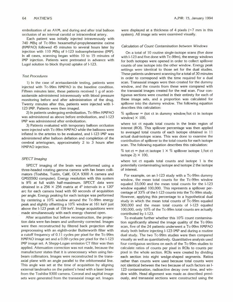
Test Procedures
1) In the case of acetazolamide testing, patients were injected with Tc-99m HMPAO in the baseline condition. Fifteen minutes later, these patients received 1 g of acetazolamide administered intravenously with blood pressure monitoring before and after administration of the drug. Twenty minutes after this, patients were injected with 1-123 IMP. Patients were then imaged.
2) In patients undergoing embolization, Tc-99m HMPAO was administered as above before embolization, and 1-123 IMP was administered after embolization.
3) Patients evaluated with temporary balloon occlusion were injected with Tc-99m HMPAO while the balloons were inflated in the arteries to be evaluated, and 1-123 IMP was administered after balloon deflation and completion of the cerebral arteriogram, approximately 2 to 3 hours after HMPAO injection.
SPECT Imaging
SPECT imaging of the brain was performed using a three-headed rotating gamma camera with fan beam collimators (Toshiba, Tustin, Calif, GCA 9300 A camera and GMS550U computer) . Energy resolution with this system is 9% at full width half-maximum. SPECT data were obtained in a 256 X 256 matrix at 4° intervals in a 120° arc for each camera head with 60 seconds of acquisition per angle. Energy peaking for each isotope was performed by centering a 10% window around the Tc-99m energy peak and slightly offsetting a 10% window at 161 keV just above the 1-123 peak of 159 ke V. Image acquisitions were made simultaneously with each energy channel open.
After acquisition but before reconstruction , the projection data were fan-beam and uniformity corrected. Images were then reconstructed by filtered back projection after preprocessing with an eighth-order Butterworth filter with a cutoff frequency of 0.11 cycles per pixel for the Tc-99m HMPAO image set and a 0 .09 cycles per pixel for the 1-123 IMP image set. A Shepp-Logan emission CT filter was then applied. Attenuation correction was not made, because the manufacturer states that it is unnecessary when using fanbeam collimators. Images were reconstructed in the transaxial plane with an angle parallel to the orbitomeatal line. This angle was set at the time of acquisition by aligning external landmarks on the patient's head with a laser beam from the Toshiba 9300 camera. Coronal and sagittal image sets were generated from the transaxial image set. Images
AJNR: 15, January 1994
were displayed at a thickness of 4 pixels (~7 mm in this system). All image sets were examined visually .
Calculation of Count Contamination between Windows
On a total of 10 routine single-isotope scans (five done with 1-123 and five done with Tc-99m), the energy windows for both isotopes were opened in order to collect spillover counts of one isotope into the other window. Energy peak settings were identical to those set for the dual studies. These patients underwent scanning for a total of 30 minutes in order to correspond with the time required for a dual scan. Transaxial images were then created for the dummy window, and the counts from these were compared with the transaxial images created for the real scan. Four contiguous sections were counted in their entirety for each of these image sets, and a proportion was calculated for spillover into the dummy window. The following equation describes this calculation:
% spillover = (tot ct in dummy window/tot ct in isotope window) X 100,
where tot ct equals total counts in the brain region of interest (ROI). This spillover percentage was then applied to averaged total counts of each isotope obtained in 11 actual dual-isotope scans. This was done to examine the contribution of spillover to the total counts for each isotope scan. The following equation describes this calculation:
% tot ct = (tot ct isotope 1 X % spillover isotope 1/tot ct isotope 2) X 100,
where tot ct equals total counts and isotope 1 is the potentially contaminating isotope and isotope 2 the isotope of interest.
For example, on an 1-123 study with a Tc-99m dummy window, the mean total counts for the Tc-99m window equaled 33,000 and the mean total counts for the 1-123 window equaled 100,000. This represents a spillover percentage of 33 % of the 1-123 counts into the Tc-99m study. However, applying this percentage to a hypothetical dual study in which the mean total counts of Tc-99m equaled 300,000 and the mean total counts of 1-123 equaled 100,000, only 10% of the Tc-99m total counts are actually contributed by 1-123.
To evaluate further whether this 10% count contamination significantly altered the image quality of the Tc-99m scan, five of the 24 patients underwent a Tc-99m HMPAO study both before injecting 1-123 IMP and during a routine dual study. The two Tc-99m studies were then compared visually as well as quantitatively. Quantitative analysis used four contiguous sections on each of the T c-99m studies to calculate ratios of counts per pixel in ROis to counts per pixel in the whole section. ROls were created by dividing each section into eight wedge-shaped segments. Ratios rather than counts were used because total counts were not identical between the two because of such factors as 1-123 contamination, radioactive decay over time, and window width. Head alignment was made as described previously, and transaxial sections were constructed using the

AJNR: 15, January 1994
same orbitomeatal angle in both studies. Sections were aligned as closely as possible to reduce artifact due to patient movement between studies. Ratio differences between studies were then calculated for a total of 60 leftand 60 right-hemisphere regions. A mean percentage difference for these 120 ROls was then calculated.
Results
Count Contamination Results
Calculations on Dummy Window Counts. Results from the dummy window showed that count spillover by Tc-99m into the 1-123 window was, on the average, 0.20% (± 0.09 SEM) (Fig 1). In a routine dual-isotope study where the average ratio of Tc-99m to 1-123 is 5:1, this could account for 0.7% (± 0.08% SEM) of the total 1-123 counts. The count spillover by 1-123 into the T c-99m window averaged 35.1% (± 1.2 SEM) (Fig 2). Again, because the dose of Tc-99m used is approximately five times greater than the 1-123 dose, 1-123 contamination averaged only 11.8% (± 0.76 SEM) of the total Tc-99m counts on the 11 dual-isotope studies in which we actually applied this calculation. Potential problems with this
- contamination could probably be even further reduced by making sure the Tc-99m-to-l-123 ratio is at least 3:1. Two studies in this group of 11 had ratios less than this and demonstrated a larger contribution of 1-123 contamination to the Tc-99m total counts in the study.
Calculations Comparing Tc-99m Studies. Results comparing the T c-99m studies done before 1-123 injection with those of the dual acquisition reveal little change in the ratio of total counts in a particular ROI to total counts in the whole section after the administration of 1-123. In comparing corresponding ROI-to-whole ratios in each
21 Ft
R L R
27
21
L
DUAL-ISOTOPE SPECT 65
Tc-99m scan, there was a mean difference of 2.83% (± 0.29 SEM) in the 60 left-hemisphere ratios and a mean difference of 2.58% (± 0.25 % SEM) in the 60 right-hemisphere ratios.
Patient Results
Acetazolamide Testing. Thirteen patients underwent acetazolamide testing (Table 1; refer to tables for patient numbers). Of these, five showed significant postacetazolamide changes (Fig 3). The patients who showed the greatest change were patients 1, 2, and 3 with bilateral vascular disease , patient 5, who had previously undergone sacrifice of a carotid artery for treatment of a cavernous carotid aneurysm, and patient 12, with a recently resected parietal A V M. Of the remaining patients, eight had minimal changes detected by visual inspection.
Based on these findings, patient 3 underwent right-carotid endarterectomy, and patient 5 underwent a right extracranial-intracranial bypass. Patients 2 and 3 were not considered surgical candidates at this time, because the former was asymptomatic on aspirin and the latter had poor access to the distal-middle cerebral artery for bypass purposes. Patient 12's perfusion defect in the area where his A V M had recently been resected was thought to reflect impaired vasereactivity, which was expected to improve with time.
Balloon-Occlusion Studies. Six patients underwent temporary balloon occlusion of a carotid artery in conjunction with baseline and ballooninflated SPECT studies (Table 2) . These studies were performed to determine if the patient could tolerate permanent carotid occlusion as a definitive treatment for either a neoplastic or vascular
Fig. 1. Brain SPECT studies comparing T c-99m transaxial sections w ith an 1-123 dummy section.
A, Representative transaxial Tc-99m brain section.
8 , Corresponding transaxial 1-123 dummy section. The change in scaling was necessary because of the extremely low counts in the dummy windows.
G0 255 60255
A B

66 MATHEWS AJNR: 15, January 1994
Fig. 2. Brain SPECT studies comparing 1- 123 IMP transaxial brain section with a corresponding Tc-99m dummy section.
A , Representative transaxial brain section .
8 , Corresponding transaxial T c-99m dummy section . A change in scaling was necessary because of the extremely low counts in the dummy windows.
R
A
7 4
21
L
p
G 0 255
A
IL l
R 'f
_.! ' -.. 1..
p
B
TABLE 1: Summary of SPECT findings using dual-isotope technique and acetazolamide
Patient
2
3
4
5 6 7 8 9
10 11
12
13
Diagnosis
Bilateral carotid occl (fi
bromuscular dysplasia)
Bilateral MCA occl (Maya
Maya)
L carotid occl R carotid
stenosis
Bilateral SDH"
R carotid occl
L CVA
L CVA
R frontal CVA
R cerebellar CV A
Brain stem AVM
R fron tal dural AVM
R par. AVM, resected
Dementia
SPECT Findings Before Acetazolamide
Small L frontal dec
Bifrontal dec L > R
L frontal dec
SDH L > R
R MCA dec
Small L frontal dec
Small L frontal dec
Small R frontal dec
R cerebellar defect, dec in poste
rior vs anterior ci rculation
L parietal inc
No defect
R parietal dec
Bilateral frontal dec
SPECT Findings After Acetazolamide
Marked bilateral dec
anterior vs posterior
Marked bifrontal dec
R> L Marked R parietal
dec Minimal change
Increased defect
Minimal change
Minimal change
Minimal change
Minimal change
Minimal change
Minimal change
Small R parietooc
cipital dec
Minimal change
21
L
Note-AVM =arteriovenous malformation ; SDH =subdural hematoma; occl =occlusion; MCA = middle
cerebral artery; CVA = cerebrovascular accident; dec = decrease; L = left; R = right; inc = increase.
• This patient underwent SPECT scanning before MR results were available. MR revealed bilateral isointense
SDH which had not been evident on CT scan.
2
I
G 0 255
lesion of the brain or skull base. Four of these patients had little or no change from baseline to balloon-inflated study (patients 14, 15, 19, and 20). Of these, only two underwent permanent carotid occlusion as a part of their treatment. Patient 14 tolerated this well; patient 15 developed a third-nerve palsy when a balloon migrated within her cavernous sinus during embolization of her carotid cavernous fistula . She was otherwise neurologically intact. Patients 17 and 18 had decrements in perfusion with the balloon inflated; however, it was not necessary to sacrifice the former patient's carotid artery during surgery, and the latter patient has not yet undergone surgery. Patient 16 had a large left -parietal arte-
riovenous fistula with mass effect, and this patient underwent temporary balloon occlusion of the left middle cerebral artery (the main contributing vessel to the fistula). In this case, perfusion to the left-temporal cortical rim as well as the rightinferior temporal lobe actually improved with the balloon inflated (Fig 4), perhaps by removing a steal phenomenon from the posterior circulation. This patient is undergoing graded embolization of the multicompartmental fistula so that he can be surgically treated in the future.
Embolization Studies. Four patients underwent pre- and postembolization studies (Table 3). At our institution, it has become standard practice to embolize large A V Ms endovascularly before

AJNR: 15, January 1994 DUAL-ISOTOPE SPECT 67
A
B
·~··· .. 1: . . . . .
!
'
Fig. 3. Brain SPECT studies before and after administration of acetazolamide in a patient with bilateral internal carotid artery occlusion from fibromuscular dysplasia.
A, Representative transaxial sections of a study obtained in baseline conditions using Tc-99m HMPAO.
B, The same transaxial sections as above obtained using 1-123 IMP after acetazolamide administration. There is diminished response in blood flow bilaterally in the anterior cerebral artery distributions and a marked decrease in the left middle cerebral artery distribution when compared with the posterior circulation. Such relative decreases are consistent with areas of poor vasoreactivity (small arrows).
TABLE 2: Summary of SPECT findings using dual-isotope technique during and after temporary balloon
occlusion of a cerebral artery
Patient Diagnosis
14 L CCA
15 R CCF
16 L AVF"
Balloon-inflated Baseline
Defect associated with aneurysm Minimal change
with mild dec L temporoparietal ctx
Slight L parieta l dec Minimal change
Large L temporoparietal dec due to Bilateral dec in tem
AVF, improvement in inferior tern- poral lobes
poral and L parieta l ctx
17 L mandibular hemangiopericytoma Slight dec L anterior circu lation Slight inc in L anterior
circu lation
18 L recurrent meningioma Dec in L MCA distribution around Slight improvement in
meningioma defect
19 Laryngeal CA Slight L parietal defect Minimal change
20 Tonsilar CA No defect Minimal change
Note.-CCA = carotid cavernous aneurysm; AVF = arteriovenous fistula; MCA = middle cerebral artery ;
L = left ; R = right; CA 'f cancer; dec = decrease; inc = increase; CCF = carotid avernous fistula; ctx = cortex.
• This patient underwent occlusion of the L MCA; all other patients underwent internal carotid artery
occlusion.
surgical excision. This is done to minimize bleeding and decrease operating time. After embolization of the A V M with either polyvinyl alcohol particles or platinum coils, three patients (22 , 23, and 24) demonstrated enlarged photopenic areas surrounding the A V M immediately after embolization (Fig 5). These changes were thought to represent diminished perfusion to the region of the AVM perhaps as a result of inadvertent proximal vessel or loss of vessels en passage. Patient 21 showed very little change from baseline to postembolii:ation study. All four patients underwent surgery, with patients 22, 23, and 24 sustaining minimal changes in neurologic status. Patient 21 developed aphasia and hemiparesis as
a result of the surgery, but these conditions now have cleared substantially.
Discussion
The technique of dual-isotope imaging has been used in a number of nuclear medicine applications including parathyroid, bone, and gastric imaging (8-1 0) . Its use in brain imaging has been reported ( 11, 12), but routine clinical use of the technique has been limited for several reasons. First, the energy peaks of the two most commonly used isotopes for brain imaging, Tc-99m and 1-123, are relatively close at 140 keV and 159 keV, respectively. Many early ')'-SPECT sys-

68 MATHEWS
Fig. 4. Brain SPECT studies during and after trial balloon occlusion of the left middle cerebral artery feeding a large left temporoparietal arteriovenous fistula.
A, Representative coronal sect ions obtained using 1-123 IMP in a baseline condition. There is a large photopenic defect in the left temporoparietal region consistent with location of the AVF (small arrows).
8, The same coronal sections as in A using Tc-99m HMPAO in the balloon-inflated condition. Note the improved flow to the inferior temporal lobes bilaterally as well as the left parietal cortical rim with the balloon inflated (large arrows). A
B
AJNR: 15, January 1994
TABLE 3: Summary of SPECT findings using dual-isotope technique during AVM embolization
Patient Location of AVM Preembolization Postembolization
21 L parietocciptital L parietoccipital A V M Minimal change
22 L frontal L frontal A V M defect Inc photopenic area
surrounding AVM
23 R frontoparietal R frontoparietal AVM Inc photopenic area
defect surrounding A V M"
24 L frontal L frontal AVM defect Inc photopenic area
surrounding A V M"
Note.-AVM =arteriovenous malformation; L =left; R =right; inc= increase.
• These patients underwent repeated SPECT scans 2 days after embolization , demonstrating reduction of
photopenic area back to preembolization size.
terns were unable to separate these peaks and to acquire images in two channels simultaneously. In addition , there has been concern that there would be excessive artifact introduced by scatter of one isotope's energy spectrum into the other 's channel. Characteristics of the imaging agents themselves also limit their usage. T c-99m HMPAO requires preparation with freshly eluted Tc-99m and administration within 30 minutes to prevent conversion of the agent to a more hydrophilic form, which accumulates in the scalp, nasopharynx, and salivary glands (13). The advantage of HMPAO, however, is that once injected, it accumulates in the brain in proportion to CBF
at that time and does not significantly redistribute (14). This allows SPECT scanning to occur several hours after actual injection. On the other hand, IMP is stable before injection, thus allowing delay in injection; once injected, however, imaging should be performed within 20 to 30 minutes, before significant redistribution (15). Finally, there has been concern over excessive radiation exposure to the patient from the combined use of both agents at one time.
In this report, we have demonstrated that dualisotope SPECT brain imaging with simultaneous image acquisition is feasible, practical, and reliable, and provides useful information in a variety

AJNR: 15, January 1994
A
B
of diagnostic and treatment settings. We are able to separate the two sets of images, which have exact section registration, because of the simultaneous acquisition. We also have demonstrated that contamination between the two windows can be minimized by slightly offsetting the 1-123 energy window and by narrowing both windows to 10%. This problem could be further handled by using an image-subtraction algorithm to reduce the percentage of count contamination from each study. Increasing the Tc-99m-to-1-123 ratio, either by increasing the Tc-99m dose or by offsetting the Tc-99m peak to just below 140 keY and at the same time increasing window width, also might decrease the relative contribution of 1-123 to the T c-99m total counts. However, as we have demonstrated in our duplicated T c-99m studies, this contamination appears to be uniformly distributed and does not substantially alter ROI to whole-section count ratios, which are presently our main means of measuring alterations in CBF in SPECT imaging of this type. In addition , simultaneous images of good quality can be obtained by minimally lengthening total scan time from 20 to 30 minutes, which most patients tolerated easily. Finally, the radiation
DUAL-ISOTOPE SPECT 69
Fig. 5. Brain SPECT studies before and after embolization of a right-frontal AV M .
A , Representative transaxial sections of a study obtained using Tc-99m HMPAO during the baseline state. The photopenic defect in the right-frontal region corresponds with the location of the AVM (small arrows).
B, The same transaxial sections as above in a study obtained using 1- 123 IMP immediately after embolization of the A V M with platinum coils and polyvinyl alcohol particles. The photopenic area surrounding the A V M increased in size, and the patient developed a mild right facial palsy consistent with ischemic changes occurring in the tissue surrounding the A V M (large arrows).
dose to these patients is approximately 0.0052 Gy total body for the doses of isotopes used. The brain exposure is 0.0085 Gy, and when pretreated with Lugol solution, the thyroid receives 0.0223 Gy (manufacturer's recommendations for Ceretec Tc-99m, Amersham, Arlington Heights, Ill; and Spectamine 1-123, IMP, Houston, Tex) . These doses are easily within tolerable limits and compare with a radiation exposure of 0.02 to 0.05 Gy during single-head CT.
In this report we also have demonstrated useful information that may be obtained from simultaneous, dual-isotope SPECT rCBF imaging. These include testing cerebrovascular reserve with acetazolamide, evaluating trial occlusion of a carotid or intracerebral artery, and examination before and after endovascular embolization of intracerebral AVMs.
Acetazolamide, a carbonic anhydrase inhibitor, has been widely used for "stress testing" of cerebral vascular reserve (2, 3). In healthy subjects , intravenous administration of this drug results in generalized cerebral vasodilation with increased CBF (1). In patients with vascular disease , baseline CBF may be symmetric; however, after the administration of acetazolamide, areas that are

70 MATHEWS
already maximally vasodilated are unable to increase flow further and thus develop focal perfusion defects. Such defects may indicate the need for surgical intervention, such as carotid endarterectomy or extracranial-intracranial bypass, as the patient has limited vascular reserve in areas of the perfusion defects and might proceed on to an ischemic event.
Temporary balloon occlusion of a carotid or intracerebral artery has proved a useful technique for evaluating a patient's tolerance to such occlusion before permanent vessel sacrifice (4-7). At a number of institutions, including our own, SPECT rCBF has been routinely incorporated into the temporary occlusion evaluation. This is done to detect those patients who may clinically tolerate a 30- to 45-minute trial occlusion but who may develop perfusion abnormalities. Several reports have suggested that these patients may develop neurologic sequelae after permanent vessel sacrifice ( 4-7). Patient 18 is just such a case. To date she has not undergone surgery because of concern that adequate collateral flow is not available, because her ipsilateral external carotid artery was sacrificed during initial resection of the meningioma.
The technique of endovascular embolization of A V Ms before surgical resection represents a major technical advance in the management of these lesions, allowing progressive rather than abrupt shunt occlusion and flow redistribution. Our overall technique and results have been described elsewhere ( 16-18). Many of these embolizations are staged over several days, and SPECT rCBF might provide useful information regarding adequacy of embolization as well as the creation of new perfusion defects or even hyperemia. Three of the four patients who underwent embolization in this study did reveal new perfusion defects, suggesting good control of A V M feeding vessels. The patient who showed little postembolization perfusion change actually had the most difficult surgical course because of intraoperative bleeding and the most prolonged postoperative recovery, although he too ultimately did well.
As we have demonstrated, a variety of diagnostic and therapeutic procedures easily can incorporate the technique of dual-isotope SPECT scanning of rCBF. Although performing sequential scans either on different days or with a second larger dose of isotope is one approach to obtaining baseline and postintervention studies, we feel that dual-isotope imaging is more convenient in that the patient undergoes only a single acquisition session, and is more accurate in comparing
AJNR : 15, January 1994
section-to-section differences because of exact registration of the brain images. It is also conceptually preferable when performing pre- and postintervention examinations to have the studies as temporally contiguous as possible. This is particularly true with pharmacologic interventions such as acetazolamide administration. Theoretically, any other situation in which baseline and postintervention evaluations of rCBF are needed could benefit from this methodology. As more CBF agents become available and highresolution scanners increase in prevalence, we believe that dual-isotope brain imaging will become a standard part of clinical nuclear medicine.
Acknowledgments
We thank Leslie Mihal for her assistance in the preparation of this manuscript.
References
1. Bonte FJ, Devous MD, Reisch JS. The effect of acetazolamide on
regional cerebral blood flow in normal human subjects as measured
by single-photon emission computed tomography. Invest Radio/
1988;23:564-568
2. Matsuda H, Higashi S, Kinuya K, et al. SPECT evaluation of brain
perfusion reserve by the acetazolamide test using Tc-99m HMPAO.
C/in Nuc/ Med 1991; 16:572-579
3. DiPiero V, Pozzilli C, Pantano P, et al. Acetazolamide effects on
cerebra l blood flow in acute reversible ischemia. Acta Neural Scand
1989;80:35-40
4. Eckard DA, Purdy PD, Bonte FJ. Temporary balloon occlusion of the
carotid artery combined with brain blood flow imaging as a test to
predict tolerance prior to permanent carotid sacrifice. AJNR Am J
Neuroradio/ 1992; 13: 1565-1569
5. Peterman SB, Taylor A , Hoffman JC. Improved detection of cerebral
hypoperfusion with internal carotid balloon test occlusion and Tc-
99m-HMPAO cerebral perfusion SPECT imaging. AJNR Am J Neu
roradiol 1991 ; 12:1035-1041
6. Moody EB, Dawson RC, Sandier MP. 99m-Tc-HMPAO SPECT im
aging in interventional neuroradiology: validation of balloon test
occlusion. AJNR Am J Neuroradiol 1991 ;12:1043-1044
7. Monsein LH , Jeffrey PJ, van Heerden BB, et al. Assessing adequacy
of collateral circu lation during balloon test occlusion of the internal
carotid artery with 99m-Tc HMPAO SPECT. AJNR Am J Neuroradio/
1991 ;12:1045-1051
8. Basarab RM , Manni A, Harrison TS. Dual isotope subtraction para
thyroid scin tigraphy in the preoperative evaluation of suspected
hyperparathyroidism. C/in Nuc/ Med 1985; 10:300-314
9. AI-Sheikh W, Sfakianakis GN, Mnaymneh W, et al. Subacute and
chronic bone infections: diagnosis using ln-111 , Ga-67, and Tc-99m
MDP bone scintigraphy and radiography. Radiology 1985;155:501-506
10. Fisher RS, Malmud LS, Bandini P, et al. Gastric emptying of a
physiologic mixed solid-liquid meal. C/in Nuc/ Med 1982;7:2 15-221
11 . Devous MD, Payne JK, Lowe, JL. Dual-isotope brain SPECT imaging
with technetium-99m and iodine-123: clinical validation using xenon-
133 SPECT. J Nuc/ Med 1992;33:1919-1924
12. Knapp WH , von Kummer R, Kubler W. Imaging of cerebral blood
flow-to-volume distribution using SPECT. J Nuc/ Med 1986;27:465-
470

AJNR: 15, January 1994
13. Matsuda H, Oba H, Seki H, et al. Determination of flow and rate
constants in a kinetic model of (99m-Tc) hexamethyl-propylene
amine oxime in the human brain. J Cereb Blood Flow Metab
1988;8:561-568
14. Podreka I, Suess E, Goldenberg G, et al. Initial experience with
technetium-99 m HM-PAO brain SPECT. J Nucl Med 1987;28: 1657-
1666
15. Kuhl DE, Barrio JR, Huang SC, et al. Quantify ing local cerebral blood
flow by N-isopropyl-p-[123-1] iodoamphetamine (IMP) tomography . J
Nucl Med 1982; 196:196-203
DUAL-ISOTOPE SPECT 71
16. Purdy PD, Samson D, Batjer HH, Risser RC. Preoperative embolization
of cerebral arteriovenous malformations with polyviny l alcohol par
ticles: experience in 51 adults. AJNR Am J Neuroradiol 1990; 11 :501-
510
17. Purdy PD, Batjer HH, Risser RC, Samson D. Arteriovenous malfor
mations of the brain: choosing embolic materials to enhance safety
and ease of exc ision. J Neurosurg 1992;77:2 17-222
18. Purdy PD, Batjer HH, Kopitnik T , Samson D. The team approach to
combined embolization and resection of arteriovenous malformations.
In : Da Pian R, ed. New trends in management of cerebrovascular
malformations. Vienna: Springer-Verlag (in press)
Top Related