

![Sensitization of Human Breast Cancer Cells to ...[CANCER RESEARCH 56, 1331-1340. March 15. 1996] Sensitization of Human Breast Cancer Cells to Cyclophosphamide and Ifosfamide by Transfer](https://static.fdocuments.in/doc/165x107/5f0d724b7e708231d43a65cf/sensitization-of-human-breast-cancer-cells-to-cancer-research-56-1331-1340.jpg)


Languages
Pages
Legal

CYCLOPHOSPHAMIDECyclophosphamide was considered by previous IARC Working Groups in 1980 and 1987 (IARC, 1981, 1987a). Since that time, new data have become available, these have been incorporated into the Monograph, and taken into consideration in the present evaluation.
1. Exposure Data
1.1 Identification of the agent
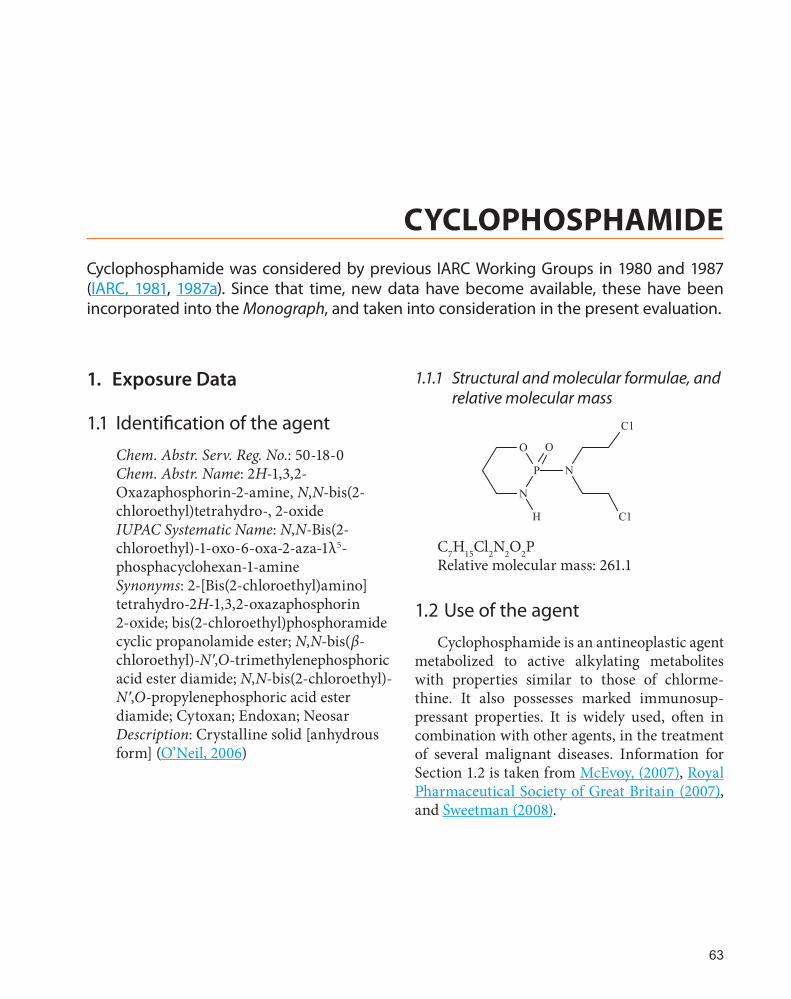
Chem. Abstr. Serv. Reg. No.: 50-18-0Chem. Abstr. Name: 2H-1,3,2-Oxazaphosphorin-2-amine, N,N-bis(2-chloroethyl)tetrahydro-, 2-oxideIUPAC Systematic Name: N,N-Bis(2-chloroethyl)-1-oxo-6-oxa-2-aza-1λ5-phosphacyclohexan-1-amineSynonyms: 2-[Bis(2-chloroethyl)amino]tetrahydro-2H-1,3,2-oxazaphosphorin 2-oxide; bis(2-chloroethyl)phosphoramide cyclic propanolamide ester; N,N-bis(β-chloroethyl)-N′,O-trimethylenephosphoric acid ester diamide; N,N-bis(2-chloroethyl)-N′,O-propylenephosphoric acid ester diamide; Cytoxan; Endoxan; NeosarDescription: Crystalline solid [anhydrous form] (O’Neil, 2006)
1.1.1 Structural and molecular formulae, and relative molecular mass
N
O
P
O
H
N
Cl
Cl
C7H15Cl2N2O2PRelative molecular mass: 261.1
1.2 Use of the agent
Cyclophosphamide is an antineoplastic agent metabolized to active alkylating metabolites with properties similar to those of chlorme-thine. It also possesses marked immunosup-pressant properties. It is widely used, often in combination with other agents, in the treatment of several malignant diseases. Information for Section 1.2 is taken from McEvoy, (2007), Royal Pharmaceutical Society of Great Britain (2007), and Sweetman (2008).
63

IARC MONOGRAPHS – 100A
1.2.1 Indications
Cyclophosphamide is used in the treatment of chronic lymphocytic leukaemia, lymphomas, soft tissue and osteogenic sarcoma, and solid tumours. It is given orally or intravenously. Cyclophosphamide is inactive until metabolized by the liver.
(a) Hodgkin lymphoma
Cyclophosphamide is used in combination regimens (e.g. bleomycin, etoposide, doxoru-bicin, cyclophosphamide, vincristine, procar-bazine, and prednisone [known as BEACOPP]) for the treatment of Hodgkin lymphoma.
(b) Non-Hodgkin lymphoma
Cyclophosphamide is used in combina-tion therapy for the treatment of non-Hodgkin lymphoma, including high-grade lymphomas, such as Burkitt lymphoma and lymphoblastic lymphoma, as well as intermediate- and low-grade lymphomas. Cyclophosphamide is commonly used with doxorubicin (hydroxydau-norubicin), vincristine (oncovin), and prednisone (known as the CHOP regimen), with or without other agents, in the treatment of various types of intermediate-grade non-Hodgkin lymphoma. Cyclophosphamide has also been used as a single agent in the treatment of low-grade lymphomas.
(c) Multiple myeloma
Cyclophosphamide is used in combination with prednisone, or as a component of combina-tion chemotherapy (i.e. vincristine, carmustine, melphalan, cyclophosphamide, and prednisone [VBMCP]) for the treatment of multiple myeloma.
(d) Leukaemia
Cyclophosphamide is used commonly for the treatment of chronic lymphocytic (lympho-blastic) leukaemia. Cyclophosphamide is used in combination with busulfan as a conditioning regimen before allogeneic haematopoietic
progenitor cell transplantation in patients with chronic myelogenous leukaemia.
Cyclophosphamide is used in the treatment of acute lymphoblastic leukaemia, especially in children. In the treatment of acute myeloid (myelogenous, non-lymphocytic) leukaemia, cyclophosphamide has been used as an additional drug for induction or post-induction therapy.
(e) Cutaneous T-cell lymphoma
Cyclophosphamide is used alone or in combi-nation regimens for the treatment of advanced mycosis fungoides, a form of cutaneous T-cell lymphoma.
(f) Neuroblastoma
Cyclophosphamide alone is used in the treatment of disseminated neuroblastoma. Combination chemotherapy that includes cyclo-phosphamide is also used for this neoplasm.
(g) Cancer of the ovary
Cyclophosphamide is used in combination chemotherapy (vincristine, actinomycin D, and cyclophosphamide [VAC]) as an alternative regimen for the treatment of ovarian germ-cell tumours.
Cyclophosphamide has been used in combi-nation with a platinum-containing agent for the treatment of advanced (Stage III or IV) epithelial ovarian cancer.
(h) Retinoblastoma
Cyclophosphamide is used in combination therapy for the treatment of retinoblastoma.
(i) Cancer of the breast
Cyclophosphamide is used alone and also in combination therapy for the treatment of breast cancer.
Combination chemotherapy with cyclo-phosphamide is used as an adjunct to surgery in premenopausal and postmenopausal women
64

Cyclophosphamide
with node-negative or -positive early (TNM Stage I or II) breast cancer. Adjuvant combina-tion chemotherapy that includes cyclophospha-mide, methotrexate, and fluorouracil has been used extensively.
Adjuvant combination chemotherapy (e.g. cyclophosphamide, methotrexate, and fluo-rouracil; cyclophosphamide, adriamycin, and fluorouracil; cyclophosphamide and adriamycin with or without tamoxifen) is used in patients with node-positive early breast cancer (Stage II) in both premenopausal and postmenopausal patients once treatment to control local disease (surgery, with or without radiation therapy) has been undertaken.
In Stage III (locally advanced) breast cancer, combination chemotherapy (with or without hormonal therapy) is used sequentially following surgery and radiation therapy for operable disease or following biopsy and radiation therapy for inoperable disease; commonly employed effective regimens include cyclophosphamide, methotrexate, and fluorouracil; cyclophospha-mide, doxorubicin, and fluorouracil; and cyclo-phosphamide, methotrexate, fluorouracil, and prednisone. These and other regimens also have been used in the treatment of more advanced (Stage IV) and recurrent disease.
(j) Small cell cancer of the lung
Cyclophosphamide is used in combination chemotherapy regimens (e.g. cyclophosphamide, adriamycin, and vincristine [CAV]; cyclophos-phamide, adriamycin, and etoposide [CAE]) for the treatment of extensive-stage small cell lung cancer.
(k) Sarcoma
Cyclophosphamide has been used in combi-nation regimens (usually with dactinomycin and vincristine) and as an adjunct to surgery and radiation therapy in the treatment of rhabdo-myosarcoma and Ewing sarcoma.
1.2.2 Dosage
Cyclophosphamide is administered orally or by intravenous injection or infusion. Less frequently, the drug has been administered intramuscularly and by intracavitary (e.g. intra-pleural, intraperitoneal) injection and direct perfusion.
In patients with no haematological deficien-cies receiving cyclophosphamide monotherapy, induction therapy in adults and children is usually initiated with an intravenous cyclophos-phamide loading dose of 40–50 mg/kg admin-istered in divided doses over 2–5 days. Other regimens for intravenous administration include 10–15 mg/kg every 7–10 days or 3–5 mg/kg twice weekly.
When cyclophosphamide is administered orally, the usual dose for induction or mainte-nance therapy is 1–5 mg/kg daily, depending on the tolerance of the patient.
A daily oral dose of 2–3 mg/kg for 60–90 days has been used in children with nephrotic syndrome, and in whom corticosteroids have been unsuccessful. In patients who are to undergo stem-cell transplantation, very high doses of cyclophosphamide such as 60 mg/kg daily for 2 days may be given as part of the conditioning regimen.
Various cyclophosphamide-containing combination chemotherapy regimens have been used in the treatment of breast cancer. One commonly employed regimen for the treatment of early breast cancer includes a cyclophospha-mide dosage of 100 mg/m2 orally on Days 1 through 14 of each cycle combined with intra-venous methotrexate at 40 mg/m2 on Days 1 and 8 of each cycle, and intravenous fluorouracil at 600 mg/m2 on Days 1 and 8 of each cycle. In patients older than 60 years of age, the initial intravenous methotrexate dosage is reduced to 30 mg/m2 and the initial intravenous fluorouracil dosage is reduced to 400 mg/m2. Dosage is also reduced if myelosuppression develops. Cycles
65

IARC MONOGRAPHS – 100A
are generally repeated monthly (i.e. allowing a 2-week rest period between cycles) for a total of 6–12 cycles (i.e. 6–12 months of therapy).
Cyclophosphamide is available as 25 and 50 mg tablets for oral administration, and as 200 mg, 500 mg, 1 g, or 2 g vials of powder for reconstitution for parenteral administration.
1.2.3 Trends in use
No information was available to the Working Group.
2. Cancer in Humans
The carcinogenicity of cyclophosphamide in humans was established initially on the basis of a large number of case reports, as well as several epidemiological studies (IARC 1981, 1987a). The interpretation of the epidemiological studies was limited by the small numbers of cases, the diffi-culty in separating the role of cyclophosphamide from other agents, or both factors.
The most substantial evidence available to previous Working Groups was a Danish study of 602 patients treated “mainly with cyclophos-phamide” for non-Hodgkin lymphoma, in which nine cases of acute myeloid leukaemia were observed compared to 0.12 expected (Pedersen-Bjergaard et al., 1985), and a case–control study of leukaemia following ovarian cancer in the former German Democratic Republic where a strong dose–response relationship was observed (Haas et al., 1987). All other studies reported at most three cases of leukaemia or bladder cancer in people who had received cyclophosphamide as the only potentially carcinogenic agent (IARC, 1981; Kinlen, 1985; Greene et al., 1986).
Subsequently, further studies have been published that have provided more detailed infor-mation on the carcinogenicity of cyclophospha-mide. This review is restricted to epidemiological studies that have used appropriate comparison
groups to investigate the role of cyclophospha-mide as the cause of specific types of cancer.
There have been several reported cohort studies in which patients treated with cyclophos-phamide were followed up, and the occurrence of second cancers investigated. Valagussa et al. (1994) followed 2465 women who had received treatment with cyclophosphamide, methotrexate and fluorouracil, a combination in which only cyclophosphamide is considered to have carcino-genic potential in humans. There were three cases of acute myeloid leukaemia observed compared to 1.3 expected, and five cases of bladder cancer compared to 2.1 expected. Statistical signifi-cance was not reported but was calculated by the Working Group to be greater than 0.05 for both types of cancer. Smith et al. (2003) followed 8563 women who had received cyclophosphamide and doxorubicin as adjuvant therapy for breast cancer and observed 43 cases of acute myeloid leukaemia or myelodysplastic syndromes (AML/MDS). The incidence of AML/MDS overall was seven times higher than expected rates in the general population, and was increased 3-fold in regimens that contained double the cumulative dose of cyclophosphamide.
Several case–control studies have also been reported. For leukaemia, Kaldor et al. (1990) investigated 114 cases of a cohort of ovarian cancer patients. The relative risks were, respec-tively, 2.2 and 4.1 in two increasing dose cate-gories of cyclophosphamide. Neither increase was reported as statistically significant. Travis et al. (1994) carried out a study involving 35 cases of leukaemia following non-hodgkin lymphoma, and found that prior treatment with cyclophosphamide was associated with a rela-tive risk of 1.8 that was not statistically signifi-cant when comparison was made to treatment with radiotherapy alone. In an investigation by Nandakumar et al. (1991) of 97 cases of myeloid leukaemia as second primary cancers, patients receiving cyclophosphamide had a relative risk of 12.6 compared to those treated surgically, and
66

Cyclophosphamide
was substantially higher when prednisone was co-administered with cyclophosphamide. Curtis et al. (1992) compared 90 women who developed acute myeloid leukaemia following breast cancer to controls, and found that the risk of leukaemia was 2.6 times greater in those who had received cyclophosphamide, compared to women who had been treated by surgery only.
There have also been two case–control studies of bladder cancer in relation to cyclo-phosphamide. Kaldor et al. (1995) investigated 63 cases of bladder cancer following ovarian cancer, and found that in comparison to surgery alone, the relative risk associated with chemo-therapy containing cyclophosphamide as the only potential bladder-cancer-causing agent was 4.2 (P = 0.025) in the absence of radiotherapy, and 3.2 (P = 0.08) with radiotherapy. Travis et al. (1995) studied 31 cases of bladder cancer and 17 cases of kidney cancer as well as matched controls within a cohort of 2-year survivors of non-Hodgkin lymphoma. The relative risk asso-ciated with cyclophosphamide treatment was 4.5 (P < 0.05) for bladder cancer, and 1.3 for kidney cancer.
2.1 Synthesis
The studies summarized above provide a comprehensive epidemiological basis for iden-tifying cyclosphosphamide as an independent cause of acute myeloid leukaemia and bladder cancer, that fully supports the conclusions drawn from earlier case reports, and more limited studies. Several studies have assessed the risk of all second primary cancers following cyclophos-phamide treatment, and some have found rates of occurrence that appear to be elevated, but have not provided evidence for risk of other specific cancer types.
3. Cancer in Experimental Animals
Cyclophosphamide has been tested for carcinogenicity by oral administration to mice and rats, by subcutaneous injection to mice, by topical application to mice, by intravenous injec-tion to rats, by intraperitoneal injection to mice and rats, and by perinatal exposure to mice.
Oral administration of cyclophosphamide resulted in skin tumours in transgenic mice (Yamamoto et al., 1996; Eastin et al., 2001), and in urinary bladder carcinoma, leukaemia, and nervous system tumours in rats (Schmähl & Habs, 1979; Habs & Schmähl, 1983). Subcutaneous injection of cyclophosphamide to mice caused a variety of neoplasms, including mammary gland carcinoma and leukaemia (Schmähl & Osswald, 1970; Walker & Bole, 1971, 1973; Walker & Anver, 1979, 1983; Petru et al., 1989).
Intravenous injection of cyclophospha-mide to rats caused both benign and malignant neoplasms (Schmähl, 1967, 1974; Schmähl & Osswald, 1970).
Intraperitoneal administration of cyclo-phosphamide increased the incidences of lung adenoma and adenocarcinoma, bladder papil-loma, and leukaemia in mice (Shimkin et al., 1966; Weisburger et al., 1975; Mahgoub et al., 1999), and mammary gland adenoma and carci-noma in rats (Weisburger et al., 1975).
Administration of cyclophosphamide to newborn mice caused lung and liver adenoma and carcinoma, and Harderian gland adenoma (Kelly et al., 1974; McClain et al., 2001).
See Table 3.1.
67

IARC M
ON
OG
RAPH
S – 100A
68
Table 3.1 Studies of cancer in experimental animals exposed to cyclophosphamide
Route Species, strain (sex), age Duration Reference
Dosing regimen Animals/group at start
Incidence of tumours Significance Comments
Oral administrationMouse, Tg ras H2/CB6F1 & B6C6F1 (M), 9 wk 26 wk Yamamoto et al. (1996)
0, 10, 30 mg/kg bw by gavage (in water, volume NR), twice/wk for 25 wk Initial number/group NR
Tg ras H2/CB6F1: Lung (adenomas)– 0/9, 3/16, 3/27 Multiplicity– 0, 0.19, 0.11 tumours/mouse
[NS]a Pharmaceutical grade
CB6F1: Lung (adenomas)– 0/6, 2/18, 2/20 Multiplicity– 0, 0.11, 0.10 tumours/mouse
[NS]a
Mouse, Tg.AC (M, F), 8–9 wk 27 wk Eastin et al. (2001)
0, 10, 30, 60 mg/kg bw by gavage (in water 50% ethanol, volume NR); twice/wk for 26 wk 15/sex/group
Skin tumours (at all sites; histologically confirmed): 5/15, 1/2, 5/5, 5/15 (M); 2/15, 5/11, 11/11, 14/15 (F)
[P < 0.0001 for 30 and 60 mg/kg bw doses in female mice]a
Purity NR; Tg.AC mice are transgenic mice that carry a v-Ha-ras oncogene
Skin tumours (squamous cell papillomas of vulva): 2/15, 4/11, 10/11, 12/15 (F)
[P ≤ 0.0003 for 30 and 60 mg/kg bw doses in female mice]a
Leukaemia (erythrocytic): 0/15, 0/15, 4/15, 1/15 (F)
P < 0.05, for 30 mg/kg bw group
Rat, Sprague-Dawley (M, F) Lifetime Schmähl & Habs (1979)
0, 0.31, 0.63, 1.25, 2.5 mg/kg bw in drinking-water, 5 ×/wk for life 40/sex/group
Malignant tumours: 4/38, 11/34, 14/36, 15/35, 13/31 (M); 5/34, 11/37, 13/37, 11/33, 9/27 (F)
[P < 0.05, for 3 highest doses]
Purity NR
Urinary bladder (carcinomas): 0/38, 2/34, 2/36, 5/35, 7/31 (M); 0/34, 0/37, 0/37, 0/33, 1/27 (F)
[P ≤ 0.02 for 2 highest doses in males]a
Lymphoid and haematopoietic tissue (leukaemia): 0/72, 3/71, 6/73, 6/68, 4/58 (M, F)
[P ≤ 0.04 for 3 highest doses for combined males and females]a
Nervous system (sarcomas): 1/72, 7/71, 5/73, 6/68, 1/58 (M, F)
[P ≤ 0.05 for 0.31 and 1.25 mg/kg doses for combined males and females]a

Cyclophosphamide69
Table 3.1 (continued)Route Species, strain (sex), age Duration Reference
Dosing regimen Animals/group at start
Incidence of tumours Significance Comments
Rat, Sprague-Dawley (M), 100 d 20 mo Habs & Schmähl (1983)
0, 2.5 mg/kg bw in drinking-water, 5 ×/wk for 20 mo 100/group
Urinary bladder (papillomas or transitional-cell carcinomas): 0/63, 24/80
[P < 0.0001]a Reported as “chemically pure”
Nervous system tumours: 1/63, 11/80
[P < 0.0076]a
Rat, Sprague-Dawley (M), 100a Lifetime Schmähl & Habs (1983)
0, 2.5 mg/kg bw in drinking-water, 5 times/wk for life 100/group
Urinary bladder (papillomas): 0/100, 15/100
[P < 0.0001]a Purity NR; only data on bladder tumours reported
Urinary bladder (transitional-cell carcinomas): 0/100, 17/100
[P < 0.0001]a
Subcutaneous injectionMouse, NMRI (F) 52 wk Schmähl & Osswald (1970)
0, 26 mg/kg bw/wk (in solvent NR), for 5 wk 50/group
Malignant tumours (primarily mammary carcinomas): 3/46, 28/46
[P < 0.001]b Purity > 98%
Mouse, New Zealand Black/New Zealand White (F) 64 wk Walker & Bole (1971)
0, 8 mg/kg bw (in saline; volume NR), daily for 64 wk 16, 10
Neoplasms (mainly lymphomas): 0/16, 6/10
P = 0.00002 Purity NR
Mouse, New Zealand Black/New Zealand White (M, F) 93 wk Walker & Bole (1973)
0, 1, 8 mg/kg bw (in 100 μL saline), daily for 93 wk 20, 10, 10 per sex
Neoplasms (mainly lymphomas): 2/16, 3/9, 8/9 (M); 1/20, 1/10, 9/9 (F)
P = 0.003 for 8 mg/kg bw males; P < 0.0001 for 8 mg/kg bw females
Purity NR
Mouse, New Zealand Black/New Zealand White (F) Lifetime Walker & Anver (1979)
0, 5.7, 16 mg/kg bw (in 100 μL saline), daily for life 15, 17, 21
Neoplasms (mainly mammary carcinomas): 0/13, 15/15, 17/19
[P < 0.0001 for 5.7 and 16 mg/kg bw groups]a
Purity NR; treatment groups not started simultaneously
Mammary carcinomas: 0/13, 5/15, 16/19
[P ≤ 0.03 for 5.7 and 16 mg/kg bw groups]a
Mouse, New Zealand Black/New Zealand White (F), 6 wk Lifetime Walker & Anver (1983)
0, 56 mg/kg bw (in 100 μL saline), weekly for life 15, 22
Neoplasms: 0/13, 17/19
[P < 0.0001]a Purity NR; groups not started simultaneously; Neoplasms were mainly mammary gland carcinomas, lung adenomas and lymphomas

IARC M
ON
OG
RAPH
S – 100A
70
Route Species, strain (sex), age Duration Reference
Dosing regimen Animals/group at start
Incidence of tumours Significance Comments
Mouse, NMRI & AKR (F), 7 wk, Lifetime Petru et al. (1989)
0, 13, 26 mg/kg bw (in saline, volume NR), weekly for life 30/group
Leukaemia (NMRI mice): 2/30, 16/30, 10/30
P ≤ 0.027 for 13 & 26 mg/kg bw groups
Purity NR [negative trend in AKR mice]
Leukaemia (AKR mice): 30/30, 25/30, 19/30
P ≤ 0.006 for 13 & 26 mg/kg bw groups
Skin applicationMouse, Tg.AC (M, F), 8–9 wk 27 wk Eastin et al. (2001)
0, 10, 30, 90 mg/kg bw (in 50% ethanol, 3.3 mL/kg bw), 2 ×/wk for 26 wk 15/sex/group
Skin tumours (at site of application): 1/15, 0/15, 2/15, 3/15 (M); 1/15, 0/15, 0/15, 2/15 (F)
[NS]a Purity NR; Tg.AC mice are transgenic mice that carry a v-Ha-ras oncogene
Skin tumours (at all skin sites): 1/15, 2/15, 3/15, 3/15 (M); 4/15, 3/15, 9/15, 14/15 (F)
[P = 0.0002 for 90 mg/kg females]a
Intravenous administrationRat, BR 46 (M) 23 mo Schmähl (1967)
0, 15 mg/kg bw (vehicle and volume NR), weekly (750 mg/kg bw total dose) 50, 40
Neoplasms (benign and malignant combined): 1/50, 14/26
[P < 0.001]b Purity > 98%
Rat, BR 46 (M) 23 mo Schmähl & Osswald (1970)
0, 13 mg/kg bw (vehicle and volume NR), weekly for 52 wk 89, 48
Neoplasms: 3/65, 4/36 (benign); 4/65, 6/36 (malignant)
[NS]b Purity > 98%
Rat, BR 46 (M) 23 mo Schmähl & Osswald (1970)
0, 33 mg/kg bw (vehicle and volume NR), 5 times every 2 wk 89, 96
Neoplasms: 3/65, 5/66 (benign); 4/65, 16/66 (malignant)
[P < 0.01, malignant tumours]b
Purity > 98%
Rat, Sprague-Dawley (M) 700 d Schmähl, (1974)
0, 13 mg/kg bw (vehicle and volume NR), weekly (670 mg/kg bw total dose) 52, 32
Neoplasms (malignant): 6/52, 14/32
[P < 0.001]b Purity > 98%
Intraperitoneal administrationMouse, dd (M, F) 48 wk Tokuoka (1965)
0 or 5 mg/kg bw (in saline 5 mL/kg), 2 injections/wk for 15 wk 20, 29
Lung (adenomas or carcinomas): 1/20, 3/29
NS Purity NR
Liver (adenomas): 0/20, 2/29
NS
Testis (interstitial cell tumours): 0/20, 4/29
NS
Mammary gland (carcinomas): 1/20, 3/29
NS
Table 3.1 (continued)

Cyclophosphamide71
Table 3.1 (continued)Route Species, strain (sex), age Duration Reference
Dosing regimen Animals/group at start
Incidence of tumours Significance Comments
Mouse, A (M, F) 48 wk Tokuoka (1965)
0 or 5 mg/kg bw (in saline 10 mL/kg), 2 injections/wk for 15 wk 16, 25
Lung (adenomas or carcinomas): 2/16, 6/25
NS Purity NR
Testis (interstitial cell tumours): 0/16, 3/25
NS
Mouse, A/J (M, F, equally split) 39 wk Shimkin et al. (1966)
0, 32.2, 129, 516, 1609 μmol/kg bw (total dose; in 200 μL water), 3 injections/wk for 4 wk 360, 30, 30, 30, 30
Lung (adenomas or adenocarcinomas): 107/339, 12/30, 11/26, 20/27, 2/4 (incidence); 0.38, 0.4, 0.6, 1.3, 2.5 (tumours per mouse)
[p < 0.001 (for 516 µmol/kg bw dose, incidence)]b
Purity NR
Mouse, Swiss-Webster-derived (M, F) 18 mo Weisburger et al. (1975)
0, 12, 25 mg/kg bw (vehicle and volume NR), 3 injections/wk for 6 mo 101, 25, 25 (M) 153, 25, 25 (F)
Lung (adenomas or adenocarcinomas): 10/101, 7/30 (M); 21/153, 10/35 (F)
P = 0.031 (M) and P = 0.027 (F) (combined 12 & 25 mg/kg bw vs control)
Purity NR; not all control mice were treated with the vehicle
Bladder (papillomas or carcinomas): 3/101 & 4/30 (M)
P = 0.048 (combined 12 & 25 mg/kg bw vs control)
Mouse, 129/Sv & 129/Sv X C57BL/6 Nf1+/+ & Nf1+/−(sex NR), 6–10 wk 15 mo Mahgoub et al. (1999)
0 or 100 mg/kg bw/wk (solvent and volume NR) for 6 wk 129/Sv Nf1+/+: 31 & 5 mice 129/Sv Nf1+/−: 46 & 12 mice 129/Sv X C57BL/6 Nf1+/+: 14 & 15 mice 129/Sv X C57Bl/6 Nf1+/−: 412 & 25 mice
Leukaemia (129/Sv Nf1+/+): 2/31, 0/5
Purity NR
Leukaemia (129/Sv Nf1+/−): 8/46, 7/12
P = 0.004
Leukaemia (129/Sv X C57BL/6 Nf1+/+): 0/14, 2/25Leukaemia (129/Sv X C57BL/6 Nf1+/−): 0/12, 7/25
Rat, Sprague-Dawley (M, F) 18 mo Weisburger et al. (1975)
0, 5, 10 mg/kg bw (vehicle and volume NR), 3 injections/wk for 6 mo 179, 25, 25 (M) 181, 25, 28 (F)
Mammary gland (adenomas): 2/105 & 24/53 (F; combined 5 & 10 mg/kg bw)
P = 0.028 Purity NR; not all control rats were treated with the vehicle
Mammary gland (carcinomas): 13/105 & 9/53 (F; combined 5 & 10 mg/kg bw)
P = 0.035

IARC M
ON
OG
RAPH
S – 100A
72
Route Species, strain (sex), age Duration Reference
Dosing regimen Animals/group at start
Incidence of tumours Significance Comments
Perinatal exposureMouse, CD-1 (M, F) 79 wk Kelly et al. (1974)
i.p. injection 0, 0.8, 4.0, 20.0 mg/kg bw (in 10 µL/kg saline), on postnatal Days 1, 3, 6 30/sex/group
Lung (adenomas): 0/28, 2/29, 4/27, 0/21 (M); 1/25, 2/27, 2/28, 3/21 (F)
P < 0.05 for 4 mg/kg bw males (life-table analysis)
Purity NR; the 20 mg/kg dose caused marked bw changes and nearly 100% mortality
Mouse, CD-1 (M, F) 1 yr McClain et al. (2001)
Oral 0, 10, 20, 40, 60 mg/kg bw by gavage (100 µL and 200 µL) on postnatal Days 8 & 15 [solvent NR] 48 (control), 24/sex
Liver (adenomas): 2/48, 2/24, 4/24, 6/24, 5/24 (M)
[P < 0.04 for 40 & 60 mg/kg bw]a
Purity NR
Liver (carcinomas): 0/48, 0/24, 1/24, 6/24, 1/24 (M)
[P = 0.0009 for 40 mg/kg bw]a
Lung (adenomas): 3/48, 0/24, 8/24, 12/24, 13/24 (M); 7/48, 3/24, 6/24, 16/24, 13/24 (F)
[P < 0.005 for 20, 40, & 60 mg/kg bw (M); 40 & 60 mg/kg bw (F)]a
Lung (carcinomas): 0/48, 1/24, 0/24, 6/24, 3/24 (M); 0/48, 1/24, 3/24, 3/24, 0/24 (F)
[P < 0.03 for 40 & 60 mg/kg bw (M); 20 & 40 mg/kg bw (F)]a
Harderian gland (adenomas): 2/48, 1/24, 1/24, 1/24, 5/24 (F)
[P < 0.04 for 60 mg/kg bw]a
Pre and postnatal exposureMouse, BR 46 (M, F) 24 mo Roschlau & Justus (1971)
i.p injection 25 mg/kg bw on gestation Day 14 [solvent and volume NR]. Male and female offspring treated every 2 wk for a total of 30 times Initial number NR
Lung (adenomas): male offspring 4/16, 2/16; female offspring 5/12 & 1/18
NS Purity NR
Lung (carcinomas): male offspring 0/16, 3/16; female offspring 0/12, 4/18
NS
a Current Working Group analysis (Fisher Exact test)b Previous Working Group analysisbw, body weight; d, day or days; F, female; i.p., intraperitoneal; M, male; mo, month or months; NR, not reported; NS, not significant; vs, versus; wk, week or weeks, yr, year or years
Table 3.1 (continued)

Cyclophosphamide
4. Other Relevant Data
4.1 Absorption, distribution, metabolism, and excretion
In most species, cyclophosphamide is rapidly absorbed, metabolized, and excreted. Its meta-bolic pathway has been studied in several species including mice, rats, hamsters, rabbit, dogs, sheep, and monkeys. Cyclophosphamide is not cytotoxic per se, because it requires metabolic activation before it can act as an alkylating agent. Activation takes place predominantly in the liver, although this may occur in other tissues (IARC, 1981).
Cyclophosphamide undergoes metabo-lism to several intermediates with alkylating activity. The principal metabolites identified are phosphoramide mustard, and acrolein. Phosphoramide mustard can undergo dephos-phoramidation to yield nornitrogen mustard, which also has alkylating activity. Metabolites of cyclophosphamide can interact with DNA and proteins, resulting in the formation of adducts. The metabolism of cyclophosphamide and DNA adducts formation are summarized in Fig. 4.1.
A minor pathway results in dechloroeth-ylation and the formation of 2-dechloroethylcy-clophosphamide and another alkylating agent, chloroacetaldehyde (Balu et al., 2002).
The other compounds such as 4-ketocyclo-phosphamide and propionic acid derivative are relatively non-toxic, and are the major urinary metabolites of cyclophosphamide in several species (IARC, 1981).
4.2 Genetic and related effects
4.2.1 Interaction with DNA
Using 4-hydroperoxycyclophosphamide as an activated form of cyclophosphamide, Mirkes et al. (1992) identified by mass spectrometric analysis the formation of the monofunctional
adduct N-(2-chloroethyl)-N-[2-(7-guaninyl)ethyl]amine (nor-G) and the bifunctional adduct N,N-bis[2-(7-guaninyl)ethyl]amine (G-nor-G) in rat embryos in in-vitro culture. The mono-functional adduct N-(2-hydroxyethyl)-N-[2-(7-guaninyl)ethyl]amine (nor-G-OH) was detected in bladder tissue of rats injected with [3H]cyclophosphamide (Benson et al., 1988). Using 32P-postlabelling analysis, a phosphotriester was shown to be formed: (1) when phosphora-mide mustard was reacted with deoxyguanosine 5′-monophosphate, (2) when cyclophospha-mide was incubated with calf thymus DNA in the presence of reconstituted cytochrome P450 (CYP) metabolizing system, and (3) in liver DNA from mice injected intraperitoneally with cyclo-phosphamide (Maccubbin et al., 1991).
Nornitrogen mustard reacts with guanosine and with guanine bases in DNA to form nor-G initially, but this is converted to a hydroxy-lated derivative (nor-G-OH), and to a cross-linked (between guanines) adduct (G-nor-G) (Hemminki, 1987). Both monofunctional adducts, but not the cross-linked adduct, were also detected when phosphoramide mustard was reacted with DNA (Cushnir et al., 1990). Acrolein reacts with DNA to form O6-(n-propanalyl)guanine, and the product of chloroacetaldehyde reaction with DNA is O6-(ethanalyl)guanine (Balu et al., 2002). Acrolein can produce exocyclic adducts in DNA, including 1,N2-hydroxypropanodeoxyguanosine and 1,N6-hydroxypropanodeoxyadenosine (Chung et al., 1984; Foiles et al., 1990; Smith et al., 1990). The former was detected in acrolein-treated human fibroblasts and in peripheral blood lymphocytes of a dog treated with cyclo-phosphamide (Wilson et al., 1991).
Nornitrogen mustard also reacts covalently with proteins, and a method for the detection of cysteine-34 residue adducts in human serum albumin has been described (Noort et al., 2002).
The single-cell gel comet assay is used to detect single-strand breaks and other alkali-labile lesions in DNA exposed to cyclophosphamide.
73

IARC M
ON
OG
RAPH
S – 100A
74 Fig. 4.1 Metabolic pathway of cyclophosphamide
O
P
NH
N
C1
C1
O
O
P
NH
N
C1
C1
O
HO
O
P
NH
N
C1
C1
O
OCYP oxidase
cyclophosphamide 4-hydroxy-cyclophosphamide 4-keto-cyclophosphamide
OH
P
H2N
N
O O
Cl
Cl
OHO
P
H2N
N
O O
Cl
Cl
aldehydeoxidase
nornitrogen mustardaldophosphamide
DNA/protein
spont
H
O
HO
P
H2N
N
O
Cl
Cl
+
acrolein phosphoramide mustard
DNADNA
Acrolein DNA adducts
A
Phosphoramide Mustard DNA adducts Nitrogene Mustard DNA adduct
carboxyphosphamide
HN
Cl
Cl
NH
NN
NN
OH
H2N
(O,S,N)-protein
Nitrogene Mustard DNA adduct
HO
NH
NN
NN
OH
H2N
C1
NH
NN
NN
OH
H2N
NN
NN
OH
H2Nnor-G-OH nor-G G-nor-G 3'-dGM P-phosphoester adduct
Phosphoramide Mustard DNA adducts
B
Acrolein DNA Adducts
8-Hydroxy-1,N2-PropanodG 7,8-Cyclic Guanine Adduct
N
N
N
NN
O
HdR
OH
N
N
N
NH2N
O
OH
dR
6-Hydroxy-1,N2-PropanodG
N
N
N
NNHO
HdR
O
NH
N
N
N
N
OH
NH2
NN
NN
OH
H2N
ONH
OH
O
P
O
HO
HO O
A. Metabolism of cyclophosphamide to phosphoramide mustard, acrolein, and nornitrogen mustard. Cyclophosphamide is metabolized by CYP enzymes to 4-hydroxycyclophosphamide, wich equilibrates with aldophosphamide to spontaneously yield phosphoramide mustard and acrolein. Aldophosphamide is also metabolized by aldehyde oxidase to carboxyphosphamide, which produces nornitrogen mustard. 4-Hydroxy-cyclophosphamide can be oxidized to the inactive 4-keto-cyclophosphamide.B. Phosphoramide mustard produces multiple monofunctional and bifunctional adducts with guanine, and acrolein forms exocyclic adducts. Nornitrogen mustard forms mono- and bifunctional adducts with guanine. From Povirk & Shuker (1994), Anderson et al. (1995), Khan et al. (1998)CYP, cytochrome P450; nor-G, N-(2-chloroethyl)-N-[2-(7-guaninyl)ethyl]amine; G-nor-G, N,N-bis[2-(7-guaninyl)ethyl]amine; nor-G-OH, N-(2-hydroxyethyl)-N-[2-(7-guaninyl)ethyl]amine; dR, deoxyribose

Cyclophosphamide
In vitro studies have demonstrated the comet-forming activity of cyclophosphamide in human hepatoma (Hep G2) cells (Uhl et al., 2000; Yusuf et al., 2000), in primary cultures of rat and human urinary bladder cells (Robbiano et al., 2002), in primary cultures of human leukocytes in the presence of metabolic activation system S9 mix (Hartmann et al., 1995; Hartmann & Speit, 1995; Frenzilli et al., 2000), and in extended-term cultures of human T-lymphocytes, also in the presence of S9 (Andersson et al., 2003). Comet formation was also detected in vivo in the urinary bladder mucosa of rats given cyclophosphamide orally (Robbiano et al., 2002), and in peripheral blood cells of patients administered the drug (Hartmann et al., 1995).
4.2.2 Genotoxic effects in humans
There are few reports of DNA-adduct formation by cyclophosphamide in humans. Acrolein-derived DNA adducts, detected by immunochemical methods, were found in blood leukocytes of cancer patients receiving cyclophosphamide (McDiarmid et al., 1991). In another study, mono-adducts and inter-strand cross-links derived from phosphoramide mustard were detected in a single patient admin-istered 1 g/m2 cyclophosphamide (Souliotis et al., 2003). Increased DNA damage (comet formation) was also observed in the lymphocytes of patients administered cyclophosphamide (Hartmann et al., 1995).
Increased frequencies of several biomarkers of genotoxicity have been observed in the lympho-cytes of patients treated with cyclophosphamide, relative to control subjects. These include muta-tions at the hypoxanthine-(guanine) phospho-ribosyl transferase (HPRT) locus (Palmer et al., 1986, 1988; Tates et al., 1994; Sanderson et al., 2001), and sister chromatide exchange (Raposa & Várkonyi, 1987; McDiarmid et al., 1990; Sardaş et al., 1994; Mertens et al., 1995; Hartmann et al., 1995).
Other studies reported positive findings for elevated chromosomal aberrations frequencies (Sessink et al., 1994; Rubes et al., 1998; Burgaz et al., 2002), and micronuclei (Yager et al., 1988; Tates et al., 1994; Zúñiga et al., 1996; Burgaz et al., 1999; Rekhadevi et al., 2007) in medical personnel exposed to cyclophosphamide. Increases in frequencies of micronuclei were also detected in buccal cells in some studies (Cavallo et al., 2005; Rekhadevi et al., 2007), but not in another (Burgaz et al., 1999).
4.2.3 Genotoxic effects in experimental systems
(a) Mutagenic effects in vitro
The previous IARC Monograph (IARC, 1987b) states that cyclophosphamide induced chromosomal aberrations, sister chromatid exchange, and DNA damage in human cells in vitro. It also induced morphological transfor-mation, chromosomal aberrations, sister chro-matid exchange, mutation, and unscheduled DNA synthesis (UDS) in rodent cells in vitro. It further induced aneuploidy, mutation, recom-bination, gene conversion, and DNA damage in fungi. It was also reported to act as a mutagen and DNA-damaging agent in bacteria.
The mutagenicity of cyclophosphamide in Salmonella typhimurium was enhanced by increased induction of CYPs in S9 liver frac-tions by a combination of β-naphthoflavone and sodium phenobarbital (Paolini et al., 1991a). Comparison of S9 from liver and kidney of preg-nant mice revealed that liver S9 was more effective in activating cyclophosphamide to mutagenic metabolites in S. typhimurium, and also in inducing sister chromatid exchange in human peripheral lymphocytes, and Chinese hamster ovary (CHO) cells (Winckler et al., 1987).
In Saccharomyces cerevisiae, higher rates of mitotic gene conversion and point mutation by cyclophosphamide were associated with induc-tion of class 2B CYPs in co-cultured epithelial cell
75
IARC MONOGRAPHS – 100A
lines from fetal mouse liver (Paolini et al., 1991b). A recombinant plasmid containing a full-length cDNA encoding the rat cytochrome CYP2B1 introduced into S. cerevisiae also increased the mutation frequency induced by cyclophospha-mide (Black et al., 1989).
CYP2B1 expressed in Chinese hamster V79-derived SD1 cell lines also potentiated cyclophosphamide mutagenesis (6-thiogua-nine resistance), whereas CYP1A1 expressed in V79-derived XEM2 cell lines did not (Doehmer et al., 1990, 1992).
Cyclophosphamide was weakly mutagenic (detected by induction of resistance to 6-thio-guanine) in differentiated Reuber hepatoma cells H4IIEC3/G-, but markedly cytotoxic and clastogenic (micronucleus formation) (Roscher & Wiebel, 1988), and also mutagenic in a Chinese hamster epithelial liver cell line (6-thioguanine resistance) (Turchi et al., 1992), and in Chinese hamster lung (CHL) cells in the presence of S9, as measured at microsatellite loci (Kikuno et al., 1995).
Using 4-hydroperoxycyclophosphamide and phosphoradiamidic mustard, the role of different repair enzymes in defining sensitivity was inves-tigated by Andersson et al. (1996) in CHO cells. Mutations in excision repair cross-comple-menting ERCC1 and ERCC4 genes caused hyper-sensitivity to the cyclophosphamide analogues.
Cyclophosphamide induced sister chromatid exchange in mouse primary bone-marrow and spleen cells (Soler-Niedziela et al., 1989), and micronuclei in mouse lymphoma in L5178Y tk+/- cells (Kirsch-Volders et al., 2003), and in parental V79 cells (Kalweit et al., 1999) in the presence of rat liver S9. Of several V79 cell lines engi-neered to express rat CYPs, increases in micro-nuclei (Ellard et al., 1991) and sister chromatid exchange (Kulka et al., 1993) were seen in the cells expressing CYP2B1. The rat hepatoma cells lines H4IIEC3/G- and 2sFou were also suscep-tible to micronuclei induction by cyclophospha-mide (Tafazoli et al., 1995).
Human T-lymphocytes were more susceptible than B-lymphocytes to both chromosomal aber-rations and sister chromatid exchange induction by cyclophosphamide in the presence of rat liver S9 (Miller 1991a, b). This difference between T- and B-lymphocytes was not found with mouse cells treated with 4-hydroxycyclophosphamide or phosphoramide mustard (Kwanyuen et al., 1990). In another study (Kugler et al., 1987), rat liver microsomal mix was more effective than rat liver S9 in activating cyclophosphamide to induce chromosomal aberrations. Human lymphocytes from women carrying mutations in the breast cancer susceptibility gene BRCA1 were more susceptible to micronuclei induction than cells from non-carriers (Trenz et al., 2003). Hep G2 human hepatoma cells were susceptible to sister chromatid exchange and micronuclei induction by cyclophosphamide (Natarajan & Darroudi, 1991) and, in analogous studies, the S9 microsomal fraction of these cells were shown to be capable in activating cyclophosphamide to induce sister chromatid exchange and micronu-clei in CHO cells (Darroudi & Natarajan, 1993). Human dental pulp cells formed chromosomal aberrations when exposed to cyclophosphamide in the presence of rat liver S9 (Tsutsui et al., 2006).
In the presence of rat liver S9, cyclophospha-mide induced morphological transformation of BALB/3T3 mouse embryonic fibroblast cells (McCarvill et al., 1990).
(b) Mutagenic effects in vivo
The previous IARC Monograph (IARC, 1987b) states that cyclophosphamide was found to bind to kidney, liver and lung DNA in mice. It also induced dominant lethality, chromosomal aber-rations, micronuclei, sister chromatid exchange, mutations, and DNA damage in rodents in vivo. In Drosophila, it induced aneuploidy, herit-able translocations, and somatic and sex-linked recessive lethal mutations. In patients adminis-tered cyclophosphamide, increased incidences of chromosomal aberrations and sister chromatid
76

Cyclophosphamide
exchange in peripheral lymphocytes and bone marrow were observed.
In Drosophila melanogaster, cyclophospha-mide tested positive for the somatic white-ivory mutation (Batiste-Alentorn et al., 1994), and produced chromosome breaks in spermatocytes (Zijlstra & Vogel, 1989).
Several studies have examined the muta-genic effects of cyclophosphamide in transgenic mice. In MutaMouse, mutation induction was observed in bone marrow (other tissues not studied) (Hoorn et al., 1993). In Big Blue mice, mutation frequencies were elevated in the liver, but not in the testis or spleen in one study (Hoyes et al., 1998), and in another study, in the lung and urinary bladder, but not in the kidney, bone-marrow or splenic T-cells (Gorelick et al., 1999). Another study compared the lacI locus in Big Blue mice with the Hprt locus in conventional B6C3F1 mice, and cyclophosphamide induced mutations in the endogenous gene in splenic lymphocytes, but not in the transgene (Walker et al., 1999). In rats, cyclophosphamide produced the ‘common deletion’ mutation in liver mito-chondrial DNA, and folic acid supplementation was found to be protective against this damage (Branda et al., 2002).
In two related studies investigating oncogene and tumour-suppressor gene expression in mice, cyclophosphamide was found to induce expres-sion of several genes, including c-Myc and Tp53, in the spleen and thymus, but not in other tissues (Ember et al., 1995; Ember & Kiss, 1997).
Many studies have investigated the cyto-genicity of cyclophosphamide in newts, rodents, dogs, and non-human primates. Results are invariably positive for this compound, and are summarized in Table 4.1.
(c) Mutagenic effects in germ cells
Anderson et al. (1995) reviewed the activity of cyclophosphamide in germ cells, and in summary, the germ cell stages that are most sensitive to cyclophosphamide are the postmeiotic stages.
Tests for germ-cell damage that examine effects in F1 progeny in which cyclophosphamide gave positive results include dominant lethality, herit-able translocations, specific locus mutations, and malformations. Although cyclophosphamide is not an effective aneugen, it causes structural and numerical chromosomal damage in second meiotic metaphases and first cleavage meta-phases, and in F1 embryos. It is also positive for inducing sister chromatid exchange in germ cells and causes abnormal sperm-head morphology. Most studies have been carried out in mice, but positive results have also been observed in rats and rabbits, e.g. induction of unscheduled DNA synthesis in the testes (reviewed in Anderson et al., 1995), and also in hamsters (Waters & Nolan, 1995).
More recent studies in mice have demon-strated the dominant lethal effects of cyclospho-sphamide (Dobrzyńska et al., 1998) as well as intrachromosomal gene conversion and mutation events primarily in meiotic stage cells (Schimenti et al., 1997). In female rats, administration of cyclophosphamide at 16 days of gestation signif-icantly increased nucleolar and synaptonemal complex fragmentation (Cusidó et al., 1995), and in male rats chronic exposure to cyclophospha-mide disrupted meiotic events before pachynema during spermatogenesis (Barton et al., 2003).
(d) Modulation of mutagenicity by other agents
A large number of studies have investigated the effects of agents in modulating the genotox-icity of cyclophosphamide, and are summarized in Table 4.2.
4.3 Mechanisms of carcinogenesis
All of the available evidence indicates that cyclophosphamide exerts its carcinogenic activity via a genotoxic mechanism (McCarroll et al., 2008). The metabolite widely thought to be responsible for the antitumour activity
77

IARC M
ON
OG
RAPH
S – 100A
78
Table 4.1 Positive cytogenicity studies of cyclophosphamide in newts, rodents, dogs, and non-human primates
Species Cytogenetic end-point investigated
Additional considerations Reference
Mouse SCE Bone-marrow cells. Reduction in frequency with increasing numbers of cell division Morales-Ramírez et al. (1990)
Mouse SCE Bone-marrow cells. A comparison of wild and laboratory mice Huang et al. (1990)Mouse MN Bladder epithelial cells Konopacka (1994)Mouse CA Bone-marrow cells. Effects of malnutrition and alcohol Terreros et al. (1995)Mouse MN Peripheral blood reticulocytes and PCE in bone marrow Hatanaka et al. (1992)Mouse MN Splenocytes Benning et al. (1992)Mouse MN Bone-marrow PCE. Comparison of i.p. and p.o. administration Wakata et al. (1989)Mouse MN 7 organs compared (bone marrow, forestomach, stomach, small intestine, large intestine,
urinary bladder, lung)Sycheva (2001)
Mouse Intrachromosomal recombination
Spleen cells Transgenic mouse model with lacZ transgenic expression depending on somatic interchromosomal inversion
Sykes et al. (1998)
Mouse MN PCE in adult bone-marrow cells and fetal liver cells. Male, female, pregnant female, and fetal mice compared
Harper et al. (1989)
Mouse MN SCE
Transplacental exposure; fetal liver cells Porter & Singh (1988)
Mouse MN CA
Bone-marrow and peripheral blood cells (CA) and peripheral blood erythrocytes (MN). Chronic ingestion of cyclosphosphamide; results positive for MN, negative for CA
Director et al. (1998)
Mouse MN Bone-marrow cells. In-vivo/in-vitro assay Odagiri et al. (1994)Mouse CA
SCEBone-marrow and spleen cells. In-vivo/in-vitro assay vs in-vivo assay Krishna et al. (1987)
Mouse SCE Bone-marrow and spleen cells. In-vivo/in-vitro assay vs in-vivo assay Krishna et al. (1988)Rat CA Liver cells of neonates exposed in utero Saxena & Singh (1998)Rat CA Bone-marrow cells. Comparison in liver cells before and after partial hepatectomy of treated
ratsRossi et al. (1987)
Rat CA SCE
Bone-marrow cells. Regenerating hepatocytes (SCE) Masuda et al. (1990)
Rat MN Peripheral blood reticulocytes and bone-marrow cells comparison Hayashi et al. (1992)Mouse MN Bone-marrow PCE (positive), hepatocytes (negative) Parton & Garriott
(1997)Rat MN Bone-marrow cells and peripheral blood reticulocytes. 14 rat strains compared Hamada et al., (2001)Rat MN Bone-marrow cells and peripheral blood reticulocytes. Effect of ageing studied Hamada et al. (2003)Rat MN Pre-estrous vaginal cells Zúñiga-González et al.
(2003)

Cyclophosphamide79
Table 4.1 (continued)Species Cytogenetic end-
point investigatedAdditional considerations Reference
Rat CA MN
Bone-marrow cells. Simultaneous evaluation of two end-points in the same animal Krishna et al. (1991)
Rat MN Bone-marrow, spleen, peripheral blood cells Abramsson-Zetterberg et al. (1999)
Rat MN Embryos, treatment during pre-implantation period Giavini et al. (1990)Rat MN
CABone-marrow and spleen cells. In-vivo/in-vitro assay Moore et al. (1995)
Newt MN Larvae exposed to agent. Red blood cells Fernandez et al. (1989)Mouse, Chinese hamster
CA SCE
Bone-marrow cells Comparison of different routes of administration
Jenderny et al. (1988)
Rat, mouse MN SCE Sperm morphology
Bone-marrow cells (MN). Splenocytes (SCE). Rats more susceptible than mice Simula & Priestly (1992)
Rat, mouse, Chinese hamster
MN SCE
Bone-marrow cells. Species comparison Susceptibility ranked into the order rat > mouse > Chinese hamster
Madle et al. (1986)
Mouse, rat, Chinese hamster, Armenian hamster, guinea-pig
CA Bone-marrow cells. Interspecies comparison Susceptibility ranked into the order guinea-pig > rat > mouse > Chinese hamster > Armenian hamster
Nersessian et al. (1992)
Dog (beagle) MN Peripheral blood reticulocytes and bone-marrow cells comparison Harper et al. (2007)Monkey MN Peripheral blood reticulocytes and bone-marrow cells comparison Hotchkiss et al. (2008)Marmoset MN Peripheral blood erythrocytes Zúñiga-González et al.
(2005)CA, chromosomal aberrations; i.p., intraperitoneal; MN, micronuclei; PCE, polychromatic erythrocytes; p.o., per oral; SCE, sister chromatid exchange; vs, versus

IARC M
ON
OG
RAPH
S – 100A
80
Table 4.2 Studies of modulation of cyclophosphamide genotoxicity in vivo and in vitro
Agent Experimental system
End-point measured Effect Reference
Retinol Retinoic acid
CHEL cells in vitro SCE Inhibitory Cozzi et al. (1990)
Apigenin Human lymphocytes + S9 in vitro
SCE CA
Inhibitory Siddique et al. (2008)
β-carotene Retinal α-tocopherol Riboflavin
Human lymphocytes + S9 in vitro
SCE Inhibitory Edenharder et al. (1998)
Vitamin C Human lymphocytes in vitro
SCE Enhancing Edenharder et al. (1998)
Vitamin K1 Human lymphocytes in vitro
SCE Inhibitory or enhancing (dependent on timing)
Edenharder et al. (1998)
Melatonin CHO cells + S9 in vitro SCE CA
Inhibitory De Salvia et al. (1999)
Melatonin CHO cells + S9 in vitro Comet formation (DNA damage)
Inhibitory Musatov et al. (1998)
O6-alkylguanine-DNA alkyltransferase (AGT)
CHO cells in vitro Hprt mutation Inhibitory Cai et al. (1999)
Buthionine sulfoximine V79 cells and CHO +S9 in vitro
SCE Enhancing Köberle & Speit (1990)
Prostaglandin E2 Mouse lymphoid L1210 leukaemia cells in vivo
SCE Enhancing Mourelatos et al. (1995)
Garlic extract Swiss albino mice in vivo
CA (bone-marrow cells) Inhibitory Shukla & Taneja (2002)
Indole-3-carbinol Swiss albino mice in vivo
CA (bone-marrow cells) Inhibitory Shukla et al. (2004)
Ascorbic acid Pregnant CBA/CaH mice in vivo
CA SCE (pre-implantation embryos)
Inhibitory (SCE no effect)
Kola et al. (1989)
Ascorbic acid Pregnant NMRI Kisslegg mice in vivo
CA SCE (pre-implantation embryos)
Inhibitory (SCE no effect) Vogel & Spielmann (1989)
β-glucan Male CD-1 mice in vivo CA (bone-marrow and spermatogonial cells)
Inhibitory Tohamy et al. (2003)

Cyclophosphamide81
Table 4.2 (continued)
Agent Experimental system
End-point measured Effect Reference
Nafenopin Male Wistar rats in vivo CA MN
Enhancing CA in bone marrow and MN in hepatocytes. Inhibitory on MN in bone marrow
Voskoboinik et al. (1997)
Prostaglandin E2 BALB/c mice inoculated with Ehrlich ascites tumour cells in vivo
SCE (Ehrlich ascites tumour cells)
Inhibitory Mourelatos et al. (1993)
Ginsenoside Rh2 Male C57BL/6 mice in vivo
MN (bone-marrow cells) Comet formation (DNA damage) (white blood cells)
Inhibitory Wang et al. (2006)
Verapamil Male BALB/c and C57BL/6 mice in vivo
CA (bone-marrow cells) Enhancing Nesterova et al. (1999)
Citrus extract Male BALB/c mice in vivo
MN (bone-marrow cells) Inhibitory Hosseinimehr & Karami (2005a)
Captopril Male NMRI mice in vivo MN (bone-marrow cells) Inhibitory Hosseinimehr & Karami (2005b)Spirulina fusiformis Male Swiss albino mice
in vivoMN (bone-marrow cells) Inhibitory Premkumar et al. (2001a)
Saffron (Crocus sativus L.) Male Swiss albino mice in vivo
MN (bone-marrow cells) Inhibitory Premkumar et al. (2001b)
Melatonin and its derivatives Male albino mice in vivo MN (bone-marrow cells) Inhibitory Elmegeed et al. (2008)Vitamin C Male Swiss albino mice
in vivoMN (bone-marrow cells) Inhibitory Ghaskadbi et al. (1992)
Malaria infection Female C57BL/6 mice MN (bone-marrow cells) Inhibitory Poça et al. (2008)Lipoic acid Male Wistar rats in vivo MN (bone-marrow cells and
peripheral blood cells)Inhibitory Selvakumar et al. (2006)
Folic acid Newborn Wistar rats (fetal exposure) in vivo
MN (peripheral blood erythrocytes)
Inhibitory Gómez-Meda et al. (2004)
Taenia taeniformis infection Sprague-Dawley rats MN (peripheral blood erythrocytes)
Enhancing Montero et al. (2003)
O6-methylguanine-DNA methyltransferase
C57BL/6 wild type and Mgmt−/− mice
Hprt mutation (splenic lymphocytes)
Inhibitory (non-significant) Hansen et al. (2007)
CA, chromosomal aberrations; CHEL, Chinese hamster epithelial liver; CHO, Chinese hamster ovary; Hprt, hypoxanthine(guanine)phosphoribosyl transferase; MN, micronuclei; SCE, sister chromatid exchange

IARC MONOGRAPHS – 100A
of cyclophosphamide is the phosphoramide mustard (Povirk & Shuker, 1994). This metabo-lite is also generally considered to be the most genotoxic, but the contribution of acrolein, which is highly toxic, to the genotoxic activity of cyclophosphamide is less clear.
It is well established that the treatment of cancer patients with cyclophosphamide results in inflammation of the urinary bladder (haem-orrhagic cystitis), which is not seen with other alkylating agents (Forni et al., 1964; Liedberg et al., 1970). In rats, cyclophosphamide treatment resulted in cystitis as well (Crocitto et al., 1996), and in mice, mutagenic activity has been detected in urine following cyclophosphamide treatment (Te et al., 1997). The ultimate alkylating metabolite of cyclophosphamide, phosphoramide mustard, is metabolized but was not shown to cause cyto-toxicity and had minimal morphological effects on the mouse bladder, but an intermediate in the formation of the acrolein metabolite, diethylcy-clophosphamide administered by intraperitoneal injection, caused severe cystitis in male rats, and less extensive inflammation in female rats (Cox, 1979). Acrolein administered to rats by intraperi-toneal injections increased urothelial cell prolif-eration (Sakata et al., 1989). Acrolein is the only metabolite of cyclophosphamide that is known to be both reactive and cytotoxic (IARC, 1995). Collectively, these data indicate that acrolein is the likely causative agent in cyclophosphamide-induced cystitis. Cystitis is an established condi-tion associated with the development of both squamous cell and urothelial bladder cancers (Michaud, 2007). However, intraperitoneal injec-tions of acrolein by itself only induced bladder hyperplasia, not cancer (Cohen et al., 1992), and oral administration studies in mice and rats did not result in carcinogenic effects (IARC, 1995). Thus it is plausible that acrolein-induced cystitis plays a promoting role in cyclophosphamide bladder tumorigenesis that is initiated by other cyclophosphamide metabolites.
The protective effect of O6-alkylguanine-DNA alkyltransferase (AGT) against cyclophospha-mide mutagenicity (Hprt mutations) (Cai et al., 1999), and cytotoxicity (Friedman et al., 1999) in CHO cells implies some involvement of acrolein-derived DNA damage. However, mice deficient in this protein (called O6-methylguanine-DNA methyl transferase [MGMT] in this study) were less susceptible to cyclophosphamide tumorigen-esis, not more (Nagasubramanian et al., 2008). Studies of sister chromatid exchange induced in human lymphocytes by acrolein and phospho-ramide mustard suggest that phosphoramide mustard is the more potent genotoxic agent (Wilmer et al., 1990). Furthermore, analysis of TP53 mutations in cyclophosphamide-associated human bladder cancers suggests that the muta-tions detected are characteristic of DNA damage caused by phosphoramide mustard, rather than by acrolein (Khan et al., 1998).
4.4 Synthesis
Cyclophosphamide, after its bioactivation to alkylating metabolites, is carcinogenic via a genotoxic mechanism.
5. Evaluation
There is sufficient evidence in humans for the carcinogenicity of cyclophosphamide. Cyclophosphamide causes cancer of the bladder, and acute myeloid leukaemia.
There is sufficient evidence in experi-mental animals for the carcinogenicity of cyclophosphamide.
Cyclophosphamide is carcinogenic to humans (Group 1).
82

Cyclophosphamide
References
Abramsson-Zetterberg L, Grawé J, Zetterberg G (1999). The micronucleus test in rat erythrocytes from bone marrow, spleen and peripheral blood: the response to low doses of ionizing radiation, cyclophosphamide and vincristine determined by flow cytometry. Mutat Res, 423: 113–124. PMID:10029688
Anderson D, Bishop JB, Garner RC et al. (1995). Cyclophosphamide: review of its mutagenicity for an assessment of potential germ cell risks. Mutat Res, 330: 115–181. PMID:7623863
Andersson BS, Sadeghi T, Siciliano MJ et al. (1996). Nucleotide excision repair genes as determinants of cellular sensitivity to cyclophosphamide analogs. Cancer Chemother Pharmacol, 38: 406–416. doi:10.1007/s002800050504 PMID:8765433
Andersson M, Agurell E, Vaghef H et al. (2003). Extended-term cultures of human T-lymphocytes and the comet assay: a useful combination when testing for genotox-icity in vitro? Mutat Res, 540: 43–55. PMID:12972057
Balu N, Gamcsik MP, Colvin ME et al. (2002). Modified guanines representing O6-alkylation by the cyclo-phosphamide metabolites acrolein and chloroacet-aldehyde: synthesis, stability, and ab initio studies. Chem Res Toxicol, 15: 380–387. doi:10.1021/tx0101503 PMID:11896686
Barton TS, Wyrobek AJ, Hill FS et al. (2003). Numerical chromosomal abnormalities in rat epididymal spermatozoa following chronic cyclophosphamide exposure. Biol Reprod, 69: 1150–1157. doi:10.1095/biol-reprod.103.016261 PMID:12773405
Batiste-Alentorn M, Xamena N, Creus A, Marcos R (1994). Further studies with the somatic white-ivory system of Drosophila melanogaster: genotoxicity testing of ten carcinogens. Environ Mol Mutagen, 24: 143–147. doi:10.1002/em.2850240210 PMID:7925328
Benning V, Depasse F, Melcion C, Cordier A (1992). Detection of micronuclei after exposure to mitomycin C, cyclophosphamide and diethylnitrosamine by the in vivo micronucleus test in mouse splenocytes. Mutat Res, 280: 137–142. doi:10.1016/0165-1218(92)90009-O PMID:1378538
Benson AJ, Martin CN, Garner RC (1988). N-(2-hydroxyethyl)-N-[2-(7-guaninyl)ethyl]amine, the putative major DNA adduct of cyclophosphamide in vitro and in vivo in the rat. Biochem Pharmacol, 37: 2979–2985. doi:10.1016/0006-2952(88)90285-7 PMID:3395373
Black SM, Ellard S, Meehan RR et al. (1989). The expres-sion of cytochrome P450IIB1 in Saccharomyces cerevi-siae results in an increased mutation frequency when exposed to cyclophosphamide. Carcinogenesis, 10: 2139–2143. doi:10.1093/carcin/10.11.2139 PMID:2680147
Branda RF, Brooks EM, Chen Z et al. (2002). Dietary modulation of mitochondrial DNA deletions and copy number after chemotherapy in rats. Mutat Res, 501: 29–36. PMID:11934435
Burgaz S, Karahalil B, Bayrak P et al. (1999). Urinary cyclophosphamide excretion and micronuclei frequen-cies in peripheral lymphocytes and in exfoliated buccal epithelial cells of nurses handling antineoplastics. Mutat Res, 439: 97–104. PMID:10029685
Burgaz S, Karahalil B, Canli Z et al. (2002). Assessment of genotoxic damage in nurses occupationally exposed to antineoplastics by the analysis of chro-mosomal aberrations. Hum Exp Toxicol, 21: 129–135. doi:10.1191/0960327102ht230oa PMID:12102538
Cai Y, Wu MH, Ludeman SM et al. (1999). Role of O6-alkylguanine-DNA alkyltransferase in protecting against cyclophosphamide-induced toxicity and muta-genicity. Cancer Res, 59: 3059–3063. PMID:10397244
Cavallo D, Ursini CL, Perniconi B et al. (2005). Evaluation of genotoxic effects induced by exposure to antineo-plastic drugs in lymphocytes and exfoliated buccal cells of oncology nurses and pharmacy employees. Mutat Res, 587: 45–51. PMID:16202645
Chung FL, Young R, Hecht SS (1984). Formation of cyclic 1-N2-propanodeoxyguanosine adducts in DNA upon reaction with acrolein or crotonaldehyde. Cancer Res, 44: 990–995. PMID:6318992
Cohen SM, Garland EM, St John M et al. (1992). Acrolein initiates rat urinary bladder carcinogenesis. Cancer Res, 52: 3577–3581. PMID:1617627
Cox PJ (1979). Cyclophosphamide cystitis–identification of acrolein as the causative agent. Biochem Pharmacol, 28: 2045–2049. doi:10.1016/0006-2952(79)90222-3 PMID:475846
Cozzi R, Bona R, Polani S, De Salvia R (1990). Retinoids as modulators of metabolism: their inhibitory effect on cyclophosphamide and 7,12-dimethylbenz[a]anthra-cene induced sister chromatid exchanges in a meta-bolically competent cell line. Mutagenesis, 5: 397–401. doi:10.1093/mutage/5.4.397 PMID:2118976
Crocitto LE, Simpson JF, Wilson TG (1996). Bladder augmentation in the prevention of cyclophosphamide-induced haemorrhagic cystitis in the rat model. Br J Urol, 78: 530–533. doi:10.1046/j.1464-410X.1996.01146.x PMID:8944508
Curtis RE, Boice JD Jr, Stovall M et al. (1992). Risk of leukemia after chemotherapy and radiation treat-ment for breast cancer. N Engl J Med, 326: 1745–1751. doi:10.1056/NEJM199206253262605 PMID:1594016
Cushnir JR, Naylor S, Lamb JH et al. (1990). Identification of phosphoramide mustard/DNA adducts using tandem mass spectrometry. Rapid Commun Mass Spectrom, 4: 410–414. doi:10.1002/rcm.1290041014 PMID:2134189
Cusidó L, Pujol R, Egozcue J, Garćia M (1995). Cyclophosphamide-induced synaptonemal complex
83

IARC MONOGRAPHS – 100A
damage during meiotic prophase of female Rattus norvegicus. Mutat Res, 329: 131–141. PMID:7603495
Darroudi F & Natarajan AT (1993). Metabolic activa-tion of chemicals to mutagenic carcinogens by human hepatoma microsomal extracts in Chinese hamster ovary cells (in vitro). Mutagenesis, 8: 11–15. doi:10.1093/mutage/8.1.11 PMID:8383795
De Salvia R, Fiore M, Aglitti T et al. (1999). Inhibitory action of melatonin on H2O2- and cyclophosphamide-induced DNA damage. Mutagenesis, 14: 107–112. doi:10.1093/mutage/14.1.107 PMID:10474831
Director AE, Tucker JD, Ramsey MJ, Nath J (1998). Chronic ingestion of clastogens by mice and the frequency of chromosome aberrations. Environ Mol Mutagen, 32: 139–147. doi:10.1002/(SICI)1098-2 2 8 0 (19 9 8)32 : 2<139 : : A I D -E M 9 >3. 0 .C O ; 2 - O PMID:9776176
Dobrzyńska MM, Lenarczyk M, Gajewski AK (1998). Induction of dominant lethal mutations after exposure of male mice to cyclophosphamide. Rocz Panstw Zakl Hig, 49: 285–291. PMID:9930021
Doehmer J, Seidel A, Oesch F, Glatt HR (1990). Genetically engineered V79 Chinese hamster cells metaboli-cally activate the cytostatic drugs cyclophosphamide and ifosfamide. Environ Health Perspect, 88: 63–65. doi:10.2307/3431052 PMID:2272335
Doehmer J, Wölfel C, Dogra S et al. (1992). Applications of stable V79-derived cell lines expressing rat cytochromes P4501A1, 1A2, and 2B1. Xenobiotica, 22: 1093–1099. doi:10.3109/00498259209051863 PMID:1441600
Eastin WC, Mennear JH, Tennant RW et al. (2001). Tg.AC genetically altered mouse: assay working group over-view of available data. Toxicol Pathol, 29: Suppl60–80. doi:10.1080/019262301753178483 PMID:11695563
Edenharder R, Kerkhoff G, Dunkelberg H (1998). Effects of beta-carotene, retinal, riboflavin, alpha-tocopherol and vitamins C and K1 on sister-chromatid exchanges induced by 3-amino-1-methyl-5H-pyrido[4,3-b]indole (Trp-P-2) and cyclophosphamide in human lymphocyte cultures. Food Chem Toxicol, 36: 897–906. doi:10.1016/S0278-6915(98)00068-4 PMID:9771550
Ellard S, Mohammed Y, Dogra S et al. (1991). The use of genetically engineered V79 Chinese hamster cultures expressing rat liver CYP1A1, 1A2 and 2B1 cDNAs in micronucleus assays. Mutagenesis, 6: 461–470. doi:10.1093/mutage/6.6.461 PMID:1800893
Elmegeed GA, Khalil WK, Raouf AA, Abdelhalim MM (2008). Synthesis and in vivo anti-mutagenic activity of novel melatonin derivatives. Eur J Med Chem, 43: 763–770. doi:10.1016/j.ejmech.2007.06.003 PMID:17706326
Ember I & Kiss I (1997). In vivo effects of cyclophospha-mide on oncogene and suppressor gene expression in a “follow up” study. Anticancer Res, 17: 3593–3597. PMID:9413208
Ember I, Raposa T, Varga C, Kiss I (1995). Effect of different cytostatic protocols on oncogene expression in CBA/Ca mice. Anticancer Res, 15: 1285–1288. PMID:7654010
Fernandez M, Gauthier L, Jaylet A (1989). Use of newt larvae for in vivo genotoxicity testing of water: results on 19 compounds evaluated by the micronucleus test. Mutagenesis, 4: 17–26. doi:10.1093/mutage/4.1.17 PMID:2654548
Foiles PG, Akerkar SA, Miglietta LM, Chung F-L (1990). Formation of cyclic deoxyguanosine adducts in Chinese hamster ovary cells by acrolein and croton-aldehyde. Carcinogenesis, 11: 2059–2061. doi:10.1093/carcin/11.11.2059 PMID:2225341
Forni AM, Koss LG, Geller W (1964). Cytological study of the effect of cyclophosphamide on the epithelium of the urinary bladder in man. Cancer, 17: 1348–1355. doi:10.1002/1097-0142(196410)17:10<1348::AID-CNCR2820171017>3.0.CO;2-0 PMID:14236768
Frenzilli G, Bosco E, Barale R (2000). Validation of single cell gel assay in human leukocytes with 18 reference compounds. Mutat Res, 468: 93–108. PMID:10882888
Friedman HS, Pegg AE, Johnson SP et al. (1999). Modulation of cyclophosphamide activity by O6-alkylguanine-DNA alkyltransferase. Cancer Chemother Pharmacol, 43: 80–85. doi:10.1007/s002800050866 PMID:9923545
Ghaskadbi S, Rajmachikar S, Agate C et al. (1992). Modulation of cyclophosphamide mutagenicity by vitamin C in the in vivo rodent micronucleus assay. Teratog Carcinog Mutagen, 12: 11–17. doi:10.1002/tcm.1770120103 PMID:1354896
Giavini E, Lemonica IP, Lou Y et al. (1990). Induction of micronuclei and toxic effects in embryos of preg-nant rats treated before implantation with anticancer drugs: cyclophosphamide, cis-platinum, adriamycin. Teratog Carcinog Mutagen, 10: 417–426. doi:10.1002/tcm.1770100507 PMID:1981952
Gómez-Meda BC, Zúñiga-González GM, Zamora-Perez A et al. (2004). Folate supplementation of cyclophos-phamide-treated mothers diminishes micronucle-ated erythrocytes in peripheral blood of newborn rats. Environ Mol Mutagen, 44: 174–178. doi:10.1002/em.20037 PMID:15278921
Gorelick NJ, Andrews JL, deBoer JG et al. (1999). Tissue-specific mutant frequencies and mutational spectra in cyclophosphamide-treated lacI trans-genic mice. Environ Mol Mutagen, 34: 154–166. doi:10.1002/(SICI)1098-2280(1999)34:2/3<154::AID-EM15>3.0.CO;2-0 PMID:10529740
Greene MH, Harris EL, Gershenson DM et al. (1986). Melphalan may be a more potent leukemogen than cyclophosphamide. Ann Intern Med, 105: 360–367. PMID:3740675
Haas JF, Kittelmann B, Mehnert WH et al. (1987). Risk of leukaemia in ovarian tumour and breast cancer patients following treatment by cyclophosphamide. Br J Cancer, 55: 213–218. PMID:3814491
84

Cyclophosphamide
Habs MR & Schmähl D (1983). Prevention of urinary bladder tumors in cyclophosphamide-treated rats by additional medication with the uroprotectors sodium 2-mercap-toethane sulfonate (mesna) and disodium 2,2′-dithio-bis-ethane sulfonate (dimesna). Cancer, 51: 606–609. doi:10.1002/1097-0142(19830215)51:4<606::AID-CNCR2820510409>3.0.CO;2-S PMID:6401591
Hamada S, Nakajima K, Serikawa T, Hayashi M (2003). The effect of aging on the results of the rat micronu-cleus assay. Mutagenesis, 18: 273–275. doi:10.1093/mutage/18.3.273 PMID:12714693
Hamada S, Yamasaki KI, Nakanishi S et al. (2001). Evaluation of the general suitability of the rat for the micronucleus assay: the effect of cyclophosphamide in 14 strains. Mutat Res, 495: 127–134. PMID:11448650
Hansen RJ, Nagasubramanian R, Delaney SM et al. (2007). Role of O6-methylguanine-DNA methyltransferase in protecting from alkylating agent-induced toxicity and mutations in mice. Carcinogenesis, 28: 1111–1116. doi:10.1093/carcin/bgl218 PMID:17116724
Harper BL, Ramanujam VM, Legator MS (1989). Micronucleus formation by benzene, cyclophospha-mide, benzo(a)pyrene, and benzidine in male, female, pregnant female, and fetal mice. Teratog Carcinog Mutagen, 9: 239–252. doi:10.1002/tcm.1770090406 PMID:2572067
Harper SB, Dertinger SD, Bishop ME et al. (2007). Flow cytometric analysis of micronuclei in peripheral blood reticulocytes III. An efficient method of monitoring chromosomal damage in the beagle dog. Toxicol Sci, 100: 406–414. doi:10.1093/toxsci/kfm241 PMID:17872896
Hartmann A, Herkommer K, Glück M, Speit G (1995). DNA-damaging effect of cyclophosphamide on human blood cells in vivo and in vitro studied with the single-cell gel test (comet assay). Environ Mol Mutagen, 25: 180–187. doi:10.1002/em.2850250303 PMID:7737135
Hartmann A & Speit G (1995). Genotoxic effects of chemicals in the single cell gel (SCG) test with human blood cells in relation to the induction of sister-chromatid exchanges (SCE). Mutat Res, 346: 49–56. doi:10.1016/0165-7992(95)90068-3 PMID:7530329
Hatanaka Y, Kitagawa Y, Toyoda Y et al. (1992). Micronucleus test with cyclophosphamide using mouse peripheral blood reticulocytes. Mutat Res, 278: 99–101. doi:10.1016/0165-1218(92)90216-M PMID:1372710
Hayashi M, Kodama Y, Awogi T et al. (1992). The micro-nucleus assay using peripheral blood reticulocytes from mitomycin C- and cyclophosphamide-treated rats. Mutat Res, 278: 209–213. doi:10.1016/0165-1218(92)90236-S PMID:1372708
Hemminki K (1987). DNA-binding products of norni-trogen mustard, a metabolite of cyclophosphamide. Chem Biol Interact, 61: 75–88. doi:10.1016/0009-2797(87)90020-2 PMID:3815587
Hoorn AJ, Custer LL, Myhr BC et al. (1993). Detection of chemical mutagens using Muta® Mouse: a transgenic
mouse model. Mutagenesis, 8: 7–10. doi:10.1093/mutage/8.1.7 PMID:8450770
Hosseinimehr SJ & Karami M (2005a). Citrus extract modulates genotoxicity induced by cyclophosphamide in mice bone marrow cells. J Pharm Pharmacol, 57: 505–509. doi:10.1211/0022357055849 PMID:15831212
Hosseinimehr SJ & Karami M (2005b). Chemoprotective effects of captopril against cyclophosphamide-induced genotoxicity in mouse bone marrow cells. Arch Toxicol, 79: 482–486. doi:10.1007/s00204-005-0655-7 PMID:15856182
Hotchkiss CE, Bishop ME, Dertinger SD et al. (2008). Flow cytometric analysis of micronuclei in peripheral blood reticulocytes IV: an index of chromosomal damage in the rhesus monkey (Macaca mulatta). Toxicol Sci, 102: 352–358. doi:10.1093/toxsci/kfn013 PMID:18211907
Hoyes KP, Wadeson PJ, Sharma HL et al. (1998). Mutation studies in lacI transgenic mice after exposure to radia-tion or cyclophosphamide. Mutagenesis, 13: 607–612. doi:10.1093/mutage/13.6.607 PMID:9862192
Huang CC, Tan JC, Sirianni SR et al. (1990). Comparison of baseline sister-chromatid exchanges (SCE), cyclo-phosphamide-, ethylnitrosourea (ENU)-induced SCE, ENU-induced cell-cycle delay and chromosome aber-rations between Peru and laboratory mice. Mutat Res, 230: 93–100. PMID:2342502
IARC (1981). Some antineoplastic and immunosuppres-sive agents. IARC Monogr Eval Carcinog Risk Chem Hum, 26: 1–411. PMID:6944253
IARC (1987a). Overall evaluations of carcinogenicity: an updating of IARC Monographs volumes 1 to 42. IARC Monogr Eval Carcinog Risks Hum Suppl, 7: 1–440. PMID:3482203
IARC (1987b). Genetic and related effects: An updating of selected IARC monographs from Volumes 1 to 42. IARC Monogr Eval Carcinog Risks Hum Suppl, 6: 1–729. PMID:3504843
IARC (1995). Dry cleaning, some chlorinated solvents and other industrial chemicals. IARC Monogr Eval Carcinog Risks Hum, 63: 1–551.
Jenderny J, Walk RA, Hackenberg U, Röhrborn G (1988). Chromosomal abnormalities and sister-chromatid exchange in bone marrow cells of mice and Chinese hamsters after inhalation and intraperitoneal admin-istration. II. Cyclophosphamide. Mutat Res, 203: 1–10. PMID:3340088
Kaldor JM, Day NE, Kittelmann B et al. (1995). Bladder tumours following chemotherapy and radiotherapy for ovarian cancer: a case-control study. Int J Cancer, 63: 1–6. doi:10.1002/ijc.2910630102 PMID:7558434
Kaldor JM, Day NE, Pettersson F et al. (1990). Leukemia following chemotherapy for ovarian cancer. N Engl J Med, 322: 1–6. doi:10.1056/NEJM199001043220101 PMID:2104664
Kalweit S, Utesch D, von der Hude W, Madle S (1999). Chemically induced micronucleus formation in V79
85
IARC MONOGRAPHS – 100A
cells–comparison of three different test approaches. Mutat Res, 439: 183–190. PMID:10023054
Kelly WA, Nelson LW, Hawkins HC, Weikel JH Jr (1974). An evaluation of the tumorigenicity of cyclo-phosphamide and urethan in newborn mice. Toxicol Appl Pharmacol, 27: 629–640. doi:10.1016/0041-008X(74)90042-8 PMID:4850539
Khan MA, Travis LB, Lynch CF et al. (1998). p53 muta-tions in cyclophosphamide-associated bladder cancer. Cancer Epidemiol Biomarkers Prev, 7: 397–403. PMID:9610789
Kikuno T, Honma M, Ogura S et al. (1995). DNA fingerprint analysis in chemically mutagenized Chinese hamster lung cells. Mutat Res, 338: 87–93. PMID:7565885
Kinlen LJ (1985). Incidence of cancer in rheumatoid arthritis and other disorders after immunosuppressive treatment. Am J Med, 78: 1A44–49. doi:10.1016/0002-9343(85)90245-1 PMID:3970040
Kirsch-Volders M, Sofuni T, Aardema M et al. (2003). Report from the in vitro micronucleus assay working group. Mutat Res, 540: 153–163. PMID:14550499
Köberle B & Speit G (1990). The effect of glutathione depletion on sister-chromatid exchange induc-tion by cytostatic drugs. Mutat Res, 243: 225–231. doi:10.1016/0165-7992(90)90095-2 PMID:2308598
Kola I, Vogel R, Spielmann H (1989). Co-administration of ascorbic acid with cyclophosphamide (CPA) to preg-nant mice inhibits the clastogenic activity of CPA in preimplantation murine blastocysts. Mutagenesis, 4: 297–301. doi:10.1093/mutage/4.4.297 PMID:2674608
Konopacka M (1994). Evaluation of frequency of micronu-clei in exfoliated cells from bladder of mice treated with benzo(a) pyrene, 2-acetylaminofluorene and cyclo-phosphamide. Cell Biol Int, 18: 669–672. doi:10.1006/cbir.1994.1094 PMID:8075628
Krishna G, Kropko ML, Ciaravino V, Theiss JC (1991). Simultaneous micronucleus and chromosome aber-ration assessment in the rat. Mutat Res, 264: 29–35. doi:10.1016/0165-7992(91)90042-3 PMID:1881414
Krishna G, Nath J, Petersen M, Ong T (1987). Cyclophosphamide-induced cytogenetic effects in mouse bone marrow and spleen cells in in vivo and in vivo/in vitro assays. Teratog Carcinog Mutagen, 7: 183–195. doi:10.1002/tcm.1770070209 PMID:2885941
Krishna G, Nath J, Petersen M, Ong T (1988). In vivo and in vivo/in vitro kinetics of cyclophosphamide-induced sister-chromatid exchanges in mouse bone marrow and spleen cells. Mutat Res, 204: 297–305. doi:10.1016/0165-1218(88)90103-6 PMID:3343979
Kugler U, Bauchinger M, Schmid E, Göggelmann W (1987). The effectiveness of S9 and microsomal mix on activation of cyclophosphamide to induce genotox-icity in human lymphocytes. Mutat Res, 187: 151–156. doi:10.1016/0165-1218(87)90082-6 PMID:3821768
Kulka U, Doehmer J, Glatt HR, Bauchinger M (1993). Cytogenetic effects of promutagens in genetically
engineered V79 Chinese hamster cells expressing cytochromes P450. Eur J Pharmacol, 228: 299–304. PMID:8482321
Kwanyuen P, Erexson GL, Bryant MF, Kligerman AD (1990). Comparison of sister-chromatid exchange frequencies in mouse T- and B-lymphocytes after expo-sure to 4-hydroxycyclophosphamide or phosphoramide mustard. Mutat Res, 245: 293–297. doi:10.1016/0165-7992(90)90159-H PMID:2266981
Liedberg CF, Rausing A, Langeland P (1970). Cyclophosphamide hemorrhagic cystitis. Scand J Urol Nephrol, 4: 183–190. doi:10.3109/00365597009137596 PMID:5518247
Maccubbin AE, Caballes L, Riordan JM et al. (1991). A cyclophosphamide/DNA phosphoester adduct formed in vitro and in vivo. Cancer Res, 51: 886–892. PMID:1988129
Madle E, Korte A, Beek B (1986). Species differences in mutagenicity testing. II. Sister-chromatid exchange and micronucleus induction in rats, mice and Chinese hamsters treated with cyclophosphamide. Mutagenesis, 1: 419–422. doi:10.1093/mutage/1.6.419 PMID:3331680
Mahgoub N, Taylor BR, Le Beau MM et al. (1999). Myeloid malignancies induced by alkylating agents in Nf1 mice. Blood, 93: 3617–3623. PMID:10339466
Masuda R, Abe S, Yoshida MC et al. (1990). Cytochrome P-450 and chromosome damage by cyclophospha-mide in LEC strain rats predisposed to hereditary hepatitis and liver cancer. Mutat Res, 244: 309–316. doi:10.1016/0165-7992(90)90078-X PMID:2385246
McCarroll N, Keshava N, Cimino M et al. (2008). An eval-uation of the mode of action framework for mutagenic carcinogens case study: Cyclophosphamide. Environ Mol Mutagen, 49: 117–131. doi:10.1002/em.20372 PMID:18240158
McCarvill JT, Lubet RA, Schechtman LM et al. (1990). Morphological transformation of BALB/3T3 cells by various procarcinogens in the presence of a rat liver S-9 activation system. Environ Mol Mutagen, 16: 304–310. doi:10.1002/em.2850160410 PMID:2253607
McClain RM, Keller D, Casciano D et al. (2001). Neonatal mouse model: review of methods and results. Toxicol Pathol, 29: Suppl128–137. doi:10.1080/019262301753178537 PMID:11695548
McDiarmid MA, Iype PT, Kolodner K et al. (1991). Evidence for acrolein-modified DNA in peripheral blood leukocytes of cancer patients treated with cyclo-phosphamide. Mutat Res, 248: 93–99. PMID:2030715
McDiarmid MA, Strickland PT, Kolodner K et al. (1990). Baseline and phosphoramide mustard-induced sister-chromatid exchanges in cancer patients treated with cyclophosphamide. Mutat Res, 241: 273–278. doi:10.1016/0165-1218(90)90024-V PMID:2366806
McEvoy GK, editor (2007). American Hospital Formulary Service Drug Information. Bethesda, MD: American Society of Health-System Pharmacists, pp. 984–988.
86

Cyclophosphamide
Mertens R, Rubbert F, Büssing A (1995). Childhood acute lymphoblastic leukemia (ALL): sister chro-matid exchange (SCE) frequency and lymphocyte subpopulations during therapy. Leukemia, 9: 501–505. PMID:7885047
Michaud DS (2007). Chronic inflammation and bladder cancer. Urol Oncol, 25: 260–268. PMID:17483025
Miller K (1991a). Sister-chromatid exchange in human B- and T-lymphocytes exposed to bleomycin, cyclophos-phamide, and ethyl methanesulfonate. Mutat Res, 247: 175–182. PMID:1706068
Miller K (1991b). Clastogenic effects of bleomycin, cyclo-phosphamide, and ethyl methanesulfonate on resting and proliferating human B- and T-lymphocytes. Mutat Res, 251: 241–251. PMID:1720874
Mirkes PE, Brown NA, Kajbaf M et al. (1992). Identification of cyclophosphamide-DNA adducts in rat embryos exposed in vitro to 4-hydroperoxycyclophosphamide. Chem Res Toxicol, 5: 382–385. doi:10.1021/tx00027a010 PMID:1504261
Montero R, Serrano L, Dávila VM et al. (2003). Infection of rats with Taenia taeniformis metacestodes increases hepatic CYP450, induces the activity of CYP1A1, CYP2B1 and COH isoforms and increases the genotox-icity of the procarcinogens benzo[a]pyrene, cyclophos-phamide and aflatoxin B(1). Mutagenesis, 18: 211–216. doi:10.1093/mutage/18.2.211 PMID:12621079
Moore FR, Urda GA, Krishna G, Theiss JC (1995). An in vivo/in vitro method for assessing micronucleus and chromosome aberration induction in rat bone marrow and spleen. 1. Studies with cyclophosphamide. Mutat Res, 335: 191–199. PMID:7477050
Morales-Ramírez P, Rodríguez-Reyes R, Vallarino-Kelly T (1990). Fate of DNA lesions that elicit sister-chromatid exchanges. Mutat Res, 232: 77–88. PMID:2117709
Mourelatos D, Kritsi Z, Mioglou E, Dozi-Vassiliades J (1993). Enhancement of antineoplastic effect and attenuation of sister chromatid exchanges by pros-taglandin E2 in Ehrlich ascites tumour cells treated with cyclophosphamide in vivo. Prostaglandins Leukot Essent Fatty Acids, 49: 707–710. doi:10.1016/0952-3278(93)90082-8 PMID:8248278
Mourelatos D, Mioglou E, Kristi Z, Dozi-Vassiliades J (1995). Enhancement of cytogenetic damage and of antineoplastic effect in lymphoid L1210 leukemia cells treated with prostaglandin E2 and cyclophosphamide in vivo. Mutat Res, 326: 125–129. PMID:7528880
Musatov SA, Anisimov VN, André V et al. (1998). Modulatory effects of melatonin on genotoxic response of reference mutagens in the Ames test and the comet assay. Mutat Res, 417: 75–84. PMID:9733925
Nagasubramanian R, Hansen RJ, Delaney SM et al. (2008). Survival and tumorigenesis in O6-methylguanine DNA methyltransferase-deficient mice following cyclophosphamide exposure. Mutagenesis, 23: 341–346. doi:10.1093/mutage/gen018 PMID:18477655
Nandakumar A, Davis S, Moolgavkar S et al. (1991). Myeloid leukaemia following therapy for a first primary cancer. Br J Cancer, 63: 782–788. PMID:2039704
Natarajan AT & Darroudi F (1991). Use of human hepatoma cells for in vitro metabolic activation of chemical mutagens/carcinogens. Mutagenesis, 6: 399–403. doi:10.1093/mutage/6.5.399 PMID:1665540
Nersessian AK, Zilfian VN, Koumkoumadjian VA (1992). Comparative investigation of cyclophosphamide action on bone marrow cells of the Armenian hamster and 4 other species of rodents. Mutat Res, 268: 211–215. PMID:1379326
Nesterova EV, Durnev AD, Seredenin SB (1999). Verapamil contributes to the clastogenic effects of acrylamide, cyclophosphamide, and dioxidine on somatic cells of BALB/C and C57BL/6 mice. Mutat Res, 440: 171–179. PMID:10209340
Noort D, Hulst AG, Jansen R (2002). Covalent binding of nitrogen mustards to the cysteine-34 residue in human serum albumin. Arch Toxicol, 76: 83–88. doi:10.1007/s00204-001-0318-2 PMID:11914777
O’Neil MJ, editor (2006). The Merck Index, 14th ed. Whitehouse Station, NJ: Merck & Co., Inc., p. 1554.
Odagiri Y, Takemoto K, Fenech M (1994). Micronucleus induction in cytokinesis-blocked mouse bone marrow cells in vitro following in vivo exposure to X-irradiation and cyclophosphamide. Environ Mol Mutagen, 24: 61–67. doi:10.1002/em.2850240108 PMID:8050417
Palmer RG, Smith-Burchnell CA, Dore CJ, Denman AM (1986). Thioguanine-resistant mutations induced by cytotoxic drugs in lymphocytes of patients with connective tissue diseases. Br J Rheumatol, 25: 376–379. doi:10.1093/rheumatology/25.4.376 PMID:3779323
Palmer RG, Smith-Burchnell CA, Pelton BK et al. (1988). Use of T cell cloning to detect in vivo mutations induced by cyclophosphamide. Arthritis Rheum, 31: 757–761. doi:10.1002/art.1780310609 PMID:3289548
Paolini M, Barone E, Corsi C et al. (1991b). Expression and inducibility of drug-metabolizing enzymes in novel murine liver epithelial cell lines and their ability to activate procarcinogens. Cancer Res, 51: 301–309. PMID:1988092
Paolini M, Sapigni E, Hrelia P et al. (1991a). Wide spec-trum detection of precarcinogens in short-term bioas-says by simultaneous superinduction of multiple forms of cytochrome P450 isoenzymes. Carcinogenesis, 12: 759–766. doi:10.1093/carcin/12.5.759 PMID:1903089
Parton JW & Garriott ML (1997). An evaluation of micro-nucleus induction in bone marrow and in hepatocytes isolated from collagenase perfused liver or from formalin-fixed liver using four-week-old rats treated with known clastogens. Environ Mol Mutagen, 29: 379–385. doi:10.1002/(SICI)1098-2280(1997)29:4<379::AID-EM6>3.0.CO;2-5 PMID:9212789
Pedersen-Bjergaard J, Ersbøll J, Sørensen HM et al. (1985). Risk of acute nonlymphocytic leukemia and
87

IARC MONOGRAPHS – 100A
preleukemia in patients treated with cyclophospha-mide for non-Hodgkin’s lymphomas. Comparison with results obtained in patients treated for Hodgkin’s disease and ovarian carcinoma with other alkylating agents. Ann Intern Med, 103: 195–200. PMID:4014901
Petru E, Berger MR, Schmähl D (1989). Long-term carci-nogenicity of cyclophosphamide in two mouse strains with different spontaneous leukemia incidence. Cancer Lett, 44: 221–226. doi:10.1016/0304-3835(89)90065-7 PMID:2924289
Poça KS, De-Oliveira AC, Santos MJ, Paumgartten FJ (2008). Malaria infection modulates effects of geno-toxic chemicals in the mouse bone-marrow micronu-cleus test. Mutat Res, 649: 28–33. PMID:17851116
Porter AJ & Singh SM (1988). Transplacental teratogenesis and mutagenesis in mouse fetuses treated with cyclo-phosphamide. Teratog Carcinog Mutagen, 8: 191–203. doi:10.1002/tcm.1770080403 PMID:2906177
Povirk LF & Shuker DE (1994). DNA damage and mutagenesis induced by nitrogen mustards. Mutat Res, 318: 205–226. PMID:7527485
Premkumar K, Abraham SK, Santhiya ST et al. (2001b). Inhibition of genotoxicity by saffron (Crocus sativus L.) in mice. Drug Chem Toxicol, 24: 421–428. doi:10.1081/DCT-100106266 PMID:11665650
Premkumar K, Pachiappan A, Abraham SK et al. (2001a). Effect of Spirulina fusiformis on cyclophosphamide and mitomycin-C induced genotoxicity and oxidative stress in mice. Fitoterapia, 72: 906–911. doi:10.1016/S0367-326X(01)00340-9 PMID:11731115
Raposa T & Várkonyi J (1987). The relationship between sister chromatid exchange induction and leukemo-genicity of different cytostatics. Cancer Detect Prev, 10: 141–151. PMID:3568006
Rekhadevi PV, Sailaja N, Chandrasekhar M et al. (2007). Genotoxicity assessment in oncology nurses handling anti-neoplastic drugs. Mutagenesis, 22: 395–401. doi:10.1093/mutage/gem032 PMID:17855733
Robbiano L, Carrozzino R, Bacigalupo M et al. (2002). Correlation between induction of DNA fragmenta-tion in urinary bladder cells from rats and humans and tissue-specific carcinogenic activity. Toxicology, 179: 115–128. doi:10.1016/S0300-483X(02)00354-2 PMID:12204548
Roscher E & Wiebel FJ (1988). Mutagenicity, clastogenicity and cytotoxicity of procarcinogens in a rat hepatoma cell line competent for xenobiotic metabolism. Mutagenesis, 3: 269–276. doi:10.1093/mutage/3.3.269 PMID:3045489
Roschlau G & Justus J (1971). Carcinogenic effect of meth-otrexate and cyclophosphamide in animal experiment. Dtsch Gesundheitsw, 26: 219–222. PMID:5095181
Rossi AM, Romano M, Zaccaro L et al. (1987). DNA synthesis, mitotic index, drug-metabolising systems and cytogenetic analysis in regenerating rat liver. Comparison with bone marrow test after ‘in vivo’
treatment with cyclophosphamide. Mutat Res, 182: 75–82. PMID:3561429
Royal Pharmaceutical Society of Great Britain (2007). British National Formulary, No. 54, London: BMJ Publishing Group Ltd./RPS Publishing.
Rubes J, Kucharová S, Vozdová M et al. (1998). Cytogenetic analysis of peripheral lymphocytes in medical personnel by means of FISH. Mutat Res, 412: 293–298. PMID:9600697
Sakata T, Smith RA, Garland EM, Cohen SM (1989). Rat urinary bladder epithelial lesions induced by acrolein. J Environ Pathol Toxicol Oncol, 9: 159–169. PMID:2732910
Sanderson BJ, Johnson KJ, Henner WD (2001). Induction of mutant lymphocytes in cyclophosphamide- and chlorambucil-treated patients. Mutagenesis, 16: 197–202. doi:10.1093/mutage/16.3.197 PMID:11320143
Sardaş S, Erdoğan F, Sardaş OS et al. (1994). Sister chromatid exchange studies for monitoring DNA damage in lymphocytes of malignant lymphoma patients under cytostatic therapy. Anticancer Drugs, 5: 487–489. doi:10.1097/00001813-199408000-00017 PMID:7524801
Saxena AK & Singh G (1998). Cyclophosphamide-induced chromosomal aberrations and associated congenital malformations in rats. In Vitro Cell Dev Biol Anim, 34: 751–752. doi:10.1007/s11626-998-0027-8 PMID:9870522
Schimenti KJ, Hanneman WH, Schimenti JC (1997). Evidence for cyclophosphamide-induced gene conver-sion and mutation in mouse germ cells. Toxicol Appl Pharmacol, 147: 343–350. doi:10.1006/taap.1997.8292 PMID:9439729
Schmähl D (1967). Carcinogenic action of cyclophospha-mide and triaziquone in rats. Dtsch Med Wochenschr, 92: 1150–1152.
Schmähl D (1974). Investigations on the influence of immunodepressive means on the chemical carcino-genesis in rats. Z Krebsforsch Klin Onkol Cancer Res Clin Oncol, 81: 211–215. doi:10.1007/BF00305020 PMID:4279516
Schmähl D & Habs M (1979). Carcinogenic action of low-dose cyclophosphamide given orally to Sprague-Dawley rats in a lifetime experiment. Int J Cancer, 23: 706–712. doi:10.1002/ijc.2910230518 PMID:572348
Schmähl D & Habs MR (1983). Prevention of cyclophos-phamide-induced carcinogenesis in the urinary bladder of rats by administration of mesna. Cancer Treat Rev, 10: Suppl A57–61. doi:10.1016/S0305-7372(83)80008-5 PMID:6414697
Schmähl D & Osswald H (1970). Experimental studies on the carcinogenic effects of anticancer chemo-therapeutics and immunosuppressive agents. Arzneimittelforschung, 20: 1461–1467. PMID:5536412
Selvakumar E, Prahalathan C, Sudharsan PT, Varalakshmi P (2006). Protective effect of lipoic acid
88

Cyclophosphamide
on micronuclei induction by cyclophosphamide. Arch Toxicol, 80: 115–119. doi:10.1007/s00204-005-0015-7 PMID:16088343
Sessink PJ, Cerná M, Rössner P et al. (1994). Urinary cyclophosphamide excretion and chromosomal aber-rations in peripheral blood lymphocytes after occupa-tional exposure to antineoplastic agents. Mutat Res, 309: 193–199. PMID:7520976
Shimkin MB, Weisburger JH, Weisburger EK et al. (1966). Bioassay of 29 Alkylating Chemicals by the Pulmonary-Tumor Response in Strain A Mice. J Natl Cancer Inst, 36: 915–935.
Shukla Y, Srivastava B, Arora A, Chauhan LK (2004). Protective effects of indole-3-carbinol on cyclo-phosphamide-induced clastogenecity in mouse bone marrow cells. Hum Exp Toxicol, 23: 245–250. doi:10.1191/0960327104ht441oa PMID:15222402
Shukla Y & Taneja P (2002). Antimutagenic effects of garlic extract on chromosomal aberrations. Cancer Lett, 176: 31–36. doi:10.1016/S0304-3835(01)00774-1 PMID:11790451
Siddique YH, Beg T, Afzal M (2008). Antigenotoxic effect of apigenin against anti-cancerous drugs. Toxicol In Vitro, 22: 625–631. doi:10.1016/j.tiv.2007.12.002 PMID:18206345
Simula AP & Priestly BG (1992). Species differences in the genotoxicity of cyclophosphamide and styrene in three in vivo assays. Mutat Res, 271: 49–58. PMID:1371829
Smith RA, Williamson DS, Cerny RL, Cohen SM (1990). Detection of 1,N6-propanodeoxyadenosine in acrolein-modified polydeoxyadenylic acid and DNA by 32P post-labeling. Cancer Res, 50: 3005–3012. PMID:2334905
Smith RE, Bryant J, DeCillis A, Anderson SNational Surgical Adjuvant Breast and Bowel Project Experience. (2003). Acute myeloid leukemia and myelodysplastic syndrome after doxorubicin-cyclophosphamide adjuvant therapy for operable breast cancer: the National Surgical Adjuvant Breast and Bowel Project Experience. J Clin Oncol, 21: 1195–1204. doi:10.1200/JCO.2003.03.114 PMID:12663705
Soler-Niedziela L, Ong T, Krishna G et al. (1989). Sister-chromatid exchange studies on direct- and indirect-acting clastogens in mouse primary cell cultures. Mutat Res, 224: 465–470. doi:10.1016/0165-1218(89)90071-2 PMID:2586544
Souliotis VL, Dimopoulos MA, Sfikakis PP (2003). Gene-specific formation and repair of DNA monoadducts and interstrand cross-links after therapeutic exposure to nitrogen mustards. Clin Cancer Res, 9: 4465–4474. PMID:14555520
Sweetman SC, editor (2008). Martindale: The Complete Drug Reference. London: Pharmaceutical Press. Available at: http://www.medicinescomplete.com/mc/
Sycheva LP (2001). Evaluation of organ specificity of mutagenic effects of cyclophosphamide in mice by
micronucleus test. Bull Exp Biol Med, 131: 374–376. doi:10.1023/A:1017916622451 PMID:11550030
Sykes PJ, Hooker AM, Harrington CS et al. (1998). Induction of somatic intrachromosomal recombination inversion events by cyclophosphamide in a transgenic mouse model. Mutat Res, 397: 209–219. PMID:9541645
Tafazoli M, Van Hummelen P, Kiefer F, Kirsch-Volders M (1995). Induction of micronuclei in metabolically competent rat hepatoma cell lines by the promutagens 7,12-dimethylbenz[a]anthracene, benzo[a]pyrene and cyclophosphamide. Mutagenesis, 10: 15–21. doi:10.1093/mutage/10.1.15 PMID:7739396
Tates AD, van Dam FJ, Natarajan AT et al. (1994). Frequencies of HPRT mutants and micronuclei in lymphocytes of cancer patients under chemo-therapy: a prospective study. Mutat Res, 307: 293–306. PMID:7513809
Te C, Gentile JM, Baguley BC et al. (1997). In vivo effects of chlorophyllin on the antitumour agent cyclophospha-mide. Int J Cancer, 70: 84–89. doi:10.1002/(SICI)1097-0215(19970106)70:1<84::AID-IJC13>3.0.CO;2-D PMID:8985095
Terreros MC, de Luca JC, Furnus CC, Dulout FN (1995). The relation between protein malnutrition, ethanol consumption and chromosomal damage induced by cyclophosphamide in bone marrow cells of mice. J Vet Med Sci, 57: 5–8. PMID:7756424
Tohamy AA, El-Ghor AA, El-Nahas SM, Noshy MM (2003). Beta-glucan inhibits the genotoxicity of cyclo-phosphamide, adriamycin and cisplatin. Mutat Res, 541: 45–53. PMID:14568293
Tokuoka S (1965). Induction of tumor in mice with N,N-bis(2-chloroethyl)-N’,O-propylenephosphoric acid ester diamide (cyclophosphamide). Gann, 56: 537–541. PMID:5893414
Travis LB, Curtis RE, Glimelius B et al. (1995). Bladder and kidney cancer following cyclophosphamide therapy for non-Hodgkin’s lymphoma. J Natl Cancer Inst, 87: 524–530. doi:10.1093/jnci/87.7.524 PMID:7707439
Travis LB, Curtis RE, Stovall M et al. (1994). Risk of leukemia following treatment for non-Hodgkin’s lymphoma. J Natl Cancer Inst, 86: 1450–1457. doi:10.1093/jnci/86.19.1450 PMID:8089863
Trenz K, Lugowski S, Jahrsdörfer U et al. (2003). Enhanced sensitivity of peripheral blood lymphocytes from women carrying a BRCA1 mutation towards the mutagenic effects of various cytostatics. Mutat Res, 544: 279–288. doi:10.1016/j.mrrev.2003.06.011 PMID:14644329
Tsutsui TW, Inaba T, Fisher LW et al. (2006). In vitro chro-mosome aberration tests using human dental pulp cells to detect the carcinogenic potential of chemical agents. Odontology, 94: 44–50. doi:10.1007/s10266-006-0065-1 PMID:16998617
Turchi G, Nardone A, Palitti F (1992). Application of an epithelial liver cell line, metabolically competent,
89

IARC MONOGRAPHS – 100A
for mutation studies of promutagens. Mutat Res, 271: 79–88. PMID:1371832
Uhl M, Helma C, Knasmüller S (2000). Evaluation of the single cell gel electrophoresis assay with human hepatoma (Hep G2) cells. Mutat Res, 468: 213–225. PMID:10882898
Valagussa P, Moliterni A, Terenziani M et al. (1994). Second malignancies following CMF-based adjuvant chemotherapy in resectable breast cancer. Ann Oncol, 5: 803–808. PMID:7848882
Vogel R & Spielmann H (1989). Beneficial effects of ascorbic acid on preimplantation mouse embryos after expo-sure to cyclophosphamide in vivo. Teratog Carcinog Mutagen, 9: 51–59. doi:10.1002/tcm.1770090107 PMID:2567069
Voskoboinik I, Drew R, Ahokas JT (1997). Peroxisome proliferator nafenopin potentiated cytotoxicity and genotoxicity of cyclophosphamide in the liver and bone marrow cells. Chem Biol Interact, 105: 81–97. doi:10.1016/S0009-2797(97)00039-2 PMID:9251722
Wakata A, Yamashita T, Tamaoki M et al. (1989). Micronucleus test with cyclophosphamide adminis-tered by intraperitoneal injection and oral gavage. Mutat Res, 223: 369–372. doi:10.1016/0165-1218(89)90088-8 PMID:2747720
Walker SE & Anver MR (1979). Accelerated appearance of neoplasms in female NZB/NZW mice treated with high-dose cyclophosphamide. Arthritis Rheum, 22: 1338–1343. doi:10.1002/art.1780221204 PMID:391238
Walker SE & Anver MR (1983). High incidence of neoplasms in female NZB/NZW mice treated with pulse doses of cyclophosphamide. Vet Immunol Immunopathol, 5: 97–104. doi:10.1016/0165-2427(83)90035-1 PMID:6606891
Walker SE & Bole GG (1971). Augmented incidence of neoplasia in female New Zealand black-New Zealand white (NZB-NZW) mice treated with long-term cyclophosphamide. J Lab Clin Med, 78: 978–979. PMID:4943505
Walker SE & Bole GG Jr (1973). Augmented incidence of neoplasia in NZB-NZW mice treated with long-term cyclophosphamide. J Lab Clin Med, 82: 619–633. PMID:4755436
Walker VE, Andrews JL, Upton PB et al. (1999). Detection of cyclophosphamide-induced mutations at the Hprt but not the lacI locus in splenic lymphocytes of exposed mice. Environ Mol Mutagen, 34: 167–181. doi:10.1002/(SICI)1098-2280(1999)34:2/3<167::AID-EM16>3.0.CO;2-O PMID:10529741
Wang Z, Zheng Q, Liu K et al. (2006). Ginsenoside Rh2 enhances antitumour activity and decreases genotoxic effect of cyclophosphamide. Basic Clin Pharmacol Toxicol, 98: 411–415. doi:10.1111/j.1742-7843.2006.pto_348.x PMID:16623867
Waters MD & Nolan C (1995). EC/US workshop report: assessment of genetic risks associated with exposure to
ethylene oxide, acrylamide, 1,3-butadiene and cyclo-phosphamide. Mutat Res, 330: 1–11. PMID:7623860
Weisburger JH, Griswold DP, Prejean JD et al. (1975). The carcinogenic properties of some of the principal drugs used in clinical cancer chemotherapy. Recent Results Cancer Res, 52: 1–17. PMID:138176
Wilmer JL, Erexson GL, Kligerman AD (1990). Effect of acrolein on phosphoramide mustard-induced sister chromatid exchanges in cultured human lymphocytes. Cancer Res, 50: 4635–4638. PMID:2369740
Wilson VL, Foiles PG, Chung FL et al. (1991). Detection of acrolein and crotonaldehyde DNA adducts in cultured human cells and canine peripheral blood lympho-cytes by 32P-postlabeling and nucleotide chromatog-raphy. Carcinogenesis, 12: 1483–1490. doi:10.1093/carcin/12.8.1483 PMID:1860170
Winckler K, Obe G, Madle S et al. (1987). Cyclophosphamide: interstrain differences in the production of mutagenic metabolites by S9-fractions from liver and kidney in different mutagenicity test systems in vitro and in the teratogenic response in vivo between CBA and C 57 BL mice. Teratog Carcinog Mutagen, 7: 399–409. doi:10.1002/tcm.1770070407 PMID:2888218
Yager JW, Sorsa M, Selvin S (1988). Micronuclei in cytoki-nesis-blocked lymphocytes as an index of occupational exposure to alkylating cytostatic drugs. IARC Sci Publ, (89)213–216. PMID:3198202
Yamamoto S, Mitsumori K, Kodama Y et al. (1996). Rapid induction of more malignant tumors by various genotoxic carcinogens in transgenic mice harboring a human prototype c-Ha-ras gene than in control non-transgenic mice. Carcinogenesis, 17: 2455–2461. doi:10.1093/carcin/17.11.2455 PMID:8968063
Yusuf AT, Vian L, Sabatier R, Cano JP (2000). In vitro detection of indirect-acting genotoxins in the comet assay using Hep G2 cells. Mutat Res, 468: 227–234. PMID:10882899
Zijlstra JA & Vogel EW (1989). Influence of metabolic factors on the mutagenic effectiveness of cyclophos-phamide in Drosophila melanogaster. Mutat Res, 210: 79–92. PMID:2491914
Zúñiga G, Torres-Bugaŕin O, Ramírez-Muñoz MP et al. (1996). Micronucleated erythrocytes in splenecto-mized patients with and without chemotherapy. Mutat Res, 361: 107–112. PMID:8980695
Zúñiga-González G, Gómez-Meda BC, Zamora-Perez A et al. (2003). Induction of micronuclei in proestrus vaginal cells from colchicine- and cyclophosphamide-treated rats. Environ Mol Mutagen, 42: 306–310. doi:10.1002/em.10202 PMID:14673876
Zúñiga-González GM, Gómez-Meda BC, Zamora-Perez AL et al. (2005). Micronucleated erythrocyte frequen-cies in old and new world primates: measurement of micronucleated erythrocyte frequencies in peripheral blood of Callithrix jacchus as a model for evaluating genotoxicity in primates. Environ Mol Mutagen, 46: 253–259. doi:10.1002/em.20154 PMID:15971258
90
Top Related