


Languages
Pages
Legal


CT PATHOLOGY
Hemorrhages involving the basal ganglia area (the putamen in particular) tend to be non-traumatic and caused by hypertension, which damages and weakens the small penetrating arteries. A mass effect with midline shift, often with secondary edema, may lead to herniation.

A blood clot is seen over the external surface of the dura. Thus, this is an epidural hematoma. Such a location for hemorrhage is virtually always the result of trauma that causes a tear in the middle meningeal artery.

Another cause for hemorrhage, particularly in persons aged 10 to 30, is a vascular malformation. Seen here is a mass of irregular, tortuous vessels over the left posterior parietal region.

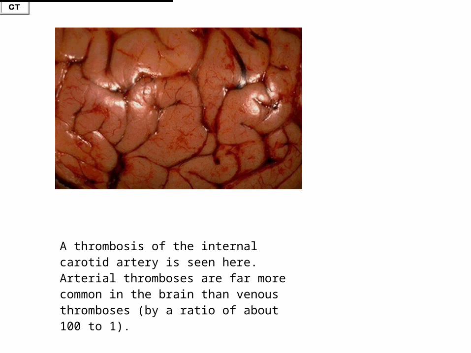
A thrombosis of the internal carotid artery is seen here. Arterial thromboses are far more common in the brain than venous thromboses (by a ratio of about 100 to 1).
A thrombosis of the internal carotid artery is seen here. Arterial thromboses are far more common in the brain than venous thromboses (by a ratio of about 100 to 1).


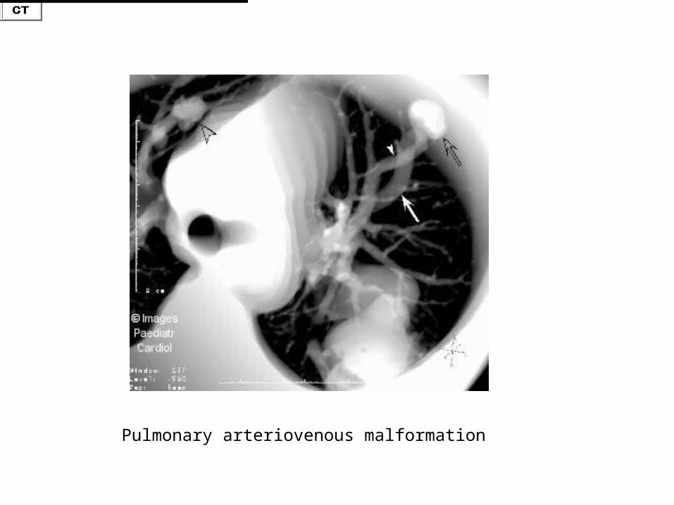
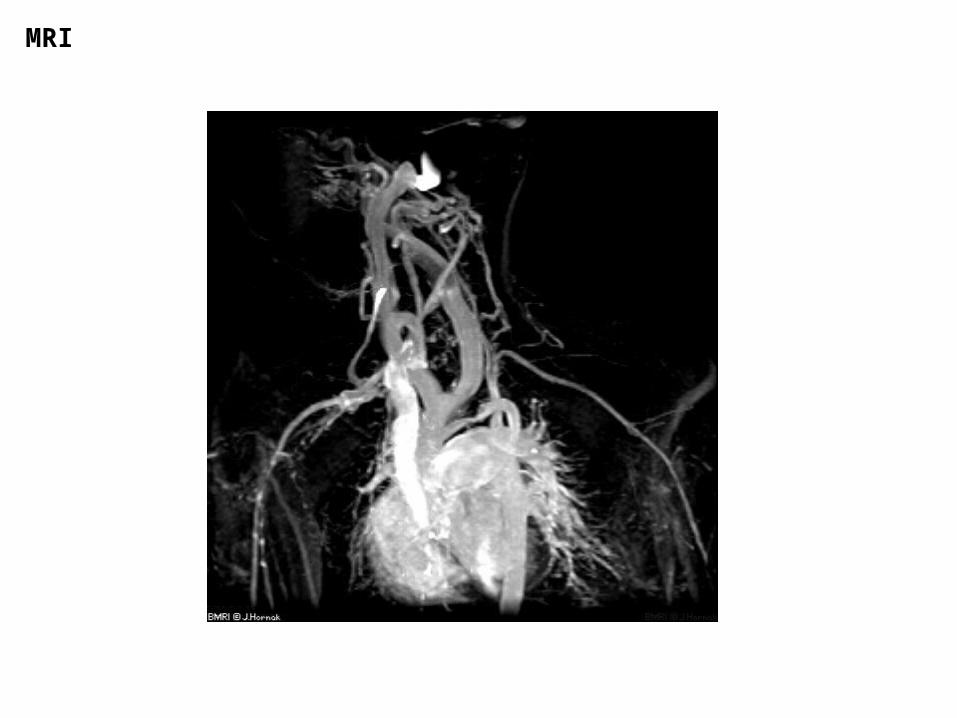
Pulmonary arteriovenous malformation


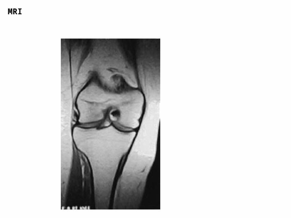
MRI

CT

CT
CT
MRI

CT
CT

CT
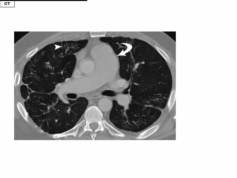
Diagnosis: Mesothelioma Brief history: 65 year old male with right upper extremity pain. Images:
CT
MRI
MRI
MRI
MRI
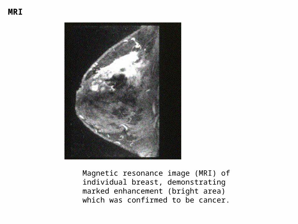
Magnetic resonance image (MRI) of individual breast, demonstrating marked enhancement (bright area) which was confirmed to be cancer.
Top Related