


Languages
Pages
Legal

M.C. Hartmann, R.M. Dwyer and M.J. Kerin
Division of Surgery, School of Medicine
National University of Ireland Galway
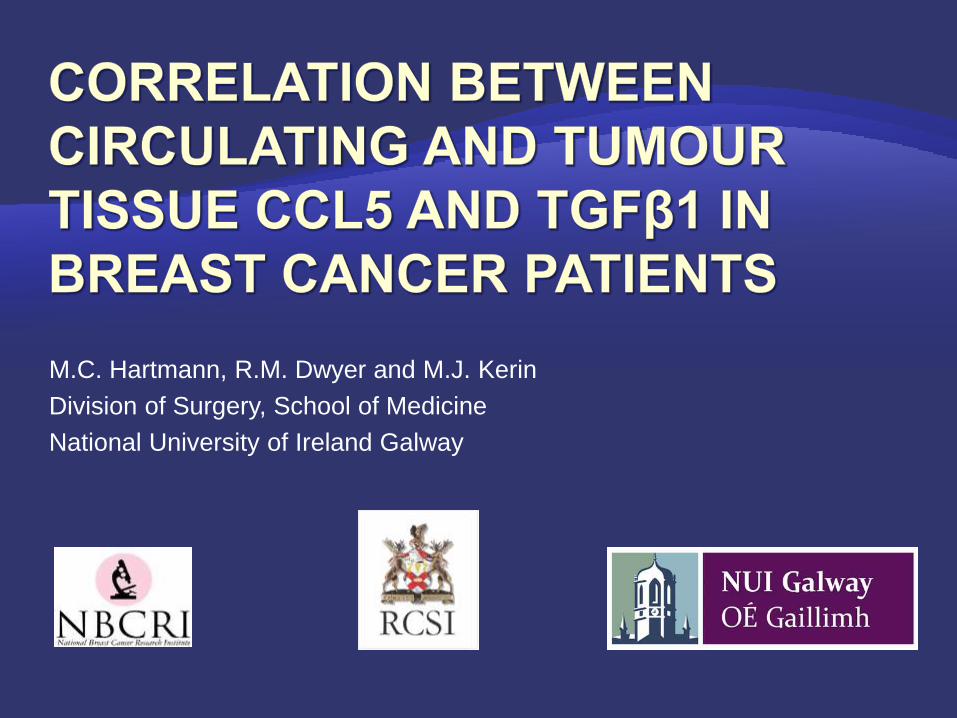
Malignancies develop at sites of chronic
inflammation - tumours considered
“wounds that do not heal”
Chemokines are chemotactic cytokines
that play a central role in the inflammatory
response through regulation of leukocyte
motility
Barrett J Rollins, European Journal of Cancer 2006

Stromal-epithelial interaction
Stromal cell
Chemokines
Chemokines facilitate stromal-epithelial interactions within primary breast tumours
Initial data highlighted potential relationship
between two factors within this microenvironment
CCL5 (RANTES) /CCR5 interactions have a
principle role in inflammation and have
been implicated in tumourigenesis
Transforming Growth Factor β 1 (TGFβ1) and its
principle receptor TGFβRII play a key role in
maintaining tissue homeostasis
cell differentiation
proliferation
chemotaxis
Tumour cell

Elevated systemic levels of both factors reported in breast cancer
patients compared to healthy controls
TGFβ1 is thought to act as a tumour suppressor in early stage breast
cancer and to promote tumourigenesis as the disease progresses
Stromal cell secretion of CCL5 within the tumour microenvironment
stimulates increased formation of lung metastasis, with no role for CCL5
reported when epithelial cells alone were used for tumour establishment
In the tumour microenvironment, loss of stromal cell expression of
TGFβ1 has been shown to support epithelial cell metastasis.
Investigation of CCL5 and TGFβ1 in breast cancer
at circulating, tumour tissue and cellular level
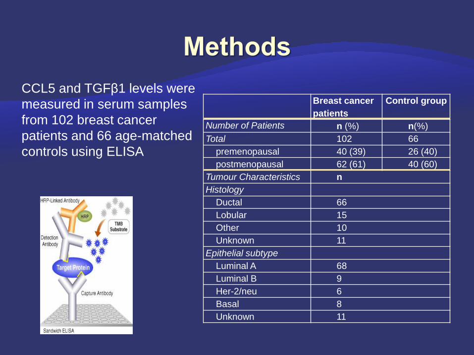
CCL5 and TGFβ1 levels were
measured in serum samples
from 102 breast cancer
patients and 66 age-matched
controls using ELISA
Breast cancer
patients
Control group
Number of Patients n (%) n(%)
Total 102 66
premenopausal 40 (39) 26 (40)
postmenopausal 62 (61) 40 (60)
Tumour Characteristics n
Histology
Ductal 66
Lobular 15
Other 10
Unknown 11
Epithelial subtype
Luminal A 68
Luminal B 9
Her-2/neu 6
Basal 8
Unknown 11
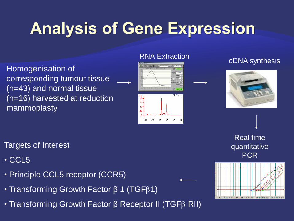
Targets of Interest
• CCL5
• Principle CCL5 receptor (CCR5)
• Transforming Growth Factor β 1 (TGF1)
• Transforming Growth Factor β Receptor II (TGF RII)
Homogenisation of
corresponding tumour tissue
(n=43) and normal tissue
(n=16) harvested at reduction
mammoplasty
RNA ExtractioncDNA synthesis
Real time
quantitative
PCR
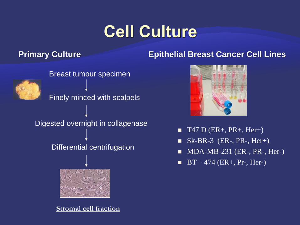
Primary Culture Epithelial Breast Cancer Cell Lines
Breast tumour specimen
Finely minced with scalpels
Digested overnight in collagenase
Differential centrifugation
Stromal cell fraction
T47 D (ER+, PR+, Her+)
Sk-BR-3 (ER-, PR-, Her+)
MDA-MB-231 (ER-, PR-, Her-)
BT – 474 (ER+, Pr-, Her-)
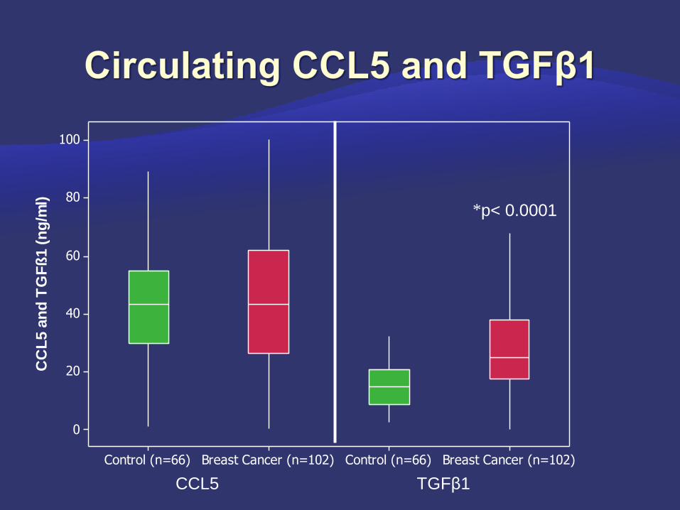
CCL5 TGFβ1
*
Breast Cancer (n=102)Control (n=66)Breast Cancer (n=102)Control (n=66)
100
80
60
40
20
0
CC
L5 a
nd
TG
Fß
1 (
ng
/ml)
p< 0.0001
(n=24)(n=22)(n=52)
120
100
80
60
40
20
0
CC
L5
(ng/m
l)
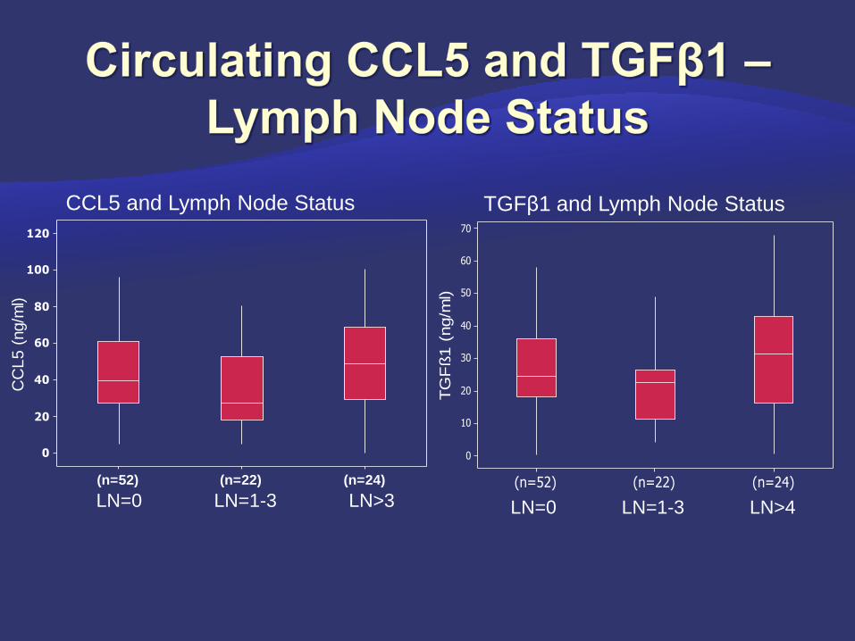
LN=0 LN=1-3 LN>3 LN=0 LN=1-3 LN>4
CCL5 and Lymph Node Status TGFβ1 and Lymph Node Status
(n=24)(n=22)(n=52)
70
60
50
40
30
20
10
0T
GF
ß1
(ng/m
l)

0
20
40
60
80
100
120
0 10 20 30 40 50 60 70
CC
L5 (
ng
/ml)
TGFβ1 (ng/ml)
r=0.43
p<0.0001
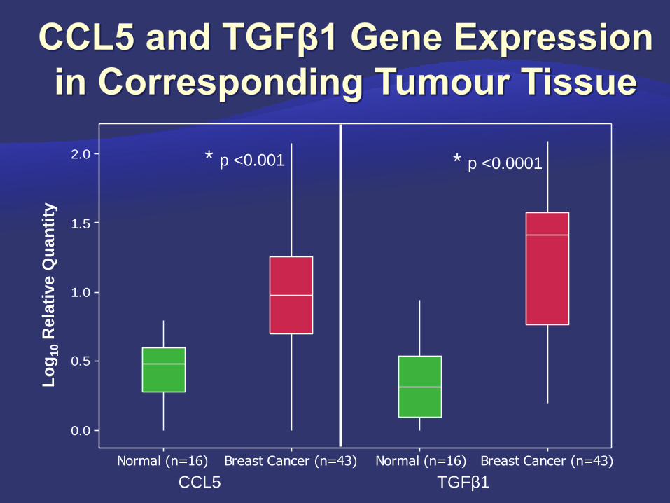
Breast Cancer (n=43) Normal (n=16)Breast Cancer (n=43)Normal (n=16)
2.0
1.5
1.0
0.5
0.0
* p <0.001 * p <0.0001
CCL5 TGFβ1
Lo
g1
0R
ela
tive Q
ua
nti
ty
0
0.5
1
1.5
2
0 0.5 1 1.5 2
Lo
g1
0R
ela
tive Q
uan
tity
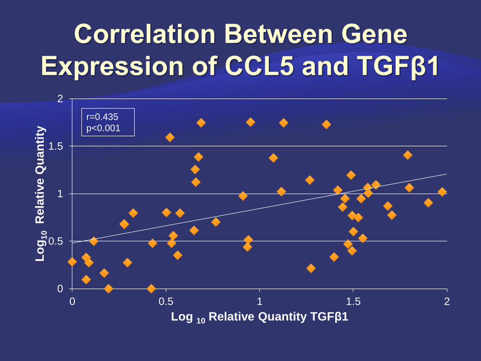
Log 10 Relative Quantity TGFβ1
r=0.435
p<0.001
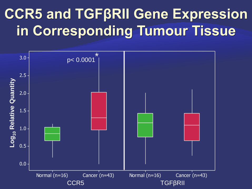
Cancer (n=43)Normal (n=16)Cancer (n=43)Normal (n=16)
3.0
2.5
2.0
1.5
1.0
0.5
0.0
Lo
g1
0R
ela
tive Q
uan
tity
*p< 0.0001
CCR5 TGFβRII

Isolated Primary Stromal Cells (n=22) Epithelial Cell Lines
TGFßRIITGFß1CCR5CCL5
3
2
1
0
-1
0
Lo
g10
Re
lati
ve
Qu
an
tity
TGFßRIITGFß1CCR5CCL5
4
3
2
1
0
-1
0
Analysis of isolated tumour stromal cell populations
(n=22) expressed relative to normal stromal cells
harvested at reduction mammoplasty (n=4)
Analysis of breast cancer epithelial cell lines
Results expressed relative to the non-
tumourigenic cell line MCF10-2A
Lo
g10
Re
lati
ve
Qu
an
tity

CCL5 and TGFβ1 levels dropped in the switch from node negative to node
positive disease and increased again as lymph node burden increased
Significant positive correlation between CCL5 and TGFβ1 at both circulating and
tissue gene expression level
CCL5, TGFβ1 and CCR5 gene expression significantly higher in tumour
compared to normal tissue
Increased expression of CCL5 in tumour compared to normal stromal cells. CCR5
was not detected in stromal cells while epithelial cell lines expressed the receptor
This study demonstrates a novel correlation between
CCL5 and TGFβ1 in breast cancer which is maintained
in early and advanced disease
The mechanisms and controlling influences warrant
further investigation and may open avenues for
therapeutic manipulation in selected patients
Top Related