


Languages
Pages
Legal


Confidential: For Review O
nly
Etiology and clinical presentation of birth defects: a
population-based study
Journal: BMJ
Manuscript ID BMJ.2017.037846
Article Type: Research
BMJ Journal: BMJ
Date Submitted by the Author: 08-Feb-2017
Complete List of Authors: Feldkamp, Marcia; University of Utah School of Medicine, Pediatrics Carey, John ; University of Utah School of Medicine, Pediatrics Byrne, Janice; University of Utah School of Medicine, Obstetrics and Gynecology; University of Utah School of Medicine, Pediatrics Krikov, Sergey; University of Utah, Division Medical Genetics
Botto, Lorenzo; University of Utah, Division Medical Genetics
Keywords: Birth Defects < Fetus/Newborn Infant, Etiology, Morphology, Pathogenesis
https://mc.manuscriptcentral.com/bmj
BMJ

Confidential: For Review O
nly
Etiology and clinical presentation of birth defects: a population-based study
Marcia L Feldkamp, John C Carey, Janice LB Byrne, Sergey Krikov, Lorenzo D Botto
Division of Medical Genetics, Department of Pediatrics, 295 Chipeta Way, Suite 2S010,
University of Utah School of Medicine, Salt Lake City, Utah, USA Marcia Feldkamp
associate professor, John Carey professor, Janice Byrne professor, Sergey Krikov
bioinformaticist, Lorenzo Botto professor, Division of Maternal-Fetal Medicine,
Department of Obstetrics and Gynecology, University of Utah School of Medicine, Salt
Lake City, Utah, USA, Janice Byrne professor
Correspondence to:
Marcia L. Feldkamp, Ph.D., P.A.
Tel: (801) 587-1955
Fax: (801) 587-7252
Email: [email protected]
Competing Interest: All authors have completed the ICMJE uniform disclosure form and declare
no support from any organisation for the submitted work, no financial relationships with any
organisations that might have an interest in the submitted work in the previous three years, and
no other relationships or activities that could appear to have influenced the submitted work.
Contributorship:
Marcia Feldkamp, John Carey, Janice Byrne, Lorenzo Botto conceived and designed the project.
Sergey Krikov and Marcia Feldkamp cleaned and conducted the analysis for the project.
All authors interpreted the data, drafted the manuscript, and assisted with manuscript revisions.
Page 1 of 34
https://mc.manuscriptcentral.com/bmj
BMJ
123456789101112131415161718192021222324252627282930313233343536373839404142434445464748495051525354555657585960
Confidential: For Review O
nly
Word Count: abstract = 274; manuscript = 2697
WHAT IS ALREADY KNOWN ON THIS TOPIC
Birth defects are common, costly, and critical. Two hospital-based studies have tried to
directly assess the proportion of birth defects with or without a known etiology.
WHAT THIS STUDY ADDS
In this large, population-based birth defect case cohort, known etiology was confirmed
in only one of every five infants. This finding underscores the gap in our understanding
of etiology among the remaining four of every five infants born with birth defects.
Determining etiology is critically important in order to focus research efforts for risk
reduction or occurrence prevention (e.g., preconception folic acid supplementation and
neural tube defects).
Page 2 of 34
https://mc.manuscriptcentral.com/bmj
BMJ
123456789101112131415161718192021222324252627282930313233343536373839404142434445464748495051525354555657585960

Confidential: For Review O
nly
Abstract
Objective Understanding the causes of birth defects is a prerequisite for effective
prevention. Although epidemiologic studies report many statistical associations, few
specific causes (genetic and environmental) are known. Moreover, the contribution of
these causes in clinically well-described population-based cohorts is uncertain. We used
a multidimensional classification that incorporates etiology, morphology, and
pathogenesis to assess causation and clinical presentation of major birth defects.
Design Population-based case cohort.
Setting We evaluated cases born 2005 through 2009 to resident women, ascertained
through Utah’s population-based surveillance system. All records underwent clinical re-
review.
Participants The final study cohort included 5,504 cases among 270,878 births
(prevalence, 2.03%). We excluded mild isolated conditions (e.g., muscular ventricular
septal defects, distal hypospadias).
Main outcome measures The primary outcomes were to determine the proportion of
birth defect cases with a known etiology (chromosomal, genetic, human teratogen,
twinning), unknown etiology, by morphology (isolated, multiple, minors only), and by
pathogenesis (sequence, developmental field defect, or known pattern of birth defects).
Results Definite cause was assigned in 20.2% (n=1,114) of cases: chromosomal or
genetic conditions accounted for 94.4% (n=1,052), teratogens 4.1% (n=46, mostly poorly
controlled pregestational diabetes), and twinning for 1.4% (n=16, conjoined or
acardiac). The remaining 79.8% (n=4,390) were classified as unknown etiology; of these
Page 3 of 34
https://mc.manuscriptcentral.com/bmj
BMJ
123456789101112131415161718192021222324252627282930313233343536373839404142434445464748495051525354555657585960

Confidential: For Review O
nly
88.2% (n=3,874) were isolated birth defects. Family history (similarly affected first
degree relative) was documented in 4.8% (n=266).
Conclusions These findings underscore the gaps in our knowledge regarding the causes
of birth defects. Even for some well-known risk factors, such as smoking, assigning
causation in individual cases remains challenging. However, the recognized impact of
some modifiable causes (i.e., maternal pregestational diabetes) highlights the potential
benefit and urgency of population-based preventive interventions.
Page 4 of 34
https://mc.manuscriptcentral.com/bmj
BMJ
123456789101112131415161718192021222324252627282930313233343536373839404142434445464748495051525354555657585960

Confidential: For Review O
nly
Birth defects are inborn errors of development. Broadly defined, birth defects include
any structural or functional anomaly with significant effects on physical, intellectual, and
social well-being.1 Birth defects represent a significant and increasing clinical and public
health challenge because of their worldwide impact on population health.
Major birth defects are common, costly and critical. Collectively, they occur in 1 in 33
births,2 which translates into an estimated 7.9 million babies every year worldwide.3 In
the US alone, the cost of care during a single year (2004) was estimated at $2.6 billion.4
This estimate does not account for the considerable indirect and lifelong personal and
societal costs. Finally, many birth defects critically impact survival. In the US, birth
defects are the leading cause of infant mortality5 and were associated in 2013 with
4,778 deaths, one in every five deaths in the first year of life.
The temporal trends are even more concerning. The occurrence of birth defects, with
very few localized exceptions (e.g., neural tube defects in countries that implemented
folic acid fortification), has not decreased for many decades. Birth defects may indeed
increase worldwide, with the alarming increase of known risk factors such as maternal
diabetes and obesity. New threats such as the Zika epidemic are emerging. Unless
significant progress is made in identifying and preventing the root causes of birth
defects, these conditions will continue to have draining effects on the survival and
health of individuals, families, and countries.
Page 5 of 34
https://mc.manuscriptcentral.com/bmj
BMJ
123456789101112131415161718192021222324252627282930313233343536373839404142434445464748495051525354555657585960

Confidential: For Review O
nly
Progress in detecting and characterizing risk factors for birth defects has come mainly
from epidemiologic studies. In fact, epidemiologic studies have produced many
associations between risk factors and groups of birth defects. However, translating
these associations to actual causes has been difficult. As a first step in filling this gap, we
evaluated the clinical and etiologic profile of birth defects in a well characterized
population-based case cohort through systematic review by clinicians, using a
multidimensional assessment tool that incorporates etiology, morphology, and
pathogenesis.
METHODS
Study Population. The data source for this study was Utah’s statewide population-based
public health surveillance system (Utah Birth Defect Network: UBDN), housed at the
Utah Department of Health. There is no patient involvement or contact as part of this
surveillance system. The UBDN monitors birth defects among all pregnancy outcomes
(live births, stillbirths, pregnancy terminations) among Utah residents. To identify
potential cases, the program utilizes multiple reporting sources, prenatal and postnatal.
Some birth defects have not been eligible for inclusion: isolated muscular ventricular
septal defects, patent foramen ovale, patent ductus arteriosus, talipes equinovarus,
congenital hip dysplasia/dislocation, congenital pulmonary airway malformation, and
cryptorchidism. Cases of fetal alcohol syndrome were included only if a major birth
defect was diagnosed. Further details of the system’s case ascertainment and medical
record abstraction have been published.6,7
Page 6 of 34
https://mc.manuscriptcentral.com/bmj
BMJ
123456789101112131415161718192021222324252627282930313233343536373839404142434445464748495051525354555657585960

Confidential: For Review O
nly
Clinical Case Review (CCR). A team of clinicians with training in medical genetics (LDB,
JCC, JLBB) review case records, including inpatient and outpatient records, laboratory
reports (e.g., genomic microarray), diagnostic evaluations (e.g., ultrasounds,
echocardiograms), operative notes, and autopsy reports. Once a case is deemed eligible,
the clinician generates a list of the case’s major and minor defects and the timing of first
diagnosis (prenatal or postnatal). Each defect is coded using the World Health
Organization International Classification of Diseases (version 9) with British Pediatric
Association extensions (ICD-9 BPA). In addition, the clinician provides three additional
classifications for each case: known etiology (yes, no); isolated vs. multiple (unrelated)
birth defect vs. syndromic (i.e., known etiology: genetic or environmental); and whether
the case is familial (yes, no). A case is considered familial if a first degree relative (parent
or sib) had a concordant phenotype.
Multidimensional Etiologic Classification. To systematically capture the clinical
presentation and etiology in the study cohort we developed and implemented a
multidimensional classification with three axes: etiology (known, unknown),
morphology (isolated, multiple majors, minors only), and pathogenesis (sequence,
developmental field, or pattern). Table 1 summarizes the system and definitions. Briefly:
1. Known etiology was assigned based on specific and conservative criteria, and
could be either genetic, environmental (teratogenic), or due to twinning.
a. Genetic. Cases were classified as having a known genetic etiology if there
was documentation of abnormal chromosomal number (trisomy) or
Page 7 of 34
https://mc.manuscriptcentral.com/bmj
BMJ
123456789101112131415161718192021222324252627282930313233343536373839404142434445464748495051525354555657585960

Confidential: For Review O
nly
structure (insertion, deletion) or a single gene condition (e.g., Noonan
syndrome).
b. Environmental. This required documentation of exposure to a recognized
human teratogen8 (e.g., medication, such as valproic acid, or
pregestational diabetes with abnormal HbA1c levels during the
periconceptional period or early pregnancy). Among mothers noted to
have diabetes (pregestational or gestational), we reviewed their timing of
diagnosis prior to or during pregnancy, medication use for control of
blood sugar, and if listed, the hemoglobin A1c testing date and level.
Women listed as gestational diabetic and diagnosed in the first trimester
were reclassified as pregestational diabetic if their hemoglobin A1c was
above 5.6. To assign diabetes as a cause, the mother had to have
evidence of poorly controlled pregestational diabetes and selected birth
defects that, based on the published literature, were evidence of diabetic
embryopathy9-11: heterotaxy, holoprosencephaly, multiple vertebral
defects, bilateral renal defects, or caudal dysgenesis. Conversely,
pregestational diabetes in cases of isolated defects such as anencephaly
or a congenital heart defect, or a major with minor defect was not
considered as a known cause for those particular infants.
c. Twinning. Abnormalities in twinning included either acardiac or
conjoined twins.
Page 8 of 34
https://mc.manuscriptcentral.com/bmj
BMJ
123456789101112131415161718192021222324252627282930313233343536373839404142434445464748495051525354555657585960

Confidential: For Review O
nly
2. Morphology: a case with a single major birth defect (with or without a minor birth
defect) was considered isolated. This definition includes isolated sequences. Some
cases would be eligible even without a major birth defect, as in some cases of
chromosomal anomalies (e.g., trisomy 21 with no reported major birth defect,
normal echocardiogram, and none of the selected list of objective minor defects).
We focused on a selected list of minor defects considered to be objective and likely
to have less variation in reporting and classification (Table 1). This list included
mainly discontinuous traits such as preauricular tags or single umbilical artery,
rather than continuous traits such as hypertelorism, which require careful
measurements and chart-based decision criteria.
3. Pathogenesis: three groups were created and defined by mechanism based on
embryology, not ICD-9 BPA codes (sequence, developmental field defect, or known
pattern of birth defects, Table 1).
Implementation of Multidimensional Classification. In this study we reviewed the
complete population-based resident cohort for five consecutive birth years (1 January
2005 through 31 December 2009). We elected to assess this five year birth cohort
because some genetic tests may be ordered well after infancy, changing a case’s
classification status. Case classification may also change as knowledge progresses. For
example, cases of CHARGE association (coloboma, heart defect, choanal atresia,
growth/developmental retardation, genital and ear abnormalities) were changed from
Page 9 of 34
https://mc.manuscriptcentral.com/bmj
BMJ
123456789101112131415161718192021222324252627282930313233343536373839404142434445464748495051525354555657585960
Confidential: For Review O
nly
‘multiple congenital anomaly’ to ‘syndrome/genetic’ after mutations in the CHD7 gene
were established as a cause in 200412 – in this situation, cases that met the established
clinical criteria for CHARGE (with or without CHD7 mutation testing) were reclassified as
‘genetic’. The classification was supported by an Access® database module that
captured both the classifications and comments from the clinical reviewers.
The cohort included 6,547 confirmed cases. We excluded 834 isolated birth defect
cases: twin related (twin-twin transfusion, TRAP-twin reversal arterial perfusion) (n=2);
pelviectasis or hydronephrosis without evidence of obstruction (n=47); small (<4mm)
secundum atrial septal defects (n=200); and distal (first degree) or megameatus type
hypospadias (n=585). We also excluded spontaneous abortions occurring at <20 weeks
gestation (n=209). After exclusions, the final study cohort included 5,504 cases.
Statistical analyses were done using SAS Enterprise Guide version 6.1 software [SAS
Institute Inc., Cary, NC, 2013].
RESULTS
The population-based study cohort included 5,504 infants with major birth defects
among 270,878 total births (live births and stillbirths), for a prevalence of 2.03%.
Compared to the underlying birth cohort, the affected cohort showed a male excess
(male 57.7%, p<0.0001), after excluding cases known to be sex specific (i.e.,
hypospadias, 47,XXY/XYY/XXX, 45,X, fragile X) or those with indeterminate sex. A family
history of a similarly affected first degree relative was documented in 4.8% (266/5504).
Page 10 of 34
https://mc.manuscriptcentral.com/bmj
BMJ
123456789101112131415161718192021222324252627282930313233343536373839404142434445464748495051525354555657585960
Confidential: For Review O
nly
Unknown etiology. Overall, 79.8% of cases (n=4,390) were classified as unknown
etiology (Table 2), 3.6% were known to be familial (isolated 3.7%; multiple 2.7%). Males
were over represented in both isolated (59.5%, p<0.0001) and multiple (55.4%, p=0.02)
case groups.
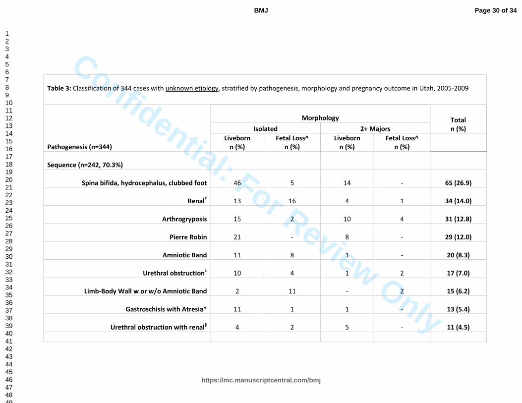
Among the unknown etiology case group, 344 (7.8% of 4391) were further classified as a
sequence (n=242, 70.3%), a developmental field defect (n=71, 20.6%), or having a
known pattern (n=31, 9.0%) (Table 3). Isolated defects represented the majority of cases
classified as a sequence (n=187, 77.3%) or developmental field defect (n=50, 70.4%),
whereas cases classified as a pattern were more likely to have multiple birth defects
(n=30, 96.8%).
Eighteen of 20 infants with birth defects consistent with VATER/VACTERL association
(vertebral, anal atresia, cardiac, tracheoesophageal fistula and/or esophageal atresia,
renal/radial and limb defects) were classified as unknown etiology.
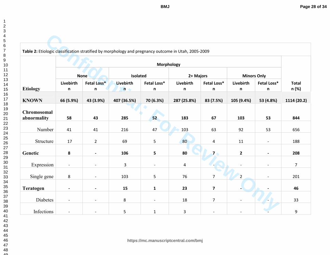
Known etiology. 20.2% (n=1,114) of cases were assigned a known etiology (Table 2). As
shown in Figure 1, 90.4% of the cases with a known etiology were represented by the
three common trisomies (21, 18, 13), Turner syndrome, structural chromosomal
abnormalities, and single gene disorders.
Page 11 of 34
https://mc.manuscriptcentral.com/bmj
BMJ
123456789101112131415161718192021222324252627282930313233343536373839404142434445464748495051525354555657585960

Confidential: For Review O
nly
The VATER/VACTERL association was observed in one case with pregestational diabetes
(teratogen) and another with partial trisomy (7q11.21 duplication) (chromosomal-
structure).
DISCUSSION
In this five-year population-based cohort with birth defects, systematic clinical review
identified a known etiology in only one of five infants—a specific etiology could not be
conclusively assigned to the majority (79.8%) of cases. We considered the etiology
known if there was conclusive evidence of one of four factors: chromosomal
abnormalities (structure or number), genetic conditions, twinning, or an established
human teratogen. Methods to determine if an environmental exposure is a human
teratogen were recently reviewed and applied to the birth defects associated with the
Zika virus.13
Based on the current science, this study revises and updates the historical findings from
two well-known hospital-based studies of infants with birth defects.14-16 The overall
conclusion remains that a specific cause cannot yet be determined for most cases of
birth defects, underscoring the current gaps in knowledge and the challenge of primary
prevention.
In this study we focused on major birth defects (excluding some common defects), for a
prevalence of 2%. Extrapolating from this conservative estimate, a minimum of 78,000
infants are estimated to born in the U.S. with a serious birth defect each year. Of these,
Page 12 of 34
https://mc.manuscriptcentral.com/bmj
BMJ
123456789101112131415161718192021222324252627282930313233343536373839404142434445464748495051525354555657585960

Confidential: For Review O
nly
63,000 would have no identifiable etiology. These are intended as minimum estimates.
Using less restrictive criteria with inclusion of milder defects, investigators have
reported higher prevalence estimates (e.g., 3.6%15 and 5.5%17).
Our estimate of a known etiology in 20.2% is conservative. As genetic technology
advances and discoveries made on the genetic causes of birth defects, the proportion
with a known cause will increase. For example, estimates of the genetic contribution to
congenital heart disease (most common birth defect) has increased, based on recent
data suggesting that copy-number variants and de novo mutations together may
account for 15% of all cases.18-20 Also, for some well-known risk factors, attributing an
exposure to a birth defect in an individual case remains challenging. The epidemiologic
metric of attributable fraction (i.e., the proportion of birth defects attributable to the
exposure when cause is known) is applicable to populations, not individual cases. It was
not possible to determine if a woman’s history of smoking directly resulted in her
infant’s oral facial cleft, since the modest odds ratio of approximately 1.3 predicts that
many children do not have an oral cleft because of the exposure. For pregestational
diabetes, however, we used data on the estimates and attributable fraction 70% for
isolated and 90% for multiple defects,10 to select certain birth defects as diabetes
related.
Determining etiology is critically important to focus research efforts for risk reduction or
occurrence prevention (e.g., preconception folic acid supplementation and neural tube
Page 13 of 34
https://mc.manuscriptcentral.com/bmj
BMJ
123456789101112131415161718192021222324252627282930313233343536373839404142434445464748495051525354555657585960
Confidential: For Review O
nly
defects). Few studies have tried to directly assess the proportion of birth defects with
or without a known etiology. Higurashi et al21 re-examined infants each month for the
first year to identify those with malformation syndromes not diagnosed at birth but did
not mention the proportion without a known etiology. Two hospital-based cohorts
employed different methodologies (e.g., inclusion criteria and diagnosis within days
after birth) to generate estimates of those infants without a known etiology.14,16 Nelson
and Holmes16 estimated 43.2% of their infants with birth defects born in a single hospital
did not have a known etiology. Cases were included if diagnosed on or before the fifth
day of life. Notably, cases considered to be familial (14.5%) or ‘multifactorial’ (23%)
were considered to be of known etiology; however, definitions were not provided and
the inclusion especially of the ‘multifactorial’ conditions is debatable. In contrast, in our
study, we defined familial cases only those with an affected first degree relative (3%).
Moreover, because of the difficulty in defining and proving multifactorial inheritance,
we did not have such a category. Of note, if we add the cases classified by Nelson and
Holmes16 as multifactorial inheritance (23%) and familial (14.5%) as unknown to those
that they classified originally as unknown (43.2%), the total adds to 80.7%, similar to our
finding.
The cause of birth defects currently without a known etiology is likely complex, and may
include interactions between the genetic profiles of parents and embryo and the
environmental milieu during preconception and early gestation. For some birth defects,
some progress has been made over the last decades, such as the contribution of
Page 14 of 34
https://mc.manuscriptcentral.com/bmj
BMJ
123456789101112131415161718192021222324252627282930313233343536373839404142434445464748495051525354555657585960

Confidential: For Review O
nly
microdeletions (e.g., deletion 22q11 in cases of heart defects and cleft palate22-25) and
novel single gene mutations (e.g., CHD7 mutations in CHARGE syndrome12). However,
while these genetic causes are relatively straightforward, it is likely that further research
will discover more complex networks accounting for genetic and environmental
contributions to birth defects etiology.
Conducting research to understand birth defect etiology requires a well-defined and
clinically characterized case group. Cases with known etiology must be carefully
identified and excluded in order to maximize the chance of discovery.26 While
commonly used birth defect classification schemes (e.g., ICD-9 or ICD-10) are valuable in
specific contexts, they are not ideal when evaluating birth defect etiologies or trends.
These coding systems are typically organized by anatomy or function rather than cause
or embryologic process. Few studies have applied birth defect-specific classifications to
population-based cohorts. One of these, the National Birth Defects Prevention Study,
has leveraged the collaboration of clinical geneticists and epidemiologists to pursue
discovery of modifiable causes of birth defects.27-29 Continued progress will require the
combined effort and a multidisciplinary approach that incorporates not only the clinical
evaluation by dysmorphologists/clinical geneticists and the methodological expertise of
epidemiologists, but includes experts in developmental biology, pharmacology,
infectious diseases, immunology, bioinformatics, in addition to a more objective
assessment of periconceptional exposures that improve upon the typical maternal self-
reports. Finally, it would be helpful to integrate etiology, morphology, and pathogenesis
Page 15 of 34
https://mc.manuscriptcentral.com/bmj
BMJ
123456789101112131415161718192021222324252627282930313233343536373839404142434445464748495051525354555657585960

Confidential: For Review O
nly
assessment into the basic framework of epidemiologic studies. Such integration will
improve precision and assist researchers to focus research initiatives and investigate
common pathways among birth defects.
This study has potential limitations. The birth prevalence of 2% reported in this study is
lower than the 3-5% reported elsewhere.2,15,17 Such lower prevalence estimate relates
to the eligibility criteria of the Utah surveillance system that excludes some common
mostly milder conditions that are variably defined and ascertained (e.g., muscular
ventricular septal defects, clubfoot, cryptorchidism). Also, because cases were classified
based on data abstracted from mother and infant medical records, there is a possibility
that critical information for appropriate classification was unavailable at the time of
medical record abstraction. In addition, we may have underestimated the proportion
due to a teratogen if an exposure (e.g, maternal pregestational diabetes) was not noted
in the medical record or not queried by the physician of record. The proportion of
teratogens may also be underestimated since we chose to have a definitive list of known
teratogens at the outset of the study.
Understanding the etiology of birth defects should be both a public health and research
priority. The findings from this study underscore the current gaps in our knowledge
regarding the causes of birth defects. Population-based surveillance integrated with
specific clinical expertise on birth defects and meaningful case classification can provide
a strong basis for effective, translational birth defect research.30 Knowing the etiology of
Page 16 of 34
https://mc.manuscriptcentral.com/bmj
BMJ
123456789101112131415161718192021222324252627282930313233343536373839404142434445464748495051525354555657585960

Confidential: For Review O
nly
birth defects can then be translated into interventions aimed at preventing birth
defects, leading to signifincantly improved child survival and long term personal and
societal outcomes.
Acknowledgments
This publication was supported by a Cooperative Agreement (Number U01DD000490)
from the Centers for Disease Control and Prevention. Its contents are solely the
responsibility of the authors and do not necessarily represent the official views of the
Centers for Disease Control and Prevention.
Data were provided by the Utah Birth Defect Network, a program within the Utah
Department of Health. This project is supported by the Health Resources and Services
Administration (HRSA) of the U.S. Department of Health and Human Services (HHS)
under grant number B04MC25374 with a title of Maternal and Child Health Services for
the amount of $3,046,261. This information or content and conclusions are those of the
author and should not be construed as the official position or policy of, nor should any
endorsements be inferred by HRSA, the U.S. Government or the Utah Department of
Health.
Page 17 of 34
https://mc.manuscriptcentral.com/bmj
BMJ
123456789101112131415161718192021222324252627282930313233343536373839404142434445464748495051525354555657585960
Confidential: For Review O
nly
References
1. World Health Organization (WHO). Sixty-third World Health Assembly. Birth Defects.
Report by the Secretariat. A63/10. April 2010.
2. Centers for Disease Control and Prevention (CDC). Update on overall prevalence of
major birth defects – Atlanta, Georgia 1978-2005. MMWR Morb Mortal Wkly Rep.
2008;57:1-5.
3. Christianson A, Howson CP, Modell B. March of Dimes Global Report on Birth Defects.
The Hidden Toll of Dying and Disabled Children. March of Dimes Birth Defects
Foundation. White Plains, New York; 2006.
4. Russo CA, Elixhauser A. Hospitalizations for Birth Defects, 2004. HCUP Statistical Brief
#24. Rockville, MD, U.S. Agency for Healthcare Research and Quality; 2007.
5. Mathews TJ, MacDorman MF, Thoma ME. Infant mortality statistics from the 2013
period linked birth/infant death data set. National vital statistics reports; Hyattsville,
MD: National Center for Health Statistics; 2015;64(9).
6. Feldkamp M, MacLeod L, Young L, Lecheminant K, Carey JC. The methodology of the
Utah Birth Defect Network: Congenital heart defects as an illustration. Birth Defects Res
A Clin Mol Teratol. 2005;73(10):693-9.
Page 18 of 34
https://mc.manuscriptcentral.com/bmj
BMJ
123456789101112131415161718192021222324252627282930313233343536373839404142434445464748495051525354555657585960

Confidential: For Review O
nly
7. Stevenson DA, Carey JC, Byrne JLB, Srisukhumbowornchai S, Feldkamp ML. Analysis of
skeletal dysplasias in the Utah population. Am J Med Genet Part A. 2012;158A(5):1046-
54.
8. Stevenson RE. Human Malformations and related anomalies. In. Human
Malformations and Related Anomalies 3rd edition. Eds: RE Stevenson, JG Hall, DB
Everman, BD Solomon. Oxford University Press; New York; 2016. p. 1-35
9. Mills JL. Malformations in infants of diabetic mothers. Birth Defects Res A Clin Mol
Teratol. 2010;88(10):769-78.
10. Correa A, Gilboa SM, Besser LM, Botto LD, Moore CA, Hobbs CA, et al. Diabetes
mellitus and birth defects. Am J Obstet Gynecol. 2008;199(3):237.e1-9.
11. Gilbert-Barnes E. Teratogenic causes of malformations. Ann Clin Lab Sci.
2010;40(2):99-114.
12. Vissers LE, van Ravenswaaij CM, Admiraal R, Hurst JA, de Vries BB, Janssen IM, et al.
Mutations in a new member of the chromodomain gene family cause CHARGE
syndrome. Nat Genet. 2004;36(9):955–57.
Page 19 of 34
https://mc.manuscriptcentral.com/bmj
BMJ
123456789101112131415161718192021222324252627282930313233343536373839404142434445464748495051525354555657585960

Confidential: For Review O
nly
13. Rasmussen SA, Jamieson DJ, Honein MA, Petersen LR. Zika virus and birth defects –
reviewing the evidence for causality. NEJM. 2016;374(20):1981-87.
14. Martinez-Frías ML, Bermejo E, Frías JL. Pathogenetic classification of a series of
27,145 consecutive infants with congenital defects. Am J Med Genet. 2000;90(3):246-9.
15. Holmes LB, Cann C, Cook C. Examination of infants for both minor and major
malformations to evaluate for possible teratogenic exposures. In: Prevention of Physical
and Mental Congenital Defects, Part B: Epidemiology, Early Detection and Therapy, and
Environmental Factors. Ed M Marois. New York:Liss; 1985; p. 59-63.
16. Nelson K, Holmes LB. Malformations due to presumed spontaneous mutations in
newborn infants. N Engl J Med. 1989;320(1):19-23.
17. Texas Department of State Health Services (TDSHS). Texas Birth Defects Registry’s
Report of Birth Defects among 1999-2011 Deliveries: Summary and Key Findings. 2015.
http://www.dshs.state.tx.us/birthdefects/data/BD_Data_99-11/Report-of-Birth-
Defects-Among-1999-2011-Deliveries.aspx.
18. Yuan S, Zaidi S, Brueckner M. Congenital heart disease: emerging themes linking
genetics and development. Curr Opin Genet Dev. 2013;23(3):352-9.
Page 20 of 34
https://mc.manuscriptcentral.com/bmj
BMJ
123456789101112131415161718192021222324252627282930313233343536373839404142434445464748495051525354555657585960

Confidential: For Review O
nly
19. Al Turki S, Manickarah AK, Mercer CL, Gerety SS, Hitz MP, Lindsay S, et al. Rare
variants in NR2F2 cause congenital heart defects in humans. Am J Med Genet.
2014;94(4):574-85.
20. Warburton D, Ronemus M, Kline J, Jobanputra V, Williams I, Anyane-Yeboa K, et al.
The contribution of de novo and rare inherited copy number changes to congenital
heart disease in an unselected sample of children with conotruncal defects or
hypoplastic left heart disease. Hum Genet. 2014;133(1):11-27.
21. Higurashi M, Iijima K, Sugimoto Y, Ishikawa N, Hoshina H, Watanabe N, Yoneyama K.
The birth prevalence of malformation syndromes in Tokyo infants: a survey of 14,430
newborn infants. Am J Med Genet. 1980;6(3):189-94.
22. de la Chapelle A, Herva R, Kolvisto M, Aula P. A deletion in chromosome 22 can
cause DiGeorge syndrome. Hum Genet. 1981;57(3):253-6.
23. Kelly RI, Zackai EH, Emanuel BS, Kistenmacher M, Greenberg F, Punnett HH. The
association of the DiGeorge anomalad with partial monosomy of chromosome 22. J
Pediatr. 1982;101(2):197-200.
Page 21 of 34
https://mc.manuscriptcentral.com/bmj
BMJ
123456789101112131415161718192021222324252627282930313233343536373839404142434445464748495051525354555657585960

Confidential: For Review O
nly
24. Amati F, Mari A, Digilio MC, Mingarelli R, Marino B, Giannotti A, et al. 22q11
deletions in isolated and syndromic patients with tetralogy of Fallot. Hum Genet.
1995;95(5):479-82.
25. Takahashi K, Kido S, Hoshino K, Ogawa K, Ohashi H, Fukushima Y. Frequency of a
22q11 deletion in patients with conotruncal cardiac malformations: a prospective study.
Eur J Pediatr. 1995;154(11):878-81.
26. Rasmussen SA, Olney RS, Holmes LB, Lin AE, Keppler-Noreuil KM, Moore CA, and the
National Birth Defects Prevention Study. Guidelines for case classification for the
National Birth Defects Prevention Study. Birth Defects Res A Clin Mol Teratol.
2003;67(3):193-201.
27. Browne ML, Van Zutphen AR, Botto LD, Louik C, Richardson S, Druschel CM.
Maternal butalbital use and selected defects in the national birth defects prevention
study. Headache. 2014;54(1):54-66.
28. Reefhuis J, Devine O, Friedman JM, Louik C, Honein MA, National Birth Defects
Prevention S. Specific SSRIs and birth defects: bayesian analysis to interpret new data in
the context of previous reports. BMJ. 2015;Jul 8;351:h3190.
Page 22 of 34
https://mc.manuscriptcentral.com/bmj
BMJ
123456789101112131415161718192021222324252627282930313233343536373839404142434445464748495051525354555657585960

Confidential: For Review O
nly
29. Botto LD, Krikov S, Carmichael SL, Munger RG, Shaw GM, Feldkamp ML; National
Birth Defects Prevention Study. Lower rate of selected congenital heart defects with
better maternal diet quality: a population-based study. Arch Dis Child Fetal Neonatal Ed.
2016;101(1):43-9.
30. Moore CA, McCabe ERB. Editorial utility of population-based birth defects
surveillance for monitoring the health of infants and as a foundation for etiologic
research. Birth Defects Res A Clin Mol Teratol. 2015;103(11):895-8.
Page 23 of 34
https://mc.manuscriptcentral.com/bmj
BMJ
123456789101112131415161718192021222324252627282930313233343536373839404142434445464748495051525354555657585960

Confidential: For Review Only
Table 1: Classification groups and definitions for etiologic classification of all cases in Utah, 2005-2009.
Etiology
Known
Anomaly of chromosome number (trisomy 21) or structure (del 22q); single gene condition (e.g., Noonan syndrome); anomaly
of gene expression (methylation-related Beckwith-Wiedemann syndrome); established human teratogen (e.g., pregestational
diabetes, valproic acid); specific twinning abnormality (i.e., acardiac or conjoined twin).
Unknown
No identifiable cause could definitively be established and documented.
Morphology
Isolated
Single major malformation, with or without a non-objective minor defect
Note: a sequence [see below for definition] if isolated is considered an isolated defect, as in the case spina bifida with clubfoot
and hydrocephalus.
Multiple
Two or more unrelated major malformations.
Minor1
Select, distinctive and objective structural defect that is not clinically or surgically significant [see list].
None
No major or distinctive minor defects were detected. A case may still be eligible in the presence of an eligible chromosomal
anomaly (e.g., a child with trisomy 21, without a major defect or one of the selected minor defects in the list of objective
minors).
Page 24 of 34
https://mc.manuscriptcentral.com/bmj
BMJ
123456789101112131415161718192021222324252627282930313233343536373839404142434445464748495051525354555657585960

Confidential: For Review Only
Pathogenesis
Sequence2
Pattern of related malformations that occur as a result of a single primary malformation. Examples include spina bifida with
hydrocephalus and clubfoot (spina bifida sequence, with spina bifida as the primary malformation). A sequence can occur as
an isolated defect (spina bifida sequence) or as multiple defect (e.g., spina bifida sequence and cleft lip).
Developmental
Field Defect3
Pattern of malformations resulting from the abnormal development of an embryonic unit (developmental field) that develops
as a single unit in early embryogenesis (e.g., during blastogenesis). Etiology of developmental field defects is typically
heterogeneous. An example is the DiGeorge anomaly, related to abnormal development and fate of populations of neural
crest cells, leading to multiple structural anomalies and potentially caused by different genetic abnormalities (e.g., deletion
22q11) or environmental factors (e.g., retinoic acid).
Pattern4
Nonrandom occurrence or pattern of multiple malformations without a known cause. Examples include the VATER/VACTERL
recurrent and variable pattern of anomalies.
1 Minor Malformations
Absent nails
Auricular tag/pit
Bifid uvula
Branchial tag/pit
Camptodactyly
Cervical ribs
Cup ear
Cutis aplasia
Cystic hygroma
Page 25 of 34
https://mc.manuscriptcentral.com/bmj
BMJ
123456789101112131415161718192021222324252627282930313233343536373839404142434445464748495051525354555657585960
Confidential: For Review Only
Ear lobe crease
Ear lobe notch
Extra nipples
Iris coloboma
Lop ear (microtia type 1)
Natal tooth
Neck webbing
Overlapping finger
Polydactyly: type B- tag involves the hand or foot
- polydactyly a major defect when involvement includes a full extra digit of hand or foot
Preauricular tag/pit
Rocker bottom feet
Single crease 5th
finger
Single transverse crease
Single umbilical artery
Syndactyly: toes
Syndactyly: hands – mild – 1st
flexion crease involvement
- syndactyly a major defect when involvement is up to/include the 2nd
flexion crease (moderate) and marked when it includes distal phalanx
2 Sequence
Amniotic band
Amniotic band with limb-body wall
Arthrogryposis
Frontonasal malformation
Gastroschisis with congenital intestinal atresia
Limb-body wall complex
Pierre Robin
Poland Urethral obstruction (i.e., posterior urethral valves, urethral atresia/stenosis)
Urethral obstruction and renal agenesis (MCDK)
Renal (agenesis, multicystic dysplastic kidney, infantile autosomal recessive polycystic kidney disease)
Spina bifida (hydrocephalus, club foot)
Sturge-Weber
3 Development Field Defect
Page 26 of 34
https://mc.manuscriptcentral.com/bmj
BMJ
123456789101112131415161718192021222324252627282930313233343536373839404142434445464748495051525354555657585960

Confidential: For Review Only
Cantrell Pentalogy
Cloacal exstrophy
DiGeorge anomaly
Holoprosencephaly
Laterality
Septo-optic dysplasia
Sirenomelia
Urorectal septum malformation
4 Pattern
Caudal dysgenesis
VACTERL: vertebral anomalies, anal atresia, cardiovascular,
tracheoesophageal fistula, esophageal atresia, renal and/or radial, limb
MURCS: Müllerian duct aplasia, renal aplasia, and
cervicothoracic somite dysplasia
Goldenhar/OAV (oculo-auricular-vertebral)
Page 27 of 34
https://mc.manuscriptcentral.com/bmj
BMJ
123456789101112131415161718192021222324252627282930313233343536373839404142434445464748495051525354555657585960
Confidential: For Review Only
Table 2: Etiologic classification stratified by morphology and pregnancy outcome in Utah, 2005-2009
Etiology
Morphology
Total
n (%)
None Isolated 2+ Majors Minors Only
Livebirth
n
Fetal Loss*
n
Livebirth
n
Fetal Loss*
n
Livebirth
n
Fetal Loss*
n
Livebirth
n
Fetal Loss*
n
KNOWN 66 (5.9%) 43 (3.9%) 407 (36.5%) 70 (6.3%) 287 (25.8%) 83 (7.5%) 105 (9.4%) 53 (4.8%) 1114 (20.2)
Chromosomal
abnormality 58 43 285 52 183 67 103 53 844
Number 41 41 216 47 103 63 92 53 656
Structure
17
2
69
5
80
4
11
-
188
Genetic 8 - 106 5 80 7 2 - 208
Expression - - 3 - 4 - - - 7
Single gene 8 - 103 5 76 7 2 - 201
Teratogen - - 15 1 23 7 - -
46
Diabetes - - 8 - 18 7 - - 33
Infections
-
-
5
1
3
-
-
-
9
Page 28 of 34
https://mc.manuscriptcentral.com/bmj
BMJ
123456789101112131415161718192021222324252627282930313233343536373839404142434445464748495051525354555657585960

Confidential: For Review Only
Medications
-
-
2
-
2
-
-
-
4
Twinning - - 1 12 1 2 - - 16
Acardia - - - 8 - - - - 8
Conjoined - - 1 4 1 2 - - 8
UNKNOWN - - 3740 (85.2%) 134 (3.1%) 462 (10.5%) 54 (1.2%) - - 4391 (79.8%)
TOTAL 66 (1.2%) 43 (0.8%) 4147 (75.3%) 204 (3.7%) 749 (13.6%) 137 (2.9%) 105 (1.9%) 53 (1.0%) 5504
* Fetal loss includes both stillbirths (20 weeks gestation and greater) and pregnancy terminations (any gestation)
MINORS ONLY (n=158):
Chromosomal (n=156)
Trisomy 21 97
Turner 29
Trisomy 18 10
45,X Mosaic 3
Klinefelter 3
Trisomy 21 Mosaic 2
Microdeletion 5
Partial trisomy 3
Del/dup 1
Del 5p 1
Del 22q 1
DiGeorge 1
Genetic (n=2)
OI type 1 1
Fragile X 1
Page 29 of 34
https://mc.manuscriptcentral.com/bmj
BMJ
123456789101112131415161718192021222324252627282930313233343536373839404142434445464748495051525354555657585960
Confidential: For Review Only
Table 3: Classification of 344 cases with unknown etiology, stratified by pathogenesis, morphology and pregnancy outcome in Utah, 2005-2009
Pathogenesis (n=344)
Morphology Total
n (%) Isolated 2+ Majors
Liveborn
n (%)
Fetal Loss^
n (%)
Liveborn
n (%)
Fetal Loss^
n (%)
Sequence (n=242, 70.3%)
Spina bifida, hydrocephalus, clubbed foot 46 5 14 - 65 (26.9)
Renal† 13 16 4 1 34 (14.0)
Arthrogryposis
15
2
10
4
31 (12.8)
Pierre Robin 21 - 8 - 29 (12.0)
Amniotic Band 11 8 1 - 20 (8.3)
Urethral obstruction‡ 10 4 1 2 17 (7.0)
Limb-Body Wall w or w/o Amniotic Band 2 11 - 2 15 (6.2)
Gastroschisis with Atresia* 11 1 1 - 13 (5.4)
Urethral obstruction with renal§ 4 2 5 - 11 (4.5)
Page 30 of 34
https://mc.manuscriptcentral.com/bmj
BMJ
123456789101112131415161718192021222324252627282930313233343536373839404142434445464748495051525354555657585960

Confidential: For Review Only
Poland Anomaly 2 - 1 - 3 (1.2)
Sturge-Weber 3 - - - 3 (1.2)
Frontonasal Malformation - - 1 - 1 (0.4)
Total 138 (57.0) 49 (20.2) 46 (19.0) 9 (3.7) 242
Developmental Field Defect (n=71, 20.6%)
Laterality 27 1 8 1 37 (52.1)
Holoprosencepahly 10 2 5 4 21 (29.6)
Septo-Optic Dysplasia
2
-
2
-
4 (5.6)
Urorectal Septum Defect 3 - 1 - 4 (5.6)
Cantrell Pentalogy 2 - - - 2 (2.8)
Sirenomelia 2 - - - 2 (2.8)
Cloacal Exstrophy 1 - - - 1 (1.4)
Total 47 (66.2) 3 (4.2) 16 (22.5) 5 (7.0) 71
Pattern (n=31, 9.0%)
VATER/VACTERL
-
-
16
2
18 (58.1)
Page 31 of 34
https://mc.manuscriptcentral.com/bmj
BMJ
123456789101112131415161718192021222324252627282930313233343536373839404142434445464748495051525354555657585960

Confidential: For Review Only
Goldenhar/OAV 1 - 8 1 10 (32.3)
Caudal Dysgenesis - - 3 - 3 (9.8)
Total 1 (3.2) 0 27 (87.1) 3 (9.6) 31
Goldenhar/OAV (oculo-auricular-vertebral)
VATER: vertebral anomalies, anal atresia, tracheoesophageal fistula, esophageal atresia, renal and/or radial, limb
VACTERL: vertebral anomalies, anal atresia, cardiovascular, tracheoesophageal fistula, esophageal atresia, renal and/or radial, limb
^ Fetal loss includes both stillbirths and pregnancy terminations
* Congenital intestinal atresia (does not include acquired atresia post-delivery) † Renal agenesis, multicystic dysplastic kidneys infantile autosomal recessive polycystic kidney disease
‡ Posterior urethral valves, urethral atresia/stenosis
§ Urethral obstruction with renal agenesis, multicystic dysplastic kidneys infantile autosomal recessive polycystic kidney disease
Page 32 of 34
https://mc.manuscriptcentral.com/bmj
BMJ
123456789101112131415161718192021222324252627282930313233343536373839404142434445464748495051525354555657585960
Confidential: For Review Only
Figure 1: Number and cumulative percent of cases with a known etiology, Utah 2005-2009
Legend:
Chromosomal
Genetic
Teratogen
Twinning
Indel: insertion deletion
Page 33 of 34
https://mc.manuscriptcentral.com/bmj
BMJ
123456789101112131415161718192021222324252627282930313233343536373839404142434445464748495051525354555657585960

Confidential: For Review O
nly
217x168mm (96 x 96 DPI)
Page 34 of 34
https://mc.manuscriptcentral.com/bmj
BMJ
123456789101112131415161718192021222324252627282930313233343536373839404142434445464748495051525354555657585960
Top Related