


Languages
Pages
Legal

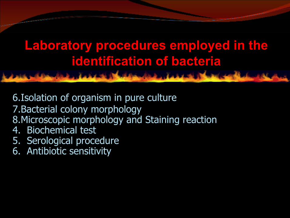
Laboratory procedures employed in the identification of bacteria
6.Isolation of organism in pure culture 7.Bacterial colony morphology8.Microscopic morphology and Staining reaction4. Biochemical test 5. Serological procedure 6. Antibiotic sensitivity

Isolation of organism in Pure Culture
• Pure culture (axenic culture)– Population of cells arising from a single cell- the approach used for the isolation of organism depends
upon the source of clinical specimen
Blood, spinal fluid and closed abscesses may yield almost pure bacterial culture
specimen of sputum, stool, materials from the skin and
body orifices usually contains mixture of organism

Bacterial colony morphology
• Bacteria grow on solid media as colonies
• colony is defined as a visible mass of microorganisms all
originating from a single mother cell.
• therefore a colony constitutes a clone of bacteria all genetically alike
• Useful in bacterial identification
• Colonies - size, shape, texture, elevation, pigmentation, effect on
growth medium

To identify the following colonial characteristics/culture characteristics:
WHOLE SHAPE OF COLONY EDGE/MARGIN OF COLONY
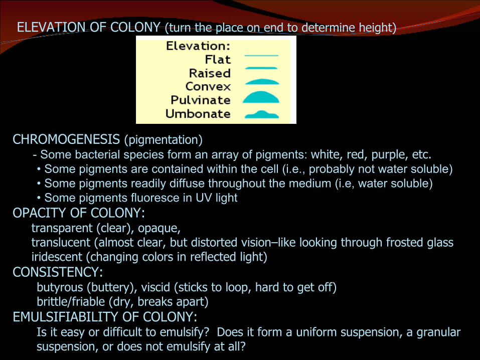
ELEVATION OF COLONY (turn the place on end to determine height)
CHROMOGENESIS (pigmentation) - Some bacterial species form an array of pigments: white, red, purple, etc.
• Some pigments are contained within the cell (i.e., probably not water soluble) • Some pigments readily diffuse throughout the medium (i.e, water soluble) • Some pigments fluoresce in UV light
OPACITY OF COLONY: transparent (clear), opaque, translucent (almost clear, but distorted vision–like looking through frosted glass iridescent (changing colors in reflected light) CONSISTENCY:
butyrous (buttery), viscid (sticks to loop, hard to get off)brittle/friable (dry, breaks apart)
EMULSIFIABILITY OF COLONY: Is it easy or difficult to emulsify? Does it form a uniform suspension, a granular suspension, or does not emulsify at all?
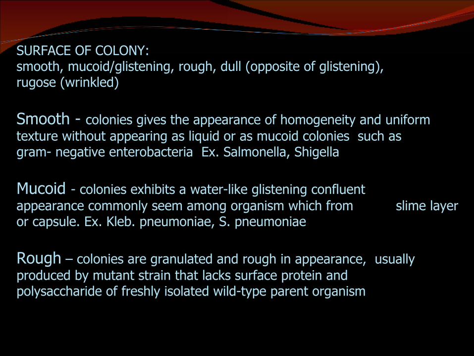
SURFACE OF COLONY: smooth, mucoid/glistening, rough, dull (opposite of glistening), rugose (wrinkled)
Smooth - colonies gives the appearance of homogeneity and uniform texture without appearing as liquid or as mucoid colonies such as gram- negative enterobacteria Ex. Salmonella, Shigella
Mucoid - colonies exhibits a water-like glistening confluent appearance commonly seem among organism which from slime layer or capsule. Ex. Kleb. pneumoniae, S. pneumoniae
Rough – colonies are granulated and rough in appearance, usually produced by mutant strain that lacks surface protein and polysaccharide of freshly isolated wild-type parent organism
Microscopic morphology
• Provide presumptive identification of an organism
Bacterial Morphology Shape Arrangement Staining reaction
Biochemical Test Various species of organism exhibits characteristic
pattern of substrate utilization, metabolic product formation and sugar fermentation Enzyme based test – based on its reaction with a substrate
Catalase, oxidase, indole, urease
Reactions in sugar fermentation broth Nitrate Broth reactions
60% of common pathogens can be identified by metabolic test
Serological procedure Antigen and antibody determination Serological Tests
Use group specific antiserum isolated from the plasma of animals that have been sensitized to the organism The antiserum contains antibody proteins that react with
antigens on the unknown organism. Procedures: agglutination, precipitation test, hemagglutination
inhibition, complement fixation, ELISA, RIA, Western blot assay Advantages:
Highly specific Does not usually require the organism to be isolated into pure
culture Can be used to identify organisms that can’t be grown on
medium
Antibiotic sensitivity antibiotic sensitivity is a term used to describe the susceptibility
of bacteria to antibiotics Antibiotic susceptibility testing (AST) is usually carried out to
determine which antibiotic will be most successful in treating a bacterial infection in vivo
Methods of testing: Broth dilution
The lower the dilution, the greater the antibiotic content Agar dilution Disk diffusion
the Kirby-Bauer test for antibiotic susceptibility, called the disc diffusion test, is a standard that has been used for years
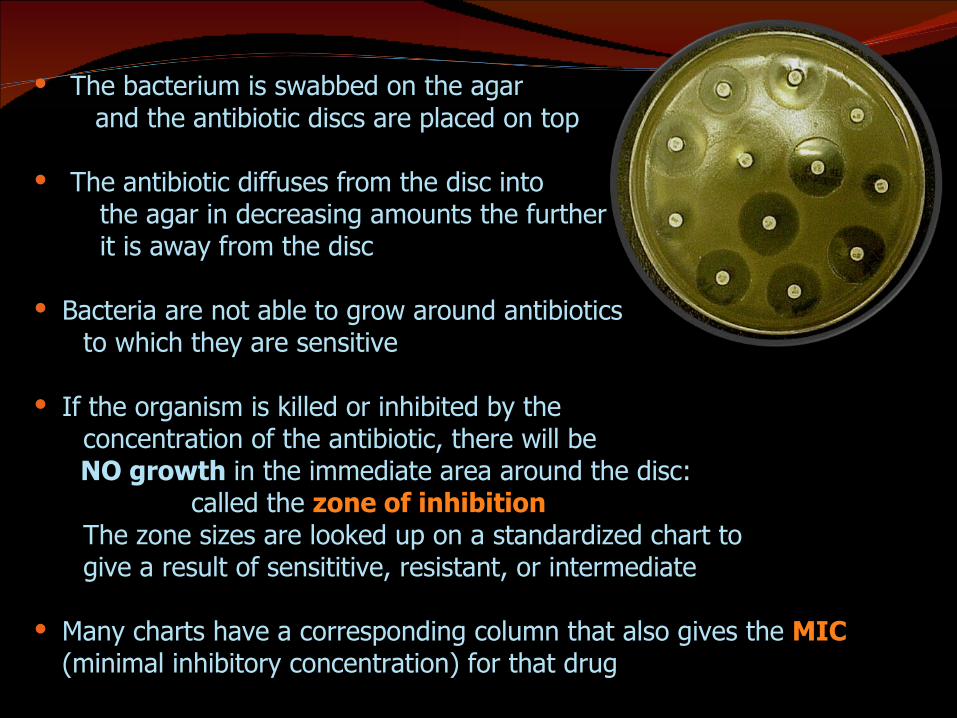
The bacterium is swabbed on the agar and the antibiotic discs are placed on top
The antibiotic diffuses from the disc into the agar in decreasing amounts the further it is away from the disc
Bacteria are not able to grow around antibiotics to which they are sensitive
If the organism is killed or inhibited by the concentration of the antibiotic, there will be NO growth in the immediate area around the disc: called the zone of inhibition The zone sizes are looked up on a standardized chart to give a result of sensititive, resistant, or intermediate
Many charts have a corresponding column that also gives the MIC (minimal inhibitory concentration) for that drug

Conventional diagnosis methods

Top Related