
Languages
Pages
Legal

Chronic Kidney Disease
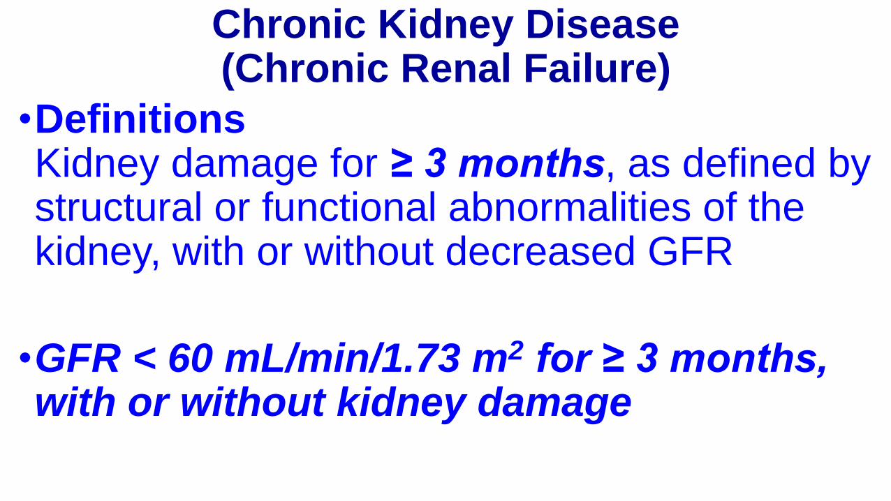
Chronic Kidney Disease(Chronic Renal Failure)
•DefinitionsKidney damage for ≥ 3 months, as defined by structural or functional abnormalities of the kidney, with or without decreased GFR
•GFR < 60 mL/min/1.73 m2 for ≥ 3 months, with or without kidney damage
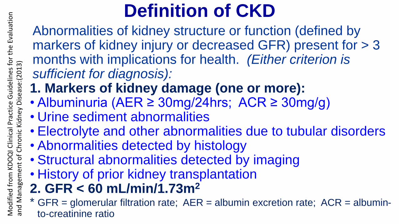
Definition of CKDAbnormalities of kidney structure or function (defined by markers of kidney injury or decreased GFR) present for > 3 months with implications for health. (Either criterion is sufficient for diagnosis): 1. Markers of kidney damage (one or more):• Albuminuria (AER ≥ 30mg/24hrs; ACR ≥ 30mg/g)• Urine sediment abnormalities• Electrolyte and other abnormalities due to tubular disorders• Abnormalities detected by histology• Structural abnormalities detected by imaging• History of prior kidney transplantation 2. GFR < 60 mL/min/1.73m2
* GFR = glomerular filtration rate; AER = albumin excretion rate; ACR = albumin-to-creatinine ratioM
od
ifie
d f
rom
KD
OQ
I Clin
ical
Pra
ctic
e G
uid
elin
es
for
the
Eval
uat
ion
an
d M
anag
emen
t o
f C
hro
nic
Kid
ney
Dis
eas
e:(2
01
3)
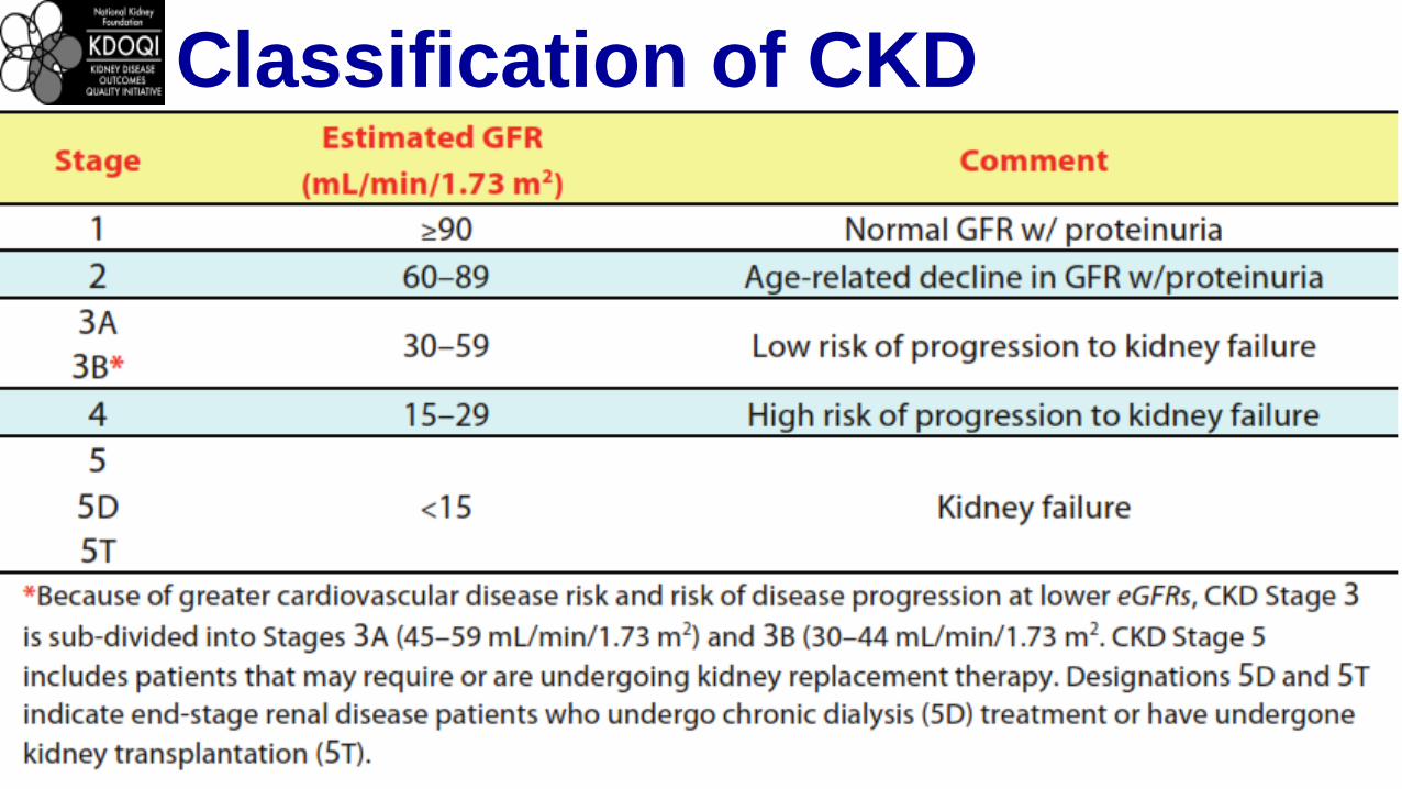
Classification of CKD
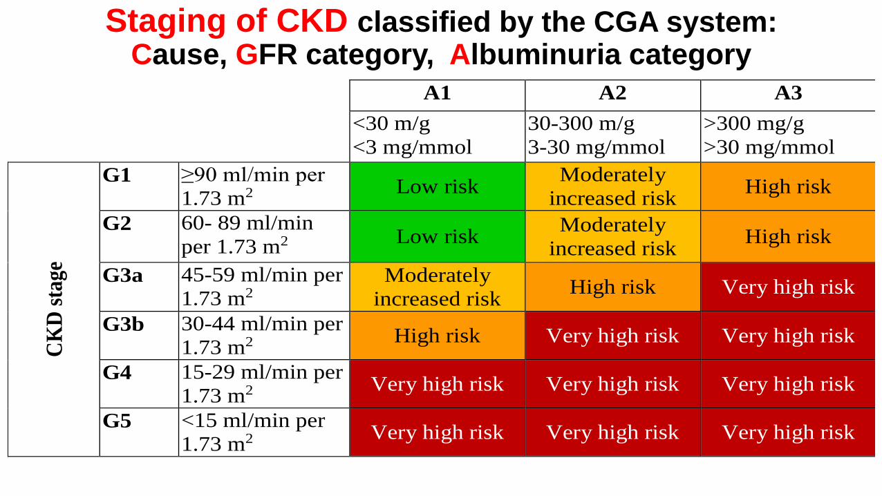
Staging of CKD classified by the CGA system:Cause, GFR category, Albuminuria category
A1 A2 A3
<30 m/g
<3 mg/mmol
30-300 m/g
3-30 mg/mmol
>300 mg/g
>30 mg/mmol
CK
D s
tag
e
G1 ≥90 ml/min per
1.73 m2 Low risk
Moderately
increased risk High risk
G2 60- 89 ml/min
per 1.73 m2 Low risk
Moderately
increased risk High risk
G3a 45-59 ml/min per
1.73 m2 Moderately
increased risk High risk Very high risk
G3b 30-44 ml/min per
1.73 m2 High risk Very high risk Very high risk
G4 15-29 ml/min per
1.73 m2 Very high risk Very high risk Very high risk
G5 <15 ml/min per
1.73 m2 Very high risk Very high risk Very high risk
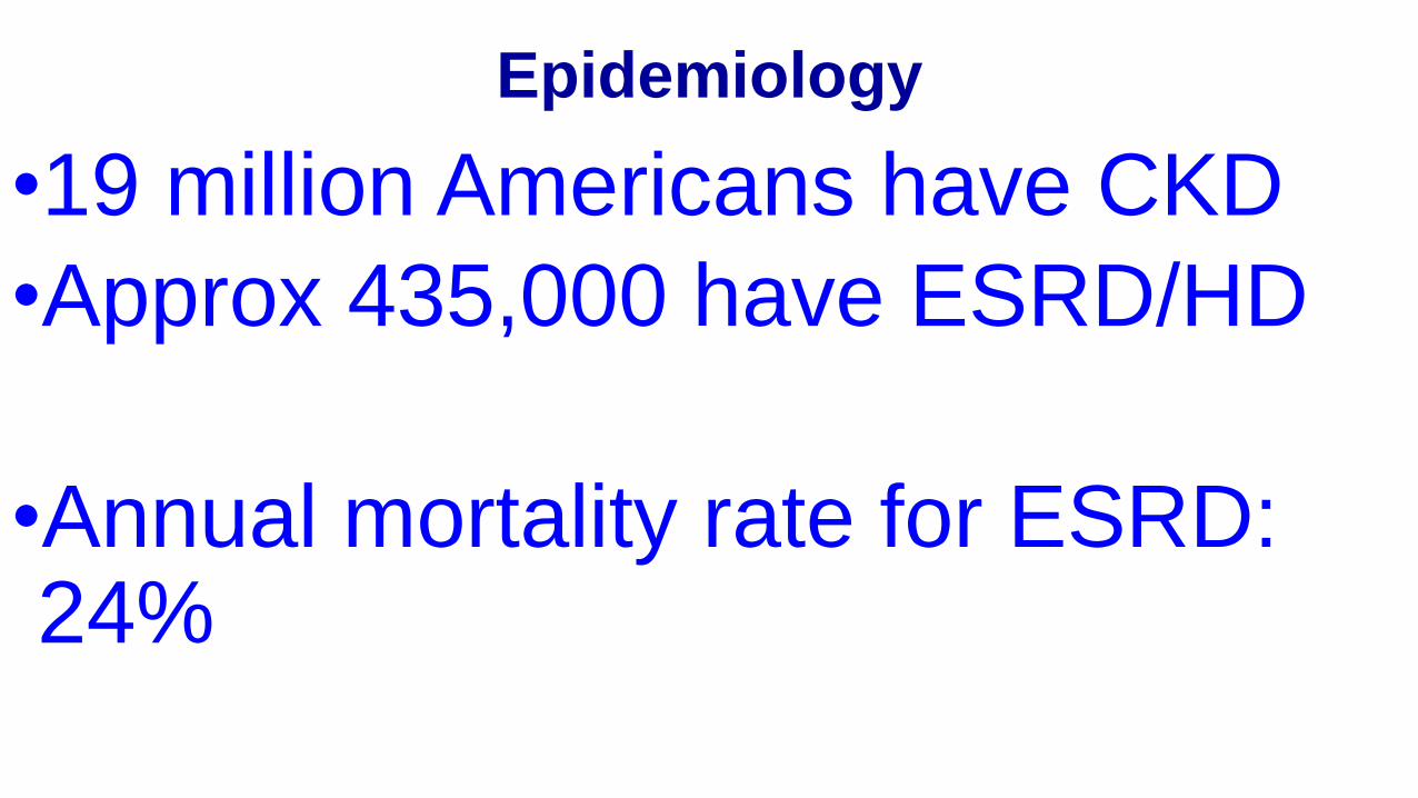
Epidemiology
•19 million Americans have CKD
•Approx 435,000 have ESRD/HD
•Annual mortality rate for ESRD: 24%
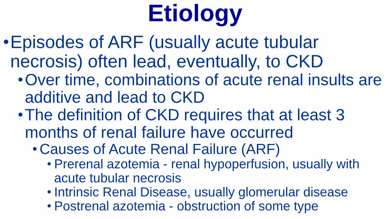
Etiology•Episodes of ARF (usually acute tubular necrosis) often lead, eventually, to CKD •Over time, combinations of acute renal insults are additive and lead to CKD
•The definition of CKD requires that at least 3 months of renal failure have occurred • Causes of Acute Renal Failure (ARF)
• Prerenal azotemia - renal hypoperfusion, usually with acute tubular necrosis
• Intrinsic Renal Disease, usually glomerular disease • Postrenal azotemia - obstruction of some type
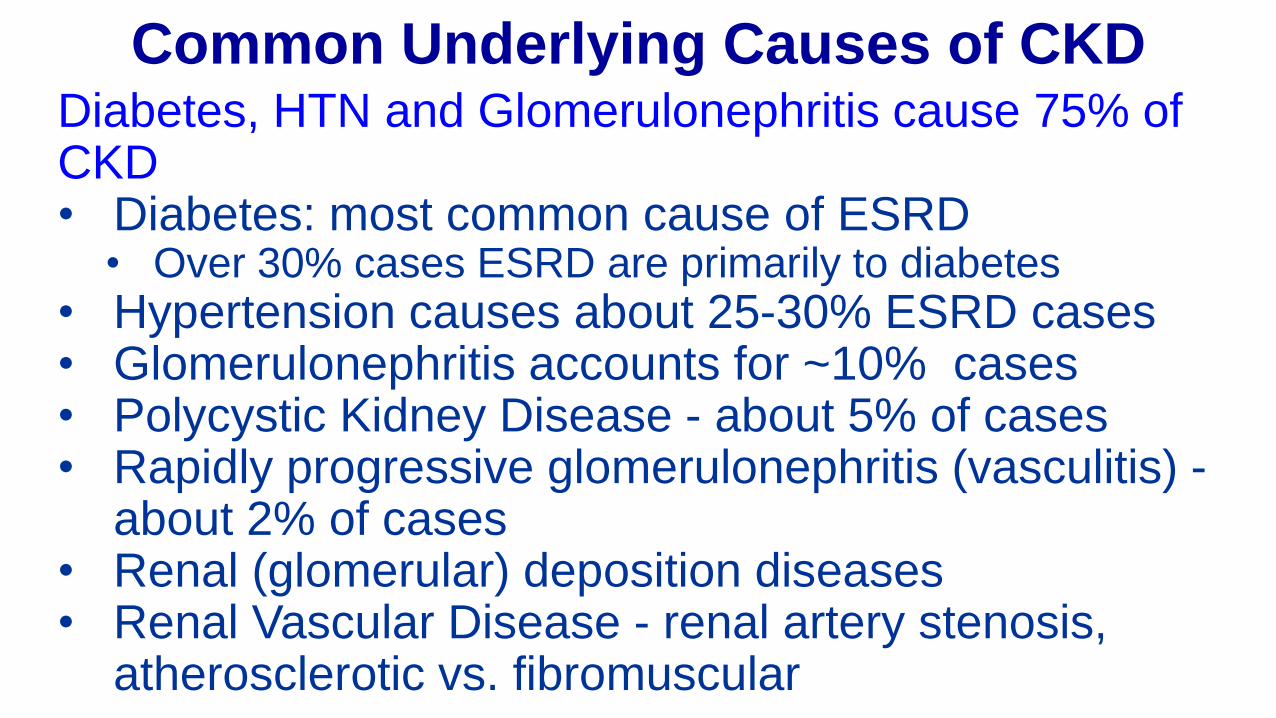
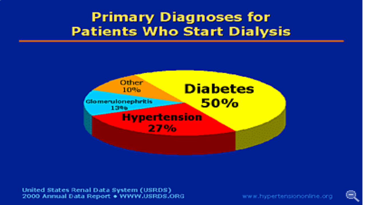
Common Underlying Causes of CKDDiabetes, HTN and Glomerulonephritis cause 75% of CKD• Diabetes: most common cause of ESRD
• Over 30% cases ESRD are primarily to diabetes
• Hypertension causes about 25-30% ESRD cases • Glomerulonephritis accounts for ~10% cases• Polycystic Kidney Disease - about 5% of cases• Rapidly progressive glomerulonephritis (vasculitis) -
about 2% of cases • Renal (glomerular) deposition diseases • Renal Vascular Disease - renal artery stenosis,
atherosclerotic vs. fibromuscular
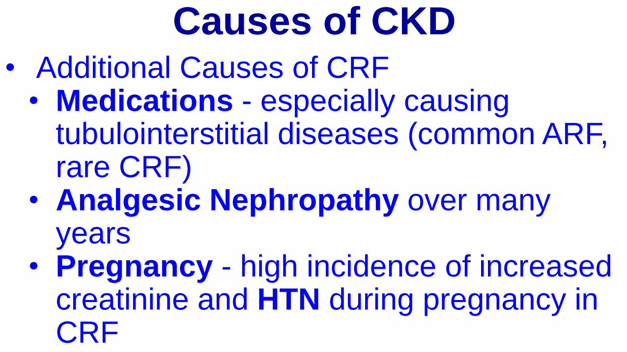
Causes of CKD• Additional Causes of CRF
• Medications - especially causing tubulointerstitial diseases (common ARF, rare CRF)
• Analgesic Nephropathy over many years
• Pregnancy - high incidence of increased creatinine and HTN during pregnancy in CRF
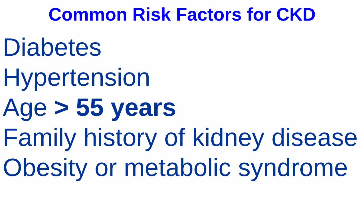
Common Risk Factors for CKD
Diabetes
Hypertension
Age > 55 years
Family history of kidney disease
Obesity or metabolic syndrome
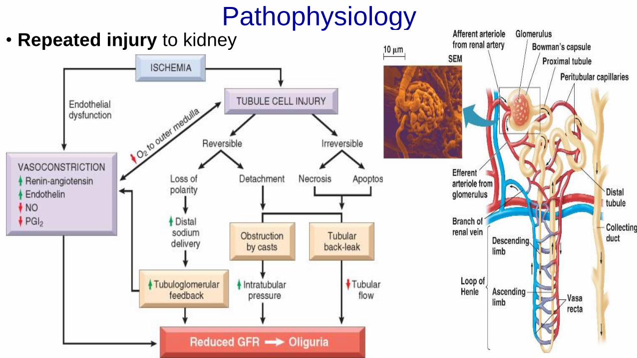
Pathophysiology• Repeated injury to kidney
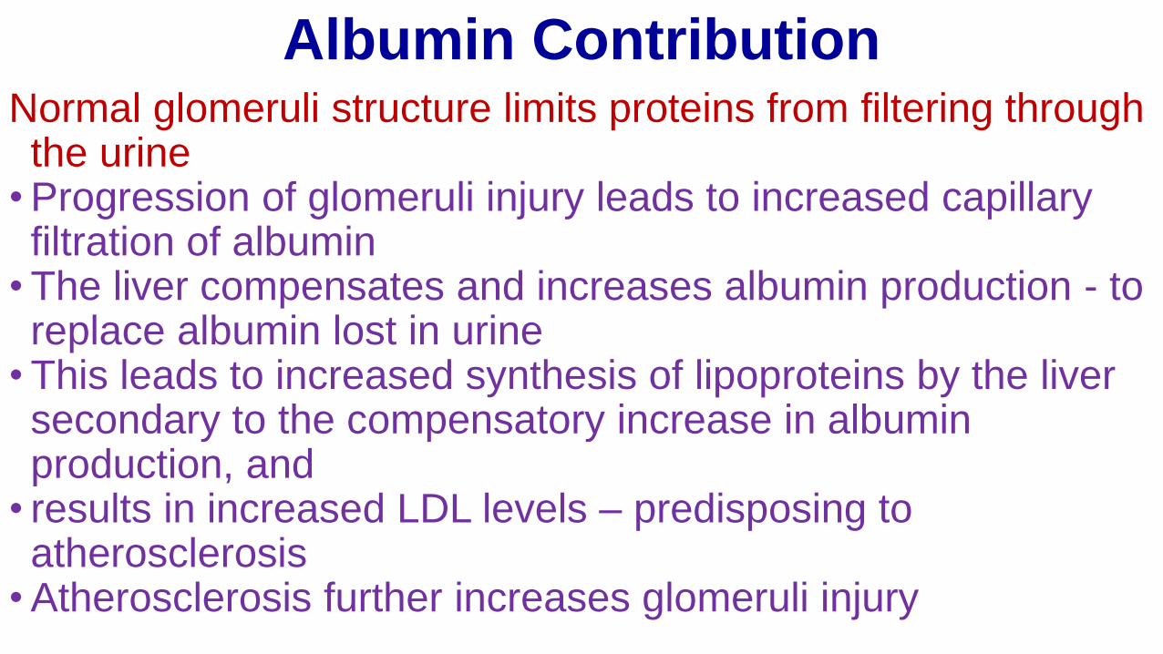
Albumin ContributionNormal glomeruli structure limits proteins from filtering through the urine
• Progression of glomeruli injury leads to increased capillary filtration of albumin
• The liver compensates and increases albumin production - to replace albumin lost in urine
• This leads to increased synthesis of lipoproteins by the liver secondary to the compensatory increase in albumin production, and
• results in increased LDL levels – predisposing to atherosclerosis
• Atherosclerosis further increases glomeruli injury
Inflammation
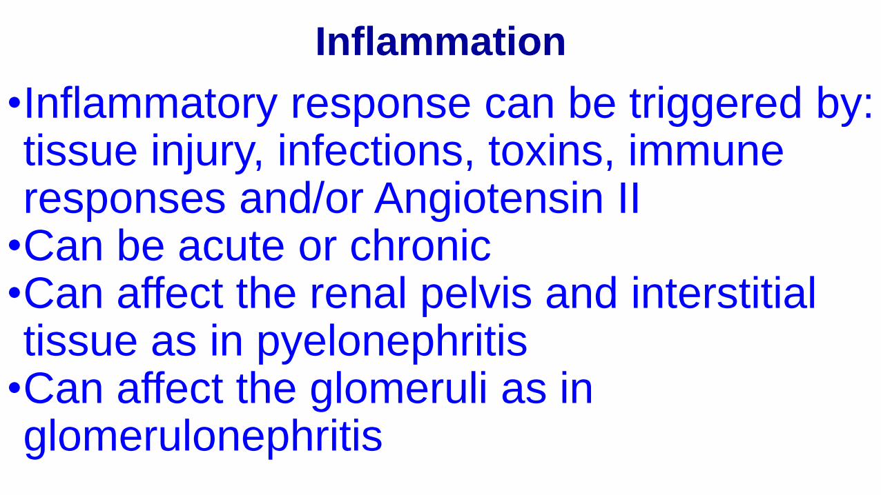
•Inflammatory response can be triggered by: tissue injury, infections, toxins, immune responses and/or Angiotensin II
•Can be acute or chronic•Can affect the renal pelvis and interstitial tissue as in pyelonephritis
•Can affect the glomeruli as in glomerulonephritis
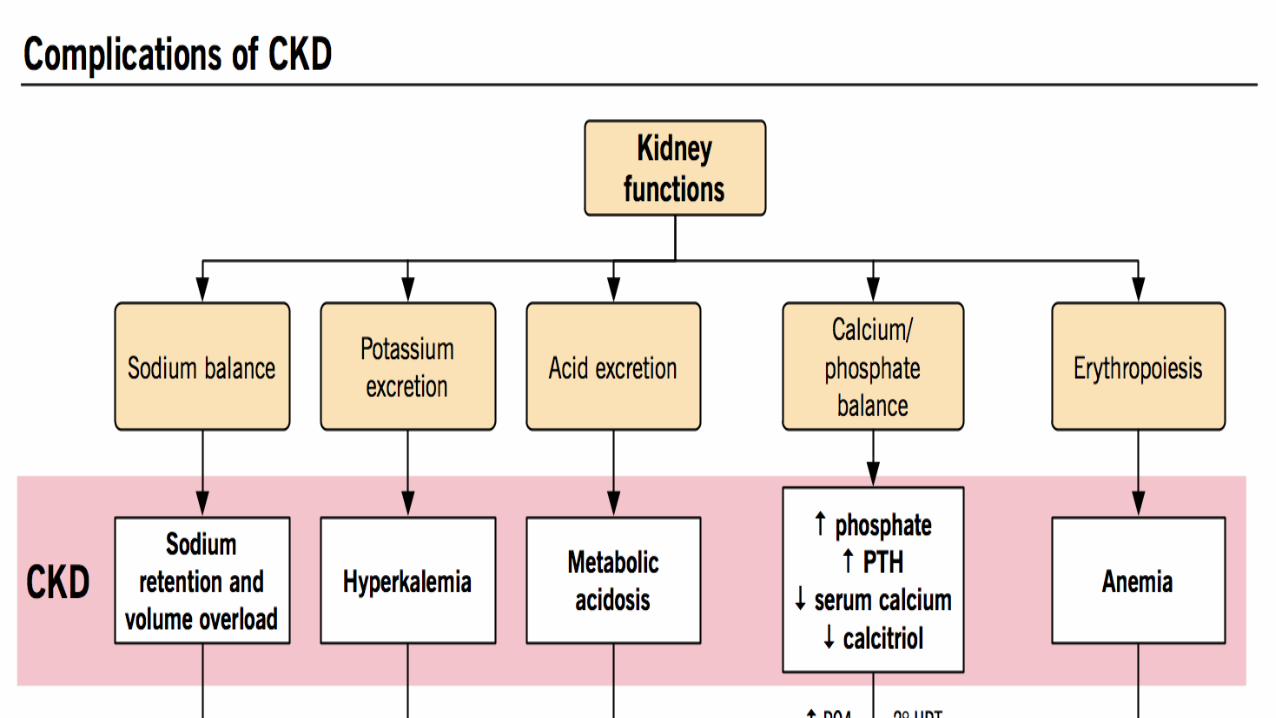
Functional Changes of CRF
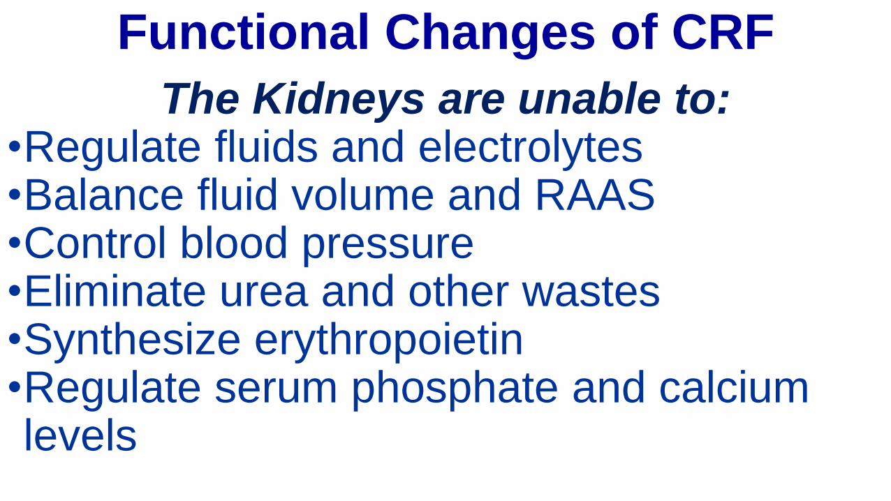
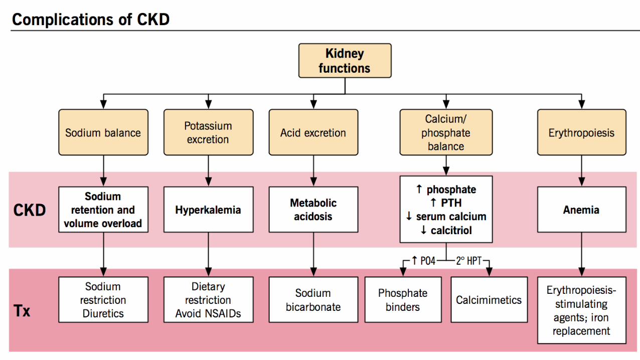
The Kidneys are unable to:•Regulate fluids and electrolytes•Balance fluid volume and RAAS•Control blood pressure•Eliminate urea and other wastes•Synthesize erythropoietin•Regulate serum phosphate and calcium levels
Uremic Syndrome
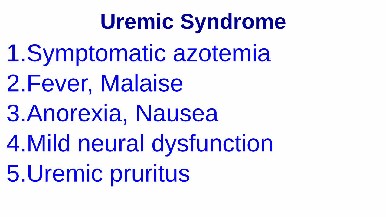
1.Symptomatic azotemia
2.Fever, Malaise
3.Anorexia, Nausea
4.Mild neural dysfunction
5.Uremic pruritus
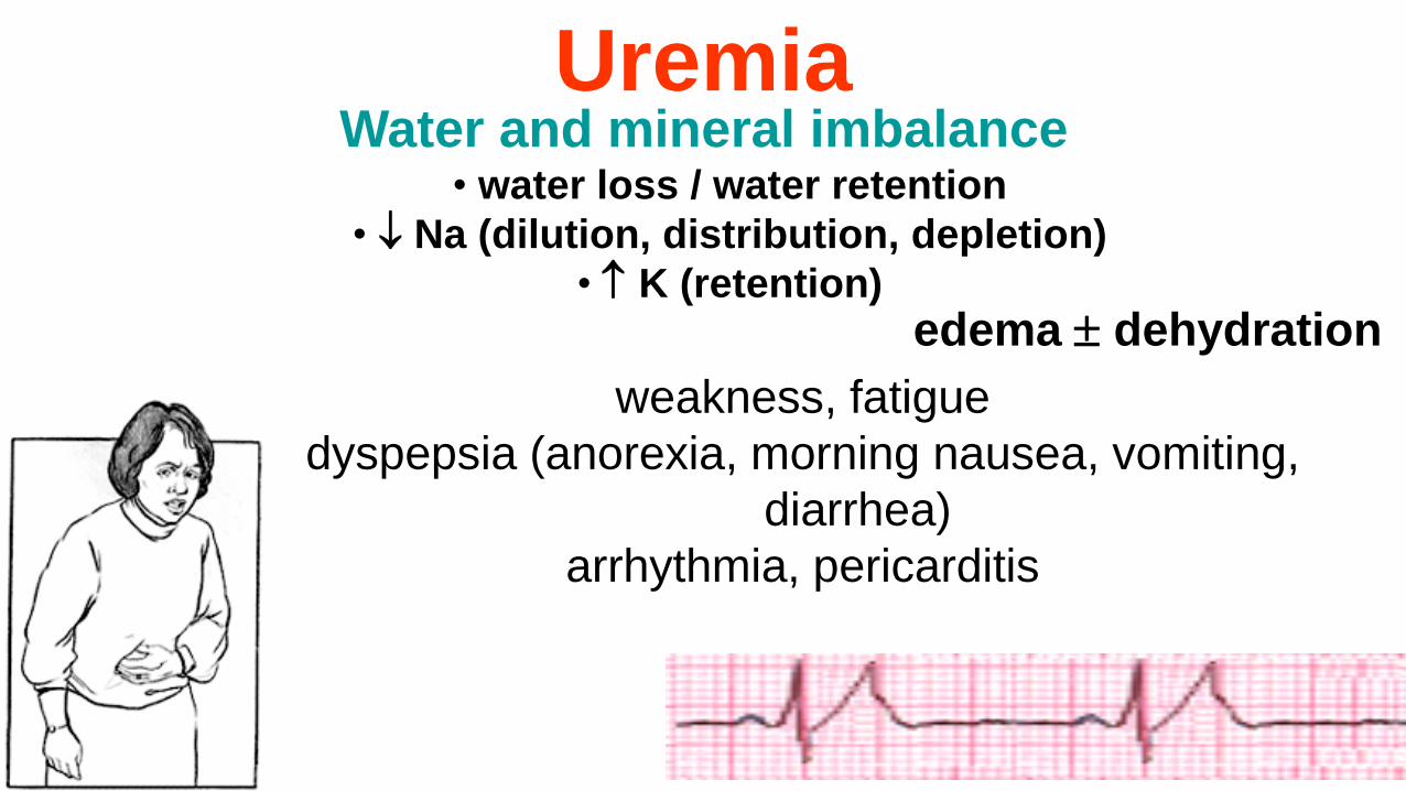
Uremia
edema dehydration
weakness, fatigue
dyspepsia (anorexia, morning nausea, vomiting,
diarrhea)
arrhythmia, pericarditis
Water and mineral imbalance• water loss / water retention
• Na (dilution, distribution, depletion)
• K (retention)
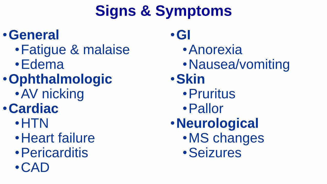
Signs & Symptoms
•General•Fatigue & malaise•Edema
•Ophthalmologic•AV nicking
•Cardiac•HTN•Heart failure•Pericarditis•CAD
•GI•Anorexia•Nausea/vomiting
•Skin•Pruritus•Pallor
•Neurological•MS changes•Seizures
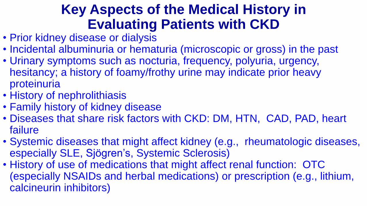
Key Aspects of the Medical History in Evaluating Patients with CKD
• Prior kidney disease or dialysis • Incidental albuminuria or hematuria (microscopic or gross) in the past • Urinary symptoms such as nocturia, frequency, polyuria, urgency,
hesitancy; a history of foamy/frothy urine may indicate prior heavy proteinuria
• History of nephrolithiasis• Family history of kidney disease• Diseases that share risk factors with CKD: DM, HTN, CAD, PAD, heart
failure• Systemic diseases that might affect kidney (e.g., rheumatologic diseases,
especially SLE, Sjögren’s, Systemic Sclerosis) • History of use of medications that might affect renal function: OTC
(especially NSAIDs and herbal medications) or prescription (e.g., lithium, calcineurin inhibitors)
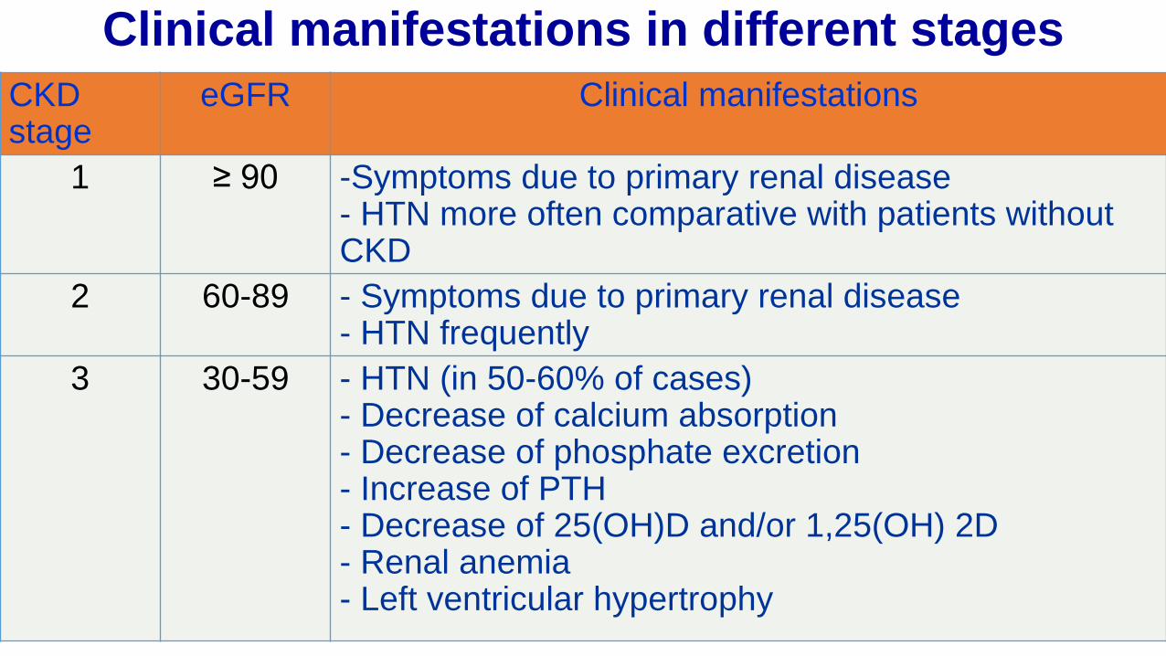
Clinical manifestations in different stages
CKD stage
eGFR Clinical manifestations
1 ≥ 90 -Symptoms due to primary renal disease- HTN more often comparative with patients without CKD
2 60-89 - Symptoms due to primary renal disease- HTN frequently
3 30-59 - HTN (in 50-60% of cases)- Decrease of calcium absorption- Decrease of phosphate excretion- Increase of PTH- Decrease of 25(OH)D and/or 1,25(OH) 2D- Renal anemia- Left ventricular hypertrophy
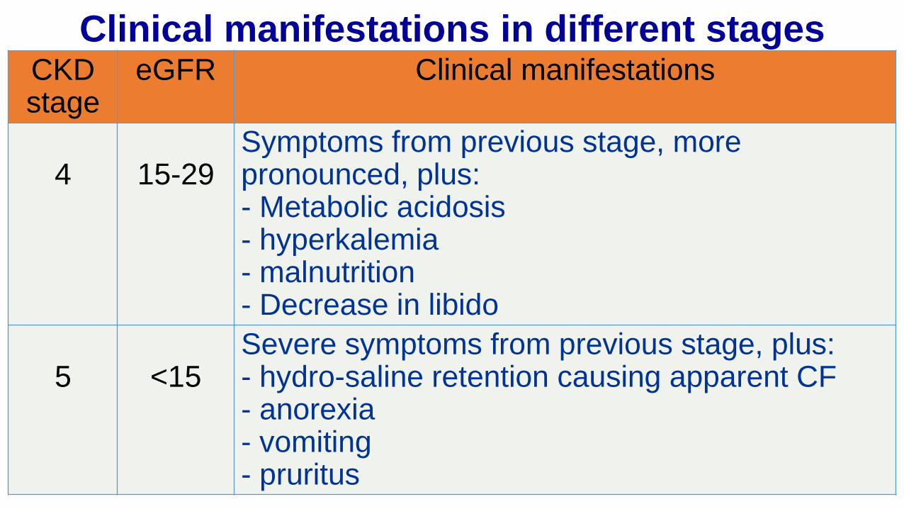
CKD stage
eGFR Clinical manifestations
4 15-29 Symptoms from previous stage, more pronounced, plus:- Metabolic acidosis- hyperkalemia- malnutrition- Decrease in libido
5 <15 Severe symptoms from previous stage, plus:- hydro-saline retention causing apparent CF- anorexia- vomiting- pruritus
Clinical manifestations in different stages
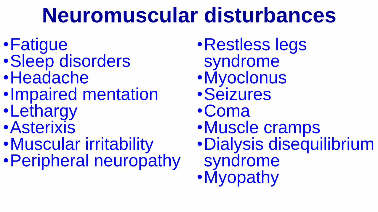
Neuromuscular disturbances
•Fatigue •Sleep disorders •Headache •Impaired mentation •Lethargy •Asterixis•Muscular irritability•Peripheral neuropathy
•Restless legs syndrome
•Myoclonus •Seizures •Coma •Muscle cramps •Dialysis disequilibrium syndrome
•Myopathy
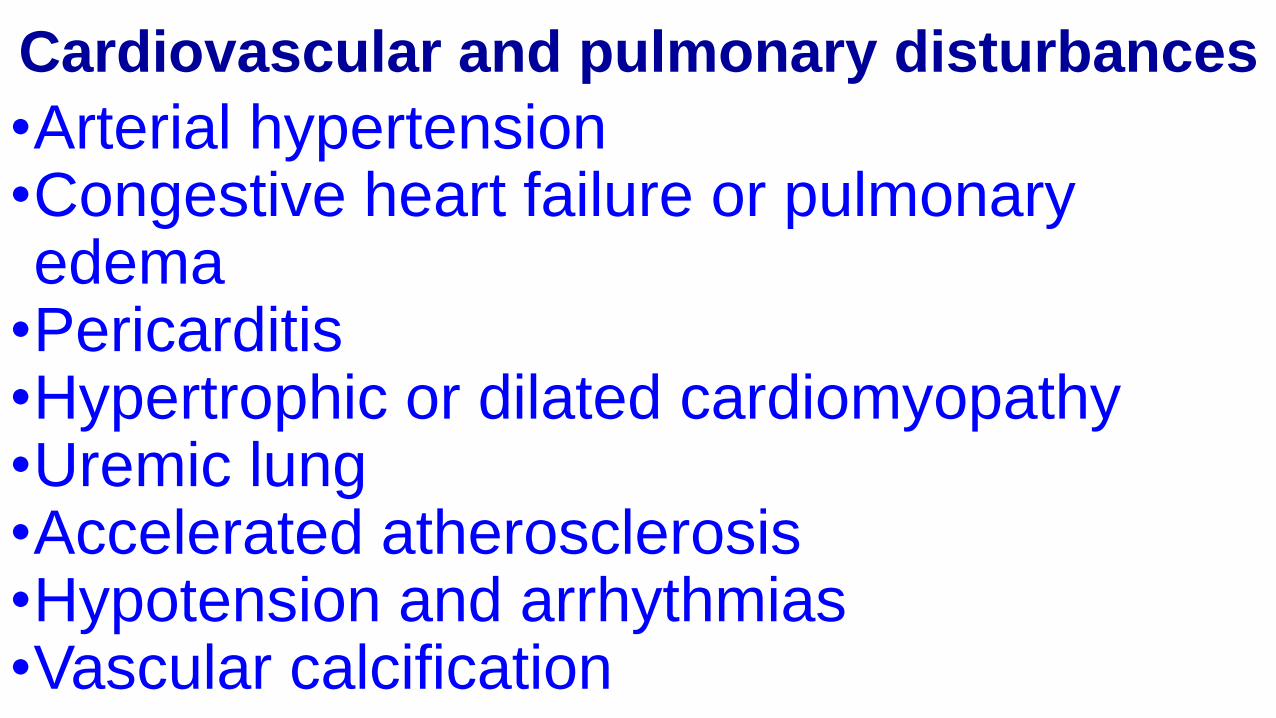
Cardiovascular and pulmonary disturbances
•Arterial hypertension •Congestive heart failure or pulmonary edema
•Pericarditis •Hypertrophic or dilated cardiomyopathy•Uremic lung •Accelerated atherosclerosis •Hypotension and arrhythmias•Vascular calcification
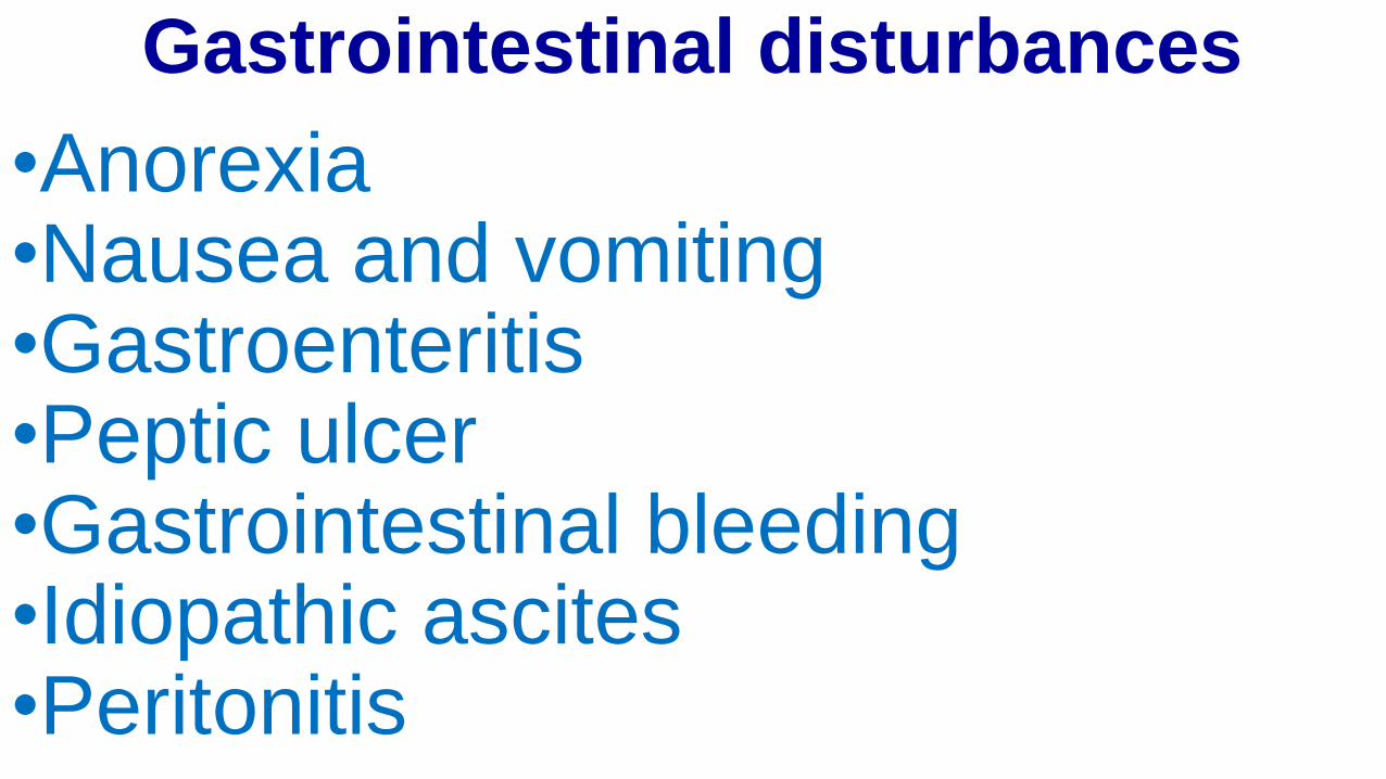
Gastrointestinal disturbances
•Anorexia •Nausea and vomiting •Gastroenteritis •Peptic ulcer •Gastrointestinal bleeding•Idiopathic ascites •Peritonitis
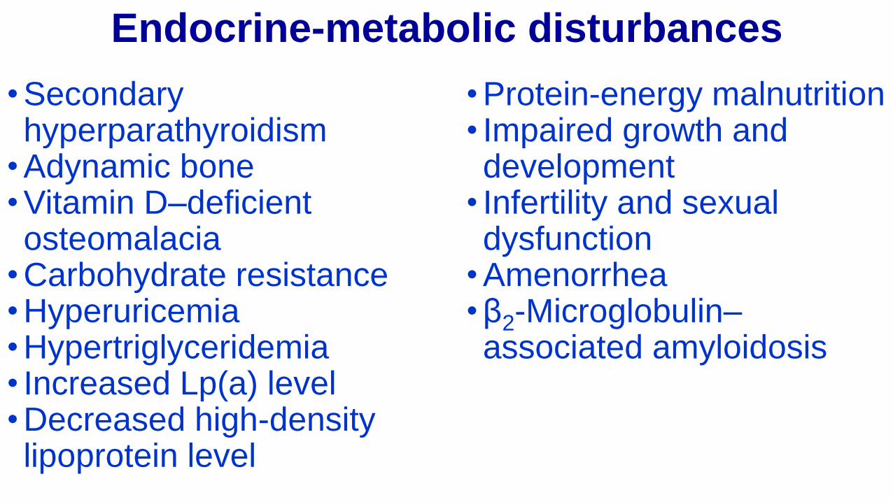
Endocrine-metabolic disturbances
• Secondary hyperparathyroidism
• Adynamic bone• Vitamin D–deficient osteomalacia
• Carbohydrate resistance• Hyperuricemia • Hypertriglyceridemia• Increased Lp(a) level• Decreased high-density lipoprotein level
• Protein-energy malnutrition• Impaired growth and development
• Infertility and sexual dysfunction
• Amenorrhea• β2-Microglobulin–associated amyloidosis
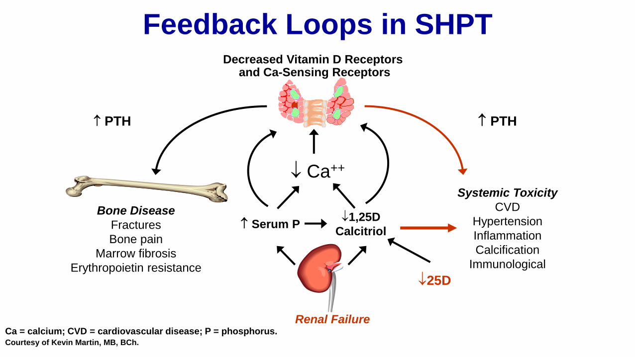
Feedback Loops in SHPT
Ca = calcium; CVD = cardiovascular disease; P = phosphorus.Courtesy of Kevin Martin, MB, BCh.
PTH
Bone Disease
Fractures
Bone pain
Marrow fibrosis
Erythropoietin resistance
Serum P1,25D
Calcitriol
Renal Failure
PTH
Systemic Toxicity
CVD
Hypertension
Inflammation
Calcification
Immunological
25D
Ca++
Decreased Vitamin D Receptors and Ca-Sensing Receptors
© 2005 The Johns Hopkins University School of Medicine.
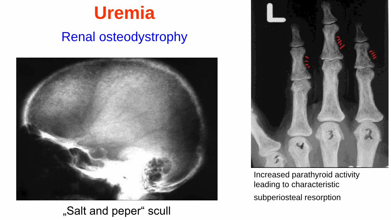
Uremia
Renal osteodystrophy
„Salt and peper“ scull
Increased parathyroid activity
leading to characteristic
subperiosteal resorption
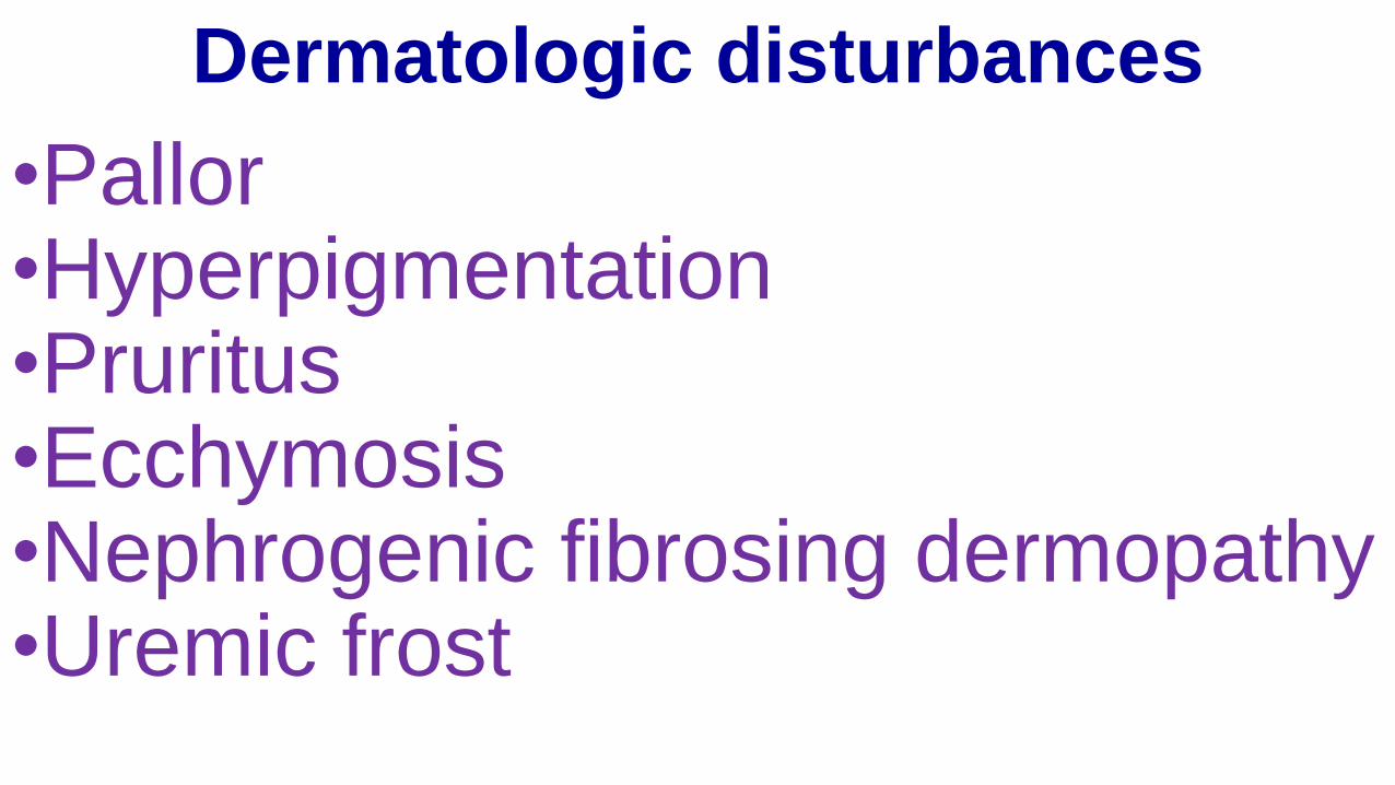
Dermatologic disturbances
•Pallor •Hyperpigmentation •Pruritus •Ecchymosis •Nephrogenic fibrosing dermopathy•Uremic frost
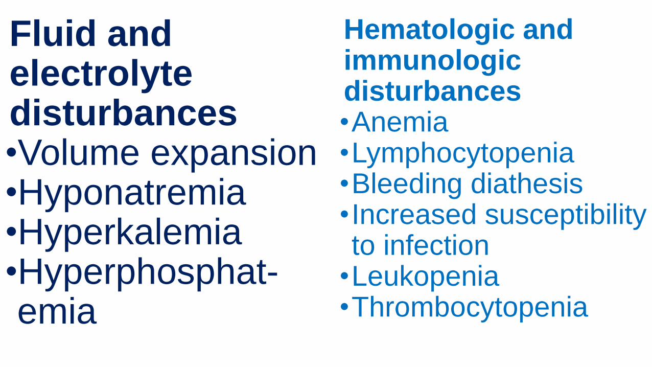
Fluid and electrolyte disturbances•Volume expansion •Hyponatremia •Hyperkalemia•Hyperphosphat-emia
Hematologic and immunologic disturbances•Anemia •Lymphocytopenia•Bleeding diathesis •Increased susceptibility to infection
•Leukopenia •Thrombocytopenia
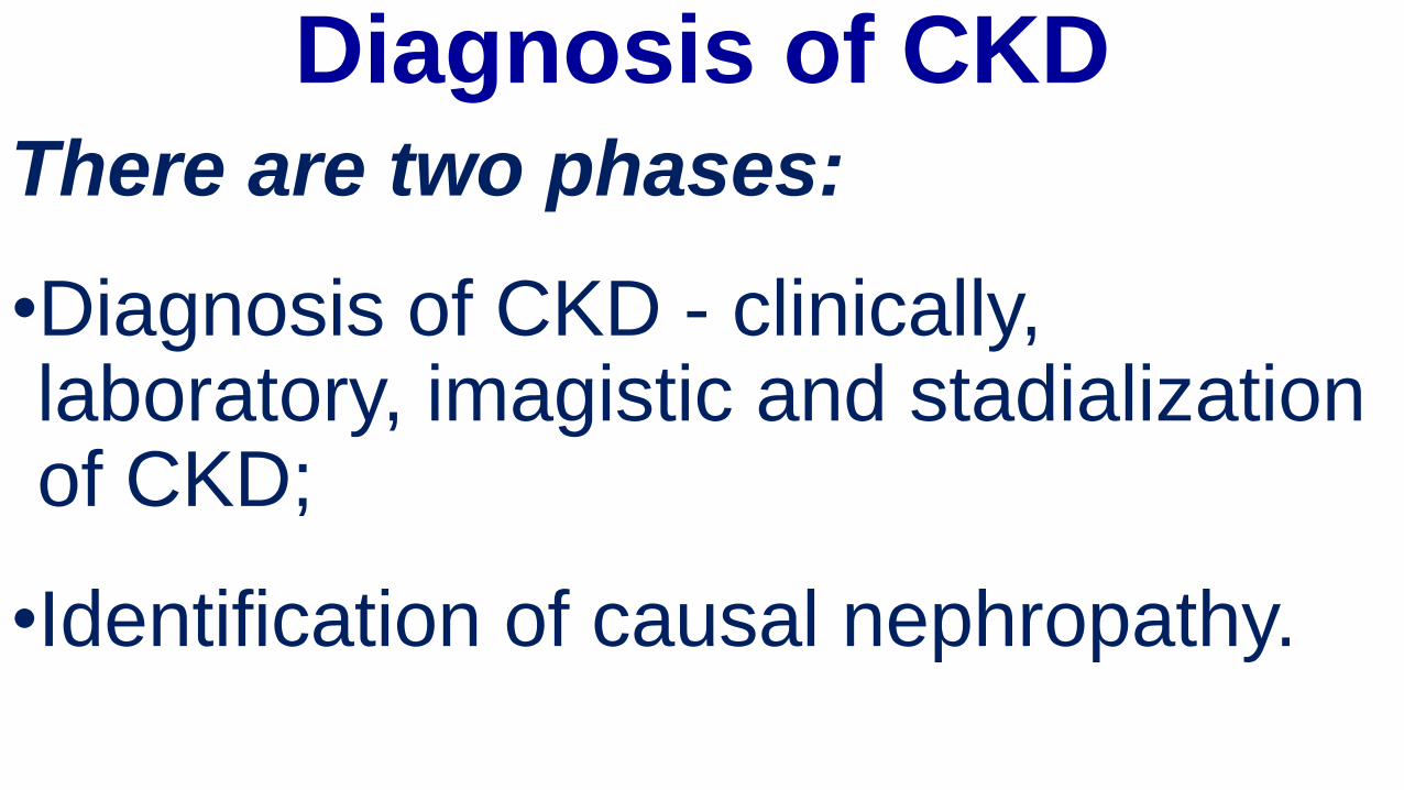
Diagnosis of CKDThere are two phases:
•Diagnosis of CKD - clinically, laboratory, imagistic and stadializationof CKD;
•Identification of causal nephropathy.
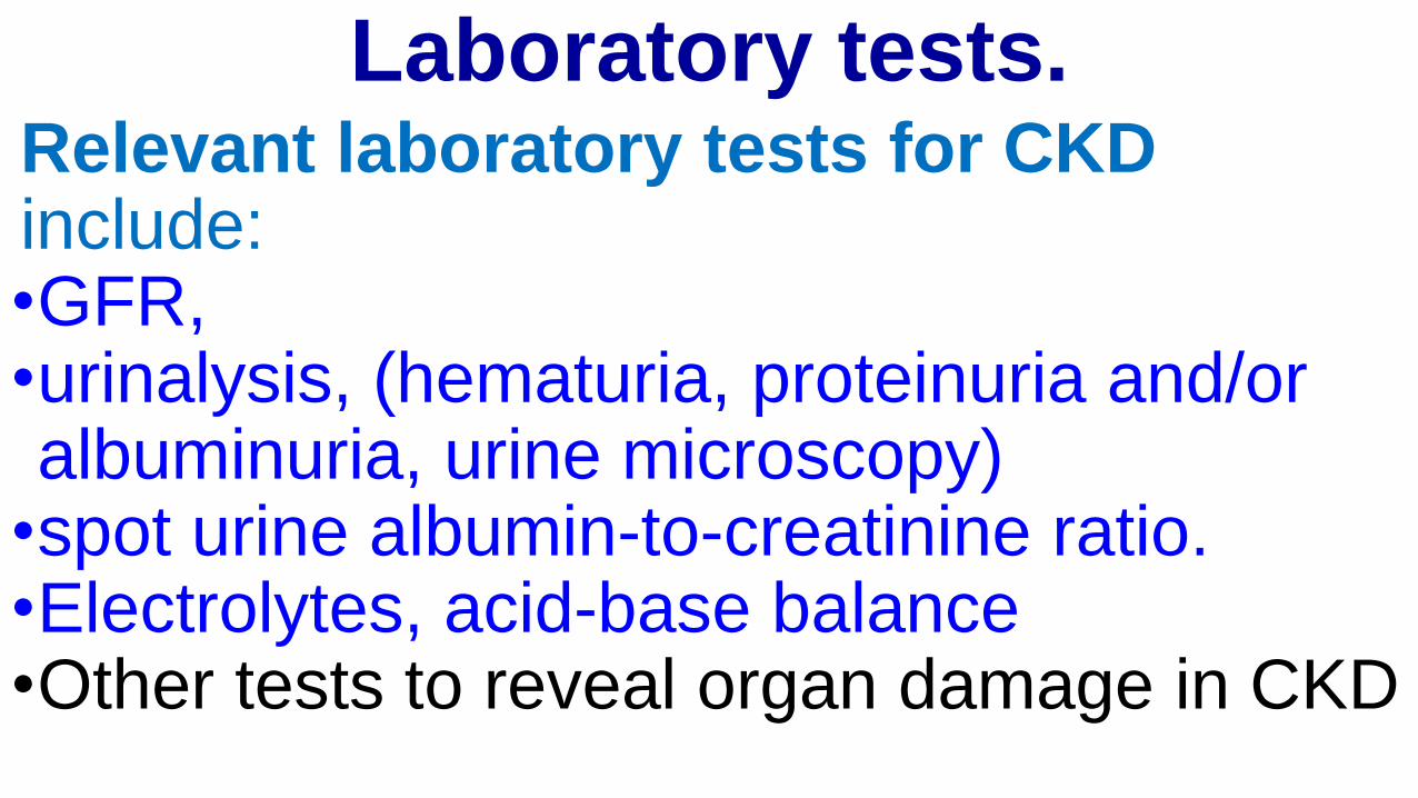
Laboratory tests. Relevant laboratory tests for CKD include: •GFR, •urinalysis, (hematuria, proteinuria and/or albuminuria, urine microscopy)
•spot urine albumin-to-creatinine ratio.•Electrolytes, acid-base balance •Other tests to reveal organ damage in CKD
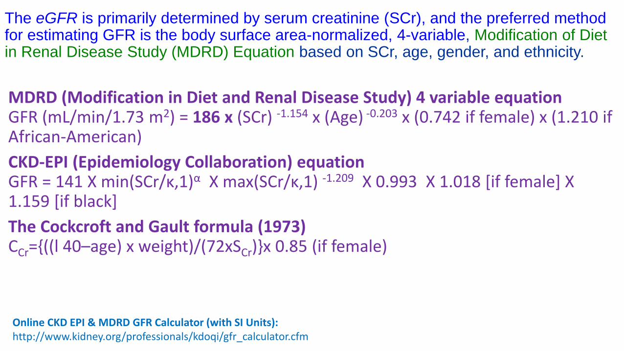
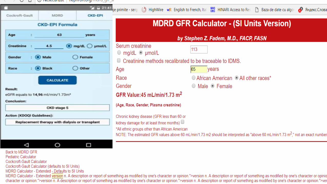
The eGFR is primarily determined by serum creatinine (SCr), and the preferred method for estimating GFR is the body surface area-normalized, 4-variable, Modification of Diet in Renal Disease Study (MDRD) Equation based on SCr, age, gender, and ethnicity.
Online CKD EPI & MDRD GFR Calculator (with SI Units):http://www.kidney.org/professionals/kdoqi/gfr_calculator.cfm
MDRD (Modification in Diet and Renal Disease Study) 4 variable equationGFR (mL/min/1.73 m2) = 186 x (SCr) -1.154 x (Age) -0.203 x (0.742 if female) x (1.210 if African-American)
CKD-EPI (Epidemiology Collaboration) equationGFR = 141 X min(SCr/κ,1)α X max(SCr/κ,1) -1.209 X 0.993 X 1.018 [if female] X 1.159 [if black]
The Cockcroft and Gault formula (1973)CCr={((l 40–age) x weight)/(72xSCr)}x 0.85 (if female)
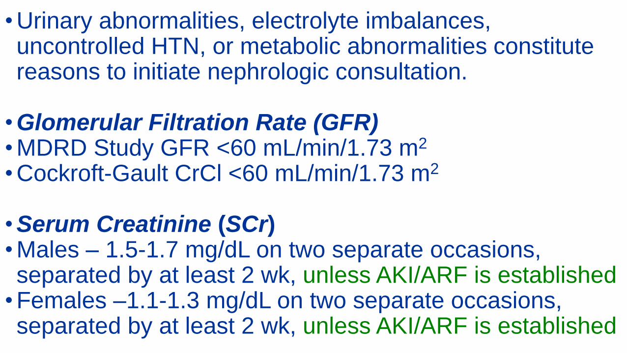
• Urinary abnormalities, electrolyte imbalances, uncontrolled HTN, or metabolic abnormalities constitute reasons to initiate nephrologic consultation.
• Glomerular Filtration Rate (GFR)• MDRD Study GFR <60 mL/min/1.73 m2
• Cockroft-Gault CrCl <60 mL/min/1.73 m2
• Serum Creatinine (SCr)• Males – 1.5-1.7 mg/dL on two separate occasions, separated by at least 2 wk, unless AKI/ARF is established
• Females –1.1-1.3 mg/dL on two separate occasions, separated by at least 2 wk, unless AKI/ARF is established
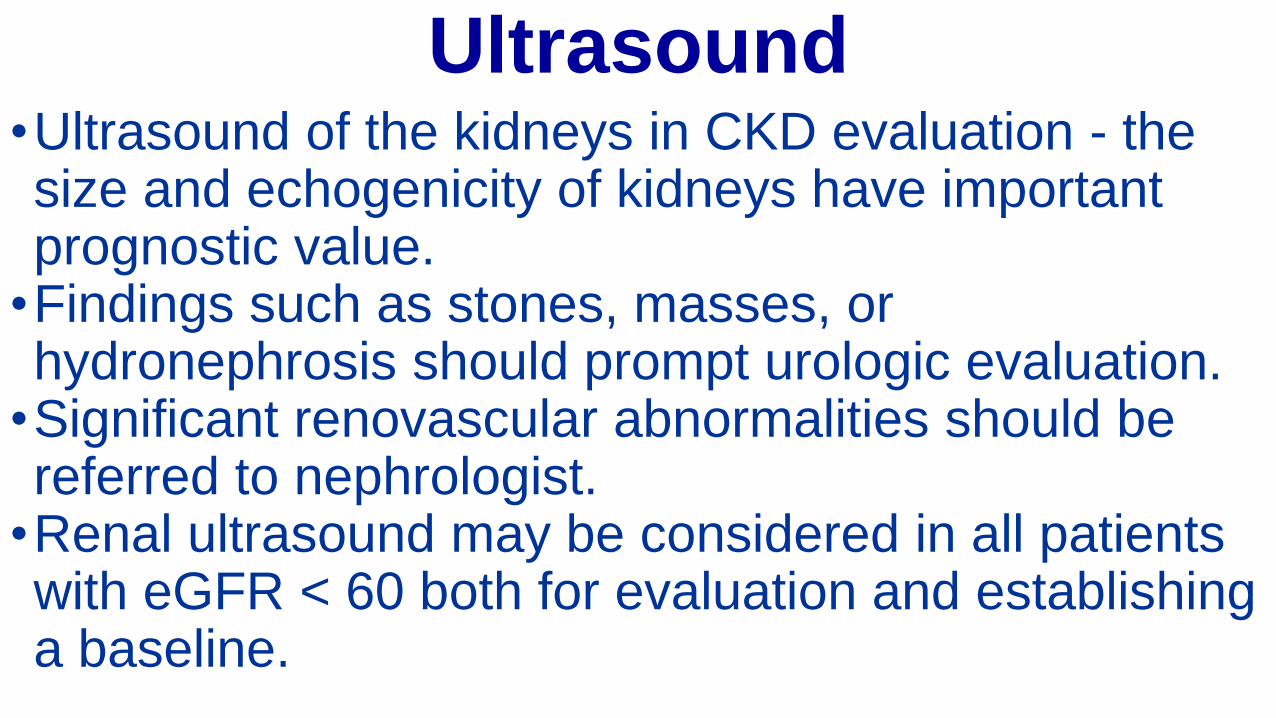
Ultrasound•Ultrasound of the kidneys in CKD evaluation - the size and echogenicity of kidneys have important prognostic value.
•Findings such as stones, masses, or hydronephrosis should prompt urologic evaluation.
•Significant renovascular abnormalities should be referred to nephrologist.
•Renal ultrasound may be considered in all patients with eGFR < 60 both for evaluation and establishing a baseline.
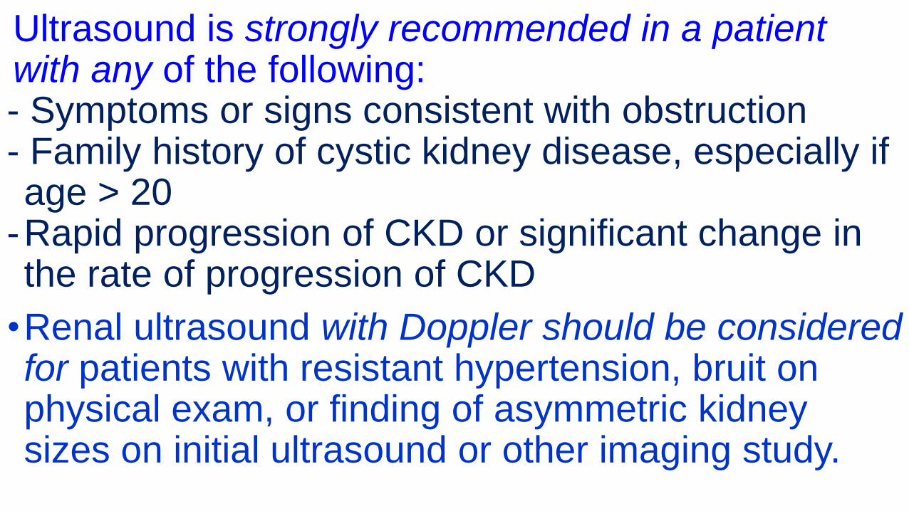
Ultrasound is strongly recommended in a patient with any of the following:- Symptoms or signs consistent with obstruction- Family history of cystic kidney disease, especially if age > 20
-Rapid progression of CKD or significant change in the rate of progression of CKD
•Renal ultrasound with Doppler should be considered for patients with resistant hypertension, bruit on physical exam, or finding of asymmetric kidney sizes on initial ultrasound or other imaging study.
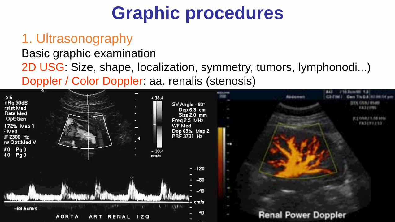
1. UltrasonographyBasic graphic examination
2D USG: Size, shape, localization, symmetry, tumors, lymphonodi...)
Doppler / Color Doppler: aa. renalis (stenosis)
Graphic procedures

Renal USG:
Enlarged kidneys (max. diam. 150 mm) with no evidence of urinary
obstruction
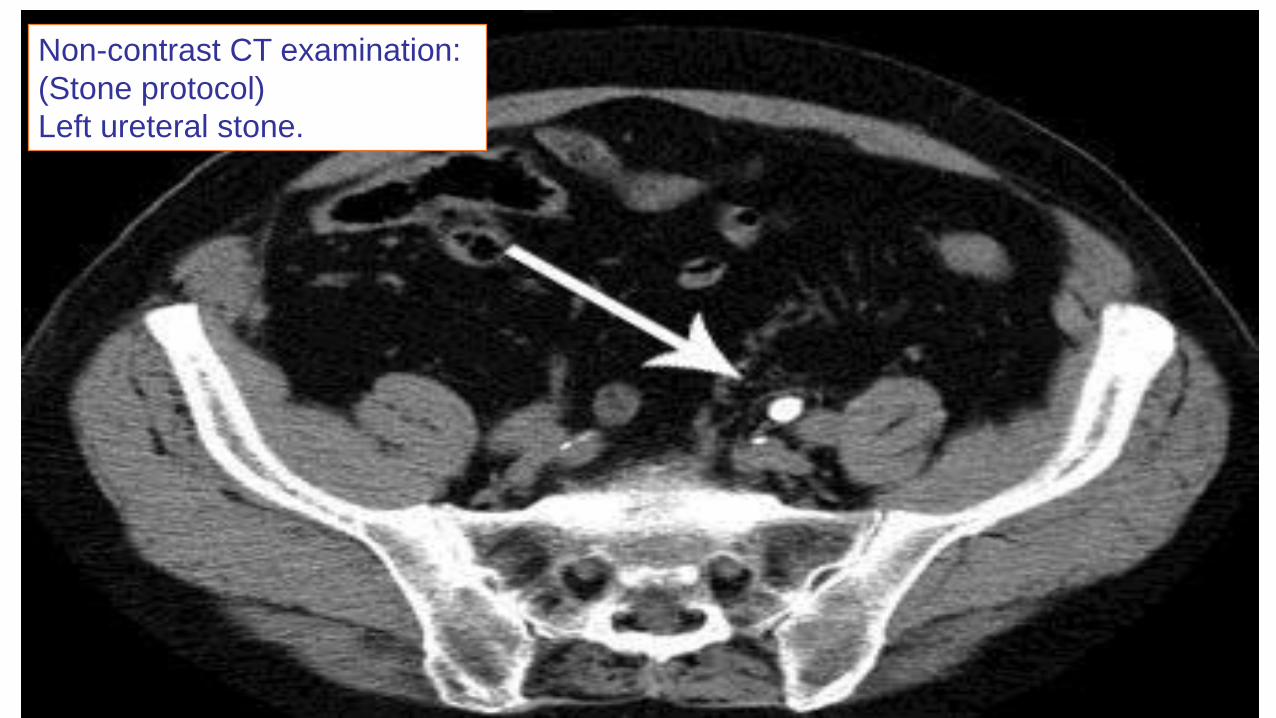
Non-contrast CT examination:
(Stone protocol)
Left ureteral stone.
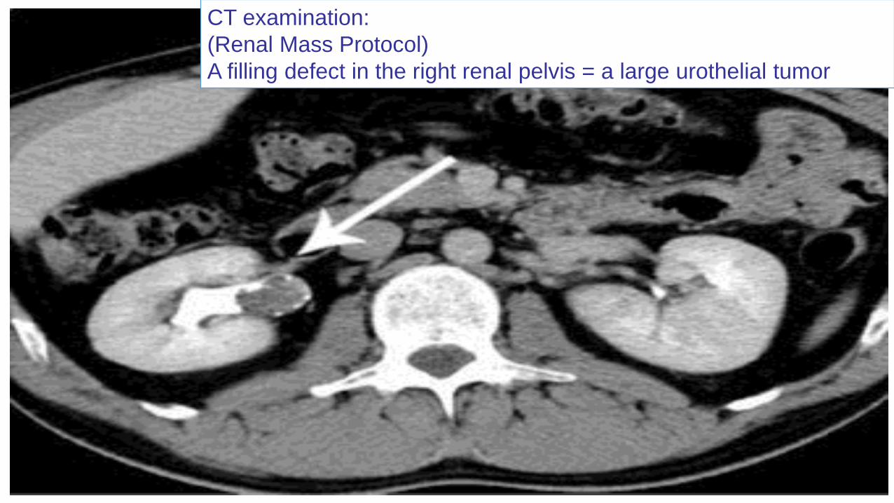
CT examination:
(Renal Mass Protocol)
A filling defect in the right renal pelvis = a large urothelial tumor
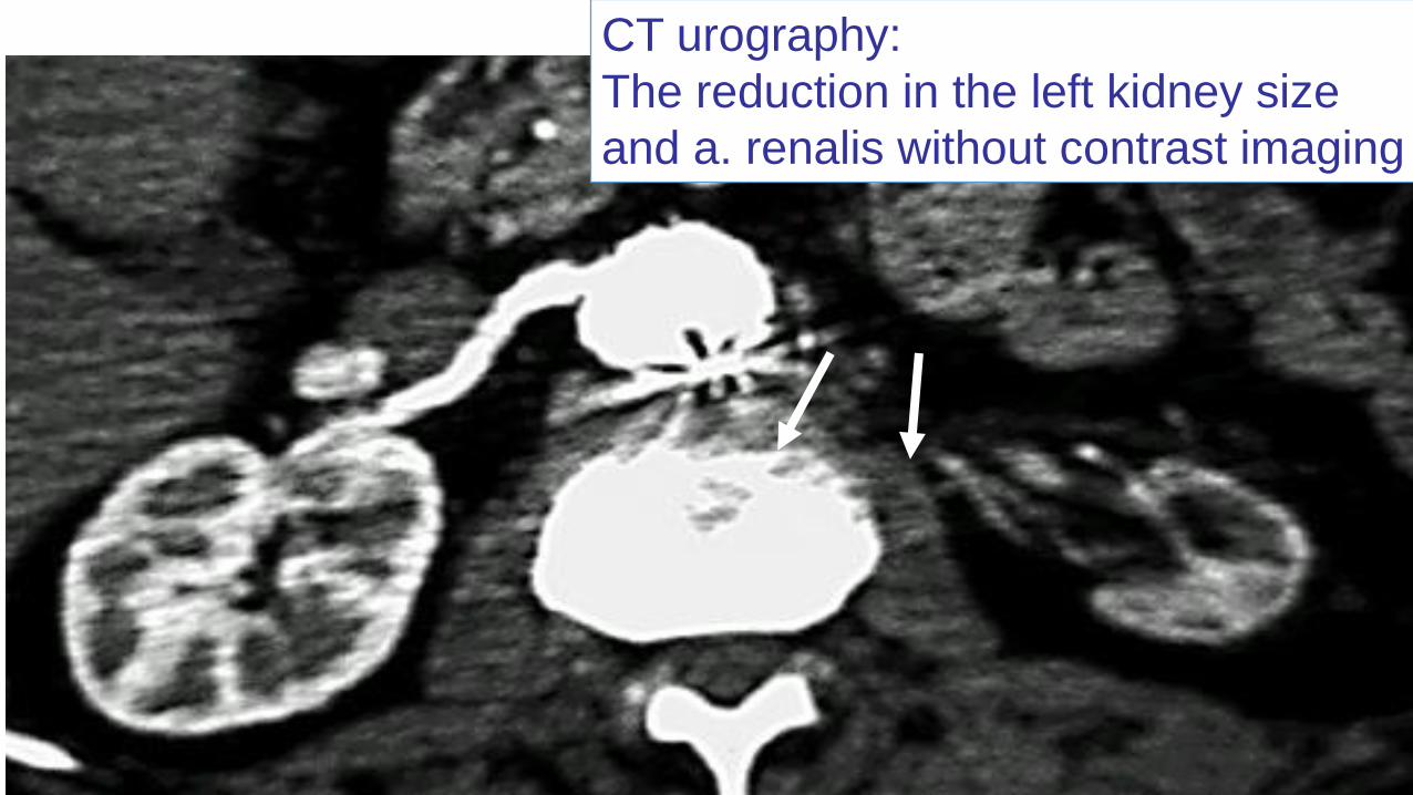
CT urography:
The reduction in the left kidney size
and a. renalis without contrast imaging
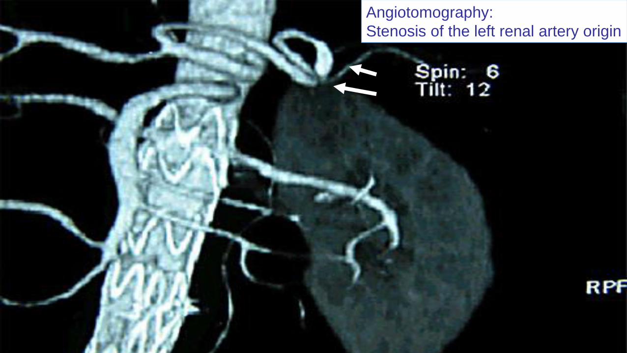
Angiotomography:
Stenosis of the left renal artery origin
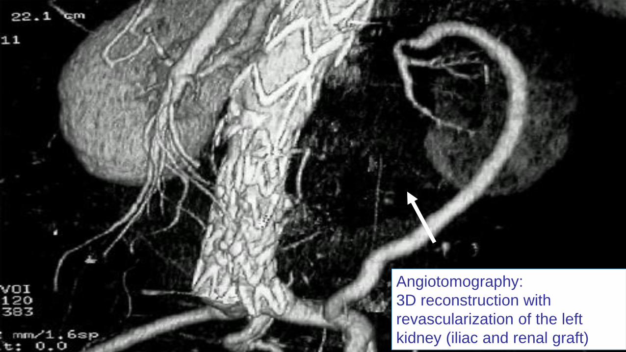
Angiotomography:
3D reconstruction with
revascularization of the left
kidney (iliac and renal graft)
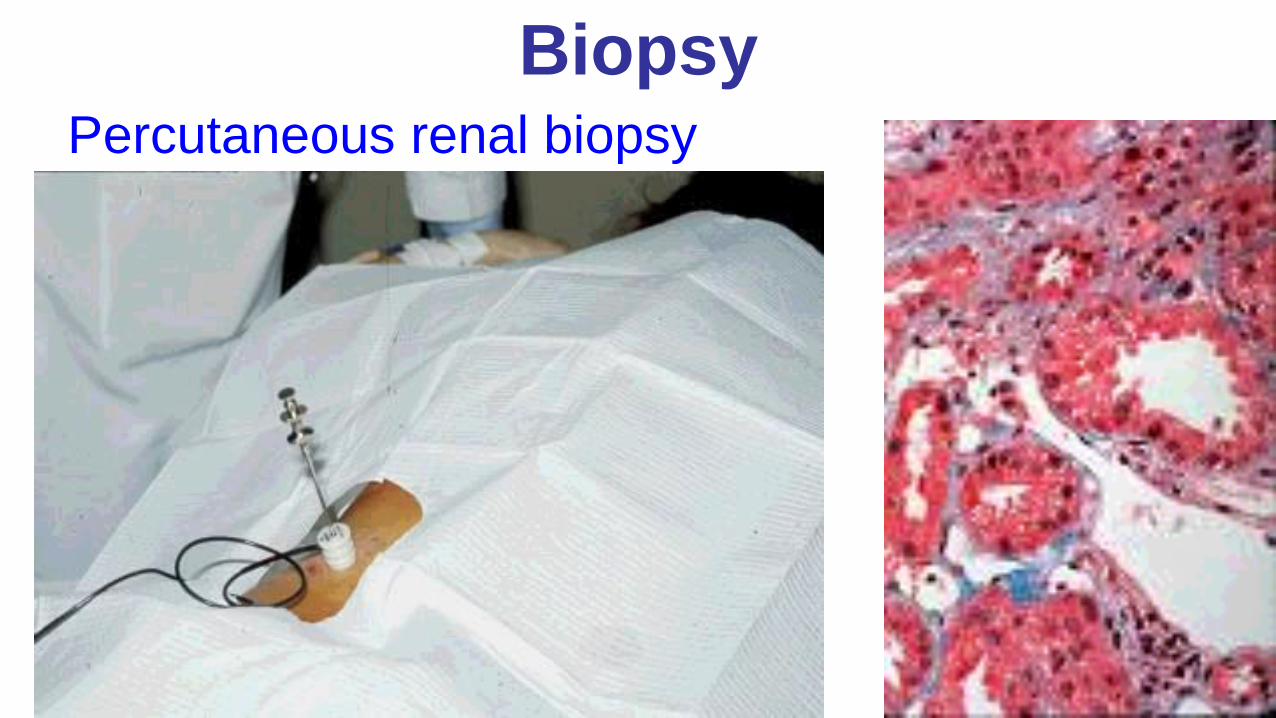
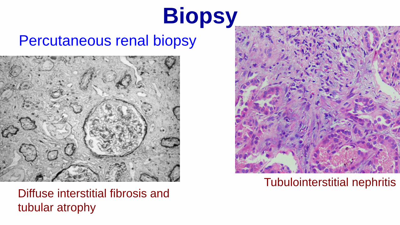
BiopsyPercutaneous renal biopsy
Biopsy
Diffuse interstitial fibrosis and
tubular atrophy
Tubulointerstitial nephritis
Percutaneous renal biopsy
Management of CKD
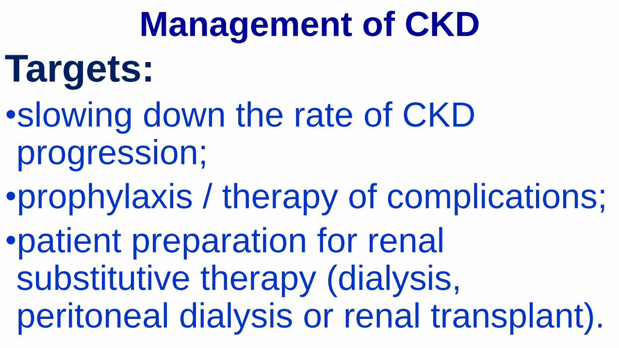
Targets:•slowing down the rate of CKD progression;
•prophylaxis / therapy of complications;
•patient preparation for renal substitutive therapy (dialysis, peritoneal dialysis or renal transplant).
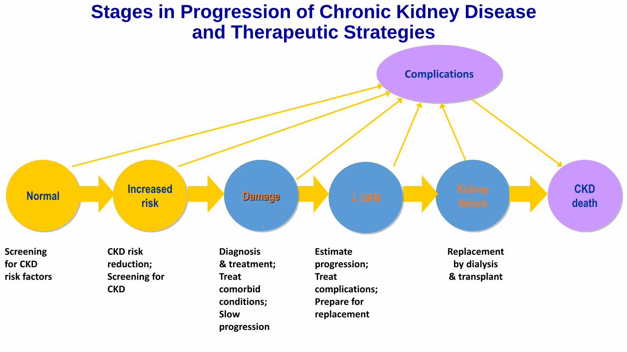
Stages in Progression of Chronic Kidney Disease and Therapeutic Strategies
CKD
death
Complications
Screening for CKDrisk factors
CKD riskreduction;Screening forCKD
Diagnosis& treatment;Treat comorbidconditions;Slow progression
Estimateprogression;Treatcomplications;Prepare forreplacement
Replacementby dialysis
& transplant
NormalIncreased
risk
Kidney
failureDamage GFR
Management of CKD•The direct management of CKD focuses on renin angiotensin aldosterone blockade (RAAS) and blood pressure control.
•Management also includes optimal management of common comorbid conditions such as diabetes and addressing cardiovascular risk factors to decrease risk for CVD.
•Also essential are patient education and a multidisciplinary approach to disease management that utilizes dieticians and social workers in addition to physicians
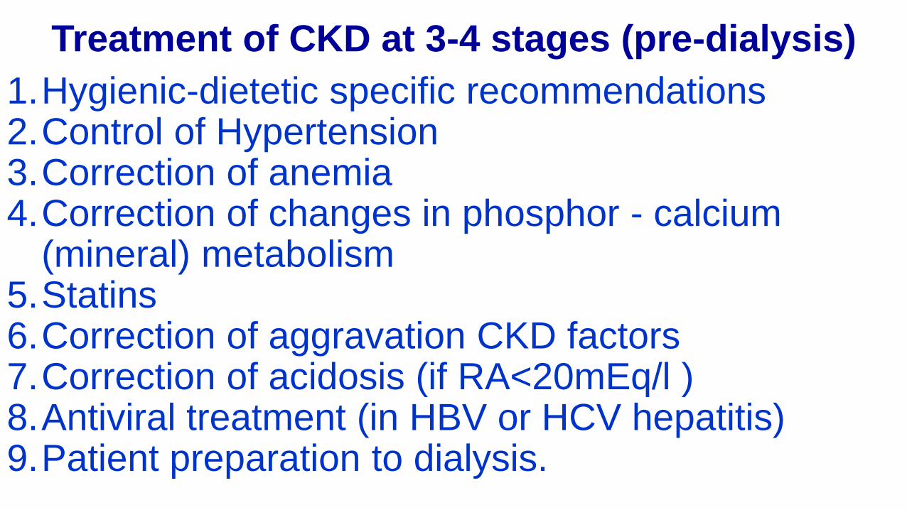
Treatment of CKD at 3-4 stages (pre-dialysis)
1.Hygienic-dietetic specific recommendations2.Control of Hypertension3.Correction of anemia4.Correction of changes in phosphor - calcium
(mineral) metabolism5.Statins6.Correction of aggravation CKD factors7.Correction of acidosis (if RA<20mEq/l )8.Antiviral treatment (in HBV or HCV hepatitis)9.Patient preparation to dialysis.
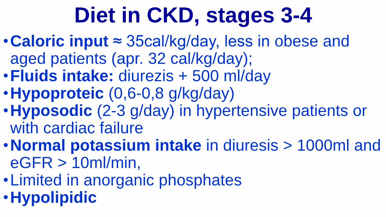
Diet in CKD, stages 3-4•Caloric input ≈ 35cal/kg/day, less in obese and aged patients (apr. 32 cal/kg/day);
•Fluids intake: diurezis + 500 ml/day•Hypoproteic (0,6-0,8 g/kg/day)•Hyposodic (2-3 g/day) in hypertensive patients or with cardiac failure
•Normal potassium intake in diuresis > 1000ml and eGFR > 10ml/min,
•Limited in anorganic phosphates•Hypolipidic
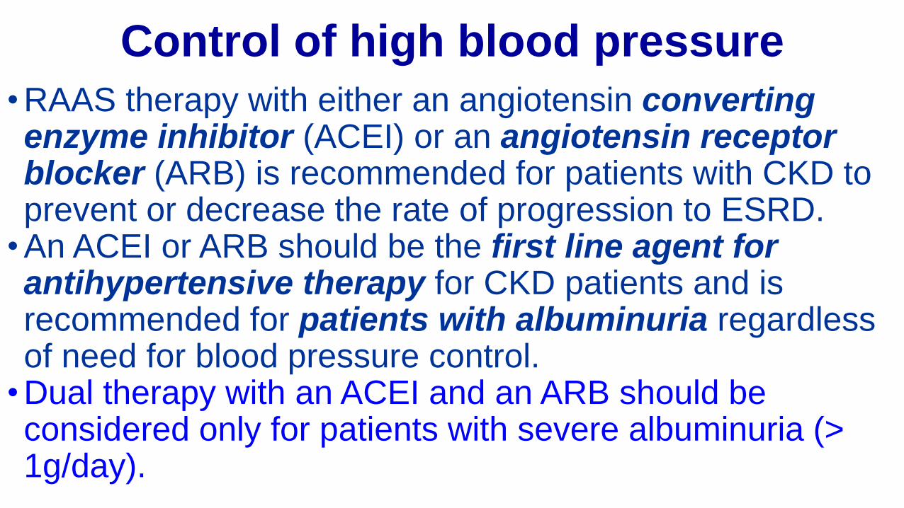
Control of high blood pressure
• RAAS therapy with either an angiotensin converting enzyme inhibitor (ACEI) or an angiotensin receptor blocker (ARB) is recommended for patients with CKD to prevent or decrease the rate of progression to ESRD.
• An ACEI or ARB should be the first line agent for antihypertensive therapy for CKD patients and is recommended for patients with albuminuria regardless of need for blood pressure control.
• Dual therapy with an ACEI and an ARB should be considered only for patients with severe albuminuria (> 1g/day).
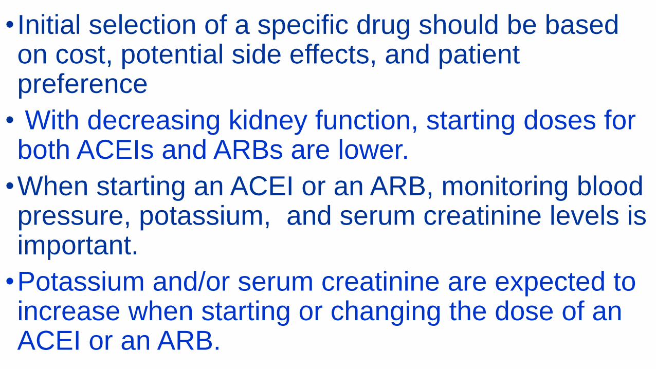
• Initial selection of a specific drug should be based on cost, potential side effects, and patient preference
• With decreasing kidney function, starting doses for both ACEIs and ARBs are lower.
•When starting an ACEI or an ARB, monitoring blood pressure, potassium, and serum creatinine levels is important.
•Potassium and/or serum creatinine are expected to increase when starting or changing the dose of an ACEI or an ARB.
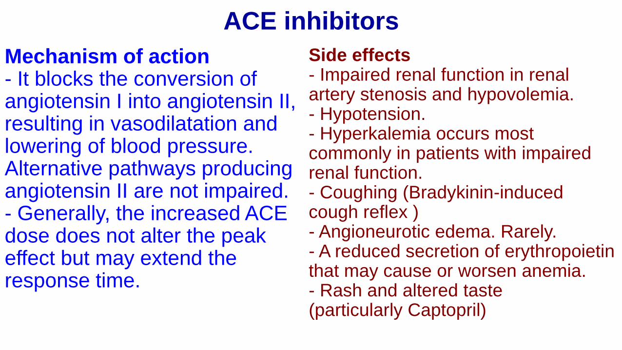
ACE inhibitors
Mechanism of action- It blocks the conversion of angiotensin I into angiotensin II, resulting in vasodilatation and lowering of blood pressure. Alternative pathways producing angiotensin II are not impaired.- Generally, the increased ACE dose does not alter the peak effect but may extend the response time.
Side effects- Impaired renal function in renal artery stenosis and hypovolemia.- Hypotension.- Hyperkalemia occurs most commonly in patients with impaired renal function.- Coughing (Bradykinin-induced cough reflex )- Angioneurotic edema. Rarely.- A reduced secretion of erythropoietin that may cause or worsen anemia.- Rash and altered taste (particularly Captopril)
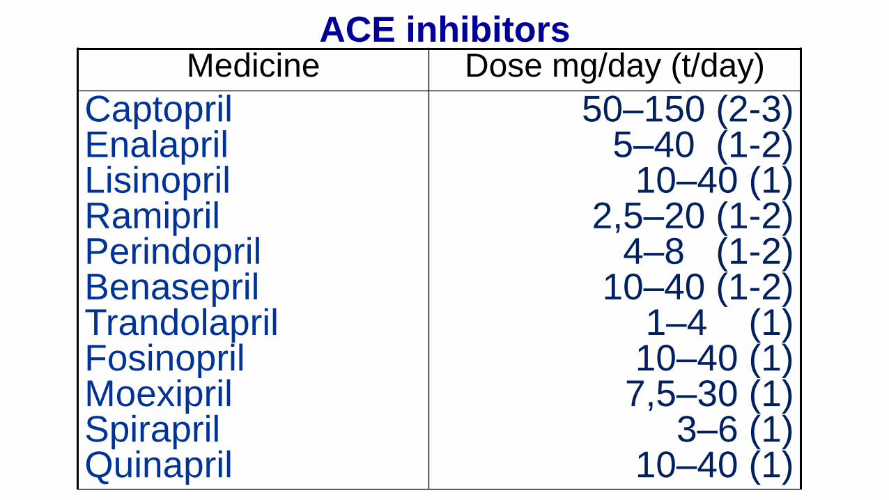
ACE inhibitorsMedicine Dose mg/day (t/day)
CaptoprilEnalaprilLisinoprilRamiprilPerindoprilBenaseprilTrandolaprilFosinoprilMoexiprilSpiraprilQuinapril
50–150 (2-3)5–40 (1-2)
10–40 (1)2,5–20 (1-2)
4–8 (1-2)10–40 (1-2)
1–4 (1)10–40 (1)
7,5–30 (1)3–6 (1)
10–40 (1)
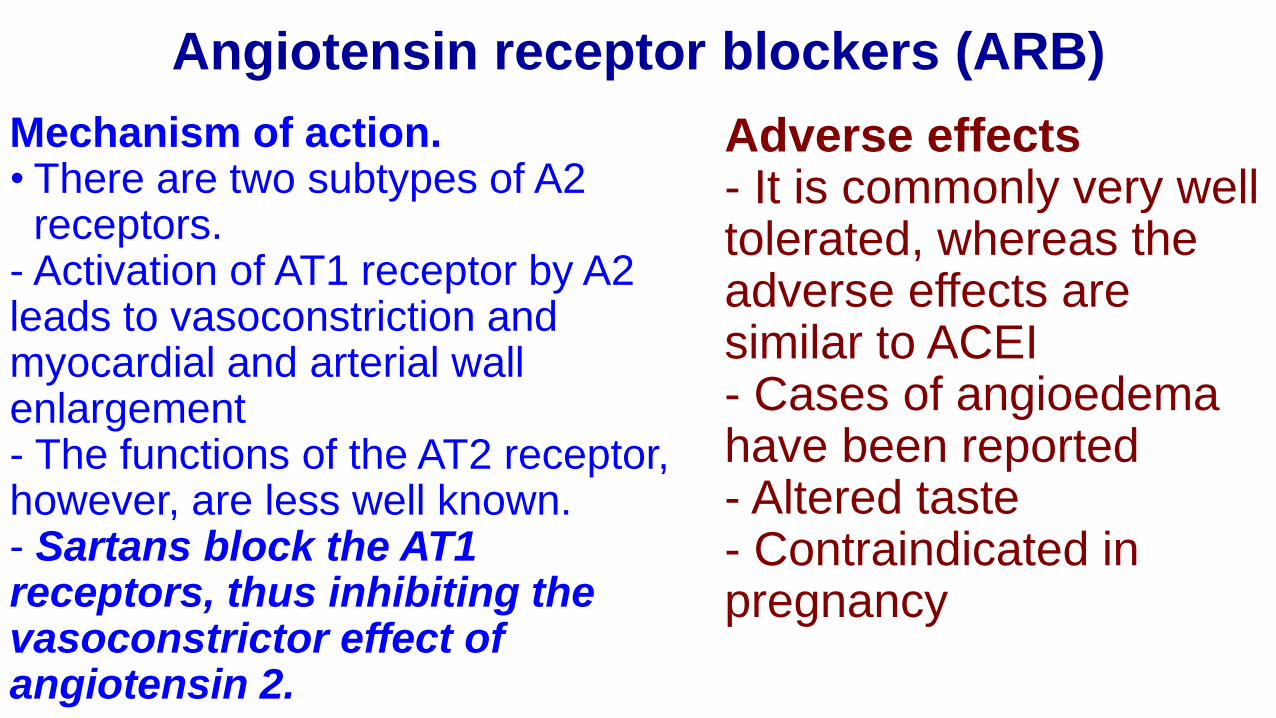
Angiotensin receptor blockers (ARB)
Mechanism of action.• There are two subtypes of A2
receptors.- Activation of AT1 receptor by A2 leads to vasoconstriction and myocardial and arterial wall enlargement- The functions of the AT2 receptor, however, are less well known. - Sartans block the AT1 receptors, thus inhibiting the vasoconstrictor effect of angiotensin 2.
Adverse effects- It is commonly very well tolerated, whereas the adverse effects are similar to ACEI- Cases of angioedema have been reported- Altered taste- Contraindicated in pregnancy
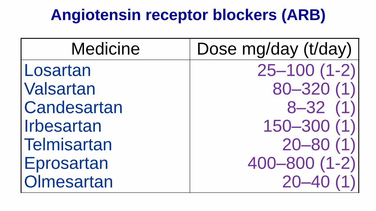
Angiotensin receptor blockers (ARB)
Medicine Dose mg/day (t/day)
LosartanValsartanCandesartanIrbesartanTelmisartanEprosartanOlmesartan
25–100 (1-2)80–320 (1)
8–32 (1)150–300 (1)
20–80 (1)400–800 (1-2)
20–40 (1)
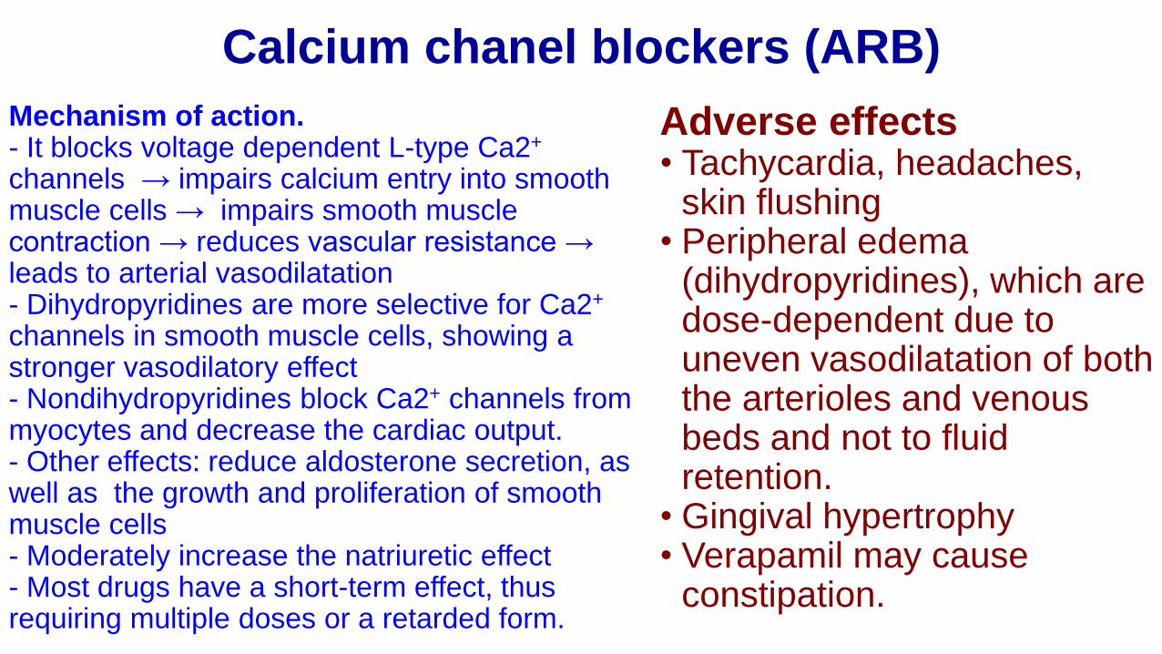
Calcium chanel blockers (ARB)
Mechanism of action.- It blocks voltage dependent L-type Ca2+
channels → impairs calcium entry into smooth muscle cells → impairs smooth muscle contraction → reduces vascular resistance → leads to arterial vasodilatation- Dihydropyridines are more selective for Ca2+
channels in smooth muscle cells, showing a stronger vasodilatory effect- Nondihydropyridines block Ca2+ channels from myocytes and decrease the cardiac output.- Other effects: reduce aldosterone secretion, as well as the growth and proliferation of smooth muscle cells- Moderately increase the natriuretic effect- Most drugs have a short-term effect, thus requiring multiple doses or a retarded form.
Adverse effects• Tachycardia, headaches,
skin flushing• Peripheral edema
(dihydropyridines), which are dose-dependent due to uneven vasodilatation of both the arterioles and venous beds and not to fluid retention.
• Gingival hypertrophy• Verapamil may cause
constipation.
Calcium chanel blockers (ARB)
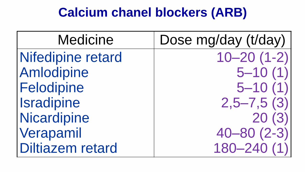
Medicine Dose mg/day (t/day)
Nifedipine retardAmlodipineFelodipineIsradipineNicardipineVerapamilDiltiazem retard
10–20 (1-2)5–10 (1)5–10 (1)
2,5–7,5 (3)20 (3)
40–80 (2-3)180–240 (1)
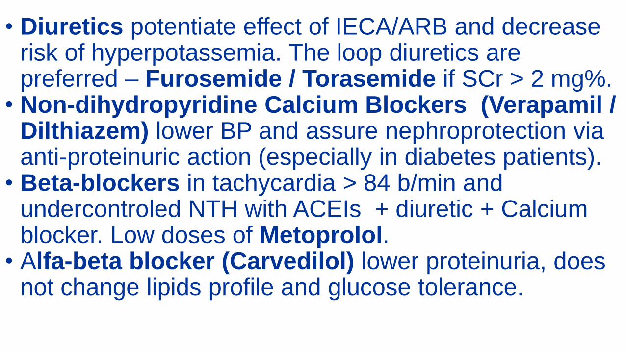
• Diuretics potentiate effect of IECA/ARB and decrease risk of hyperpotassemia. The loop diuretics are preferred – Furosemide / Torasemide if SCr > 2 mg%.
• Non-dihydropyridine Calcium Blockers (Verapamil / Dilthiazem) lower BP and assure nephroprotection via anti-proteinuric action (especially in diabetes patients).
• Beta-blockers in tachycardia > 84 b/min and undercontroled NTH with ACEIs + diuretic + Calcium blocker. Low doses of Metoprolol.
• Alfa-beta blocker (Carvedilol) lower proteinuria, does not change lipids profile and glucose tolerance.
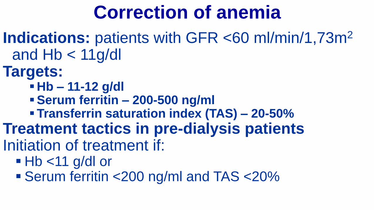
Indications: patients with GFR <60 ml/min/1,73m2
and Hb < 11g/dlTargets:
Hb – 11-12 g/dlSerum ferritin – 200-500 ng/mlTransferrin saturation index (TAS) – 20-50%
Treatment tactics in pre-dialysis patientsInitiation of treatment if: Hb <11 g/dl or Serum ferritin <200 ng/ml and TAS <20%
Correction of anemia
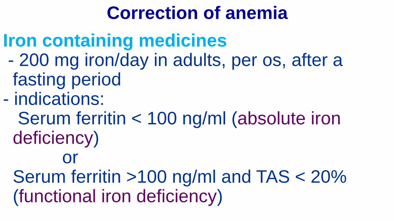
Iron containing medicines- 200 mg iron/day in adults, per os, after a fasting period
- indications:Serum ferritin < 100 ng/ml (absolute iron deficiency)
orSerum ferritin >100 ng/ml and TAS < 20% (functional iron deficiency)
Correction of anemia
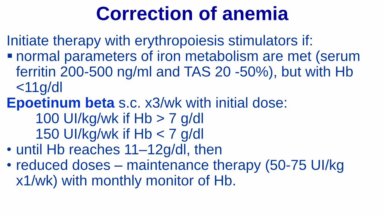
Initiate therapy with erythropoiesis stimulators if: normal parameters of iron metabolism are met (serum
ferritin 200-500 ng/ml and TAS 20 -50%), but with Hb<11g/dl
Epoetinum beta s.c. x3/wk with initial dose:100 UI/kg/wk if Hb > 7 g/dl150 UI/kg/wk if Hb < 7 g/dl
• until Hb reaches 11–12g/dl, then• reduced doses – maintenance therapy (50-75 UI/kg
x1/wk) with monthly monitor of Hb.
Correction of anemia
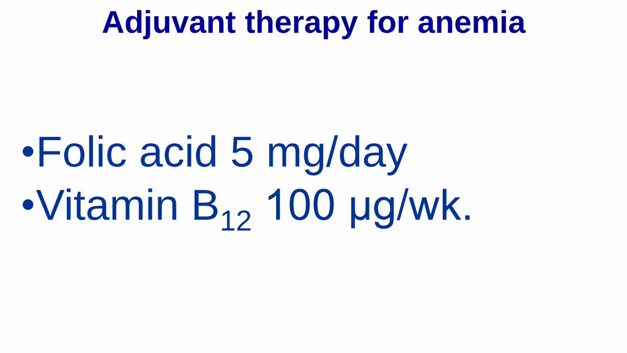
•Folic acid 5 mg/day
•Vitamin B12 100 µg/wk.
Adjuvant therapy for anemia
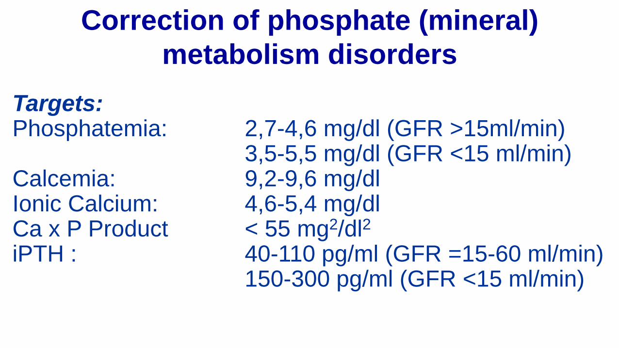
Targets:Phosphatemia: 2,7-4,6 mg/dl (GFR >15ml/min)
3,5-5,5 mg/dl (GFR <15 ml/min)Calcemia: 9,2-9,6 mg/dlIonic Calcium: 4,6-5,4 mg/dlCa x P Product < 55 mg2/dl2
iPTH : 40-110 pg/ml (GFR =15-60 ml/min)150-300 pg/ml (GFR <15 ml/min)
Correction of phosphate (mineral)
metabolism disorders
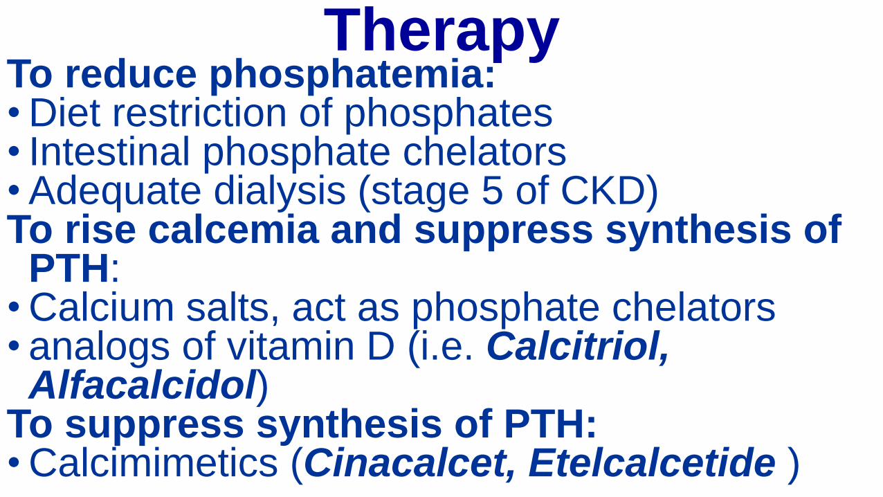
TherapyTo reduce phosphatemia:• Diet restriction of phosphates• Intestinal phosphate chelators• Adequate dialysis (stage 5 of CKD)To rise calcemia and suppress synthesis of PTH:
• Calcium salts, act as phosphate chelators• analogs of vitamin D (i.e. Calcitriol, Alfacalcidol)
To suppress synthesis of PTH:• Calcimimetics (Cinacalcet, Etelcalcetide )
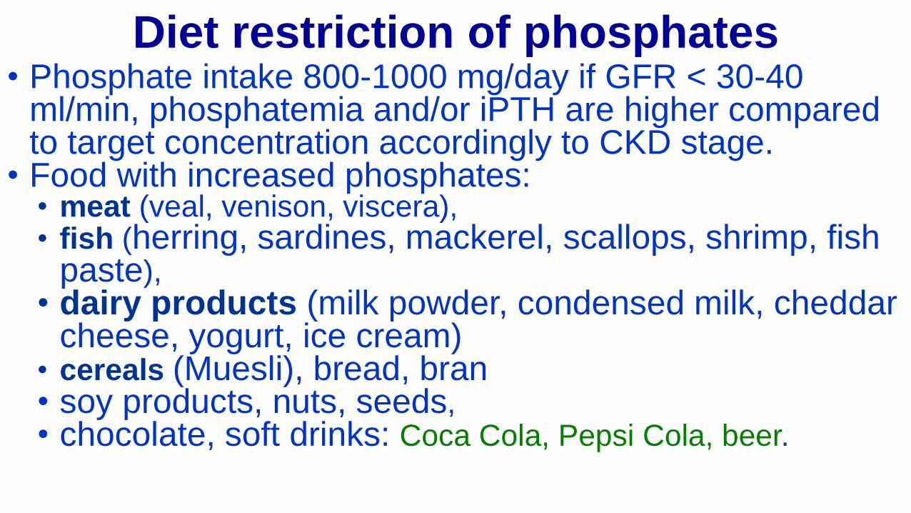
• Phosphate intake 800-1000 mg/day if GFR < 30-40 ml/min, phosphatemia and/or iPTH are higher compared to target concentration accordingly to CKD stage.
• Food with increased phosphates: • meat (veal, venison, viscera), • fish (herring, sardines, mackerel, scallops, shrimp, fish
paste), • dairy products (milk powder, condensed milk, cheddar
cheese, yogurt, ice cream)• cereals (Muesli), bread, bran• soy products, nuts, seeds, • chocolate, soft drinks: Coca Cola, Pepsi Cola, beer.
Diet restriction of phosphates
•Calcium salts
•Aluminum salts
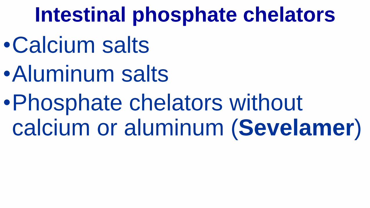
•Phosphate chelators without calcium or aluminum (Sevelamer)
Intestinal phosphate chelators
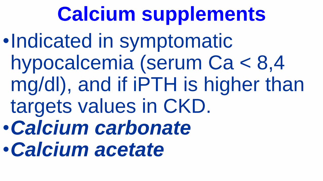
•Indicated in symptomatic hypocalcemia (serum Ca < 8,4 mg/dl), and if iPTH is higher than targets values in CKD.
•Calcium carbonate•Calcium acetate
Calcium supplements
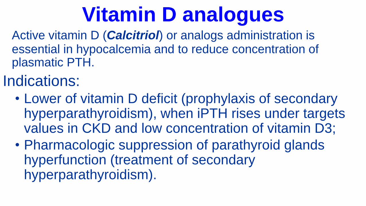
Active vitamin D (Calcitriol) or analogs administration is essential in hypocalcemia and to reduce concentration of plasmatic PTH.
Indications:• Lower of vitamin D deficit (prophylaxis of secondary
hyperparathyroidism), when iPTH rises under targets values in CKD and low concentration of vitamin D3;
• Pharmacologic suppression of parathyroid glands hyperfunction (treatment of secondary hyperparathyroidism).
Vitamin D analogues
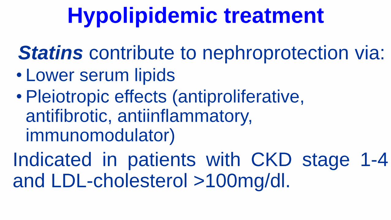
Statins contribute to nephroprotection via:• Lower serum lipids
• Pleiotropic effects (antiproliferative, antifibrotic, antiinflammatory, immunomodulator)
Indicated in patients with CKD stage 1-4and LDL-cholesterol >100mg/dl.
Hypolipidemic treatment
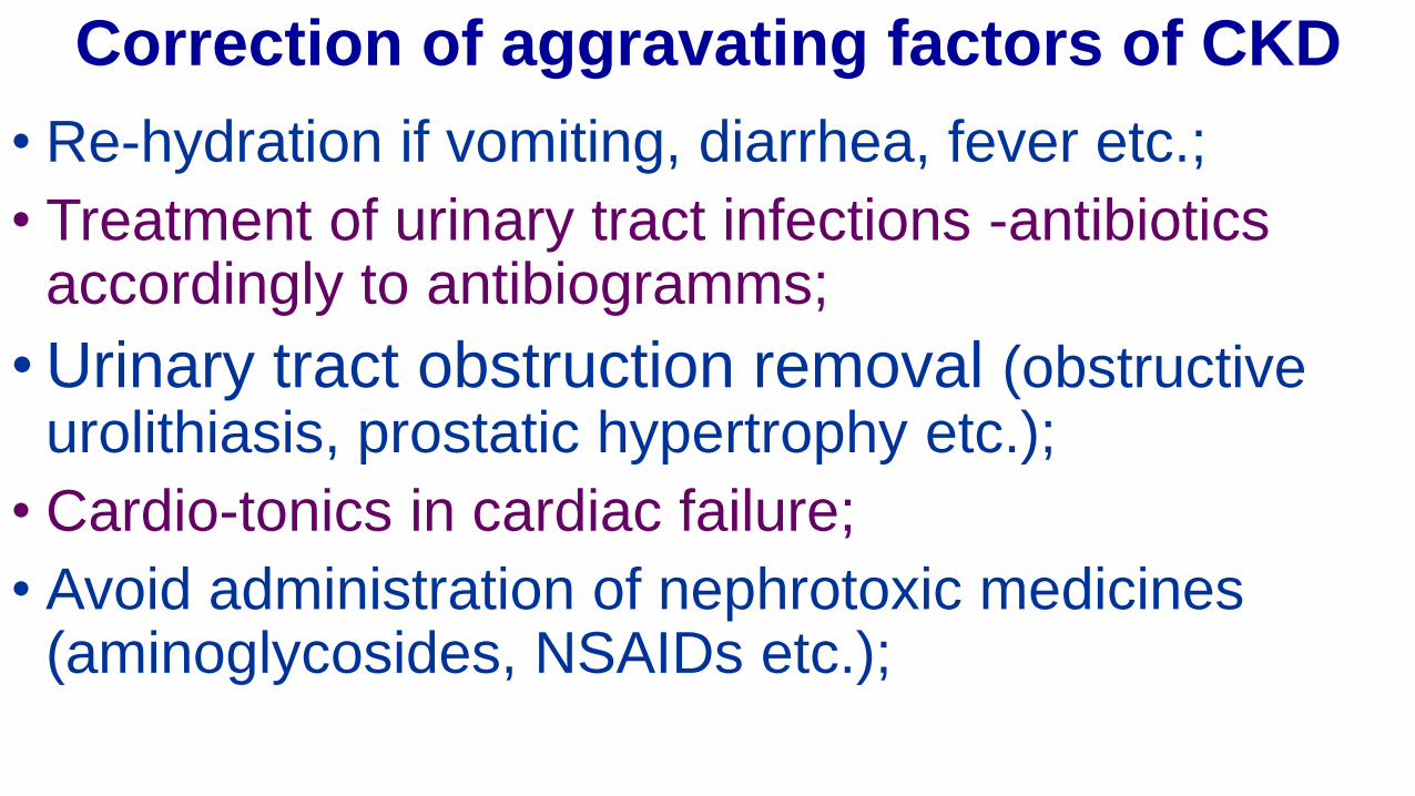
• Re-hydration if vomiting, diarrhea, fever etc.;
• Treatment of urinary tract infections -antibiotics accordingly to antibiogramms;
• Urinary tract obstruction removal (obstructive urolithiasis, prostatic hypertrophy etc.);
• Cardio-tonics in cardiac failure;
• Avoid administration of nephrotoxic medicines (aminoglycosides, NSAIDs etc.);
Correction of aggravating factors of CKD
Treatment of metabolic acidosis by alkali therapy is usually indicated to raise and maintain the plasma pH to greater than 7.20.
Indication: serum bicarbonate <22 mEq/LMedicines:
• Calcium carbonate 6–12g/day• Sodium bicarbonate 10–15g/day - per os• Sodium bicarbonate 14‰ – i.v.Formula to determine bicarbonate dose:
HCO3- deficit = deficit/L (desired serum HCO3
- - measured HCO3-) × 0.5 ×
body weight (volume of distribution for HCO3-)
(100 ml sol. NaHCO3 14‰ = 16,8mmol bicarbonate).
Correction of acidosis
•Psychological counseling;
•Training of patient with CKD;
•Avoid forearm vein due the need of arteriovenous fistula to start dialysis;
• Installation of arteriovenous fistula when eGFR is 20-25 ml/min/1,73 m2.
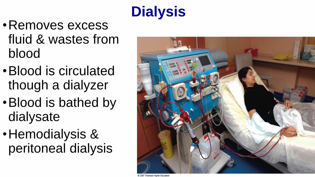
Dialysis•Removes excess fluid & wastes from blood
•Blood is circulated though a dialyzer
•Blood is bathed by dialysate
•Hemodialysis & peritoneal dialysis
Dialysis
•Hemodialysis• Lasts 3-4 hours
• 3 times/week
• Complications• Infections
• Blood clotting
• Hypotension
• Muscle cramping
• Headaches, weakness
• Nausea & vomiting
• Agitation
•Peritoneal dialysis• Vascular access not required
• Fewer dietary restrictions
• Can be scheduled when convenient
•Acute failure• Continuous renal replacement therapy (CRRT)
Kidney Transplants
•Restores function
•Allows a more liberal diet
•Frees patient from
dialysis
• Immunosuppressive
drug therapy• Many side effects
affecting nutrition
•Protein & energy
requirements increase
•Control CHO & lipids
•Sodium, potassium, &
phosphorus intakes
liberalized
•Calcium supplementation
•Be alert for potential food
borne infection
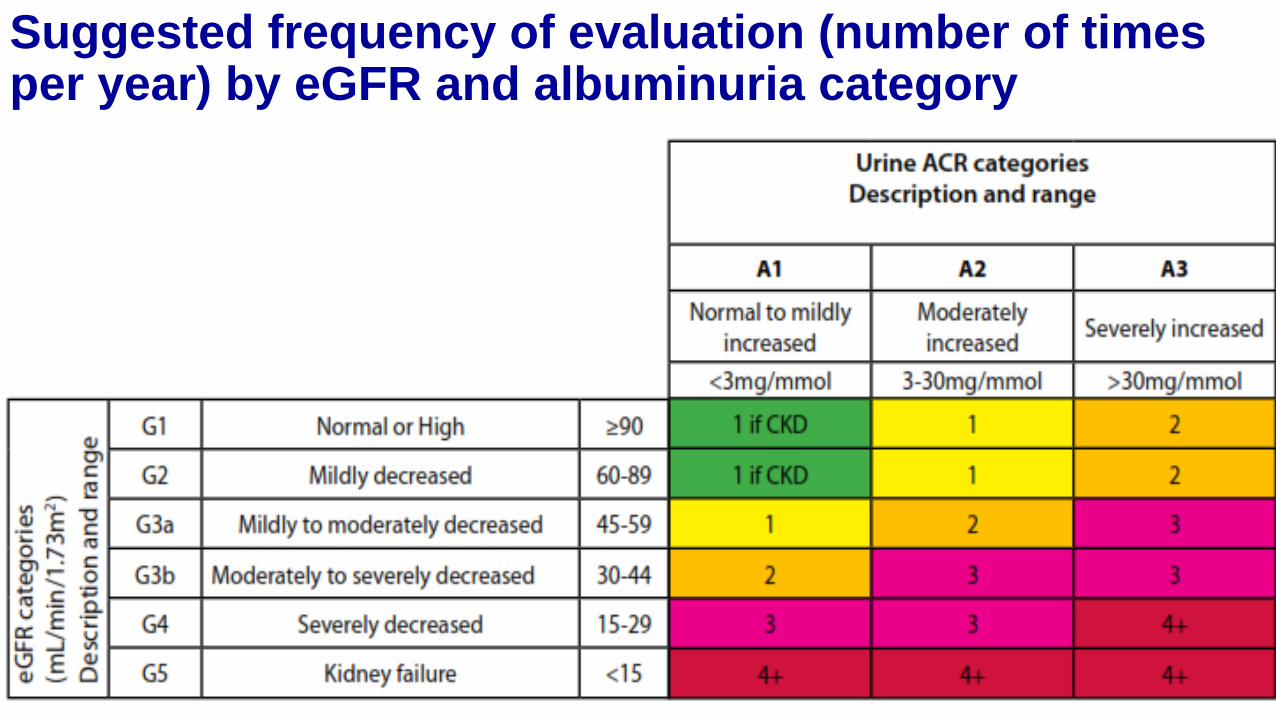
Suggested frequency of evaluation (number of times per year) by eGFR and albuminuria category
Top Related