


Languages
Pages
Legal

Chin Ptosis: Classification, Anatomy,and CorrectionEvan S. Garfein, M.D.,1 and Barry M. Zide, D.M.D., M.D.1
ABSTRACT
For years, the notion of chin ptosis was somehow integrated with the concept ofwitch’s chin. That was a mistake on many levels because chin droop has four major causes,all different and with some overlap. With this article, the surgeon can quickly diagnosewhich type and which therapeutic modality would work best. In some cases the problem is asimple fix, in others the droop can only be stabilized, and in the final two, definite correctiveprocedures are available. Of note, in certain situations two types of chin ptosis may overlapbecause both the patient and the surgeon may each contribute to the problems.For example, in dynamic ptosis, a droop that occurs with smile in the unoperated patientcan be exacerbated and further produced by certain surgical methods also. This paperclassifies the variations of the problems and explains the anatomy with the final emphasison long-term surgical correction, well described herein. This article is the ninth on thissubject and a review of them all would be helpful (greatly) for understanding the enigmasof the lower face.
KEYWORDS: Lip incompetence, chin ptosis, witch’s chin, chin droop, mentalis muscle
All chin ptosis patients are not alike. Theproper diagnosis of the type of chin ptosis places thepatient into one of four categories, which will deter-mine who can and who cannot be helped and by whichmethods. The four types of chin ptosis are: (1) illusory,(2) developmental, (3) iatrogenic, and (4) dynamic. Thelast two—iatrogenic and dynamic—may overlap.
As a general rule, chin ptosis describes the descentof the soft tissues of the presymphyseal soft tissues belowthe inferior border of the mandible. It is not the witch’schin, which projects forward. Chin ptosis cannot beaddressed by standard rhytidectomies, and certaindeformities may become more noticeable if othermisdirected procedures are performed, for example, asubmental lipectomy in the presence of dynamic ptosis.The goal of this article is to describe the normal and
abnormal anatomy, diagnosis, and management of thefour types of chin ptosis, as well as how to managedynamic ptosis in the presence of other problems—surgeon-caused or not.
ESSENTIAL CHIN ANATOMYThe mentalis muscles are paired elevators of the centrallower lip that originate from the mandible at the leveljust beneath the attached gingiva of the central andlateral incisors.1 They usually overlap and insert intothe deep dermis of the chin pad. Often some fat may liebetween the separate origins, but the height of theorigins is 8 to 10 mm.
A good way to think of the mentalis muscles is assearchlights that usually overlap to insert into the chin
1Institute of Reconstructive Plastic Surgery, New York UniversitySchool of Medicine, New York, New York.
Address for correspondence and reprint requests: Barry M. Zide,D.M.D., M.D., Institute of Reconstructive Plastic Surgery, New YorkUniversity School of Medicine, New York, NY 10016 (e-mail: [email protected]).
Craniomaxillofac Trauma Reconstruction 2008;1:1–14. Copyright #
2008 by Thieme Medical Publishers, Inc., 333 Seventh Avenue,New York, NY 10001, USA. Tel: +1(212) 584-4662.DOI 10.1055/s-0028-1098968. ISSN 1943-3875.
1
pad dermis. A chin cleft is essentially a muscle-deficientzone, where the ‘‘searchlights’’ are not overlapping; thatis, a ‘‘muscle-free’’ area. When the zone of overlap ispredominantly at the superior chin pad, the result is aninferior chin cleft with upper chin pad muscle excess.Patients with considerable superior overlap tend to havea deep labiomental fold with a high takeoff; that is, abulge in the upper portion of the pad.2 The true insertionof an individual’s mentalis muscles can often be seen inpatients with Bell’s palsy, where muscle atrophy and lossof lip elevation are seen on the affected side (Fig. 1).
To understand iatrogenic and developmental pto-sis, the surgeon must better understand the anatomy andaction of the mentalis muscles. On a sagittal cut throughthe midportion of the face, certain crucial anatomicallandmarks must be recognized and their importanceknown: (1) the horizontal upper fibers maintain lip
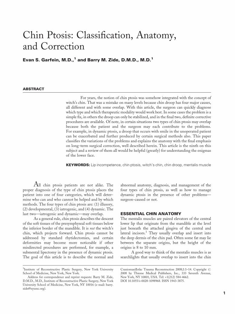
position—this part arises from just below the attachedgingival; and (2) the oblique fibers whose direction andvector of pull are responsible for lower lip elevation,pouting, and the ability to firmly press the lips together.The junction of the upper mentalis and the orbicularisoris muscles forms the labiomental fold. If an overlapoccurs, the fold is indistinct. If the orbicularis is thin ornarrow, the fold is high. A distinct junction leads to adistinct fold. Thus the labiomental fold marks the pointof junction of the lower lip stabilizers with the marginallip pursers (orbicularis oris) (Fig. 2).
The depressors of the lower lip include the twostrong depressors (the triangularis and the quadratus)and the ancillary depressor (i.e., the anterior platysma).The lower lip does not come down on the affected side incases of marginal mandibular palsy, and the mentalis onthat side is paretic as well.
Figure 1 Patient with Bell’s palsy. (A) Note atrophy of mentalis muscle with loss of bulk on the patient’s right. (B) Note
inability to elevate lower lip on affected side (right side) and loss of dimpling with attempted contraction of mentalis muscles.
Figure 2 (A) Arrow points to the most medial of the lip depressors, which consist of triangularis, quadratus, anterior
platysma. Shiny tissues medially are the mentalis insertions into the chin pad. (B) The level at which the orbicularis oris and the
mentalis muscles intersect leads, in this cadaver, to a high labiomental fold (arrowhead). The distinctiveness of the fold is
determined by the amount of overlap. The upper part of the mentalis muscle contains horizontally directed fibers that stabilize
the lower lip (arrow). The oblique lower fibers allow the lip to pout.
2 CRANIOMAXILLOFACIAL TRAUMA & RECONSTRUCTION/VOLUME 1, NUMBER 1 2008
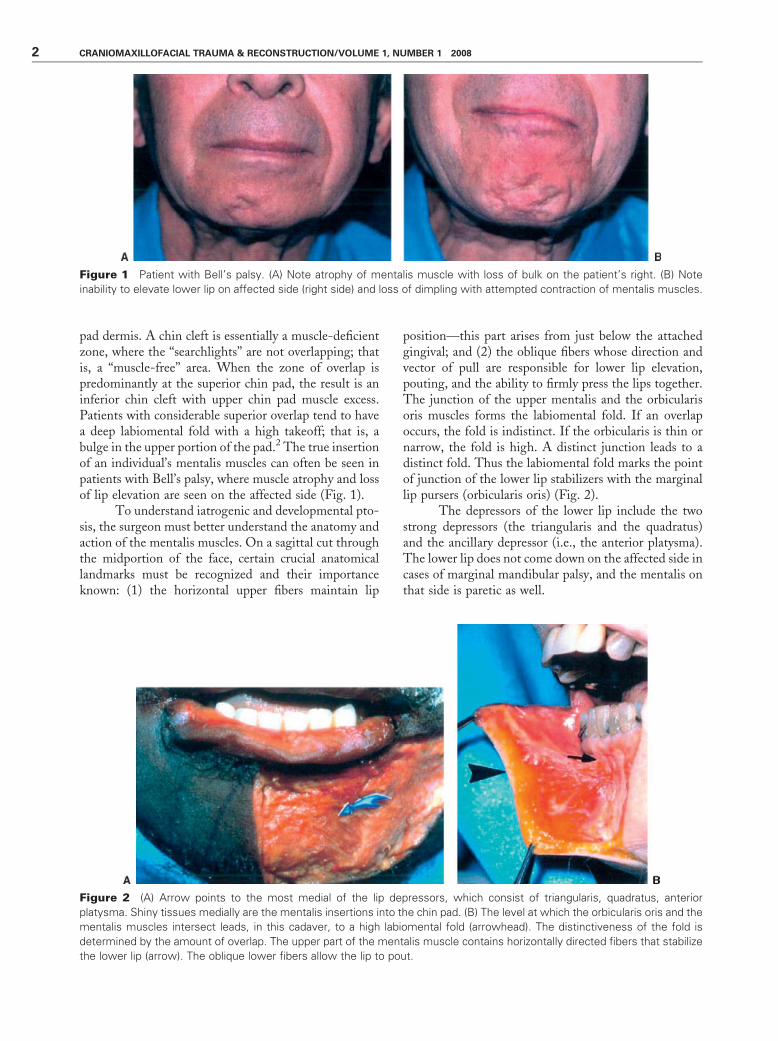
ABNORMAL ANATOMYThe key in managing the issue of chin ptosis rests onmaking the correct diagnosis and having a clear under-standing of the involved anatomy of each type. Thesurgeon must look at the chin as starting at the lower lipand continuing onto the submental region. A prominentsubmental crease, when combined with lack of boneyprojection and ptosis of the soft tissues of the chin pad,constitutes the witch’s chin deformity described byGonzalez-Ulloa in 19723; but, in truth, his nomencla-ture was inaccurate. In the illusory type of chin ptosis, lipposition is normal, the intraoral sulcus is normal, and themovement is normal. Illusory ptosis, as the name suggests,describes the perceived descent of the chin without thisactually being the case. It is caused by an exaggeratedcrease that may extend cephalad over the inferior man-dibular border on each side. The patient has usually nothad surgery. The solution for this is straightforward:Eliminate the submental crease (by excision or augmen-tation) and undermine the soft tissues up onto the face toallow redraping.
Lesavoy,4 Peterson,5 and Feldman6 all describe avariety of techniques to address the exaggerated sub-mental crease. The senior author’s preferred technique isto either excise the fold as an ellipse or to deepithelialize
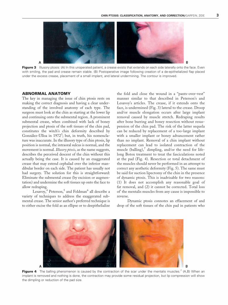
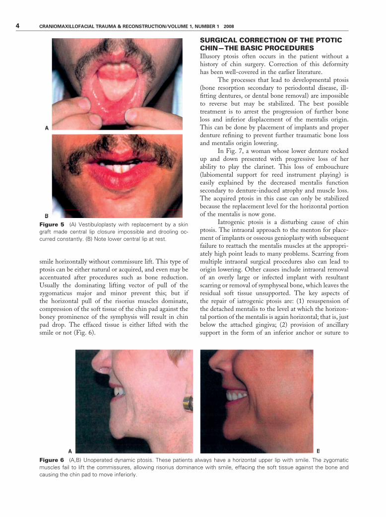
the fold and close the wound in a ‘‘pants-over-vest’’manner similar to that described in Peterson’s andLesavoy’s articles. The crease, if it extends onto theface, is undermined (Fig. 3) lateral to the crease. Droopand/or muscle elongation occurs after large implantremoval caused by muscle stretch. Redraping resultsafter bone burring and boney resection without resus-pension of the chin pad. The risk of the latter sequelacan be reduced by replacement of a too-large implantwith a smaller implant or boney advancement ratherthan no implant. Removal of a chin implant withoutreplacement can lead to isolated contraction of themuscle (balling),7 dimpling, and/or the need for life-long Botox treatment to treat the fasciculations notedat the pad (Fig. 4). Resection or total detachment ofthe muscles should never be performed in an attempt tocorrect any aesthetic deformity (Fig. 5). The same mustbe said for suction lipectomy of the chin in the presenceof dynamic ptosis. This is inadvisable for two reasons:(1) It does not accomplish any reasonable goal offat removal, and (2) it cannot be corrected. Total lossof’ the mentalis muscles from any cause is impossible toreverse.
Dynamic ptosis connotes an effacement of anddrop of the soft tissues of the chin pad in patients who
Figure 3 Illusory ptosis: (A) In this unoperated patient, a crease exists that extends on each side laterally onto the face. Even
with smiling, the pad and crease remain stable. (B) Postoperative image following creation of a de-epithelialized flap placed
under the excess crease, placement of a small implant, and lateral undermining. The contour is improved.
Figure 4 The balling phenomenon is caused by the contraction of the scar under the mentalis muscles.7 (A,B) When an
implant is removed and nothing is done, the contraction may provide some residual projection, but lip compression will show
the dimpling or reduction of the pad size.
CHIN PTOSIS: CLASSIFICATION, ANATOMY, AND CORRECTION/GARFEIN, ZIDE 3
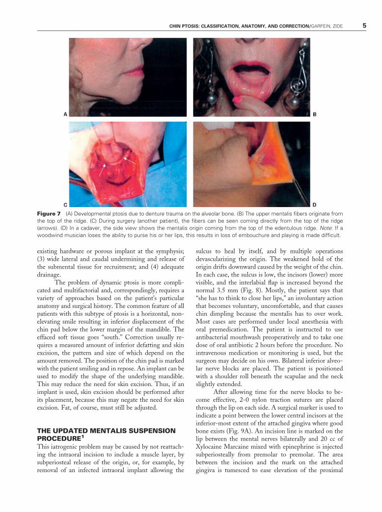
smile horizontally without commissure lift. This type ofptosis can be either natural or acquired, and even may beaccentuated after procedures such as bone reduction.Usually the dominating lifting vector of pull of thezygomaticus major and minor prevent this; but ifthe horizontal pull of the risorius muscles dominate,compression of the soft tissue of the chin pad against theboney prominence of the symphysis will result in chinpad drop. The effaced tissue is either lifted with thesmile or not (Fig. 6).
SURGICAL CORRECTION OF THE PTOTICCHIN—THE BASIC PROCEDURESIllusory ptosis often occurs in the patient without ahistory of chin surgery. Correction of this deformityhas been well-covered in the earlier literature.
The processes that lead to developmental ptosis(bone resorption secondary to periodontal disease, ill-fitting dentures, or dental bone removal) are impossibleto reverse but may be stabilized. The best possibletreatment is to arrest the progression of further boneloss and inferior displacement of the mentalis origin.This can be done by placement of implants and properdenture refining to prevent further traumatic bone lossand mentalis origin lowering.
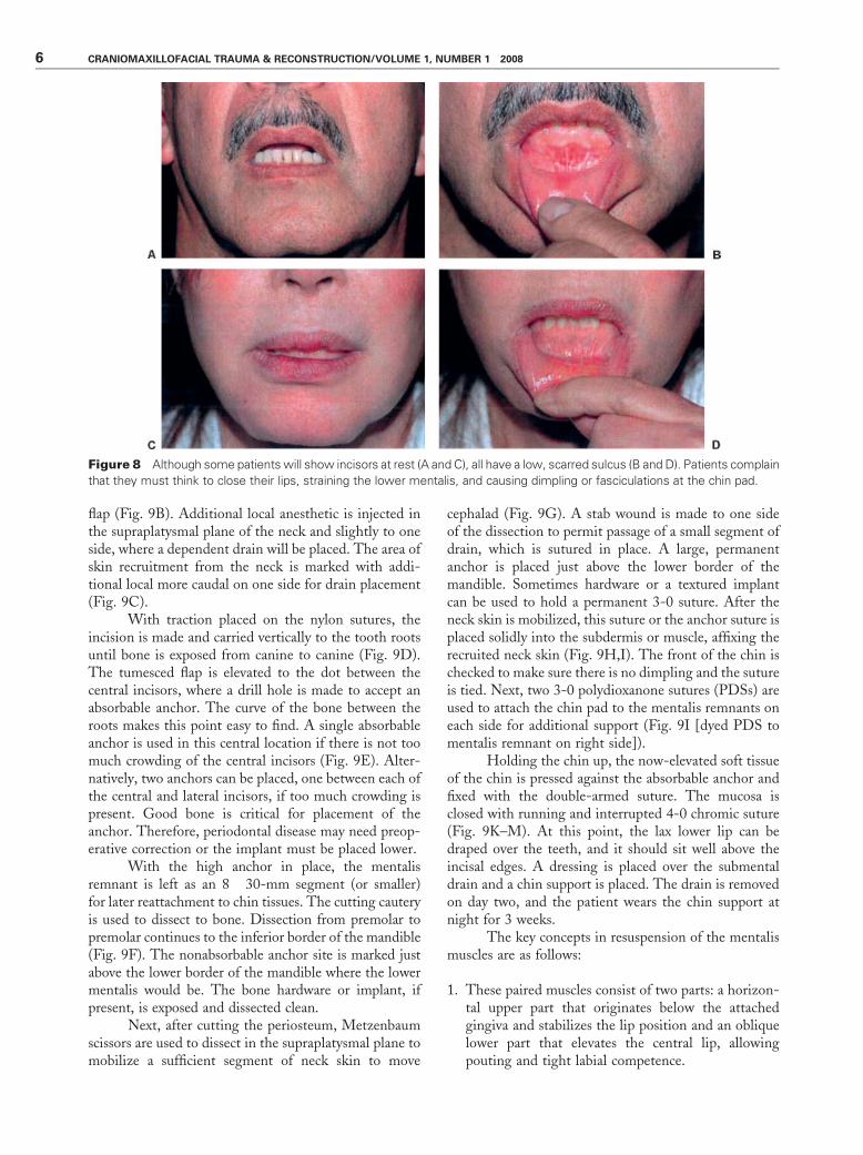
In Fig. 7, a woman whose lower denture rockedup and down presented with progressive loss of herability to play the clarinet. This loss of embouchure(labiomental support for reed instrument playing) iseasily explained by the decreased mentalis functionsecondary to denture-induced atrophy and muscle loss.The acquired ptosis in this case can only be stabilizedbecause the replacement level for the horizontal portionof the mentalis is now gone.
Iatrogenic ptosis is a disturbing cause of chinptosis. The intraoral approach to the menton for place-ment of implants or osseous genioplasty with subsequentfailure to reattach the mentalis muscles at the appropri-ately high point leads to many problems. Scarring frommultiple intraoral surgical procedures also can lead toorigin lowering. Other causes include intraoral removalof an overly large or infected implant with resultantscarring or removal of symphyseal bone, which leaves theresidual soft tissue unsupported. The key aspects ofthe repair of iatrogenic ptosis are: (1) resuspension ofthe detached mentalis to the level at which the horizon-tal portion of the mentalis is again horizontal; that is, justbelow the attached gingiva; (2) provision of ancillarysupport in the form of an inferior anchor or suture to
Figure 5 (A) Vestibuloplasty with replacement by a skin
graft made central lip closure impossible and drooling oc-
curred constantly. (B) Note lower central lip at rest.
Figure 6 (A,B) Unoperated dynamic ptosis. These patients always have a horizontal upper lip with smile. The zygomatic
muscles fail to lift the commissures, allowing risorius dominance with smile, effacing the soft tissue against the bone and
causing the chin pad to move inferiorly.
4 CRANIOMAXILLOFACIAL TRAUMA & RECONSTRUCTION/VOLUME 1, NUMBER 1 2008
existing hardware or porous implant at the symphysis;(3) wide lateral and caudal undermining and release ofthe submental tissue for recruitment; and (4) adequatedrainage.
The problem of dynamic ptosis is more compli-cated and multifactorial and, correspondingly, requires avariety of approaches based on the patient’s particularanatomy and surgical history. The common feature of allpatients with this subtype of ptosis is a horizontal, non-elevating smile resulting in inferior displacement of thechin pad below the lower margin of the mandible. Theeffaced soft tissue goes ‘‘south.’’ Correction usually re-quires a measured amount of inferior defatting and skinexcision, the pattern and size of which depend on theamount removed. The position of the chin pad is markedwith the patient smiling and in repose. An implant can beused to modify the shape of the underlying mandible.This may reduce the need for skin excision. Thus, if animplant is used, skin excision should be performed afterits placement, because this may negate the need for skinexcision. Fat, of course, must still be adjusted.
THE UPDATED MENTALIS SUSPENSIONPROCEDURE1
This iatrogenic problem may be caused by not reattach-ing the intraoral incision to include a muscle layer, bysubperiosteal release of the origin, or, for example, byremoval of an infected intraoral implant allowing the
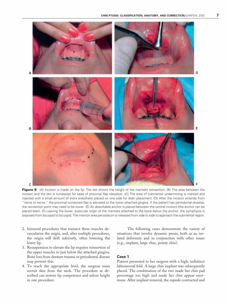
sulcus to heal by itself, and by multiple operationsdevascularizing the origin. The weakened hold of theorigin drifts downward caused by the weight of the chin.In each case, the sulcus is low, the incisors (lower) morevisible, and the interlabial flap is increased beyond thenormal 3.5 mm (Fig. 8). Mostly, the patient says that‘‘she has to think to close her lips,’’ an involuntary actionthat becomes voluntary, uncomfortable, and that causeschin dimpling because the mentalis has to over work.Most cases are performed under local anesthesia withoral premedication. The patient is instructed to useantibacterial mouthwash preoperatively and to take onedose of oral antibiotic 2 hours before the procedure. Nointravenous medication or monitoring is used, but thesurgeon may decide on his own. Bilateral inferior alveo-lar nerve blocks are placed. The patient is positionedwith a shoulder roll beneath the scapulae and the neckslightly extended.
After allowing time for the nerve blocks to be-come effective, 2-0 nylon traction sutures are placedthrough the lip on each side. A surgical marker is used toindicate a point between the lower central incisors at theinferior-most extent of the attached gingiva where goodbone exists (Fig. 9A). An incision line is marked on thelip between the mental nerves bilaterally and 20 cc ofXylocaine Marcaine mixed with epinephrine is injectedsubperiosteally from premolar to premolar. The areabetween the incision and the mark on the attachedgingiva is tumesced to ease elevation of the proximal
Figure 7 (A) Developmental ptosis due to denture trauma on the alveolar bone. (B) The upper mentalis fibers originate from
the top of the ridge. (C) During surgery (another patient), the fibers can be seen coming directly from the top of the ridge
(arrows). (D) In a cadaver, the side view shows the mentalis origin coming from the top of the edentulous ridge. Note: If a
woodwind musician loses the ability to purse his or her lips, this results in loss of embouchure and playing is made difficult.
CHIN PTOSIS: CLASSIFICATION, ANATOMY, AND CORRECTION/GARFEIN, ZIDE 5
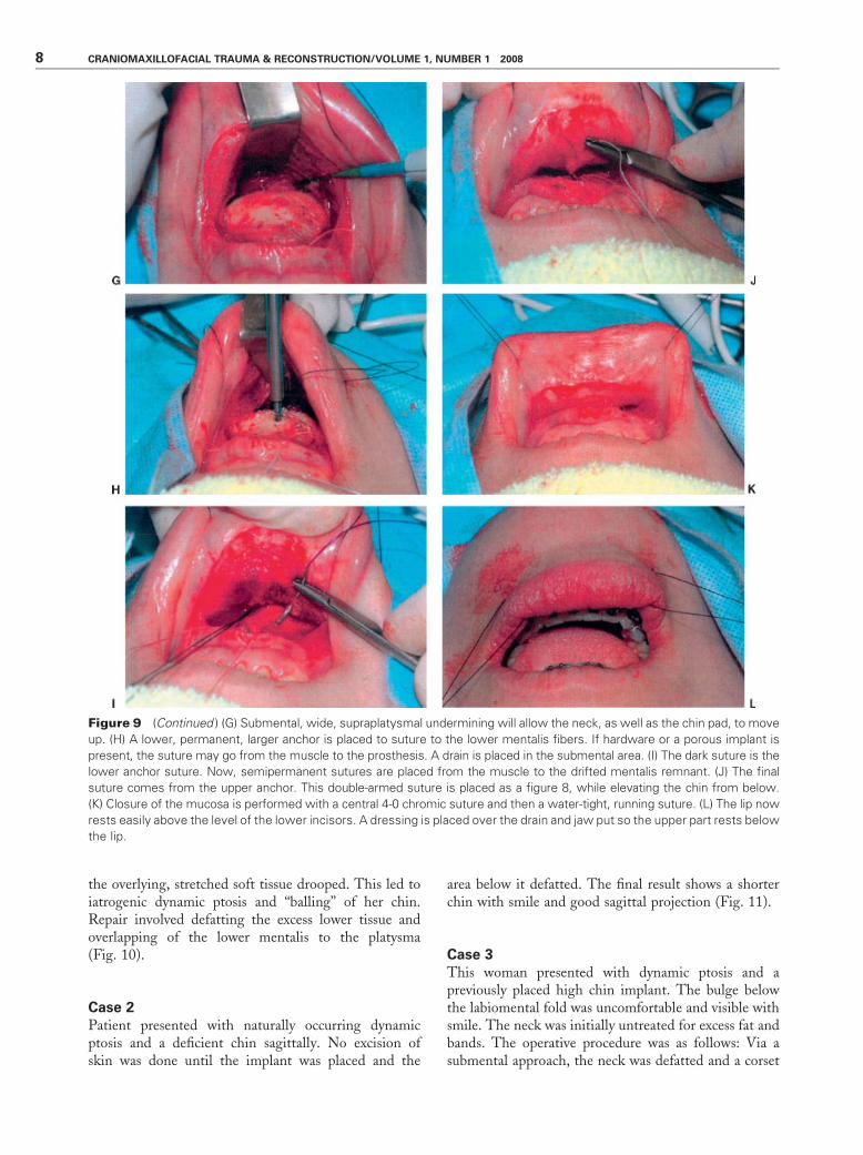
flap (Fig. 9B). Additional local anesthetic is injected inthe supraplatysmal plane of the neck and slightly to oneside, where a dependent drain will be placed. The area ofskin recruitment from the neck is marked with addi-tional local more caudal on one side for drain placement(Fig. 9C).
With traction placed on the nylon sutures, theincision is made and carried vertically to the tooth rootsuntil bone is exposed from canine to canine (Fig. 9D).The tumesced flap is elevated to the dot between thecentral incisors, where a drill hole is made to accept anabsorbable anchor. The curve of the bone between theroots makes this point easy to find. A single absorbableanchor is used in this central location if there is not toomuch crowding of the central incisors (Fig. 9E). Alter-natively, two anchors can be placed, one between each ofthe central and lateral incisors, if too much crowding ispresent. Good bone is critical for placement of theanchor. Therefore, periodontal disease may need preop-erative correction or the implant must be placed lower.
With the high anchor in place, the mentalisremnant is left as an 8� 30-mm segment (or smaller)for later reattachment to chin tissues. The cutting cauteryis used to dissect to bone. Dissection from premolar topremolar continues to the inferior border of the mandible(Fig. 9F). The nonabsorbable anchor site is marked justabove the lower border of the mandible where the lowermentalis would be. The bone hardware or implant, ifpresent, is exposed and dissected clean.
Next, after cutting the periosteum, Metzenbaumscissors are used to dissect in the supraplatysmal plane tomobilize a sufficient segment of neck skin to move
cephalad (Fig. 9G). A stab wound is made to one sideof the dissection to permit passage of a small segment ofdrain, which is sutured in place. A large, permanentanchor is placed just above the lower border of themandible. Sometimes hardware or a textured implantcan be used to hold a permanent 3-0 suture. After theneck skin is mobilized, this suture or the anchor suture isplaced solidly into the subdermis or muscle, affixing therecruited neck skin (Fig. 9H,I). The front of the chin ischecked to make sure there is no dimpling and the sutureis tied. Next, two 3-0 polydioxanone sutures (PDSs) areused to attach the chin pad to the mentalis remnants oneach side for additional support (Fig. 9I [dyed PDS tomentalis remnant on right side]).
Holding the chin up, the now-elevated soft tissueof the chin is pressed against the absorbable anchor andfixed with the double-armed suture. The mucosa isclosed with running and interrupted 4-0 chromic suture(Fig. 9K–M). At this point, the lax lower lip can bedraped over the teeth, and it should sit well above theincisal edges. A dressing is placed over the submentaldrain and a chin support is placed. The drain is removedon day two, and the patient wears the chin support atnight for 3 weeks.
The key concepts in resuspension of the mentalismuscles are as follows:
1. These paired muscles consist of two parts: a horizon-tal upper part that originates below the attachedgingiva and stabilizes the lip position and an obliquelower part that elevates the central lip, allowingpouting and tight labial competence.
Figure 8 Although some patients will show incisors at rest (A and C), all have a low, scarred sulcus (B and D). Patients complain
that they must think to close their lips, straining the lower mentalis, and causing dimpling or fasciculations at the chin pad.
6 CRANIOMAXILLOFACIAL TRAUMA & RECONSTRUCTION/VOLUME 1, NUMBER 1 2008
2. Intraoral procedures that transect these muscles de-vascularize the origin, and, after multiple procedures,the origin will drift inferiorly, often lowering thelower lip.
3. Resuspension to elevate the lip requires reinsertion ofthe upper muscles to just below the attached gingiva.Bone loss from denture trauma or periodontal diseasemay prevent this.
4. To reach the appropriate level, the surgeon mustrecruit skin from the neck. The procedure as de-scribed can restore lip competence and sulcus heightin one procedure.
The following cases demonstrate the variety ofsituations that involve dynamic ptosis, both as an iso-lated deformity and in conjunction with other issues(e.g., implant, large chin, pointy chin).
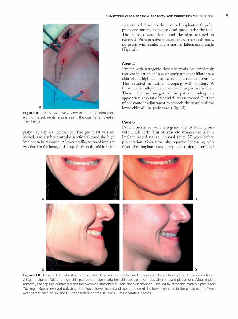
Case 1
Patient presented to her surgeon with a high, indistinctlabiomental fold. A large chin implant was subsequentlyplaced. The combination of the two made her chin padpercentage too high and made her chin appear enor-mous. After implant removal, the capsule contracted and
Figure 9 (A) Incision is made on the lip. The dot shows the height of the mentalis reinsertion. (B) The area between the
incision and the dot is tumesced for ease of proximal flap elevation. (C) The area of submental undermining is marked and
injected with a small amount of extra anesthetic placed on one side for drain placement. (D) After the incision extends from
‘‘nerve to nerve,’’ the proximal tumesced flap is elevated to the lower attached gingiva. If the patient has periodontal disease,
the reinsertion point may need to be lower. (E) An absorbable anchor is placed between the central incisors (the anchor can be
placed later). (F) Leaving the lower, avascular origin of the mentalis attached to the bone below the anchor, the symphysis is
exposed from bicuspid to bicuspid. The menton area periosteum is released from side to side to approach the submental region.
CHIN PTOSIS: CLASSIFICATION, ANATOMY, AND CORRECTION/GARFEIN, ZIDE 7
the overlying, stretched soft tissue drooped. This led toiatrogenic dynamic ptosis and ‘‘balling’’ of her chin.Repair involved defatting the excess lower tissue andoverlapping of the lower mentalis to the platysma(Fig. 10).
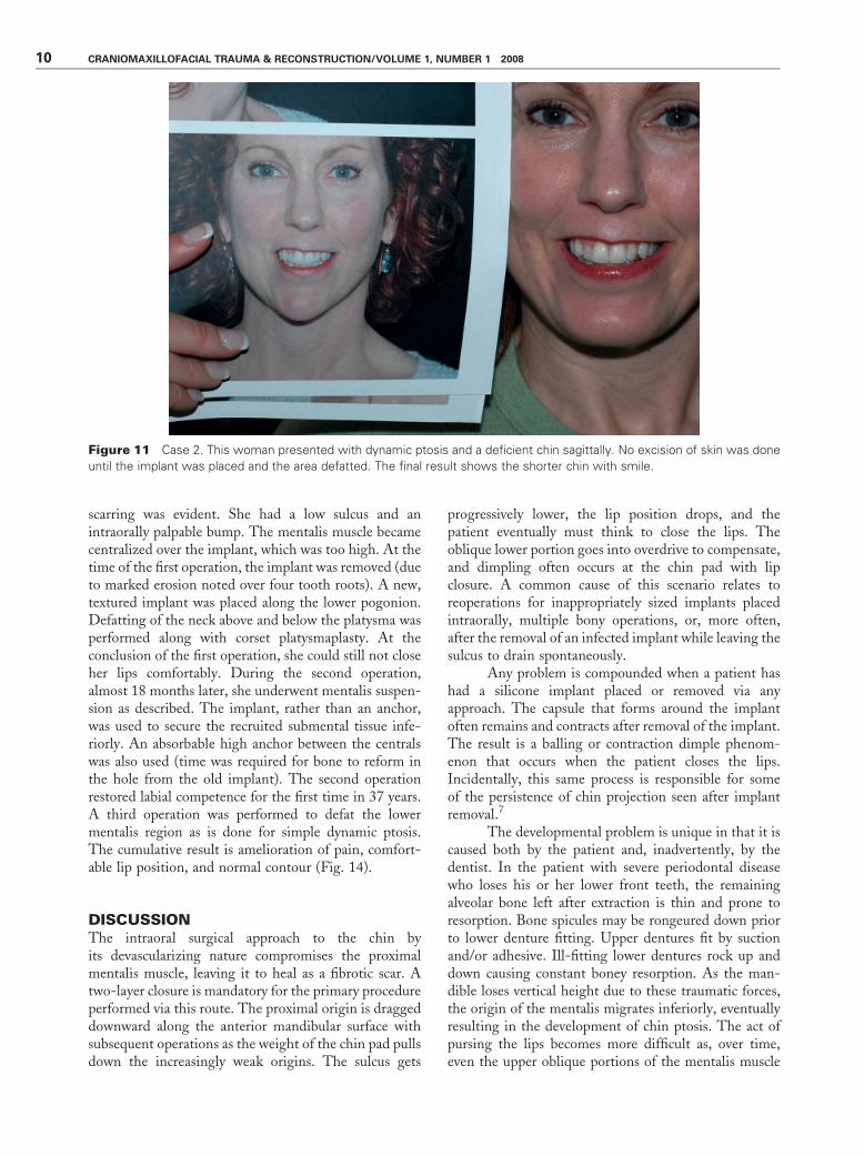
Case 2
Patient presented with naturally occurring dynamicptosis and a deficient chin sagittally. No excision ofskin was done until the implant was placed and the
area below it defatted. The final result shows a shorterchin with smile and good sagittal projection (Fig. 11).
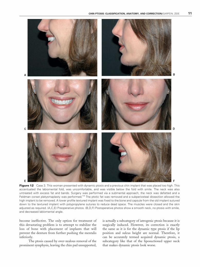
Case 3
This woman presented with dynamic ptosis and apreviously placed high chin implant. The bulge belowthe labiomental fold was uncomfortable and visible withsmile. The neck was initially untreated for excess fat andbands. The operative procedure was as follows: Via asubmental approach, the neck was defatted and a corset
Figure 9 (Continued ) (G) Submental, wide, supraplatysmal undermining will allow the neck, as well as the chin pad, to move
up. (H) A lower, permanent, larger anchor is placed to suture to the lower mentalis fibers. If hardware or a porous implant is
present, the suture may go from the muscle to the prosthesis. A drain is placed in the submental area. (I) The dark suture is the
lower anchor suture. Now, semipermanent sutures are placed from the muscle to the drifted mentalis remnant. (J) The final
suture comes from the upper anchor. This double-armed suture is placed as a figure 8, while elevating the chin from below.
(K) Closure of the mucosa is performed with a central 4-0 chromic suture and then a water-tight, running suture. (L) The lip now
rests easily above the level of the lower incisors. A dressing is placed over the drain and jaw put so the upper part rests below
the lip.
8 CRANIOMAXILLOFACIAL TRAUMA & RECONSTRUCTION/VOLUME 1, NUMBER 1 2008
platysmaplasty was performed. The ptotic fat was re-moved, and a subperiosteal dissection allowed the highimplant to be removed. A lower profile, textured implantwas fixed to the bone, and a capsule from the old implant
was sutured down to the textured implant with poly-propylene sutures to reduce dead space under the fold.The muscles were closed and the skin adjusted asrequired. Postoperative pictures show a smooth neck,no ptosis with smile, and a normal labiomental angle(Fig. 12).
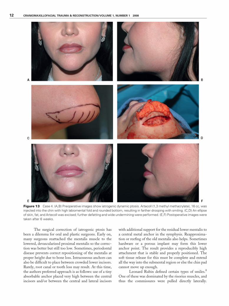
Case 4
Patient with iatrogenic dynamic ptosis had previouslyreceived injection of 16 cc of semipermanent filler into achin with a high labiomental fold and rounded bottom.This resulted in farther drooping with smiling. Afull-thickness elliptoid skin excision was performed first.Then, based on images of the patient smiling, anappropriate amount of fat and filler was excised. Furtherminor contour adjustment to smooth the margin of thelower chin will be performed (Fig. 13).
Case 5
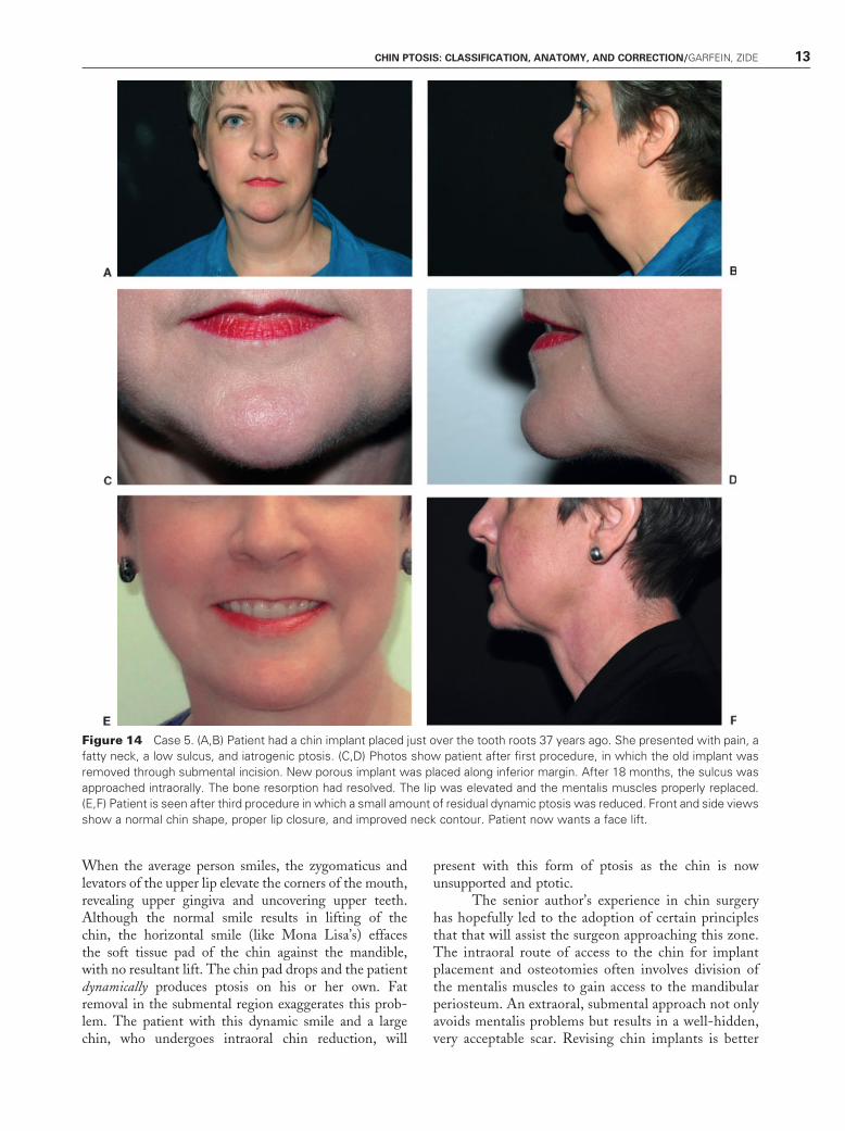
Patient presented with iatrogenic and dynamic ptosiswith a full neck. This 56-year-old woman had a chinimplant placed via an intraoral route 37 years beforepresentation. Over time, she reported increasing painfrom the implant (secondary to erosion). Intraoral
Figure 10 Case 1. This patient presented with a high labiomental fold and removal of a large chin implant. The combination of
a high, indistinct fold and high chin pad percentage made her chin appear enormous after implant placement. After implant
removal, the capsule contracted and the overlying stretched muscle and skin drooped. This led to iatrogenic dynamic ptosis and
‘‘balling.’’ Repair involved defatting the excess lower tissue and reorientation of the lower mentalis to the platysma in a ‘‘vest
over pants’’ fashion. (A and C) Preoperative photos. (B and D) Postoperative photos.
Figure 9 (Continued ) (M) A view of the dependent drain
exiting the submental area is seen. The drain is removed in
1 or 2 days.
CHIN PTOSIS: CLASSIFICATION, ANATOMY, AND CORRECTION/GARFEIN, ZIDE 9
scarring was evident. She had a low sulcus and anintraorally palpable bump. The mentalis muscle becamecentralized over the implant, which was too high. At thetime of the first operation, the implant was removed (dueto marked erosion noted over four tooth roots). A new,textured implant was placed along the lower pogonion.Defatting of the neck above and below the platysma wasperformed along with corset platysmaplasty. At theconclusion of the first operation, she could still not closeher lips comfortably. During the second operation,almost 18 months later, she underwent mentalis suspen-sion as described. The implant, rather than an anchor,was used to secure the recruited submental tissue infe-riorly. An absorbable high anchor between the centralswas also used (time was required for bone to reform inthe hole from the old implant). The second operationrestored labial competence for the first time in 37 years.A third operation was performed to defat the lowermentalis region as is done for simple dynamic ptosis.The cumulative result is amelioration of pain, comfort-able lip position, and normal contour (Fig. 14).
DISCUSSIONThe intraoral surgical approach to the chin byits devascularizing nature compromises the proximalmentalis muscle, leaving it to heal as a fibrotic scar. Atwo-layer closure is mandatory for the primary procedureperformed via this route. The proximal origin is draggeddownward along the anterior mandibular surface withsubsequent operations as the weight of the chin pad pullsdown the increasingly weak origins. The sulcus gets
progressively lower, the lip position drops, and thepatient eventually must think to close the lips. Theoblique lower portion goes into overdrive to compensate,and dimpling often occurs at the chin pad with lipclosure. A common cause of this scenario relates toreoperations for inappropriately sized implants placedintraorally, multiple bony operations, or, more often,after the removal of an infected implant while leaving thesulcus to drain spontaneously.
Any problem is compounded when a patient hashad a silicone implant placed or removed via anyapproach. The capsule that forms around the implantoften remains and contracts after removal of the implant.The result is a balling or contraction dimple phenom-enon that occurs when the patient closes the lips.Incidentally, this same process is responsible for someof the persistence of chin projection seen after implantremoval.7
The developmental problem is unique in that it iscaused both by the patient and, inadvertently, by thedentist. In the patient with severe periodontal diseasewho loses his or her lower front teeth, the remainingalveolar bone left after extraction is thin and prone toresorption. Bone spicules may be rongeured down priorto lower denture fitting. Upper dentures fit by suctionand/or adhesive. Ill-fitting lower dentures rock up anddown causing constant boney resorption. As the man-dible loses vertical height due to these traumatic forces,the origin of the mentalis migrates inferiorly, eventuallyresulting in the development of chin ptosis. The act ofpursing the lips becomes more difficult as, over time,even the upper oblique portions of the mentalis muscle
Figure 11 Case 2. This woman presented with dynamic ptosis and a deficient chin sagittally. No excision of skin was done
until the implant was placed and the area defatted. The final result shows the shorter chin with smile.
10 CRANIOMAXILLOFACIAL TRAUMA & RECONSTRUCTION/VOLUME 1, NUMBER 1 2008
become ineffective. The only option for treatment ofthis devastating problem is to attempt to stabilize theloss of bone with placement of implants that willprevent the denture from further pushing the mentalisinferiorly.
The ptosis caused by over-zealous removal of theprominent symphysis, leaving the chin pad unsupported,
is actually a subcategory of iatrogenic ptosis because it issurgically induced. However, its correction is exactlythe same as it is for the dynamic type ptosis if the lipposition and sulcus height are normal. Therefore, itcan be accurately termed acquired dynamic ptosis, asubcategory like that of the liposuctioned upper neckthat makes dynamic ptosis look worse.
Figure 12 Case 3. This woman presented with dynamic ptosis and a previous chin implant that was placed too high. This
accentuated the labiomental fold, was uncomfortable, and was visible below the fold with smile. The neck was also
untreated with excess fat and bands. Surgery was performed via a submental approach; the neck was defatted and a
Feldman corset platysmaplasty was performed.14 The ptotic fat was removed and a subperiosteal dissection allowed the
high implant to be removed. A lower profile textured implant was fixed to the bone and capsule from the old implant sutured
down to the textured implant with polypropylene sutures to reduce dead space. The muscles were closed and the skin
adjusted as required. (A,C,E) Preoperative photos. (B,D,F) Postoperative photos show a smooth neck, no ptosis with smile,
and decreased labiomental angle.
CHIN PTOSIS: CLASSIFICATION, ANATOMY, AND CORRECTION/GARFEIN, ZIDE 11
The surgical correction of iatrogenic ptosis hasbeen a dilemma for oral and plastic surgeons. Early on,many surgeons reattached the mentalis muscle to thelowered, devascularized proximal mentalis so the correc-tion was better but still too low. Sometimes, periodontaldisease prevents correct repositioning of the mentalis atproper height due to bone loss. Intraosseous anchors canalso be difficult to place between crowded lower incisors.Rarely, root canal or tooth loss may result. At this time,the authors preferred approach is as follows: use of a tinyabsorbable anchor placed very high between the centralincisors and/or between the central and lateral incisors
with additional support for the residual lower mentalis toa central metal anchor in the symphysis. Reapproxima-tion or reefing of the old mentalis also helps. Sometimeshardware or a porous implant may form this loweranchor point. The result provides a reproducibly highattachment that is stable and properly positioned. Thesoft tissue release for this must be complete and extendall the way into the submental region or else the chin padcannot move up enough.
Leonard Rubin defined certain types of smiles.8
One of these was dominated by the risorius muscles, andthus the commissures were pulled directly laterally.
Figure 13 Case 4. (A,B) Preoperative images show iatrogenic dynamic ptosis. Artecoll (1,3 methyl methacrylate), 16 cc, was
injected into the chin with high labiomental fold and rounded bottom, resulting in farther drooping with smiling. (C,D) An ellipse
of skin, fat, and Artecoll was excised; further defatting and wide undermining were performed. (E,F) Postoperative images were
taken after 6 weeks.
12 CRANIOMAXILLOFACIAL TRAUMA & RECONSTRUCTION/VOLUME 1, NUMBER 1 2008
When the average person smiles, the zygomaticus andlevators of the upper lip elevate the corners of the mouth,revealing upper gingiva and uncovering upper teeth.Although the normal smile results in lifting of thechin, the horizontal smile (like Mona Lisa’s) effacesthe soft tissue pad of the chin against the mandible,with no resultant lift. The chin pad drops and the patientdynamically produces ptosis on his or her own. Fatremoval in the submental region exaggerates this prob-lem. The patient with this dynamic smile and a largechin, who undergoes intraoral chin reduction, will
present with this form of ptosis as the chin is nowunsupported and ptotic.
The senior author’s experience in chin surgeryhas hopefully led to the adoption of certain principlesthat that will assist the surgeon approaching this zone.The intraoral route of access to the chin for implantplacement and osteotomies often involves division ofthe mentalis muscles to gain access to the mandibularperiosteum. An extraoral, submental approach not onlyavoids mentalis problems but results in a well-hidden,very acceptable scar. Revising chin implants is better
Figure 14 Case 5. (A,B) Patient had a chin implant placed just over the tooth roots 37 years ago. She presented with pain, a
fatty neck, a low sulcus, and iatrogenic ptosis. (C,D) Photos show patient after first procedure, in which the old implant was
removed through submental incision. New porous implant was placed along inferior margin. After 18 months, the sulcus was
approached intraorally. The bone resorption had resolved. The lip was elevated and the mentalis muscles properly replaced.
(E,F) Patient is seen after third procedure in which a small amount of residual dynamic ptosis was reduced. Front and side views
show a normal chin shape, proper lip closure, and improved neck contour. Patient now wants a face lift.
CHIN PTOSIS: CLASSIFICATION, ANATOMY, AND CORRECTION/GARFEIN, ZIDE 13
accomplished with a well-secured implant or osteotomyadvancement rather than with no implant. Removalalone can lead to dimpling and ptosis of the chin.Management of iatrogenic ptosis involves making anincision on the buccal surface of the lower lip, stablyanchoring the mentalis muscles to the alveolus at theappropriate level, with wide undermining of the softtissues beyond the submentum to the platysma to allowredraping. Stable, superior resuspension of the mentalismuscles with anchors fixed to the alveolus will avoiddescent of the origin of the muscles. This articlerepresents the next to last one in the 10-part series onchin surgery.9–13 For a clear understanding of chinsurgery, we recommend that you review all of them.They will leave the reader with a finer understanding ofthe chin and its nuances which, like the nose, deservesgreat respect.
REFERENCES
1. Zide BM, McCarthy J. The mentalis muscle: an essentialcomponent of chin and lower lip position. Plast ReconstrSurg 1989;83(3):413–420
2. Zide BM. The mentalis muscle: an essential component ofchin and lower lip position. Plast Reconstr Surg 2000;105(3):1213–1215
3. Gonzalez-Ulloa M. Ptosis of the chin: the witches’ chin.Plast Reconstr Surg 1972;50:54–57
4. Lesavoy MA, Creasman C, Schwartz RI. A technique forcorrecting witch’s chin deformity. Plast Reconstr Surg 1996;97(4):842–846
5. Peterson RA. Correction of the senile chin by derma-fatflaps, chin implant, and platysma plication. 1982 FifteenthAnnual Meeting of the American Society for AestheticPlastic Surgery; Las Vegas, NV, April 22
6. Feldman JJ. The ptotic (witch’s) chin deformity: an excisionalapproach. Plast Reconstr Surg 1992;90:207–217
7. Friedland JA, Coccaro PJ, Converse JM. Retrospectivecephalometric analysis of mandibular bone absorption undersilicone rubber chin implants. Plast Reconstr Surg 1976;57(2):144–151
8. Rubin LR. The anatomy of a smile: its importance in thetreatment of facial paralysis. Plast Reconstr Surg 1974;53(4):384–387
9. Zide BM, Pfeifer TM, Longaker MT. Chin surgery: I.Augmentation—the allures and the alerts. Plast ReconstrSurg 1999;104(6):1843–1845
10. Zide BM, Longaker MT. Chin surgery: II. Submentalostectomy and soft-tissue excision. Plast Reconstr Surg 1999;104(6):1854–1860
11. Zide BM, Boutros S. Chin surgery: III. Revelations. PlastReconstr Surg 2003;111(4):1542–1550
12. Zide BM, Warren SM, Spector JA. Chin surgery: IV. Thelarge chin—key parameters for successful chin reduction.Plast Reconstr Surg 2007;120(2):530–537
13. Warren SM, Spector JA, Zide BM. Chin surgery: V.Treatment of the long. nonprojecting chin. Plast ReconstrSurg 2007;120(3):760–768
14. Feldman JJ. Corset platysmaplasty. Plast Reconstr Surg 1990;85(3):333–343
14 CRANIOMAXILLOFACIAL TRAUMA & RECONSTRUCTION/VOLUME 1, NUMBER 1 2008
Top Related