


Languages
Pages
Legal


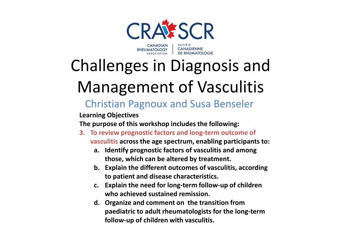
Challenges in Diagnosis and
Management of VasculitisChristian Pagnoux and Susa Benseler
Learning Objectives
The purpose of this workshop includes the following:
1. To review challenging clinical presentations of vasculitis in
children and adults so that participants will be able to
complete the following:
a. Compare and contrast clinical presentations of
vasculitis in children as compared to adults.
b. Conduct an appropriate diagnosis work-up for
vasculitis.
c. Diagnose some more challenging vasculitides of
children and/or adults.

Challenges in Diagnosis and
Management of VasculitisChristian Pagnoux and Susa Benseler
Learning Objectives
The purpose of this workshop includes the following:
2. To discuss evidence-based treatment approaches to
vasculitis (case-based) so that participants will be able to
complete the following:
a. Evaluate the form and severity of vasculitis prior to
deciding treatment.
b. Establish an adequate therapeutic scheme for patients,
integrating their individual characteristics, such as age.
c. Understand the typical therapies used for vasculitis.
Challenges in Diagnosis and
Management of VasculitisChristian Pagnoux and Susa Benseler
Learning Objectives
The purpose of this workshop includes the following:
3. To review prognostic factors and long-term outcome of
vasculitis across the age spectrum, enabling participants to:
a. Identify prognostic factors of vasculitis and among
those, which can be altered by treatment.
b. Explain the different outcomes of vasculitis, according
to patient and disease characteristics.
c. Explain the need for long-term follow-up of children
who achieved sustained remission.
d. Organize and comment on the transition from
paediatric to adult rheumatologists for the long-term
follow-up of children with vasculitis.
Disclosure Statement
• Susa Benseler
– Nothing to disclose
• Christian Pagnoux
– Consulting and speaker fees: Hoffmann-La Roche,
GSK
– Educational subventions (CanVasc): Hoffmann-La
Roche, Euroimmun
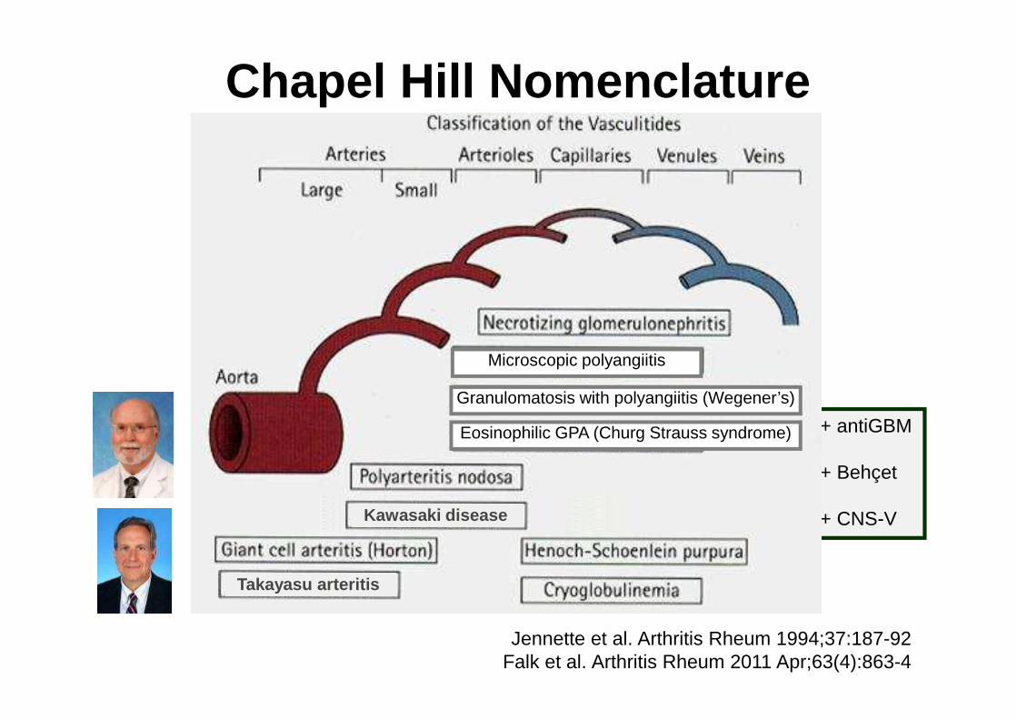
+ antiGBM
+ Behçet
+ CNS-V
Chapel Hill Nomenclature
Jennette et al. Arthritis Rheum 1994;37:187-92Falk et al. Arthritis Rheum 2011 Apr;63(4):863-4
Kawasaki disease
Takayasu arteritis
Granulomatosis with polyangiitis (Wegener’s)
Microscopic polyangiitis
Eosinophilic GPA (Churg Strauss syndrome)

EULAR/PRINTO/PRES classification
Ozen et al. Ann Rheum Dis.
2006;65(7):936-41

Patient: 5-year old girl (2009)
• March 13, 2009
– Presentation to the ER at Sickkids with severe mid-
abdominal pain, normal bowel movements, no
blood in stool, no vomitting, low grade fever for 3
days
– Bloodwork: raised ESR, CRP
– Ultrasound: critical SMA stenosis
� Admission for workup

MRA
• Marked proximal stenosis and vessel wall thickening of the SMA and its branches with contrast enhancement
• Proximal stenosisof the left renal artery

• 6 months “induction therapy”:
– Cyclophosphamide IV monthly 750-1000mg/m2
– High dose corticosteroids 2mg/kg, slow taper
– Enoxaparin
Treatment March-Sept 2009

MRA
• Improvement ofthe focal renal artery stenosis
• Stable appearanceof enhancingvessel wall thickening of SMA and branches
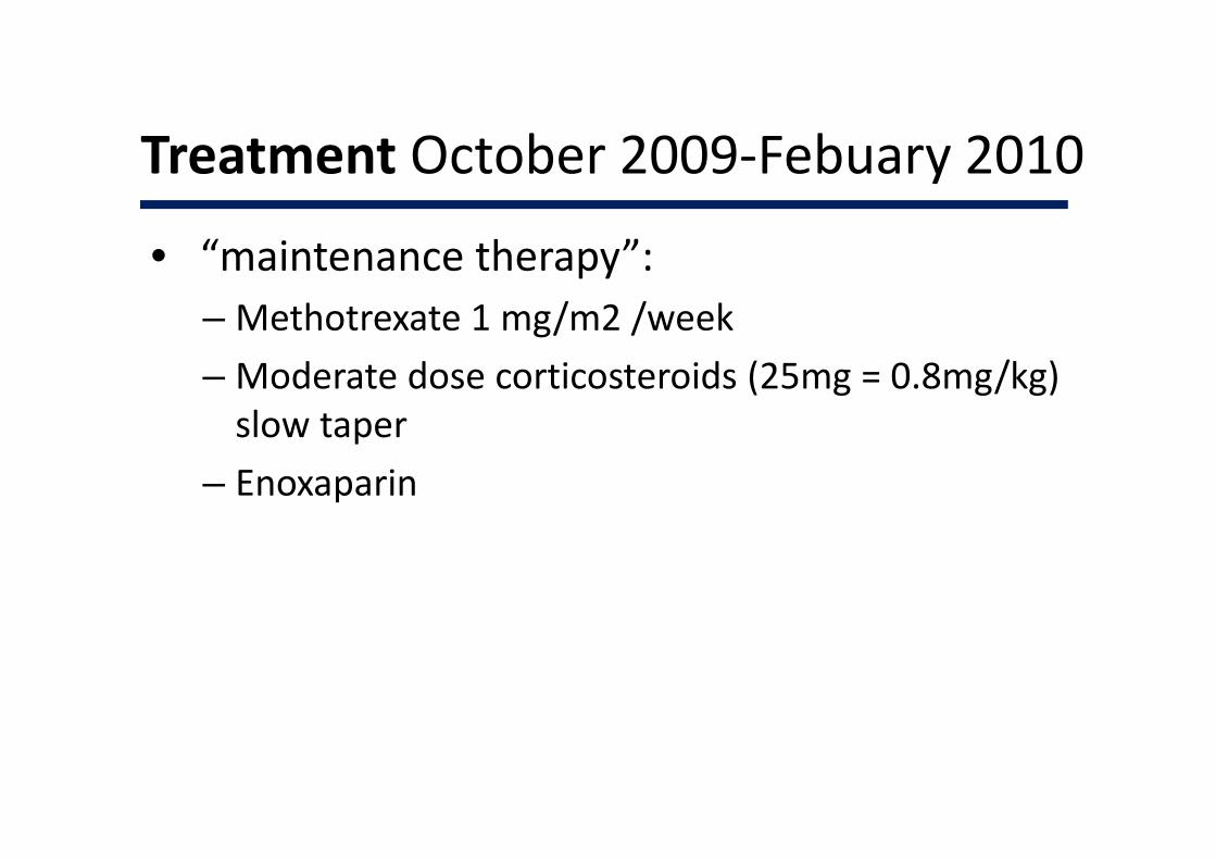
• “maintenance therapy”:
– Methotrexate 1 mg/m2 /week
– Moderate dose corticosteroids (25mg = 0.8mg/kg)
slow taper
– Enoxaparin
Treatment October 2009-Febuary 2010
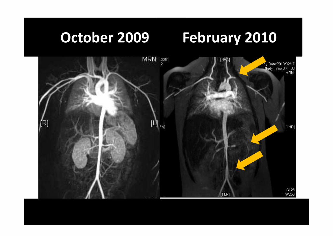
October 2009 February 2010

•“Infliximab rescue therapy” 5mg/kg
monthly IV in addition
• Methotrexate 1 mg/m2 /week
• Corticosteroids (10mg)
• Enoxaparin
Treatment February-May 2010
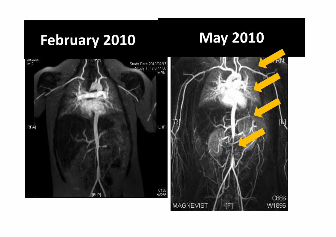
May 2010February 2010

• Daily oral cyclophosphamide (50mg/day,
2mg/kg ) plus high dose corticosteroids
(60mg/day)
• Enoxaparin
Treatment May-August 2010

August 2010May 2010

• Daily oral cyclophosphamide (total 10 months)
plus high dose corticosteroids (taper monthly)
• Enoxaparin
Treatment August 2010-Febuary 2011
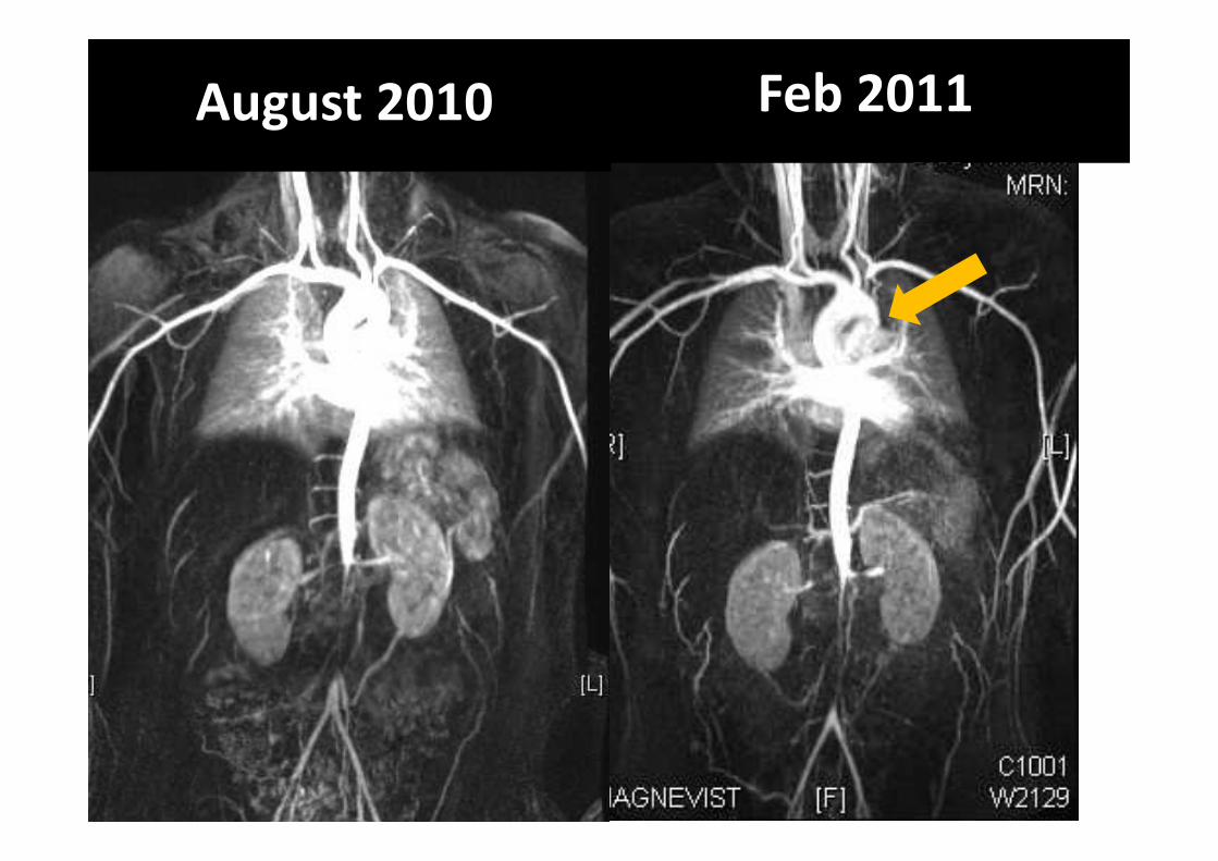
Feb 2011August 2010

Inflammatory markers

Inflammatory markers
IV CYPHD PRED
MTXMD PRED
αTNFHD PRED
ORAL CYPHD PRED
AZALD PRED
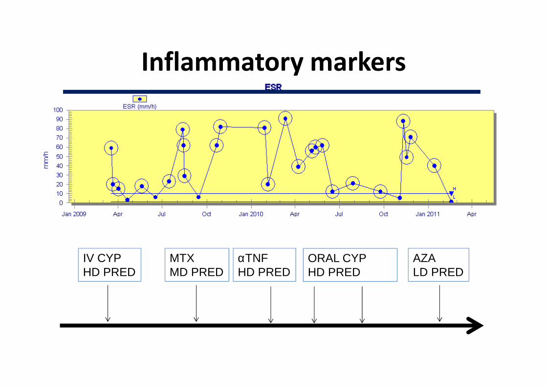
Inflammatory markers
IV CYPHD PRED
MTXMD PRED
αTNFHD PRED
ORAL CYPHD PRED
AZALD PRED
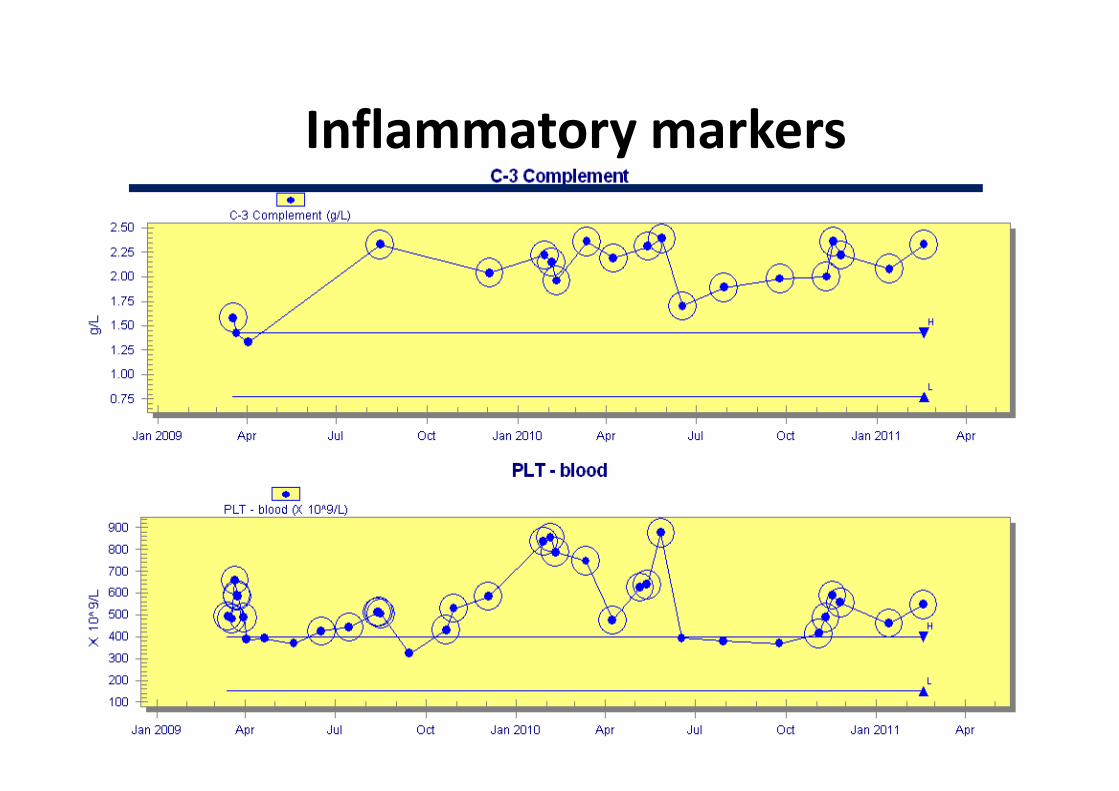
Inflammatory markers

• Off Prednisone, on Imuran maintenance
• MRA stable, clinically claudication
• Exposure to 6 months of IV and 10 month of
oral cyclophosphamide (cumulative dose: 17g)
• Moderate to high dose corticosteroids for 24
months (vertebral fractures, cataracts)
Treatment March 2012


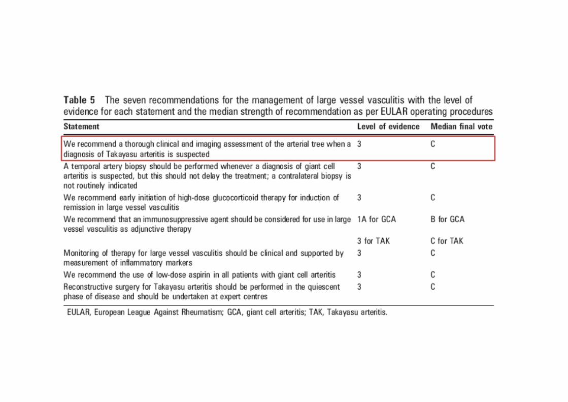
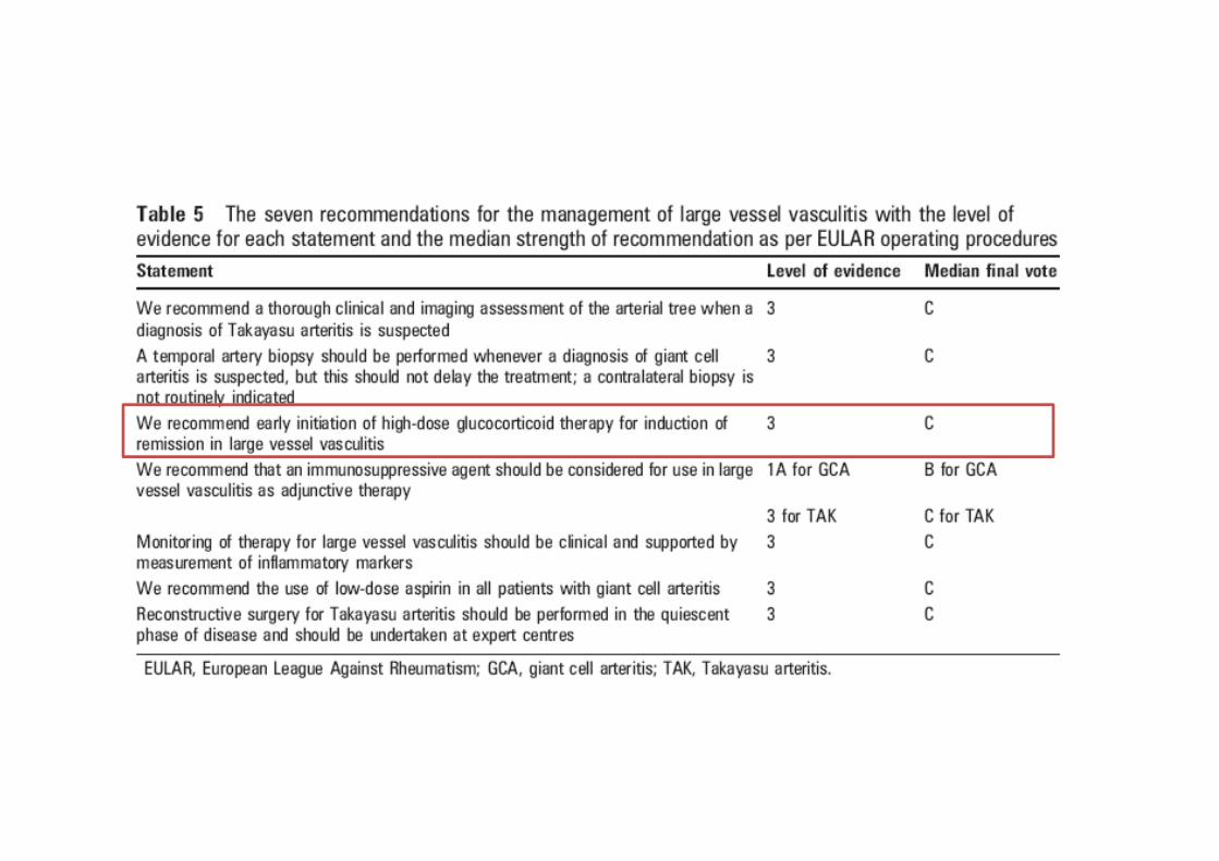
Large vessel vasculitis
Takayasu arteritis

Diagnosis TA: Angiography positive plus at least on e criterion
Takayasu arteritis PRES/EULAR 2005
Ozen, ANR 2005

TA Histology
• Inflammatory infiltrate and
concentric thickening of
intima, media and adventitia
• Mononuclear infiltrate: T
cells, macrophages

Treatment of TA in children

?
Treatment of TA in adults


Treatment of refractory TA

Rationale:
anti-TNF in Takayasu Arteritis
• TNFα � granuloma formation
• Increased serum TNFα in patients with
Takayasu
• Higher TNFα production from CD3+ T cells in
patients with active disease
Wallis. Semin Arthritis Rheum.2005;34:34-8Park. Rheumatology. 2006;45:545-8
Tripathy. Clin Immunol 2006;118:154-8

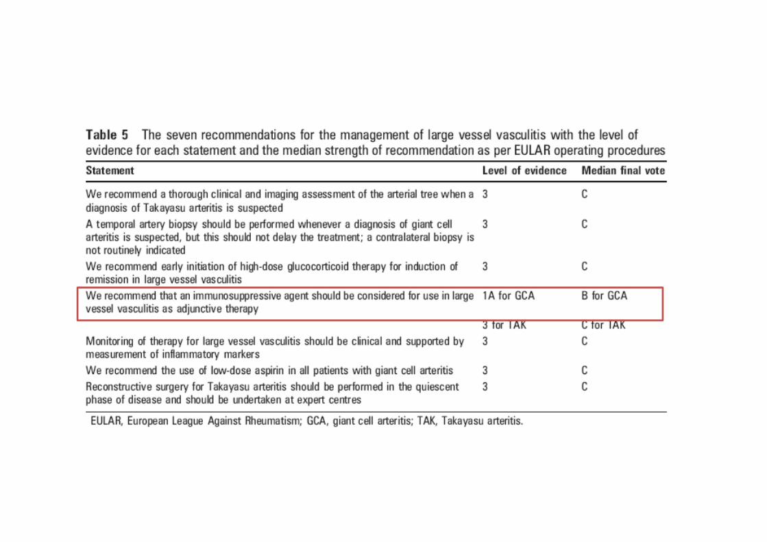
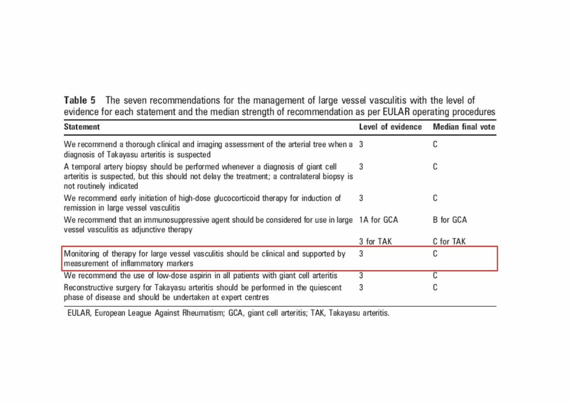
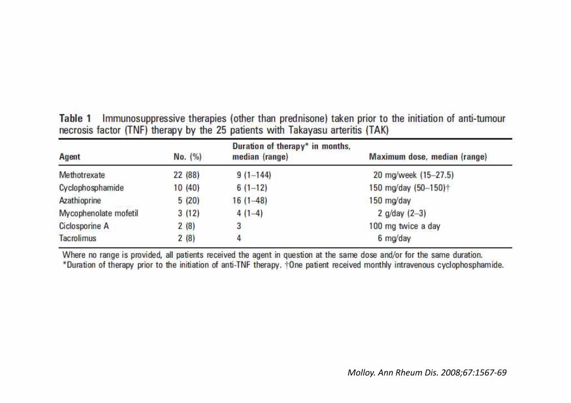
• Retrospective single-centre study of 25 patients with refractory Takayasu Arteritis
– Stable remission could not be achieved with use of low-dose prednisone (<10mg/day)
• Outcomes:
– Partial or complete remission
– Disease relapse
– Adverse events associated with anti-TNF therapy
Molloy. Ann Rheum Dis. 2008;67:1567-69

Remission
• Complete and sustained remission:
– Absence of features of active disease
– Absence of new lesions on imaging studies
– No glucocorticoid therapy for at least 6 months
• Partial remission:
– Glucocorticoid dose reduced by at least 50%
Molloy. Ann Rheum Dis. 2008;67:1567-69

Patients
• 22/25 were female (88%)
• Mean age 35 years (range 15-64)
• Mean age of disease onset 25 years (range
10-53)

Outcome Etanercept
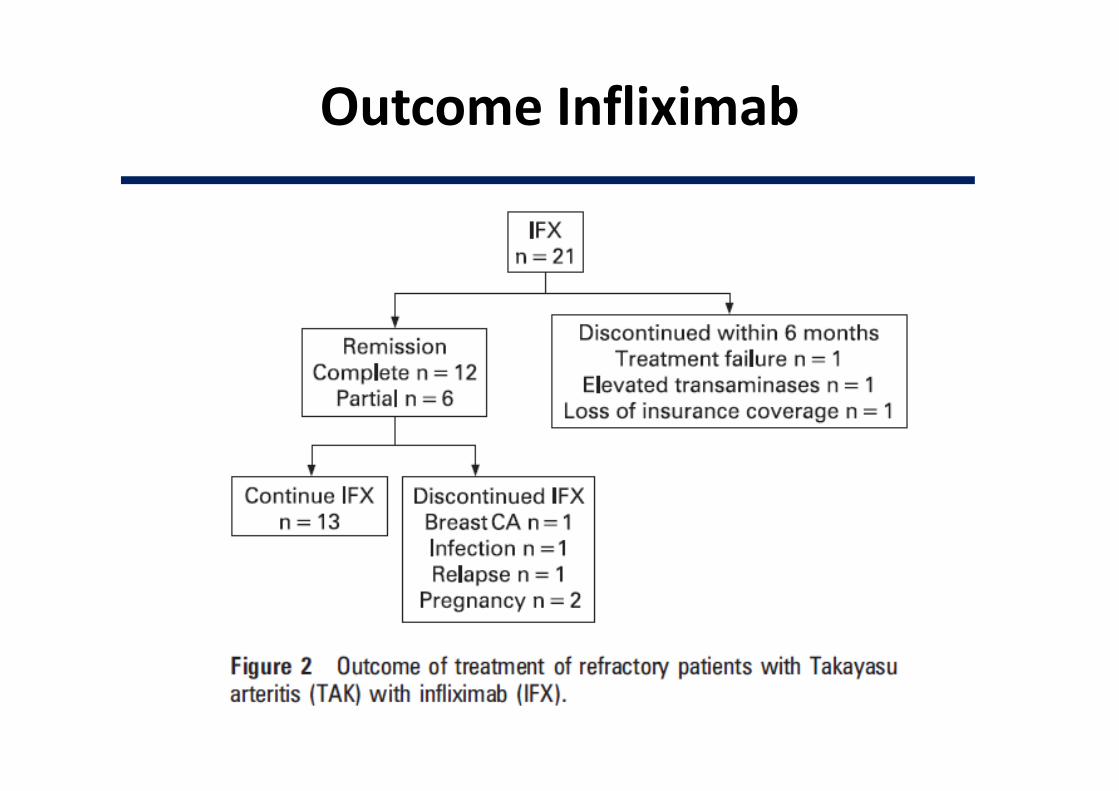
Outcome Infliximab

Prednisone therapy
• Discontinued in 60%
– After a median interval of 10 months (range 0-72)
– Prednisone-free remission maintained for a
median duration of 30 months (range 6-82)
• Tapered <10mg/day in 28%

Relapses during anti-TNF therapy
• Four patients with major disease relapse
– New stenotic lesions
– Elevated inflammatory markers
– Three patients achieved remission on higher doses
of anti-TNF therapy
Adverse events
• Abnormal liver function tests
• Primary histoplasmosis (after 2 infusions)
• Breast cancer (after 41 months of therapy)

Christian, 32 years-old
• No family history
• Minor asthma (salbutamol puffs, PRN)
• Married, 4 healthy children
• Smoker (5 packs-year)
• 2009: R temporal artery prominence,
then R temporal headaches and fatigue

Christian, 32 years-old
• BP 120/80 symmetrical, normal auscultation
• All pulses +, with prominent R>L temporal
arteries and behind ears, not tender
• Normal ESR and CBC
• Doppler-US: enlarged R TA 1.1 x 0.6 cm, versus
L 0.25


Christian, 32 years-old
• Diagnosis?
1. « Temporal Takayasu? »
2. « GCA/LVV of the youth? »
3. Other systemic vasculitis with TA involvement?
4. TA fibromuscular dysplasia?
5. Ehlers-Danlos (type IV)?
6. Other?
• Biopsy?
• Treatment?
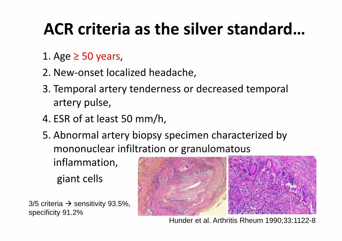
ACR criteria as the silver standard…
1. Age ≥ 50 years,
2. New-onset localized headache,
3. Temporal artery tenderness or decreased temporal
artery pulse,
4. ESR of at least 50 mm/h,
5. Abnormal artery biopsy specimen characterized by
mononuclear infiltration or granulomatous
inflammation,
giant cells
3/5 criteria � sensitivity 93.5%, specificity 91.2%
Hunder et al. Arthritis Rheum 1990;33:1122-8

1990 Criteria for the Classification of
Takayasu Arteritis
1. Age at disease onset < 40 years
2. Claudication of extremities
3. Decreased brachial artery pulse
4. BP difference >10 mm Hg
5. Bruit over subclavian arteries or aorta
6. Arteriographic narrowing or occlusion of the entire
aorta, its primary branches, or large arteries in the
proximal upper or lower extremities
≥3 criteria � sensitivity 90.5%, specificity 97.8%
Bloch et al. Arthritis Rheum. 1990 Aug;33(8):1068-73.

Ehlers-Danlos Syndrome Type IV
• Hereditary, DA, with COL3A1 mutation (50% de novo)
• Major diagnostic criteria:
– Arterial rupture– Intestinal rupture
– Uterine rupture during pregnancy
– Family history of EDS type IV
• Minor diagnostic criteria:– Thin, translucent skin (especially noticeable on the chest/abdomen)
– Characteristic facial appearance (thin lips and philtrum, small chin, thin nose, large eyes)
– Acrogeria
– Arteriovenous carotid-cavernous sinus fistula
– Hypermobility of small joints
– Tendon/muscle rupture
– Early-onset varicose veins
– Pneumothorax/pneumohemothorax
– Easy bruising Chronic joint subluxations/dislocations
– Congenital dislocation of the hips
– Talipes equinovarus (clubfoot)
– Gingival recession

Christian, 32 years-old
• 2010-2011: L temporal artery increased in
size, with some occasional tenderness

Christian, 32 years-old
• 2010-2011: L temporal artery increased in
size, with some occasional tenderness
• Left TABx:
– mixed infiltration with eosinophils, lymphocytes
and rare plasma cells
– disruption of elastic layers
– intimal proliferation and fibrosis
– organizing thrombus filling the lumen,
with recanalization
– no giant cells, granuloma or necrosis
Ito et al. 2008

Juvenile temporal arteritis (JTA)
• 1975, Lie et al. (JAMA)
• « Nodules » in the TA region of children or
young adults
• Bx: occlusion of lumen by intimal proliferation
as well as intra- and peri-vascular eosinophil
infiltration (no giant cell)
• Main differential diagnoses:
– Kimura disease
– Angiolymphoid hyperplasia with eosinophilia
– Thromboangiitis obliterans with eosinophilia
Nesher et al. Semin Arthritis Rheum 39

Juvenile temporal arteritis (JTA)
• Diagnostic criteria
– Children or young adults (� 7-44 years)
– Absence of associated features (myalgias, visual
disturbance, fever, anemia)
– Manifested as painless temporal nodule
– Normal ESR (� mild eosinophilia)
– Eosinophilic panarteritis and thrombosis with or without
microaneurysmal disruption of the artery
– Intimal proliferation, disruption of the media and extensive
infiltrate consisting of lymphocytes, eosinophils and
plasma cells
– Absence of granulomatous infiltration and giant cells
Tomlinson et al. Mayo Clin Proc 1994
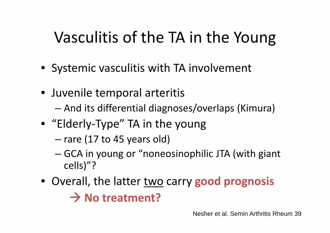
Vasculitis of the TA in the Young
• Systemic vasculitis with TA involvement
• Juvenile temporal arteritis
– And its differential diagnoses/overlaps (Kimura)
• “Elderly-Type” TA in the young
– rare (17 to 45 years old)
– GCA in young or “noneosinophilic JTA (with giant cells)”?
• Overall, the latter two carry good prognosis
���� No treatment?Nesher et al. Semin Arthritis Rheum 39
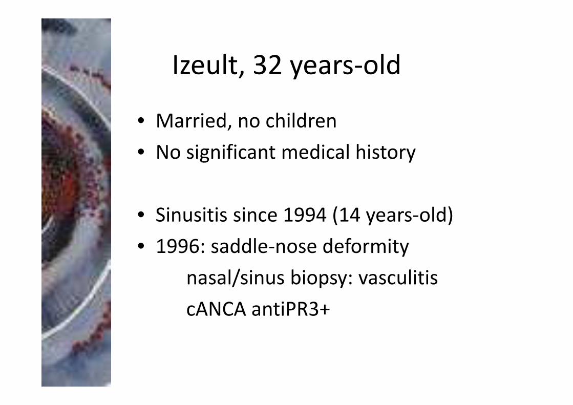
Izeult, 32 years-old
• Married, no children
• No significant medical history
• Sinusitis since 1994 (14 years-old)
• 1996: saddle-nose deformity
nasal/sinus biopsy: vasculitis
cANCA antiPR3+


Izeult, 32 years-old
• Sinusitis since 1994 (14 years old)
• 1996: saddle-nose deformity
nasal/sinus biopsy: vasculitis
cANCA antiPR3+
� Methotrexate + Prednisone

Izeult, 32 years-old
• Do you agree with this therapeutic choice?
1. YES
2. NO
3. I am OK with this choice, but I would have
treated her with a different drug/agent
4. I have no idea

EARLY SYSTEMIC GPA (<150 µM)
NORAM
– Methotrexate vs oral Cyclophosphamide for induction
– Non-inferiority trial (d=15%) for remission at 6 months
– 100 p. with “early systemic” GPA for 12 months
Remission at 6 moMTX 89.8% CYC 93.5% (P=0.04)
Relapse at 18 moMTX 69.5% CYC 46.5% (P=0.02)
CYC LeukopeniaMTX liver enzymes
CS at M188.8 g MTX vs 6.2 CYC(P<0.01)
de Groot et al. Arthritis Rheum 2005;52:2461–9
CYC

Izeult, 32 years-old
• Methotrexate + Prednisone until 2004
• 2005: sinusitis, recurrent lacrimal duct
obstruction then voice hoarseness & stridor…
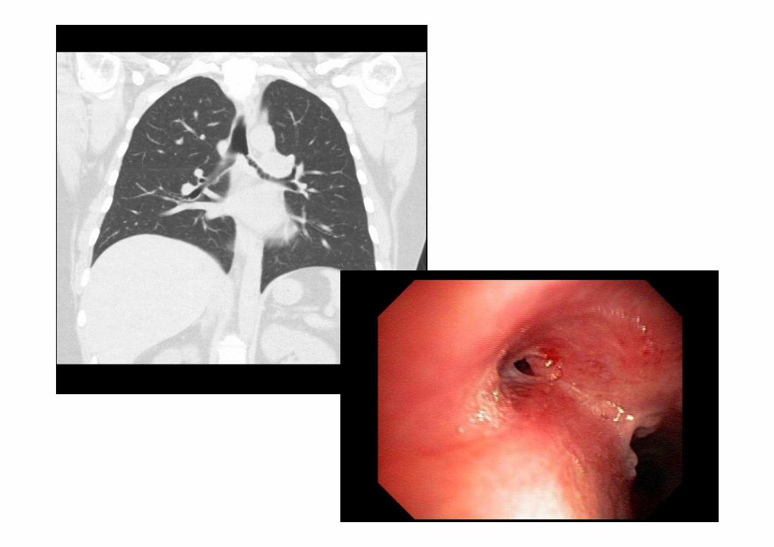
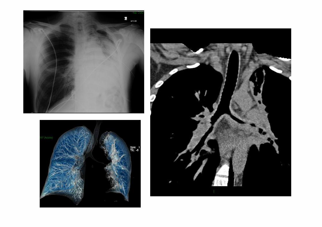
Bronchial stenoses in GPA
• 7 GPA patients with endobronchial stenoses
(1991-2004 in 4 French centers – 5F/2M)
• Cough and dyspnea (all), minor hemoptysis (4),
some “stridor” (4) … lung collapse
Hervier et al. Rev Med Interne 2006;27:453–457

Bronchial stenoses in GPA
• 7 GPA patients with endobronchial stenoses
(1991-2004 in 4 French centers – 5F/2M)
• Cough and dyspnea (all), minor hemoptysis (4),
some “stridor” (4)
• With SGS or low tracheal involvement in 3
Hervier et al. Rev Med Interne 2006;27:453–457


SGS in GPA
Elective location, 1-2 cm below the vocal cords (junction of 2 embryological segments?)*
*Eliachar et al, Cleve Clin J Med 69 Supp.2:149-51 Cabral et . Arthritis Rheum 2009;60:3413–34Lebovics et al, Laryngoscope 1992; 102:1341-5 Langford et al, Arthritis Rheum 1996; 39:1754-60Fowler et al. (Cleveland) ACR 2012, Chicago #1531 Eustaqio et al. Arch Otol Head N Surg 2011;137:480-5
3-23%, F>M, ~35 y-old 3% FVSG 500 p. F58% 9.3% VCRC 268 p. F68% 16% NIH 1992 158 p.23% Cleveland 1996, 43% isolated at Dx, aged 26, F 63%
14% ARChiVe (n=65)25% Denver (n=28)50% Cleveland (n=28)!!!
Bronchial stenoses in GPA
• 7 GPA patients with endobronchial stenoses
(1991-2004 in 4 French centers – 5F/2M)
• Cough and dyspnea (all), minor hemoptysis (4),
some “stridor” (4)
• With SGS or low tracheal involvement in 3
• Not isolated at Dx, but isolated during 2 relapses
• Bx: inflammation (7), granuloma (5), vasculitis (2)
Hervier et al. Rev Med Interne 2006;27:453–457
Izeult, 32 years-old
• Local treatment?
1. Yes
2. No
3. I don’t know
• Systemic treatment?
1. Prednisone + cyclophosphamide
2. Prednisone + methotrexate
3. Prednisone alone
4. Prednisone + rituximab
5. Can not decide…

Subglottic stenosis
Local treatment
• Dilations (balloons or bougies)
• Corticosteroid injections
• Topical mitomycine (in vitro antifibroblastic)
• Laser deobstruction � secondary stenosis
• Diathermy deobstruction
• Stents
• Surgery (reconstructive procedures, permanent tube-free speech-ready tracheostomy)
*Eliachar et al, Cleve Clin J Med 69 Supp.2:149-51Lebovics et al, Laryngoscope 1992; 102:1341-5Langford et al, Arthritis Rheum 1996; 39:1754-60Wolter et al. Laryngoscope. 2010 Dec;120(12):2452-5

Systemic treatment
Corticosteroids….
+ ???

CYCLOPS• Open label RCT
• 149 AASV (40% GPA)
• No Iº hypothesis
• Pulse (IV or oral) vs
continuous oral CYC
• Remission at 9 mo
Pulse 88.1%
Continuous 87.7%
• IV pulse = lower rate of
leukopenia HR 0.41
[CI, 0.23 to 0.71]
• At 18 mo:
14.5% relapsed
(18.8% IV vs. 9.4% PO) de Groot et al, Ann Intern Med 2009;150:670-680.
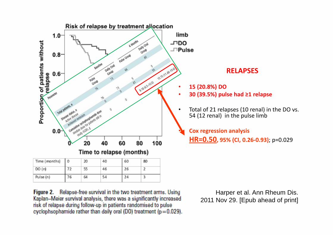
RELAPSES
• 15 (20.8%) DO
• 30 (39.5%) pulse had ≥1 relapse
• Total of 21 relapses (10 renal) in the DO vs. 54 (12 renal) in the pulse limb
• Cox regression analysis
HR=0.50, 95% (CI, 0.26-0.93); p=0.029
Harper et al. Ann Rheum Dis. 2011 Nov 29. [Epub ahead of print]
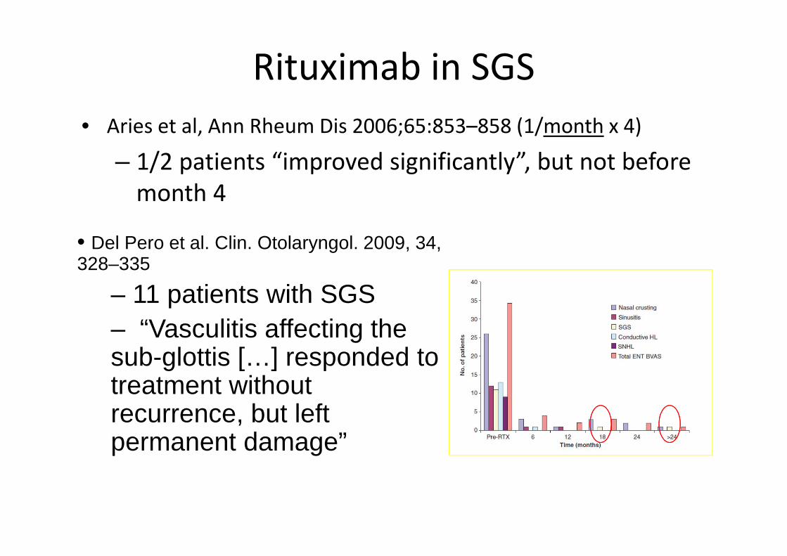
Rituximab in SGS
• Aries et al, Ann Rheum Dis 2006;65:853–858 (1/month x 4)
– 1/2 patients “improved significantly”, but not before
month 4
• Del Pero et al. Clin. Otolaryngol. 2009, 34, 328–335
– 11 patients with SGS– “Vasculitis affecting the sub-glottis […] responded to treatment without recurrence, but left permanent damage”

Rituximab in SGS
Holle et al. Ann Rheum Dis. 2012 Mar;71(3):327-33
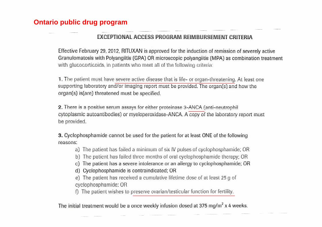
Ontario public drug program

Patient: 5 year old girl
• HPI:
– Presented to Sickkids ER with headache, behavior
change, vomiting, low grade fever
– Cluster of seizures in ER, admission to ICU in
seizure status
Patient: 5 year old girl
• pmHx:
– Healthy
– No exposures

Examination
• O/E
– Vital signs: tachycardia, Temp 38.0°C,
normal blood pressure, unwell
– General physical examination: normal
– Neurological examination:
• hyperreflexic, photophobic
• severe headaches
• Seizure status, continues bilateral seizures

Laboratory tests
• ↑↑↑↑ ESR 48 mm/h, ↑↑↑↑ CRP 32mg/dl, ↑↑↑↑ WBC 28, normal diff
• CSF: 31 WBC, 90% lymph, (↑↑↑↑ ) protein
• ↑↑↑↑ Opening pressure 38 cm H2O (N<20)
• Infectious, rheumatologic, metabolic w/u negative

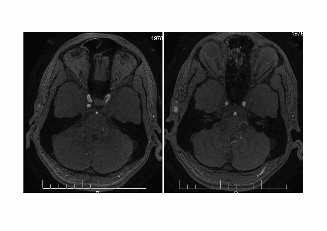
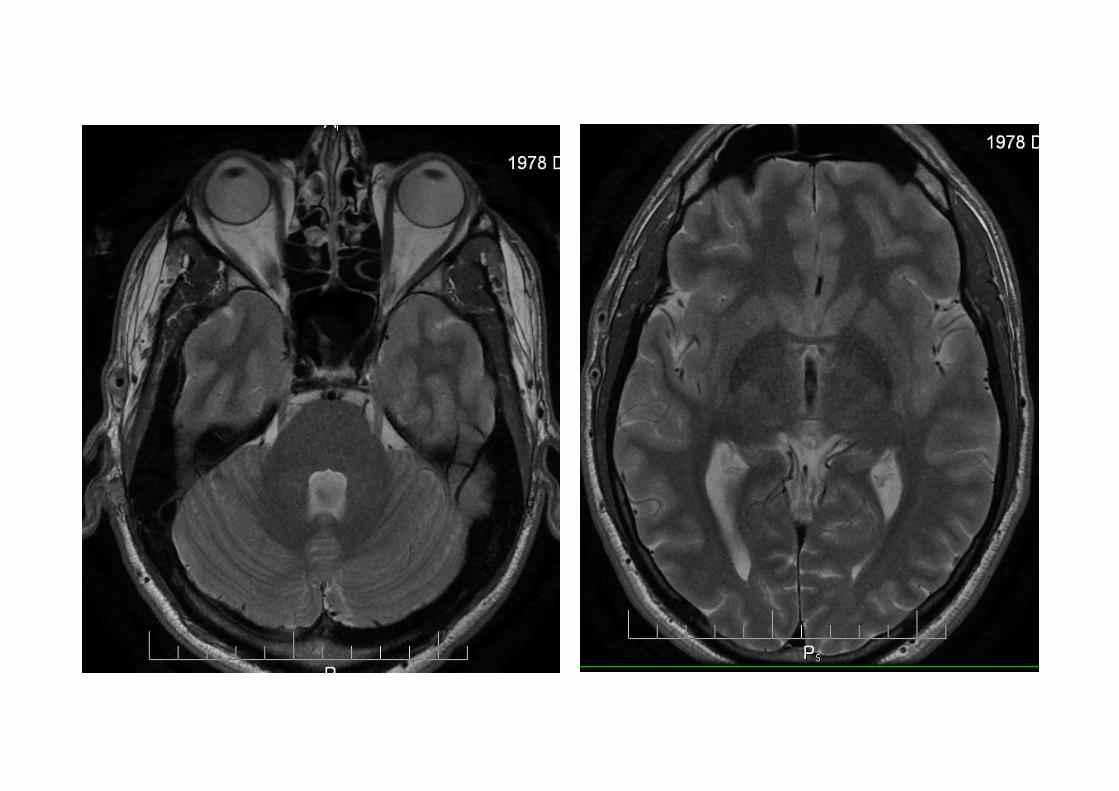
• White and grey matter lesions,
• Leptomeningealcontrast enhancement
MRI

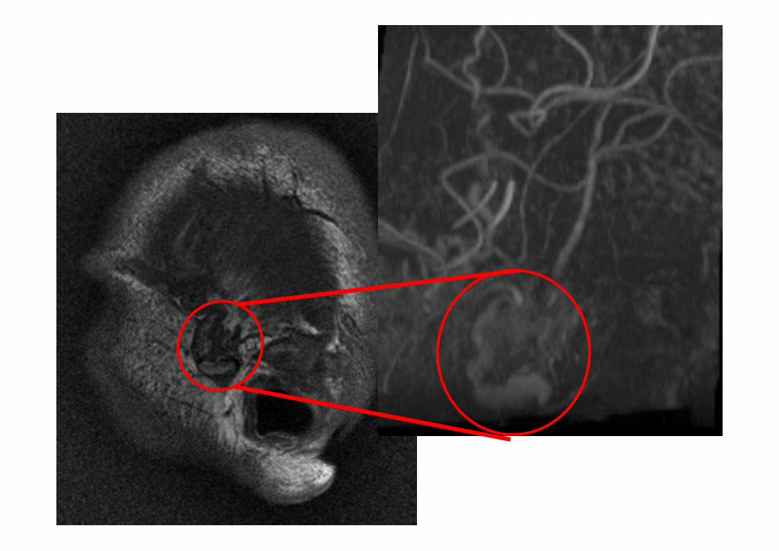
MRA, Conventional Angiography
normal

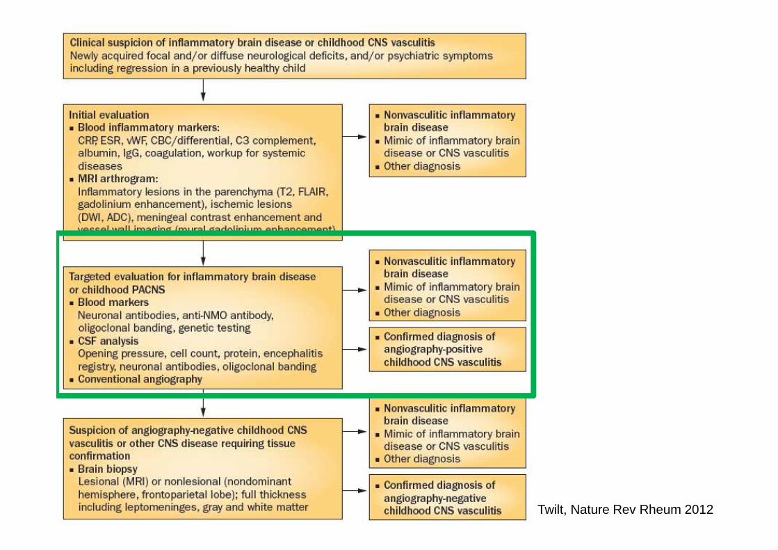
Twilt, Nature Rev Rheum 2012

Twilt, Nature Rev Rheum 2012
Twilt, Nature Rev Rheum 2012
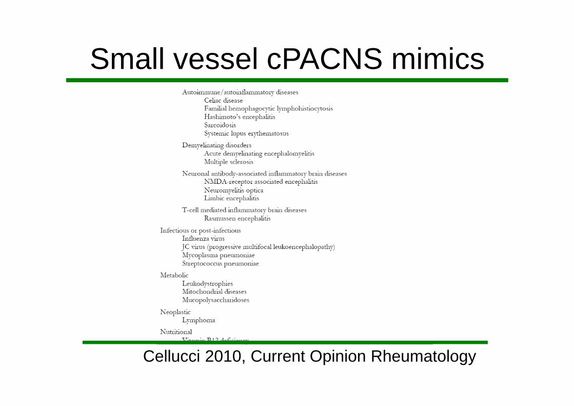
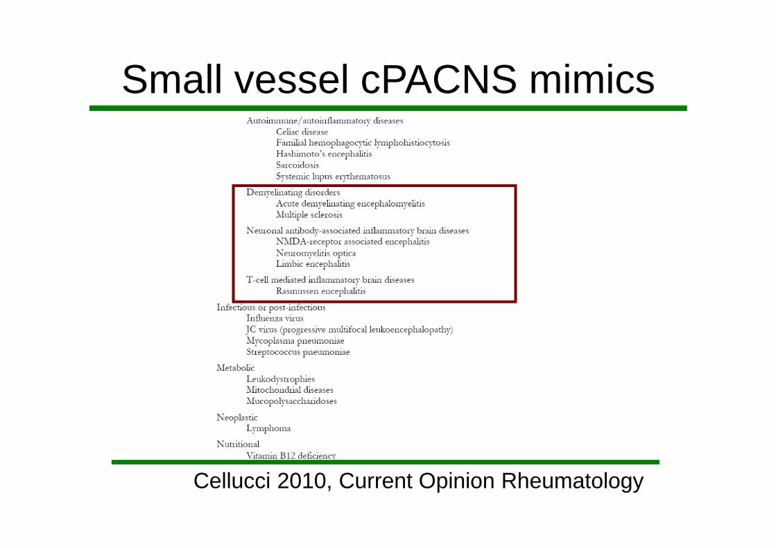
Small vessel cPACNS mimics
Cellucci 2010, Current Opinion Rheumatology
Small vessel cPACNS mimics
Cellucci 2010, Current Opinion Rheumatology
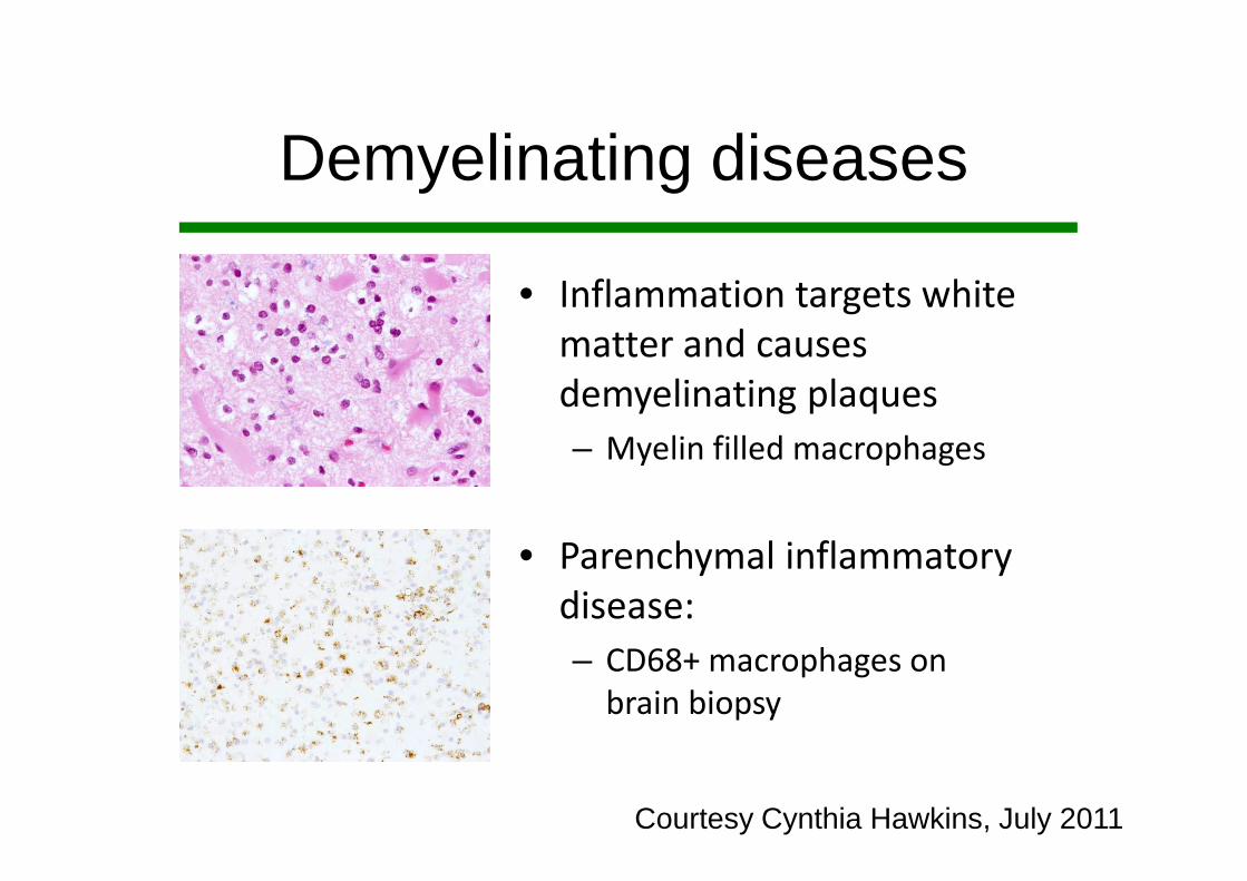
Courtesy Cynthia Hawkins, July 2011
• Inflammation targets white
matter and causes
demyelinating plaques
– Myelin filled macrophages
• Parenchymal inflammatory
disease:
– CD68+ macrophages on
brain biopsy
Demyelinating diseases

• Direct antibody
binding
• Targets: cell surface
receptors, channels,
enzymes
• No complement in
brain parenchyma
Neuronal antibody associated IBrainD
Dalmau 2005

Anti-NMDAR Encephalitis
Dalmau et al. Lancet Neurology 2010
Presence of tumour
Absence of tumour
N=400
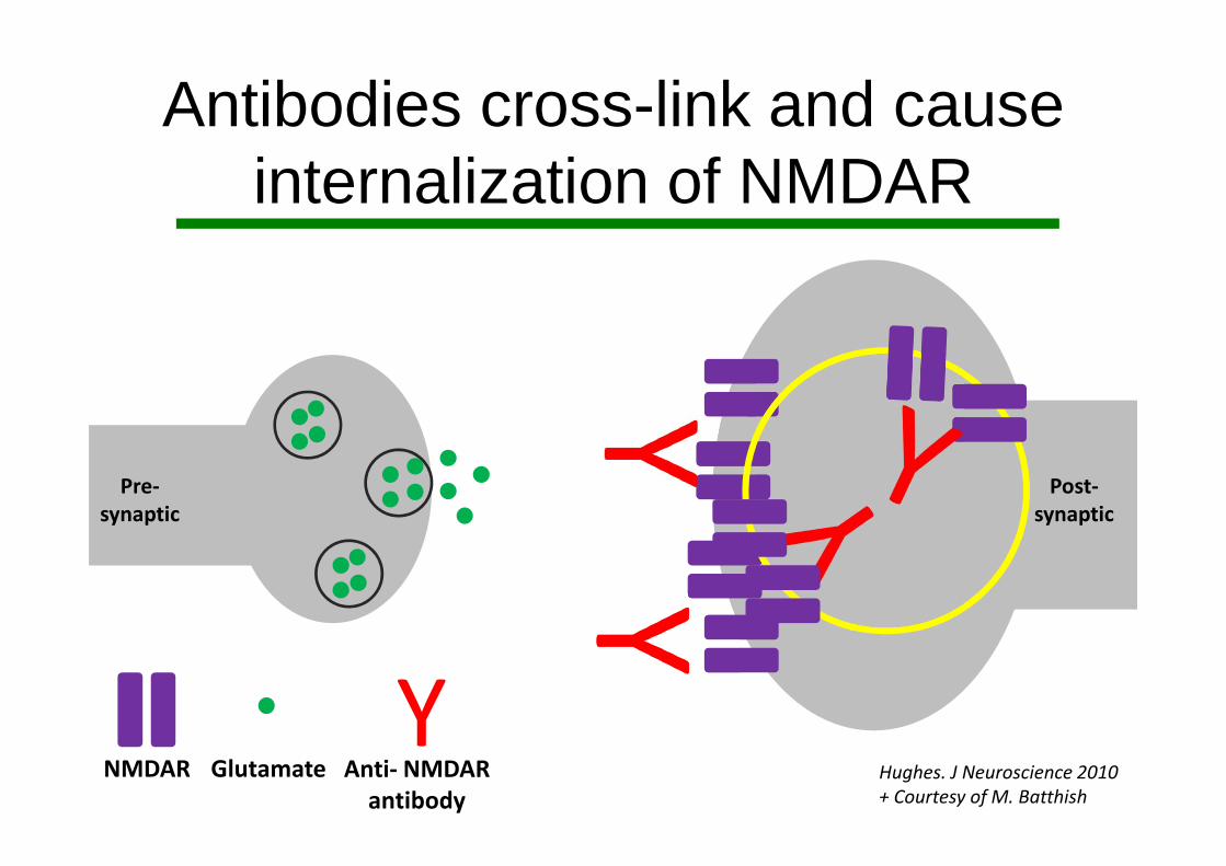
Hughes. J Neuroscience 2010
+ Courtesy of M. Batthish
NMDAR
YAnti- NMDAR
antibody
Pre-
synaptic
Post-
synaptic
Glutamate
Antibodies cross-link and cause internalization of NMDAR
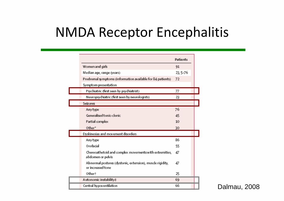
NMDA Receptor Encephalitis
Dalmau, 2008

Neuromyelitis optica NMO
Graber, 2008
• Auto-antibodies
against
Aquaporin4
• CSF ± serum
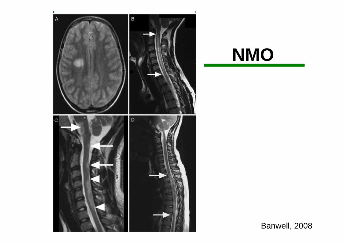
NMO
Banwell, 2008

Limbic encephalitis
• Paraneoplastic antibodies– Hu, Ma
• Auto-antibodies against LGI– Secreted antigen associated
with Voltage-gated potassium
channels
– AMP: anchor protein for LGI
– AMP-binding protein
Dalmau 2005, 2010Haberlandt, Bien 2010
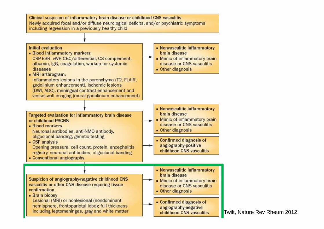
Twilt, Nature Rev Rheum 2012

Brain biopsy
Lymphocytic small vessel vasculitis

Diagnosis:
Primary CNS Vasculitis of childhood
Angiography-negative,
small vessel SVcPACNS
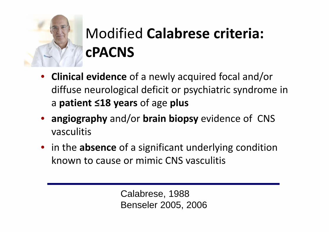
Modified Calabrese criteria:
cPACNS
• Clinical evidence of a newly acquired focal and/or
diffuse neurological deficit or psychiatric syndrome in
a patient ≤18 years of age plus
• angiography and/or brain biopsy evidence of CNS
vasculitis
• in the absence of a significant underlying condition
known to cause or mimic CNS vasculitis
Calabrese, 1988Benseler 2005, 2006

Primary CNS Vasculitis of Childhood
Angiography-positivecPACNS
Large vessel disease
Angiography-negativecPACNS
Small vessel disease
Benseler 2005, 2005Elbers, 2011
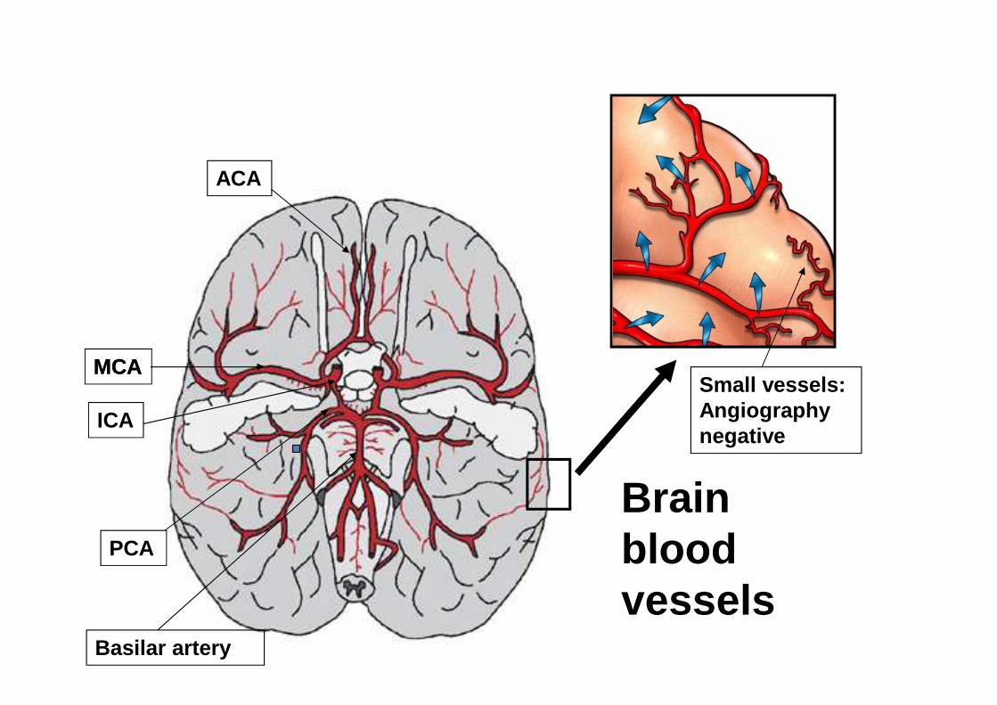
Brainblood vessels
MCA
ACA
PCA
MCA
ICA
Basilar artery
Small vessels: Angiography negative
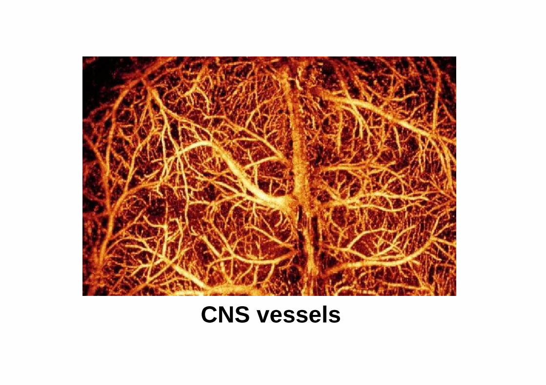
CNS vessels

SVcPACNS protocol

SVcPACNS treatment protocol
Hutchinson, 2010 Lancet Neurology
Anticoagulation Heparin/LMWH x 2 weeksAspirin 3-5mg/kg/d
Immunosuppression Induction therapy x 6 monthsIV cyclophosphamid pulse(500-750mg/m2 monthly x 7)
Prednisone 2mg/kg/d x 1 month(monthly taper: 60-50-40-30-25-20-17.5-15-12.5-10-7.5-5-2.5mg)
Maintenance therapy x18 month MMF 800-1200mg/m2/d
(Azathioprine 2mg/kg/d max 150mg)
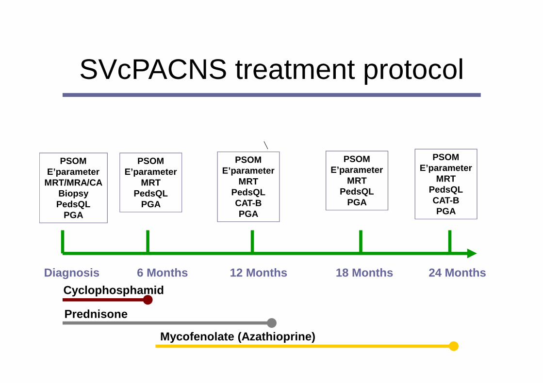
SVcPACNS treatment protocol
Diagnosis 6 Months 12 Months 18 Months 24 Months
PSOME’parameter
MRT/MRA/CABiopsyPedsQL
PGA
PSOME’parameter
MRTPedsQL
PGA
PSOME’parameter
MRTPedsQLCAT-BPGA
PSOME’parameter
MRTPedsQL
PGA
PSOME’parameter
MRTPedsQLCAT-BPGA
Cyclophosphamid
Prednisone
Mycofenolate (Azathioprine)

Hutchinson, 2010 Lancet Neurology
at 24 months� 69% had NOfunctional neurological deficit

SVcPACNS Flares
Cyclophophamid
Prednisone
MMF
Azathioprine Relapse: ON
Diagnosis 6 Months 12 Months 18 Months 24 Months

Conclusions
• Similarities and differences between children and adult vasculitides
• Collaboration is needed and beneficial
• Long-term follow-up of patients is essential
– Relapsing diseases
– Long-term damage and delayed complications
� Transition clinics

Get on board!
Top Related