


Languages
Pages
Legal


Ch 12 The Nervous System
and Nervous Tissue

Divisions of the Nervous System
Efferent Afferent
An Introduction to the Nervous System
Organs of the Nervous System
Brain and spinal cord
Sensory receptors of sense organs (eyes,
ears, etc.)
Nerves connect nervous system with other
systems
An Introduction to the Nervous System
The Nervous System
Includes all neural tissue in the body
Neural Tissue
Contains two kinds of cells
Neurons:
– cells that send and receive signals
Neuroglia (glial cells):
– cells that support and protect neurons

Nervous Tissue
Neurons perform all communication, information
processing, and control functions of the nervous
system
Neuroglia preserve physical and biochemical
structure of neural tissue and are essential to
survival and function of neurons

12-1 Structure and Function of the
Nervous System
Anatomical Divisions of the Nervous System
Central nervous system (CNS)
Peripheral nervous system (PNS)

Divisions of the Nervous System
The Central Nervous System (CNS)
spinal cord and brain
Contains neural tissue, connective tissues,
and blood vessels
Functions of the CNS
Are to process and coordinate:
– sensory data: from inside and outside body
– motor commands: control activities of peripheral organs
(e.g., skeletal muscles)
– higher functions of brain: intelligence, memory, learning,
emotion
Divisions of the Nervous System
The Peripheral Nervous System (PNS)
Includes all neural tissue outside the CNS
Functions of the PNS
Deliver sensory information to the CNS- Afferent
Division
Carry motor commands to peripheral tissues and
systems- Efferent Division
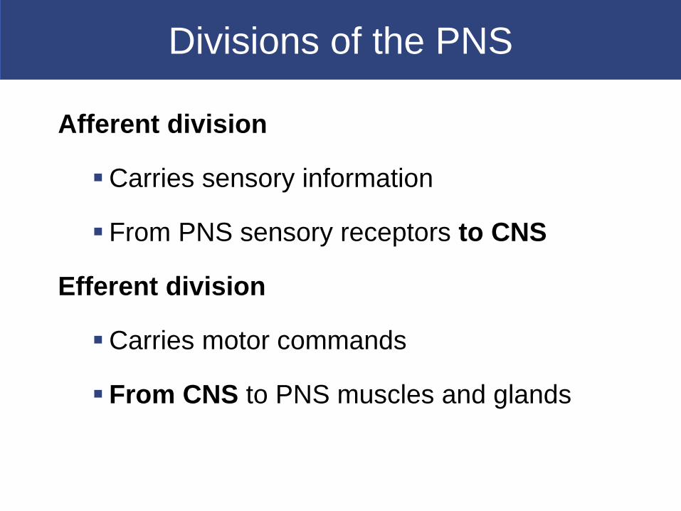
Divisions of the PNS
Afferent division
Carries sensory information
From PNS sensory receptors to CNS
Efferent division
Carries motor commands
From CNS to PNS muscles and glands

Nerves
also called peripheral nerves
Bundles of axons with connective tissues
and blood vessels
Carry sensory information and motor
commands in PNS:
–cranial nerves—connect to brain
–spinal nerves—attach to spinal cord
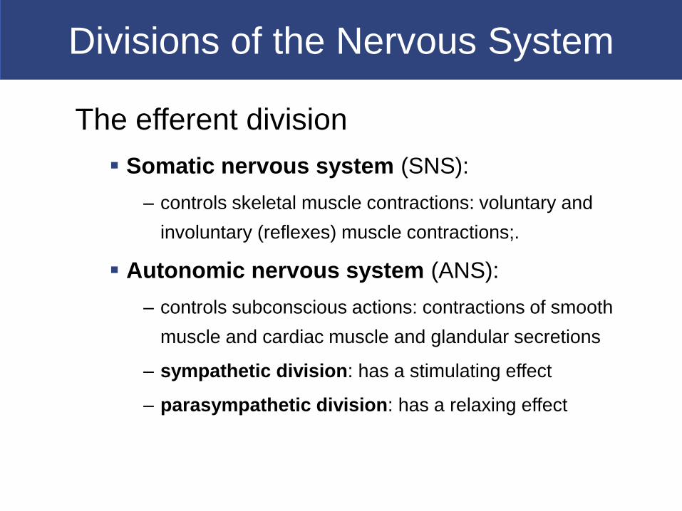
Divisions of the Nervous System
The efferent division
Somatic nervous system (SNS):
– controls skeletal muscle contractions: voluntary and
involuntary (reflexes) muscle contractions;.
Autonomic nervous system (ANS):
– controls subconscious actions: contractions of smooth
muscle and cardiac muscle and glandular secretions
– sympathetic division: has a stimulating effect
– parasympathetic division: has a relaxing effect
12-2 Nervous Tissue
The basic functional units of the nervous
system- neurons -carry action potential
system

Neurons
The Axon
Is long
Carries electrical signal (action potential) to target
Axon structure is critical to function
Nissl bodies
Dense areas of RER and ribosomes
Make neural tissue appear gray (gray matter)
Dendrites
Highly branched
Dendritic spines: many fine processes
– receive information from other neurons

Neurons
Major Organelles of the Cell Body
Large nucleus and nucleolus
Perikaryon (cytoplasm)
Mitochondria (produce energy)
RER and ribosomes (produce neurotransmitters)
Cytoskeleton
Neurofilaments and neurotubules: in place of
microfilaments and microtubules
Neurofibrils: bundles of neurofilaments that provide support
for dendrites and axon

Neurons
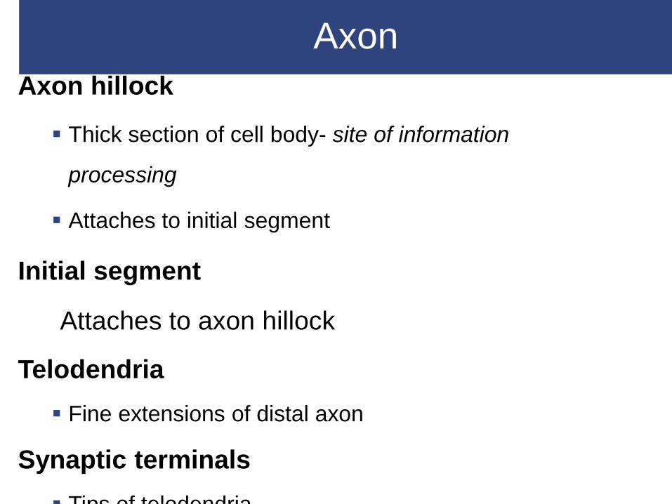
Axon
Axon hillock
Thick section of cell body- site of information
processing
Attaches to initial segment
Initial segment
Attaches to axon hillock
Telodendria
Fine extensions of distal axon
Synaptic terminals
Tips of telodendria

Neurons
Types of Neurons
Bipolar neurons
Found in special sensory organs (sight, smell,
hearing)
Unipolar neurons
Found in sensory neurons of PNS
Multipolar neurons
Common in the CNS
Include all skeletal muscle motor neurons

Neuron Classification
Bipolar Neurons
Are small
One dendrite, one
axon
Unipolar Neurons
Have very long axons
Fused dendrites and
axon
Cell body to one side
Multipolar Neurons
Have very long axons
Multiple dendrites, one
axon- Common in
CNS

Neurons
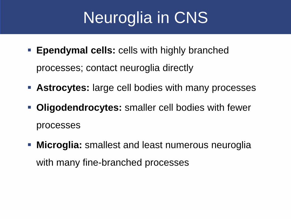
Neuroglia in CNS
Ependymal cells: cells with highly branched
processes; contact neuroglia directly
Astrocytes: large cell bodies with many processes
Oligodendrocytes: smaller cell bodies with fewer
processes
Microglia: smallest and least numerous neuroglia
with many fine-branched processes
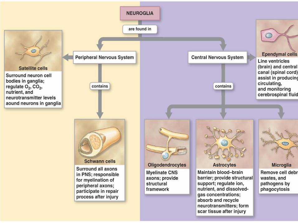
Neuroglia

Neuroglia in CNS

Neuroglia of CNSEpendymal cells - form epithelium called ependymal/choroid
plexus - Line ventricles of brain:
– secrete cerebrospinal fluid (CSF)
– have cilia or microvilli that circulate CSF
– monitor CSF and contain stem cells for repair
Astrocytes - maintain blood–brain barrier
(isolates CNS)
Create three-dimensional framework for CNS
Repair damaged neural tissue and guides neuron
development
Neuroglia of CNS
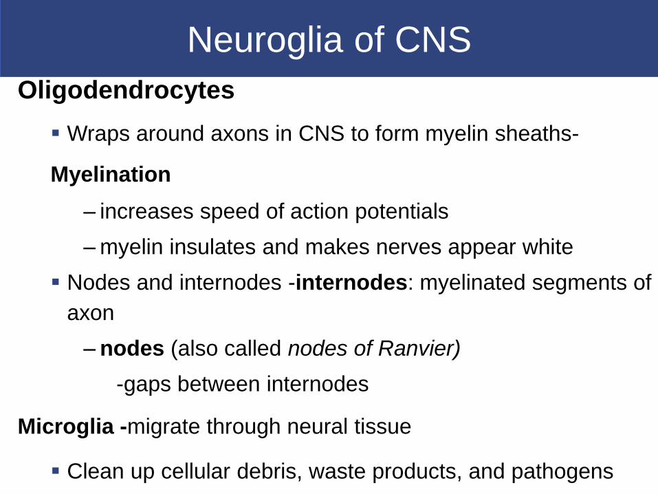
Oligodendrocytes
Wraps around axons in CNS to form myelin sheaths-
Myelination
– increases speed of action potentials
– myelin insulates and makes nerves appear white
Nodes and internodes -internodes: myelinated segments of
axon
– nodes (also called nodes of Ranvier)
-gaps between internodes
Microglia -migrate through neural tissue
Clean up cellular debris, waste products, and pathogens

Neuroglia of PNS
Satellite cells
Surround ganglia (neuron cell bodies)
Regulate environment around neuron
Schwann cells
Also called neurilemmocytes
Form myelin sheath (neurilemma) around
peripheral axons
One Schwann cell sheaths one segment of
axon:
–many Schwann cells sheath entire axon
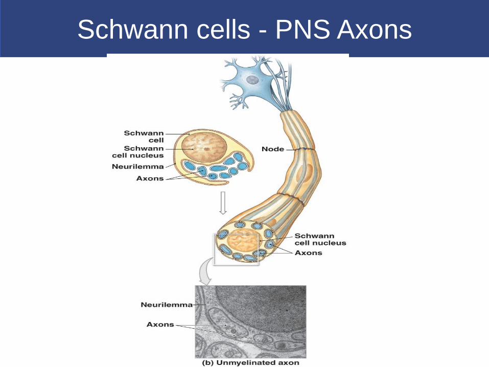
Schwann cells - PNS Axons

12-3 Function of Nervous Tissue
Functional Divisions of the PNS
Afferent division
Carries sensory information
From PNS sensory receptors to CNS
Efferent division
Carries motor commands
From CNS to PNS muscles and glands
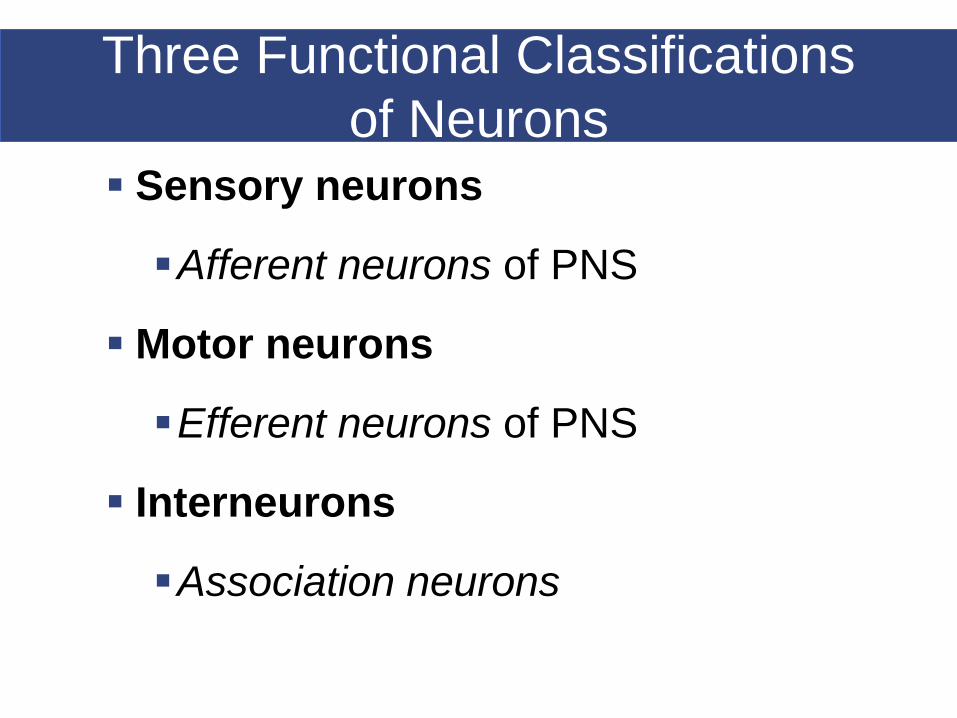
Three Functional Classifications
of Neurons
Sensory neurons
Afferent neurons of PNS
Motor neurons
Efferent neurons of PNS
Interneurons
Association neurons

Receptors and Effectors
Receptors:
–detect changes or respond to stimuli
–neurons and specialized cells (skin)
–complex sensory organs (e.g., eyes,
ears)
Effectors:
–respond to motor (efferent) signals
–cells and organs

Sensory Neurons
Functions of Sensory Neurons
Monitor internal environment (visceral sensory
neurons)
Monitor effects of external environment (somatic
sensory neurons)
Structures of sensory neurons
Unipolar
Cell bodies grouped in sensory ganglia
Processes (afferent fibers) extend from sensory
receptors to CNS

Motor Neurons
Motor Neurons
Carry instructions from CNS to peripheral effectors
Two major efferent systems
Somatic nervous system (SNS):
– includes all somatic motor neurons that innervate skeletal muscles
Autonomic (visceral) nervous system (ANS):
– visceral motor neurons innervate all other peripheral effectors
(automatic-involuntary)
– e.g., smooth muscle, cardiac muscle, glands, adipose tissue
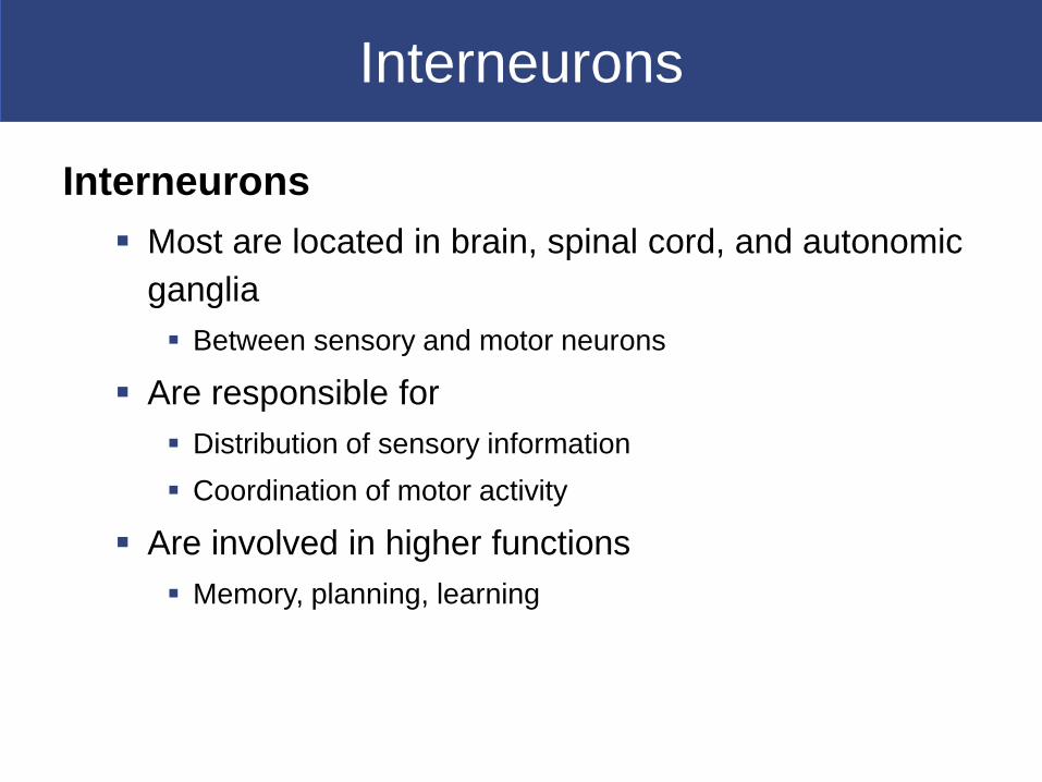
Interneurons
Interneurons
Most are located in brain, spinal cord, and autonomic
ganglia
Between sensory and motor neurons
Are responsible for
Distribution of sensory information
Coordination of motor activity
Are involved in higher functions
Memory, planning, learning
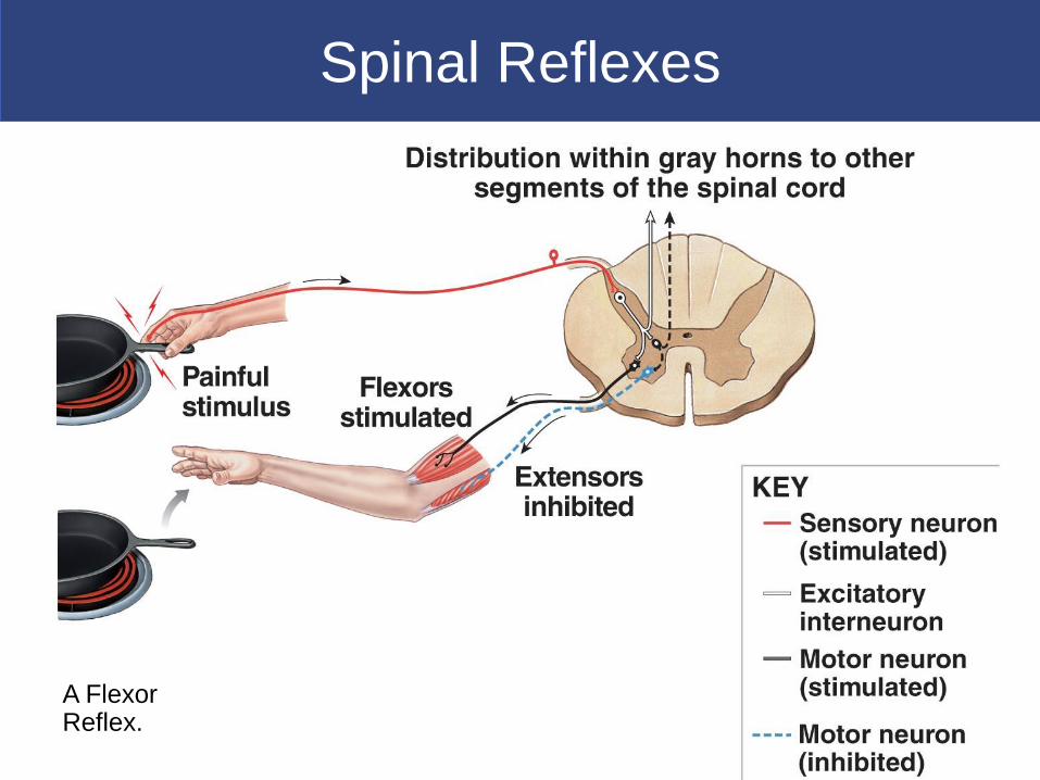
Spinal Reflexes
A Flexor Reflex.
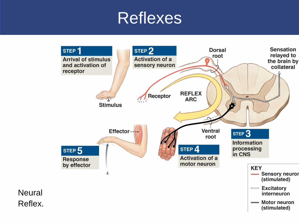
Reflexes
Neural
Reflex.

12-4 The Action Potential 13 min
Ion Movements and Electrical Signals
Plasma (cell) membrane produces
electrochemical signals by ion movements and
establishing electrical differences across the
neurolemma. This is called transmembrane
potential.
Transmembrane potential is particularly
important to neurons- Excitability
Transmembrane Potential
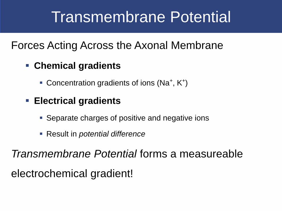
Forces Acting Across the Axonal Membrane
Chemical gradients
Concentration gradients of ions (Na+, K+)
Electrical gradients
Separate charges of positive and negative ions
Result in potential difference
Transmembrane Potential forms a measureable
electrochemical gradient!
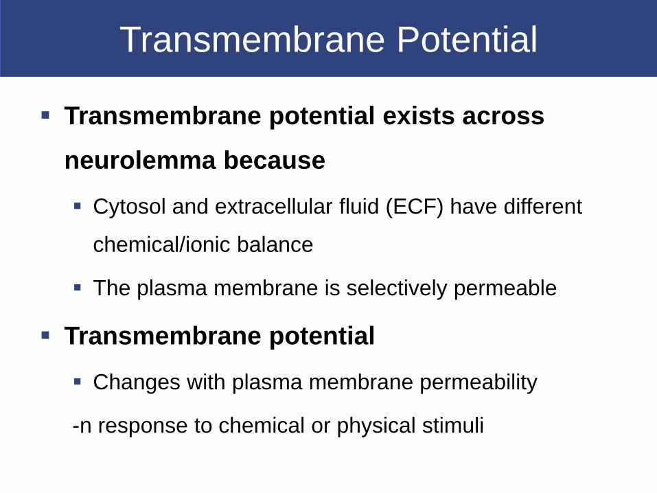
Transmembrane Potential
Transmembrane potential exists across
neurolemma because
Cytosol and extracellular fluid (ECF) have different
chemical/ionic balance
The plasma membrane is selectively permeable
Transmembrane potential
Changes with plasma membrane permeability
-n response to chemical or physical stimuli
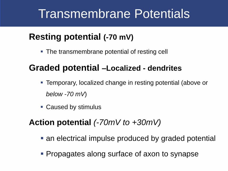
Transmembrane Potentials
Resting potential (-70 mV)
The transmembrane potential of resting cell
Graded potential –Localized - dendrites
Temporary, localized change in resting potential (above or
below -70 mV)
Caused by stimulus
Action potential (-70mV to +30mV)
an electrical impulse produced by graded potential
Propagates along surface of axon to synapse

Transmembrane Potential
Resting Potential.
Transmembrane Potential
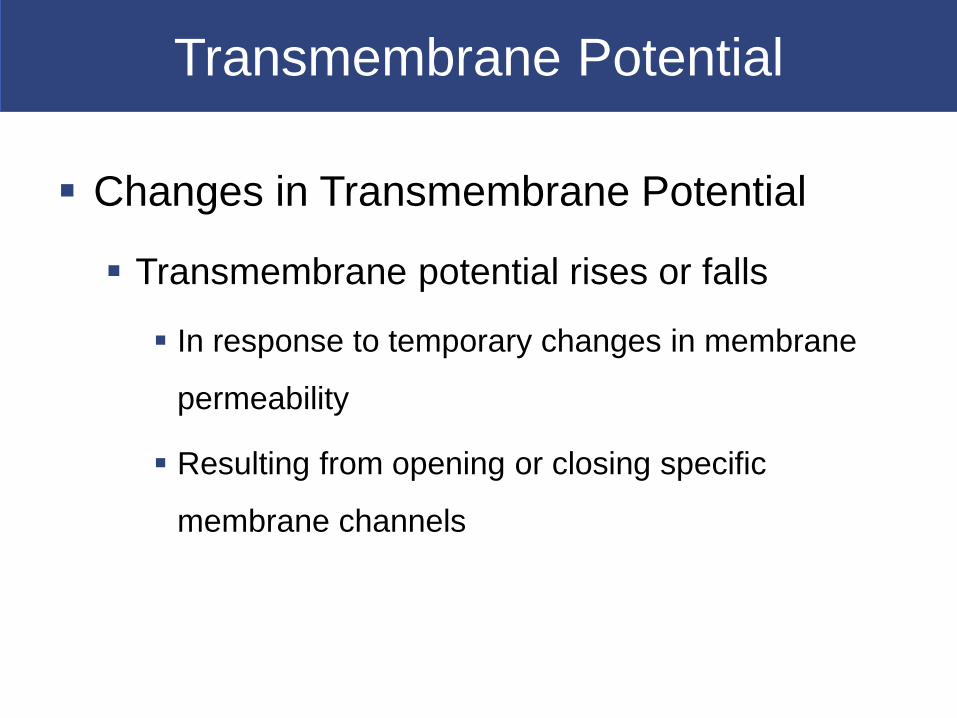
Changes in Transmembrane Potential
Transmembrane potential rises or falls
In response to temporary changes in membrane
permeability
Resulting from opening or closing specific
membrane channels

Transmembrane Potential
Three Classes of Gated Channels
Chemically gated channels
Open in presence of specific chemicals (e.g., ACh) at a binding site
Found on neuron cell body and dendrites
Voltage-gated channels
Respond to changes in transmembrane potential
Have activation gates (opens) and inactivation gates (closes)
Characteristic of excitable membrane
Found in neural axons, skeletal muscle sarcolemma, cardiac muscle
Mechanically gated channels
Respond to membrane distortion
Found in sensory receptors (touch, pressure, vibration)
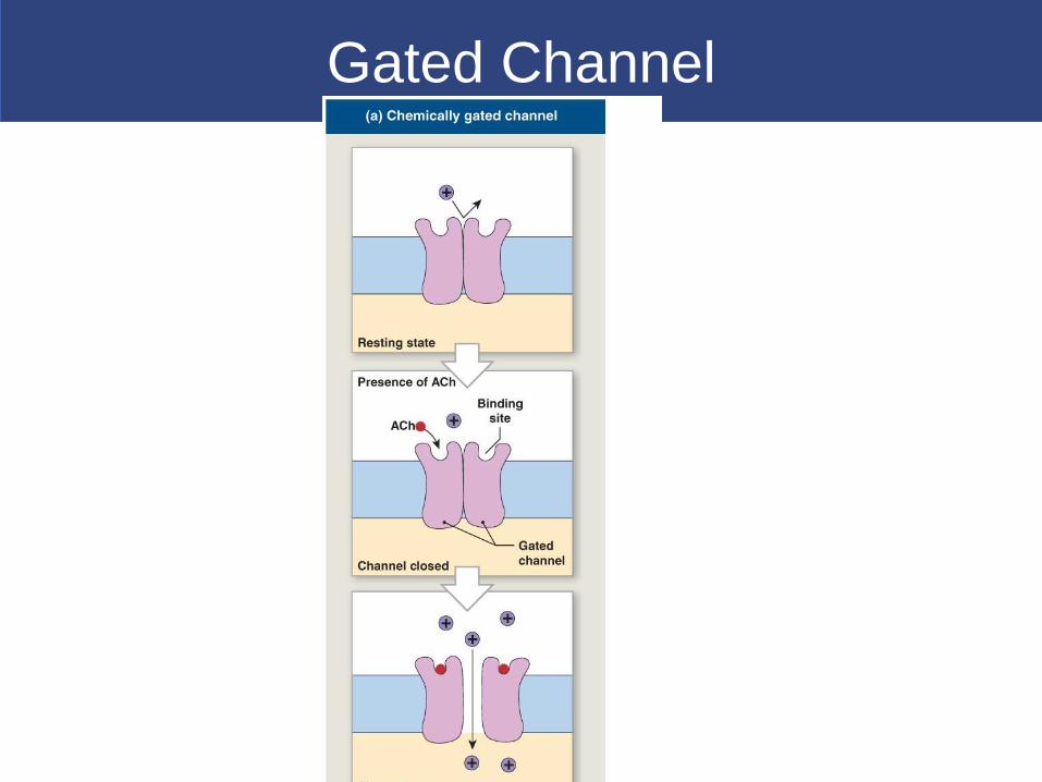
Gated Channel

Gated Channels
Na+

Transmembrane Potential
Sodium and Potassium Channels
Membrane permeability to Na+ and K+ determines
transmembrane potential
They are either passive or active
Passive channels: (leak channels)
– are always open
– permeability changes with conditions
Active channels (also called gated channels):
– open and close in response to stimuli
– at resting potential, most gated channels are closed

Resting Potential
Active Forces Across the Membrane
Sodium–potassium ATPase (exchange pump)
Is powered by ATP
Carries 3 Na+ out and 2 K+ in
Balances passive forces of diffusion
Maintains resting potential (–70 mV)

Sodium-Potassium Exchange Pump
-Powering the Sodium-Potassium Exchange Pump
To maintain concentration gradients of Na+ and K+
over time
Requires energy (1 ATP for each 2K+/3 Na+
exchange)
Without ATP
Neurons stop functioning

Transmembrane Potential
(Resting State).
Graded and Action Potentials
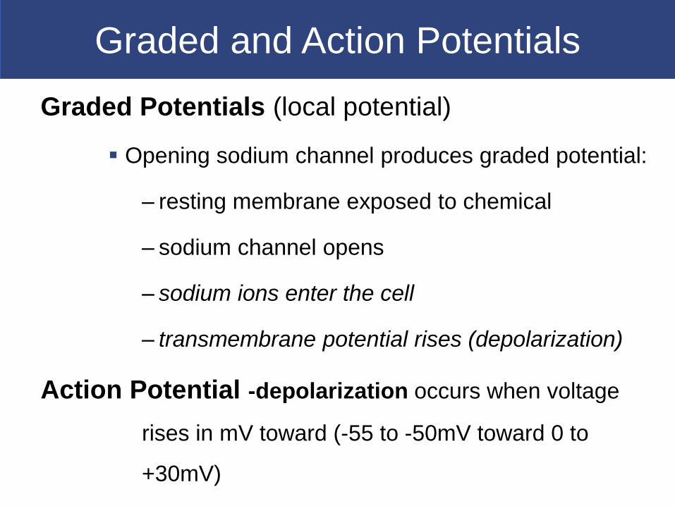
Graded Potentials (local potential)
Opening sodium channel produces graded potential:
– resting membrane exposed to chemical
– sodium channel opens
– sodium ions enter the cell
– transmembrane potential rises (depolarization)
Action Potential -depolarization occurs when voltage
rises in mV toward (-55 to -50mV toward 0 to
+30mV)
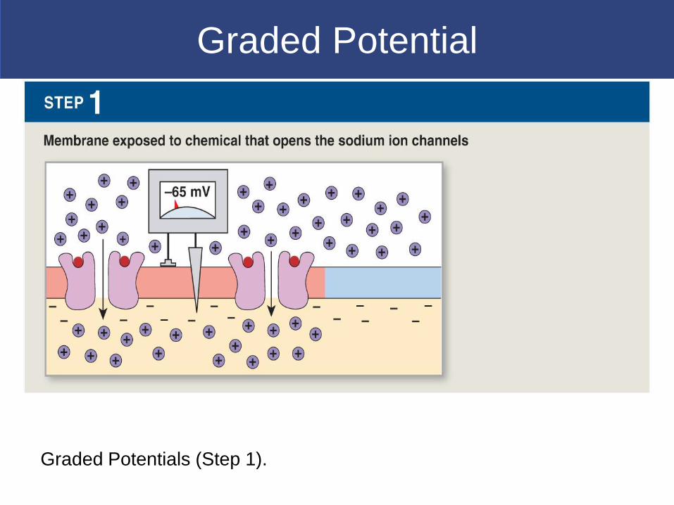
Graded Potential
Graded Potentials (Step 1).
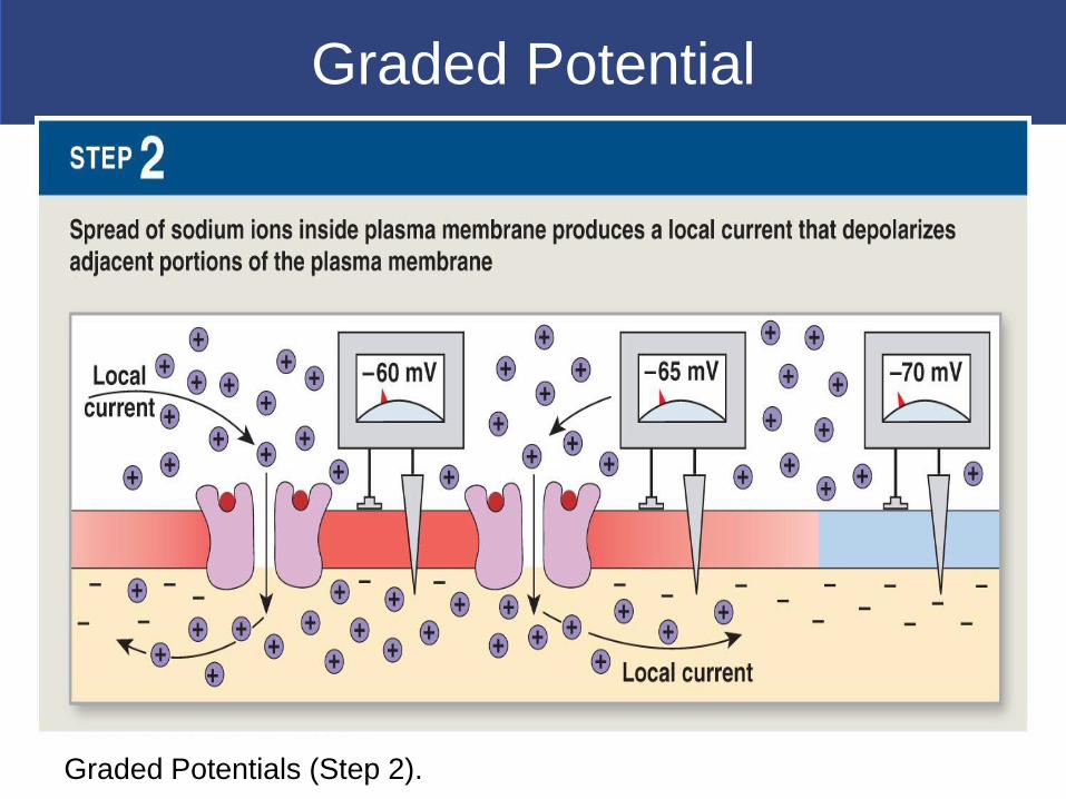
Graded Potential
Graded Potentials (Step 2).

Depolarization-
Any shift in transmembrane potential toward
0 mV:
– movement of Na+ through channel
– produces local current
– depolarizes nearby plasma membrane (graded
potential)
– change in potential is proportional to stimulus

Potentials
Repolarization
When the stimulus is removed, transmembrane
potential returns to normal
Hyperpolarization
Increasing the negativity of the resting potential
Result of opening a potassium channel
Opposite effect of opening a sodium channel
Positive ions move out, not into cell

Graded Potential
Depolarization, Repolarization, and Hyperpolarization.

Action Potential
Threshold- membrane depolarized 10-15 mV
above resting value- Begins-
Initiating Action Potential
Initial stimulus
A graded depolarization of axon hillock large enough
(10 to 15 mV) to change resting potential
(-70 mV) to threshold level of voltage-gated sodium
channels (–60 to –55 mV)
Action Potential
All-or-none principle
If a stimulus exceeds threshold amount:
– the action potential is the same
–no matter how large the stimulus
Action potential is either triggered, or not
Action Potential
Four Steps in the Generation of Action Potentials
Step 1: Depolarization to threshold
Step 2: Activation of Na+ channels
Rapid depolarization
Na+ ions rush into cytoplasm
Inner membrane changes from negative to positive

Action Potential

Generating Action Potentials
The Generation of an Action Potential

Action Potential
Step 3: Inactivation of Na+ & K+ channels
At +30 mV -Inactivation gates close (Na+ channel
inactivation)
K+ channels open and repolarization begins
Step 4: Return to normal permeability
K+ channels begin to close: when membrane reaches
normal resting potential (–70 mV)
K+ channels finish closing: membrane is hyperpolarized to -
90 mV transmembrane potential returns to resting level:
– action potential is over

Action Potential
The Refractory Period
The time period -From beginning of action potential to
return to resting state
During which membrane will not respond normally to
additional stimuli
Absolute refractory period
Sodium channels open or inactivated
No action potential possible
Relative refractory period
Membrane potential almost normal
Very large stimulus can initiate action potential

Action Potential
Propagations of Action Potential
Propagation
Moves action potentials generated in axon hillock
Along entire length of axon
A series of repeated actions, not passive flow
Two methods of propagating action potentials
Continuous conduction: unmyelinated axons
Saltatory conduction: myelinated axons

Continuous Propagation
Action potentials along an unmyelinated axon
Affects one segment of axon at a time
Steps in a propagation
Step 1: Action potential in segment 1:
– depolarizes membrane to +30 mV
Step 2: Depolarizes second segment to threshold:
– second segment develops action potential
Step 3: First segment enters refractory period
Step 4: Local current depolarizes next segment
Cycle repeats -Action potential travels in one direction (1m/sec)

Action Potential
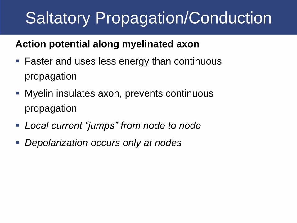
Saltatory Propagation/Conduction
Action potential along myelinated axon
Faster and uses less energy than continuous
propagation
Myelin insulates axon, prevents continuous
propagation
Local current “jumps” from node to node
Depolarization occurs only at nodes
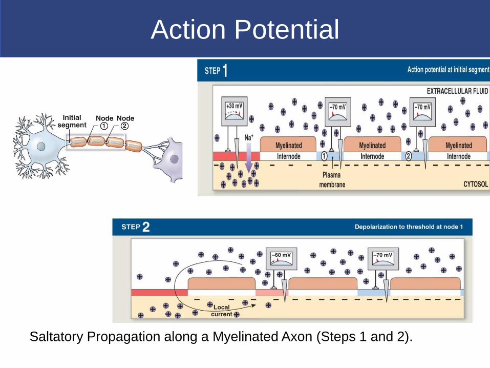
Action Potential
Saltatory Propagation along a Myelinated Axon (Steps 1 and 2).

Saltatory Propagation
Saltatory Propagation along a Myelinated Axon (Steps 3 and 4).

Axon Diameter and Speed
Axon Diameter and Conduction/Propagation Speed
Ion movement is related to cytoplasm concentration
Axon diameter affects action potential speed
The larger the diameter, the lower the resistance
Three Groups of Axons
Type A fibers Type B fibers Type C fibers
These groups are classified by
Diameter
Myelination
Speed of action potentials
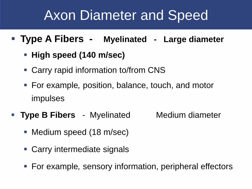
Axon Diameter and Speed
Type A Fibers - Myelinated - Large diameter
High speed (140 m/sec)
Carry rapid information to/from CNS
For example, position, balance, touch, and motor
impulses
Type B Fibers - Myelinated Medium diameter
Medium speed (18 m/sec)
Carry intermediate signals
For example, sensory information, peripheral effectors

Axon Diameter and Speed
Type C Fibers Unmyelinated Small diameter
Slow speed (1 m/sec) Continuous propagation
Carry slower information
For example, involuntary muscle, gland controls
“Information” travels within the nervous system as
propagated electrical signals (action potentials)
The most important information (vision, balance, motor
commands) is carried by large-diameter, myelinated
axons
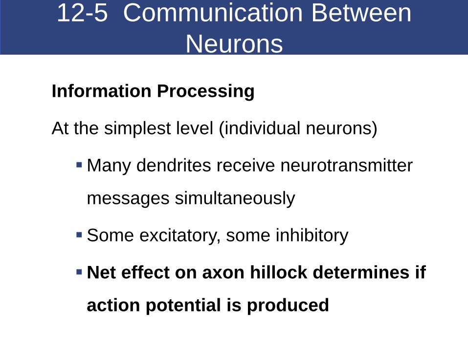
12-5 Communication Between
Neurons
Information Processing
At the simplest level (individual neurons)
Many dendrites receive neurotransmitter
messages simultaneously
Some excitatory, some inhibitory
Net effect on axon hillock determines if
action potential is produced

Chemical Synapses
Are found in most synapses between neurons
and all synapses between neurons and other
cells
Cells not in direct contact
Action potential may or may not be
propagated to postsynaptic cell, depending on
Amount of neurotransmitter released
Sensitivity of postsynaptic cell

Synaptic Activity
Action potentials (nerve impulses)
Are transmitted from presynaptic neuron
To postsynaptic neuron (or other postsynaptic cell)
across a synapse
Axon Terminal - area of axon of presynaptic neuron
Contains synaptic vesicles of neurotransmitters
Neurotransmitters:
– are released at presynaptic membrane
– affect receptors of postsynaptic membrane
– are broken down by enzymes
– are reassembled at axon terminal

The Synapse Area where a neuron communicates with another cell
Presynaptic cell: neuron that sends message
Postsynaptic cell: cell that receives message
Synaptic cleft: small gap that separates the presynaptic
membrane and the postsynaptic membrane

Action Potentials
Frequency of Action Potentials
Information received by a postsynaptic cell may be
simply the frequency of action potentials received
Rate of Generation of Action Potentials
Frequency of action potentials
Depends on degree of depolarization above
threshold
Holding membrane above threshold level
Has same effect as a second, larger stimulus
Reduces relative refractory period

Postsynaptic Potentials
Postsynaptic Potentials
Graded potentials developed in a postsynaptic cell
In response to neurotransmitters
Two Types of Postsynaptic Potentials
Excitatory postsynaptic potential (EPSP)
Graded depolarization of postsynaptic membrane
Inhibitory postsynaptic potential (IPSP)
Graded hyperpolarization of postsynaptic membrane

Action Potentials
Inhibition
A neuron that receives many IPSPs
Is inhibited from producing an action potential
Because the stimulation needed to reach threshold
is increased
Summation
To trigger an action potential
One EPSP is not enough
EPSPs (and IPSPs) combine through summation:

Neurotransmitters
Effect of a Neurotransmitter on a Postsynaptic
Membrane
Depends on the receptor
Not on the neurotransmitter
For example, acetylcholine (ACh)
Usually promotes action potentials
But inhibits cardiac neuromuscular junctions

Neurotransmitters
Two Classes of Neurotransmitters
Excitatory neurotransmitters
Cause depolarization of postsynaptic membranes
Promote action potentials
Inhibitory neurotransmitters
Cause hyperpolarization of postsynaptic
membranes
Suppress action potentials
Groups of Neurotransmitters
Cholinergic Synapses (from Acetylocholine)
Any synapse that releases ACh
All neuromuscular junctions with skeletal muscle fibers
Many synapses in CNS
All neuron-to-neuron synapses in PNS
All neuromuscular and neuroglandular junctions of
ANS parasympathetic division

Cholinergic Synapses Events
Action potential arrives, depolarizes synaptic (axon) terminal
Calcium ions enter synaptic knob, trigger exocytosis of Ach
ACh binds to receptors, depolarizes postsynaptic membrane
AChE breaks ACh into acetate and choline and are sent
back to axon terminal for reentry and reuse.

Neurotransmitters
Other Neurotransmitters
At least 50 neurotransmitters other than ACh,
including
Some amino acids
Biogenic Amines
Neuropeptides
Prostaglandins, ATP, Some dissolved gases
Examples -Biogenic Amine
Neurotransmitters Serotonin
A CNS neurotransmitter
Affects attention and emotional states
Norepinephrine (NE)
Released by adrenergic synapses
Excitatory and depolarizing effect
Widely distributed in brain and portions of ANS
Dopamine
A CNS neurotransmitter
May be excitatory or inhibitory
Involved in Parkinson disease, cocaine use

Neurotransmitters
Amino Acid-Gamma Aminobutyric Acid
(GABA)
Inhibitory effect
Functions in CNS -Not well understood
Neuropeptides ex Endorphins
in the CNS
Bind to the same receptors as opium or morphine
Relieve pain

Neurotransmitter Effects
Direct effects on membrane channels
For example, ACh, glutamate, aspartate
Indirect effects via G proteins (2nd messenger)
For example, E, NE, dopamine, histamine,
GABA
Indirect effects via intracellular enzymes
For example, lipid-soluble gases (NO, CO)
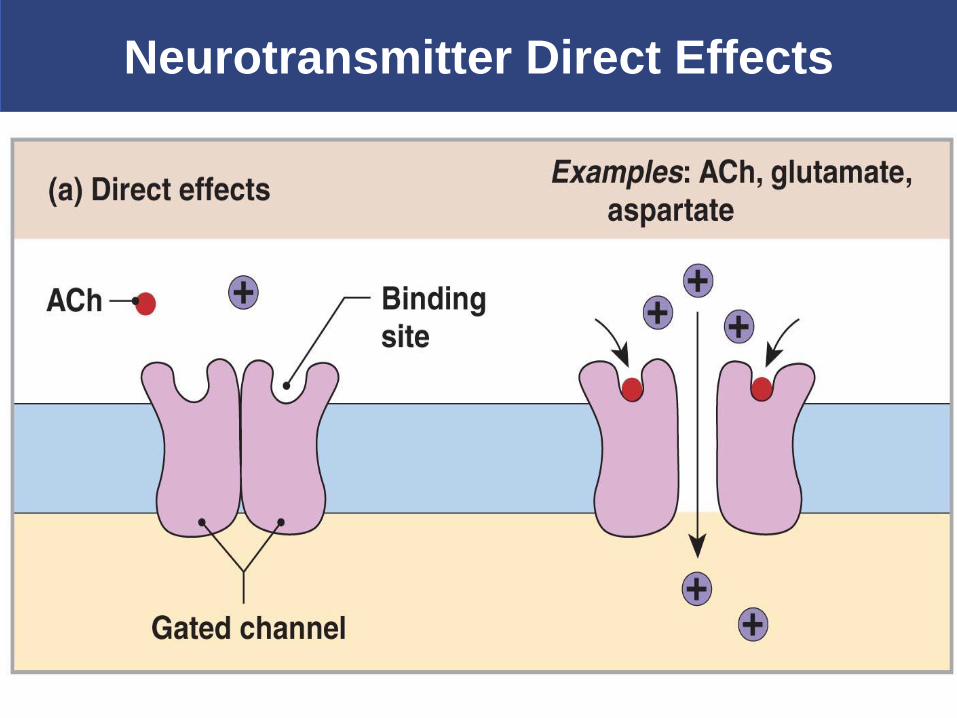
Neurotransmitter Direct Effects
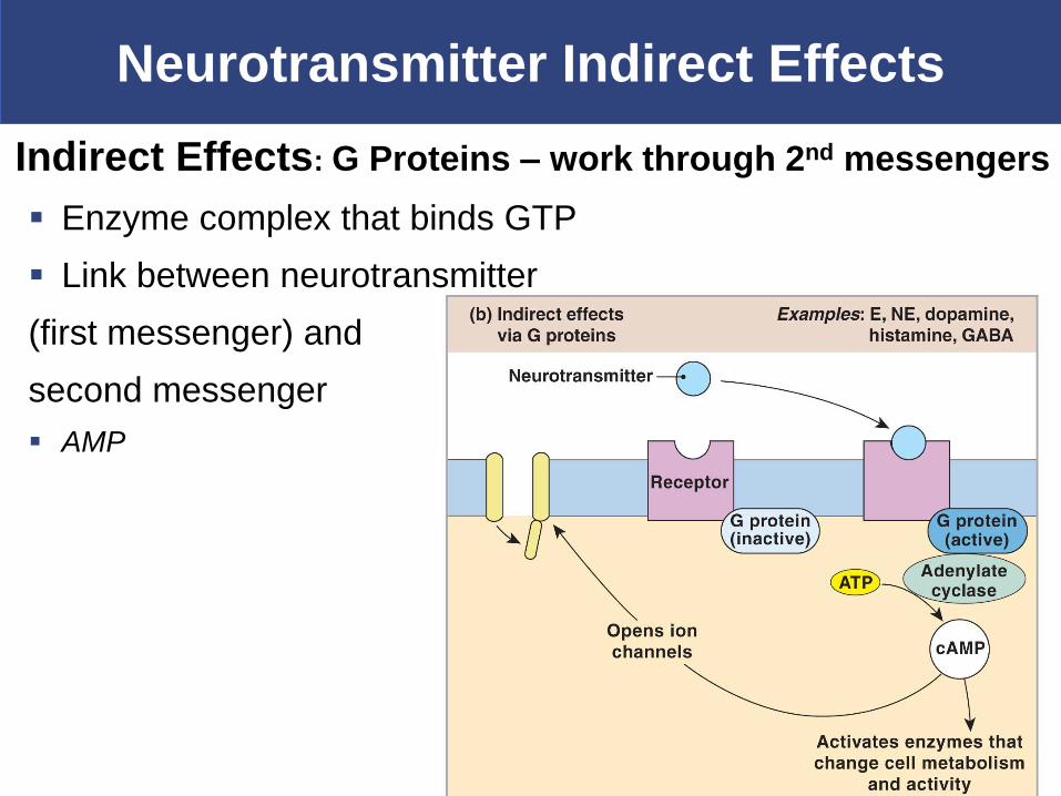
Neurotransmitter Indirect Effects
Indirect Effects: G Proteins – work through 2nd messengers
Enzyme complex that binds GTP
Link between neurotransmitter
(first messenger) and
second messenger
AMP

Neurotransmitter Indirect Effect
Indirect Effects: Intracellular enzymes
Lipid–soluble gases (NO, CO)
Bind to enzymes in brain cells
Top Related