


Languages
Pages
Legal


CCA-fMRI Toolbox - SPM 5
User's Manual Version 2.01


CCA-fMRI Toolbox - Version 2.01
i
Content Trademarks ......................................................................................................................................... ii Credits ................................................................................................................................................ iii
1. BACKGROUND ................................................................................................................................1
2. REQUIREMENTS.............................................................................................................................2
Hardware requirements.......................................................................................................................2 Software requirements .........................................................................................................................2 Restrictions...........................................................................................................................................2
3. INSTALLATION & UPGRADE .....................................................................................................3
4. START UP ..........................................................................................................................................4
5. THE FILE MENU .............................................................................................................................6
6. ANALYSIS WORKFLOW...............................................................................................................8
7. PREPROCESSING ...........................................................................................................................9
8. SETTING UP AN EXPERIMENT............................................................................................... 10
THE EXPERIMENT MENU......................................................................................................................... 10 Setup new experiment….................................................................................................................... 10 Paradigm design…............................................................................................................................ 12 Response model settings ................................................................................................................... 17 Steerable filters….............................................................................................................................. 19 Image set …....................................................................................................................................... 22
9. DATA ANALYSIS.......................................................................................................................... 24
10. RESULTS.................................................................................................................................... 26
The File Menu ................................................................................................................................... 30 The Flip Menu................................................................................................................................... 31 The Projection Menu ........................................................................................................................ 32
11. SCRIPTING................................................................................................................................ 33
Script for 2-dimensional analysis ..................................................................................................... 34 Script for 3-dimensional analysis ..................................................................................................... 38
12. APPENDIX A – SCRIPTING WRAPPER FUNCTIONS....................................................... I
scrCreateHemodynModel() ................................................................................................................. I scrFilterVolumes() .............................................................................................................................. II scrCalcMeanImage() .........................................................................................................................III scrRCCA() ..........................................................................................................................................IV scrRCCAPass1() ................................................................................................................................. V scrRCCAPass2() ................................................................................................................................VI
13. APPENDIX B - SOFTWARE LICENSE................................................................................... I
THE GNU GENERAL PUBLIC LICENSE (GPL) .......................................................................................... II
CCA-fMRI Toolbox - Version 2.01
ii
Trademarks
Matlab® is a registered trademark of The MathWorks, Inc.
(http://www.mathworks.com).
Linux® is the registered trademark of Linus Torvalds in the U.S. and other countries.
Windows® is registered trademarks of Microsoft Corporation in the U.S.A and/or
other countries.

CCA-fMRI Toolbox - Version 2.01
iii
Credits
Author
The toolbox and its documentation were developed by Ph.D. Nils Paulsson, Center for
Medical Image Science and Visualization (CMIV), Linköping University, with much
appreciated input from Professor Magnus Borga and Ph.Lic. Joakim Rydell,
Department of Biomedical Engineering, Linköping University, Sweden.
Scientific work
This toolbox is based on the work in, among others, the following references:
1. Borga M., Learning Multidimensional Signal Processing., Ph.D. thesis,
Linköping University, SE-581 83 Linköping, Sweden, 1998. Dissertation No
531, ISBN 91-7219-202-X,
http://www.imt.liu.se/mi/Publications/Papers/M_Borga_thesis.pdf
2. Buxton R., Wong E. and Frank L., Dynamics of Blood Flow and Oxygenation
Changes During Brain Activation: the Balloon Model.,Magnetic Resonance in
Medicine, 39(6):855-864, 1998.
3. Das S. and Sen P., Asymptotic distribution of restricted canonical correlations
and relevant resampling methods., Journal of Multivariate Analysis, 56(1):1-
19, 1996.
4. Friman O., Borga M., Lundberg P. and Knutsson H., Adaptive Analysis of
fMRI Data, NeuroImage 19(3):837-845, July 2003.
5. Friman O., Borga M., Lundberg P. and Knutsson H, Detecting Neural Activity
in fMRI Using Maximum Correlation Modeling, NeuroImage, 15(2):386-395,
February 2002.
6. Friston K. J., Mechelli A., Turner R. and Price C. J., Nonlinear responses in
fMRI: the balloon model, Volterra kernels,and other hemodynamics.
NeuroImage, 12:466-477, 2000.
7. Hotelling H., Relations between two sets of variates., Biometrika, 28:321-377,
1936.
8. Rydell J., Adaptive Spatial Filtering of fMRI Data, Linköping Studies in
Science and Technology, Thesis No. 1200, Linköping University, SE-581 83
Linköping, Sweden, LiU-TEK-LIC-2005:55, ISBN 91-85457-43-4.
9. Zheng Y., Martindale J., Johnston D., Jones M., Berwick J. and Mayhew J., A
model of the hemodynamic response and oxygen delivery to brain.,
Neuroimage 16: 617-37, 2002.


CCA-fMRI Toolbox - Version 2.01
1
1. Background The CCA-fMRI Toolbox implements the use of canonical correlation analysis (CCA)
for detecting brain activity patterns recorded by functional magnetic resonance
imaging (fMRI). CCA was developed by Hotelling [7] and is a method for finding the
maximum correlation between linear combinations of two sets of variables. In the
CCA-fMRI Toolbox CCA is used in a two step process. In the first step, CCA is used to
construct a low pass filter that adapts to the environment of each voxel in the brain
volume analyzed. In the second step, CCA compare the temporal intensity change of
the filtered voxels with an expected activation pattern to determine the level of
correlation and, hence, the level of activation. The method is thoroughly described in
reference [4].
SPM is a well known and free (GNU General Public License) software package for
analysis of brain imaging data sequences. It has a long history and has continuously
been released in new versions since at least 1994.

CCA-fMRI Toolbox - Version 2.01
2
2. Requirements
Hardware requirements
− Any hardware platform that is supported by both Matlab® version 7.4 and SPM 5.
− Minimum 1 GB RAM. For 3-dimensional analysis a minimum of 1.5 GB RAM is
recommended.
− CPU running at 2 GHz or above.
− Minimum 1.5 GB available temporary disk storage.
Software requirements
− Matlab® 7.4 or later.
− SPM software package version 5 (http://www.fil.ion.ucl.ac.uk/spm/).
− Linux®, Windows® XP or any other operating system that is supported by both
SPM 5 and Matlab® 7.4.
Restrictions
− The CCA-fMRI Toolbox version 2.xx was written for SPM 5 and does not support
SPM 2. For SPM 2 support, please use version 1.x of the toolbox.
− The CCA-fMRI Toolbox was developed and tested using 32-bit Matlab®, versions
7.4. Correct operation under 64-bit Matlab® has neither been tested nor verified.
− SPM 5 is officially only supported on Matlab® versions 6.5 - 7.3. However, besides
having successfully developed the CCA-fMRI Toolbox using Matlab® 7.4 and SPM
5, there are several external sources reporting that this is indeed a working
combination.

CCA-fMRI Toolbox - Version 2.01
3
3. Installation & Upgrade Installation and upgrade both follow the exact same procedure as outlined below.
1. If not already done, install Matlab® and SPM 5 according to their respective
installation instructions.
2. Unzip the CCA-fMRI Toolbox zip-file in a temporary directory.
3. Move the extracted folder, named CCA_fMRI, to the toolbox folder in the
installed SPM 5 directory tree, e.g.:
<MatlabPath>\toolbox\spm5\toolbox.
Be sure to have write access to the toolbox folder. If prompted to overwrite or
replace an existing CCA_fMRI directory and its files acknowledge doing so.
4. Start Matlab® and add the path of the CCA-fMRI Toolbox-folder, e.g.
<MatlabPath>\toolbox\spm5\toolbox\CCA_fMRI,to the Matlab®
path by selecting Set Path… from the File-menu in Matlab's® console
window.
5. Install the CCA-fMRI Toolbox by entering:
>> CCA_fMRI('install');
at the Matlab® prompt. When the installation is done the following text will be
displayed:
CCA-fMRI for SPM has now been installed and the
toolbox can be reached from within SPM.
The CCA-fMRI Toolbox is now installed and ready to be used. In case SPM 5 is used
on a computer where each user has a profile of their own, step 5 above has to be
repeated for all users that want to use the CCA-fMRI Toolbox. In other words, repeat
the following steps for each user:
1. Log on the user
2. Start Matlab®
3. Run CCA_fMRI('install');
For more information regarding usage of the CCA-fMRI Toolbox please see the
following chapters in this User's Manual.

CCA-fMRI Toolbox - Version 2.01
4
4. Start up The CCA-fMRI Toolbox can be reached from the SPM GUI in the same way as any
other SPM toolbox. First, start SPM from the Matlab® prompt:
>> spm
Click the fMRI time-series button in the main window:
Three new windows are opened. Open the drop down list named Toolboxes… in the
lower left corner of the fMRI main window and select CCAfMRI.

CCA-fMRI Toolbox - Version 2.01
5
The CCA-fMRI Toolbox starts up and opens the main window. The toolbox is now
ready to be used.

CCA-fMRI Toolbox - Version 2.01
6
5. The File Menu The File menu's main purpose is to provide the abilities to save and load experiment
settings (see Setting up an experiment), import/export paradigms and optimize memory
usage.
The Load Experiment… loads a previously saved experiment and makes it the
currently defined experiment.
The Save Experiment… menu stores the current experiment setup to an external
Matlab® file (MAT-format) using a user provided file name. The file holds a Matlab®
structure named Experiment having the following fields:
Experiment
ParadigmDesign: [1x1 struct]
BalloonLimits: [1x1 struct]
GammaDiffLimits: [1x1 struct]
FilterSettings: [1x1 struct]
ImageFiles: [1x1 struct]
ParadigmDesign - holds the parameters defining the paradigm of the
experiment. For more informaton see ValidateDesign.m and the paradigm
design dialog box.
BalloonLimits - specifies the base and threshold values of the
hemodynamic response model used in the experiment for cases when being
based on the balloon model. For more information see
ValidateBalloonLimits.m and the balloon settings dialog box.
GammDiffLimmits - specifies the base and threshold values of the
hemodynamic response model used in the experiment for cases when being
based on the differential gamma function. For more information see
ValidateGammaDiffLimits.m and the GammaDiff settings dialog box.

CCA-fMRI Toolbox - Version 2.01
7
FilterSettings - specifies the filter settings of the experiment. For more
information see SteerableBasisFilter3D.m and the steerable filter
settings dialog box.
ImageFiles - specifies the set of image files that are analyzed in the
experiment. For more information see frm_ImageFiles.m.
At times the same paradigm is used in several experiments. To facilitate the
experiment setup the two menu items Import paradigm… and Export
paradigm… allows the user to exclusively save and load the paradigm part of an
experiment.
The Settings menu opens up the dialog box for managing memory usage and FFT
optimization levels. There are different settings depending on if 2-dimensional or 3-
dimensional analysis is performed (see Setting up an experiment). For 2-dimensional
analysis Virtual memory usage has to be set to be large enough to hold the
entire image set of an experiment.
For 3-dimensional analysis the Virtual memory usage filtering parameter
has to be set to be large enough to hold the entire image set of an experiment. The
Virtual memory usage RCCA setting has to be large enough to hold at least
one image file. Finally, the FFT Optimization level sets the level of
optimization performed during filtering with steerable filters. The lowest level is 1 and
the highest level is 4. The higher levels may produce lower initial performance than the
lower levels but over time Matlab®'s FFT module will learn how to perform an
optimal Fourier transform. To determine the size of an image in bytes use the
following formula ImageSizeX * ImageSizeY * ImageSizeZ * 4. The image size is
expressed as number of pixels. To get the size of an image set just multiply the image
size with the number of images.

CCA-fMRI Toolbox - Version 2.01
8
6. Analysis workflow The workflow of the CCA-fMRI analysis is straight forward and consists of
preprocessing, experiment setup, data processing and finally evaluation of the result.
Analysis work
flow
Preprocess image
data
Setup new or load
existing
experiment
Process data
Evaluate and save
results
Done
In the preprocessing step the raw image data is realigned, normalized, etc. to facilitate
the subsequent data processing. However, lowpass filtering must not be performed.
That would significantly degrade the quality of the results. The CCA-fMRI Toolbox
performs its own lowpass filtering using adaptive filters. In the second step,
experiment setup, parameters such as paradigm, BOLD model settings, filter properties
and image data set are specified. Experiments can also be stored/loaded from
secondary storage (hard drive, CD-ROM, etc). The step of processing data consists of
several phases but the process is entirely automatic. Once started there is no need for
user interaction until the processing has finished. This is usually the most time
consuming step. A 2D-analysis usually takes less than 10 minutes and a 3D-analysis
less than an hour, depending on size of data set, amount of available RAM, etc. In the
last step the result can be visualized as well as exported to secondary storage. With the
exception of preprocessing, all steps are performed within the CCA-fMRI Toolbox user
interface.

CCA-fMRI Toolbox - Version 2.01
9
7. Preprocessing The CCA-fMRI Toolbox will usually produce better results when analyzing
preprocessed data. Commonly applied preprocessing procedures in SPM are:
− Setting origin
− Realignment
− Reorientation
The only prerequisite of using preprocessed images in the toolbox is that the data is
available as standard SPM 5 image files. This is seldom a problem since SPM usually
applies preprocessing to the image data files directly or by creating new preprocessed
versions of existing image files.
NOTE
Smoothing, or low pass filtering, must not be applied to the data set during
preprocessing. Doing so would interfere with the adaptive filtering applied later on by
the CCA-fMRI Toolbox and significantly degrade the quality of the final analysis
results.

CCA-fMRI Toolbox - Version 2.01
10
8. Setting up an experiment An analysis setup is defined in terms of an experiment, which defines the parameters
necessary for the analysis of a certain fMRI data set. An experiment is defined by the
four main property groups:
− Paradigm design
− Response model (BOLD) settings
− Settings of the steerable filters
− Name and location of the image data set to be analyzed
All properties are accessible from the Experiment menu in the CCA-fMRI Toolbox
main window.
The paradigm design specifies at what time points and for how long the subject is
exposed to stimuli. The response model describes how the expected BOLD response,
resulting from the stimuli, should look like. The steerable filter settings give the shape
and size of the adaptive low pass filters used during the data analysis and the data set
points out which image files to analyze.
The Experiment menu
Setup new experiment…
This menu item takes you through all the steps necessary to setup a new experiment,
i.e. the paradigm design, the response model (BOLD) settings, the filter settings and
the image data file selection. The setup workflow is as follows:

CCA-fMRI Toolbox - Version 2.01
11
Specify paradigm
design
Specify BOLD
model parameters
Optionally
Specify steerable
filter settings
Select image data
set
Setup new
experiment...
New experiment
defined
Setting up a new experiment this way is entirely transactional, meaning that the user
has to respond Ok to all the dialog boxes that appear during the setup procedure. If the
user selects Cancel, i.e. click the Cancel-button, in any of the steps above, none of
the settings are saved and any previously specified experiment and results are still
intact.
The separate settings are also available under their corresponding menu headline in the
Experiment menu. Consequently, an experiment can also be setup in a more manual
fashion by accessing the menu items Paradigm design…, Response model
settings, Steerable filters and Image set…. Please see below for more
information about how to use the separate settings dialog boxes.
CCA-fMRI Toolbox - Version 2.01
12
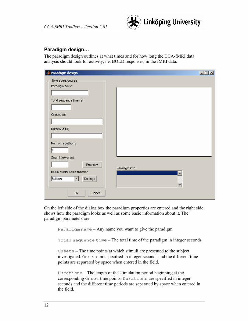
Paradigm design…
The paradigm design outlines at what times and for how long the CCA-fMRI data
analysis should look for activity, i.e. BOLD responses, in the fMRI data.
On the left side of the dialog box the paradigm properties are entered and the right side
shows how the paradigm looks as well as some basic information about it. The
paradigm parameters are:
Paradigm name – Any name you want to give the paradigm.
Total sequence time – The total time of the paradigm in integer seconds.
Onsets – The time points at which stimuli are presented to the subject
investigated. Onsets are specified in integer seconds and the different time
points are separated by space when entered in the field.
Durations – The length of the stimulation period beginning at the
corresponding Onset time points. Durations are specified in integer
seconds and the different time periods are separated by space when entered in
the field.

CCA-fMRI Toolbox - Version 2.01
13
Num of repetitions – Specifies the number of times the
Onset/Duration settings should be repeated within the total sequence time.
As such, the Onset/Duration settings may not be repeated beyond the total
sequence time.
Scan interval – The sampling interval at which fMRI image volumes are
recorded by the MR scanner. Scan interval is given in decimal seconds.
BOLD Model basis function – Specifies what mathematical model
should be used to approximate the BOLD response. The two options are the
Balloon model and the Differential gamma model. Default model selection is
the Balloon model.
The dialog box also has four buttons. Besides the standard Ok/Cancel-buttons, there
is also a Preview-button for plotting the resulting paradigm design and a Setting-
button that allows direct access to the dialog boxes for adjusting the parameters of the
selected BOLD model basis function. Please see menu item Response model
settings for more information.
CCA-fMRI Toolbox - Version 2.01
14
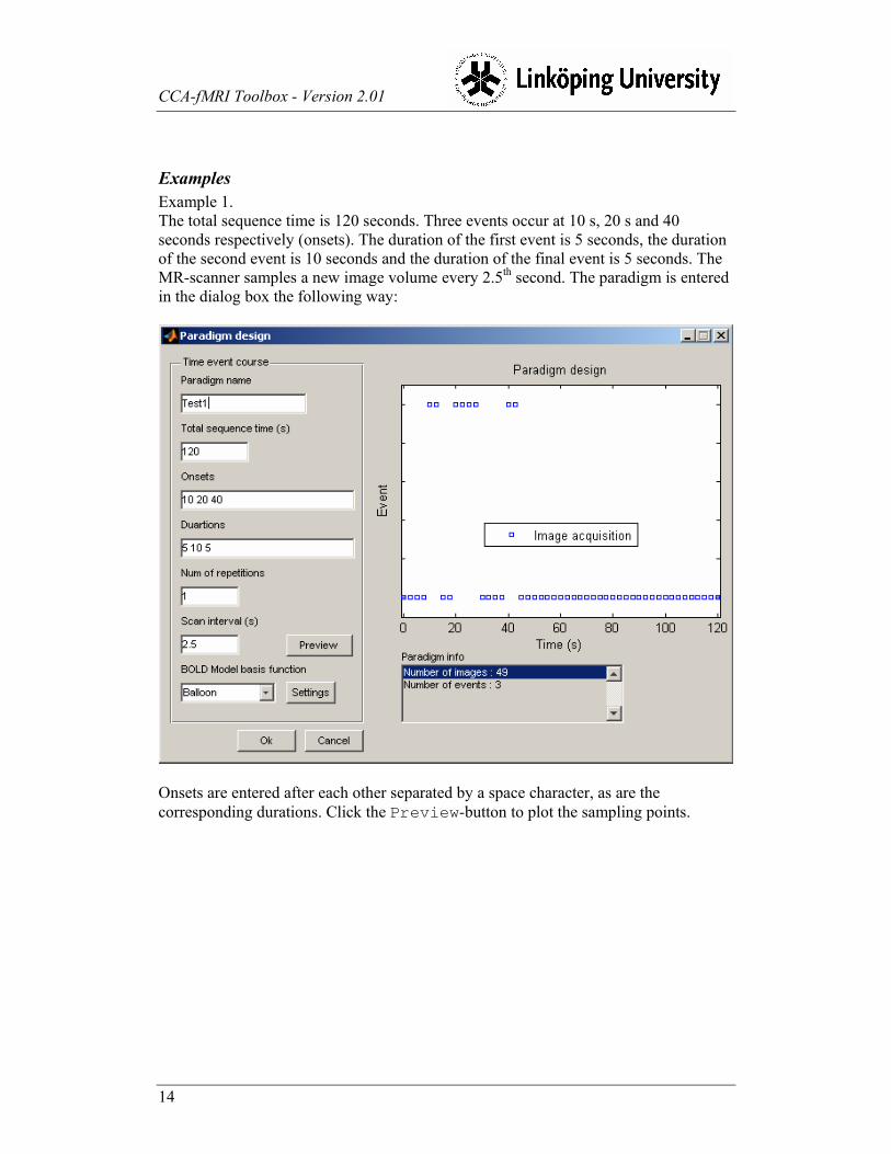
Examples
Example 1.
The total sequence time is 120 seconds. Three events occur at 10 s, 20 s and 40
seconds respectively (onsets). The duration of the first event is 5 seconds, the duration
of the second event is 10 seconds and the duration of the final event is 5 seconds. The
MR-scanner samples a new image volume every 2.5th second. The paradigm is entered
in the dialog box the following way:
Onsets are entered after each other separated by a space character, as are the
corresponding durations. Click the Preview-button to plot the sampling points.

CCA-fMRI Toolbox - Version 2.01
15
Example 2.
In this example almost all parameters are the same as in Example 1 with the notable
exception of the Num of repetitions. This time we would like to repeat the
Onset/Duration sequence owing to repetitive patterns of stimuli presentations. By
setting Num of repetitions to 2 the Onset/Duration sequence will be repeated
one time directly after the last data point of the last stimuli event, ending at 45 seconds
(Onset 40 s + Duration 5 s = 45 seconds).
Again, click the Preview-button to plot the new paradigm. The limitation of the Num
of repetitions-parameter is that the Onset/Duration sequence can't be
repeated beyond the Total sequence time-setting.

CCA-fMRI Toolbox - Version 2.01
16
Example 3
A consequence of having the possibility to repeat the sequence of events, the setting
Onset = [10 30 50 70] and Duration = [10 10 10 10] can also be defined as
Onset=10 s, Duration=10 s and Num of repetitions=4 :

CCA-fMRI Toolbox - Version 2.01
17
Response model settings
The response model describes the expected BOLD response for a given paradigm. In
other words, the model is considered to be the approximate true activation pattern for
any subject exposed to the stimuli paradigm used in the experiment. The level of
activation in a brain voxel is, simplified, determined by comparing the temporal
intensity change of the voxel with the intensity change of the response model. High
correlation means high level of activation and vice versa. This comparison is
performed for each voxel in the sample data.
There are two different BOLD basis functions available in the toolbox; the Balloon
model and the Differential Gamma model, also called GammaDiff in the toolbox. Of
the two, the Balloon model is considered more accurate but also somewhat more
demanding for the computer to generate. For a fairly up to date computer there is little
incentive not to use the Balloon model.
The two models have their own sets of adjustable parameters, accessible from the
Response model settings menu. Disregarding what basis function is used, the
BOLD model is generated in the same way. First, 500 plausible response curves are
generated by randomizing the values of corresponding parameters. To assure plausible
curves the parameters are only allowed random variations within a specific tolerance
interval. The 500 plausible responses are then reduced, by principal component
analysis, to a compressed format, which is also the expected BOLD response used in
the subsequent data analysis.
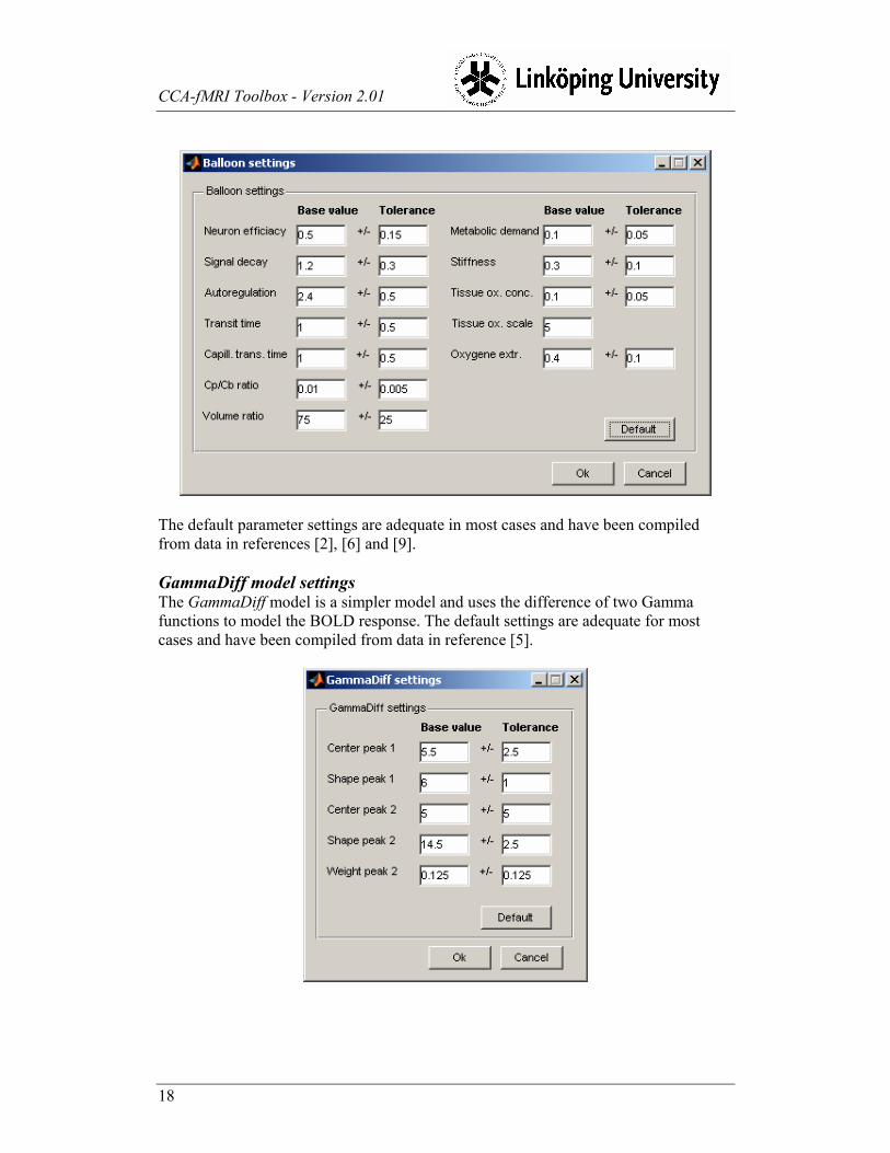
Balloon model settings
The Balloon model is a fairly complicated model having a number of adjustable
parameters corresponding to, among other things, properties of an expanding blood
vessel forming a local balloon of oxygenated blood. The model, and its parameters, is
explained in references [2] and [6].
CCA-fMRI Toolbox - Version 2.01
18
The default parameter settings are adequate in most cases and have been compiled
from data in references [2], [6] and [9].
GammaDiff model settings The GammaDiff model is a simpler model and uses the difference of two Gamma
functions to model the BOLD response. The default settings are adequate for most
cases and have been compiled from data in reference [5].
CCA-fMRI Toolbox - Version 2.01
19
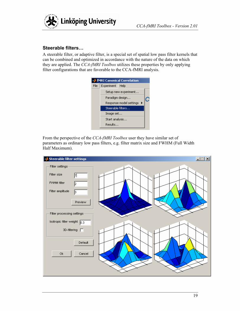
Steerable filters…
A steerable filter, or adaptive filter, is a special set of spatial low pass filter kernels that
can be combined and optimized in accordance with the nature of the data on which
they are applied. The CCA-fMRI Toolbox utilizes these properties by only applying
filter configurations that are favorable to the CCA-fMRI analysis.
From the perspective of the CCA-fMRI Toolbox user they have similar set of
parameters as ordinary low pass filters, e.g. filter matrix size and FWHM (Full Width
Half Maximum).

CCA-fMRI Toolbox - Version 2.01
20
In the left side of the dialog box the filter and processing parameters are set and the
right side shows previews of the filter kernel shapes. The filter kernel views are
updated by clicking on the Preview-button using the currently entered values.
NOTE
Even when selecting 3-dimensional analysis the filter kernels will still be drawn as 2-
dimensional kernels in the previews.
Filter settings
The filter parameters are:
Filter size – The size of the adaptive filter given as the number of pixels
(or voxels) of one side if the filter kernel. The filter size is symmetric in all
directions forming a squared matrix or cube depending on whether 2-
dimensional or 3-dimensional analysis should be performed. Only odd sizes
are allowed.
FWHM filter – The full width half maximum of the filter kernels expressed
as number of pixels. This is the same as the effective width of the filter.
Filter amplitude – The amplitude of the final filter.
Large settings for FilterSize and FWHM filter tend to favor large regions of
activation and suppress small. Small settings allow small regions of activation to
remain but also increase the spatial noise level.
Filter processing settings
The filter processing parameters defines how the adaptive filter is applied during the
data analysis phase. The parameters are:
Isotropic filter weight – Defines the importance of the center voxel
during filtering. Weight 0.0 means that the center voxel is given no special
importance, i.e. only anisotropic filtering, and weight 1.0 gives the center voxel
the same importance as the anisotropic filter.
3D-filtering – Enables 3-dimensional filtering. When unchecked, 2-
dimensional filtering is used.
Using 3-dimensional filtering is the preferred setting since that would take into account
the entire 3-dimensional neighborhood. 2-dimensional only takes into account the
neighborhood in the X/Y-plane, which typically is the horizontal plane through the
brain.
The drawback of using 3-dimensional filtering is that the complexity of the fMRI
analysis grows exponentially meaning significantly longer processing time, compared
to 2-D filtering. On a fairly modern computer a 2-dimensional analysis would take 5-

CCA-fMRI Toolbox - Version 2.01
21
10 minutes. The same analysis performed in 3 dimensions would take from 40 minutes
up to an hour.
NOTE
The most important factor for maximizing the 3-D performance is to have a lot of
random access memory (RAM). The analysis generates between 1 and 2 GB of
temporary data. Large amount of RAM prevents data from being written to disk during
the analysis, hence increasing performance. See Requirements for more information
regarding the recommended hardware platform.

CCA-fMRI Toolbox - Version 2.01
22
Image set …
The Image set window is used for selecting the image set to be analyzed by the
CCA-fMRI Toolbox. The data set is defined by clicking on the Select files
button.
The CCA-fMRI Toolbox uses the standard SPM file dialog box to select the image set.
In case the images have been preprocessed be sure to select the image files having the
correct name prefix.

CCA-fMRI Toolbox - Version 2.01
23
Information about separate image files in the set, selectable from the drop down list
Current file, is displayed in the panel. The currently selected image can also be
viewed using the Preview-button.

CCA-fMRI Toolbox - Version 2.01
24
9. Data analysis The data analysis is an automated process with little need for manual intervention.
Depending on whether 2-dimensional or 3-dimensional analysis has been chosen one
of the dialog boxes below will be opened.
Besides the Start-button, which commences the analysis process, there is also an
Abort-button for interrupting an analysis in progress and a progress bar showing the
progress of each separate phase.
2-Dimensional analysis 3-Dimensional analysis
The analysis is divided into separate phases, shown in the dialog boxes above. The
phases are listed in the order of execution and each phase has a status displayed to the
right. The valid statuses are Waiting…, Working…, Done and Aborted. The
toolbox keeps track of these statuses meaning that when a previously aborted analysis

CCA-fMRI Toolbox - Version 2.01
25
is restarted only the phases not yet finished, i.e. not having the Done-status, will be
reprocessed. Some parts of the analysis process may be reprocessed in cases when
experiment settings influencing the phase have been changed. Which phases to
reprocess is entirely handled by the CCA-fMRI Toolbox. Once the analysis has finished
the result is reachable from the Results… dialog box. The results are also stored in
two standard SPM image files in the same directory as the original image file sets
analyzed. The two images are named CorrelationMap_#TIMESTAMP# and
MeanVolume_#TIMESTAMP#, where #TIMESTAMP# is on the format MMM-DD-
HH-MM-SS, e.g. CorrelationMap_Jan-15_14-27-23.img (or .hdr).
NOTE
When aborting an analysis the toolbox usually takes a while before actually aborting.
The reason is that owing to limitations in Matlab® the abort of the analysis has to be
synchronized with an update of the horizontal progress bar which only happens a
limited number of times during each phase. The toolbox will, however, in time abort
the analysis process.

CCA-fMRI Toolbox - Version 2.01
26
10. Results The Results… menu provide basic functionality for displaying, printing and saving
the analysis results from the current experiment.
The Results dialog box displays the correlation as color coded levels superimposed
on a background image of the brain analyzed. The brain is segmented along the X-, Y-
and Z- dimensions forming 3 image segments. The lower left image is the Y/X-
segment, the upper left is the Y/Z-segment and the right is the X/Z-segment.

CCA-fMRI Toolbox - Version 2.01
27
The background image is the mean image of the entire image set. The projection
honors reorientation and other preprocessing procedures applied in advance of the
canonical correlation analysis.
In the lower right area information about the current voxel is displayed, i.e. voxel
coordinate in millimeters as well as its corresponding correlation coefficient. The
current voxel is selected using the mouse to select and clicking in one of the three
projections. The voxel information and hair cross are updated accordingly. The vertical
slider is used to manually determine the smallest correlation threshold value for which
correlation should be superimposed on the background image. The threshold level can
also be inverted by checking the Invert threshold checkbox. This can be useful
in cases where e.g. anticorrelation is of interest.
Inverted threshold

CCA-fMRI Toolbox - Version 2.01
28
Before the CCA-calculation the image volume is segmented with respect to the brain.
To reduce the possibility of accidentally excluding brain voxels the segmentation
algorithm usually extracts a volume slightly larger than the brain itself. As a result, the
analysis also includes voxels slightly outside the brain. Using the horizontal Mask
size slider the segmentation mask can be reduced in size allowing the correlation
map to better correlate with the brain tissue.
Original brain segmentation mask
The original brain segmentation mask is used when the Mask size-slider is
positioned to the far left. Moving the slider to the right reduces the mask size stepwise.

CCA-fMRI Toolbox - Version 2.01
29
Reduced brain segmentation mask
The Mask size-slider only affects the displayed results and not the segmentation
mask stored in the result's data file.
CCA-fMRI Toolbox - Version 2.01
30
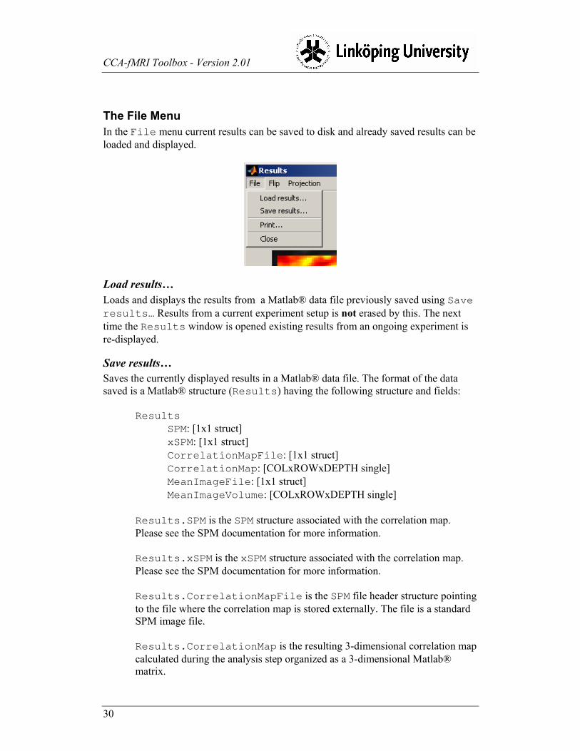
The File Menu
In the File menu current results can be saved to disk and already saved results can be
loaded and displayed.
Load results…
Loads and displays the results from a Matlab® data file previously saved using Save
results… Results from a current experiment setup is not erased by this. The next
time the Results window is opened existing results from an ongoing experiment is
re-displayed.
Save results…
Saves the currently displayed results in a Matlab® data file. The format of the data
saved is a Matlab® structure (Results) having the following structure and fields:
Results
SPM: [1x1 struct]
xSPM: [1x1 struct]
CorrelationMapFile: [1x1 struct]
CorrelationMap: [COLxROWxDEPTH single]
MeanImageFile: [1x1 struct]
MeanImageVolume: [COLxROWxDEPTH single]
Results.SPM is the SPM structure associated with the correlation map.
Please see the SPM documentation for more information.
Results.xSPM is the xSPM structure associated with the correlation map.
Please see the SPM documentation for more information.
Results.CorrelationMapFile is the SPM file header structure pointing
to the file where the correlation map is stored externally. The file is a standard
SPM image file.
Results.CorrelationMap is the resulting 3-dimensional correlation map
calculated during the analysis step organized as a 3-dimensional Matlab®
matrix.

CCA-fMRI Toolbox - Version 2.01
31
Results.MeanImageFile is the SPM file header structure pointing to the
file where the mean image of all the image volumes in the set is stored
externally. The file is a standard SPM image file.
Results.MeanImage is the resulting 3-dimensional mean image calculated
during the analysis step organized as a 3-dimensional Matlab® matrix.
Print…
Prints a copy of the currently displayed results.
Close
Closes the results dialog box and returns to the main window.
The Flip Menu
The Flip menu provides the ability to flip the dimensions of the displayed projections
180 degrees. The Reset menu item resets the dimensions to default orientation.

CCA-fMRI Toolbox - Version 2.01
32
The Projection Menu
The Projection menu allows to change between various ways of displaying the
results. Currently only segmented projection is supported.

CCA-fMRI Toolbox - Version 2.01
33
11. Scripting Great efforts have been made to keep the graphical user interface separated from the
actual image processing modules. As a consequence it is straight forward to use the
CCA-fMRI Toolbox, and its modules, in Matlab® scripts to simplify repetitive
analyses. An advantage of running the toolbox in script mode is that repetitive tasks
for large analysis series can be automated. Script mode also reduce the memory
footprint significantly thereby allowing larger datasets to be analyzed than is the case
with the graphical user interface.
Below are two examples of how to use the toolbox in scripts. The first example is a 2-
dimensional analysis and the second example is a 3-dimensional analysis. The two
sample scripts are located in the SampleScripts folder located within the CCA-
fMRI Toolbox directory. The wrapper functions are located in the main toolbox
directory and have names prefixed by scr.
NOTE
Make sure that the path to the sample directory has been added to the Matlab® path
before trying to use the samples.

CCA-fMRI Toolbox - Version 2.01
34
Script for 2-dimensional analysis
The script Sample2DAnalysis.m uses analysis wrappers functions, also provided in the toolbox, for the actual processing. Their usage and
calling parameters and are described in Appendix A – Scripting wrapper functions. The wrapper functions used for 2-dimensional analysis are:
1. scrCreateHemodynModel() 2. scrRCCA()
function [CorrelationMap MeanImageVolume] = Sample2DAnalysis()
% GET IMAGE VOLUME FILES TO PROCESS
disp('Getting image files...');
% Query the user for the files in the data set. The is not the usual way
% of specifying what files to analyze in a script...
warning off;
% ImageFiles = spm_get([],'.img','Select image files',pwd);
[ImageFiles Cancel] = spm_select([1 Inf], 'image','Select image files', '',pwd);
spm_Headers = spm_vol(ImageFiles);
warning on;
% CREATE HEMODYNAMIC RESPONSE MODEL
disp('Creating hemodyn model...');
% Setup parameters for balloon model
BalloonLimits.NeuronEffBase = 0.5;
BalloonLimits.NeuronEffMinMax = 0.15;
BalloonLimits.SigDecayBase = 1.2;
BalloonLimits.SigDecayMinMax = 0.3;
BalloonLimits.AutoRegBase = 2.4;
BalloonLimits.AutoRegMinMax = 0.5;
BalloonLimits.TransTimeBase = 1;
BalloonLimits.TransTimeMinMax = 0.5;
BalloonLimits.CapTransTimeBase = 1.0;
BalloonLimits.CapTransTimeMinMax = 0.5;
BalloonLimits.CpbRatioBase = 0.01;
BalloonLimits.CpbRatioMinMax = 0.005;
BalloonLimits.VolRatioBase = 75;

CCA-fMRI Toolbox - Version 2.01
35
BalloonLimits.VolRatioMinMax = 25;
BalloonLimits.MetabolicBase = 0.1;
BalloonLimits.MetabolicMinMax = 0.05;
BalloonLimits.StiffnessBase = 0.3;
BalloonLimits.StiffnessMinMax = 0.1;
BalloonLimits.TissueOxConcBase = 0.1;
BalloonLimits.TissueOxConcMinMax = 0.05;
BalloonLimits.TOScale = 5;
BalloonLimits.RestExtractBase = 0.4;
BalloonLimits.RestExtractMinMax = 0.1;
% In case default values are appropriate use the following instead
% BalloonLimits = GetDefBalloon();
% Setup the paradigm. The model basis function can also be set to 'Gamma
% diff' in which case the structure GammaDiffLimits should replace
% BalloonLimits below.
ParadigmDesign.ModelBasisFunction = 'Balloon';
ParadigmDesign.Name = 'Test paradigm';
ParadigmDesign.TotalSequenceTime = 360;
ParadigmDesign.NumberOfRepetitions = 1;
ParadigmDesign.SamplingInterval = 2.7;
Onsets = [40 120 200 280];
Durations = [40 40 40 40];
ParadigmDesign.RelaxStimEvents = Compatibility('OnsetDuration2EventTime', Onsets, Durations);
HemoDynRespModel = scrCreateHemodynModel(ParadigmDesign, BalloonLimits);
% LOAD IMAGE DATA SET
% Check that the number of files is equal to the number of expected data
% points in the paradigm
NumberOfFiles = size(spm_Headers,1);
SampleCount = size(HemoDynRespModel,1);
if (NumberOfFiles > SampleCount)
% Adjust the number of images
disp ('Clipping trailing image files');
spm_Headers = spm_Headers(1:SampleCount);
elseif (NumberOfFiles < SampleCount);
% Not enough image files
disp ('Not enough image files');

CCA-fMRI Toolbox - Version 2.01
36
return;
end;
% Init image storage
disp ('Initiating image storage...');
MemFileName = 'c:\temp\memfile.dat';
ImageStorage = InitImageFileStorage(MemFileName, spm_Headers);
% Load images into memory mapped file
disp ('Loading images...');
LoadImageFiles(spm_Headers, ImageStorage, 0);
% GET MEAN IMAGE AND BRAIN SEGMENTATION MASK OF ORIGINAL IMAGE VOLUMES
disp('Calculating mean image...');
% Calculate the mean image volume
[MeanImageVolume SegMask] = scrCalcMeanImage(spm_Headers);
% SETUP FILTERS
disp('Setting up filters...');
FilterSettings.FilterSize = 7;
FilterSettings.FWHMLowPass = 5;
FilterSettings.FWHMFilter = 2;
FilterSettings.IsoFilterWeight = 0.3;
FilterSettings.Filter3D = false;
% PROCESS THE IMAGE DATA
disp('Processing images...');
CorrelationMap = scrRCCA(HemoDynRespModel, FilterSettings, ImageStorage, SegMask);
% FINISH UP
disp ('Saving results...');
% Put together the results. Warning is disabled to prevent numerous
% "Warning: Cant get default Analyze orientation - assuming
% flipped" -messages in case SPM isn't currently running.
warning off;
Results = GenerateResults(ParadigmDesign, spm_Headers, CorrelationMap, MeanImageVolume, SegMask);
warning on;

CCA-fMRI Toolbox - Version 2.01
37
% Save the results to a file that is readable by the CCA-fMRI toolbox's
% Results window.
ResultsFileName = 'c:\temp\Results2D.mat';
save (ResultsFileName, 'Results');
% Done
disp('Done!!!');

CCA-fMRI Toolbox - Version 2.01
38
Script for 3-dimensional analysis
The script Sample3DAnalysis.m uses analysis wrapper functions, also provided in the toolbox, for the actual processing. Their usage and
calling parameters are described in Appendix A – Scripting wrapper functions. The wrapper functions 3-dimensionala analysis are:
3. scrCreateHemodynModel() 4. scrFilterVolumes() 5. scrCalcMeanImage() 6. scrRCCAPass1() 7. scrRCCAPass2()
function [CorrelationMap MeanImageVolume] = Sample3DAnalysis()
% GET IMAGE VOLUME FILES TO PROCESS
disp('Get image files...');
% Query the user for the files in the data set. The is not the usual way
% of specifying what files to analyze in a script.
warning off;
ImageFiles = spm_get([],'.img','Select image files',pwd);
spm_Headers = spm_vol(ImageFiles);
warning on;
% CREATE HEMODYNAMIC RESPONSE MODEL
disp('Create hemodyn model...');
% Setup parameters for balloon model
BalloonLimits.NeuronEffBase = 0.5;
BalloonLimits.NeuronEffMinMax = 0.15;
BalloonLimits.SigDecayBase = 1.2;
BalloonLimits.SigDecayMinMax = 0.3;
BalloonLimits.AutoRegBase = 2.4;
BalloonLimits.AutoRegMinMax = 0.5;
BalloonLimits.TransTimeBase = 1;
BalloonLimits.TransTimeMinMax = 0.5;
BalloonLimits.CapTransTimeBase = 1.0;
BalloonLimits.CapTransTimeMinMax = 0.5;

CCA-fMRI Toolbox - Version 2.01
39
BalloonLimits.CpbRatioBase = 0.01;
BalloonLimits.CpbRatioMinMax = 0.005;
BalloonLimits.VolRatioBase = 75;
BalloonLimits.VolRatioMinMax = 25;
BalloonLimits.MetabolicBase = 0.1;
BalloonLimits.MetabolicMinMax = 0.05;
BalloonLimits.StiffnessBase = 0.3;
BalloonLimits.StiffnessMinMax = 0.1;
BalloonLimits.TissueOxConcBase = 0.1;
BalloonLimits.TissueOxConcMinMax = 0.05;
BalloonLimits.TOScale = 5;
BalloonLimits.RestExtractBase = 0.4;
BalloonLimits.RestExtractMinMax = 0.1;
% In case default values are appropriate use the following instead
% BalloonLimits = GetDefBalloon();
% Setup the paradigm
ParadigmDesign.TotalSequenceTime = 360;
Onsets = [40 120 200 280];
Durations = [40 40 40 40];
ParadigmDesign.RelaxStimEvents = Compatibility('OnsetDuration2EventTime', Onsets, Durations);
ParadigmDesign.NumberOfRepetitions = 1;
ParadigmDesign.SamplingInterval = 2.7;
HemoDynRespModel = scrCreateHemodynModel(ParadigmDesign, BalloonLimits);
% SETUP FILTERS
disp('Setup filters...');
FilterSettings.FilterSize = 7;
FilterSettings.FWHMLowPass = 5;
FilterSettings.FWHMFilter = 2;
FilterSettings.IsoFilterWeight = 0.3;
FilterSettings.Filter3D = true;
% Create the 3D-filters
[IsoFilt AnisoFilt_1 AnisoFilt_2 AnisoFilt_3 AnisoFilt_4 AnisoFilt_5 AnisoFilt_6] = GetFilters3D(FilterSettings);
Filters3D = {IsoFilt AnisoFilt_1 AnisoFilt_2 AnisoFilt_3 AnisoFilt_4 AnisoFilt_5 AnisoFilt_6};
% SETUP TEMPORARY STORAGE
disp('Setup temporary storage...');

CCA-fMRI Toolbox - Version 2.01
40
% Specify names of temporary storage used during the processing
FilteredFileName = 'C:\temp\memmapfile1.dat';
RCCAFileName = 'C:\temp\memmapfile2.dat';
% Setup the temporary files (memory mapped files)
VolumeSize = spm_Headers(1).dim(1:3);
VolumeCount = size(spm_Headers,1);
[FilteredStorage RCCAStorage] = InitImageFileStorage3D(VolumeSize, VolumeCount, FilteredFileName, RCCAFileName);
% GET MEAN IMAGE AND BRAIN SEGMENTATION MASK OF ORIGINAL IMAGE VOLUMES
disp('Calculate mean image...');
% Calculate the mean image volume
[MeanImageVolume SegMask] = scrCalcMeanImage(spm_Headers);
% FILTER VOLUMES
disp('Filter image volumes...');
% Filter all volumes with all filters
scrFilterVolumes(spm_Headers, Filters3D, FilteredStorage, RCCAStorage);
% Clean up
clear Filters3D;
clear FilteredStorage;
% RUN 1ST RCCA PASS
disp('1st RCCA pass...');
% Perform 1st RCCA pass reusing the temporary file used during the
% filtering step as output file
SizeRCCAArray = scrRCCAPass1(HemoDynRespModel, FilterSettings.IsoFilterWeight, ...
RCCAStorage, FilteredFileName, VolumeCount, VolumeSize, SegMask);
clear RCCAStorage;
% RUN 2ND RCCA PASS
disp('2nd RCCA pass...');
% Perform 2nd RCCA pass using the input file from the 1st pass
CorrelationMap = scrRCCAPass2(HemoDynRespModel, FilteredFileName, SizeRCCAArray, VolumeSize, SegMask);

CCA-fMRI Toolbox - Version 2.01
41
% FINISH UP
disp ('Saving results...');
% Put together the results. Warning is disabled to prevent numerous
% "Warning: Cant get default Analyze orientation - assuming
% flipped"-messages in case SPM isn't currently running
warning off;
Results = GenerateResults(ParadigmDesign, spm_Headers, CorrelationMap, MeanImageVolume, SegMask);
warning on;
% Save the results to a file that is readable by the CCA-fMRI toolbox's
% Results window.
ResultsFileName = 'c:\temp\scrResults3D.mat';
save (ResultsFileName, 'Results');
% Done
disp('Done!!!');


CCA-fMRI Toolbox - Version 2.01
I
12. Appendix A – Scripting wrapper functions
scrCreateHemodynModel()
Definition [HemoDynRespModel] = scrCreateHemodynModel(ParadigmDesign,
BalloonLimits)
Description
Creates a hemodynamic response model based on the settings of
ParadigmDesign. The model is returned in HemoDynRespModel. The
ParadigmDesign parameter has the following fields: ParadigmDesign.TotalSequenceTime;
ParadigmDesign.RelaxStimEvents;
ParadigmDesign.NumberOfRepetitions;
ParadigmDesign.SamplingInterval;
The hemodynamic model created is based on the balloon model using the
parameters specified in the BalloonLimits structure having the following
fields:
BalloonLimits.NeuronEffBase;
BalloonLimits.NeuronEffMinMax;
BalloonLimits.SigDecayBase;
BalloonLimits.SigDecayMinMax;
BalloonLimits.AutoRegBase;
BalloonLimits.AutoRegMinMax;
BalloonLimits.TransTimeBase;
BalloonLimits.TransTimeMinMax;
BalloonLimits.CapTransTimeBase;
BalloonLimits.CapTransTimeMinMax;
BalloonLimits.CpbRatioBase;
BalloonLimits.CpbRatioMinMax;
BalloonLimits.VolRatioBase;
BalloonLimits.VolRatioMinMax;
BalloonLimits.MetabolicBase;
BalloonLimits.MetabolicMinMax;
BalloonLimits.StiffnessBase;
BalloonLimits.StiffnessMinMax;
BalloonLimits.TissueOxConcBase;
BalloonLimits.TissueOxConcMinMax;
BalloonLimits.TOScale;
BalloonLimits.RestExtractBase;
BalloonLimits.RestExtractMinMax
See Also ValidateDesign.m
ValidateBalloonLimits.m

CCA-fMRI Toolbox - Version 2.01
II
scrFilterVolumes()
Definition scrFilterVolumes(spm_Headers, Filters3D, FilteredStorage,
RCCAStorage)
Description
Applies the 3-dimensional filter kernels Filters3D to the image volumes
defined by spm_Headers. scrFilterVolumes() uses the memory
mapped file FilteredStorage for intermediate storage. After the filtering
has been performed the final results are rearranged and copied to the memory
mapped file RCCAStorage, which is used by the subsequent canonical
correlation analysis.
spm_Headers is a standard SPM image file header vector. Filters3D is a
cell array holding the different filter kernels that should be applied to the image
volumes. FilteredStorage and RCCAStorage are structures of the type
ImageStorage having the following fields:
ImageStorage.TotalSize
ImageStorage.TotNumOfObjects
ImageStorage.ObjectSize
ImageStorage.CurrentObjectIndex
ImageStorage.RepeatValue
ImageStorage.MemFileHandle
ImageStorage.MemFileHandle.data[].Object
ImageStorage.MemFileHandle.format
ImageStorage.MemFileHandle.offset
ImageStorage.MemFileHandle.repeat
ImageStorage.MemFileHandle.writable
See Also InitImageFileStorage3D.m
GetFilters3D.m
Filter3D.m
spm_get()
spm_vol()

CCA-fMRI Toolbox - Version 2.01
III
scrCalcMeanImage()
Definition [MeanImageVol SegMask] = scrCalcMeanImage(spm_Headers)
Description
Calculates the mean image volume and the brain segmentation mask for the
image files specified by spm_Headers. The resulting mean image is returned
as a standard Matlab® matrix in MeanImageVol and the segmentation mask
is returned as a logical matrix in SegMask. The spm_Headers is the same
headers as returned by e.g. spm_vol().
See Also

CCA-fMRI Toolbox - Version 2.01
IV
scrRCCA()
Definition [CorrelationMap MeanImageVolume] = scrRCCA(HemoDynRespModel,
FilterSettings, ImageStorage, Segmask)
Description
Performs a 2-dimensional correlation analysis and returns the correlation map
in CorrelationMap and the mean image in MeanImageVolume.
HemoDynRespModel is the hemodynamic response model to which the
MRI-data should be compared. FilterSettings specifies the size and
shape of the steerable filters and have the following fields:
FilterSettings.FilterSize; FilterSettings.FWHMLowPass; FilterSettings.FWHMFilter; FilterSettings.IsoFilterWeight; FilterSettings.Filter3D;
ImageStorage specifies the memory mapped file into which the images to
analyzed have been loaded and SegMask is the brain segmentation mask as
returned by scrCalcMeanImage().
See Also ValidateSteerableFilter.m
InitImageFileStorage.m
LoadImageFiles.m

CCA-fMRI Toolbox - Version 2.01
V
scrRCCAPass1()
Definition [SizeRCCAArray] = scrRCCAPass1(HemoDynRespModel,
IsoFilterWeight, RCCAStorage, OutputFileName, VolumeCount,
ValidVolumeSize, SegMask)
Description
Performs the first phase of the 3-dimensional canonical correlation analysis and
returns the size of the output data from phase 1 in SizeRCCAArray.
HemoDynRespModel is the hemodynamic response model to which the
MRI-data should be compared. IsoFilterWeight specifies the amount of
isotropic filtering to add to the anisotropic filtered images (see headline
Steerable Filters… for more information about this parameters).
RCCAStorage is the very same memory mapped file storage used by
scrFilterVolumes() to store filtered image volumes. OutputFileName
is the name of a temporary file in which scrRCCAPass1() can store
intermediate results that subsequently will be processed by the second phase of
the canonical correlation analysis. If the file doesn't exists it will be created
during the first phase. VolumeCount specifies the number of image
volumes/files in the original SPM data file set, i.e.
size(spm_Headers,1). ValidVolumeSize specifies the size of the
volumes that are valid after filtering. The valid volume size can be calculated
the following way using the first image data file in the set to get the original
image size:
SkipVoxels = fix(FilterSettings.FilterSize/2); ValidVolumeSize = spm_Headers(1).dim(1:3) - SkipVoxels * 2;
SegMask is the brain segmentation mask as returned by
scrCalcMeanImage().
See Also spm_get()
spm_vol()
Filter3D.m
RestrictedCCA.m
CCA-fMRI Toolbox - Version 2.01
VI
scrRCCAPass2()
Definition [CorrelationMap] = scrRCCAPass2(HemoDynRespModel,
InputFileName, SizeRCCAArray, ValidVolumeSize, SegMask)
Description
Performs the second and final step of the 3-dimensional canonical correlation
analysis and returns the correlation map in CorrelationMap. The
correlation map has the same size as an image volume in the original SPM data
set analyzed. HemoDynRespModel is the hemodynamic response model
earlier created by scrCreateHemodynModel(). InputFileName is the
name of the temporary file used in phase 1 as output file
(OutputFileName). SizeRCCAArray is the size of the input data object
as returned by scrRCCAPass1(). ValidVolumeSize is the same valid
volume size used by scrRCCAPass1(). SegMask is the brain segmentation
mask as returned by scrCalcMeanImage().
See Also RestrictedCCA.m

CCA-fMRI Toolbox - Version 2.01
I
13. Appendix B - Software License COPYRIGHT © 2007
− Department of Biomedical Engineering (http://www.imt.liu.se/mi/), Linköping
University, Sweden.
− Center for Medical Image Science and Visualization (http://cmiv.liu.se), Linköping
University, Sweden.
This program is free software; you can redistribute it and/or modify it under the terms
of the GNU General Public License as published by the Free Software Foundation;
either version 2 of the License, or (at your option) any later version.
This program is distributed in the hope that it will be useful, but WITHOUT ANY
WARRANTY; without even the implied warranty of MERCHANTABILITY or
FITNESS FOR A PARTICULAR PURPOSE. See the GNU General Public License
for more details.
You should have received a copy of the GNU General Public License along with this
program; if not, write to the Free Software Foundation, Inc., 51 Franklin Street, Fifth
Floor, Boston, MA 02110-1301, USA.
CCA-fMRI Toolbox - Version 2.01
II
The GNU General Public License (GPL)
Version 2, June 1991
Copyright (C) 1989, 1991 Free Software Foundation, Inc.
59 Temple Place, Suite 330, Boston, MA 02111-1307 USA
Everyone is permitted to copy and distribute verbatim copies
of this license document, but changing it is not allowed.
Preamble
The licenses for most software are designed to take away your freedom
to share and change it. By contrast, the GNU General Public License
is intended to guarantee your freedom to share and change free
software--to make sure the software is free for all its users. This
General Public License applies to most of the Free Software
Foundation's software and to any other program whose authors commit
to using it. (Some other Free Software Foundation software is covered
by the GNU Library General Public License instead.) You can apply it
to your programs, too.
When we speak of free software, we are referring to freedom, not
price. Our General Public Licenses are designed to make sure that you
have the freedom to distribute copies of free software (and charge
for this service if you wish), that you receive source code or can
get it if you want it, that you can change the software or use pieces
of it in new free programs; and that you know you can do these
things.
To protect your rights, we need to make restrictions that forbid
anyone to deny you these rights or to ask you to surrender the
rights. These restrictions translate to certain responsibilities for
you if you distribute copies of the software, or if you modify it.
For example, if you distribute copies of such a program, whether
gratis or for a fee, you must give the recipients all the rights that
you have. You must make sure that they, too, receive or can get the
source code. And you must show them these terms so they know their
rights.
We protect your rights with two steps: (1) copyright the software,
and (2) offer you this license which gives you legal permission to
copy, distribute and/or modify the software.
Also, for each author's protection and ours, we want to make certain
that everyone understands that there is no warranty for this free
software. If the software is modified by someone else and passed on,
we want its recipients to know that what they have is not the
original, so that any problems introduced by others will not reflect
on the original authors' reputations.
Finally, any free program is threatened constantly by software
patents. We wish to avoid the danger that redistributors of a free

CCA-fMRI Toolbox - Version 2.01
III
program will individually obtain patent licenses, in effect making
the program proprietary. To prevent this, we have made it clear that
any patent must be licensed for everyone's free use or not licensed
at all.
The precise terms and conditions for copying, distribution and
modification follow.
TERMS AND CONDITIONS FOR COPYING, DISTRIBUTION AND MODIFICATION
0. This License applies to any program or other work which contains a
notice placed by the copyright holder saying it may be distributed
under the terms of this General Public License. The "Program", below,
refers to any such program or work, and a "work based on the Program"
means either the Program or any derivative work under copyright law:
that is to say, a work containing the Program or a portion of it,
either verbatim or with modifications and/or translated into another
language. (Hereinafter, translation is included without limitation in
the term "modification".) Each licensee is addressed as "you".
Activities other than copying, distribution and modification are not
covered by this License; they are outside its scope. The act of
running the Program is not restricted, and the output from the
Program is covered only if its contents constitute a work based on
the Program (independent of having been made by running the Program).
Whether that is true depends on what the Program does.
1. You may copy and distribute verbatim copies of the Program's
source code as you receive it, in any medium, provided that you
conspicuously and appropriately publish on each copy an appropriate
copyright notice and disclaimer of warranty; keep intact all the
notices that refer to this License and to the absence of any
warranty; and give any other recipients of the Program a copy of this
License along with the Program.
You may charge a fee for the physical act of transferring a copy, and
you may at your option offer warranty protection in exchange for a
fee.
2. You may modify your copy or copies of the Program or any portion
of it, thus forming a work based on the Program, and copy and
distribute such modifications or work under the terms of Section 1
above, provided that you also meet all of these conditions:
a) You must cause the modified files to carry prominent notices
stating that you changed the files and the date of any change.
b) You must cause any work that you distribute or publish, that in
whole or in part contains or is derived from the Program or any part
thereof, to be licensed as a whole at no charge to all third parties
under the terms of this License.
c) If the modified program normally reads commands interactively when
run, you must cause it, when started running for such interactive use
in the most ordinary way, to print or display an announcement
including an appropriate copyright notice and a notice that there is

CCA-fMRI Toolbox - Version 2.01
IV
no warranty (or else, saying that you provide a warranty) and that
users may redistribute the program under these conditions, and
telling the user how to view a copy of this License. (Exception: if
the Program itself is interactive but does not normally print such an
announcement, your work based on the Program is not required to print
an announcement.)
These requirements apply to the modified work as a whole. If
identifiable sections of that work are not derived from the Program,
and can be reasonably considered independent and separate works in
themselves, then this License, and its terms, do not apply to those
sections when you distribute them as separate works. But when you
distribute the same sections as part of a whole which is a work based
on the Program, the distribution of the whole must be on the terms of
this License, whose permissions for other licensees extend to the
entire whole, and thus to each and every part regardless of who wrote
it.
Thus, it is not the intent of this section to claim rights or contest
your rights to work written entirely by you; rather, the intent is to
exercise the right to control the distribution of derivative or
collective works based on the Program.
In addition, mere aggregation of another work not based on the
Program with the Program (or with a work based on the Program) on a
volume of a storage or distribution medium does not bring the other
work under the scope of this License.
3. You may copy and distribute the Program (or a work based on it,
under Section 2) in object code or executable form under the terms of
Sections 1 and 2 above provided that you also do one of the
following:
a) Accompany it with the complete corresponding machine-readable
source code, which must be distributed under the terms of Sections 1
and 2 above on a medium customarily used for software interchange;
or,
b) Accompany it with a written offer, valid for at least three years,
to give any third party, for a charge no more than your cost of
physically performing source distribution, a complete machine-
readable copy of the corresponding source code, to be distributed
under the terms of Sections 1 and 2 above on a medium customarily
used for software interchange; or,
c) Accompany it with the information you received as to the offer to
distribute corresponding source code. (This alternative is allowed
only for noncommercial distribution and only if you received the
program in object code or executable form with such an offer, in
accord with Subsection b above.)
The source code for a work means the preferred form of the work for
making modifications to it. For an executable work, complete source
code means all the source code for all modules it contains, plus any
associated interface definition files, plus the scripts used to
control compilation and installation of the executable. However, as a

CCA-fMRI Toolbox - Version 2.01
V
special exception, the source code distributed need not include
anything that is normally distributed (in either source or binary
form) with the major components (compiler, kernel, and so on) of the
operating system on which the executable runs, unless that component
itself accompanies the executable.
If distribution of executable or object code is made by offering
access to copy from a designated place, then offering equivalent
access to copy the source code from the same place counts as
distribution of the source code, even though third parties are not
compelled to copy the source along with the object code.
4. You may not copy, modify, sublicense, or distribute the Program
except as expressly provided under this License. Any attempt
otherwise to copy, modify, sublicense or distribute the Program is
void, and will automatically terminate your rights under this
License. However, parties who have received copies, or rights, from
you under this License will not have their licenses terminated so
long as such parties remain in full compliance.
5. You are not required to accept this License, since you have not
signed it. However, nothing else grants you permission to modify or
distribute the Program or its derivative works. These actions are
prohibited by law if you do not accept this License. Therefore, by
modifying or distributing the Program (or any work based on the
Program), you indicate your acceptance of this License to do so, and
all its terms and conditions for copying, distributing or modifying
the Program or works based on it.
6. Each time you redistribute the Program (or any work based on the
Program), the recipient automatically receives a license from the
original licensor to copy, distribute or modify the Program subject
to these terms and conditions. You may not impose any further
restrictions on the recipients' exercise of the rights granted
herein. You are not responsible for enforcing compliance by third
parties to this License.
7. If, as a consequence of a court judgment or allegation of patent
infringement or for any other reason (not limited to patent issues),
conditions are imposed on you (whether by court order, agreement or
otherwise) that contradict the conditions of this License, they do
not excuse you from the conditions of this License. If you cannot
distribute so as to satisfy simultaneously your obligations under
this License and any other pertinent obligations, then as a
consequence you may not distribute the Program at all. For example,
if a patent license would not permit royalty-free redistribution of
the Program by all those who receive copies directly or indirectly
through you, then the only way you could satisfy both it and this
License would be to refrain entirely from distribution of the
Program.
If any portion of this section is held invalid or unenforceable under
any particular circumstance, the balance of the section is intended
to apply and the section as a whole is intended to apply in other
circumstances.

CCA-fMRI Toolbox - Version 2.01
VI
It is not the purpose of this section to induce you to infringe any
patents or other property right claims or to contest validity of any
such claims; this section has the sole purpose of protecting the
integrity of the free software distribution system, which is
implemented by public license practices. Many people have made
generous contributions to the wide range of software distributed
through that system in reliance on consistent application of that
system; it is up to the author/donor to decide if he or she is
willing to distribute software through any other system and a
licensee cannot impose that choice.
This section is intended to make thoroughly clear what is believed to
be a consequence of the rest of this License.
8. If the distribution and/or use of the Program is restricted in
certain countries either by patents or by copyrighted interfaces, the
original copyright holder who places the Program under this License
may add an explicit geographical distribution limitation excluding
those countries, so that distribution is permitted only in or among
countries not thus excluded. In such case, this License incorporates
the limitation as if written in the body of this License.
9. The Free Software Foundation may publish revised and/or new
versions of the General Public License from time to time. Such new
versions will be similar in spirit to the present version, but may
differ in detail to address new problems or concerns.
Each version is given a distinguishing version number. If the Program
specifies a version number of this License which applies to it and
"any later version", you have the option of following the terms and
conditions either of that version or of any later version published
by the Free Software Foundation. If the Program does not specify a
version number of this License, you may choose any version ever
published by the Free Software Foundation.
10. If you wish to incorporate parts of the Program into other free
programs whose distribution conditions are different, write to the
author to ask for permission. For software which is copyrighted by
the Free Software Foundation, write to the Free Software Foundation;
we sometimes make exceptions for this. Our decision will be guided by
the two goals of preserving the free status of all derivatives of our
free software and of promoting the sharing and reuse of software
generally.
NO WARRANTY
11. BECAUSE THE PROGRAM IS LICENSED FREE OF CHARGE, THERE IS NO
WARRANTY FOR THE PROGRAM, TO THE EXTENT PERMITTED BY APPLICABLE LAW.
EXCEPT WHEN OTHERWISE STATED IN WRITING THE COPYRIGHT HOLDERS AND/OR
OTHER PARTIES PROVIDE THE PROGRAM "AS IS" WITHOUT WARRANTY OF ANY
KIND, EITHER EXPRESSED OR IMPLIED, INCLUDING, BUT NOT LIMITED TO, THE
IMPLIED WARRANTIES OF MERCHANTABILITY AND FITNESS FOR A PARTICULAR
PURPOSE. THE ENTIRE RISK AS TO THE QUALITY AND PERFORMANCE OF THE
PROGRAM IS WITH YOU. SHOULD THE PROGRAM PROVE DEFECTIVE, YOU ASSUME
THE COST OF ALL NECESSARY SERVICING, REPAIR OR CORRECTION.

CCA-fMRI Toolbox - Version 2.01
VII
12. IN NO EVENT UNLESS REQUIRED BY APPLICABLE LAW OR AGREED TO IN
WRITING WILL ANY COPYRIGHT HOLDER, OR ANY OTHER PARTY WHO MAY MODIFY
AND/OR REDISTRIBUTE THE PROGRAM AS PERMITTED ABOVE, BE LIABLE TO YOU
FOR DAMAGES, INCLUDING ANY GENERAL, SPECIAL, INCIDENTAL OR
CONSEQUENTIAL DAMAGES ARISING OUT OF THE USE OR INABILITY TO USE THE
PROGRAM (INCLUDING BUT NOT LIMITED TO LOSS OF DATA OR DATA BEING
RENDERED INACCURATE OR LOSSES SUSTAINED BY YOU OR THIRD PARTIES OR A
FAILURE OF THE PROGRAM TO OPERATE WITH ANY OTHER PROGRAMS), EVEN IF
SUCH HOLDER OR OTHER PARTY HAS BEEN ADVISED OF THE POSSIBILITY OF
SUCH DAMAGES.
END OF TERMS AND CONDITIONS
Top Related