


Languages
Pages
Legal


Cardiac CT, FDG-PET and Cardiac MRI in infective endocarditis
Juan C. Lopez-Mattei, M.D., FACC, FASE, FSCCT, FSCMR
Associate Professor of Medicine and Radiology
Co-Director of MD Anderson Cardiac Radiology Services

Objectives
• Understand the diagnostic value of other techniques in the diagnosis of infective endocarditis (IE)
• Understand strengths and limitation of each technique for Dx of IE
• Being able to apply the appropriate technique to the appropriate clinical indication (prosthetic valve vs. native valve).

Why other modalities in IE?
• Pandemic of COVID 19
• Highly contagious disease and diminish staff exposure
• Donning and doffing are common methods of contamination
• Limited PPE availability
• Spectrum of strength in indications
• Some cases are just not clarified by echo

Modified Duke’s Criteria
Clinical Infectious Diseases 2000;30:633–8

VEG score and NPV for exclusion of IE with TTE
Connolly, et al. J Am Soc Echocardiogr 2019;32:1551-7

N Engl J Med. 1991 Mar 21;324(12):795-800.
N=118

J Am Coll Cardiol 2009;53:436–44

Comparison of CT & TEE for Dx of IE
Circulation: Cardiovascular Imaging. 2018;11:e006986

CCT performs well in identifying abscess
International Journal of Cardiovascular Imaging (2018) 34:1155–1163

Paravalvular abscess in CCT

Clin Infect Dis. 2020 Feb 3;70(4):583-594.

Focal Uptake
Clin Infect Dis. 2020 Feb 3;70(4):583-594.

Total 303 pts
Clin Infect Dis. 2020 Feb 3;70(4):583-594.
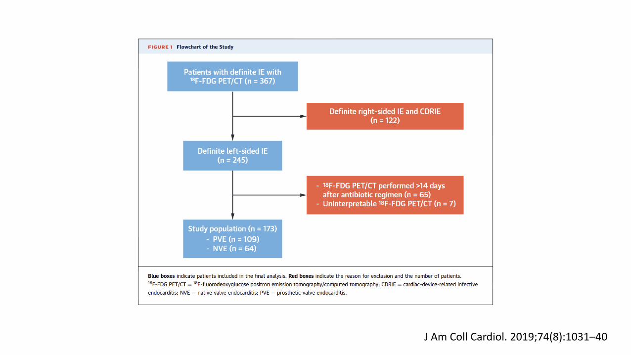
J Am Coll Cardiol. 2019;74(8):1031–40

J Am Coll Cardiol. 2019;74(8):1031–40

ESC 2015 Guidelines for IE
European Heart Journal (2015) 36, 3075–3123

CMR evidence for IE is in case series realm
Diagn Interv Radiol 2014

But…
Methodist Debakey Cardiovasc J. 2013 Jul-Sep;9(3):142-8.

And…
Methodist Debakey Cardiovasc J. 2013 Jul-Sep;9(3):142-8.

ASE 2017 Valve Regurgitation Guidelines

Putting it all together….
Suspected endocarditis with native valve
Suspected endocarditis with prosthetic valve
PUI or COVID+ COVID –ve or low risk for COVID
1. Consider clarity/Quality of TTE (VEG score)
2. Cardiac CT3. TEE with full PPE and minimal
ancillary staff
1. Consider clarity/Quality of TTE (VEG score)
2. TEE with full PPE
PUI or COVID+ COVID –ve or low risk for COVID
1. Quality of TTE2. PET FDG / Cardiac CT3. TEE with full PPE and minimal ancillary staff
1. Quality of TTE2. TEE with full PPE

Thanks!
Top Related