


Languages
Pages
Legal

Electronic Supplementary Material for Analytical MethodsThis journal is © The Royal Society of Chemistry 2014
1
SupplementaryInformention for:
Synthesis and evaluation of a silica-bonded concanavalin A material for lectin
affinity enrichment of N-linked glycoproteins and glycopeptides
Yujie Liua, Dongmei Fua,b, Yuansheng Xiaoa, Zhimou Guoa, Long Yua,b *, Xinmiao
Lianga *
a Key Laboratory of Separation Science for Analytical Chemistry, Dalian Institute of
Chemical Physics, Chinese Academy of Sciences, Dalian 116023, Chinab National Glycoengineering Research Center, Shandong University
Tel: +86-411-8437-9519
Fax: +86-411-8437-9539
E-mail: [email protected] [email protected]
Electronic Supplementary Material (ESI) for Analytical Methods.This journal is © The Royal Society of Chemistry 2014

Electronic Supplementary Material for Analytical MethodsThis journal is © The Royal Society of Chemistry 2014
2
1. Materials and Methods
Silica gel (particle size 5μm; pore size 300Å; surface area 60 m2 /g) was purchased
from Fuji Silysia Chemical (Kasugai, Japan). Glutaric anhydride, APTES, EDC, NHS,
mono-ethanolamine, hydroxyethyl piperazine ethanesulfonic acid (HEPES), and
trifluoroacetic acid (TFA) were purchased from J&K Scientific Ltd. Con A, methyl α-
D-mannopyranoside, chicken ovalbumin (OVA), RNase B, human serum albumin
(HSA), bovine fetuin, dithiothreitol (DTT), iodoacetic acid (IAA), ammonium
bicarbonate (NH4HCO3), and tetrahydrofuran (THF) were purchased from Sigma (St.
Louis, Mo). BCA Protein Assay Reagent (bicinchoninic acid) was purchased from
Pierce (Rockford, IL). Trypsin was purchased from Promega (Madison, WI). Tris
(hydroxymethyl) aminomethane (Tris), and formic acid (FA) were purchased from
Acros (Geel, Belgium). Toluene and N, N-dimethyl formamide (DMF) were
purchased from Kemiou Chemical Reagent Co. Ltd. (Tianjin, China). Acetonitrile
(CH3CN, HPLC gradient) was from Merk (Darmstadt, Germany). Water was purified
by the Milli-Q system (Milford, MA). Materials for desalting were prepared in our lab.
All the solvents were dried with 4 Å sieves before use.
LC-MS analysis was performed on Waters 2998-Masslynx system. Elemental
analysis was performed on a Vario EL III system (Elementar, Germany), Fourier
transform infrared spectrum (FT-IR) was performed on EQUINOX 55(Oregon, USA),
and the content of protein was detected on Bio-tek ELx800 (California, USA).
2. Preparation of SG Con A and packing of the column
Silica gel was dried at 150 oC for 8h before the reaction. In the presence of
equivalent Et3N (0.63 μL), 3-aminopropyltriethoxy silane 1 (APTES) (4.5 mmol, 1.1
mL) was added to the solution of the glutaric anhydride 2 (5.4 mmol, 616 mg) in 10
mL anhydrous THF, stirring for 4h at room temperature to generate the silane
coupling reagent 3, then silica gel (2.5 g) in 30 mL dry toluene was added, stirred for
48 h at 80 oC to obtain matrix 4, which was filtered and washed by THF, toluene,
methanol, water and methanol successively, then dried at 80 oC overnight.
At room temperature, matrix 4 (2.0 g) was suspended in dried DMF (20 mL) with
0.2 M EDC and 0.4 M NHS, the mixture was stirred for 2 h and filtered, followed by
washing with DMF and 10 mM HEPES buffer (pH 7.4). At room temperature, the
obtained NHS-silica matrix 5 was re-suspended in 20 mM HEPES buffer (10 mL),
followed by adding Con A (236 mg) in 50 mL HEPES buffer containing 0.2 M
methyl α-D-mannopyranoside as the inhibitor to protect the binding sites. After

Electronic Supplementary Material for Analytical MethodsThis journal is © The Royal Society of Chemistry 2014
3
stirring gently for 2h, 0.2 M ethanolamine was added to block the reaction for 1 h to
obtain the material 6 (SG Con A), which was washed with HEPES buffer until there
was no UV absorption peak being found under 280 nm.
The obtained SG Con A (1.5 g) in HEPES buffer (15 mL) was treated in an
ultrasonic bath for 5 min before pouring into stainless steel column (150 mm × 3.0
mm), with 20 mM HEPES buffer as slurry solvent under the pressure of 32 MPa. The
column can be kept in 20 mM HEPES buffer containing 0.02% NaN3 at 4 oC for 12
month without affecting its trapping efficiency.
3. Chromatographic conditions
Glycoproteins were separated by SG Con A column, gradient elution that
performed on Alliance HPLC system consisted of Waters 2695 pump and Waters
2996 DAD detector (Waters, Milford, MA, USA). The mobile phase A was 10 mM
HEPES buffer (pH 7.4, containing 0.15 M sodium chloride, 1 mM calcium chloride,
1mM manganese chloride, 1mM magnesium chloride, 0.02% sodium azide), and the
mobile phase B was 0.2 M methyl α-D-mannopyranoside in 10 mM HEPES buffer.
The column temperature was held at 20 oC at a flow rate of 0.2 mL/min.
A sample of commercial glycoprotein with the concentration of 10 μg/μL,
including HSA, ovalbumin, and RNase B, was loaded onto the SG Con A column,
respectively, and the fractioned glycoproteins were dialyzed against water to remove
the salts.
4. Deglycosylation of N-linked glycoproteins
The release of glycans from the obtained fractions was performed following the
previously described 2. Briefly, the glycoprotein fractions (100 μg) was dissolved in
100 μL 20 mM ammonium bicarbonate (pH 8.5), 0.1 μL PNGase F (500 units per μL)
was added to the solution and incubated overnight at 37 oC for N-glycan release. After
heating to 95 oC for 5 min to stop the reaction, the glycans were directly enriched with
C18 and Click Maltose sequentially for MS analysis.
5. Enrichment of released glycans from OVA
Firstly, C18 (about 1 mg) was packed into the GE Loader tip with an inert sieve
plug up the end, the resulting microcolumn was first equilibrated with
H2O/CH3CN/FA (80:20:0.1(v/v), 90 μL). Then PNGase F digest (10 μL) was dried, re-
dissolved in H2O/CH3CN/FA (80:20:0.1 (v/v), 10μL) and loaded onto the column. The
column was rinsed with H2O (90 μL) to remove glycans and salts. The remaining
glycoproteins were eluted with CH3CN/H2O/FA (50:50:0.1(v/v), 90 μL), dried and

Electronic Supplementary Material for Analytical MethodsThis journal is © The Royal Society of Chemistry 2014
4
collected.
Secondly, Click Maltose (about 1 mg) was packed into the GE Loader tip with an
inert sieve plug up the end, the resulting micro-column was first equilibrated with
CH3CN/H2O/FA (80:20:0.1(v/v), 90 μL). Then the mixture of glycans was dried, re-
dissolved in CH3CN/H2O/FA (80:20:0.1 (v/v), 10μL), and loaded onto the column.
The column was rinsed with CH3CN/H2O/FA (80:20:0.1(v/v), 60 μL) to remove salts.
The glycans were eluted with CH3CN/H2O/FA (70:30:0.1, 30 μL for two times),
collected and dried. The enrichment process was repeated at least three times.
6. Enrichment of RNase B glycopeptides with SG Con A and Agarose Con A
According to the procedure stated in our previous work3, RNase B glycoproteins
were digested with trypsin, enriched with C18 and Click Maltose sequentially for MS
analysis4.
In detail, SG Con A (about 1 mg) was packed into the GE Loader tip with an inert
sieve plug up the end, the resulting micro-column was equilibrated with HEPES
buffer (pH 7.4) before the tryptic digest (10 μL) was loaded onto the column. Then
the column was rinsed with HEPES buffer (pH 7.4, 60 μL) to remove the non-
glycopeptides. While the glycopeptide fraction was eluted with 0.2 M methyl α-D-
mannopyranoside in 10 mM HEPES buffer (30 μL for two times), collected and dried.
The enrichment process was repeated at least three times.
When it turns to Agarose Con A, three replicate enrichment procedures were
repeated just by the replacement of material SG Con A.
In order to desalination, C18 (about 1 mg) was packed into the GE Loader tip with
an inert sieve plug up the end, the resulting micro-column was first equilibrated with
H2O/CH3CN/FA (80:20:0.1(v/v), 90 μL). Then tryptic digest (10 μL) was dried, re-
dissolved in H2O/CH3CN/FA (80:20:0.1 (v/v), 10μL), and loaded onto the column.
The column was rinsed with H2O/CH3CN /FA (80:20:0.1(v/v), 45 μL) to remove non-
glycopeptides as well as salts. The glycopeptide fraction was eluted with
CH3CN/H2O/FA (60:40:0.1, 30 μL for two times), collected and dried, and three
replicate has been performed for enrichment.
Then, Click Maltose (about 1 mg) was packed into the GE Loader tip with an inert
sieve plug up the end, the resulting micro-column was first equilibrated with
CH3CN/H2O/FA (80:20:0.1(v/v), 90 μL). The desalted peptides from C18 were re-
dissolved in CH3CN/H2O/FA (80:20:0.1 (v/v), 10μL) and loaded onto the column.
The column was rinsed with CH3CN/H2O/FA (80:20:0.1(v/v), 60 μL) to remove non-

Electronic Supplementary Material for Analytical MethodsThis journal is © The Royal Society of Chemistry 2014
5
glycopeptides, and eluted with CH3CN/H2O/FA (70:30:0.1, 30 μL for two times) to
enrich the glycopeptides, and three replicate has been performed for enrichment.

Electronic Supplementary Material for Analytical MethodsThis journal is © The Royal Society of Chemistry 2014
6
7. Mass Spectrum experiments
MS experiments were performed on a quadrupole time-of-flight (Q-TOF) mass
spectrometer (Waters, Manchester, UK). The nano-ESI source was operated under
positive ion mode with nanospray voltage at 2.1 kV. MS data were acquired at m/z
1000-2000 for ovalbumin fractions and 600-2000 for RNase B fractions.

Electronic Supplementary Material for Analytical MethodsThis journal is © The Royal Society of Chemistry 2014
7
Table S1
Stationary phase C% N% Surface coverage
(μmol/m2)
Silica particles 4 6.1 0.7 8.1
Silica particles 5 7.4 1.1 6.6

Electronic Supplementary Material for Analytical MethodsThis journal is © The Royal Society of Chemistry 2014
8
Fig. S1 LC-MS analysis of the synthesized silane reagent 3.

Electronic Supplementary Material for Analytical MethodsThis journal is © The Royal Society of Chemistry 2014
9
Fig. S2 Fourier Transform Infrared Spectroscopy (FT-IR) of silica particles 4 and silica particles 5.
Electronic Supplementary Material for Analytical MethodsThis journal is © The Royal Society of Chemistry 2014
10
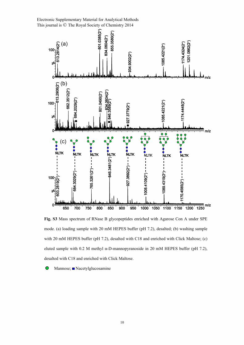
Fig. S3 Mass spectrum of RNase B glycopeptides enriched with Agarose Con A under SPE
mode. (a) loading sample with 20 mM HEPES buffer (pH 7.2), desalted; (b) washing sample
with 20 mM HEPES buffer (pH 7.2), desalted with C18 and enriched with Click Maltose; (c)
eluted sample with 0.2 M methyl α-D-mannopyranoside in 20 mM HEPES buffer (pH 7.2),
desalted with C18 and enriched with Click Maltose.
Mannose; Nacetylglucosamine

Electronic Supplementary Material for Analytical MethodsThis journal is © The Royal Society of Chemistry 2014
11
Fig. S4 Mass spectrum of RNase B glycopeptides enriched with SG ConA. (a) loading sample
with 10 mM HEPES buffer (pH 7.2), desalted with C18; (b) washing sample with 10 mM
HEPES buffer (pH 7.2), desalted with C18 and enriched with Click Maltose; (c) elution of
sample with 0.2 M methyl α-D-mannopyranoside in 10 mM HEPES buffer (pH 7.2), desalted
with C18 and enriched with Click Maltose.
Mannose; Nacetylglucosamine
Electronic Supplementary Material for Analytical MethodsThis journal is © The Royal Society of Chemistry 2014
12
Reference
1. J. Baranauskiene, J. Kazlauske, S. Gustaite, B. Niemeyer and J. Liesiene, Journal of Liquid
Chromatography & Related Technologies, 2014, 37, 1847-1861.
2. A. L. Tarentino, C. M. Gomez and T. H. Plummer Jr, Biochemistry, 1985, 24, 4665-4671.
3. L. Yu, X. Li, J. Dong, X. Zhang, Z. Guo and X. Liang, Analytical Methods, 2010, 2, 1667-
1670.
4. Q. Fu, Z. Guo, T. Liang, X. Zhang, Q. Xu and X. Liang, Analytical Methods, 2010, 2, 217-224.
Top Related