


Languages
Pages
Legal


Breast MRI: New and Abbreviated
Protocols
Christopher Comstock M.D.
Department of Radiology
Memorial Sloan-Kettering Cancer Center

Topics
• What is our goal?
• Current status of screening
• How do we change screening
• Abbreviated Breast MRI (AB-MR)
• EA1141 AB-MR Trial
• Multiparametric Breast MRI
Beyond the scope of this talk!
• The debate over screening the benefit
of mammography, particularly for
women in their forties.

What is Our Goal?
• Decrease breast cancer mortality
• Reduction in the morbidities associated
with surgery and chemotherapy
• Finding breast cancers at a smaller size
and earlier stage leads to a reduction in
mortality and the use of less aggressive
therapies

Reservoir of Breast Cancer Present
in 1000 Women Being Screened
• Is it 30, 40, 50, 60 or more breast cancers
per 1000 women?
• Depends on risk of population
• Detection level (size and stage) depends
on modality and frequency of screening

Reservoir of Breast Cancer Present
in 1000 Women Being Screened
Tomo plus WBUS

The Dissemination of Medical
Technologies into Clinical Practice
• Innovations medical in technology and
quality of information are the sole driving
force in the acceptance and adoption of
new technologies
• The dissemination of medical technologies
depends on the social, political and
ideological context into which they are
introduced
Much Can Be Learned From the
History of Mammography
• Despite improvements in technology,
mammography languished from 1930s to 1970 – 1930-1950 Stafford L. Warren, Jacob Gershon-Cohen and Raul
Leborgne
– 1950s Improved techniques, Robert Egan
• The production of better data alone did not
eliminate the role that economics, authority and
ideology played
“TO SEE TODAY WITH THE EYES OF TOMORROW” A HISTORY OF SCREENING
MAMMOGRAPHY. Barron H. Lerner, MD, PhD
Background Paper for the Institute of Medicine report: “Mammography and Beyond: Developing
Technologies for the Early Detection of Breast Cancer” March 2001
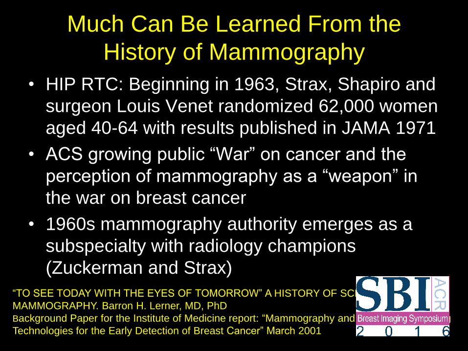
Much Can Be Learned From the
History of Mammography
• HIP RTC: Beginning in 1963, Strax, Shapiro and
surgeon Louis Venet randomized 62,000 women
aged 40-64 with results published in JAMA 1971
• ACS growing public “War” on cancer and the
perception of mammography as a “weapon” in
the war on breast cancer
• 1960s mammography authority emerges as a
subspecialty with radiology champions
(Zuckerman and Strax)
“TO SEE TODAY WITH THE EYES OF TOMORROW” A HISTORY OF SCREENING
MAMMOGRAPHY. Barron H. Lerner, MD, PhD
Background Paper for the Institute of Medicine report: “Mammography and Beyond: Developing
Technologies for the Early Detection of Breast Cancer” March 2001
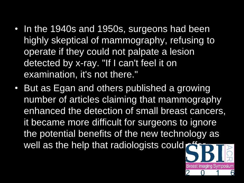
• In the 1940s and 1950s, surgeons had been
highly skeptical of mammography, refusing to
operate if they could not palpate a lesion
detected by x-ray. "If I can't feel it on
examination, it's not there."
• But as Egan and others published a growing
number of articles claiming that mammography
enhanced the detection of small breast cancers,
it became more difficult for surgeons to ignore
the potential benefits of the new technology as
well as the help that radiologists could offer.

MSKCC breast surgeon Jerome A. Urban
wrote to Zuckerman in 1964, "I think this is
an exciting finding and represents the third
carcinoma which we personally did not
strongly suspect on clinical examination."
Urban closed his letter by stating "More
power to you" (Urban, 1964).
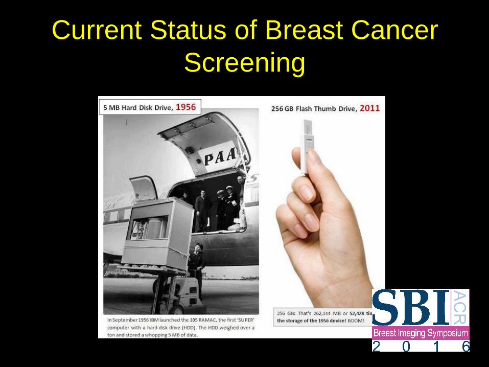
Current Status of Breast Cancer
Screening

What have we done with breast cancer
screening the since 1960s?
1
3
AB-MR Working Group
1969 1975 2013

Limitations of Mammography
1. 38% sensitivity in women with dense breast 1
2. 17 % interval cancer rate 2
3. Interval cancers with higher grade histologies 2
Group Meeting • May 8-
10, 2014 1
4
1) Pisano ED, Gatsonis C, Hendrick E, et al. Diagnostic performance of digital
versus film mammography for breast-cancer screening. Digital Mammographic
Imaging Screening Trial (DMIST) Investigators Group.
N Engl J Med. 2005 Oct 27;353(17):1773-83.
2) Porter PL, El-Bastawissi AY, Mandelson MT, et al.: Breast tumor characteristics
as predictors of mammographic detection: comparison of interval- and screen-
detected cancers. J Natl Cancer Inst 91 (23): 2020-8, 1999.
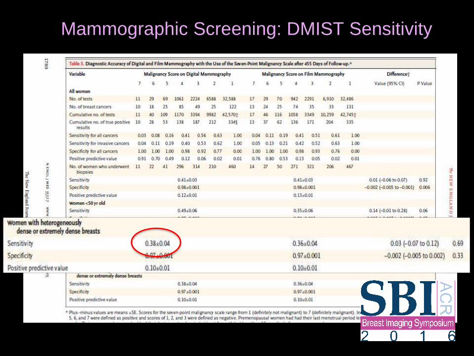
Mammographic Screening: DMIST Sensitivity
AB-MR Working Group 1
5

Digital Breast Tomosynthesis (DBT): A little better….
1. 3D mammographic technique
2. Possible increase in caner sensitivity and
reduced call-back rates compared to FFDM
AB-MR Working Group 1
6
MG MG +DBT
Skaane et al, 2013 6.1/1000 8.0/1000
Ciatto et al, 2013 5.3/1000 8.1/1000
Haas et al, 2013 5.2/1000 5.7/1000
Friedwald et al, 2014 4.2/1000 5.4/1000

Tomosynthesis
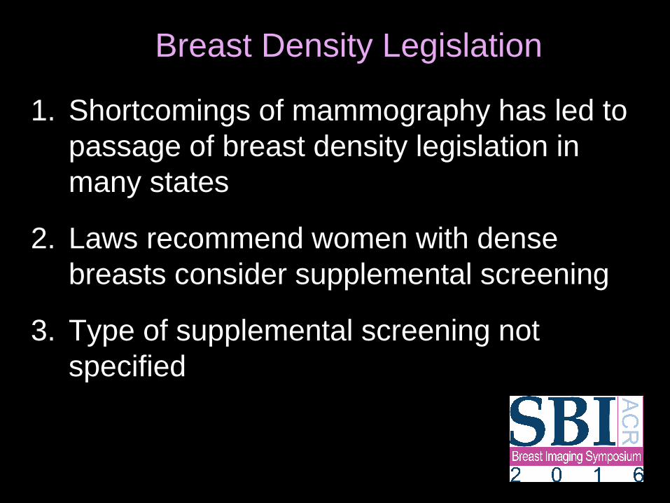
Breast Density Legislation
1. Shortcomings of mammography has led to
passage of breast density legislation in
many states
2. Laws recommend women with dense
breasts consider supplemental screening
3. Type of supplemental screening not
specified
AB-MR Working Group 1
8

Breast Density Legislation
Group Meeting • May 8-
10, 2014 1
9

Whole Breast Screening Ultrasound
1. Default supplemental screening modality
due to relatively low cost and wide
availability
2. Supplemental cancer yield: 3-4/1000
3. Limitation of WBUS include:
– Low PPV (8-9%)
– High frequency of short-term follow
recommendations
– Time consuming
Group Meeting • May 8-
10, 2014 2
0
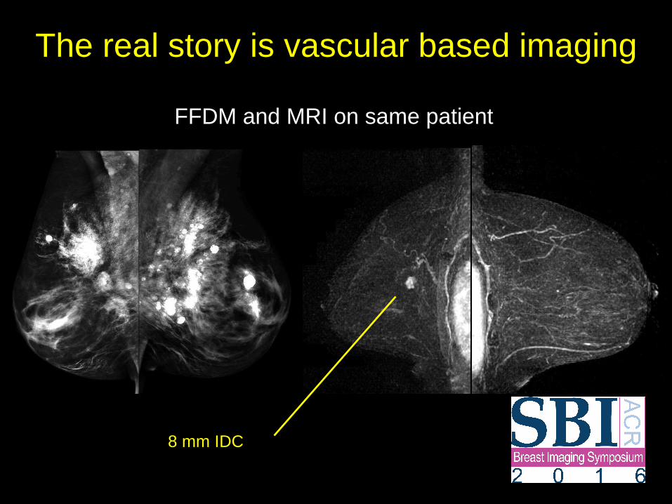
The real story is vascular based imaging
Group Meeting • May 8-
10, 2014 2
1
FFDM and MRI on same patient
8 mm IDC

The Use of MRI for Breast Cancer
Screening
1. Not limited by breast density
2. No ionizing radiation
3. Most sensitive test for breast cancer screening
4. PPV similar to mammography
5. Preferentially detects higher grade lesions
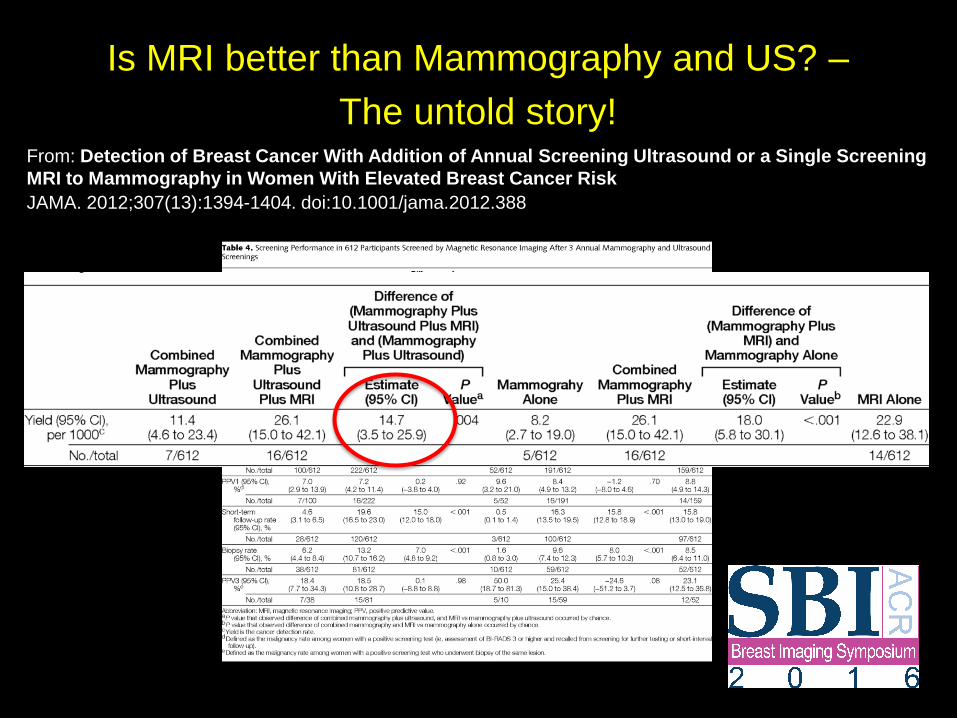
Is MRI better than Mammography and US? –
The untold story!
Group Meeting • May 8-
10, 2014 2
3
From: Detection of Breast Cancer With Addition of Annual Screening Ultrasound or a Single Screening
MRI to Mammography in Women With Elevated Breast Cancer Risk
JAMA. 2012;307(13):1394-1404. doi:10.1001/jama.2012.388

EVA Trial
Group Meeting • May 4-6,
2012 2
4
Kuhl C, Weigel S, Schrading S, et al. Prospective multicenter
cohort study to refine management recommendations for
women at elevated familial risk of breast cancer: the EVA trial. J
Clin Oncol. 2010 Mar 20;28(9):1450-7.
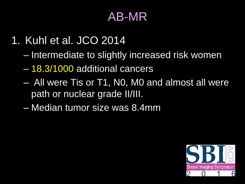
AB-MR
1. Kuhl et al. JCO 2014
– Intermediate to slightly increased risk women
– 18.3/1000 additional cancers
– All were Tis or T1, N0, M0 and almost all were
path or nuclear grade II/III.
– Median tumor size was 8.4mm
Group Meeting • May 4-6,
2012 2
5

Why have we ignored MRI except for
extremely high-risk women?
1. Cost
2. Time
3. Perceived low PPV
Group Meeting • May 8-
10, 2014 2
6

Why Have We Ignored MRI So
Many Years?
• Breast MRI has remained relatively
unchanged for over 20 years
• However, breast density advocacy and
legislation has changed the current
landscape
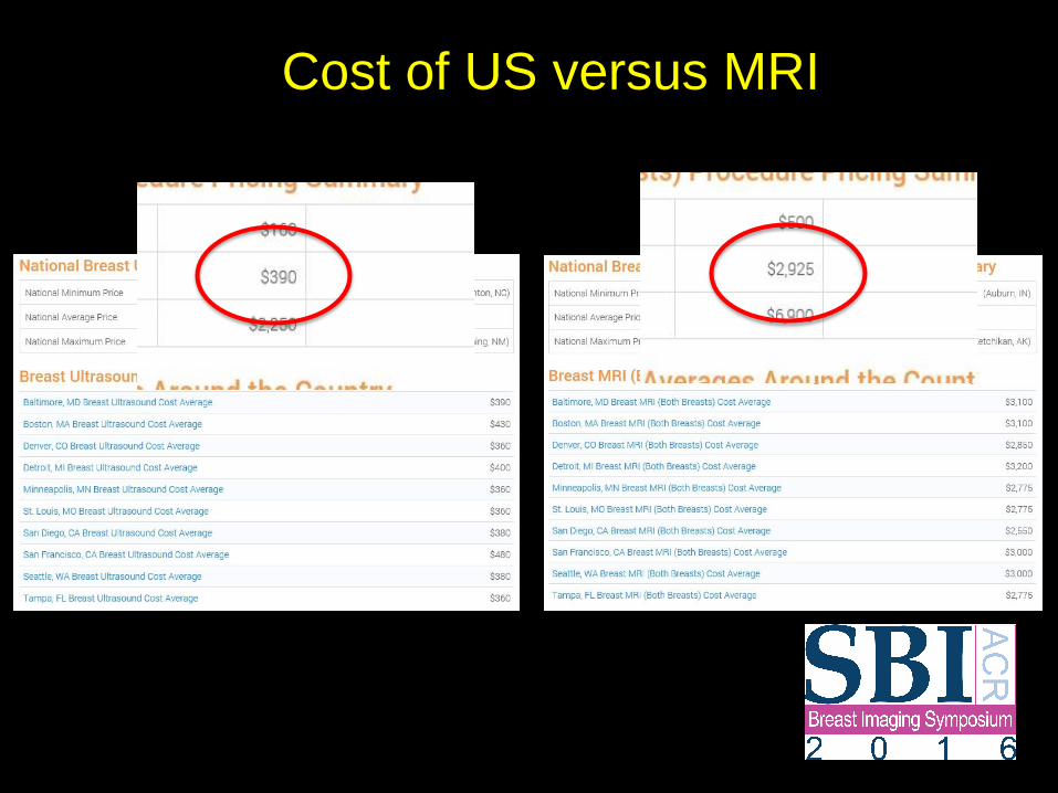
Cost of US versus MRI
Group Meeting • May 8-
10, 2014 2
8

How Do We Increase Access to
Breast MRI?
• Change the mindset that MRI should be
limited to only women extremely high-risk
women.
– Create a “different” breast MRI exam that is
cheaper, faster and relatively accurate
– Allow administrators a “distinction” in billing
between full and AB-MR
– Evaluate this “new” modality in a phase II
multicenter NCI trial

Abbreviated MRI (AB-MR)
1. Low cost ($300-$500)
2. Quick (less than 10min)
3. PPV similar to mammography (20-30%)
4. 150% increase in cancer detection
5. Optimal screening interval 1-3 yrs?
6. Quality accreditation
7. Reader qualifications
8. Interpretation guidelines
Group Meeting • May 8-
10, 2014 3
0
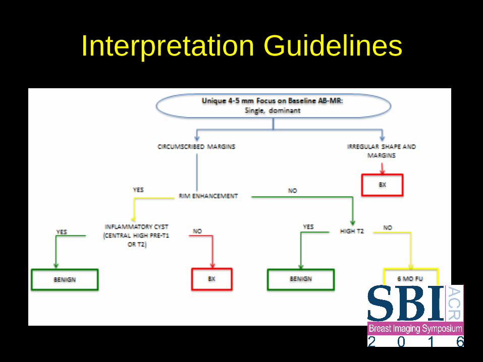
Interpretation Guidelines

Interpretation Guidelines
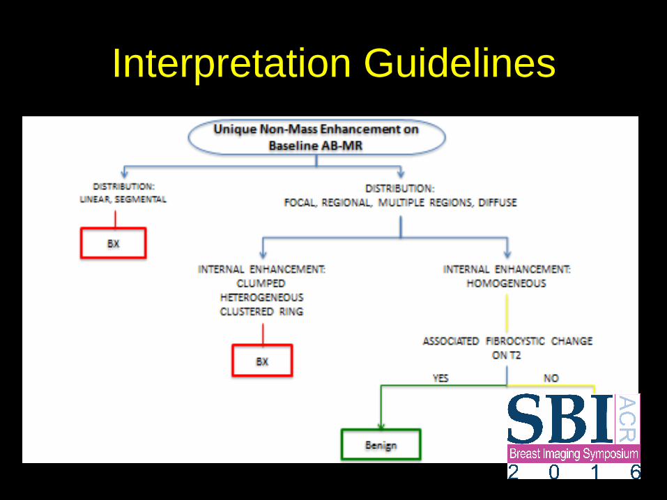
Interpretation Guidelines
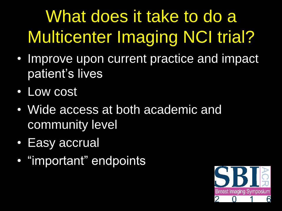
What does it take to do a
Multicenter Imaging NCI trial? • Improve upon current practice and impact
patient’s lives
• Low cost
• Wide access at both academic and
community level
• Easy accrual
• “important” endpoints

CTEP

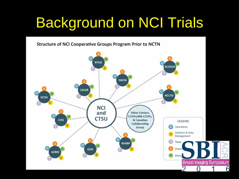
Background on NCI Trials

Background on NCI Trials
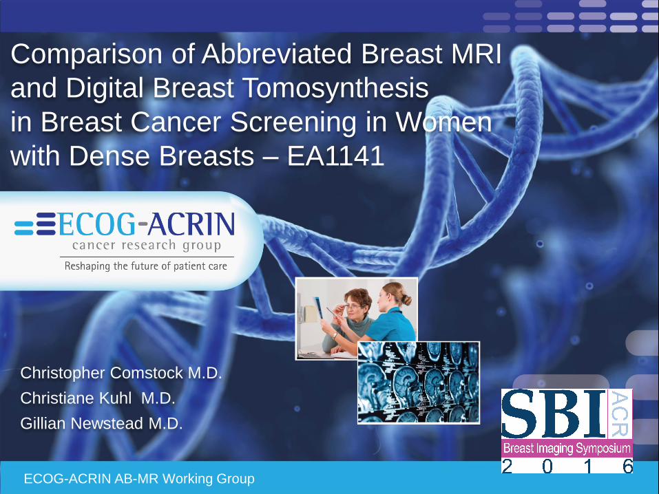
Comparison of Abbreviated Breast MRI
and Digital Breast Tomosynthesis
in Breast Cancer Screening in Women
with Dense Breasts – EA1141
ECOG-ACRIN AB-MR Working Group
Christopher Comstock M.D.
Christiane Kuhl M.D.
Gillian Newstead M.D.

AB-MR Trial Concept
Comparison of Abbreviated Breast MRI and
Digital Breast Tomosynthesis in Breast Cancer
Screening Women with Dense Breasts
Primary aim: To compare the invasive cancer
detection rate of AB-MR to DBT.
Group Meeting • May 8-
10, 2014 4
0

AB-MR Trial Concept
Secondary aims:
1. To compare the tumor biologies of invasive cancers and DCIS detected on AB-MR and DBT
2. To compare the PPV of biopsies, call back rates, and short-term follow up rates of AB-MR and DBT on both the initial and 1 year follow up studies
3. To evaluate the accuracy and interval cancer rate of AB-MR and DBT at 1 year
4. To perform a comparative cost analysis between DBT and AB-MR
Group Meeting • May 8-
10, 2014 4
1

Inclusion
1. Women over the age of 40 and scheduled for screening DBT;
2. Asymptomatic women;
3. Does not qualify for high-risk Breast MR screening as defined by the ACS recommendations.
4. No known breast cancer;
5. Have not had a breast US within the prior 12 months.
6. No prior MRI
7. Women who agree to not have screening US for the study period.
Group Meeting • May 8-
10, 2014 4
2

Schema
Group Meeting • May 8-
10, 2014 4
3
Women ages 40-75 with dense breasts already scheduled for routine screening DBT
Randomization
Arm A (DBT first) Years 0 and 1 DBT followed by AB-MR. Year 0 PRO/QOL assessments to be completed approximately 2 weeks after screening
Arm B (AB-MR first) Years 0 and 1 AB-MR followed by DBT. Year 0 PRO/QOL assessments to be completed approximately 2 weeks after screening
Return to routine mammographic screening and follow up for 3 years
Accrual Goal= 1450 1. Suspicious lesions detected on one or both of the modalities at the Year 0 or 1 time points will
be biopsied as per local standard practice 2. Tissue collection and analysis for all cancers detected

Statistical Considerations
1. The table shows that 1363 cases with complete data from
both tests and pathology are needed to ensure power
90% for a difference in the rates of invasive cancer
detection as low as 9/1000.
2. Assuming that inadequate information will be available on
up to 6% of cases, a sample size of 1450 will provide
power 90% to compare the diagnostic yield in invasive
cancer of the two modalities.
Group Meeting • May 8-
10, 2014 4
4
Power Sample
size
Difference in invasive
cancer rates (ABMR –DBT)
Proportion of
discordant cases
0.90 1191 0.009 0.010
0.90 1363 0.009 0.011
0.90 1552 0.009 0.012
0.90 1057 0.010 0.011
0.90 1197 0.010 0.012
0.90 949 0.011 0.012
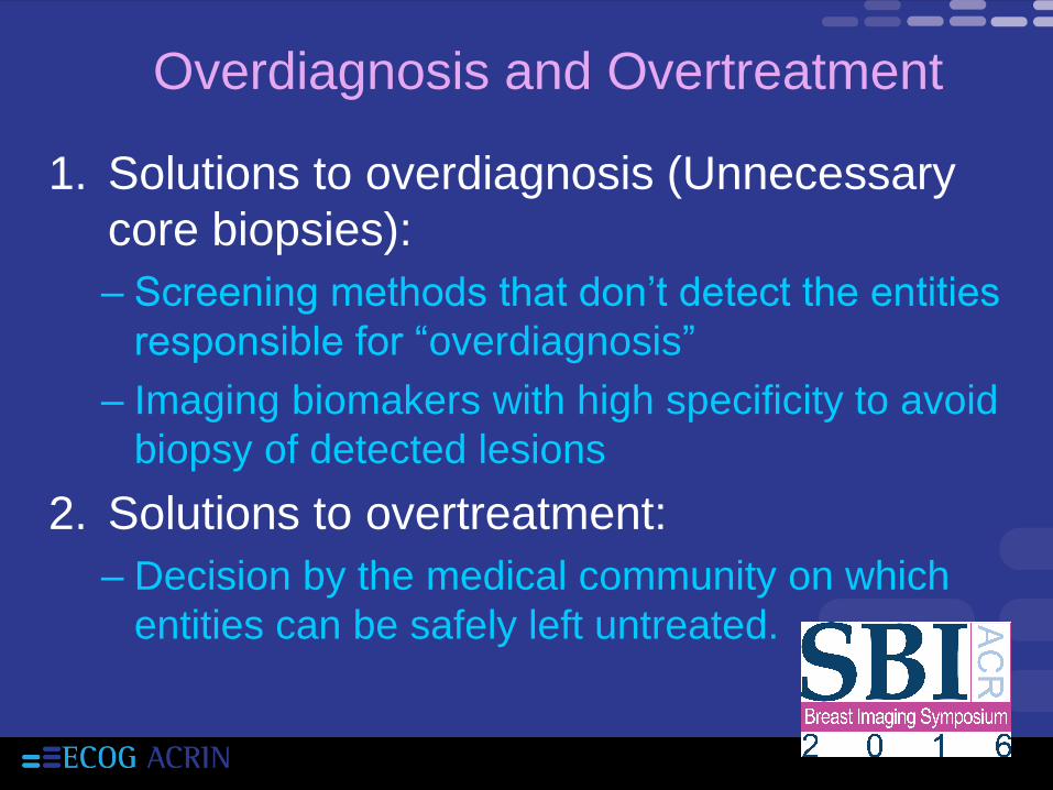
Overdiagnosis and Overtreatment
1. Solutions to overdiagnosis (Unnecessary
core biopsies):
– Screening methods that don’t detect the entities
responsible for “overdiagnosis”
– Imaging biomakers with high specificity to avoid
biopsy of detected lesions
2. Solutions to overtreatment:
– Decision by the medical community on which
entities can be safely left untreated.
Group Meeting • May 8-
10, 2014 4
5

Tumor Biology
1. Exploration of the differences in the
biological detection profiles (BDP) of
Tomosynthesis and AB-MR. (PAM50 for
invasive CA and DCIS score for DCIS)
Group Meeting • May 8-
10, 2014 4
6

AB-MR Working Group
• Chris Comstock M.D.
• Christiane Kuhl M.D.
• Gillian Newstead M.D.
• Liz Morris M.D.
• Connie Lehman M.D. PhD.
• Linda Moy M.D.
• Constantine Gatsonis PhD
• Bob Nishikawa PhD
• Nancy Sauers MS
Group Meeting • May 8-
10, 2014 4
7
• Seema Khan M.D
• Brian Leyland-Jones MB BS PhD
• Larry Solin M.D.
• Lori Goldstein M.D.
• David Brenin M.D.
• Toncred Styblo M.D.
Kathy Miller M.D.

Different Techniques and
Equipment
Technique and
Protocols

Requires Gadolinium contrast to detect cancer

Dynamic Contrast Enhanced MRI
of the Breast (DCE MRI)
• Gadolinium pools in the interstitial space
of lesions
• Gadolinium shortens T1 relaxation thereby
increasing signal (enhancement)
• Multiple post-injection scans are
performed to evaluate lesion enhancement
over time (kinetics)

Gadolinium
• 20 ml bolus (0.5 mmol/ml)
• 20 sec prior to scan
• 0.1 mmol/kg

When to schedule Breast MR
• During 2nd week of cycle
• May have to repeat a small
percentage of cases due to
hormonal effects

Technical Considerations
• Field Intensity 1.5 or 3.0 Tesla
• Plane of acquisition Axial vs. Sagittal
• 2D or 3D 3D
• One breast / both at once Both
• High Res / Dynamic Both (Morphology /
Kinetics)
• Fat Saturation Yes
• Subtraction Yes
• Registration Helpful
• MIPs Helpful

Timing and K-space
• Cancer peak 60-120 (90) sec
• Optimal to time center of K-
space at 90 sec post contrast
• Is vendor specific and may
have to adjust injection timing
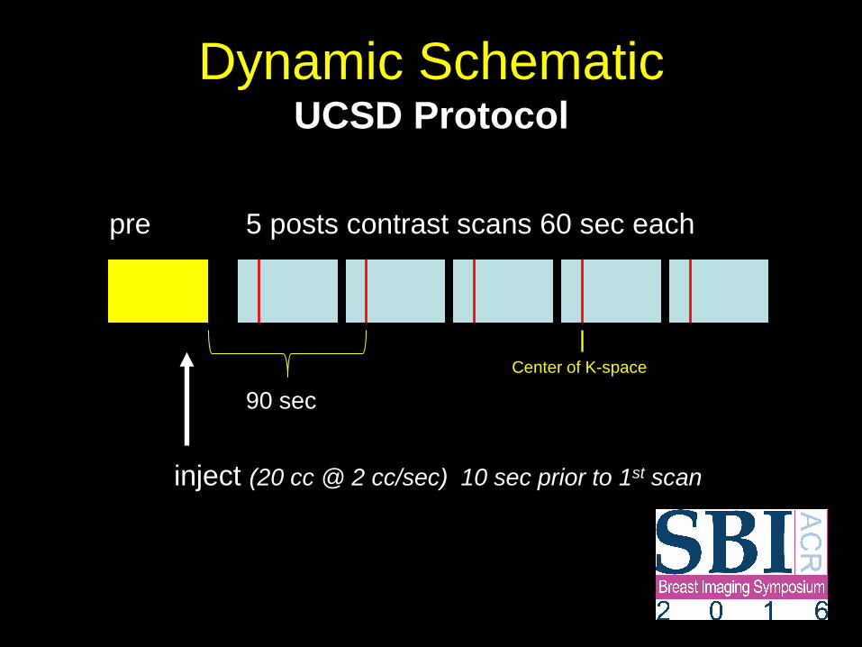
Dynamic Schematic UCSD Protocol
inject (20 cc @ 2 cc/sec) 10 sec prior to 1st scan
pre 5 posts contrast scans 60 sec each
90 sec
Center of K-space

Dynamic Schematic MSKCC Protocol
inject (20 cc @ 2 cc/sec) 20 sec prior to 1st scan
pre 3 posts contrast scans 95 sec each
90 sec
Center of K-space

Our Technique
• 1mm slices (3D data set)
• Contiguous with no gap
• In-plane 300x300 matrix
• 300mm FOV
• 1x1x1mm voxel size
• Maintain in plane resolution w larger FOV

Protocol (20 minutes)
Localizer
Bilat pre-contrast T1 non fat sup
Bilat T2-weighted fat-sat
3D GRE non-fat-sat
3D fat-sat dynamic (Pre+3)
body coil
bilateral breast coil
bilateral breast coil
bilateral breast coil
bilateral breast coil

Spatial and Temporal Resolution
• Spatial resolution: Morphology (In-plane
and slice thickness)
• Temporal resolution: Kinetics (Scan time)
• Bilateral scanning
• Most current systems can achieve both
high spatial and temporal resolution
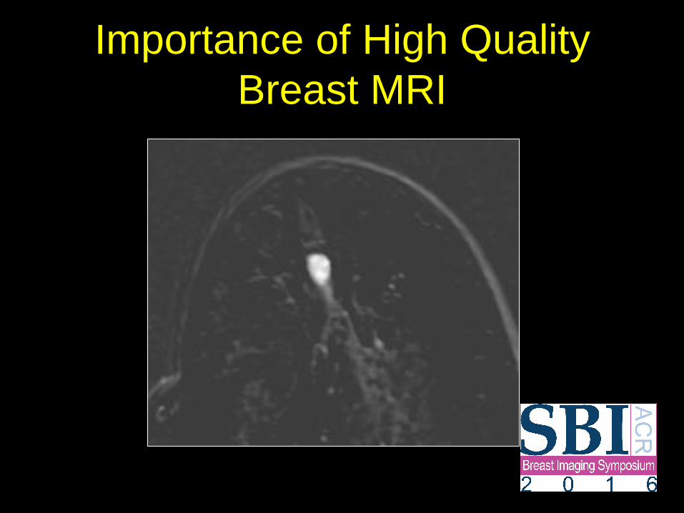
Importance of High Quality
Breast MRI
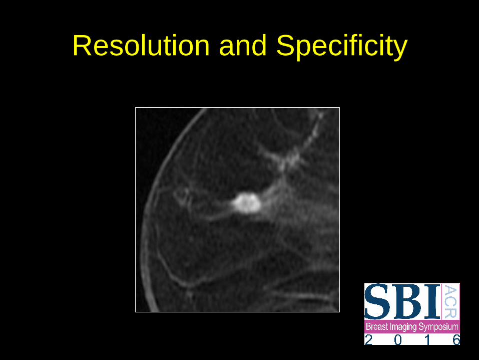
Resolution and Specificity

Effect of Higher Resolution


Non-Isovolumetric
Original Sagittal Reconstructed Axial
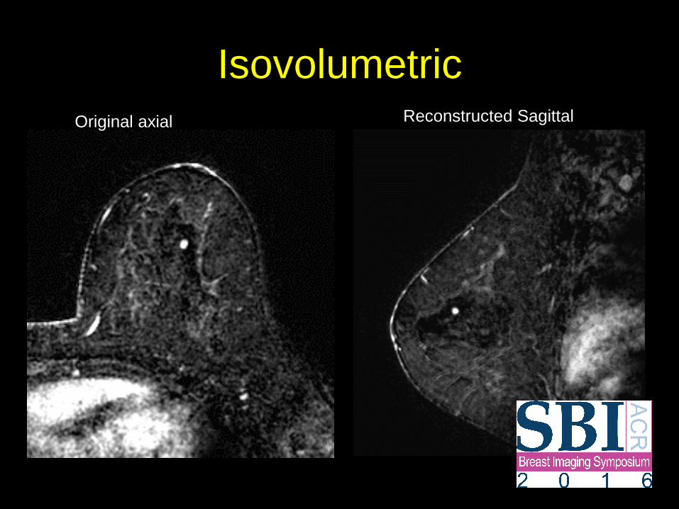
Isovolumetric
Original axial Reconstructed Sagittal

1.5T versus 3T
• Potential for higher spatial resolution
• Shimming more difficult
• Software/coil optimization in early stages
• Limited clinical data
• Improved MR spectroscopy
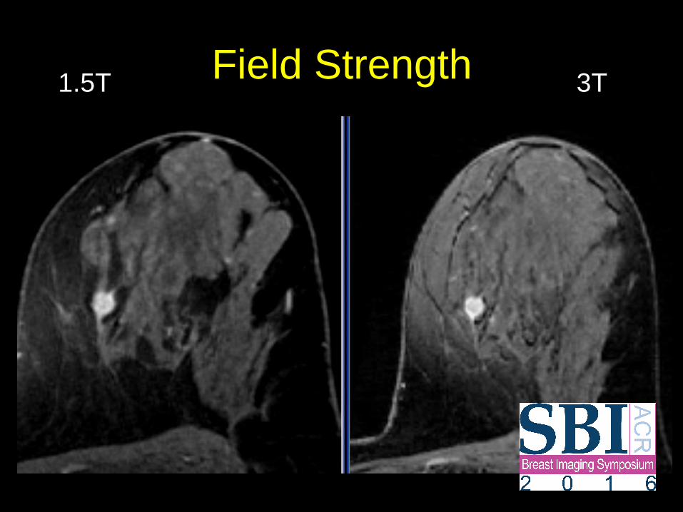
Field Strength 1.5T 3T

Field Strength 1.5T 3T
512X348
2.6mm every 1.6mm 2.6mm every 1.3mm

Fat Saturation and Subtraction
Unsubtracted
No fat Sat

Image
Subtraction
Fat
suppressed
GRE

KPBS NOVA 2001
Kinetics
Kinetics
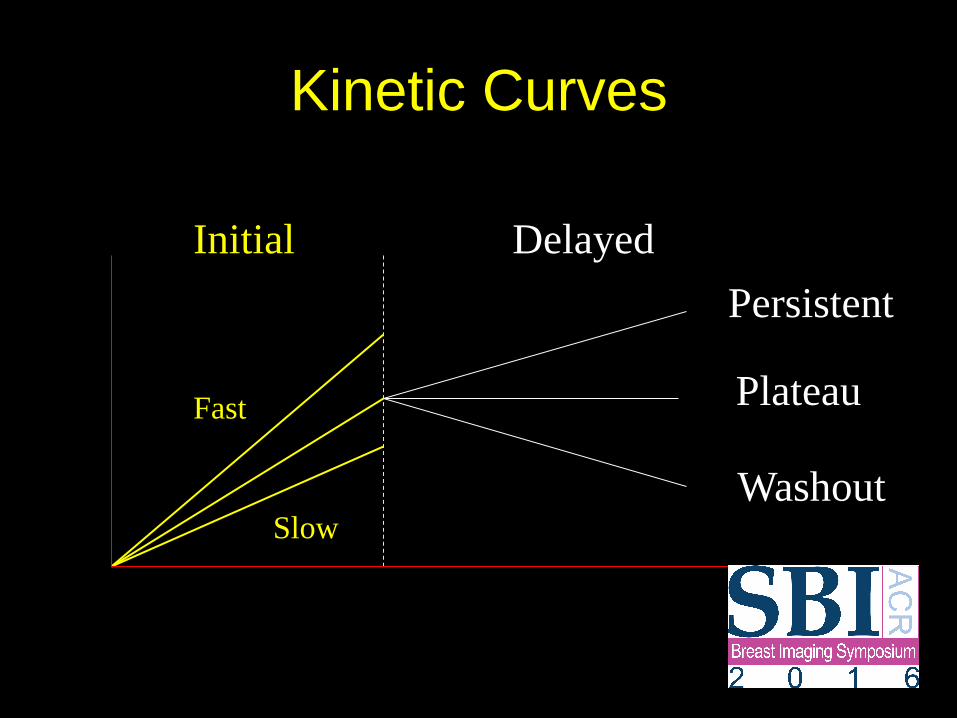
Kinetic Curves
Persistent
Plateau
Initial Delayed
Washout
Fast
Slow
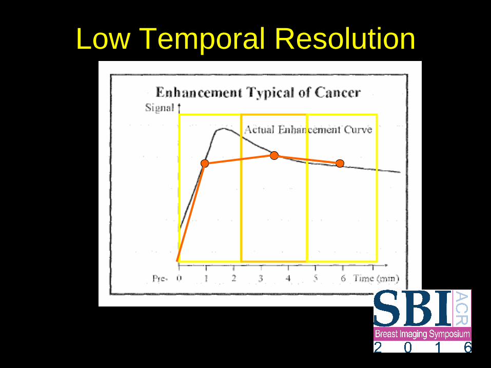
Low Temporal Resolution

Higher Temporal Resolution
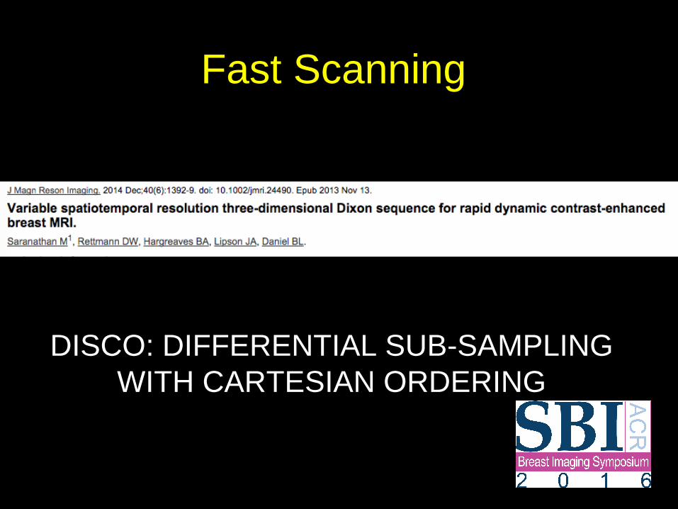
DISCO: DIFFERENTIAL SUB-SAMPLING
WITH CARTESIAN ORDERING
Fast Scanning
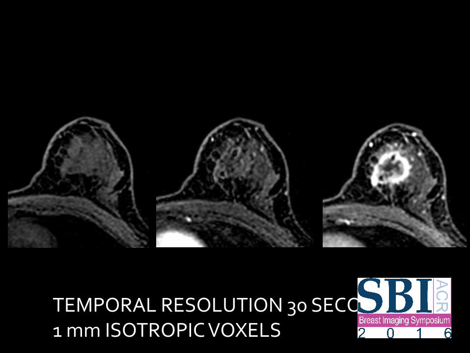
TEMPORAL RESOLUTION 30 SECONDS 1 mm ISOTROPIC VOXELS

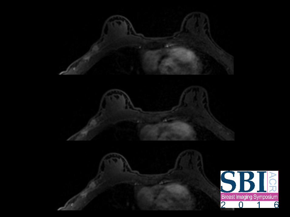

TEMPORAL RESOLUTION 10 SECONDS 1 mm ISOTROPIC VOXELS

DWI
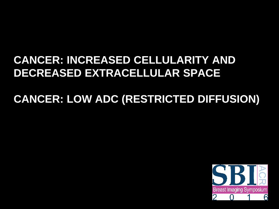
CANCER: INCREASED CELLULARITY AND
DECREASED EXTRACELLULAR SPACE
CANCER: LOW ADC (RESTRICTED DIFFUSION)

DWI
• POSSIBLE NON-CONTRAST ALTERNATIVE
• LIMITATIONS:
– LOW SNR
– POOR SPATIAL RESOLUTION
– MOTION
– INHOMOGENEOUS FAT SUPPRESSION

IMPROVING SPATIAL RESOLUTION
OF DWI

HIGH RESOLUTION MULTI B-VALUE DWI
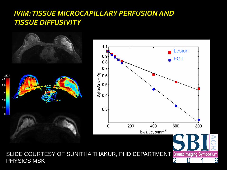
T1w post
ADC
DWI b = 800 s/mm2
L R Lesion
FGT
ADC8b, Dd and Dp in 10-3 mm2/s
SLIDE COURTESY OF SUNITHA THAKUR, PHD DEPARTMENT OF MEDICAL
PHYSICS MSK
IVIM: TISSUE MICROCAPILLARY PERFUSION AND TISSUE DIFFUSIVITY

PET-
MRI
MULTIPARAMETRIC MRI
SLIDE COURTESY OF K PINKER, MD Department of Biomedical Imaging and
Image-guided Therapy, Division of Molecular and Gender Imaging, Medical University
Vienna
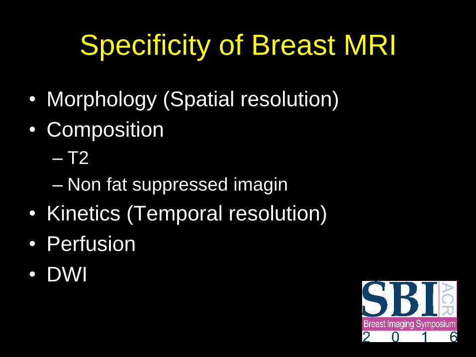
Specificity of Breast MRI
• Morphology (Spatial resolution)
• Composition
– T2
– Non fat suppressed imagin
• Kinetics (Temporal resolution)
• Perfusion
• DWI
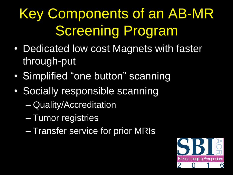
Key Components of an AB-MR
Screening Program • Dedicated low cost Magnets with faster
through-put
• Simplified “one button” scanning
• Socially responsible scanning
– Quality/Accreditation
– Tumor registries
– Transfer service for prior MRIs

In the End
• Access to MRI will be widely expanded
• Women with dense breasts will have a
faster, more sensitive and more accurate
option to WBUS.
• AB-MR will be a misnomer: With
competition, breast MRI will revert back to
a singular study incorporating kinetics,
perfusion, T2, and DWI all within an
approximate 12-15min exam.

Thank You
• EA1141 AB-MR Trial
• ECOG-ACRIN Roster
Top Related