


Languages
Pages
Legal

Brain TumorsBrain TumorsMaria RountreeMaria Rountree
Most common types of Most common types of brain tumorsbrain tumors
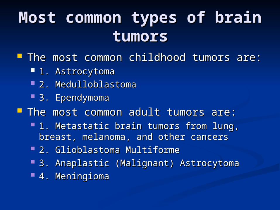
The most common childhood tumors are: The most common childhood tumors are: 1. Astrocytoma 1. Astrocytoma 2. Medulloblastoma 2. Medulloblastoma 3. Ependymoma 3. Ependymoma
The most common adult tumors are: The most common adult tumors are: 1. Metastatic brain tumors from lung, breast, 1. Metastatic brain tumors from lung, breast,
melanoma, and other cancers melanoma, and other cancers 2. Glioblastoma Multiforme 2. Glioblastoma Multiforme 3. Anaplastic (Malignant) Astrocytoma 3. Anaplastic (Malignant) Astrocytoma 4. Meningioma 4. Meningioma
Incidence of brain Incidence of brain tumorstumors
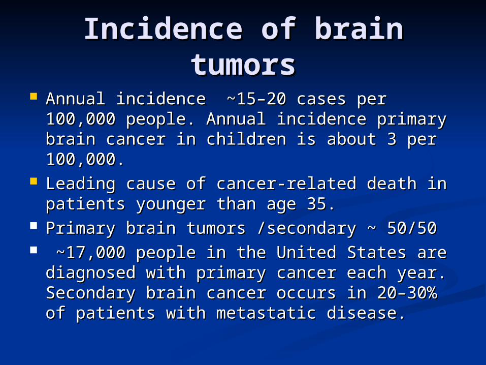
Annual incidence ~15–20 cases per 100,000 Annual incidence ~15–20 cases per 100,000 people. Annual incidence primary brain people. Annual incidence primary brain cancer in children is about 3 per 100,000. cancer in children is about 3 per 100,000.
Leading cause of cancer-related death in Leading cause of cancer-related death in patients younger than age 35. patients younger than age 35.
Primary brain tumors /secondary ~ 50/50Primary brain tumors /secondary ~ 50/50 ~17,000 people in the United States are ~17,000 people in the United States are
diagnosed with primary cancer each year. diagnosed with primary cancer each year. Secondary brain cancer occurs in 20–30% of Secondary brain cancer occurs in 20–30% of patients with metastatic disease. patients with metastatic disease.
Clinical Presentation of Clinical Presentation of brain tumorsbrain tumors
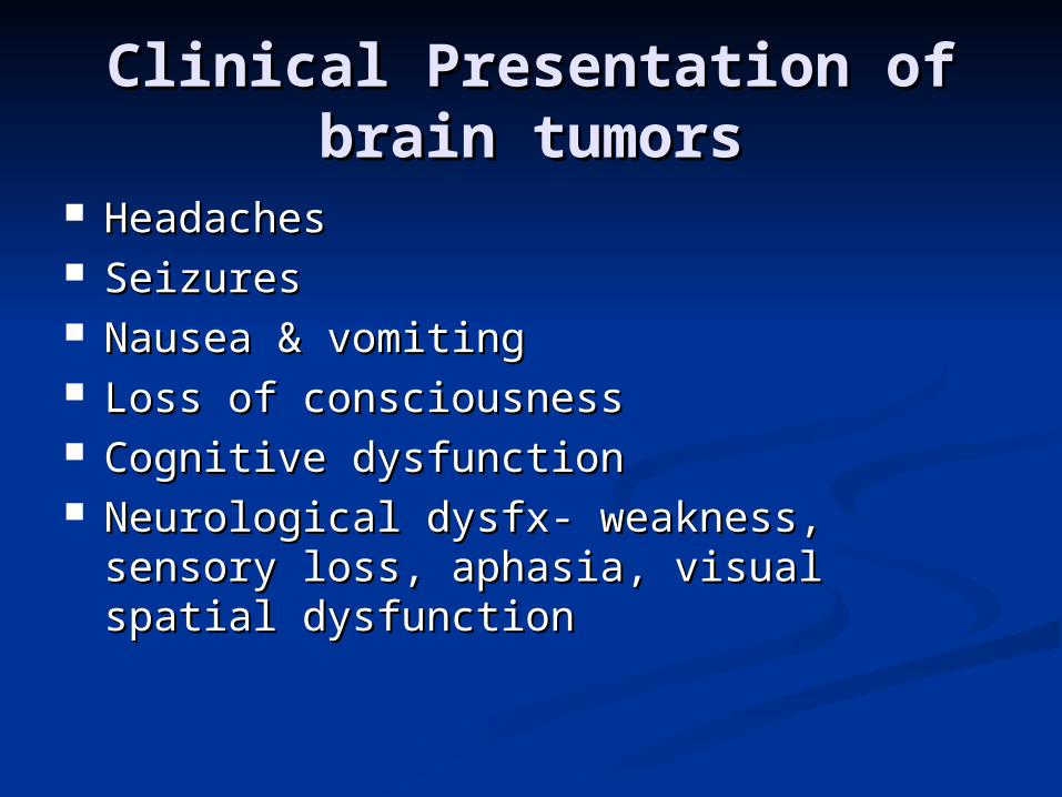
HeadachesHeadaches SeizuresSeizures Nausea & vomitingNausea & vomiting Loss of consciousnessLoss of consciousness Cognitive dysfunctionCognitive dysfunction Neurological dysfx- weakness, sensory Neurological dysfx- weakness, sensory
loss, aphasia, visual spatial dysfunctionloss, aphasia, visual spatial dysfunction

Cognitive dysfunctionCognitive dysfunction Includes memory problem, mood or Includes memory problem, mood or
personality disorderspersonality disorders It is the presenting symptom in 30-35% of It is the presenting symptom in 30-35% of
patients with brain metastasis.patients with brain metastasis. Patients symptoms often subtle, complain Patients symptoms often subtle, complain
of fatigue, urge to sleep and loss of of fatigue, urge to sleep and loss of interest in daily activities. Confused with interest in daily activities. Confused with depression.depression.
Consider neuroimaging in patients who Consider neuroimaging in patients who present with new onset of depressive present with new onset of depressive symptoms or without obvious cause.symptoms or without obvious cause.

Case:Case: 76 yo old female presented with increased 76 yo old female presented with increased
irritability with her family, sleeplessness irritability with her family, sleeplessness and reckless spending.and reckless spending.
PMH: HTN, breast cancerPMH: HTN, breast cancer PE, labs –wnlPE, labs –wnl MSE notable for loud rapid speech, flight MSE notable for loud rapid speech, flight
of ideas, no delusions or hallucinationsof ideas, no delusions or hallucinations CT revealed a 3 cm intraventricular lesionCT revealed a 3 cm intraventricular lesion Meningioma was removed and sxs slowly Meningioma was removed and sxs slowly
abatedabated
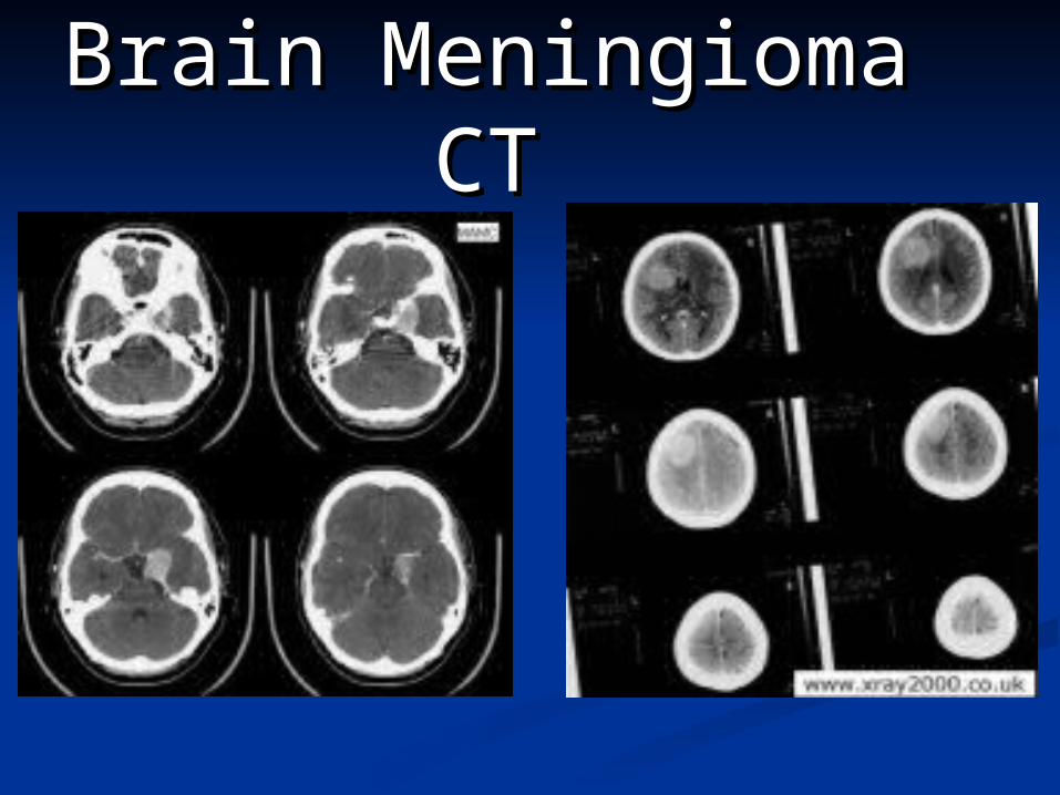
Brain Meningioma Brain Meningioma CTCT
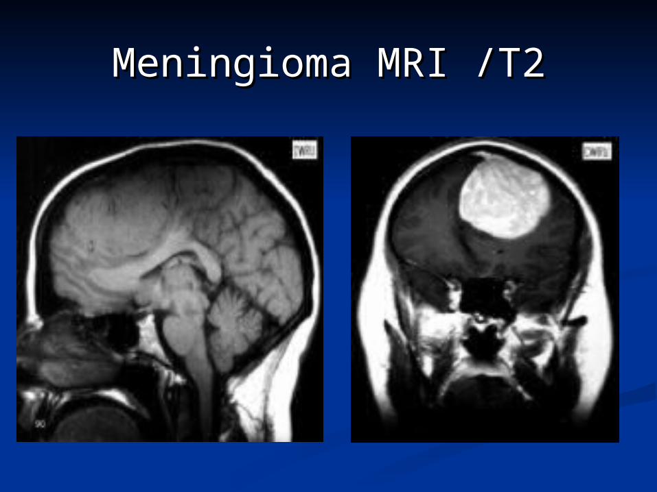
Meningioma MRI /T2Meningioma MRI /T2

Figure 1a. CT scan showing an astrocytic lesion (arrows), one of
the glial tumors, in the left frontal lobe.
Figure 1b. Intraoperative photograph of exposed brain tumor showing the pale lesion
pouting out of the brain surface after opening the overlying dura.
Figure 1c. Photomicrograph of a smear preparation showing
astrocytic hypercellularity (more cells than normal) and nuclear
pleomorphism (abnormal variability of the nuclei in cells), in
keeping with a malignant astrocytoma.


Neuroimaging of brain Neuroimaging of brain tumorstumors
Major diagnostic modality. Useful for Major diagnostic modality. Useful for preoperative planningpreoperative planning
The diagnosis of a primary brain tumor is The diagnosis of a primary brain tumor is best made by cranial MRI. This should be best made by cranial MRI. This should be the first test obtained in a patient with the first test obtained in a patient with signs or symptoms suggestive of an signs or symptoms suggestive of an intracranial mass. The MRI scan should intracranial mass. The MRI scan should always be obtained both with and without always be obtained both with and without contrast material (gadolinium).contrast material (gadolinium).
MRI superior to CT scan for evaluating MRI superior to CT scan for evaluating meninges, subarachnoid space, posterior meninges, subarachnoid space, posterior fossa and defining the vascular fossa and defining the vascular abnormality of the lesionabnormality of the lesion

NeuroimagingNeuroimaging High-grade or malignant gliomas appear High-grade or malignant gliomas appear
as contrast-enhancing mass lesions, which as contrast-enhancing mass lesions, which arise in white matter and are surrounded arise in white matter and are surrounded by edema by edema
Multifocal malignant gliomas are seen in Multifocal malignant gliomas are seen in ~ 5% of patients.~ 5% of patients.
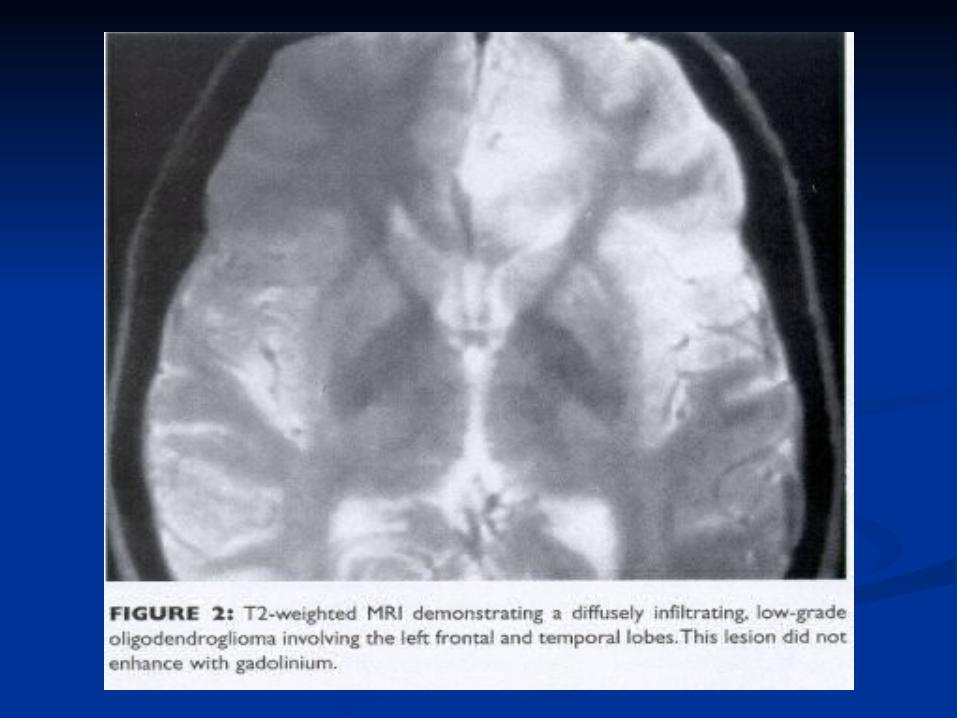
Low-grade gliomas typically are Low-grade gliomas typically are nonenhancing lesions that diffusely nonenhancing lesions that diffusely infiltrate brain tissue and may involve a infiltrate brain tissue and may involve a large region of brain. Low-grade gliomas large region of brain. Low-grade gliomas are usually best appreciated on T2-are usually best appreciated on T2-weighted MRI scans.weighted MRI scans.
NeuroimagingNeuroimaging A contrast-enhanced CT scan may be used if A contrast-enhanced CT scan may be used if
MRI is unavailable. CT may be false-negative MRI is unavailable. CT may be false-negative in patients with a low-grade tumor and can in patients with a low-grade tumor and can have significant artifact through the posterior have significant artifact through the posterior fossa, which may obscure a lesion in this area. fossa, which may obscure a lesion in this area.
Calcification, which may suggest the Calcification, which may suggest the diagnosis of an oligodendroglioma, is often diagnosis of an oligodendroglioma, is often better appreciated on CT than on MRI. better appreciated on CT than on MRI.
CT useful if there is a question of bone or CT useful if there is a question of bone or vascular involvement, or for detecting mets to vascular involvement, or for detecting mets to skull base. Also, in ER situation or if MRI is skull base. Also, in ER situation or if MRI is contraindicated.contraindicated.

Radiologic features of Radiologic features of metastatic diseasemetastatic disease
-Multiple lesions-Localization at the grey-white junction-More circumscribed margins-Relatively large amount of edema compared to size of lesion

SourcesSources
Wen, Patrick Y. Overview of Brain Wen, Patrick Y. Overview of Brain Metastases. UptoDate version 13.3.Metastases. UptoDate version 13.3.
Wong, Eric T. Clinical presentation Wong, Eric T. Clinical presentation and diagnosis of brain tumors. and diagnosis of brain tumors. UptoDate version 13.3.UptoDate version 13.3.
Ma, Julie. Mania Resulting from Ma, Julie. Mania Resulting from Brain Tumor. Clinical Vignette UCLA Brain Tumor. Clinical Vignette UCLA Department of Medicine.Department of Medicine.
Top Related