


Languages
Pages
Legal


CASE PRESENTATION HOPI 56 F with ESRD 2/2 known idiopathic membranous GN on HD MWF , h/o Non compliance with hd/ phos.binders. She presented with h/o worsening rt hip pain for several months and maroon colored stools×2 for one day.she was previously seen in Bellevue ed in Feb 2012 with similar complaints, Xray showed right hip fracture. She was advised surgery,however ,left AMA PMH/PSH:ESRD secondary to Idiopathic GN h/o surgery av fistula placement HOME MEDICATIONS : phoslo 2 cap tid Niferex 150 mg daily Calcitriol 1 mc tiw w/ hd epo 2000 units tiw w/ hd
PHYSICAL EXAMINATION
Vs stable
GENERAL: middle aged AA female , nad
Chest: Clear to auscultation
cvs: s1s2,rrr
abd: soft,nt ,nd bs+
Extr: 1+ pitting edema bl le

Laboratory Test
YEAR IPTH Ca Ph Vit D ALP 2001-02 181 7.4-8.5 2.4-7 2003-04 171-244 7.6-8.2 3.9-6.3 62-100 2005-06 112-440 7.2-8.4 4.6-7.2 91-150 2007 278 8-9 4.5-5.7 2008 166-273 7.5-9 3.8-6.6 2009 332-215 8-9 4.6-6.3 2010 230-303 8.4-10 4.8-5.7 94-136 2011 240-365 8-9.4 3.2-5.4 94-130 2012 174-240 7.4-9 2.6-5.5 29 Bonealp-58

PELVIC MRI
MRI impression: Well defined erosive excavations are seen on B/L hips with Surrounding low T1, intermediate T2 soft tissue lobulations with a pathological Fracture extending from Rt femoral neck to the intertrochanteric region D/D Arthropathy related to long term dialysis Amyloid deposition disease Crystal deposition disease

X-RAY HAND

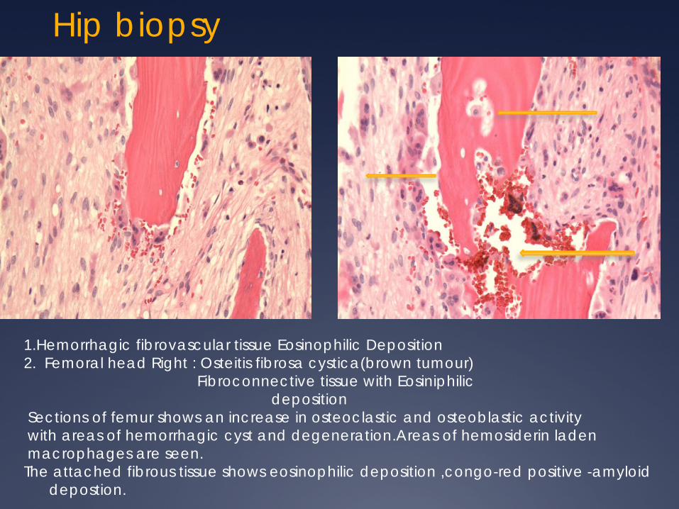
Hip biopsy
1.Hemorrhagic fibrovascular tissue Eosinophilic Deposition 2. Femoral head Right : Osteitis fibrosa cystica(brown tumour) Fibroconnective tissue with Eosiniphilic deposition Sections of femur shows an increase in osteoclastic and osteoblastic activity with areas of hemorrhagic cyst and degeneration.Areas of hemosiderin laden macrophages are seen. The attached fibrous tissue shows eosinophilic deposition ,congo-red positive -amyloid
depostion.

BONE DISEASE IN CHRONIC KIDNEY
DISEASE
SONIKA PURI
BONE DISEASE IN CKD
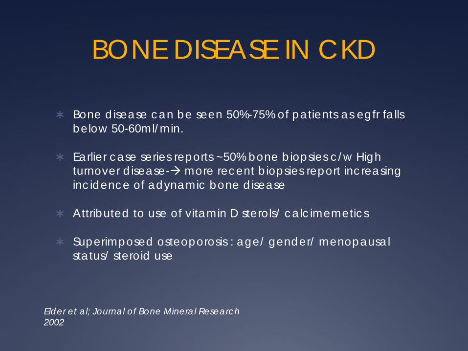
Bone disease can be seen 50%-75% of patients as egfr falls below 50-60ml/min.
Earlier case series reports ~50% bone biopsies c/w High turnover disease- more recent biopsies report increasing incidence of adynamic bone disease
Attributed to use of vitamin D sterols/ calcimemetics
Superimposed osteoporosis : age/ gender/ menopausal status/ steroid use
Elder et al; Journal of Bone Mineral Research 2002

Elder et al; Journal of Bone Mineral Research 2002

Evolution of ROD distribution pattern over time (modified after [6]).
Brandenburg V M , Floege J NDT Plus 2008;1:135-147
© The Author [2008]. Published by Oxford University Press on behalf of ERA-EDTA. All rights reserved. For Permissions, please e-mail: [email protected]
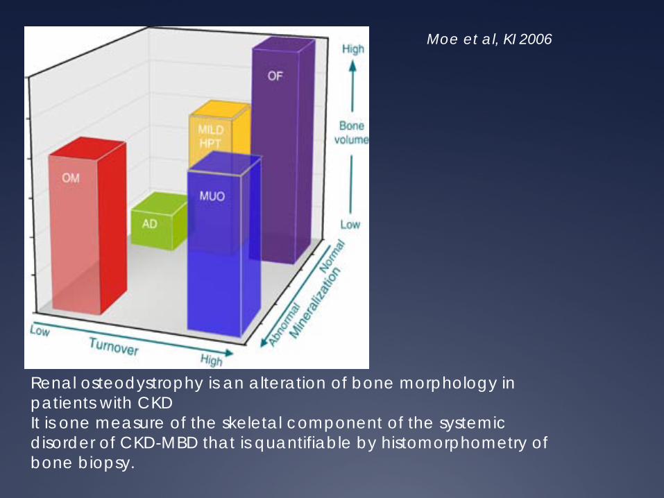
Renal osteodystrophy is an alteration of bone morphology in patients with CKD It is one measure of the skeletal component of the systemic disorder of CKD-MBD that is quantifiable by histomorphometry of bone biopsy.
Moe et al, KI 2006
COMPONENTS FOR CLASSIFICATION OF ROD
TURNOVER : coupled process of bone resorption and bone formation
-assessed by histiomorphometry using double-tetracycline labelling
-Represented by Bone formation rate and activation frequency
-Bone rebsorption rate cannot be measured directly
MINERALIZATION: Calcification of bone collagen
-Reflected in osteoid volume; dynamic measure – tetracycline labelling : measurement of osteoid maturation time.
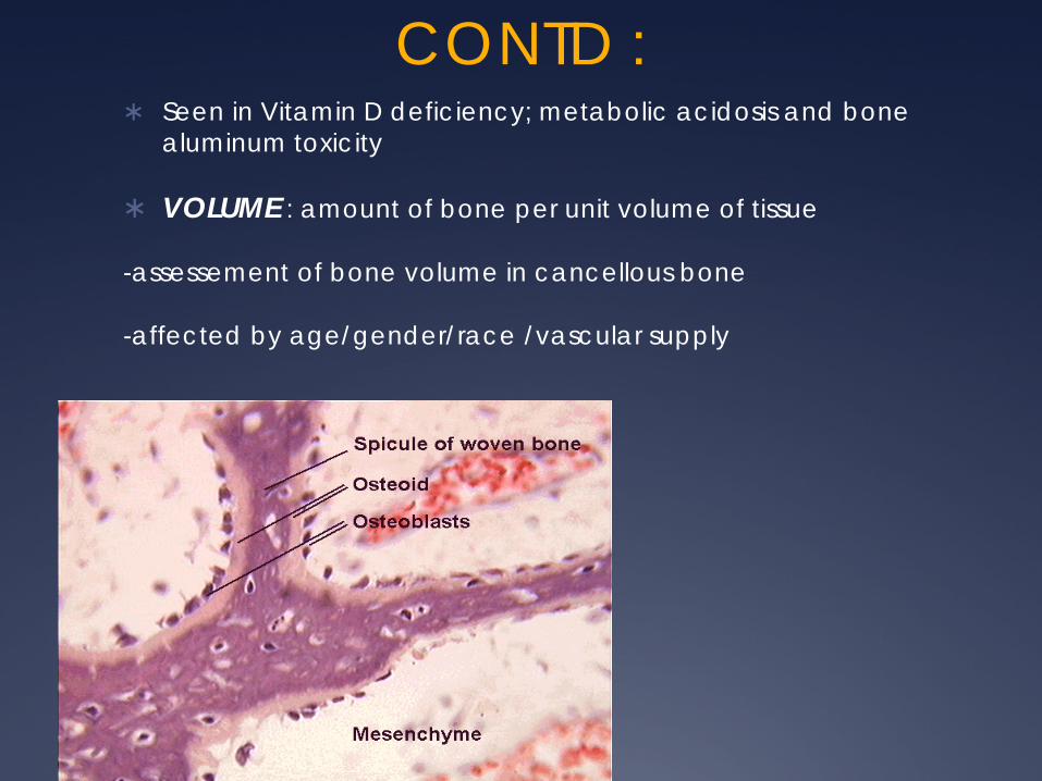
CONTD : Seen in Vitamin D deficiency; metabolic acidosis and bone
aluminum toxicity
VOLUME : amount of bone per unit volume of tissue
-assessement of bone volume in cancellous bone
-affected by age/gender/race /vascular supply

Pediatric Nephrology, 2000
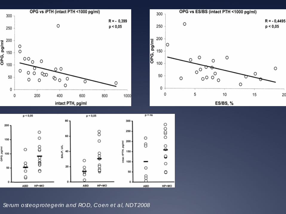
Serum osteoprotegerin and ROD, Coen et al, NDT 2008
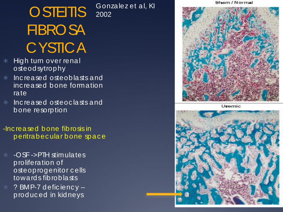
OSTEITIS FIBROSA CYSTICA
High turn over renal osteodsytrophy
Increased osteoblasts and increased bone formation rate
Increased osteoclasts and bone resorption
-Increased bone fibrosis in peritrabecular bone space
-OSF ->PTH stimulates proliferation of osteoprogenitor cells towards fibroblasts
? BMP-7 deficiency – produced in kidneys
Gonzalez et al, KI 2002
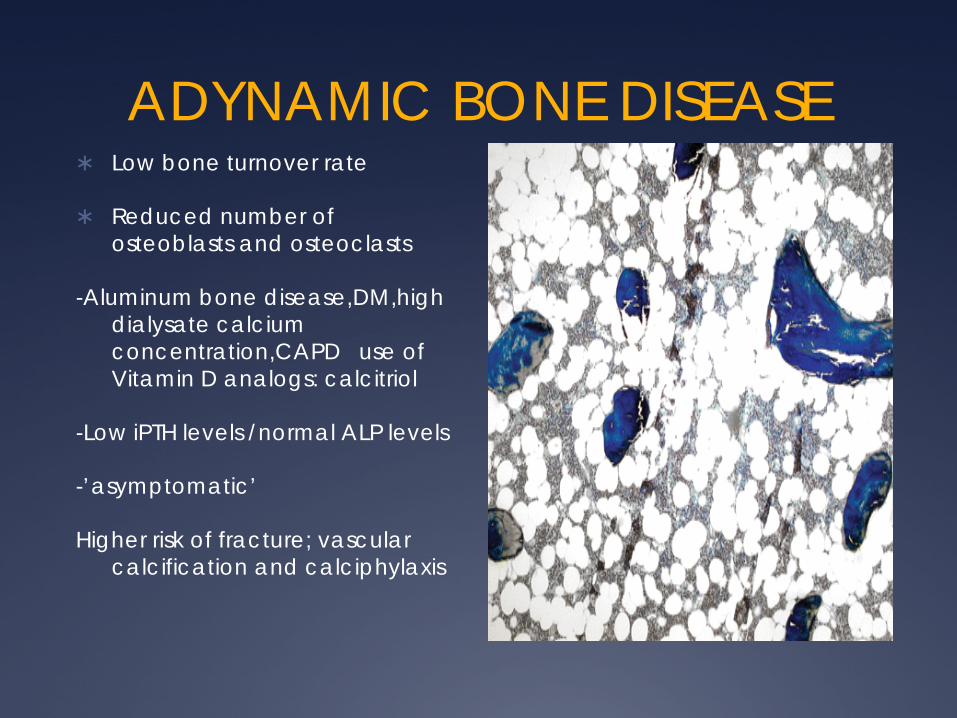
ADYNAMIC BONE DISEASE Low bone turnover rate
Reduced number of osteoblasts and osteoclasts
-Aluminum bone disease,DM,high dialysate calcium concentration,CAPD use of Vitamin D analogs: calcitriol
-Low iPTH levels /normal ALP levels
-’asymptomatic’
Higher risk of fracture; vascular calcification and calciphylaxis

Histomorphometric parameters of bone turnover.
Malluche H H et al. JASN 2012;23:525-532
©2012 by American Society of Nephrology

Microstructural parameters of bone are lower in patients with low than in patients with normal or high turnover.
Malluche H H et al. JASN 2012;23:525-532
©2012 by American Society of Nephrology

Malluche et al, KI 1976
-EOFS % of trabecular surface with fibrosis -HO fraction of trabecular surface covered with osteoclasts -OSF % of trab covered by unmineralized osteoid
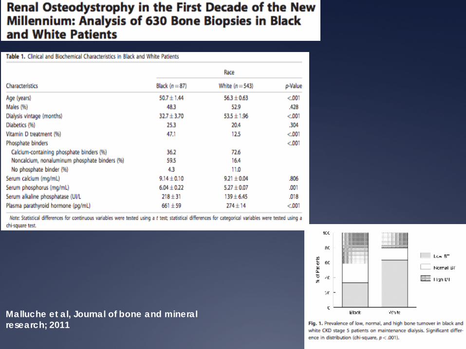
Malluche et al, Journal of bone and mineral research; 2011
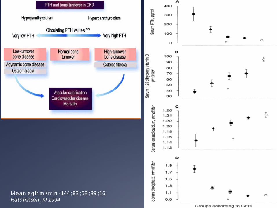
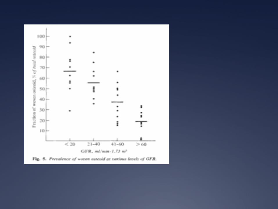
Mean egfr ml/min -144 ;83 ;58 ;39 ;16 Hutchinson, KI 1994
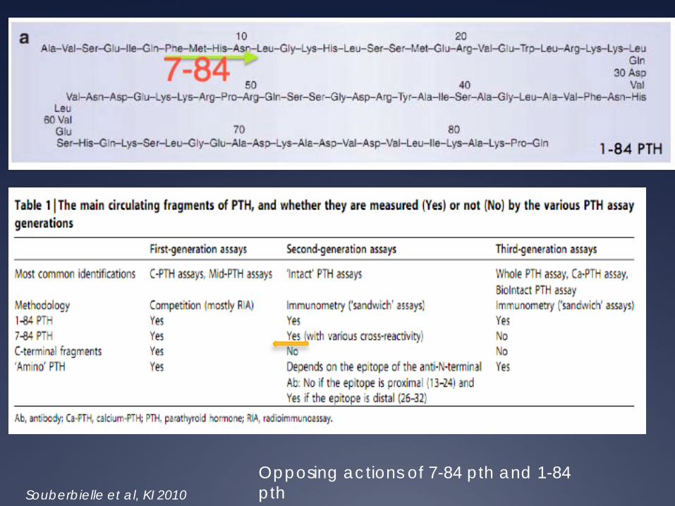
Souberbielle et al, KI 2010 Opposing actions of 7-84 pth and 1-84 pth

KDIGO 2009 GUIDELINES
-CKD Stage 3-5 : suggestion to evaluate pts with PTH levels above the upper reference limit of the assay for hyperphosphatemia/hypocalcemia and Vitamin d defic, no pth target -Ckd 5D- pth target 130-600pg/ml
PTH as a marker for bone disease
Joly et al, AJKD 2008
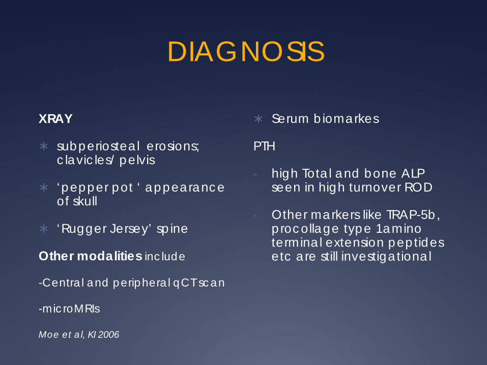
DIAGNOSIS
XRAY
subperiosteal erosions; clavicles/ pelvis
‘pepper pot ‘ appearance of skull
‘Rugger Jersey’ spine
Other modalities include
-Central and peripheral qCT scan
-microMRIs
Serum biomarkes
PTH
- high Total and bone ALP seen in high turnover ROD
- Other markers like TRAP-5b, procollage type 1amino terminal extension peptides etc are still investigational
Moe et al, KI 2006

FRACTURE RISK/ BONE DENSITY MEASUREMENT
-4 fold increase in risk of hip fracture in pt’s on dialysis -yearly incidence rate of fracture by site is 1% for hip, 2.4% for other sites (0.07-0.22%) -No correlation between type of ROD and Dexa scan
Nickolas; KI 2008
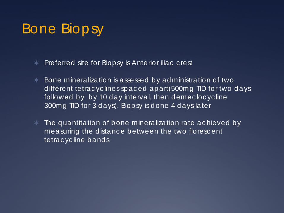
Bone Biopsy
Preferred site for Biopsy is Anterior iliac crest
Bone mineralization is assessed by administration of two different tetracyclines spaced apart(500mg TID for two days followed by by 10 day interval, then demeclocycline 300mg TID for 3 days). Biopsy is done 4 days later
The quantitation of bone mineralization rate achieved by measuring the distance between the two florescent tetracycline bands

INDICATIONS OF BONE BIOPSY
Inconsistencies among biochemical parameters that preclude a definitive interpretation.
Unexplained skeletal fracture or bone pain
Severe progressive
Unexplained hypercalcemia
Suspicion of overload or toxicity from aluminium
Before parathyroidectomy if there has been significant exposure to aluminium in the past or if the results of biochemical determinations are not c/w sec. or 3 hyperpth
Severe progressive VC

Beta-2 amyloidosis -As gfr declines b2 microglobulin levels can be elevated 30-50 times -B2M is major component of DRA,however needs other proteins for stabilization -presence of lysophospholipids in cartilage promotes stabilization/ monomerization of B2M fibrils
-Japanese study : DRA seen in pts on hd for >20yrs Incidence of surgical intervention for Carpal tunnel/ Spondyloarthropathies/ joint arthropathy was 25%,66% and 78% for dialysis vintage 20-24yrs, 25-29yrs and >30yrs resp.
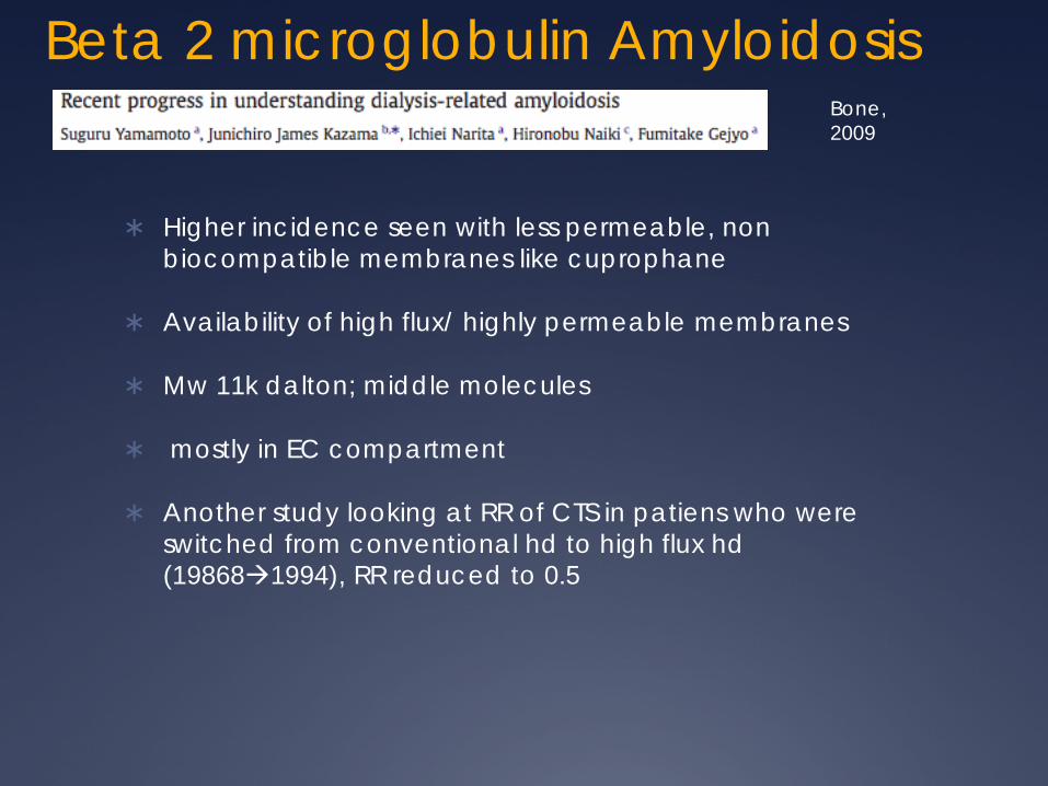
Beta 2 microglobulin Amyloidosis
Higher incidence seen with less permeable, non biocompatible membranes like cuprophane
Availability of high flux/ highly permeable membranes
Mw 11k dalton; middle molecules
mostly in EC compartment
Another study looking at RR of CTS in patiens who were switched from conventional hd to high flux hd (198681994), RR reduced to 0.5
Bone, 2009
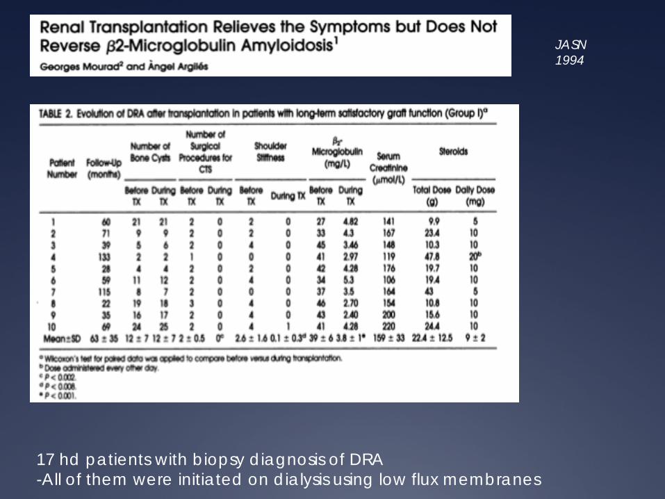
JASN 1994
17 hd patients with biopsy diagnosis of DRA -All of them were initiated on dialysis using low flux membranes
Top Related