


Languages
Pages
Legal

ISSN: 0973-4945; CODEN ECJHAO
E-Journal of Chemistry
http://www.e-journals.net 2010, 7(4), 1334-1339
Biosynthesis of Gold Nanoparticles (Green-Gold)
Using Leaf Extract of Terminalia Catappa
BALAPRASAD ANKAMWAR
Department of Chemistry,
University of Pune, Ganeshkhind, Pune-411007, India. [email protected]
Received 5 February 2010; Accepted 2 April 2010
Abstract: The synthesis of eco-friendly nanoparticles is evergreen branch of
nanoscience for biomedical application. Low cost of synthesis and non toxicity
are main features make it more attractive potential option for biomedical field
and elsewhere. Here, we report the synthesis of gold nanoparticles in aqueous
medium using Terminalia catappa (Almond) leaf extract as the reducing and
stabilizing agent. On treating chloroauric acid solutions with Terminalia
catappa (TC) leaf extract rapid reduction of chloroaurate ions is observed
leading to the formation of highly stable gold nanoparticles in solution. TEM
analysis of the gold nanoparticles indicated that they ranged in size from 10 to
35 nm with average size of 21.9 nm.
Keywords: Biosynthesis, Terminalia catappa, Gold nanoparticles, Green-gold.
Introduction
Currently, there is growing need to develop eco-friendly and body benign nanoparticle
synthesis processes without use of toxic chemicals in the synthesis protocols to avoid
adverse effects in biomedical applications. Obviously, researchers in this field paid their
attention towards the use of biological systems for the synthesis of biocompatible metal and
semiconductor nanostructures. Some well-known examples of bio-organisms synthesizing
inorganic materials include magnetotactic bacteria (synthesizing magnetite nanoparticles)1
diatoms (synthesizing siliceous materials)2 and S-layer bacteria (producing gypsum and
calcium carbonate layers)3.
Many biotechnological applications such as remediation of toxic metals employ
microorganisms such as bacteria4 and yeast
5. Nair and Pradeep
6 have synthesized nano-
crystals of gold, silver and their alloys by reaction of the corresponding metal ions within
cells of lactic acid bacteria present in buttermilk. The bacteria7 and algae
8 are exploited for
synthesis of gold nanoparticle.

1335 B. ANKAMWAR
The extra cellular synthesis of gold nanoparticles of about 8 nm diameter has also been
reported by using the alkalothermophilic actinomycete Thermomonospora sp9. As can be
seen from the above, the use of microorganisms in the deliberate and controlled synthesis of
nanoparticles is a relatively new and exciting area of research with considerable potential for
development. Recently synthesis of Au and Ag nanoparticles using extracts of cinnamomum
camphora leaf10
, phyllanthin11
and edible mushroom12
as a reducing and capping agent has
been reported.
While the microorganisms such as bacteria, actinomycete and fungi continue to be
investigated in metal nanoparticle synthesis, the use of parts of whole plants, similarly in
nanoparticle synthesis methodologies is an exciting possibility that is relatively
unexplored and underexploited. Using plants for synthesis of nanoparticles could be
advantageous over other environmentally benign biological processes by eliminating the
elaborate process of maintaining cell cultures. It can also be suitably scaled up for large-
scale synthesis of nanoparticles. Recently, Jose-Yacaman and co-workers13
demonstrated
the synthesis of gold and silver nanoparticles within live alfalfa plants in solid media.
Moreover, agricultural biomass has been used to reduce Cr(VI) to Cr(III) ions14
indicating
that biological methods can be very efficient in decontaminating polluted waters and soil
polluted with heavy metal ions.
In our earlier reports, synthesis of gold nanoparticles have been shown by the reduction
of aqueous AuCl4- ions using extracts from Emblica officinalis (Indian Gooseberry) fruit
15
and Tamarindus indica16
leaf. Recently, we had demonstrated the biological synthesis of
triangular gold nanoprisms by a single step, room temperature reduction of aqueous
chloroaurate ions by the extract of the plant, lemongrass17
. There is still much scope for
improvement in bio-based methods for metal nanoparticle synthesis, particularly in relation
to improving the monodispersity of the nanoparticles and modulating their size and shape, as
well as in reducing the time required for nanoparticle synthesis. On a more fundamental
level, it would be interesting to study the nature of nanoparticles formed using extracts from
different parts of a plant. The main idea behind selection of TC extract is due to its
anticancer18
, antibacterial19
and antioxidant activities20-22
,
In this paper we demonstrate method for the synthesis of gold nanoparticles by the
reduction of aqueous chloroaurate ions by using Terminalia catappa leaf extract.
Experimental Reagents and Chemicals
Tetrachloroauric acid (HAuCl4.XH2O) was obtained from Sigma Aldrich. Freshly prepared
double distilled water was used through out the experimental work.
Biological synthesis of gold nanoparticles
The broth used for the reduction of Au3+
ions to Au0 was prepared by taking 10 g of
thoroughly washed and finely cut Terminalia catappa leaves in a 500 mL Erlenmeyer flasks
with 40 mL of sterile distilled water and then was boiled it for 15 min. In a typical
experiment, 0.2 mL of broth was added to 50 mL of 10-3
M aqueous chloroauric acid
(HAuCl4) solution. Within an hour (50 minutes) cherry red solution was obtained.
UV-Vis spectroscopy studies
UV-Vis spectroscopy measurement of the Terminalia catappa leaf extract reduced gold
nanotriangles was carried out on a JASCO dual-beam spectrophotometer (model V-570)
operated at a resolution of 1 nm.
Wavenumber, cm-1
Wavenumber, cm-1
Wavelength, nm
Ab
sorb
ance
Tra
nsm
itta
nce
(au
)
Biosynthesis of Gold Nanoparticles Using Leaf Extract 1336
X-ray diffraction (XRD) measurement
XRD measurements of the bioreduced chloroauric acid solution drop-coated on glass were done on a Phillips PW 1830 instrument operating at a voltage of 40 KV and current of 20 mA with Cu Kα radiation.
Fourier transform infrared (FTIR) spectroscopy measurements
For FTIR spectroscopy measurements, drop coated samples on Si (111) wafers were prepared. After complete reduction of AuCl4
- ions by the Terminalia catappa leaf broth and formation of
gold nanoparticles was centrifuged at 9000 rpm for 10 min to isolate the gold nanoparticles from free proteins or other compounds present in the solution. The gold nanoparticle pellets obtained after centrifugation were redispersed in water prior to FTIR analysis centrifuged again at 9000 rpm for 10 min to isolate the gold nanoparticles from traces of free proteins or other compounds present in the solution if any. FTIR measurements of TC leaf extract reduced gold nanoparticles were carried out on a Perkin-Elmer FTIR Spectrum One spectrophotometer in the diffuse reflectance mode operating at a resolution of 4 cm
-1.
TEM measurements TEM samples of the gold nanoparticles synthesized by the biological reduction were prepared by placing a drop over carbon coated copper grids and allowing the solvent to evaporate. TEM measurements were performed on a JEOL model 1200EX instrument operated at an accelerating voltage at 80 kV.
Results and Discussion
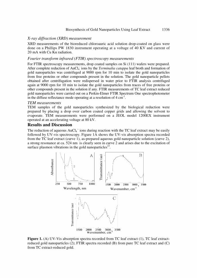
The reduction of aqueous AuCl4-- ions during reaction with the TC leaf extract may be easily
followed by UV-vis spectroscopy. Figure 1A shows the UV-vis absorption spectra recorded from the TC leaf extract (curve 1), as-prepared aqueous gold nanoparticle solution (curve 2), a strong resonance at ca. 524 nm is clearly seen in curve 2 and arises due to the excitation of surface plasmon vibrations in the gold nanoparticles
23.
Figure 1. (A) UV-Vis absorption spectra recorded from TC leaf extract (1), TC leaf extract-
reduced gold nanoparticles (2); FTIR spectra recorded (B) from pure TC leaf extract and (C)
from TC extract-reduced gold.
Tra
nsm
itta
nce
(au
)

Particle size, nm 20, degrees
1337 B. ANKAMWAR
It should be noted here that there is no time-dependent change in the UV – vis absorption
spectra. Curve 2 in Figure 1A clearly indicating that the gold nanoparticles in aqueous phase are
extremely stable with no precipitation observed even after four months. The stability for such a
long period seems to be due to antimicrobial19
and antioxidant20-22
properties of TC leaf extract.
FTIR measurements were carried out to identify the possible biomolecules in the TC leaf extract responsible for the reduction of AuCl4
— ions and also the capping agents responsible
for the stability of the biogenic nanoparticle solution. Chen at al24
reported that leaves of TC contains 21% tannin, whereas Rayudu and Rajdurai
25 analyzed the polyphenols and carboxylic
compounds of TC. Figure 1B represents the FTIR spectrum of TC leaf extract which shows prominent absorption bands at 1718 cm
-1, 1441 cm
-1 and 3372 cm
-1. The shoulder at 1718 cm
–1
is characteristic of carbonyl stretch vibrations from carboxylic acid and phenols, while the stretch at 1441 arises due to the C-O stretching and O-H deformation possibly from the acid groups present in the TC leaf extract
26, 27. The broad stretching at 3372 cm
-1 arises due to the
free O-H groups present in the phenols. Figure 1C represents the FTIR spectrum of the TC leaf extract reduced gold with the absorption bands at 1420 cm
-1, 1715 cm
-1 and 3145 cm
-1. The
shift in the carbonyl stretch frequency (1718 cm-1
) to lower wavenumbers (1715 cm-1
) followed by the disappearance of the 1718 cm
-1 resonance may be due to its binding with the
gold nanoparticle surface. The shift in the C-O stretching and O-H deformation frequency (1441 cm
-1) to lower wavenumbers (1420 cm
-1) followed by the disappearance of the 1441 cm
-1
resonance indicate the facilitation of the binding of O-H group of phenols with the gold nanoparticle surface. In addition to above supportive evidence the 3372 cm
-1 feature shifts to
3145 cm-1
due to the binding of the hydroxyl group with gold nanoparticle surface26
.
Figure 2A and 2B show a TEM and low magnification TEM image recorded from the biologically synthesized gold nanoparticles at the end of the reaction with TC leaf extract respectively, while Figure 2C is a plot of particle size distribution (PSD) histogram measured from an analysis of 120 particles from Figure 2A. The TEM image shows that the gold nanoparticles are predominantly spherical in morphology with their size ranging from 10 to 35 nm with an average size of about 21.9 nm. A Gaussian fit to the PSD histogram yielded a particle size of 21.9 ± 2 nm.
Figure 2. Representative TEM images of TC leaf extract-reduced gold nanoparticles at low
magnification (A) and at higher magnification (B). The lower panels in the image (A) shows
the corresponding particle size distribution histograms (C). The solid lines in the lower
panels is Gaussian fits to the histogram. (D) XRD pattern of a solution-cast film of the TC
leaf extract-reduced gold nanotriangles deposited on a glass substrate. The Bragg reflections
are identified in the XRD pattern.
% o
f par
ticl
es
Inte
nsi
ty,
(au
)

Biosynthesis of Gold Nanoparticles Using Leaf Extract 1338
The formation of gold nanoparticles synthesized using TC leaf extract was further
supported by X-ray diffraction (XRD) measurements (Figure 2D). The Bragg reflections
corresponding to the (111), (200), (220), (311) and (222) sets of lattice planes are observed
that may be indexed on the basis of the fcc structure of gold. The (200), (220), (311) and
(222) Bragg reflections are weak and considerably broadened relative to the intense (111)
reflection. This interesting feature indicates that gold nanocrystals are in the film are
predominantly (111)-oriented.
The antibacterial19
and antioxidant20-22
properties of biomolecules present in the TC leaf
extract have facilitated excellent stability of the nanoparticles. The size of the nanoparticles
being in the range 10 - 35 nm with average size of 21.9 nm makes circulation into blood
vessels feasible. In addition to the size and stability, the anticancer18
, antibacterial19
,
antioxidant20-2
properties of TC leaf extract could have important application in the use of
the biogenic gold nanoparticles in cancer therapy, and is currently being pursued.
Conclusion
The rapid synthesis of stable gold nanoparticles using TC leaf extract has been
demonstrated. The reduction of the metal ions and the stabilization of the Au nanoparticles
is believed to occur by the various acids and hydrolysable tannins present in the TC leaf.
The nanoparticles are extremely stable with time. The anticancer, antibacterial and
antioxidant properties of TC leaf extract could be exploited in the use of the biogenic gold
nanoparticles in cancer therapy.
Acknowledgments
The author thanks the Indian Academy of Sciences, Bangalore for a Summer Research
Fellowship. The author also thanks the Director, National Chemical Laboratory (NCL), Pune
and Dr. Murali Sastry, Scientist, NCL for permission to carry out major work of this
research at NCL.
References
1. (a) Lovley D R, Stolz J F, Nord G L and Phillips E J P, Nature, 1987, 330, 252; (b)
Dickson D P E, J Magn Matrer., 1999, 203, 46.
2. (a) Mann S, Nature, 1993, 365, 499; (b) Oliver S, Kupermann A, Coombs N, Lough
A and Ozin G A, Nature, 1995, 378, 47.
3. (a) Pum D and Sleytr U B, Trends Biotechnol., 1999, 17, 8; (b) Sleytr U B, Messner
P, Pum D and Sara M, Angew Chem Int Ed., 1999, 38, 1034.
4. Stephen J R and Maenaughton S J, Curr Opin Biotechnol., 1999, 10, 230.
5. Mehra R K and Wingre D R, J Cell Biochem., 1991, 45, 30.
6. Nair B and Pradeep T, Cryst Growth Des., 2002, 2, 293-298.
7. (a) Southam G and Beveridge T J, Geochim. Cosmochim Acta., 1996, 60, 4369; (b)
Beveridge T J and Murray R G E, J Bacteriol., 1980, 141, 876.
8. Robinson M G, Brown L N and Beverley D, Biofouling, 1997, 11, 59-79.
9. Ahmad A, Senapati S, Khan M I, Kumar R and Sastry M, Langmuir, 2003, 19, 3550.
10. Huang J, Li Q, Sun D, Lu Y, Su Y, Yang X, Wang H, Wang Y, Shao W, He N, Hong J
and Chen C, Nanotechnology, 2007, 18(10), 105104.
11. Kasthuri J, Kathiravan K and Rajendiran N, J Nanopart Res., 2009, 11(5), 1075-1085.
12. Daizy P, Spectrochimica Acta, Part A: Molecular and Biomolecular Spectroscopy,
2009, 73A(2), 374-381.
1339 B. ANKAMWAR
13. Gardea-Torresdey J L, Gomez E, Peralta-Videa J R, Parsons J G, Troiani H E and
Jose-Yacaman M, Langmuir, 2003, 19, 1357.
14. Gardea-Torresdey J L, Tiemann K J, Armendariz V, Bess-Oberto L, Chianelli R R,
Rios J, Parsons J G and Gomez G, J Hazard Mater., 2000, 80, 175.
15. Ankamwar B, Damle C, Ahmad A and Sastry M, J Nanosci Nanotech., 2005, 5,
1665-1671.
16. Ankamwar B, Chaudhary M and Sastry M, Syn React Inorg Metal Org Nano-Metal
Chem., 2005, 35, 19–26.
17. Shiv Shankar S, Rai A, Ankamwar B, Singh A, Ahmad A and Sastry M, Nature
Mater., 2004, 3, 482–488.
18. Kandil F E, Soliman A M, Skodack S R and Mabry T J, Asian J Chem., 1999, 11(3),
1001-1004.
19. Pawar S P and Pal S C, Indian J Med Sci., 2002, 56(6), 276-278.
20. Ko T F , Weng Y M and Chiou R Y, J Agric Food Chem., 2002, 11(9), 5343-5348.
21. Lin C C, Hsu Y F and Lin T C, Anticancer Res., 2001, 21(1A), 237-243.
22. Chyou C C, Tsai S Y, Ko P T and Mau J L, Food Chem., 2002, 78(4), 483-488.
23. Underwood S and Mulavaney P, Langmuir, 1994, 10, 3427.
24. Chen P S, Li J H, Liu T Y and Lin T C, Cancer Letters, 2000, 152(2), 115-122.
25. Rayudu G V N and Rajdurai S, Leather Sci., 1966, 13(10), 289.
26. Vogel A I, Textbook of Practical Organic Chemistry; Addison Wesley Longman:
Harlow, England, 1989, 1219.
27. Silverstein, R M and Basseler G C, Spectrometric Identification of Organic
Compounds; Ch. 4; Wiley: New York, 1967, 111-125.
Submit your manuscripts athttp://www.hindawi.com
Hindawi Publishing Corporationhttp://www.hindawi.com Volume 2014
Inorganic ChemistryInternational Journal of
Hindawi Publishing Corporation http://www.hindawi.com Volume 2014
International Journal ofPhotoenergy
Hindawi Publishing Corporationhttp://www.hindawi.com Volume 2014
Carbohydrate Chemistry
International Journal of
Hindawi Publishing Corporationhttp://www.hindawi.com Volume 2014
Journal of
Chemistry
Hindawi Publishing Corporationhttp://www.hindawi.com Volume 2014
Advances in
Physical Chemistry
Hindawi Publishing Corporationhttp://www.hindawi.com
Analytical Methods in Chemistry
Journal of
Volume 2014
Bioinorganic Chemistry and ApplicationsHindawi Publishing Corporationhttp://www.hindawi.com Volume 2014
SpectroscopyInternational Journal of
Hindawi Publishing Corporationhttp://www.hindawi.com Volume 2014
The Scientific World JournalHindawi Publishing Corporation http://www.hindawi.com Volume 2014
Medicinal ChemistryInternational Journal of
Hindawi Publishing Corporationhttp://www.hindawi.com Volume 2014
Chromatography Research International
Hindawi Publishing Corporationhttp://www.hindawi.com Volume 2014
Applied ChemistryJournal of
Hindawi Publishing Corporationhttp://www.hindawi.com Volume 2014
Hindawi Publishing Corporationhttp://www.hindawi.com Volume 2014
Theoretical ChemistryJournal of
Hindawi Publishing Corporationhttp://www.hindawi.com Volume 2014
Journal of
Spectroscopy
Analytical ChemistryInternational Journal of
Hindawi Publishing Corporationhttp://www.hindawi.com Volume 2014
Journal of
Hindawi Publishing Corporationhttp://www.hindawi.com Volume 2014
Quantum Chemistry
Hindawi Publishing Corporationhttp://www.hindawi.com Volume 2014
Organic Chemistry International
Hindawi Publishing Corporationhttp://www.hindawi.com Volume 2014
CatalystsJournal of
ElectrochemistryInternational Journal of
Hindawi Publishing Corporation http://www.hindawi.com Volume 2014
Top Related