


Languages
Pages
Legal

Discovery of the effect of telomeres on
chromosomes.
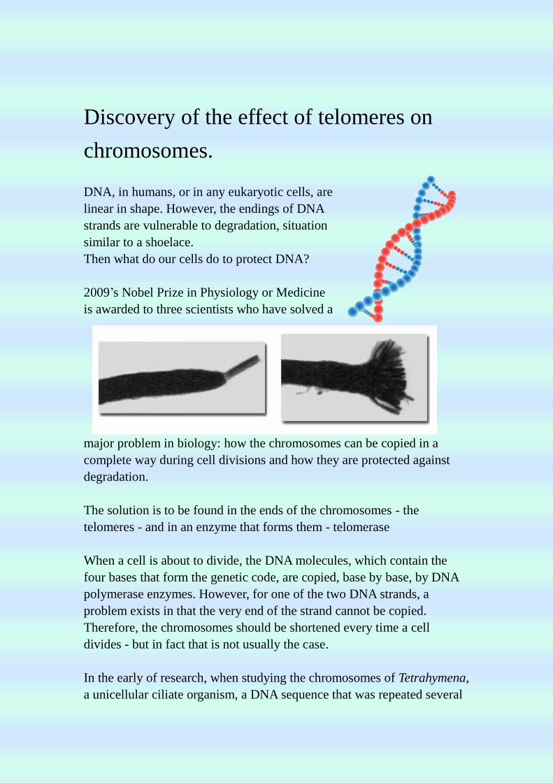
DNA, in humans, or in any eukaryotic cells, are
linear in shape. However, the endings of DNA
strands are vulnerable to degradation, situation
similar to a shoelace.
Then what do our cells do to protect DNA?
2009’s Nobel Prize in Physiology or Medicine
is awarded to three scientists who have solved a
major problem in biology: how the chromosomes can be copied in a
complete way during cell divisions and how they are protected against
degradation.
The solution is to be found in the ends of the chromosomes - the
telomeres - and in an enzyme that forms them - telomerase
When a cell is about to divide, the DNA molecules, which contain the
four bases that form the genetic code, are copied, base by base, by DNA
polymerase enzymes. However, for one of the two DNA strands, a
problem exists in that the very end of the strand cannot be copied.
Therefore, the chromosomes should be shortened every time a cell
divides - but in fact that is not usually the case.
In the early of research, when studying the chromosomes of Tetrahymena,
a unicellular ciliate organism, a DNA sequence that was repeated several
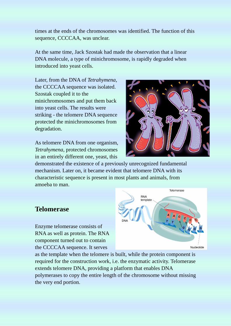
times at the ends of the chromosomes was identified. The function of this
sequence, CCCCAA, was unclear.
At the same time, Jack Szostak had made the observation that a linear
DNA molecule, a type of minichromosome, is rapidly degraded when
introduced into yeast cells.
Later, from the DNA of Tetrahymena,
the CCCCAA sequence was isolated.
Szostak coupled it to the
minichromosomes and put them back
into yeast cells. The results were
striking - the telomere DNA sequence
protected the minichromosomes from
degradation.
As telomere DNA from one organism,
Tetrahymena, protected chromosomes
in an entirely different one, yeast, this
demonstrated the existence of a previously unrecognized fundamental
mechanism. Later on, it became evident that telomere DNA with its
characteristic sequence is present in most plants and animals, from
amoeba to man.
Telomerase
Enzyme telomerase consists of
RNA as well as protein. The RNA
component turned out to contain
the CCCCAA sequence. It serves
as the template when the telomere is built, while the protein component is
required for the construction work, i.e. the enzymatic activity. Telomerase
extends telomere DNA, providing a platform that enables DNA
polymerases to copy the entire length of the chromosome without missing
the very end portion.
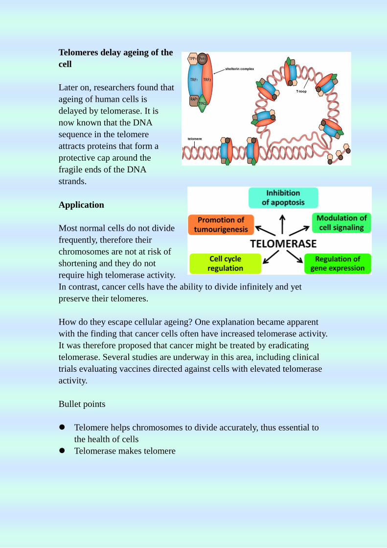
Telomeres delay ageing of the
cell
Later on, researchers found that
ageing of human cells is
delayed by telomerase. It is
now known that the DNA
sequence in the telomere
attracts proteins that form a
protective cap around the
fragile ends of the DNA
strands.
Application
Most normal cells do not divide
frequently, therefore their
chromosomes are not at risk of
shortening and they do not
require high telomerase activity.
In contrast, cancer cells have the ability to divide infinitely and yet
preserve their telomeres.
How do they escape cellular ageing? One explanation became apparent
with the finding that cancer cells often have increased telomerase activity.
It was therefore proposed that cancer might be treated by eradicating
telomerase. Several studies are underway in this area, including clinical
trials evaluating vaccines directed against cells with elevated telomerase
activity.
Bullet points
Telomere helps chromosomes to divide accurately, thus essential to
the health of cells
Telomerase makes telomere
Children with shorter telomere length are more susceptible
towards catching a cold
If telomeres are allowed to shorten, cells struggle to multiply
properly. This makes it tough to rebuild and repair bodily
tissue. Shortened telomeres open the door to disease.
Telomere deterioration is linked to many common symptoms
and health conditions, many of which are the precursors of
diseases that are not presumed to be related. High blood
pressure, high levels of triglycerides and high blood sugar
levels
Discovery of Biology – Blue light
Waking up early for school, struggling to stay awake in morning
assembly, they seem to be the most common problem for students. Well,
not anymore! Researchers at Rensselaer Polytechnic Institute have just
found out the solution to all these troubles---Blue Light.
As a matter of fact, the blue light is just
nothing high-tech or complicated, it is just
light that provide blue visual sensation.
For example, the blue sky. The research
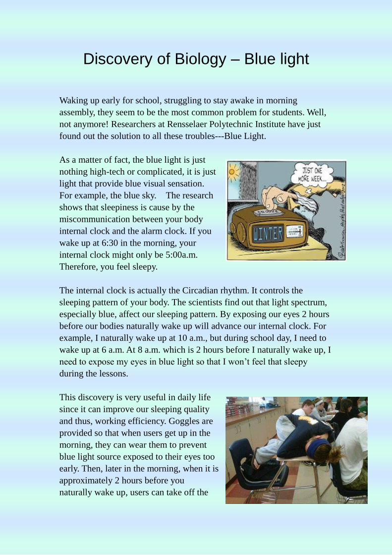
shows that sleepiness is cause by the
miscommunication between your body
internal clock and the alarm clock. If you
wake up at 6:30 in the morning, your
internal clock might only be 5:00a.m.
Therefore, you feel sleepy.
The internal clock is actually the Circadian rhythm. It controls the
sleeping pattern of your body. The scientists find out that light spectrum,
especially blue, affect our sleeping pattern. By exposing our eyes 2 hours
before our bodies naturally wake up will advance our internal clock. For
example, I naturally wake up at 10 a.m., but during school day, I need to
wake up at 6 a.m. At 8 a.m. which is 2 hours before I naturally wake up, I
need to expose my eyes in blue light so that I won’t feel that sleepy
during the lessons.
This discovery is very useful in daily life
since it can improve our sleeping quality
and thus, working efficiency. Goggles are
provided so that when users get up in the
morning, they can wear them to prevent
blue light source exposed to their eyes too
early. Then, later in the morning, when it is
approximately 2 hours before you
naturally wake up, users can take off the

goggles and be exposed to blue light. For example, blue screens in
computers.
This is a huge milestone for humans, the research found by Rensselaer
Polytechnic Institute solve a huge inconvenience in our lives. By getting
blue light at the right time, it can change our sleeping pattern. Especially
for shift workers and people with sleep disorder.
GREEN FLUORESCENT PROTEIN
Introduction
Green Fluorescent Protein (GFP) and GFP-like proteins have become the
microscope of the twenty-first
century. Every month more than
200 papers are published reporting
yet another way GFP has been put
to work. In most cases, GFP can be
used in a way very similar to a
microscope; it can show us when a
protein is made, and what its
movements are.
In honor of the 2008 Nobel Prize in Chemistry,
the whole October 2009 issue of Chemical
Society Reviews (Vol. 38, pp. 2813-2963) is
devoted to GFP. This is an excellent resource for
anyone wanting more detailed information about
GFP than is presented in this module.
Aequorea Bioluminescence
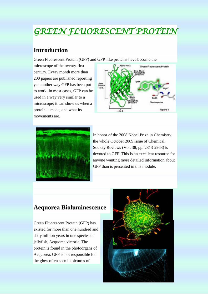
Green Fluorescent Protein (GFP) has
existed for more than one hundred and
sixty million years in one species of
jellyfish, Aequorea victoria. The
protein is found in the photoorgans of
Aequorea. GFP is not responsible for
the glow often seen in pictures of
jellyfish - that "fluorescence" is actually due to the reflection of the flash used to
photograph the jellies.
Aequorea victoria
Aequorea victoria photoorgans
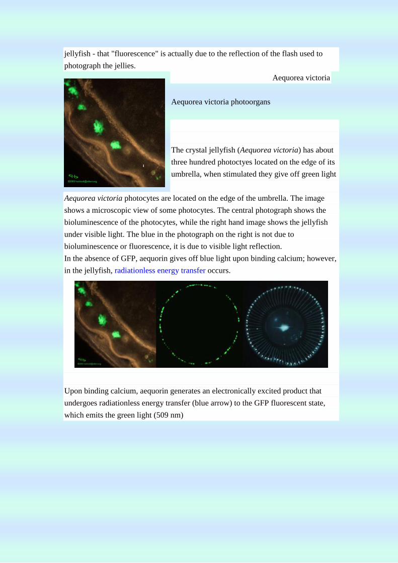
The crystal jellyfish (Aequorea victoria) has about
three hundred photoctyes located on the edge of its
umbrella, when stimulated they give off green light
Aequorea victoria photocytes are located on the edge of the umbrella. The image
shows a microscopic view of some photocytes. The central photograph shows the
bioluminescence of the photocytes, while the right hand image shows the jellyfish
under visible light. The blue in the photograph on the right is not due to
bioluminescence or fluorescence, it is due to visible light reflection.
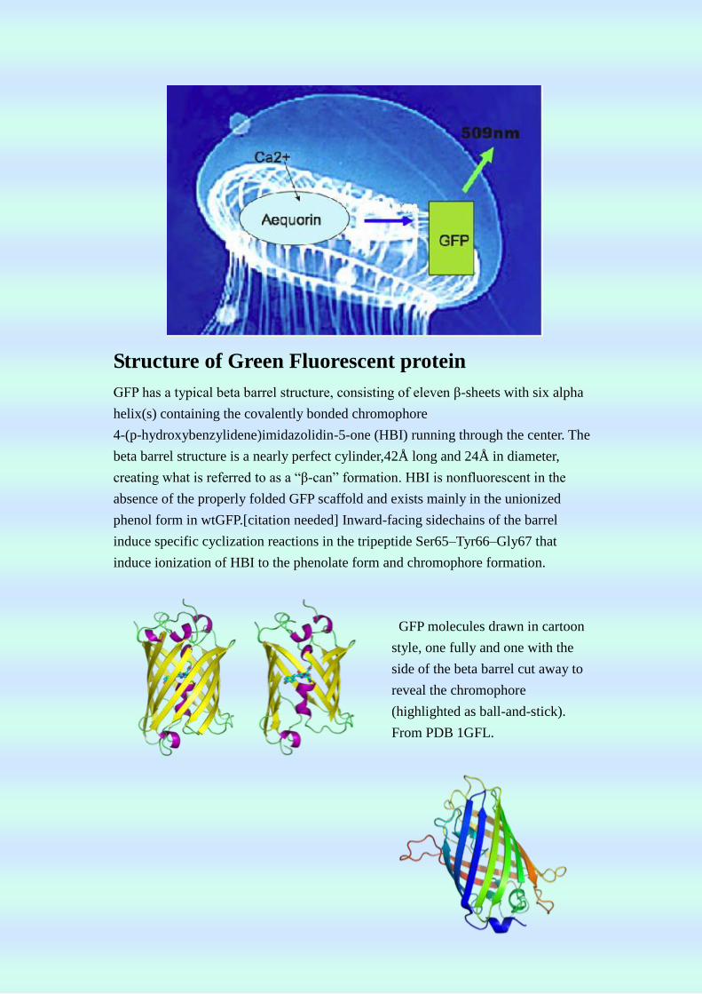
In the absence of GFP, aequorin gives off blue light upon binding calcium; however,
in the jellyfish, radiationless energy transfer occurs.
Upon binding calcium, aequorin generates an electronically excited product that
undergoes radiationless energy transfer (blue arrow) to the GFP fluorescent state,
which emits the green light (509 nm)
Structure of Green Fluorescent protein
GFP has a typical beta barrel structure, consisting of eleven β-sheets with six alpha
helix(s) containing the covalently bonded chromophore
4-(p-hydroxybenzylidene)imidazolidin-5-one (HBI) running through the center. The
beta barrel structure is a nearly perfect cylinder,42Å long and 24Å in diameter,
creating what is referred to as a “β-can” formation. HBI is nonfluorescent in the
absence of the properly folded GFP scaffold and exists mainly in the unionized
phenol form in wtGFP.[citation needed] Inward-facing sidechains of the barrel
induce specific cyclization reactions in the tripeptide Ser65–Tyr66–Gly67 that
induce ionization of HBI to the phenolate form and chromophore formation.
GFP molecules drawn in cartoon
style, one fully and one with the
side of the beta barrel cut away to
reveal the chromophore
(highlighted as ball-and-stick).
From PDB 1GFL.

GFP ribbon diagram. From PDB 1EMA.
This process of post-translational modification is referred to as maturation. The
hydrogen-bonding network and electron-stacking interactions with these sidechains
influence the color, intensity and photostability of GFP and its numerous derivatives.
The tightly packed nature of the barrel excludes solvent molecules, protecting the
chromophore fluorescence from quenching by water.
Application of Green Fluorescent protein
1. Fluorescence Microscopy
The availability of GFP and its derivatives has
thoroughly redefined fluorescence microscopy
and the way it is used in cell biology and other
biological disciplines. For instance,
GFP is used to express the protein in small sets of specific cells. This allows
researchers to optically detect specific types of cells in vitro (in a dish), or even in
vivo (in the living organism).
A novel possible use of GFP includes using it as a
sensitive monitor of intracellular processes via an eGFP
laser system made out of a human embryonic kidney cell
line.
GFP is used widely in cancer research to
label and track cancer cells. GFP-labeled
cancer cells have been used to model
metastasis, the process by which cancer cells
spread to distant organs.
2. Transgenic pets
Mice
expre
ssing
GFP
under UV light (left & right), compared to normal mouse (center)
GloFish, the first pet sold with these proteins artificially present.
3. GFP in fine art
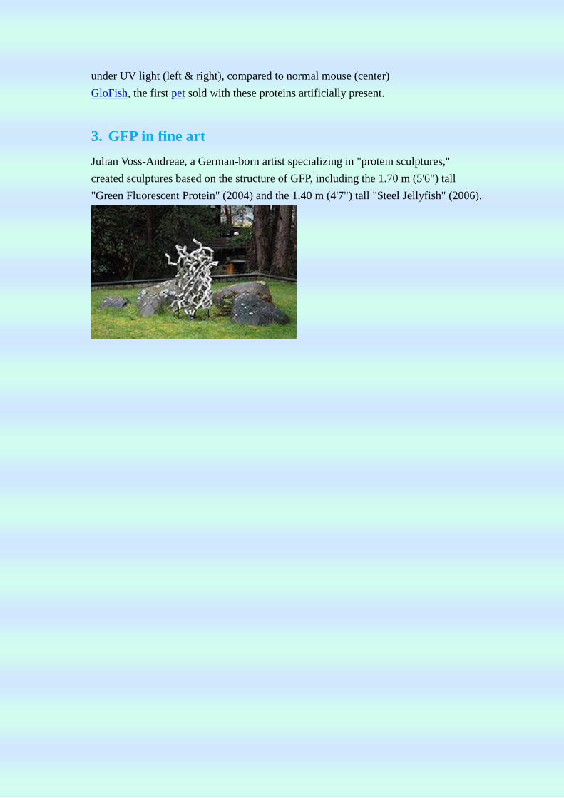
Julian Voss-Andreae, a German-born artist specializing in "protein sculptures,"
created sculptures based on the structure of GFP, including the 1.70 m (5'6") tall
"Green Fluorescent Protein" (2004) and the 1.40 m (4'7") tall "Steel Jellyfish" (2006).
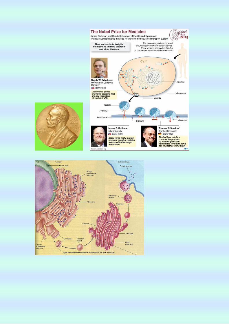
Machinery Regulating Vesicle Traffic, a Major Transport System in our Cells
INTRODUCTION
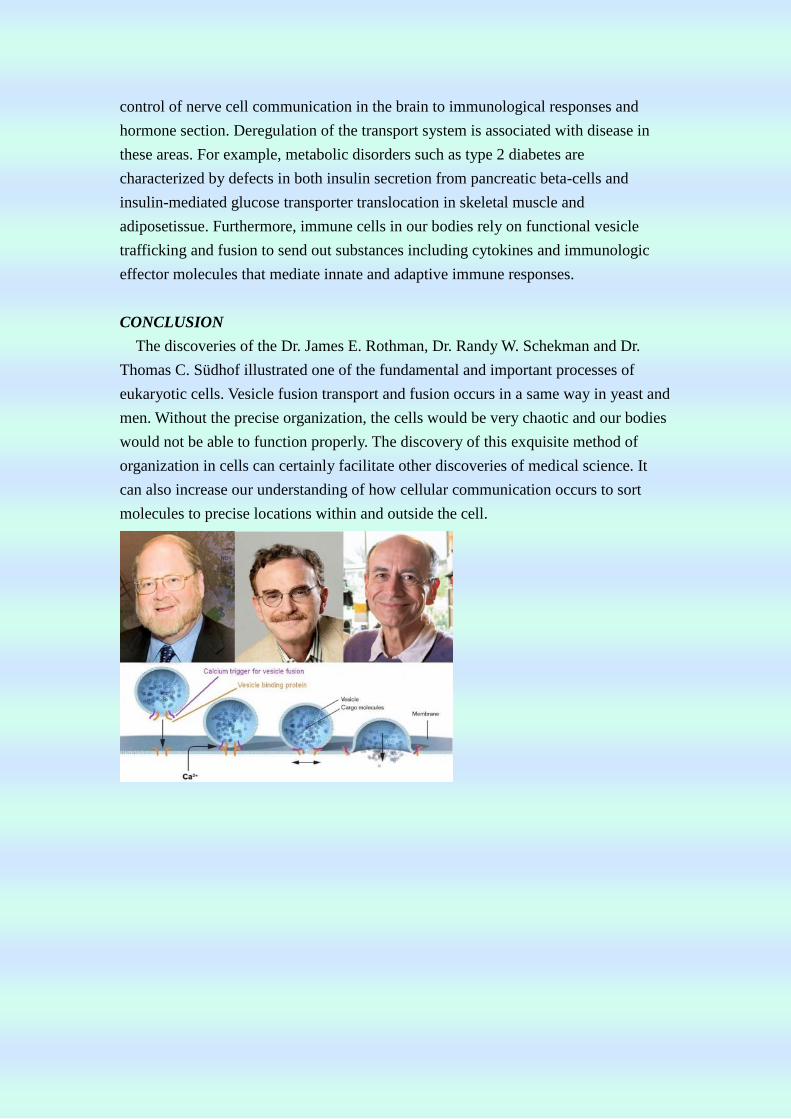
The 2013 Nobel Prize in Physiology or Medicine is awarded to Dr. James E.
Rothman, Dr. Randy W. Schekman and Dr. Thomas C. Südhof for their discoveries of
machinery regulating vesicle traffic, a major transport system in our cells. Each cell in
our bodies has a complex organization where specific cellular functions are separated
into different compartments called organelles. Molecules produced in the cell are
packaged in vesicles and transported with special and temporal precision to the
correct locations within and outside the cell. This is called cellular
compartmentalization. Mysteries of cellular compartmentalization have long intrigued
scientists.
THE PROCESS OF DISCOVERY
Dr. Randy W. Schekman discovered genes encoding proteins that are key regulators
of vesicle traffic. Comparing normal with genetically mutated yeast cells in which
vesicle traffic was disturbed, he identified genes that control transport to different
compartments and to the cell surface.
Dr. James E. Rothman discovered that a protein complex enables vesicles to fuse
with their target membranes. Proteins on the vesicle bind to specific complementary
proteins on the target membrane, ensuring that the vesicle fuses at the right location
and that cargo molecules are delivered to the correct destination.
Dr. Thomas C. Südhof studied how signals are transmitted from one nerve cell to
another in the brain, and how calcium (Ca2+) controls this process. He identified the
molecular machinery that senses calcium ions and converts this information to vesicle
fusion, thereby explaining how temporal precision is achieved and how vesicles can
be released on command.
IMPORTANCE OF THE DISCOVERY
The work of Rothman, Schekman and Südhof has unraveled machinery that is
essential for routing of cargo in cells in organisms as distantly related as yeast and
man. These discoveries have had a major impact on our understanding of how
molecules are correctly sorted to precise locations in cell. In the light of this, it comes
as no surprise that defects at any number of steps in the machinery controlling vesicle
transport and fusion are associated with disease.
Vesicle transport and fusion is essential for physiological processes ranging from
control of nerve cell communication in the brain to immunological responses and
hormone section. Deregulation of the transport system is associated with disease in
these areas. For example, metabolic disorders such as type 2 diabetes are
characterized by defects in both insulin secretion from pancreatic beta-cells and
insulin-mediated glucose transporter translocation in skeletal muscle and
adiposetissue. Furthermore, immune cells in our bodies rely on functional vesicle
trafficking and fusion to send out substances including cytokines and immunologic
effector molecules that mediate innate and adaptive immune responses.
CONCLUSION
The discoveries of the Dr. James E. Rothman, Dr. Randy W. Schekman and Dr.
Thomas C. Südhof illustrated one of the fundamental and important processes of
eukaryotic cells. Vesicle fusion transport and fusion occurs in a same way in yeast and
men. Without the precise organization, the cells would be very chaotic and our bodies
would not be able to function properly. The discovery of this exquisite method of
organization in cells can certainly facilitate other discoveries of medical science. It
can also increase our understanding of how cellular communication occurs to sort
molecules to precise locations within and outside the cell.
Top Related