


Languages
Pages
Legal


Berkeley Feb 2004

Outer membrane
Inter-membrane space
Inner membrane
Matrix
Cristae


ATP
ADP + Pi
H+H+
ATP hydrolysis

++--
ATP
ADP + Pi
H+ H+
Energy (redox energy, photons etc)
The proton circuit: Continuous generation
of ATP

Electrons enter the respiratory chain at a redox potential
of–300mV
Ubiquinone pool at 0mV
Cytochrome c at +250mV H2O
at +800mV
Complex I
Complex IIComplex III
Complex IV
ATP synthase
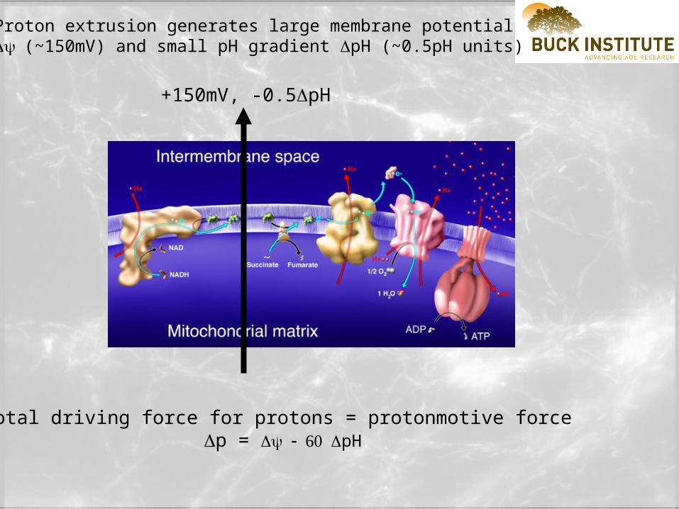
Proton extrusion generates large membrane potential (~150mV) and small pH gradient pH (~0.5pH units)
+150mV, -0.5pH
Total driving force for protons = protonmotive force p = pH

Electrical circuit
Vo
ltag
e 1
.5V
Electron current
Mitochondrion
Vo
ltag
e 0
.2V
Proton current
Mitochondrial membrane
+
_
+
_
Mitochondria work like an electrical circuit.
•The 'battery' is the respiratory chain
Mitochondrion
Vo
ltag
e 0
.2V
Proton current
Mitochondrial membrane
+
_
I III IV
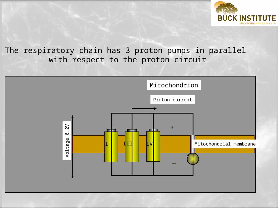
The respiratory chain has 3 proton pumps in parallel with respect to the proton circuit
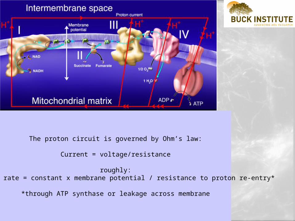
The proton circuit is governed by Ohm’s law:
Current = voltage/resistance
roughly:Respiration rate = constant x membrane potential / resistance to proton re-entry*
*through ATP synthase or leakage across membrane

m
Matrix Ca 2+
ATP generation
Reactiveoxygenspecies
NADP+
REDUCTION
Glutathionereduction
Respiratory chain

Mitochondria and ‘stress’ i.e. under which circumstances might theMitochondria’s ability to maintain a high ATP/ADP ratio and a reducing
environment fail?
A. Oxygen limitation (stroke, heart attack, near drowning)

synaptic cleft 1M
cytoplasm 10mM
synaptic vesicles 100mM
'Classic EM images from the lab of John Heuser (Washington University)'
GLUTAMATE COMPARTMENTATION

'Classic EM images from the lab of John Heuser (Washington University)'
GLUTAMATE EXCITOTOXICITY
1. (BIOENERGETIC DEFICIT RESULTING FROM OXYGEN
DEPROIVATION CAUSES ATP COLLAPSE, FAILURE OF
PLASMA MEMBRANE SODIUM PUMPS AND
MASSIVE GLUTAMATE RELEASE)

'Classic EM images from the lab of John Heuser (Washington University)'
GLUTAMATE EXCITOTOXICITY
2. POST-SYNAPTIC NMDA RECEPTORS
PATHOLOGICALLY ACTIVATED

'Classic EM images from the lab of John Heuser (Washington University)'
GLUTAMATE EXCITOTOXICITY
3. MASSIVE Ca 2+ ENTRY AND ACCUMULATION BY
MITOCHONDRIA

'Classic EM images from the lab of John Heuser (Washington University)'
GLUTAMATE EXCITOTOXICITY
4. MITOCHONDRIAL Ca 2+ LOADING CAN INITIATE DELAYED CELL DEATH

Mitochondria and ‘stress’ i.e. under which circumstances might theMitochondria’s ability to maintain a high ATP/ADP ratio and a reducing
environment fail?
B. Respiratory chain restriction

I
II
III IVQ
Rotenone
Malonate
Some specific mitochondrial inhibitors

Langston JW, Ballard P, Tetrud JW, Irwin I (1983) Chronic Parkinsonism in humans due to a product of meperidine-analog synthesis. Science 219: 979-980.
Four persons developed marked parkinsonism after using an illicit drug intravenously. Analysis of the substance injected by two of these patients revealed primarily 1-methyl-4-phenyl-1,2,5,6-tetrahydropyridine (MPTP) with trace amounts of 1-methyl-4-phenyl-4-propionoxy-piperidine (MPP+). it is proposed that this chemical selectively damages cells in the substantia nigra
Nicklas WJ, Vyas I, Heikkila RE (1985) Inhibition of NADH-linked oxidation in brain mitochondria by 1-methyl-4-phenyl-pyridine, a metabolite of the neurotoxin, 1-methyl-4-phenyl-1,2,5,6-tetrahydropyridine. Life Sci 36: 2503-2508. (MPP+), a major metabolite of the neurotoxin, (MPTP) inhibited the oxidation of NADH-linked substrates by brain mitochondrial preparations. Compromise of mitochondrial oxidative capacity by MPP+ could be an important factor in mechanisms underlying the toxicity of MPTP

Betarbet R, Sherer TB, MacKenzie G, Garcia-Osuna M, Panov AV, Greenamyre JT (2000) Chronic systemic pesticide exposure reproduces features of Parkinson's disease. Nat Neurosci 3: 1301-1306.
chronic, systemic inhibition of complex I by the lipophilic pesticide, rotenone, causes highly selective nigrostriatal dopaminergic degeneration. These results indicate that chronic exposure to a common pesticide can reproduce the anatomical, neurochemical, behavioral and neuropathological features of PD.

Beal MF, Brouillet E, Jenkins B, Henshaw R, Rosen B, Hyman BT (1993) Age-dependent striatal excitotoxic lesions produced by the endogenous mitochondrial inhibitor malonate. J Neurochem 61: 1147-1150.
Abstract: Intrastriatal injection of malonate, a reversible inhibitor of succinate dehydrogenase (SDH), produced age dependent striatal lesions… The results strengthen the possibility that a subtle impairment of energy metabolism may play a role in the pathogenesis of Huntington's disease.

Mitochondria and ‘stress’ i.e. under which circumstances might theMitochondria’s ability to maintain a high ATP/ADP ratio and a reducing
environment fail?
C. Ca2+ overload of mitochondria and activation of the permeability transition

extr
a-m
itoch
ondr
ial f
ree
C
a c
onc
ent
ratio
n (
M)
2+
mitochondria
Ca2+
Ca2+
Ca2+
'set-point'
Ca 2+
Permeability transition

Permeability transition leads to matrix swelling, unfolding of inner membrane, bursting of outer membrane and release of cytochrome
c

Mitochondria and ‘stress’ i.e. under which circumstances might theMitochondria’s ability to maintain a high ATP/ADP ratio and a reducing
environment fail?
D. Mitochondria and pro-apoptotic stress

PlasmaMembrane
OuterMitochondrial
Membrane
InnerMitochondrial
Membrane
PutativeBAx/Bid channel
CQ
9-05
Deathreceptor
Procaspase-8 Caspase-8
Bid t-Bid
Bax
Bax*
C
Apaf-1+
Procaspase-9
Caspase-9
Activation of downstreameffector caspases
e.g. caspase-3
Bcl-2(-)(+)

Ca 2+
Reactive Oxygen Species
Cell Death
Apoptoticsignal
Cytochrome c
Caspase activation
Cell Death
Apoptosis Necrosis

Mitochondria and ‘stress’ i.e. under which circumstances might theMitochondria’s ability to maintain a high ATP/ADP ratio and a reducing
environment fail?
E. Mitochondria and oxidative stress

superoxide
superoxide
Reactive oxygen species (ROS): superoxide anion produced by complexes I and III (increased at high membrane potential)
O2 + e- = O2.-
cf:
O2 + 4e- + 4H+ = 2H2O
O2.-
H2O2
Superoxide dismutases; SOD1, CuZn, cytoplasmic
SOD2, Mn, mitochondrial matrix
SOD
Glutathione peroxidase
H2O
GSH GSSG
NADPH NADP+
NADH NAD+
Glutathione reductase
transhydrogenase
NO
ONOO-

Mitochondria and ‘stress’ i.e. under which circumstances might the mitochondria’s ability to maintain a high ATP/ADP ratio and a
reducing environment fail?
F. Defects in the mitochondrial genome

cyt b
ND5
16s rRNA
12s rRNA
ND6
LHON 1448
4C
LHON 14459A
O (origin)H
ND4
ND4LND3
CO
XIIIATPase 6
ATP
ase 8
CO
XII
COXI
ND2
ND1MELAS 3243G
LHON 11778A
NA
RP 8993G
/C
ME
RR
F 8344G
LHON 3460A
common d
eletio
n
O (origin)L
9-06
Heavy
chain
Light
chain

Table 9.1 some mitochondrial mutations
location
Pearson's syndrome, Kearns-Sayresyndrome, chronic progressiveexternal ophthalmoplegia (CPEO)
‘Common deletion’ of4977 base pairsbetween A8 and ND5.
All mt protein synthesisabolished due to lack of tRNAs
MELAS (mitochondrialencephalomyopathy, lactic acidosisand stroke-like episodes).
tRNAleu(UUR)
All mt protein synthesisabolished due to lack of tRNAs
MERRF (myoclonus, epilepsy,with ragged-red fibers)
tRNAlys All mt protein synthesisabolished due to lack of tRNAs
Leber’s hereditary opticneuropathy (LHON),
6 point mutations inComplex I ND genes
loss of Complex I activity
NARP (neuropathy, ataxia andretinitis pigmentosa).
A6 Inhibition of ATP synthase

Schapira AHV (1998) Mitochondrial dysfunction in neurodegenerative disorders. Biochim Biophys Acta Bio-Energetics 1366: 225-233
Mutations of mitochondrial DNA (mtDNA) are associated with a wide spectrum of disorders encompassing the myopathies, encephalopathies and cardiomyopathies, in addition to organ specific presentations such as diabetes mellitus and deafness. Parkinson's disease , Huntington's disease , Friedreich's ataxia. In any event, mitochondria present an important target for future strategies for 'neuroprotection' to prevent or retard neurodegeneration.

•Paperback: 288 pages •Publisher: Academic Press; ISBN: 0125181213; •$49.95

Top Related