


Languages
Pages
Legal


Basic Illuminating Light Paths and Proper Microscope
AlignmentE. D. Salmon
University of North Carolina at Chapel Hill
Today
• Answers to last lecture’s questions
• Light Paths for Trans illumination
• Koehler Illumination
• Action of Field and Condenser Diaphragms
• Conjugate Planes in Properly Aligned Microscope
• Major components of Research Microscope

Homework
1.
A beam of light in glass hits a surface at an angle. At what angle does the light just become total internally reflected if the glass has a refractive Index of 1.52 and the interface has a refractive index of :
a. Airb. Waterc. Immersion oil
In each case, what is the numerical aperture (NA) of the beam relative to the normal to the interface?
Snell' Law: n1 sin(q1) = n2sin (q2); q2 = 90 degrees, sin (90) = 1; n1sin(q1) = n2*1; q1 = sin-1(n1/n2)
n(glass) = 1.52n2 angle
air 1 41.1381water 1.33 61.04289oil 1.515 85.34851
q1 90
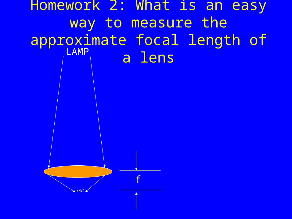
Homework 2: What is an easy way to measure the approximate focal length of
a lens
f
LAMP
LAMP
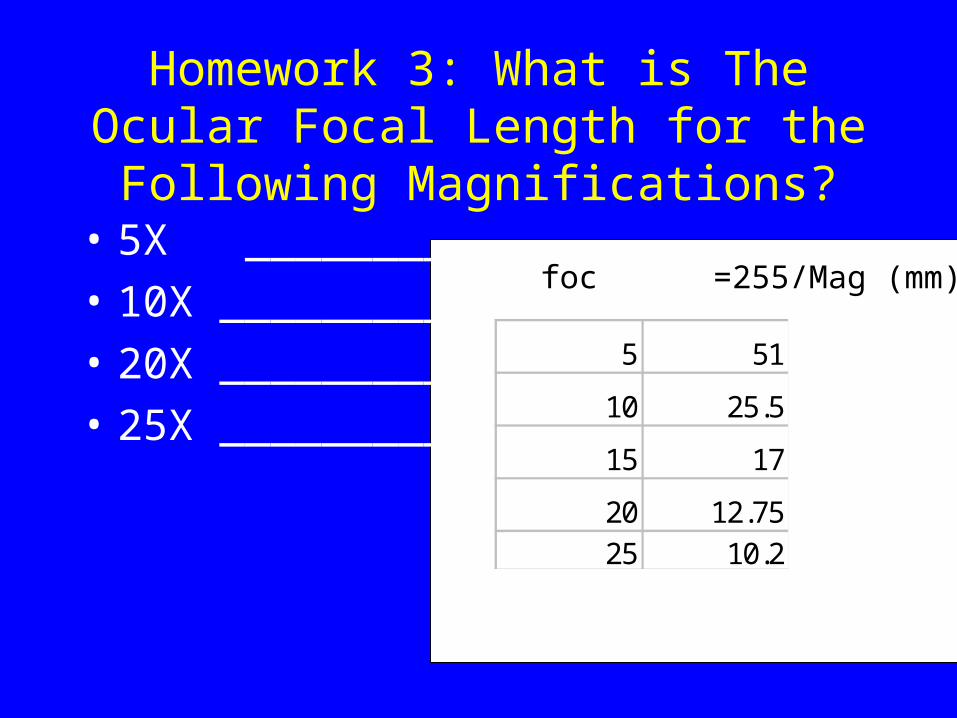
Homework 3: What is The Ocular Focal Length for the Following
Magnifications?• 5X _________
• 10X _________
• 20X _________
• 25X _________
5 51
10 25.5
15 17
20 12.7525 10.2
foc =255/Mag (mm)
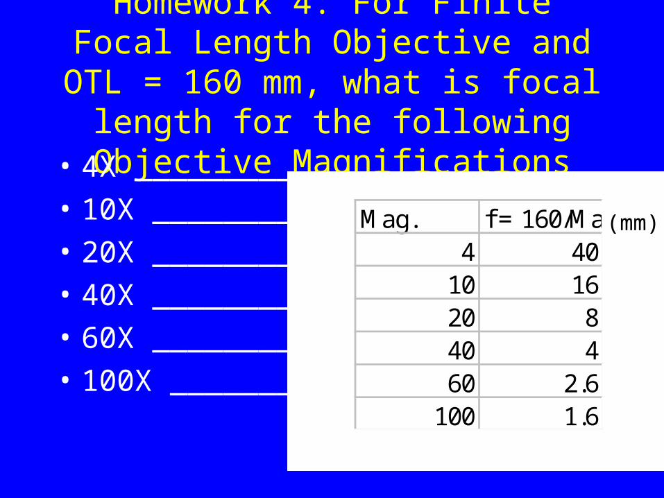
Homework 4: For Finite Focal Length Objective and OTL = 160 mm, what is focal length for the following Objective
Magnifications• 4X _________
• 10X ________
• 20X ________
• 40X ________
• 60X ________
• 100X _______
Mag. f = 160/Mag4 40
10 1620 840 460 2.6
100 1.6
(mm)

A Lamp Collector Lens and Microscope Condenser Lens are Used to Concentrate Light
on the Specimen
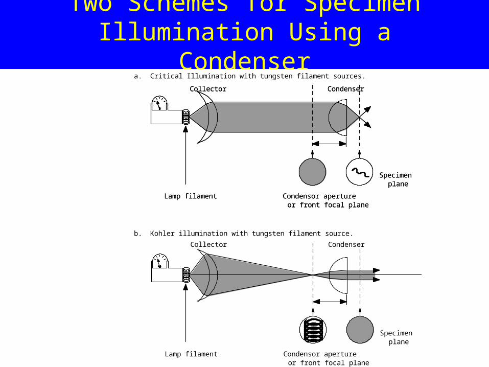
Two Schemes for Specimen Illumination Using a Condenser
a. Critical Illumination with tungsten filament sources.
b. Kohler illumination with tungsten filament source.
Condensor apertureor front focal plane
Specimenplane
Lamp filament
Collector Condenser
Condensor apertureor front focal plane
Specimenplane
Lamp filament
Collector Condenser
Condensor apertureor front focal plane
Specimenplane
Lamp filament
Collector Condenser

Critical Illumination
a. Critical Illumination with tungsten filament sources.
b. Kohler illumination with tungsten filament source.
Condensor apertureor front focal plane
Specimenplane
Lamp filament
Collector Condenser
Condensor apertureor front focal plane
Specimenplane
Lamp filament
Collector Condenser
Condensor apertureor front focal plane
Specimenplane
Lamp filament
Collector Condenser
Light source out-of-focus at condenser aperture and in-focus at specimen.Produces bright, but un-even illumination of specimen.
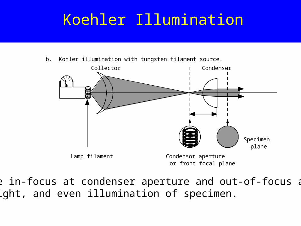
Koehler Illumination
Light source in-focus at condenser aperture and out-of-focus at specimen.Produces bright, and even illumination of specimen.
a. Critical Illumination with tungsten filament sources.
b. Kohler illumination with tungsten filament source.
Condensor apertureor front focal plane
Specimenplane
Lamp filament
Collector Condenser
Condensor apertureor front focal plane
Specimenplane
Lamp filament
Collector Condenser
Condensor apertureor front focal plane
Specimenplane
Lamp filament
Collector Condenser
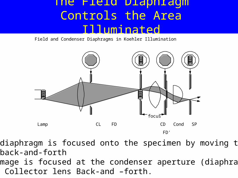
The Field Diaphragm Controls the Area Illuminated
The field diaphragm is focused onto the specimen by moving the condenser back-and-forthThe lamp image is focused at the condenser aperture (diaphragm) by moving the Collector lens Back-and –forth.
Lamp CL FD CD Cond SP OBJ OBA
FD' CD'
focus
Field and Condenser Diaphragms in Koehler Illumination

The Condenser Diaphragm Controls the Illumination NA
An image of the Condenser Diaphragm is in-focus in the Objective Back Focal Plan (Aperture). As the condenser diaphragm is opened, the illumination NA increases without changing the area of specimenIlluminated (area controlled by Field Diaphragm).
Condenser and Objective Apertures
obm
Qcond
CD Cond SP OBJ OB FFP BFP

Summary of Koehler Illumination
• The lamp filament and collector lens must both be centered on the same optical axis in the lamp housing.
• The collector lens is used to project an image of the lamp centered and in-focus at the condenser diaphragm.
• The condenser lens is used to project an image of the field diaphragm centered and in-focus on the specimen.

Summary of Koehler Illumination (cont.)
• A telescope is used to view the objective back back focal plane (aperture) in order to:
a) Adjust the opening of the condenser diaphragm so that the diameter of its image is equal to or slightly less than the diameter of the objective aperture;
b) Adjust the focus and x-y position of the lamp image so it is centered and in-focus at the objective back aperture
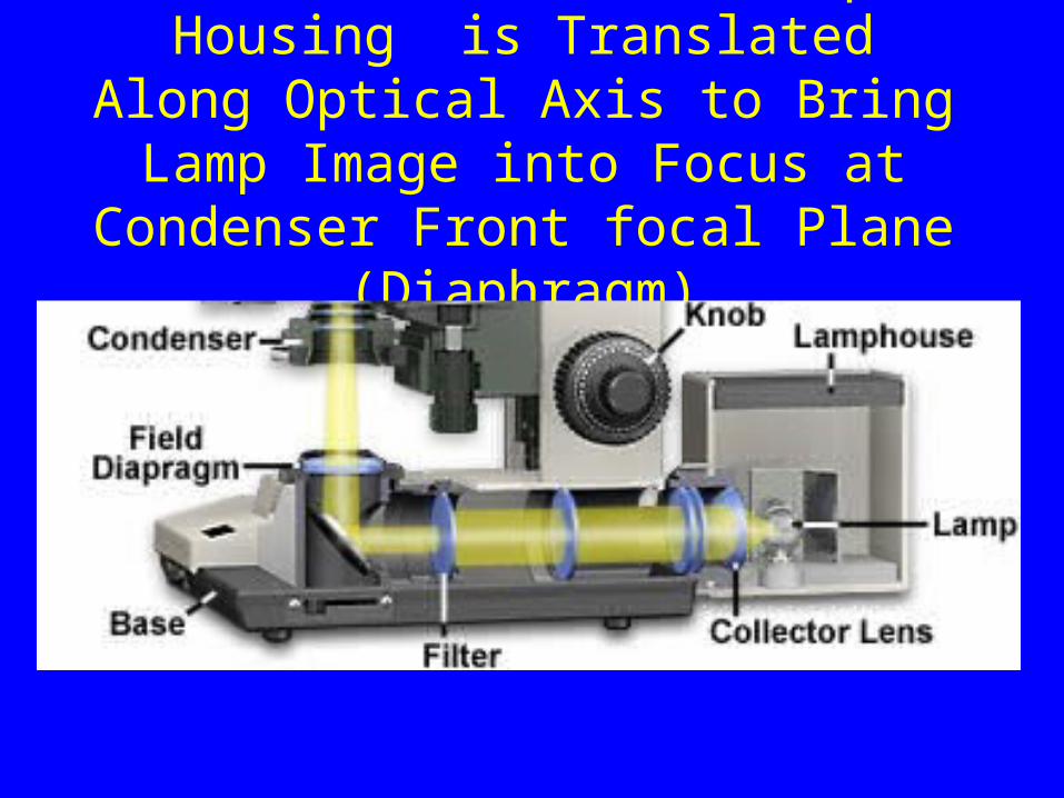
Collector Lens in Lamp Housing is Translated Along Optical Axis to Bring Lamp Image into Focus at Condenser
Front focal Plane (Diaphragm)
Condenser focus
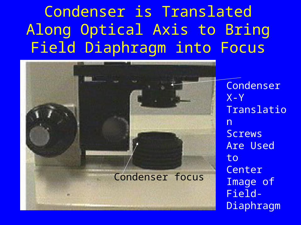
Condenser is Translated Along Optical Axis to Bring Field Diaphragm into
Focus
Condenser focus
CondenserX-Y TranslationScrewsAre Used toCenter Image of Field-Diaphragm
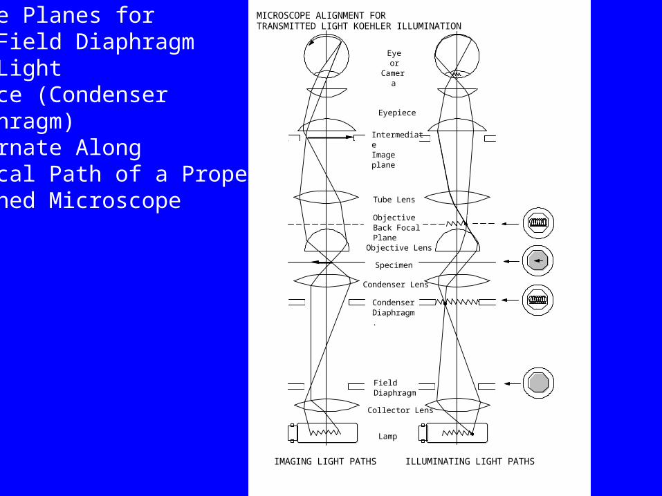
IMAGING LIGHT PATHS ILLUMINATING LIGHT PATHS
Eyeor
Camera
Eyepiece
IntermediateImage plane
Objective Lens
Specimen
Condenser Lens
CondenserDiaphragm.
FieldDiaphragm
Collector Lens
Lamp
Objective Back FocalPlane
Tube Lens
MICROSCOPE ALIGNMENT FORTRANSMITTED LIGHT KOEHLER ILLUMINATIONImage Planes for
The Field DiaphragmAnd Light Source (Condenser Diaphragm)Alternate AlongOptical Path of a ProperlyAligned Microscope

The Light Path is Extended or Re-Directed Using Projection Lenses,
Mirrors and Prisms and Beam-Switches
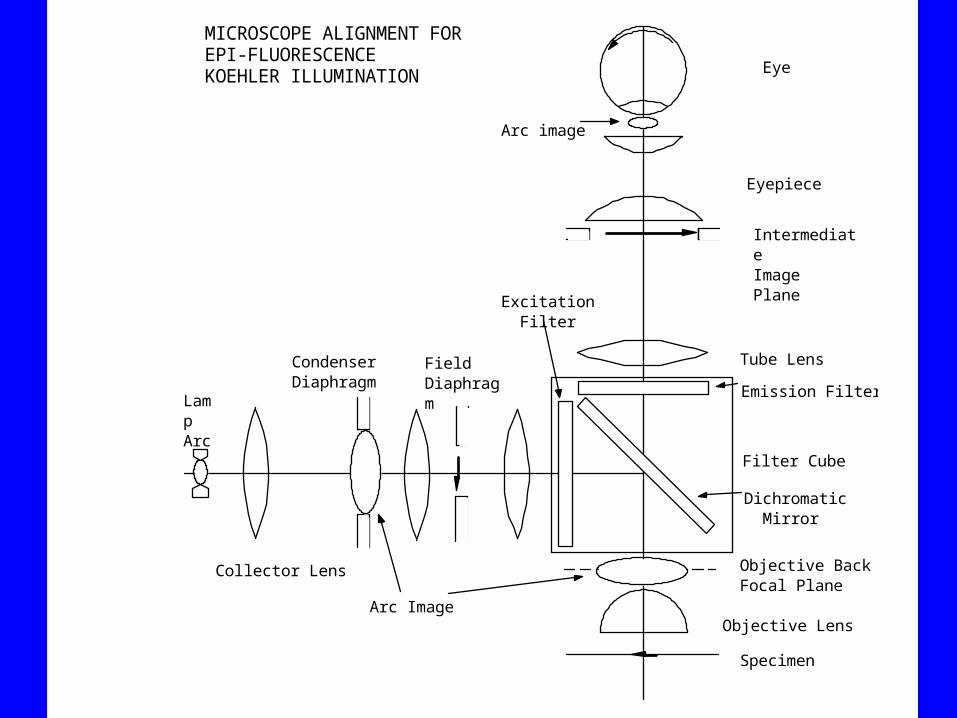
Objective Lens
Specimen
Objective BackFocal Plane
Eye
Eyepiece
Tube Lens
IntermediateImage Plane
Emission Filter
Filter Cube
Dichromatic Mirror
Excitation Filter
LampArc
Arc Image
Condenser Diaphragm
FieldDiaphragm
Arc image
MICROSCOPE ALIGNMENT FOR EPI-FLUORESCENCE KOEHLER ILLUMINATION
Collector Lens
Homework Problem 5
The light source is a 3-mm square tungsten filament. The design
of the illumination system requires that (1) the filament be 300 mm away from the condenser diaphragm, (2) the image of the filament must be in focus at the condenser diaphragm and (3) the filament must be 15-mm square to fill the condenser aperture with light. Assuming the lamp collector lens is an ideal thin lens, determine the focal length, and theposition of the collector lens between the lamp filament and the condenser diaphragm.

Homework Problem 6
A field diaphragm or iris is placed in front of the collector lens as shown for the Koehler illumination system. The field iris is used to control the illuminated area of the specimen. The condenser lens is translated back and forth along the central axis until an image of the field diaphragm is in sharp focus on the specimen. When the opening of the field diaphragm is 20 mm, the image on the specimen must be 2 mm in diameter. In addition, the field diaphragm is placed 160 mm away from the condenser lens. What is the focal length of the condenser needed to meet these requirements?
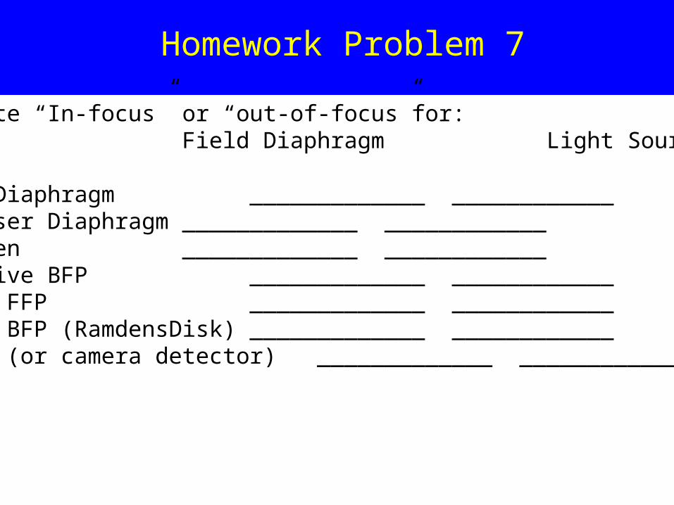
Homework Problem 7
Indicate “In-focus” or “out-of-focus”for:Field Diaphragm Light Source
at:Field Diaphragm _____________ ____________Condenser Diaphragm _____________ ____________Specimen _____________ ____________Objective BFP _____________ ____________Ocular FFP _____________ ____________Ocular BFP (RamdensDisk) _____________ ____________Retina (or camera detector) _____________ ____________

Homework 8
Work through the Microscope Illumination Section under Microscope Anatomy at: http://micro.magnet.fsu.edu/primer/index.html

Camera
Camera AdapterBinocular
Eyepiece
Beam Switch
Filter Cube ChangerSlot for Analyzer
Slot for DIC Prism
Objective Nosepiece
Objective
Stage
Condenser: Diaphragm&Turret Centering Focus
Field Diaphragm
Coarse/Fine Specimen Focus
Filtersand Diffuser
Lamp: Focus, Centering
Mirror:Focus andCentering
Mirror:Focus andCentering
Focus, Centering
Trans-Lamp Housing
Epi-Lamp HousingEpi-Field Diaphragm
Epi-CondenserDiaphragm
ShutterFilters& Centering
Slot for Polarizer
Upright Microscope Stand
Body Tube
MICROSCOPE COMPONENTS
Magnification Changer
Homework Problem 9:
IdentifyMajor ComponentsAndTheir LocationsAnd FunctionsWithinModern Research LightMicroscope(See Salmon And Canman,2000, CurrentProtocols in CellBiology, 4.1)
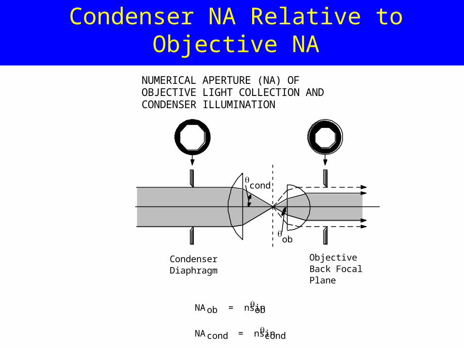
Condenser NA Relative to Objective NA
ob
cond
NUMERICAL APERTURE (NA) OF OBJECTIVE LIGHT COLLECTION AND CONDENSER ILLUMINATION
CondenserDiaphragm
Objective Back Focal Plane
NAob = nsinob
NAcond = nsincond
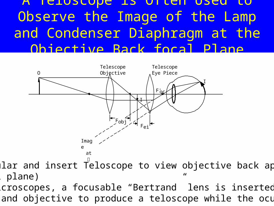
A Teloscope is Often Used to Observe the Image of the Lamp and Condenser Diaphragm at
the Objective Back focal Plane
-Remove Ocular and insert Teloscope to view objective back aperture(back focal plane) -In some microscopes, a focusable “Bertrand” lens is inserted between the ocular and objective to produce a teloscope while the ocular
I'
Image
at
I
O
F'oc
Telescope TelescopeObjective Eye Piece
FeiFobj
Top Related