


Languages
Pages
Legal

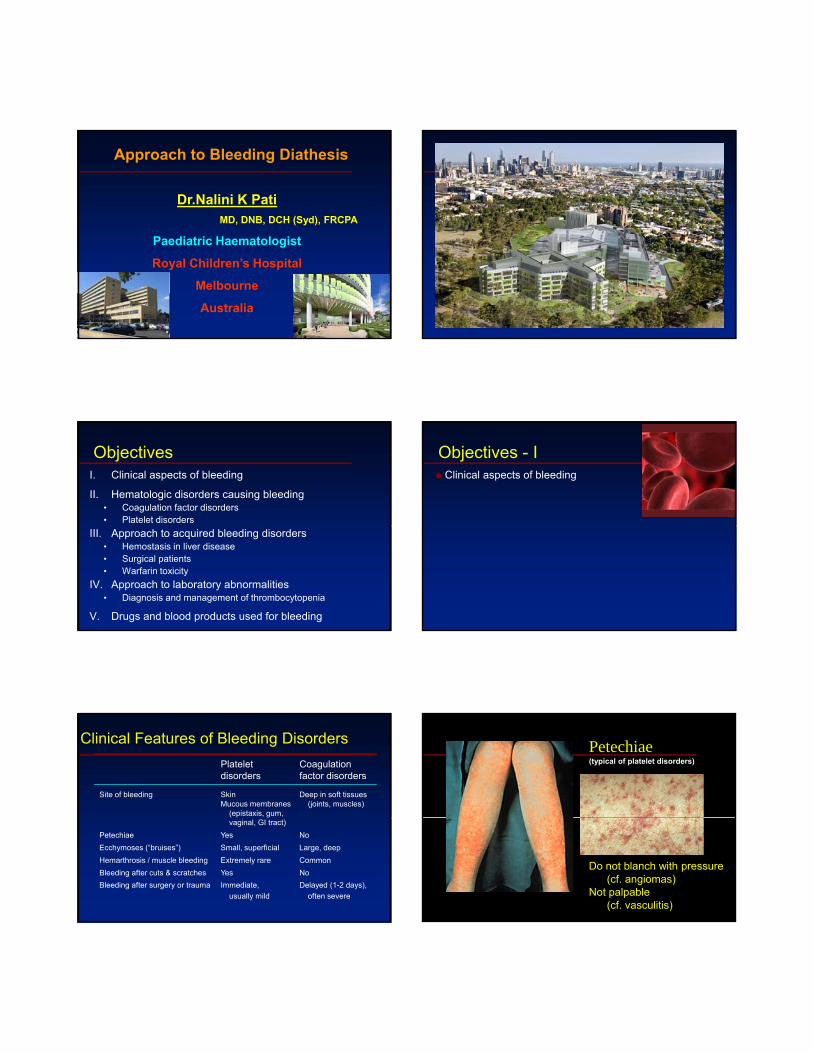
Dr.Nalini K PatiMD, DNB, DCH (Syd), FRCPA
Approach to Bleeding Diathesis
Paediatric Haematologist
Royal Children’s Hospital
Melbourne
Australia
ObjectivesI. Clinical aspects of bleeding
II. Hematologic disorders causing bleeding• Coagulation factor disorders• Platelet disorders
III. Approach to acquired bleeding disorders• Hemostasis in liver disease• Surgical patients• Warfarin toxicity
IV. Approach to laboratory abnormalities• Diagnosis and management of thrombocytopenia
V. Drugs and blood products used for bleeding
Objectives - IClinical aspects of bleeding
Clinical Features of Bleeding DisordersPlatelet Coagulation disorders factor disorders
Site of bleeding Skin Deep in soft tissuesMucous membranes (joints, muscles)
(epistaxis, gum,vaginal, GI tract)
Petechiae Yes NoEcchymoses (“bruises”) Small, superficial Large, deepHemarthrosis / muscle bleeding Extremely rare CommonBleeding after cuts & scratches Yes NoBleeding after surgery or trauma Immediate, Delayed (1-2 days),
usually mild often severe
Petechiae(typical of platelet disorders)
Do not blanch with pressure(cf. angiomas)
Not palpable(cf. vasculitis)
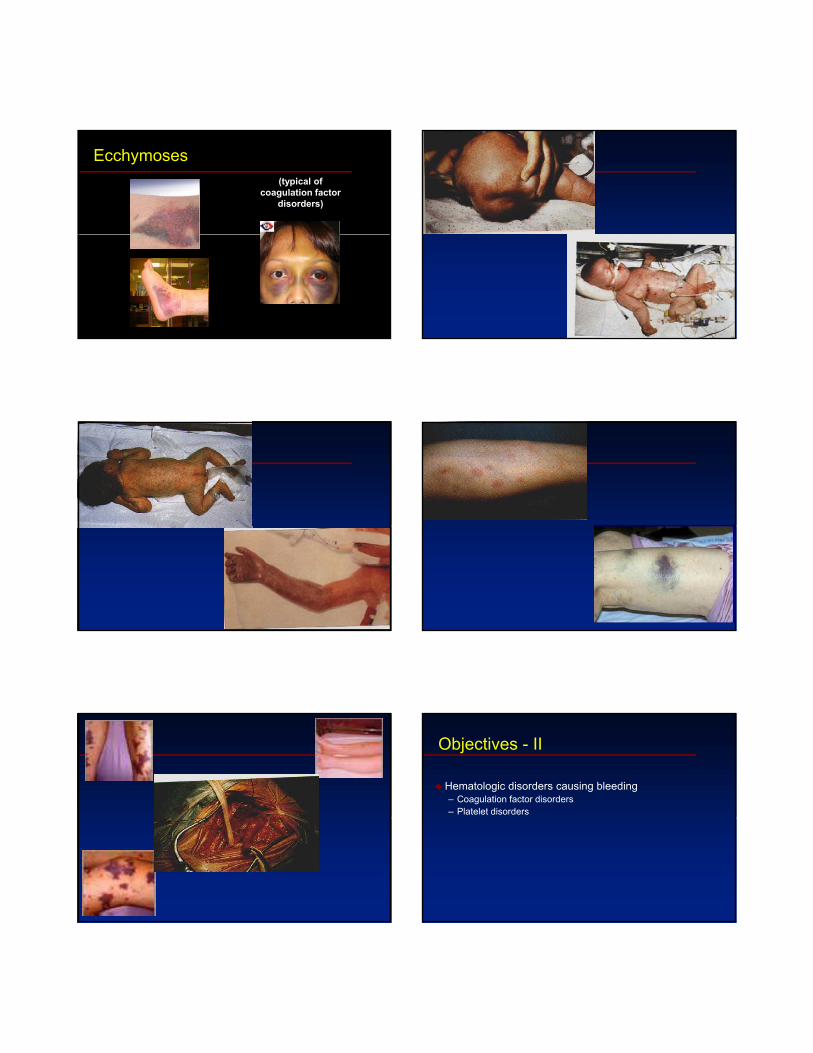
Ecchymoses(typical of
coagulation factor disorders)
Objectives - II
Hematologic disorders causing bleeding– Coagulation factor disorders– Platelet disorders
Coagulation factor disordersInherited bleeding disorders– Hemophilia A and B– vonWillebrands disease
Acquired bleeding disorders– Liver disease– Vitamin K
– Other factor deficiencies deficiency/warfarin overdose
– DIC
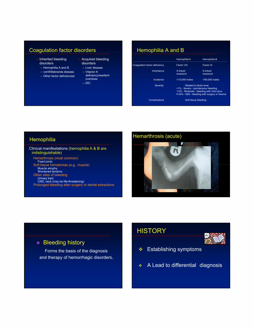
Hemophilia A and BHemophilia A Hemophilia B
Coagulation factor deficiency Factor VIII Factor IX
Inheritance X-linked X-linkedi irecessive recessive
Incidence 1/10,000 males 1/50,000 males
Severity Related to factor level<1% - Severe - spontaneous bleeding1-5% - Moderate - bleeding with mild injury5-25% - Mild - bleeding with surgery or trauma
Complications Soft tissue bleeding
HemophiliaClinical manifestations (hemophilia A & B are
indistinguishable)Hemarthrosis (most common)
Fixed jointsSoft tissue hematomas (e g muscle)Soft tissue hematomas (e.g., muscle)
Muscle atrophyShortened tendons
Other sites of bleedingUrinary tractCNS, neck (may be life-threatening)
Prolonged bleeding after surgery or dental extractions
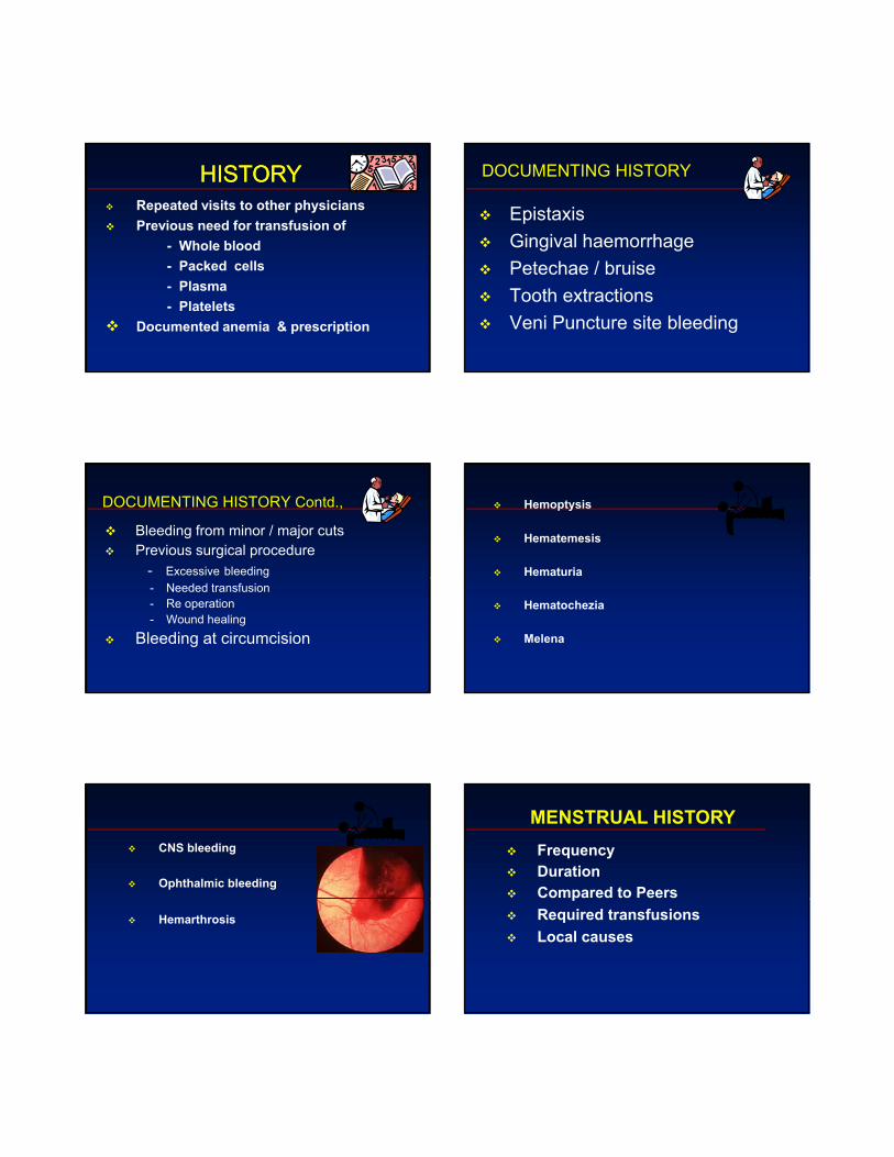
Hemarthrosis (acute)
Bleeding history Forms the basis of the diagnosis
and therapy of hemorrhagic disorders.
HISTORY
Establishing symptoms
A Lead to differential diagnosis
Repeated visits to other physiciansPrevious need for transfusion of
- Whole blood
HISTORYHISTORY
- Packed cells- Plasma- Platelets
Documented anemia & prescription
DOCUMENTING HISTORY
EpistaxisGingival haemorrhagePetechae / bruiseTooth extractionsVeni Puncture site bleeding
DOCUMENTING HISTORY Contd.,
Bleeding from minor / major cutsPrevious surgical procedure
- Excessive bleeding- Needed transfusion- Re operation- Wound healing
Bleeding at circumcision
Hemoptysis
Hematemesis
Hematuria
Hematochezia
Melena
CNS bleeding
Ophthalmic bleeding
Hemarthrosis
FrequencyDuration Compared to Peers
MENSTRUAL HISTORY
pRequired transfusionsLocal causes

PregnanciesSpontaneous / Induced abortionsEstimated blood lossDocumented anaemia
CHILD BIRTH
Required . Transfusion
. D & C
. Hysterectomy
. Iron therapy
Neonatal history
Medicines
Dietary history
Family history / pedigree chart
EXAMINATION
Age
Sex
General Examination
Systemic examination
THROMBOCYTOPENIA
Congenital. Is the history since birth
I bl di th t. Is bleeding worse than count. Is there a family history
. Are there any physicalcongenital abnormalities
THROMBOCYTOPENIA Contd.,
Other differential diagnosis must be considered- Additional laboratory abnormalities
WBC - abnormal cells, neutropeniaRBC - Macrocytosis, Fragmentation
Are there additional clinical feat res- Are there additional clinical featuresLymphadenopathy, splenomegalyBone pain, LimpingSick child
*In the presence of above findings consider
marrow examination
Underlying disorders - SLE
. Auto antibody screen - ANF. Auto antibody screen ANF and ds DNA
Anti-phospholipid syndromeHIV infection
Treatment of hemophilia A
Intermediate purity plasma products– Virucidally treated– May contain von Willebrand factor
High purity (monoclonal) plasma productsHigh purity (monoclonal) plasma products– Virucidally treated– No functional von Willebrand factor
Recombinant factor VIII– Virus free/No apparent risk– No functional von Willebrand factor
Dosing guidelines for hemophilia AMild bleeding– Target: 30% dosing q8-12h; 1-2 days (15U/kg)– Hemarthrosis, oropharyngeal or dental, epistaxis, hematuria
Major bleeding– Target: 80-100% q8-12h; 7-14 days (50U/kg)– CNS trauma, hemorrhage, lumbar puncture– Surgery– Retroperitoneal hemorrhage– GI bleeding
Adjunctive therapy– ε-aminocaproic acid (Amicar) or DDAVP (for mild disease only)
Complications of therapyFormation of inhibitors (antibodies)– 10-15% of severe hemophilia A patients– 1-2% of severe hemophilia B patients
Viral infections– Hepatitis B Human parvovirus– Hepatitis C Hepatitis A– HIV Other
Viral infections in hemophiliacs
HIV -positive HIV-negative(n=382) (n=345)
53% 47%Hepatitis serology % positive % negativeHepatitis serology % positive % negative
Negative 1 20Hepatitis B virus only 1 1Hepatitis C virus only 24 45Hepatitis B and C 74 34
Blood 1993:81;412-418
Treatment of hemophilia B
Agent – High purity factor IX– Recombinant human factor IX
Dose– Initial dose: 100U/kg– Subsequent: 50U/kg every 24 hours
von Willebrand Disease: Clinical Features
von Willebrand factor– Synthesis in endothelium and megakaryocytes– Forms large multimer – Carrier of factor VIII
A h l l b d h li– Anchors platelets to subendothelium– Bridge between platelets
Inheritance - autosomal dominantIncidence - 1/10,000Clinical features - mucocutaneous bleeding
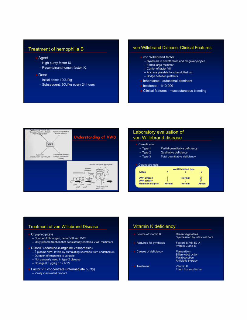
Understanding of VWDLaboratory evaluation of von Willebrand disease
Classification– Type 1 Partial quantitative deficiency– Type 2 Qualitative deficiency– Type 3 Total quantitative deficiency
Diagnostic tests:vonWillebrand type
Assay 1 2 3
vWF antigen ⇓ Normal ⇓⇓vWF activity ⇓ ⇓ ⇓⇓Multimer analysis Normal Normal Absent
Treatment of von Willebrand Disease
Cryoprecipitate– Source of fibrinogen, factor VIII and VWF– Only plasma fraction that consistently contains VWF multimers
DDAVP (deamino-8-arginine vasopressin)DDAVP (deamino 8 arginine vasopressin)– ↑ plasma VWF levels by stimulating secretion from endothelium– Duration of response is variable– Not generally used in type 2 disease– Dosage 0.3 µg/kg q 12 hr IV
Factor VIII concentrate (Intermediate purity)– Virally inactivated product
Vitamin K deficiencySource of vitamin K Green vegetables
Synthesized by intestinal flora
Required for synthesis Factors II, VII, IX ,XProtein C and S
Causes of deficiency MalnutritionBiliary obstructionMalabsorptionAntibiotic therapy
Treatment Vitamin KFresh frozen plasma
Common clinical conditions associated withDisseminated Intravascular Coagulation
Sepsis
Trauma
Obstetrical complications– Amniotic fluid embolism– Abruptio placentae
Activation of both coagulation and fibrinolysisTriggered by
– Head injury– Fat embolism
Malignancy
b up o p ace ae
Vascular disorders
Reaction to toxin (e.g. snake venom, drugs)
Immunologic disorders– Severe allergic reaction– Transplant rejection
Disseminated Intravascular Coagulation (DIC)Mechanism
Systemic activationof coagulation
Intravasculardeposition of fibrin
Depletion of plateletsand coagulation factors
BleedingThrombosis of smalland midsize vessels
with organ failure
Pathogenesis of DIC
Coagulation Fibrinolysis
Fib i
Release of thromboplastic
material intocirculation
Consumption ofcoagulation factors;
presence of FDPs↑ aPTT↑ PT
Fibrinogen
FibrinMonomers
FibrinClot
(intravascular)
Fibrin(ogen)Degradation
Products
Plasmin
Thrombin Plasmin↑ TT
↓ Fibrinogen
Presence of plasmin↑ FDP
Intravascular clot↓ Platelets
Schistocytes
Laboratory Evaluation of DIC
1. ASSESS DEPLETION OF COAGULATIONFACTORS
2. ASSESS FIBRINOLYSIS
3. BLOOD FILM FOR MICRO-ANGIOPATHY
Laboratory Evaluation of DIC- I
• Assess Depletion Of Coagulation Factors
• PLATELET COUNT
• PROTHROMBIN TIME
• PARTIAL THROMBOPLASTIN TIME
• THROMBIN TIME
• FIBRINOGEN
Laboratory Evaluation of DIC-II
Tests Of Fibrinolysis
CLOT LYSIS
EUGLOBIN LYSIS TIME
FIBRIN PLATE LYSIS
PARA-COAGULATION
FDP
D DIMER
Laboratory Evaluation of DIC-III
• Prothrombin activation peptide [Fl.2]
• Thrombin anti-thrombin complexes
• AT III levels
These tests are not available for routine
clinical management.
Disseminated Intravascular CoagulationTreatment approaches
Treatment of underlying disorder
Anticoagulation with heparin
Platelet transfusion
Fresh frozen plasma
Coagulation inhibitor concentrate (ATIII)
Classification of platelet disorders
Quantitative disorders
– Abnormal distribution– Dilution effect
Qualitative disorders
– Inherited disorders (rare)A i d di d– Decreased production
– Increased destruction
– Acquired disorders» Medications» Chronic renal failure» Cardiopulmonary bypass
Thrombocytopenia
Immune-mediatedIdioapthicDrug-inducedDrug inducedCollagen vascular diseaseLymphoproliferative diseaseSarcoidosis
Non-immune mediatedDICMicroangiopathic hemolytic anemia
Approach to the thrombocytopenic patient
History– Is the patient bleeding?– Are there symptoms of a secondary illness? (neoplasm,
infection, autoimmune disease)Is there a history of medications alcohol use or recent– Is there a history of medications, alcohol use, or recent transfusion?
– Are there risk factors for HIV infection?– Is there a family history of thrombocytopenia?– Do the sites of bleeding suggest a platelet defect?
Assess the number and function of platelets– CBC with peripheral smear– Bleeding time or platelet aggregation study
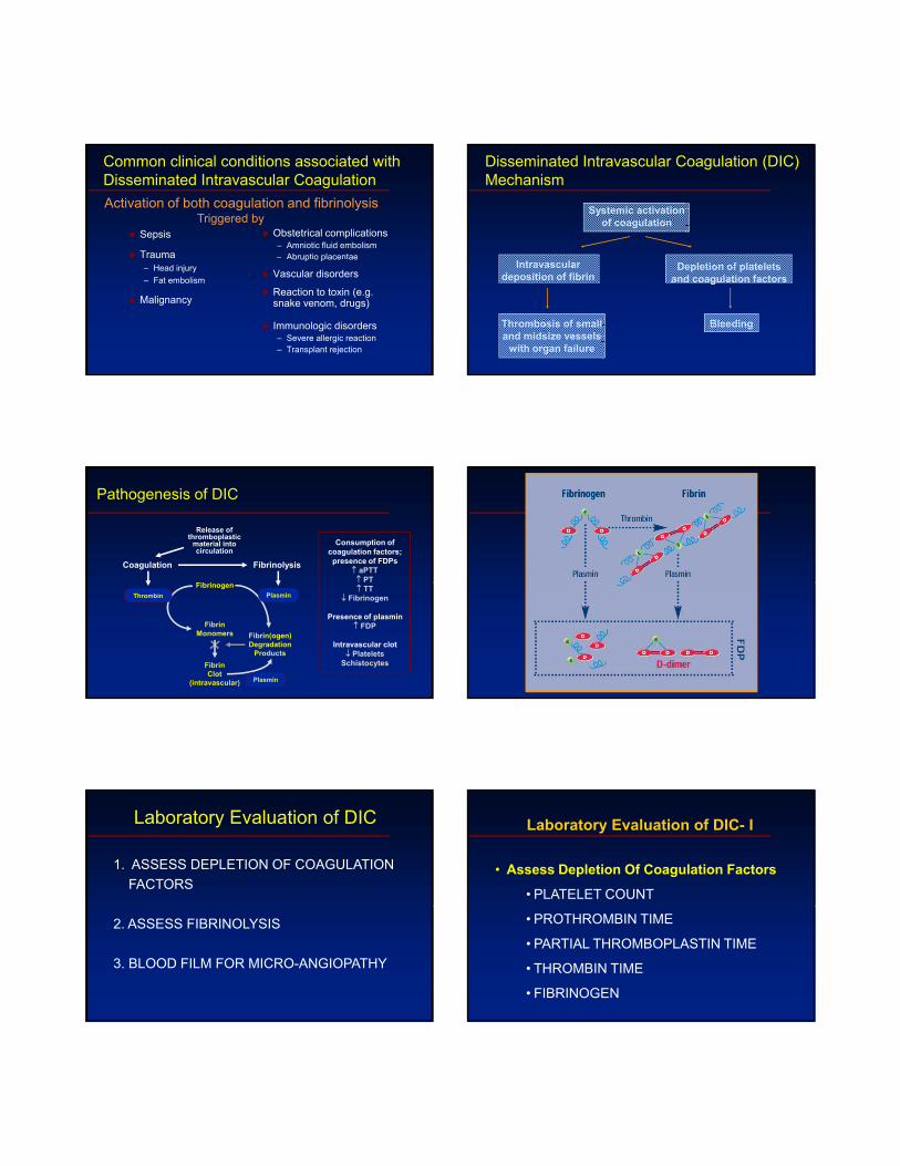
Bleeding time and bleeding5-10% of patients have a prolonged bleeding time
Most of the prolonged bleeding times are due to aspirin or drug ingestion
Prolonged bleeding time does not predict excess surgical blood loss
Not recommended for routine testing in preoperative patients
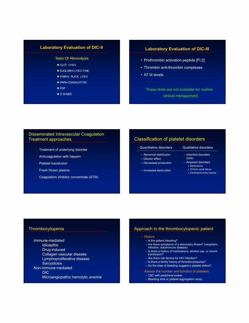
Features of Acute and Chronic ITP
Features Acute ITP Chronic ITP
Peak age Children (2-6 yrs) Adults (20-40 yrs)Female:male 1:1 3:1Antecedent infection Common RareOnset of symptoms Abrupt Abrupt-indolentPlatelet count at presentation <20,000 <50,000Duration 2-6 weeks Long-termSpontaneous remission Common Uncommon
Initial Treatment of ITP
Platelet count Symptoms Treatment(per µl)
>50,000 None
20-50,000 Not bleeding NoneBleeding Glucocorticoids
IVIG
<20,000 Not bleeding GlucocorticoidsBleeding Glucocorticoids
IVIGHospitalization
Summary of case series with ITP
Variable No./total (%)
Complete responseWith glucocorticoids 370/1447 (26%)Wi h l 81/88 (66%)With splenectomy 581/885 (66%)
Death from hemorrhage 78/1761 (4%)
Healthy at last observation 1027/1606 (64%)
George, JN. N Engl J Med: 1994;331; 1207
Long-term morbidity and mortalityin adults with ITP
134 patients with severe ITP studied for mean of 10.5 yrs
– CR and PR patients (85%)No increased mortality compared to control population» No increased mortality compared to control population
– Non-responders/maintenance therapy» Increased morbidity due to ITP-related hospitalizations » Increased mortality related equally to bleeding and infection
Portielje JE et al. Blood 2001:97;2549
Objectives - III
Approach to acquired bleeding disorders– Hemostasis in liver disease– Surgical patients
Warfarin toxicity– Warfarin toxicity
Liver Disease and Hemostasis
1. Decreased synthesis of II, VII, IX, X, XI, and fibrinogen
2. Dietary Vitamin K deficiency (Inadequate intake or malabsortion)
3. Dysfibrinogenemia4. Enhanced fibrinolysis (Decreased alpha-2-
antiplasmin)5. DIC6. Thrombocytoepnia due to hypersplenism
Management of Hemostatic Defects in Liver Disease
Treatment for prolonged PT/PTTVitamin K 10 mg SQ x 3 days - usually ineffective
Fresh-frozen plasma infusion25 30% f l l (1200 1500 l)25-30% of plasma volume (1200-1500 ml) immediate but temporary effect
Treatment for low fibrinogenCryoprecipitate (1 unit/10kg body weight)
Treatment for DIC (Elevated D-dimer, low factor VIII, thrombocytopenia
Replacement therapy
Vitamin K deficiency due to warfarin overdoseManaging high INR values
Clinical situation Guidelines
INR therapeutic-5 Lower or omit next dose;Resume therapy when INR is therapeutic
INR 5-9; no bleeding Lower or omit next dose;Resume therapy when INR is therapeutic
Omit dose and give vitamin K (1-2.5 mg po)
Rapid reversal: vitamin K 2-4 mg po (repeat)
INR >9; no bleeding Omit dose; vitamin K 3-5 mg po; repeat as necessaryResume therapy at lower dose when INR therapeutic
Chest 2001:119;22-38s (supplement)
Vitamin K deficiency due to warfarin overdoseManaging high INR values in bleeding patients
Clinical situation Guidelines
INR > 20; serious bleeding Omit warfarinVitamin K 10 mg slow IV infusionFFP or PCC (depending on urgency)R t it i K i j ti 12 h d dRepeat vitamin K injections every 12 hrs as needed
Any life-threatening bleeding Omit warfarinVitamin K 10 mg slow IV infusionPCC ( or recombinant human factor VIIa)Repeat vitamin K injections every 12 hrs as needed
Chest 2001:119;22-38s (supplement)
Approach to Post-operative bleeding
1. Is the bleeding local or due to a hemostatic failure?1. Local: Single site of bleeding usually rapid with minimal
coagulation test abnormalities2. Hemostatic failure: Multiple site or unusual pattern with
abnormal coagulation tests
2 Evaluate for causes of peri-operative hemostatic failure2. Evaluate for causes of peri operative hemostatic failure1. Preexisting abnormality2. Special cases (e.g. Cardiopulmonmary bypass)
3. Diagnosis of hemostatic failure1. Review pre-operative testing2. Obtain updated testing

Objectives - IV
Approach to laboratory abnormalities– Diagnosis and management of thrombocytopenia
Laboratory Evaluation of BleedingOverview
CBC and smear Platelet count ThrombocytopeniaRBC and platelet morphology TTP, DIC, etc.
Coagulation Prothrombin time Extrinsic/common pathwaysPartial thromboplastin time Intrinsic/common pathwaysCoagulation factor assays Specific factor deficiencies50:50 mix Inhibitors (e.g., antibodies)Fibrinogen assay Decreased fibrinogenThrombin time Qualitative/quantitative
fibrinogen defectsFDPs or D-dimer Fibrinolysis (DIC)
Platelet function von Willebrand factor vWDBleeding time In vivo test (non-specific)Platelet function analyzer (PFA) Qualitative platelet disorders
and vWDPlatelet function tests Qualitative platelet disorders
Blood Film
XIIaXIIa
Coagulation cascadeIntrinsic system Intrinsic system (surface contact)(surface contact)
XIIXII
XIXI XIa
Tissue factorTissue factor
Extrinsic system Extrinsic system (tissue damage)(tissue damage)
IIa
IXIX IXa VIIa VIIVII
VIIIVIII VIIIaVIIIa
XX
VV VaVa
IIII
FibrinogenFibrinogen FibrinFibrin
(Thrombin)(Thrombin)IIa
Vitamin K dependant factorsVitamin K dependant factorsXa
Laboratory Evaluation of the Coagulation PathwaysPartial thromboplastin time
(PTT)Prothrombin time
(PT)Surface activating agent
(Ellagic acid, kaolin)PhospholipidCalcium
ThromboplastinTissue factorPhospholipid
Calcium
Intrinsic pathway Extrinsic pathway
Common pathwayThrombin timeThrombin
Fibrin clot
Pre-analytic errorsProblems with blue-top tube– Partial fill tubes– Vacuum leak and citrate
evaporation
Biological effects– Hct ≥55 or ≤15– Lipemia, hyperbilirubinemia,
hemolysis
Problems with phlebotomy– Heparin contamination– Wrong label– Slow fill– Underfill– Vigorous shaking
Laboratory errors– Delay in testing– Prolonged incubation at 37°C– Freeze/thaw deterioration
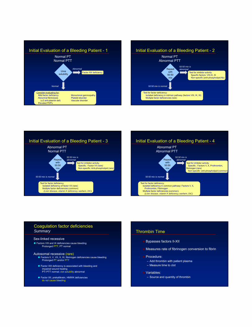
Initial Evaluation of a Bleeding Patient - 1Normal PT
Normal PTT
UreaAbnormal
Factor XIII deficiency
Consider evaluating for:Mild factor deficiency Monoclonal gammopathyAbnormal fibrinolysis Platelet disorder
(α2 anti-plasmin def) Vascular disorderElevated FDPs
solubility
Normal
Factor XIII deficiency
Initial Evaluation of a Bleeding Patient - 2Normal PT
Abnormal PTT
Repeatwith
50:50 mix is abnormal
Test for inhibitor activity:
Test for factor deficiency:Isolated deficiency in intrinsic pathway (factors VIII, IX, XI)Multiple factor deficiencies (rare)
50:50mix
50:50 mix is normal
Test for inhibitor activity:Specific factors: VIII,IX, XINon-specific (anti-phospholipid Ab)
Initial Evaluation of a Bleeding Patient - 3Abnormal PTNormal PTT
Repeatwith
50:50 mix is abnormal
Test for inhibitor activity:
Test for factor deficiency:Isolated deficiency of factor VII (rare)Multiple factor deficiencies (common)
(Liver disease, vitamin K deficiency, warfarin, DIC)
50:50mix
50:50 mix is normal
Test for inhibitor activity:Specific: Factor VII (rare)Non-specific: Anti-phospholipid (rare)
Initial Evaluation of a Bleeding Patient - 4Abnormal PT
Abnormal PTT
Repeatwith
50:50 mix is abnormal
Test for inhibitor activity:
Test for factor deficiency:Isolated deficiency in common pathway: Factors V, X,
Prothrombin, FibrinogenMultiple factor deficiencies (common)
(Liver disease, vitamin K deficiency, warfarin, DIC)
50:50mix
50:50 mix is normal
Test for inhibitor activity:Specific : Factors V, X, Prothrombin,
fibrinogen (rare)Non-specific: anti-phospholipid (common)
Coagulation factor deficienciesSummary
Sex-linked recessiveFactors VIII and IX deficiencies cause bleeding
Prolonged PTT; PT normal
Autosomal recessive (rare)Autosomal recessive (rare)Factors II, V, VII, X, XI, fibrinogen deficiencies cause bleedingProlonged PT and/or PTT
Factor XIII deficiency is associated with bleeding andimpaired wound healingPT/ PTT normal; clot solubility abnormal
Factor XII, prekallikrein, HMWK deficienciesdo not cause bleeding
Thrombin Time
Bypasses factors II-XII
Measures rate of fibrinogen conversion to fibrin
Procedure:– Add thrombin with patient plasma– Measure time to clot
Variables:– Source and quantity of thrombin
Causes of prolonged Thrombin Time
HeparinHypofibrinogenemiaDysfibrinogenemiaElevated FDPs or paraproteinThrombin inhibitors (Hirudin)Thrombin antibodies
Objectives - V
Drugs and blood products used for bleeding
Treatment Approaches tothe Bleeding Patient
Red blood cellsPlatelet transfusionsFresh frozen plasmaCryoprecipitateCryoprecipitateAmicarDDAVPRecombinant Human factor VIIa
RBC transfusion therapyIndications
Improve oxygen carrying capacity of blood
– Bleeding
– Chronic anemia that is symptomatic
– Peri-operative management
Red blood cell transfusionsSpecial preparation
CMV-negative CMV-negative patients Prevent CMV transmission
Irradiated RBCs Immune deficient recipient Prevent GVHDor direct donoror direct donor
Leukopoor Previous non-hemolytic Prevents reactiontransfusion reaction
CMV negative patients Prevents transmission
Washed RBC PNH patients Prevents hemolysisIgA deficient recipient Prevents anaphylaxis
Red blood cell transfusionsAdverse reactions
Immunologic reactions
Hemolysis RBC incompatibilityAnaphylaxis Usually unknown; rarely against IgAFebrile reaction Antibody to neutrophilsFebrile reaction Antibody to neutrophilsUrticaria Antibody to donor plasma proteinsNon-cardiogenic Donor antibody to leukocytespulmonary edema
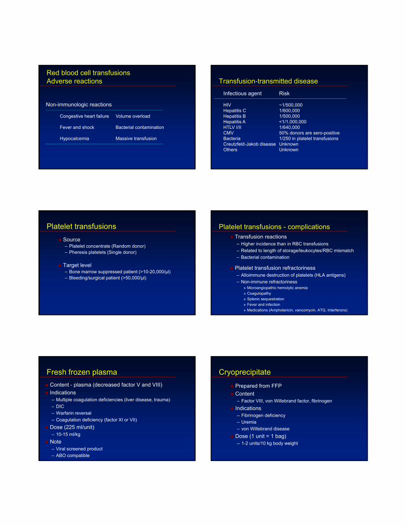
Red blood cell transfusionsAdverse reactions
Non-immunologic reactions
Congestive heart failure Volume overloadCongestive heart failure Volume overload
Fever and shock Bacterial contamination
Hypocalcemia Massive transfusion
Transfusion-transmitted disease
Infectious agent Risk
HIV ~1/500,000Hepatitis C 1/600,000Hepatitis B 1/500 000Hepatitis B 1/500,000Hepatitis A <1/1,000,000HTLV I/II 1/640,000CMV 50% donors are sero-positiveBacteria 1/250 in platelet transfusionsCreutzfeld-Jakob disease UnknownOthers Unknown
Platelet transfusionsSource– Platelet concentrate (Random donor)– Pheresis platelets (Single donor)
Target level– Bone marrow suppressed patient (>10-20,000/µl)– Bleeding/surgical patient (>50,000/µl)
Platelet transfusions - complicationsTransfusion reactions– Higher incidence than in RBC transfusions– Related to length of storage/leukocytes/RBC mismatch– Bacterial contamination
Platelet transfusion refractoriness– Alloimmune destruction of platelets (HLA antigens)– Non-immune refractoriness
» Microangiopathic hemolytic anemia» Coagulopathy» Splenic sequestration» Fever and infection» Medications (Amphotericin, vancomycin, ATG, Interferons)
Fresh frozen plasmaContent - plasma (decreased factor V and VIII)Indications– Multiple coagulation deficiencies (liver disease, trauma)– DIC– Warfarin reversal– Coagulation deficiency (factor XI or VII)
Dose (225 ml/unit)– 10-15 ml/kg
Note– Viral screened product– ABO compatible
Cryoprecipitate
Prepared from FFPContent– Factor VIII, von Willebrand factor, fibrinogen
IndicationsIndications– Fibrinogen deficiency– Uremia– von Willebrand disease
Dose (1 unit = 1 bag)– 1-2 units/10 kg body weight
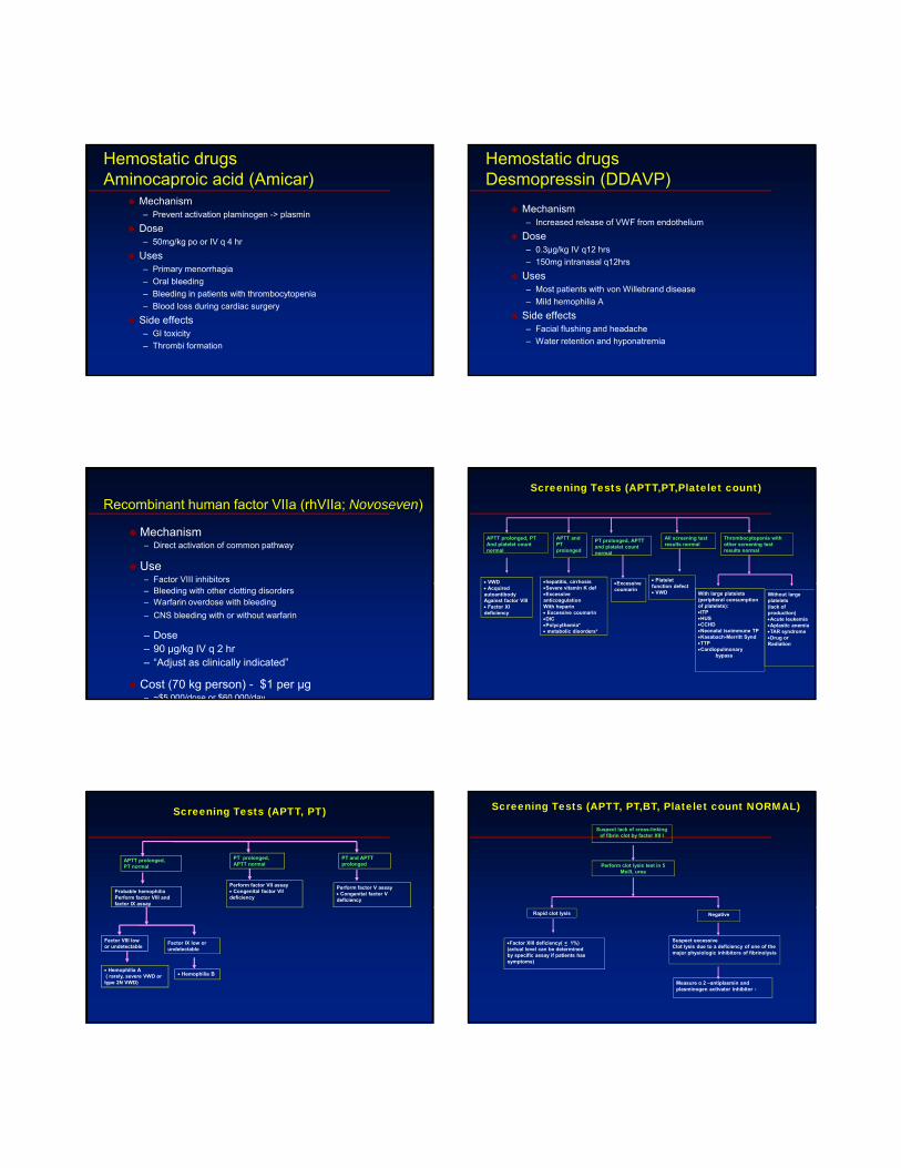
Hemostatic drugsAminocaproic acid (Amicar)
Mechanism– Prevent activation plaminogen -> plasmin
Dose– 50mg/kg po or IV q 4 hr
Uses– Primary menorrhagia– Oral bleeding– Bleeding in patients with thrombocytopenia– Blood loss during cardiac surgery
Side effects– GI toxicity– Thrombi formation
Hemostatic drugsDesmopressin (DDAVP)
Mechanism– Increased release of VWF from endothelium
Dose– 0.3µg/kg IV q12 hrs
150 i t l 12h– 150mg intranasal q12hrs
Uses– Most patients with von Willebrand disease– Mild hemophilia A
Side effects– Facial flushing and headache– Water retention and hyponatremia
Recombinant human factor VIIa (rhVIIa; Novoseven)
Mechanism– Direct activation of common pathway
Use– Factor VIII inhibitors– Bleeding with other clotting disorders– Warfarin overdose with bleeding – CNS bleeding with or without warfarin
– Dose– 90 µg/kg IV q 2 hr – “Adjust as clinically indicated”
Cost (70 kg person) - $1 per µg– ~$5,000/dose or $60,000/day
APTT prolonged, PTAnd platelet count normal
APTT and PT prolonged
PT prolonged, APTT and platelet count normal
All screening test results normal
Thrombocytopenia with other screening test results normal
• VWD •hepatitis, cirrhosis •Excessive • Platelet
Screening Tests (APTT,PT,Platelet count)
VWD• Acquired autoantibodyAgainst factor VIII• Factor XI deficiency
p ,•Severe vitamin K def•Excessive anticoagulationWith heparin• Excessive coumarin•DIC•Polycythemia*• metabolic disorders*
•Excessive coumarin function defect
• VWD With large platelets(peripheral consumption of platelets):•ITP•HUS•CCHD•Neonatal isoimmune TP •Kasabach-Merritt Synd•TTP•Cardiopulmonary
bypass
Without large platelets(lack of production)•Acute leukemia•Aplastic anemia•TAR syndrome•Drug or Radiation
APTT prolonged,PT normal
Probable hemophiliaPerform factor VIII and factor IX assay
PT prolonged,APTT normal
Perform factor VII assay• Congenital factor VII deficiency
PT and APTT prolonged
Perform factor V assay• Congenital factor V deficiency
Screening Tests (APTT, PT)
Factor VIII low or undetectable
Factor IX low or undetectable
• Hemophilia A( rarely, severe VWD or type 2N VWD)
• Hemophilia B
Suspect lack of cross-linking of fibrin clot by factor XII I
Perform clot lysis test in 5 Mol/L urea
Screening Tests (APTT, PT,BT, Platelet count NORMAL)
Rapid clot lysis Negative
•Factor XIII deficiency( < 1%)(actual level can be determinedby specific assay if patients has symptoms)
Suspect excessiveClot lysis due to a deficiency of one of the major physiologic inhibitors of fibrinolysis
Measure α 2 –antiplasmin and plasminogen activator inhibitor -
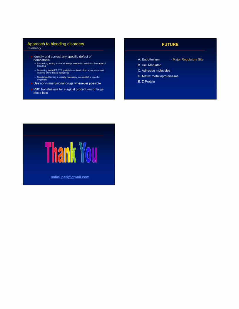
Approach to bleeding disordersSummary
Identify and correct any specific defect of hemostasis– Laboratory testing is almost always needed to establish the cause of
bleeding
– Screening tests (PT,PTT, platelet count) will often allow placement into one of the broad categories
– Specialized testing is usually necessary to establish a specific diagnosis
Use non-transfusional drugs whenever possible
RBC transfusions for surgical procedures or large blood loss
FUTURE
A. Endothelium - Major Regulatory Site
B. Cell Mediated
C. Adhesive molecules
D. Matrix metalloproteinases
E. Z-Protein
Top Related