


Languages
Pages
Legal


Apoptosis and Oncotic Necrosis:The final common pathways of liver injury
Hartmut Jaeschke
Department of Pharmacology, Toxicology & Therapeutics
University of Kansas Medical CenterKansas City, KS
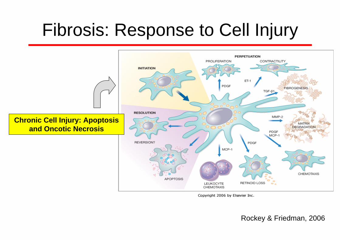
Fibrosis: Response to Cell Injury
Rockey & Friedman, 2006
Chronic Cell Injury: Apoptosis and Oncotic Necrosis

• cellular condensation (cell shrinkage)
• membrane blebbing, but no loss of integrity
• aggregation of chromatin at the nuclear membrane
• formation of membrane bound vesicles (apoptotic bodies)
• no disintegration of organelles; organelles remain intact
• swelling of the cell and lysis
• loss of membrane integrity
• flocculation of chromatin
• no vesicle formation, complete lysis
• disintegration (swelling) of cell organelles
Morphology
Apoptosis Oncotic Necrosis

Hepatocellular Apoptosis
H & E
Galactosamine(500 mg/kg; 6 hr)

Hepatocellular Oncotic Necrosis and Inflammation
H & E
Bile Duct Ligation3 days

Apoptotic Cell Death: Caspases
Proenzyme (zymogen)
prodomain large subunit linker domain small subunit
Peptidase1 496
DED DED Pro-Caspase 8Initiator Caspase
Death effector domainDED
Peptidase1 277 Pro-Caspase 3
Effector Caspase
Cysteine-dependentaspartase domainPeptidase
PeptidaseCARD1 416
Pro-Caspase 9Initiator Caspase
Caspase recruitment domainCARD
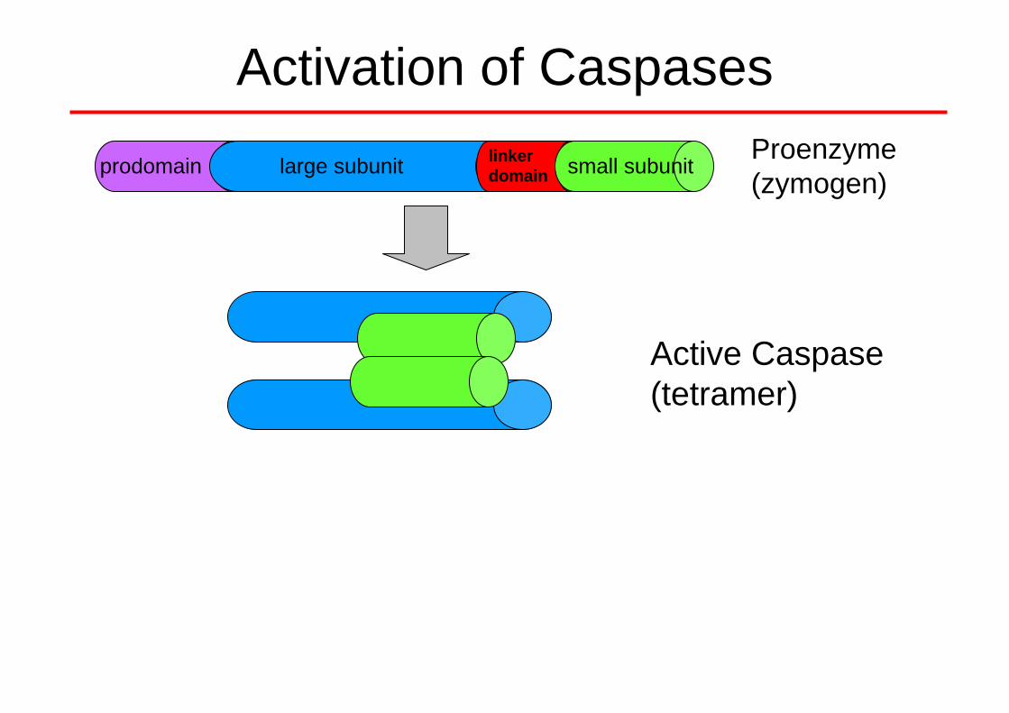
Activation of CaspasesProenzyme(zymogen)
Active Caspase(tetramer)
prodomain large subunit linker domain small subunit

Activation of CaspasesProenzyme(zymogen)
Active Caspase(tetramer)
prodomain large subunit linker domain small subunit
0 20 40 90 120 Time (min)
p32
p11Fas-AbJo-2
Proenzyme
Active Fragment

0
2 0 0
4 0 0
6 0 0
Activation of Caspases
0 20 40 90 120 Time (min)
p32
p11
(ΔF/min/mg protein)
600
400
200
0
*
*
*

DDDD DD
FAS-L FAS-L
DDDDDD
FAD
DFA
DD
FAD
DFA
DD
Cas8
FAS-L
DDDDDD
FAD
DFA
DD
FAD
DFA
DD
Cas10
Cas
p10
Casp 6
Casp
8Bid
BaxBak
C
C
C
C
C
C
C
CC
C
C
CC
C
Casp 7Casp 3A
paf1
Apa
f1
Cas
p9
Cas
p9
C
CC
C
CAD
Endo
GEn
doG
CAD
Endo
G
AIF
AIF
Dia
blo
Diablo
C
Bak Bak
Bax
Bax
CaspaseSubstrates
CAD
CARD
C
C
CARD
DISC
FAS-LFAS-L
dATP
dATP
Fas-mediated Apoptosis Signaling in Hepatocytes
CAD ICADCAD

Bid
Bcl-2
Bak
C
C
CC
C
C
C
C
C
C
C
C Casp 3
C
C
CC
EndoG EndoG
EndoG
EndoG
AIF
Diablo
C
Caspase 3 SubstratesAIF
cIAPDiablo
Intrinsic Pathway of Apoptosis
Ca
Ca
Ca
Ca
AIF
Diablo
Diablo C
AIF
C
C
C
C
C
C
C C
CC
C
C
BaxBakBax
Bak
AIFDiablo
CaCa
Ca
Ca
CaCa
CaCa
CaCa
PTP
Bcl-XL
Bcl-2 Bax
Bax
Bax
28S
eIF2α
PTP
Cathepsin
C
Apa
f1CARD
Apaf1CA
RD
Apaf1 CARD
Apaf1
CARD
Casp 9
Casp 9Casp
9
C
CC
BaxBakBax
Bak
CaCa
Ca
Ca
Casp 9
C C
m-Calpain
Casp 12
p53Bax

Caspase Targets in ApoptosisCasp10
Casp 6
Casp 8
Casp 7
Casp 3
Apa
f1
Apa
f1
Cas
p9
Cas
p9
CARDC
CARD
CARD Casp 2
MDM2Fodrin
Prese-nelin2 Gelsolin
Actin Lamin A
DNA fragmentation andchromatin condensation
CytoskeletonCell shape and membrane
blebbing
CAD
Trans-glutaminase
Topo-isomerase
PARPDNA-PKEndoGICAD
Keratin-18
Gas2
Cell cycle and other
p21
FAK
hnRNP
β-catenin
NuMASREB1
Calpastatin
EMAP II
Rock-1

DNA Fragmentation
1 2 3 4 5 6 7 8 9 10 11 12 13
MW Controls G/ET
0
200
400
600
800
1000
(% Vmax)
00 20 40 90 120
600
400
200
800
1000 *
*
*
Time (min)
DNA LadderAnti-Histone ELISA
CADCAD

DNA Strand Breaks: TUNEL Assay
Terminal deoxynucleotidyl transferase-mediated dUTP nick end labeling
GalactosamineGalactosamine (500 mg/kg; 6 hr)(500 mg/kg; 6 hr)

Characteristic Features of Oncotic Necrosis
• Morphology: cell swelling, cell contents release, karyolysis
Mechanism is dependent on the insult

Oncotic Necrosis: Acetaminophen- induced Hepatotoxicity
Cell Swelling, Karyolysis
Cell Contents Release : Plasma ALT > 3000 U/L)

DNA Strandbreaks during OncoticNecrosis
TUNEL Assay
CV
4 h APAP 6 h APAP

DNA Strandbreaks: Oncotic Necrosis vs Apoptosis
TUNEL Assay
CV
6 h Gal/ET 6 h APAP

DNA Fragmentation:Apoptosis vs Oncotic Necrosis
1 2 3 4 5 6 7 8 9 10 11 12 13
MW Controls G/ET
DNA LadderAnti-Histone ELISA
APAP
DN
A F
ragm
enta
tion
(% c
ontro
l)
0
500
1000
1500
2000
C G/E G/E ZVAD
AAP AAP ZVAD
AAP GSH
*
* *#
#

1 2 3 4 5 6 7 8 9
0 0.5 1 2 3 4 6 G/E
Acetaminophen (h)
p32
p11
Caspase-3 Processing during Acetaminophen Toxicity
Lawson et al., Toxicol Appl Pharmacol 156: 179-86, 1999 Gujral et al., Toxicol Sci 67: 322-8, 2002

BaxBak
C
C
C
C
C
C
C
CC
C
C
CC
C
C
CC
C
EndoG
Endo
GEn
doG
EndoG
Endo
G
EndoG
AIF
AIF
Dia
blo
Diablo
C
Bak Bak
Bax
Bax
AIF
C
DiabloAIF
EndoG
DNA Fragmentation: Nuclear Translocation of Mitochondrial Intermembrane Proteins
Diablo
C C
Nucleus
Mitochondria
Control
5 mM AAP, 6 h
Endonuclease G
Bajt et al., Toxicol Sci 94: 217-225,2006

Cytochrome c
Bax
BidtBid
Contro
lAPAP 2
h
APAP 4 h
Contro
lAPAP 2
hAPAP 4
h
Mitochondria Cytosol
APAP-induced Bax and tBid Translocation to Mitochondria
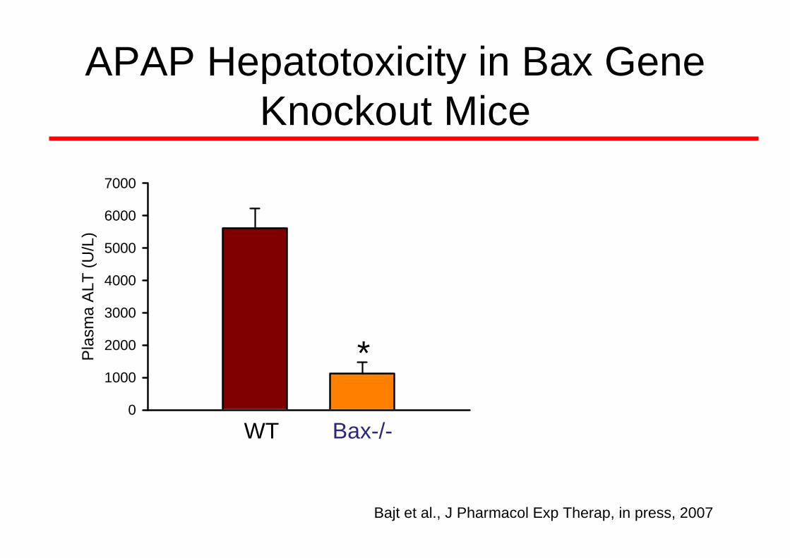
APAP Hepatotoxicity in Bax Gene Knockout Mice
Pla
sma
ALT
(U/L
)
0
1000
2000
3000
4000
5000
6000
7000
*
WT Bax-/-
Bajt et al., J Pharmacol Exp Therap, in press, 2007

APAP Hepatotoxicity in Bax Gene Knockout Mice
Pla
sma
ALT
(U/L
)
0
1000
2000
3000
4000
5000
6000
7000
*
WT Bax-/-
Bajt et al., J Pharmacol Exp Therap, in press, 2007
WT
Bax-/-

NAPQI APAPGSH ↓
Protein Arylation
P4501.
2.
Bax
Cyt c / SmacAIF Endonuclease G
ONOO-
Nucleus
DNA-Strandbreaks
ATP ↓
CaspaseActivation
ATP ↓
PARP Activation
DNA Repair
DNA-Fragmentation
Chromatin Condensation
NAD+ Depletion
ATP ↓ ⇐ MPT
?Mechanism of Mechanism of APAP ToxicityAPAP Toxicity
Toxicol Sci 89: 31-41, 2006
O2–
NO
Bax MPT
Protein Nitration

Apoptosis vs Oncotic Necrosis
Initiation Execution
Apoptosis ApoptosisATP high
Apoptosis Secondary NecrosisATP low
Oncosis Oncotic NecrosisATP low
OncosisATP high Secondary Apoptosis ?
Same process or replaced by different mode of cell death?
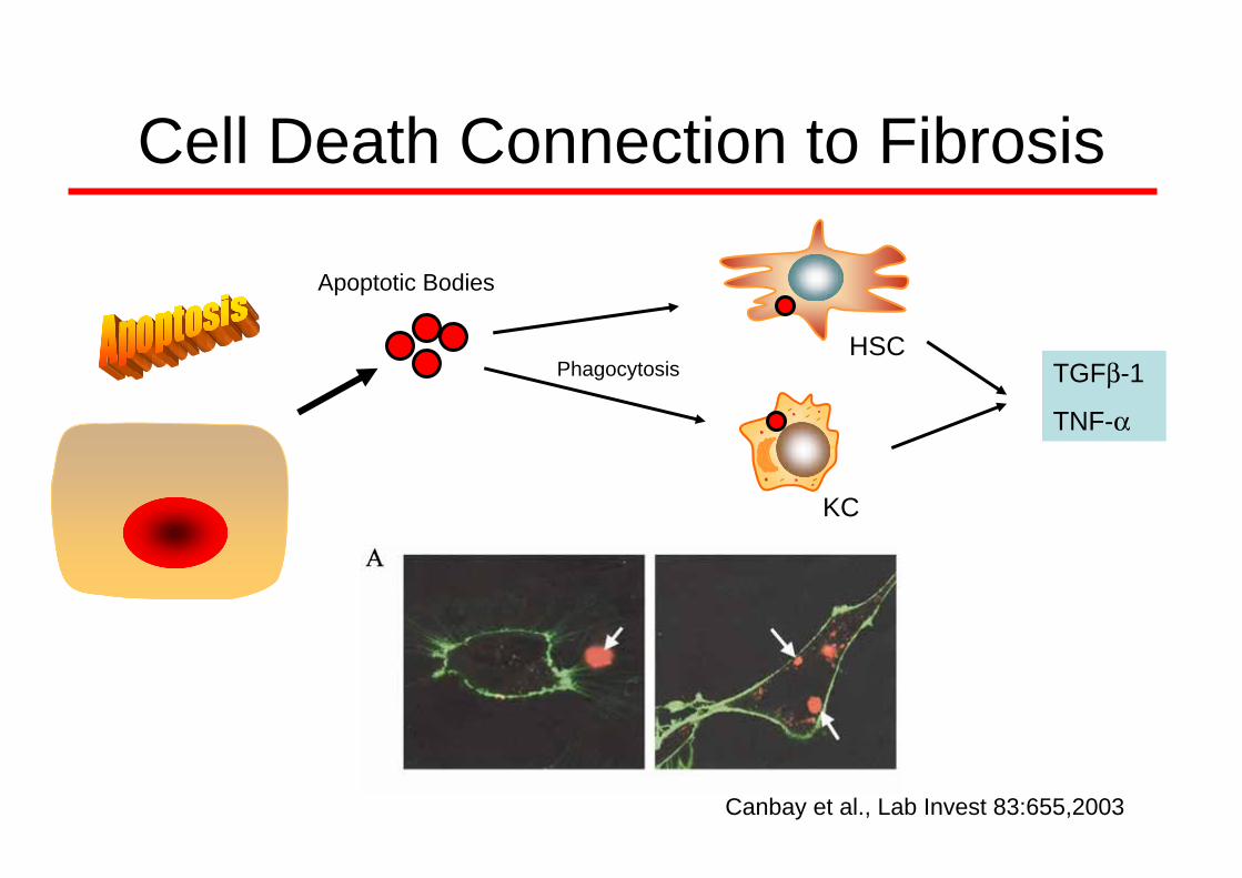
Cell Death Connection to Fibrosis
Apoptotic Bodies
PhagocytosisHSC
KC
TGFβ-1
TNF-α
Canbay et al., Lab Invest 83:655,2003

Cell Death Connection to Fibrosis
Phagocytosis
HSC
KC
TGFβ-1
TNF-α
HMGB1
Cell Debris
HSC
KC
TLR4TGFβ-1
TNF-α

Fibrosis: Response to Cell Injury
Rockey & Friedman, 2006
Apoptosis
Oncotic Necrosis
Top Related