


Languages
Pages
Legal


Anchorage condition during
canine retraction using
transpalatal arch with
continuous and segmented
arch mechanics.
Adel Alhadlaq; Thamer Alkhadra; Tarek
El-
Bialy.
Angle Orthodontist, Vol 86, No 3, 2016

INTRODUCTION:
Tooth extraction is an important
step in orthodontic treatment.
The mechanics of closing the
extraction spaces depend on the
diagnostic criteria that dictate the
required type of anchorage.
Introduction Continues…
Maximum Anchorage means when less
than one-third of the extraction space is
lost by forward movement of the
posterior teeth.
Moderate anchorage when up to half of
the extraction space is lost by forward
movement of the posterior teeth
Minimum anchorage when more than
two-thirds of the extraction space is lost
by forward movement of the posterior
teeth.

Introduction Continues…
The TPA has been widely used in orthodontics either with Continuous or segmented arch mechanics to minimize anchorage loss and/or control rotation of the upper first molars.
Introduction Continues…
It has been recently
suggested that TPA can
control anchorage,
especially when it is
combined with TAD’s.
Introduction Continues…
In segmented arch
mechanics, canine retraction
T-loops have been used to
control anchorage during
space closure by modulating
moments of the posterior
teeth part of the T-loop.

Introduction Continues…
The aim of this study was to
compare anchorage loss between
two groups of patients who were
treated either with continuous arch
or segmented arch technique
using T-loops to close extraction
spaces while TPA was used to
support the upper first molars.

MATERIALS AND METHODS:
Records of 20 orthodontic patients treated either by continuous arch technique (n =10) or segmented arch technique using T-loops and TPA were studied.
Analyzed records included lateral cephalometric radiographs before treatment (T0) and immediately after complete canine retraction (T1).
This study was approved by the Health Ethics Review Board at the University of Alberta, Canada (protocol Pro00041075).
MATERIALS AND METHODS:
The bracket system used in all
patients was the Synergy bracket
system (0.022 × 0.025 inches;
RMO,Denver, Colo).
The TPA was fabricated from 0.036
stainless steel wire soldered to
previously fit upper first molar bands.
MATERIALS AND METHODS:
In the continuous arch wire group,
sliding of upper canines was
performed along 0.018 × 0.025-inch
stainless steel wire using an
elastomeric chain connected between
the upper canines and upper molars’
band hooks (Energy chain, RMO).
MATERIALS AND METHODS:
In the segmented arch technique
cases, initial leveling within the buccal
segment was performed using 0.018-
inch round nickel titanium wire (RMO),
and then the posterior teeth (second
premolar to second molar) were
stabilized by rigid 0.018 × 0.025-inch
stainless steel wires (RMO)

MATERIALS AND METHODS:
Canine retraction was
performed using a T-loop
fabricated from 0.019 ×
0.025-inch titanium-
molybdenum alloy (RMO).
MATERIALS AND METHODS:
The anterior part of the T-loop (alpha)
was bent 35˚ apical, while the
posterior part (beta) was bent 60˚
apical to produce a posterior moment-
to-force ratio of about 12 in the
posterior segment.

MATERIALS AND METHODS:
The anterior segment would
produce a moment-to-force ratio of
approximately 6 at 6-mm
activation of the T-loop that is
positioned initially off center
mesially.
MATERIALS AND METHODS:
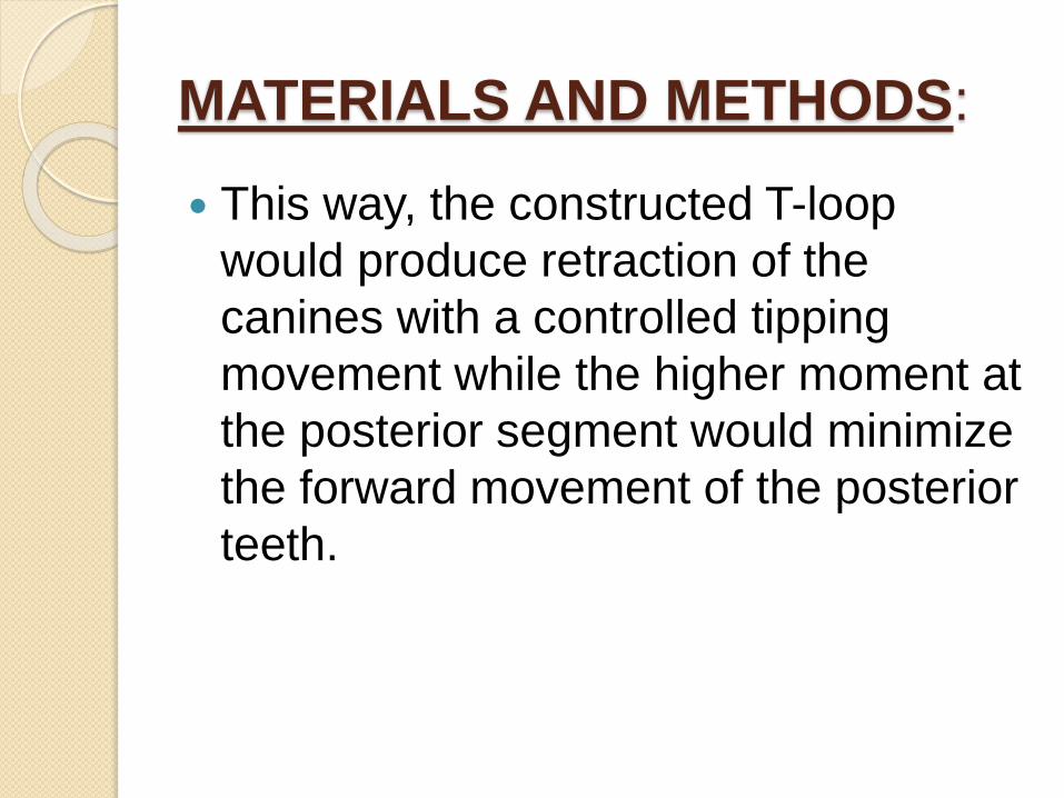
This way, the constructed T-loop
would produce retraction of the
canines with a controlled tipping
movement while the higher moment at
the posterior segment would minimize
the forward movement of the posterior
teeth.
MATERIALS AND METHODS:
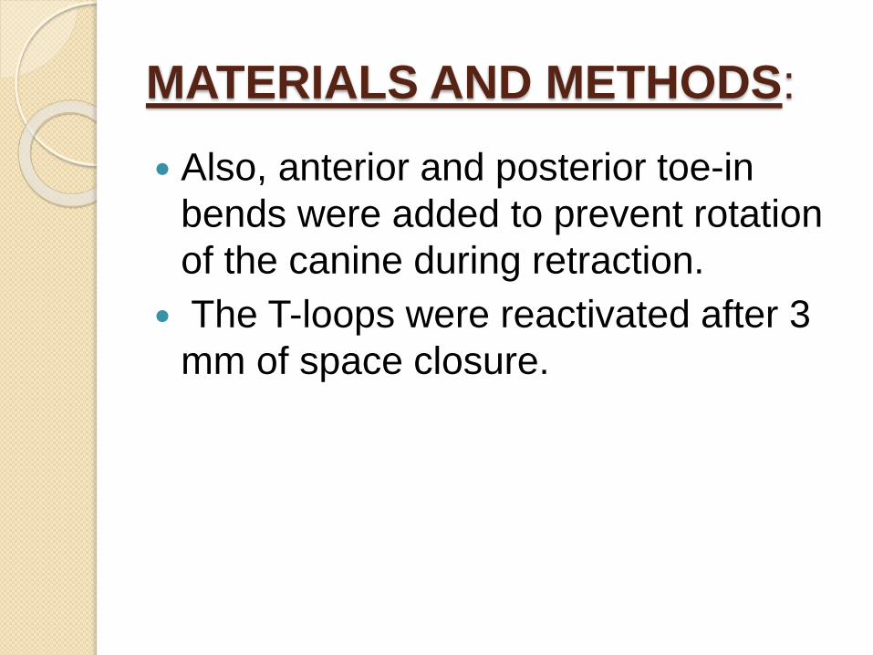
Also, anterior and posterior toe-in
bends were added to prevent rotation
of the canine during retraction.
The T-loops were reactivated after 3
mm of space closure.
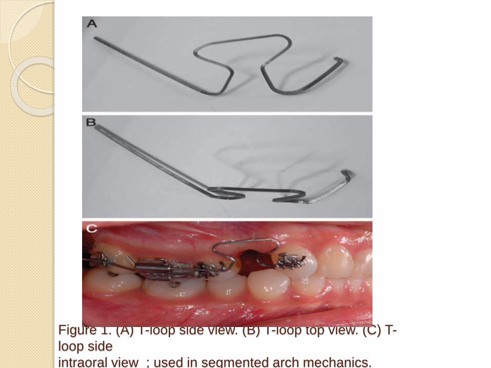
Figure 1. (A) T-loop side view. (B) T-loop top view. (C) T-
loop side
intraoral view ; used in segmented arch mechanics.

MATERIALS AND METHODS:
Cephalometric radiographs were
obtained at the beginning of the
treatment and after space closure.
All cephalometric radiographs were
digitized using Dolphin imaging
software (Dolphin Imaging &
Management Solution, Chatsworth,
Calif), and Ricketts cephalometric
analysis was used.

• Anchorage was assessed by
evaluating the anteroposterior
movement of the distal surface of
the upper first molar to a vertical
line drawn from the Pt point
perpendicular to the Frankfurt
plane


RESULTS: In the continuous arch group, cephalometric
analysis and superimposition showed that the upper first molars moved forward significantly (4.5±3, P<.05) compared with the segmented arch group. (-.7±1.4, P<05)
The molar relationship has become more class II in the continuous arch group compared with the segmented arch groupdue to the forward movement of the upper molars ( loss of anchorage).
The Frankfurt-Mandibular plane angle (FMA) showed a greater increase after canine retraction in the continuous arch group than in the segmented arch group, but the difference was not statistically significant

Figure 4. Graph showing anchorage loss in
both segmented and continuous arch
mechanics.
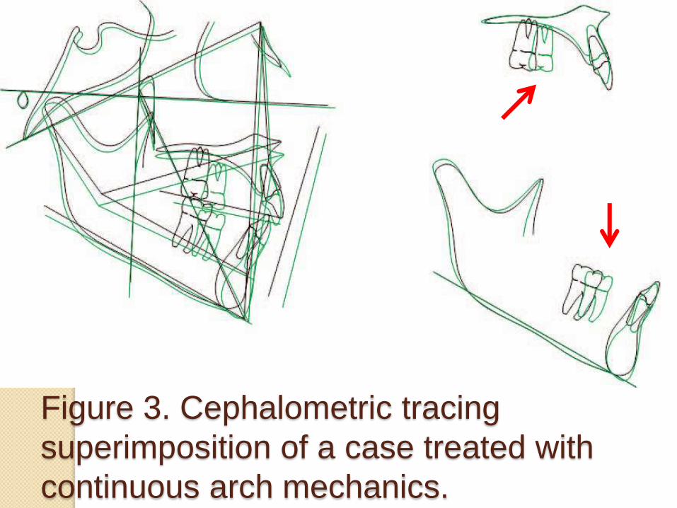
Figure 3. Cephalometric tracing
superimposition of a case treated with
continuous arch mechanics.

Figure 2. Cephalometric tracing
superimposition of a case treated with
segmented arch mechanics.

DISCUSSION:
Although TADs have shown significant anchorage control in the literature, their risk of failure and complications, including loosening or fracture of the TADs, pain, and soft tissue inflammation, remain a concern for some clinicians.
Segmented arch mechanics have not received wide acceptance in orthodontic clinical practice, possibly because of their complexity and challenges facing the clinician in maintaining a continuous and reproducible force system.

The minimum anchorage loss when
using the segmented arch mechanics
in our study agrees with previous
reports that showed anchorage control
using the beta bend in the retraction T-
loop while using TPA mainly to prevent
the rotation of upper molars.
Despite the maximum anchorage
control shown in our study when
retracting the upper canines with the
segmented arch technique, other
reports have shown greater
anchorage conservation when an en
masse vs two-step retraction
approach has been used for maximum
anchorage treatment.
The increased FMA after canine
retraction in the continuous arch group
confirms anchorage loss. When the
upper molars move forward, there is
always tendency for them to tip
mesially, which leads to extrusion of
their distal part and consequently
backward rotation of the mandible—
hence, increased FMA.

This study is a retrospective study with
a small number of cases.
More prospective controlled clinical
trials may be needed to confirm these
results with a larger sample size and
wide distribution of cases with respect
to their facial forms and anchorage
requirements.

CONCLUSION:
The use of a TPA when combined with
segmented arch mechanics results in
more anchorage control than when
used with continuous arch sliding
mechanics during upper canine
retraction.

REFERENCES: 1. Burstone CJ. Rationale of the segmented arch. Am J Orthod.
1962;48:805–822.
2. Dalessandri D, Salgarello S, Dalessandri M, et al. Determinants for success rates of temporary anchorage devices in orthodontics: a meta-analysis (n . 50). Eur J Orthod. 2014; 36:303–313.
3. Burstone CJ. The segmented arch approach to space closure. Am J Orthod. 1982;82:361–378.
4. Mezomo M, de Lima ES, de Menezes LM, Weissheimer A Allgayer S. Maxillary canine retraction with self-ligating and conventional brackets. Angle Orthod. 2011;81:292–297.
5. Oz AA, Arici N, Arici S. The clinical and laboratory effects of bracket type during canine distalization with sliding mechanics. Angle Orthod. 2011;82:326–332.
6. Burrow SJ. Canine rvs conventional edgewise brackets. Angle Orthod. 2010;80: 438–445.
7. Machibya FM, Bao X, Zhao L, Hu M. Treatment time, outcome, and anchorage loss comparisons of self-ligating and conventional brackets. Angle Orthod. 2013;83:280–285.etraction rate with self-ligating brackets

8. de Almeida MR, Herrero F, Fattal A, Davoody AR, Nanda R, Uribe F. A comparative anchorage control study between conventional and self-ligating bracket systems using differential moments. Angle Orthod. 2013;83:937–942.
9. Moninia AC, Juniorb LG, Martinsc RP, Vianna AP. Canine retraction and anchorage loss: self-ligating versus conventional brackets in a randomized split-mouth study. Angle Orthod. 2014;84:846–852.
10. Kuhlberg AJ, Burstone CJ. T-loop position and anchorage control. Am J Orthod Dentofacial Orthop. 1997;112:12–18.
11. Braun S, Marcotte MR. Rationale of the segmented approach to orthodontic treatment. Am J Orthod Dentofacial Orthop. 1995;108:1–8.
12. Katona TR, Isikbay SC, Chen J. Effects of first- and secondorder gable bends on the orthodontic load systems produced by T-loop archwires. Angle Orthod. 2014;84:350–357.
13. Burstone CJ, Koenig HA. Precision adjustment of the transpalatal lingual arch: computer arch form predetermination. Am J Orthod. 1981;79:115–134.
14. Gollner P, Bantleon HP, Ingervall B. Force delivery from a transpalatal arch for the correction of unilateral first molar cross-bite. Eur J Orthod. 1993;15:411–420.

15. Ingervall B, Honigl KD, Bantleon H. Moments and forces delivered by transpalatal arches for symmetrical first molar rotation. Eur J Orthod. 1996;18:131–139.
16. Ten Hoeve A. Palatal bar and lip bumper in nonextraction treatment. J Clin Orthod. 1985;19:272–291.
17. Gunduz E, Zachrisson BU, Honigl KD, CrismaniAG, Bantleon HP. An improved transpalatal bar design. Part I. Comparison of moments and forces delivered by two bar designs for symmetrical molar derotation.
18. Dahlquist A, Gebauer U, Ingervall B. The effect of a transpalatal arch for the correction of first molar rotation. Eur J Orthod. 1996;18:257–267.
19. Ingervall B, Gollner P, Gebauer U, Frohlich K. A clinical investigation of the correction of unilateral first molar crossbite with a transpalatal arch. Am J OrthodDentofacial Orthop. 1995;107:418–425.

20. Kuhlberg AJ, Priebe D. Testing force systems and biomechanics— measured tooth movements from differential moment closing loops. Angle Orthod. 2003;73:270–280.
21. Zablocki HL, McNamara JA Jr, Franchi L, Baccetti T. Effect of the transpalatal arch during extraction treatment. Am J Orthod DentofacialOrthop. 2008;133:852–860.
22. Bobak V, Christiansen RL, Hollister SJ, Kohn DH. Stressrelated molar responses to the transpalatal arch: a finite element analysis. Am J Orthod Dentofacial Orthop. 1997; m112:512–518.
23. Ingervall B, Go¨ llner P, Gebauer U, Fro¨ hlichK. A clinical investigation of the correction of unilateral first molar crossbite with a transpalatal arch. Am J Orthod Dentofacial

24. Borsos G, Voko´ Z, Gredes T, Kunert-Keil C, VeghA. Tooth movement using palatal implant supported anchorage compared to conventional dental anchorage. Ann Anat. 2012;194:556–560.
25. Lee J, Miyazawa K, Tabuchi M, Sato T, Kawaguchi M, Goto S. Effectiveness of en-masse retraction using midpalatal miniscrews and a modified transpalatal arch: treatment duration and dentoskeletal changes. Korean J Orthod. 2014; 44:88–95.
26. Wilmes B, Olthoff G, Drescher D. Comparison of skeletal and conventional anchorage methods in conjunction with pre-operative decompensation of a skeletal class III malocclusion. J Orofac Orthop. 2009;70:297–305.
27. Ricketts RM. New perspectives on orientation and their benefits to clinical orthodontics—part I. Angle Orthod. 1975; 45:238–248.
28. Xu TM, Zhang X, Oh HS, Boyd RL, Korn EL, Baumrind S. Randomized clinical trial comparing control of maxillary anchorage with 2 retraction techniques. Am J Orthod

THANK YOU
Top Related