


Languages
Pages
Legal


ADENOIDS Disclaimer: The pictures used in this presentation have been obtained from a number of sources. Their use is purely for academic and teaching purposes. The contents of this presentation do not have any intended commercial use. In case the owner of any of the pictures has any objection and seeks their removal please contact at [email protected]. The se pictures will be removed immediately.

Embryology
The formation of the adenoids begins in the
3rd month of fetal development. This starts
with glandular primordia in the posterior
nasopharynx becoming associated with
infiltrating lymphocytes.
In the 5th month sagittal folds are formed
which are the beginnings of pharyngeal
crypts. The surface is covered with
pseudostratified ciliated epithelium.
By the 7th month of development the
adenoids are fully formed.

Anatomy
The lymphoid tissue of the nasopharynx and
oropharynx is composed of the adenoids,
the tubal tonsils, the lateral bands, the
palatine tonsils, and the lingual tonsils.
There are also lymphoid collections in the
posterior pharyngeal wall and in the
laryngeal ventricles.
These structures form a ring of tissue named
Waldeyer’s ring after the German anatomist
who described them.
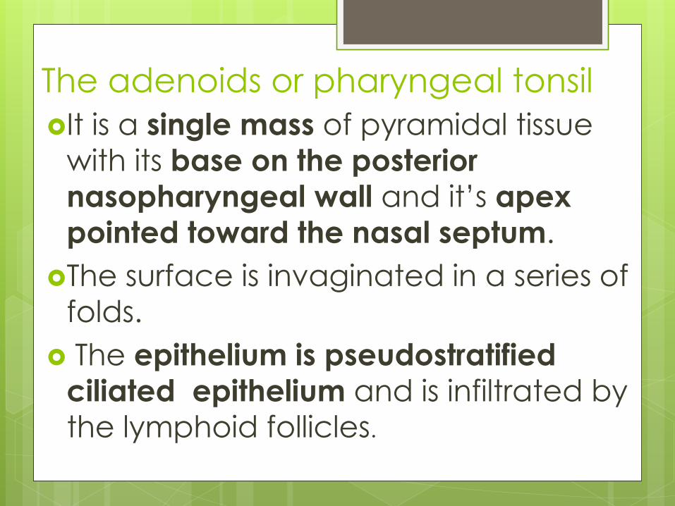
The adenoids or pharyngeal tonsil
It is a single mass of pyramidal tissue
with its base on the posterior
nasopharyngeal wall and it’s apex
pointed toward the nasal septum.
The surface is invaginated in a series of
folds.
The epithelium is pseudostratified
ciliated epithelium and is infiltrated by
the lymphoid follicles.

Blood supply is from
the:
Ascending palatine
branch of the facial
artery
Pharyngeal branch
of the internal
maxillary artery
Artery of the
pterygoid canal
Ascending cervical
branch of the
thyrocervical trunk.

Venous drainage is through the pharyngeal plexus and the pterygoid plexus flowing ultimately into the facial and internal jugular veins.
Innervation is derived from the glossopharyngeal and vagus nerves.
Efferent lymphatics drain to the retropharyngeal nodes and the upper deep cervical nodes.

Function and Immunology
The tonsils and adenoids are part of the
secondary immune system.
Without afferent lymphatics the lymphoid
nodules in these structures are exposed to
antigen only in the crypts of the palatine
tonsils and the folds of the adenoids where it
is transported through the epithelial layer.
These are involved in the production of
mostly secretory IgA, which is transported to
the surface providing local immune
protection.

Acute adenoiditis symptoms include purulent
rhinorrhea, nasal obstruction, fever, and sometimes
otitis media.
This can be difficult to differentiate from an acute
upper respiratory infection but tends to have a
longer and more severe course.

Acute adenoiditis symptoms include
purulent rhinorrhea,
nasal obstruction, fever, and
sometimes otitis
media.
This can be difficult
to differentiate from
an acute upper respiratory infection
but tends to have a
longer and more
severe course.
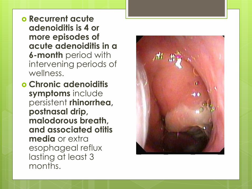
Recurrent acute adenoiditis is 4 or more episodes of acute adenoiditis in a 6-month period with intervening periods of wellness.
Chronic adenoiditis symptoms include persistent rhinorrhea, postnasal drip, malodorous breath, and associated otitis media or extra esophageal reflux lasting at least 3 months.

Obstructive adenoid hyperplasia includes symptoms of chronic nasal obstruction, rhinorrhea, snoring, mouth breathing, and a hyponasal voice.
Obstructive sleep apnea in children is clinically marked by loud snoring, apneic episodes while sleeping, daytime somnolence, behavioral problems, and enuresis

Adenoid facies or “long face
syndrome”. It is the long, open-mouthed,
face of children with adenoid
hypertrophy.
The mouth is always open
because upper airway
congestion has made patients
obligatory mouth breathers.
The most common presenting
symptoms are chronic mouth
breathing and snoring.
The most dangerous symptom is
sleep apnea

The characteristic
facial appearance
consists of:
Underdeveloped
thin nostrils
Short upper lip
Prominent upper
teeth
Crowded teeth
Narrow upper
alveolus

High-arched palate
Hypoplastic maxilla
Eustachian blockage
causing glue ear-
deafness
The deafness and
inattentiveness
interferes with the
learning
Child grows with
lowered intelligence
and understanding

Diagnosis

Posterior Rhinoscopy

Nasopharygoscopy

Nasopharygoscopy
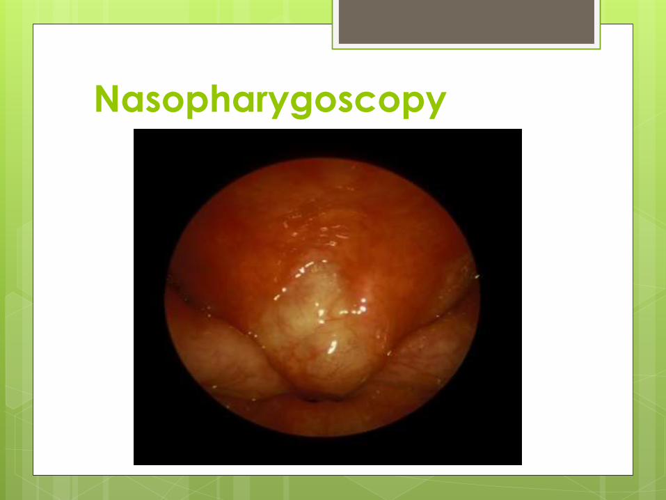
Nasopharygoscopy

X-Ray soft tissue nasopharynx-
lateral view

X-Ray soft tissue nasopharynx-
lateral view

CT Scan

Adenoidectomy-Indications
Four or more episodes of recurrent purulent rhinorrhea in prior 12 months in a child <12. One episode documented by intranasal examination or diagnostic imaging.
Persisting symptoms of adenoiditis after 2 courses of antibiotic therapy.
Sleep disturbance with nasal airway obstruction persisting for at least 3 months.
Hyponasal or nasal speech

Adenoidectomy-Indications Otitis media with effusion >3 months or
second set of tubes
Dental malocclusion or orofacial growth disturbance documented by orthodontist.
Cardiopulmonary complications including cor pulmonale, pulmonary hypertension, right ventricular hypertrophy associated with upper airway obstruction.
Otitis media with effusion over age 4.

Contraindications A submucous cleft palate which may lead to
velopharyngeal insufficiency after surgery. If
the adenoid obstruction is severe enough,
then only superior half adenoidectomy is performed.
Avoid surgery in patients with hemoglobin less
than 10.
Perform surgery at least 2 weeks after the last
attack of acute tonsillitis.
Wait at least 6 weeks after polio vaccination.
Avoid surgery in patients with uncontrolled
systemic diseases (ie. leukemia).

Jennings's Mouth Gag

St. Claire Thomson Adenoid
Curette
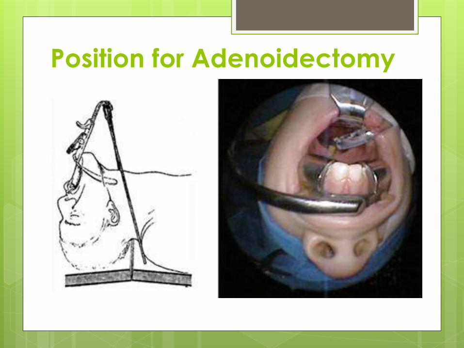
Position for Adenoidectomy

Adenoidectomy

Adenoidectomy Specimen

Complications
The incidence of mortality from
adenotonsillar surgery ranges from 1 in
16,000 to 1 in 35,000 cases.
Anesthetic complications and hemorrhage
cause the majority of deaths.
The prevalence of hemorrhage ranges from
0.1% to 8.1%.
It is divided into primary bleeding, in the first
24 hours, and secondary bleeding, around
7-10 days post operatively.

Other risks include:
Vomiting
Dehydration
Airway obstruction due to edema
Pulmonary edema
Fever, velopharyngeal insufficiency
Dental injury
Burns
Nasopharyngeal stenosis.

Atlantoaxial subluxation can occur in
patients with Down syndrome.
Atlantoaxial joint laxity because of Grisel’s
syndrome. This is vertebral body
decalcification and laxity of the anterior
transverse ligament between the atlas and
the axis from inflammation or infection in the
nasopharynx.
Spontaneous subluxation occurs about 1
week post operatively with pain and
torticollis.
Top Related