


Languages
Pages
Legal

British Journal of Plastic Surgery (1990), 43,42&430 0 1990 The Trustees of British Association of Plastic Surgeons
A new protocol for the treatment of stage I cutaneous malignant melanoma; interim results of the first 806 patients treated
J. EVANS and B. G. MCCANN
Derriford Hospital, Plymouth, and the Norfolk and Norwich Hospital, Norwich
Summary-A protocol is described which has been used in a prospective study of 806 patients with stage I malignant melanoma. Excisional biopsies are performed as soon as possible and Breslow thickness and Clark’s levels determined. Patients are allocated to low, medium and high risk groups and definitive excision is carried out with a clearance of 220 and 50 mm respectively.
Preliminary results are reported on the 806 patients treated over 7 years in three plastic surgery units. No local recurrences have occurred so far in the low and medium risk groups, which suggests that the protocol is safe.
Although standard 5 cm excision of cutaneous malignant melanoma was originally recommended by Handley (1907) it was not until Petersen et al. recommended similar treatment in 1962 that wide excision of these tumours was adopted as standard practice by surgeons in the United Kingdom. Subsequent histopathological studies demonstrated a close relationship between prognosis and the level to which a primary malignant melanoma had penetrated into the dermis (Clark, 1969) and the maximum thickness of the tumour (Breslow, 1970).
Using the prognostic indicators of Clark and Breslow, obtained from excisional biopsies, several protocols have been developed in recent years which link the margin of excision of a primary cutaneous malignant melanoma to the risk of recurrence (Bagley et al., 198 1; Taylor et al., 1984 ; Veronesi et al., 1988).
During 1982 a retrospective study was initiated of 500 cases of cutaneous malignant melanoma treated by the Plastic Surgery Department in Norwich. Prior to this date it was not standard practice to provide an assessment of tumour thickness or level of invasion in the histological report.
In many cases only one tissue block had been cut and we could not be sure that the tumour had been cut through its maximum thickness. Details of history and presentation, which are now thought to be important, had not always been recorded. Consequently the exercise was abandoned, and a new protocol for a prospective study was designed.
The objectives of the protocol were based on the following requirements :
1.
2.
3.
4.
5.
6.
To minimise the cosmetic disfigurement follow- ing surgical excision of primary malignant melanomas. Cassileth et al. (1983) reported the substantial psychological distress which can be caused to patients who have wide excisions of melanoma and grafting. To minimise the risk of local recurrence after excision of the primary tumour. This has been borne out by protocols published by Taylor and Veronesi, which suggests that limited excisions of superficial and thin tumours are safe in this respect. To take account of the varying thickness of skin in different parts of the body and in different age groups which might affect the depth of penetra- tion of a tumour in relation to its maximum thickness. The protocol must be simple to follow and practical for use in a busy plastic surgery unit so that data collection is reliable in a large series of patients. The protocol should not cause any delay to treatment that might worsen prognosis. It should be inexpensive to run,
The protocol
1. Biopsy
All patients who are referred with suspicious pigmented lesions should be seen at the next
426
A NEW PROTOCOL FOR THE TREATMENT OF STAGE I CUTANEOUS MALIGNANT MELANOMA 427
outpatient clinic, or sooner if necessary. No patient should wait for more than a week. For patients in whom malignant melanoma is a differential diag- nosis the lesion should be photographed and excisional outpatient biopsy performed within 48 hours of the clinic attendance. Even clinically obvious malignant melanomas must be biopsied in order to obtain accurate histological typing and staging. In participating units, it should be possible to have the histopathological report within 48 hours of the biopsy.
joint. Lesions on the face have a 20 mm clearance whenever possible though a wider excision is allowable if there is indication for cosmetic benefit to the patient (e.g. a cosmetic facial unit).
The biopsy must be a whole tumour biopsy on a superficial subdermal plane including a narrow margin of skin beyond the tumour of 2-3 mm.
Biopsy sites were originally left open. This policy has subsequently been changed and biopsy sites may now be closed by direct suturing whenever possible.
In the high risk group, a 50 mm clearance is not possible for subungual and facial lesions and may not be possible for lesions of the perineum and genitalia. In these sites, excision should be as wide as is anatomically possible. Subungual tumours are again treated by amputation through the IP joint. Abdomino-perineal resection may be indicated for peri-anal melanoma. It is essential that any devia- tion from the plan of definitive treatment is meticulously recorded.
2. Histopathological staging
The histopathologist is required to assess the depth of invasion (Clark’s level) and to measure the maximum tumour thickness (Breslow). The latter is done by using an eye-piece micrometer which is the only special piece of equipment required for this protocol. Based on the histopathological de- scription of the tumour the patient is placed into one of three risk groups :
Table 1 Three risk groups as defined by histopathologi- cal staging
There is no requirement for prophylactic regional node dissection except where a lesion lies directly over the regional nodes when en bloc removal of the regional nodes with the primary tumour may be indicated. The protocol excludes adjuvant chemo- therapy.
4. Follow-up including management qf recurrences
Follow-up must be planned for 20 years via an outpatient follow-up clinic for the first 10 years and via mailed enquiries from a Cancer Registry or the hospital department for the next 10 years. Recur- rences should be treated by conventional methods according to site and extent.
Low risk Less than 0.76 mm thick, Clark’s level II or III.
Medium risk Less than 0.76 mm thick, Clark’s level IV. All turnours between 0.76 and 1.5 mm.
High risk Greater than 1.5 mm thick, Clark’s level IV. All Clark’s level V.
Patients should be taught to self-examine and must be encouraged to return to the unit between follow-up appointments if they have any anxieties. Such an open-door policy has already proved very supportive to some patients. General practitioners should be kept regularly informed.
5. Documentation
3. Definitive treatment
According to the risk group into which the tumour falls, definitive treatment is prescribed as follows :
Table 2
Risk group
LOW Medium High
Clearance margin
2 mm (i.e. biopsy) 20 mm 50 mm
A three-sheet proforma has been developed for data collection. It has been kept brief in order to maximise the reliability of data collection. Tables 1 and 2 are produced on a single sheet and should be readily available in the participating units, including theatres, for the instruction of new staff. One person should be responsible for holding a register of all patients who are entered into the protocol.
Interim report on the first 806 patients admitted to the protocol
The only exceptions where anatomy precludes The Plastic Surgery Units involved
wider excisions are as follows. In the medium risk This protocol was initiated in 1982 by the Plastic group, subungual lesions should be treated by Surgery Unit in Norwich which has clinical amputation through the distal interphalangeal responsibility for most of East Anglia and Ipswich.
428 BRITISH JOURNAL OF PLASTIC SURGERY
Since then, the protocol has been adopted by two more units: Leicester in 1983 (serving South Trent and Lincolnshire) and Plymouth in 1987 (serving Cornwall and most of Devon).
All clinicians have been asked frequently to report any known recurrences. The operating registers and histopathology data banks have been reviewed every 3 months to ensure that recurrences have not been missed.
The patients
All patients were those with stage I disease at the time of presentation. Patients with stage II disease and those who had had incisional biopsies were excluded as, too, were all patients with early lesions where there was pathological doubt about the diagnosis of malignant melanoma.
Results
For the most part, patients have been referred to the plastic surgeons by general practitioners and dermatologists. General surgeons have referred an average of one patient per surgeon, per year, often after excisional biopsy had been performed. Ap- proximately 5% of patients were referred by GPs after the melanoma had been excised for histology; these were usually cases where the diagnosis of malignant melanoma was thought to be unlikely. Although photographs are lacking for some of these patients, they were included in the protocol pro- vided that excision had appeared adequate histo- logically.
In the time from April 1982 to November 1988,806 patients between the ages of 16 and 93 were treated according to the protocol described. Epidemiologi- cal data have been collected from these patients, and will be published at a later date with the results of this long-term follow-up.
Recurrences (Table 4)
Follow-up
All patients in the high risk group have been followed up by plastic surgeons at least until a first recurrence. Some patients who developed wide- spread disease were subsequently referred to oncol- ogists, general practitioners or terminal care specialists.
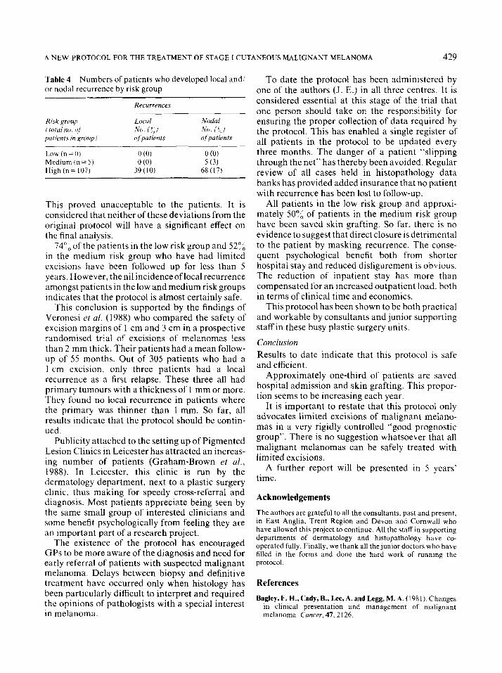
Local recurrence has been defined as any secondary deposit of malignant melanoma found between the original site of the lesion and the regional lymph nodes. To date, 39 patients have developed “local recurrences” and 73 patients have developed regional lymph node metastases. All but 5 of these patients belong to the high risk group. There has been no evidence of local recurrence among patients in the low and medium risk groups and no evidence of lymph node metastases among patients in the low risk group.
Discussion
In the low and medium risk groups, all patients were followed up either by staff of the plastic surgery units or (particularly in the low risk group) by those of co-operating departments of dermatol- ogy. Special melanoma clinics have been set up in Leicester and Norwich for this purpose.
Two modifications have had to be made to the protocol as originally defined. First, biopsy wounds are now routinely closed rather than being left open, as was originally planned. Second, the protocol originally dictated that three tattoo marks should be made at 5 cm from the centre of the tumour to demonstrate how much skin would have been removed by traditional primary treatment.
Table 3 Distribution of 806 patients treated according to the protocol between April 1982 and November 1988 by risk group and by year of treatment
Year Low risk
1982 5 1983 9 1984 17 1985 22 1986 29 1987 89 1988 85
Totals 256
%pa
(13) (10) (24) (32) (32) (38) (38)
Medium risk %pa High risk
I (19) 25 22 (25) 57 19 (27) 35 14 (20) 33 18 (20) 44 38 (17) 101 49 (22) 88
167 383
%pa
(68) (65) (49) (48) (48) (45) (40)
Total
37 88 71 69 91
228 222
806
A NEW PROTOCOL FOR THE TREATMENT OF STAGE I CUTANEOUS MALIGNANT MELANOMA 429
Table 4 Numbers of patients who developed local and/ or nodal recurrence by risk group
Recurrences
Risk grrmp f total no. of patients in ,qroup I -__
Low (n = 0) Medium (n = 5) High (n= 107)
L‘ocul Nodal No. I”;] No. i f, i of patients ofpatients
0 (0) 0 (0) 0 (0) 5 (3)
39(lO) 68 (17)
This proved unacceptable to the patients. It is considered that neither of these deviations from the original protocol will have a significant effect on the final analysis.
74”, of the patients in the low risk group and 5296 in the medium risk group who have had limited excisions have been followed up for less than 5 years. However, the nil incidence of local recurrence amongst patients in the low and medium risk groups indicates that the protocol is almost certainly safe.
This conclusion is supported by the findings of Veronesi et al. (1988) who compared the safety of excision margins of 1 cm and 3 cm in a prospective randomised trial of excisions of melanomas less than 2 mm thick. Their patients had a mean follow- up of 55 months. Out of 305 patients who had a 1 cm excision, only three patients had a local recurrence as a first relapse. These three all had primary tumours with a thickness of 1 mm or more. They found no local recurrence in patients where the primary was thinner than 1 mm. So far, all results indicate that the protocol should be contin- ued.
Publicity attached to the setting up of Pigmented Lesion Clinics in Leicester has attracted an increas- ing number of patients (Graham-Brown et al., 1988). In Leicester, this clinic is run by the dermatology department, next to a plastic surgery clinic, thus making for speedy cross-referral and diagnosis. Most patients appreciate being seen by the same small group of interested clinicians and some benefit psychologically from feeling they are an important part of a research project.
The existence of the protocol has encouraged GPs to be more aware of the diagnosis and need for early referral of patients with suspected malignant melanoma. Delays between biopsy and definitive treatment have occurred only when histology has been particularly difficult to interpret and required the opinions of pathologists with a special interest in melanoma.
To date the protocol has been administered by one of the authors (J. E.) in all three centres. It is considered essential at this stage of the trial that one person should take on the responsibility for ensuring the proper collection of data required by the protocol. This has enabled a single register of all patients in the protocol to be updated every three months. The danger of a patient “slipping through the net” has thereby been avoided. Regular review of all cases held in histopathology data banks has provided added insurance that no patient with recurrence has been lost to follow-up.
All patients in the low risk group and approxi- mately 5096 of patients in the medium risk group have been saved skin grafting. So far, there is no evidence to suggest that direct closure is detrimental to the patient by masking recurrence. The conse- quent psychological benefit both from shorter hospital stay and reduced disfigurement is obvious. The reduction of inpatient stay has more than compensated for an increased outpatient load. both in terms of clinical time and economics.
This protocol has been shown to be both practical and workable by consultants and junior supporting staff in these busy plastic surgery units.
Conclusion
Results to date indicate that this protocol is safe and efficient.
Approximately one-third of patients are saved hospital admission and skin grafting. This propor- tion seems to be increasing each year.
It is important to restate that this protocol only advocates limited excisions of malignant melano- mas in a very rigidly controlled “good prognostic group”. There is no suggestion whatsoever that all malignant melanomas can be safely treated with limited excisions.
A further report will be presented in 5 years’ time.
Acknowledgements
The authors are grateful to all the consultants, past and present, in East Anglia. Trent Region and Devon and Cornwall who have allowed this project to continue. All the staff in supporting departments of dermatology and histopathology have CO-
operated fully. Finally, we thank all the junior doctors who have filled in the forms and done the hard work of running the protocol.
References
Bagley, F. H., Cady, B., Lee, A. and Legg, M. A. (198 1). Changes in clinical presentation and management of malignant melanoma. Cancer, 47, 2126.
430 BRITISH JOURNAL OF PLASTIC SURGERY
Breslow, A. (1970). Thickness, cross-sectional areas and depth Taylor, B. A., Hughes, L. E. and Williams, G. T. (1984). of invasion in the prognosis of cutaneous melanoma. Annals Improving prognosis for malignant melanoma in Britain. of Surgery, 172,902. British Journalof Surgery, 71,950.
Cassileth, B. R., Lusk, E. J. and Tenaglia, A. N. (1983). Patients’ Veronesi, U. et al. (1988). Thin stage I primary cutaneous perception of the cosmetic impact of melanoma resection. malignant melanoma. Comparison of excision with margins Plastic and Reconstructive Surgery, 71, 73. of 1 or 3 cm. New England Journal ofMedicine, 318, 1159.
Clark, W. H. (1969). From Bernardino, E. A. and Mihm, M. C. The histogenesis and biologic behaviour of primary human malignant melanomas of skin. Cancer Research, 29,705. The Authors
Graham-Brown, R. A. C., Osborne, J. E., London,S. M., Fletcher, A.. Kirk. P.. White. J. and Tomlinson. J. (1988). The effects of Judith Evans, FRCSEd, Senior Registrar in Plastic Suraerv, , ,, I ~
a public education campaign for early diagnosis of malignant Derriford Hospital, Plymouth, De;on PL6 8DH. - . melanoma on workload and outcome--the Leicester experi- B. G. McCann, FRCPath, Consultant Histopathologist, The ence. British Journalof Dermatology, 119, (Suppl. 33), 23. Norfolk and Norwich Hospital, Norwich, Norfolk NRl 3SR.
HandIey, W. S. (1907). The pathology of melanotic growths in relation to their operative treatment. Luncet, I, 927 and 996. Requests for reprints to Dr Evans.
Petersen, N. C., Bodenham, D. C. and Lloyd, 0. C. (1962). Malignant melanomas of the skin. Part II. British Journal of Paw received 2 August 1989. Plastic Surgery, 15,97. Accepted 18 December 1989.
Top Related