
![Chaps 1 3 Ai Prolog[1]](https://static.fdocuments.in/doc/165x107/546a5e4eb4af9f20138b4591/chaps-1-3-ai-prolog1.jpg)


Languages
Pages
Legal


2.6 Adaptive Mechanisms ofExtreme Alkaliphiles
Koki Horikoshi# Springer 201
Terry Ann Krulwich1 . Jun Liu1 . Masato Morino2 . Makoto Fujisawa2 .
Masahiro Ito2 . David B. Hicks1
1Mount Sinai School of Medicine, New York, NY, USA2Toyo University, Ora-gun, Gunma, Japan
Prologue . . . . . . . . . . . . . . . . . . . . . . . . . . . . . . . . . . . . . . . . . . . . . . . . . . . . . . . . . . . . . . . . . . . . . . . . . . . . . . . . . . 120
Introduction . . . . . . . . . . . . . . . . . . . . . . . . . . . . . . . . . . . . . . . . . . . . . . . . . . . . . . . . . . . . . . . . . . . . . . . . . . . . . . 120
Cytoplasmic pH Homeostasis: The Central Challenge . . . . . . . . . . . . . . . . . . . . . . . . . . . . . . . . . . . 121
Growth of Extreme Alkaliphiles at Alkaline Cytoplasmic pH Values Not Tolerated by
Neutralophiles . . . . . . . . . . . . . . . . . . . . . . . . . . . . . . . . . . . . . . . . . . . . . . . . . . . . . . . . . . . . . . . . . . . . . . . . . . . . 123
Na+/H+ Antiporters: Key Participants in Cytoplasmic pH Homeostasis of Alkaliphiles . 124
Redundancy in the Cation/Proton Antiporter Complements of Alkaliphiles, and the
Importance of Mrp-Type Antiporters in Alkaliphily . . . . . . . . . . . . . . . . . . . . . . . . . . . . . . . . . . . . . . 125
Additional Structural, Enzymatic, and Metabolic Strategies for Cytoplasmic pH
Homeostasis, and Their Built-In Redundancies . . . . . . . . . . . . . . . . . . . . . . . . . . . . . . . . . . . . . . . . . . 130
A Low Bulk Protonmotive Force (pmf) Resulting from Successful pH Homeostasis,
and Its Bioenergetic Consequences . . . . . . . . . . . . . . . . . . . . . . . . . . . . . . . . . . . . . . . . . . . . . . . . . . . . . . . 131
Strategies for Oxidative Phosphorylation at Low Protonmotive Force . . . . . . . . . . . . . . . . . . . 132
Conclusions . . . . . . . . . . . . . . . . . . . . . . . . . . . . . . . . . . . . . . . . . . . . . . . . . . . . . . . . . . . . . . . . . . . . . . . . . . . . . . . 135
Cross-References . . . . . . . . . . . . . . . . . . . . . . . . . . . . . . . . . . . . . . . . . . . . . . . . . . . . . . . . . . . . . . . . . . . . . . . . . . 135
(ed.), Extremophiles Handbook, DOI 10.1007/978-4-431-53898-1_2.6,
1

120 2.6 Adaptive Mechanisms of Extreme Alkaliphiles
Prologue
" ‘‘Most engineers accept the ‘no free lunch’ principle, which states that any mechanism that
increases robustness in one setting (i.e., to one type of perturbation, or with respect to one
type of output) always compromises it in another’’ (Lander et al. 2009).
Introduction
Extreme alkaliphiles, like extremophiles in general, possess numerous structural, metabolic,
physiological, and bioenergetic adaptations that enable them to function well under their
particular ‘‘extreme’’ condition or, in the case of poly-extremophiles, under several extreme
conditions at once (see also >Chaps. 2.1 Introduction and History of Alkaliphiles,> 2.4 Anaerobic Alkaliphiles and Alkaliphilic Poly-Extremophiles). If they are facultative
extremophiles, many of the adaptations are present even under non-extreme growth condi-
tions. That is, the adaptations to the extreme condition are ‘‘hard-wired’’ although their expres-
sion may increase further when the bacteria confront the extreme condition(s). The constitutive
hard-wiring is presumed to be a mechanism that anticipates the need to survive and grow upon
a sudden shift to the extreme condition(s). Here, we will summarize a number of different
adaptations of alkaliphiles that support their ability to grow optimally at pH values well above
9.0. Some of these species or strains are obligate alkaliphiles that exhibit little or no growth at pH
values closer to neutral. Other, facultative alkaliphiles, grow in a range from pH 7.5 to �11
(Guffanti and Hicks 1991; Yumoto 2007). The facultative alkaliphiles exhibit trade-offs of the
type predicted by the ‘‘no free lunch’’ principle defined above. They have a remarkable capacity
for growth at pH values much higher than the outer limit of pH 8.5–9.0 for growth of typical
neutralophilic bacteria. Facultative alkaliphiles also transition almost seamlessly through
a sudden shift from near neutral pH to extremely alkaline pH (Krulwich 1995; Krulwich et al.
2007; Padan et al. 2005; Slonczewski et al. 2009). However, these alkaliphiles exhibit a cost of
these remarkable alkaliphilic properties. This deficit is reflected in lower growth rates at near
neutral pH than at high pH even though there are greater energy costs for growth at alkaline
pH, for example, pH homeostasis and ATP synthesis (Krulwich et al. 2007). We suggest that
extreme, obligate alkaliphiles represent extreme examples of the ‘‘no free lunch’’ principle
in having entirely lost the capacity to grow at neutral pH while excelling at highly alkaline
pH values.
Each alkaliphile strain that has been examined in some detail displays multiple adaptations
that address specific aspects of the challenge of growth at very high pH. For example, they have
multiple types of strategies and apparently redundant transporters or enzymes to achieve
alkaline pH homeostasis (Padan et al. 2005; Slonczewski et al. 2009). This is in accord with
a corollary of the ‘‘no free lunch’’ principle which posits that such multiple strategies and their
built-in redundancies confer robustness upon the system under the specific condition targeted
by the strategies, that is, survival and growth at alkaline pH in our case. It further predicts that
a cost will be exacted in the form of low robustness relative to other organisms under other
conditions, that is, non-alkaline conditions in our case. Thus the ‘‘no free lunch’’ principle is
also called the principle of ‘‘conservation of fragility’’ in the engineering literature (Lander et al.
2009). There follows a set of examples of how this conceptualization of alkaliphily explains data
emerging from physiological and bioenergetic studies of several alkaliphiles.We note that a deeper

Adaptive Mechanisms of Extreme Alkaliphiles 2.6 121
and broader understanding of the design principles underpinning alkaliphily awaits more wide-
spread studies of different extreme alkaliphiles in which mutations can be made in the native
setting. Genetically tractable strains are almost entirely unavailable for most extreme alkaliphile
types, including extreme Gram-negative alkaliphiles and poly-extremophiles that are alkaliphilic
as well as thermophilic and/or halophilic (Ma et al. 2004; Mesbah et al. 2007). Without many
more genetically tractable and extensively characterized alkaliphile strains, we cannot test the
hypotheses about models of robustness and adaptation that are developed from biochemical,
genome, transcriptome, and proteome data. The ‘‘omics’’ studies can be complemented by
studies of mutations of specific molecules in heterologous settings. However, the impact of
mutational alterations in specific molecules will often be affected by systems-level adaptations
in the native host in ways that cannot be anticipated by work outside the native setting.
Cytoplasmic pH Homeostasis: The Central Challenge
A small number of alkaliphilic bacteria were first described in the 1920s (>Chap. 2.1 Intro-
duction and History of Alkaliphiles). Only a few studies appeared after those first reports until
the 1970s when Koki Horikoshi initiated work that spurred a sustained interest in these
extremophiles, their physiology, and the potential products they might yield (Horikoshi
1991). It was immediately clear to bioenergeticists that alkaliphiles would require a capacity
for robust cytoplasmic pH homeostasis. This expectation has been validated in numerous
species and strains of alkaliphiles (Cook et al. 1996; Guffanti andHicks 1991; Olsson et al. 2003;
Sturr et al. 1994; Yumoto 2002). Among the most intensively studied strains is alkaliphilic
Bacillus pseudofirmus OF4, an extreme facultative alkaliphile that is genetically tractable and
grows well over a pH range from 7.5 to at least 11.4 (Krulwich et al. 2007; Sturr et al. 1994). B.
pseudofirmus OF4 is able to maintain a cytoplasmic pH of 8.2 when the external pH is 10.5,
a pH gradient of 2.3 pH units, inside acidic relative to the outside (> Fig. 2.6.1, upper left);
similar capacities for pH homeostasis are found in other extremely alkaliphilic Bacillus species,
such as the facultative alkaliphile B. halodurans C-125 (Ito and Aono 2002), the obligate
alkaliphile B. alcalophilus (Guffanti and Hicks 1991), as well as the alkaliphilic cyanobacterium
Spirulina platensis (Pogoryelov et al. 2003) (> Fig. 2.6.1, upper right). In accord with the notion
that robust adaptation to one perturbation tempers the robustness in the face of other
challenges, thermophilic alkaliphiles typically do not exhibit cytoplasmic pH homeostasis
that is as robust as that of non-thermophilic extreme alkaliphiles. Examples of this difference
are in> Fig. 2.6.1, which shows the cytoplasmic pH at external pH values near the upper end of
the optimal pH range for alkaliphilic B. pseudofirmusOF4, B. haloduransC-125, and S. platensis
in comparison with thermoalkaliphiles Clostridium paradoxum (Cook et al. 1996) and Bacillus
sp. TA2.A1 (recently proposed to be the TA2.A1 strain of Caldalkalibacillus thermarum
(McMillan et al. 2009). It has been suggested that even more ‘‘poly-extremophilic’’ bacteria,
such as alkaliphilic, thermophilic, and halophilic Natranaerobius thermophilus (Mesbah et al.
2007), may approach a physico-chemical limit of adaptations to the multiple stresses (Bowers
et al. 2009) (see >Chap. 2.4 Anaerobic Alkaliphiles and Alkaliphilic Poly-Extremophiles).
The detailed pH homeostasis profile for B. pseudofirmusOF4, and its relationship to growth
rate, was revealed by a set of assays on cells growing in continuous culture onmalate-containing
media at different rigorously controlled pH values from 7.5 to 11.4 (Sturr et al. 1994). These
analyses showed that: (1) the growth rate was lower at pH 7.5, at which the doubling time was
54min, than in the range of external pH from 8.5–10.5, at which the doubling time was 38min;

O.M. Trichome
Respiration ATP synthase
SCWP
Mrp andother CPAs
Mot PS
CheACheWMCP
NavBPNa+
Na+
Na+ Na+
Na+-coupledmotility
Na+/solutesymporters
Na+ Solutes
pH 6.4 pH 8.2
Thylakoid
ATPsynthase
ATP
ADP + Pi
Photosystemsand
cytochromes
Na+/solutesymporters
Na+-coupled ATPaseNa+
SCWP
ADP + PiADP + Pi
55°C65°C
− pH 7.8
+ pH 9.1
Na+
Na+
Na+/solutesymporters
Solutes Na+
Na+/solutesymporters
SolutesNa+/protonantiporters
and otherCPAs
Na+ H+
− pH 8.4
+ pH 9.5
pH 8.2
pH 10.5
−
+Na+
Na+
?Na+
Mot?
ATPATP
Clostridium paradoxum
Solute
pH 10.0
Spirulina(Arthrospira)
platensis
CPAs
H+
H+ H+ H+
Respiration ATP synthase
SCWP
H+
H+
H+
CPAs
Bacillus sp. TA2.A1(Caldalkalibacillus thermarum TA2.A1)
H+
H+
H+
NDH
ADP + Pi
ATP
MrpBacillus pseudofirmus OF4/Bacillus halodurans C-125
H+H+
H+
F0
F0
F0F0
F1
F1
F1F1
Na+
MQ
. Fig. 2.6.1
Diagrammatic illustration of the pH homeostasis capacity of two alkaliphiles and two
thermoalkaliphiles and elements of their membrane-associated Na+ and H+ translocation
pathways. The cytoplasmic pH at a high external pH that supports optimum growth is shown in
the top two panels for extreme alkaliphiles, Bacillus pseudofirmus OF4, Bacillus halodurans C-125
(which have comparable patterns), and Spirulina platensis, and in the bottom two panels for two
thermoalkaliphiles, aerobic Bacillus sp. TA2.A1 and anaerobic Clostridium paradoxum. Elements of
their Na+ and H+ pathways are shown schematically and are described in the text. The two
extremely alkaliphilic B. pseudofirmus OF4 and B. halodurans C-125 have robust Mrp-dependent
pH homeostasis and have voltage-gated sodium channels that play a role in Na+ circulation in
support of pH homeostasis as well as chemotaxis (Fujinami et al. 2009; Ito et al. 2004; Ren et al.
2001). For C. paradoxum, motility has been shown (Li et al. 1993) while Na+-coupling of motility or
of solute uptake have not yet been directly shown but are proposed (Ferguson et al. 2006). The
arcs outside the membrane are shown to indicate secondary cell wall polymers (SCWP)(Schaffer
and Messner 2005) or outer membrane (OM) and trichome layers (Ciferri 1983)
122 2.6 Adaptive Mechanisms of Extreme Alkaliphiles
(2) the cytoplasmic pH was maintained at about pH 7.5 at external pH values �9.5. At more
alkaline pH values the cytoplasmic pH rose, but still remained between 2.3 and 1.8 pH units
lower than the outside pH values up to the highest pH tested, pH>11 (see > Fig. 2.6.2); (3) at
pH�10.5, the doubling time increased roughly in parallel with the increasing cytoplasmic pH;
and (4) the cells were still capable of growth at pH 11.4, at which the cytoplasmic pH was 9.6.

6
7
8
9
10
5 6 7 8 9 10 11 12pHout
pHin
NeutralophilesAlkaliphiles
. Fig. 2.6.2
Alkaline pH homeostasis by representative alkaliphilic and neutralophilic bacteria. Reported
cytoplasmic pH data at the indicated external pH values are shown for two model neutralophilic
bacteria, B. subtilis ( ) (Shioi et al. 1980) and Escherichia coli ( ) (Padan et al. 1981) and for the
following alkaliphilic bacteria: B. pseudofirmus OF4 (■) (Sturr et al. 1994); B. halodurans C-125 ( )
(Ito and Aono 2002); B. cohnii YN-2000 ( ) (Suigyama et al. 1986); B. pseudofirmus RAB ( ) (Kitada
et al. 1982), B. alcalophilus ATCC 27647( ) (Hoffmann and Dimroth 1991)
Adaptive Mechanisms of Extreme Alkaliphiles 2.6 123
The centrality of pH homeostasis for alkaliphiles is demonstrated by the direct relationship
between the decrease in growth rate (monitored as increasing doubling time) and the increase
in cytoplasmic pH beyond 8.2. It is also notable that, in keeping with the ‘‘no free lunch’’
principle, B. pseudofirmusOF4 grows better at pH 10.5, where the cytoplasmic pH is 8.2 than at
pH of 7.5 where the cytoplasmic pH is 7.5. The optimal cytoplasmic pH for typical
neutralophiles is 7.5–7.6 (see the Escherichia coli and B. subtilis patterns in > Fig. 2.6.2)
(Padan et al. 2005; Slonczewski et al. 2009). For B. pseudofirmus OF4, an optimal growth rate
is observed together with a cytoplasmic pH of 7.5 only when the external pH is higher than 7.5,
that is, in the pH 8.5–9.5 range. This suggests that at external pH values near neutral, factors on
the outer surface limit growth of this alkaliphile.
Growth of Extreme Alkaliphiles at Alkaline Cytoplasmic pH ValuesNot Tolerated by Neutralophiles
An unanticipated finding was that optimal growth of alkaliphiles would occur at cytoplasmic
pH values such as 8.2 (Sturr et al. 1994) (see> Fig. 2.6.1). When neutralophilic bacteria such as
E. coli and B. subtilis are exposed to alkaline conditions that lead to cytoplasmic pH values
above 8, growth arrest results (Padan et al. 2005; Slonczewski et al. 2009). Thus it was even
more surprising that slower but significant growth of B. pseudofirmus OF4 persists when the
cytoplasmic pH is as high as 9.6 (> Fig. 2.6.2) (Krulwich et al. 2007; Sturr et al. 1994).
124 2.6 Adaptive Mechanisms of Extreme Alkaliphiles
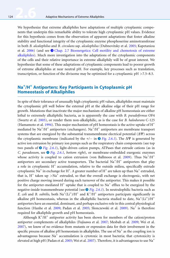
We hypothesize that extreme alkaliphiles have adaptations of multiple cytoplasmic compo-
nents that underpin this remarkable ability to tolerate high cytoplasmic pH values. Evidence
for this hypothesis comes from the observation of apparent adaptations that foster alkaline
stability and functional integrity of the cytoplasmic enzyme phosphoserine aminotransferase
in both B. alcalophilus and B. circulans ssp. alcalophilus (Dubnovitsky et al. 2005; Kapetaniou
et al. 2006) (and see >Chap. 2.7 Bioenergetics: Cell motility and chemotoxis of extreme
alkaliphiles). Much more investigation into the adaptations of the cytoplasmic components
of the cells and their relative importance in extreme alkaliphily will be of great interest. We
hypothesize that some of these adaptations of cytoplasmic components lead to poorer growth
of extreme alkaliphiles at near neutral pH. For example, key processes such as translation,
transcription, or function of the divisome may be optimized for a cytoplasmic pH >7.5–8.5.
Na+/H+ Antiporters: Key Participants in Cytoplasmic pHHomeostasis of Alkaliphiles
In spite of their tolerance of unusually high cytoplasmic pH values, alkaliphiles must maintain
the cytoplasmic pH well below the external pH at the alkaline edge of their pH range for
growth. Mutations that inactivate the major mechanism of alkaline pH homeostasis are either
lethal to extremely alkaliphilic bacteria, as is apparently the case with B. pseudofirmus OF4
(Swartz et al. 2005), or render them non-alkaliphilic, as is the case for B. halodurans C-125
(Hamamoto et al. 1994). This major mechanism of pH homeostasis is the active uptake of H+
mediated by Na+/H+ antiporters (exchangers). Na+/H+ antiporters are membrane transport
systems that are energized by the substantial transmembrane electrical potential (DC) across
the cytoplasmic membrane (indicated by the +/� in > Fig. 2.6.1). The DC is generated by
active ion extrusion by primary ion pumps such as the respiratory chain components (see top
two panels of > Fig. 2.6.1), light-driven cation pumps, ATPases that extrude cations (as in
C. paradoxum, see > Fig. 2.6.1, bottom right), or membrane-embedded exergonic enzymes
whose activity is coupled to cation extrusion (von Ballmoos et al. 2009). Thus Na+/H+
antiporters are secondary active transporters. The bacterial Na+/H+ antiporters that play
a role in cytoplasmic H+ accumulation, relative to the outside milieu, specifically extrude
cytoplasmic Na+ in exchange for H+. A greater number of H+ are taken up than Na+ extruded,
that is, H+ taken up >Na+ extruded, so that the overall exchange is electrogenic, with net
positive charge moving inward during each turnover of the antiporter. This makes it possible
for the antiporter-mediated H+ uptake that is coupled to Na+ efflux to be energized by the
negative-inside transmembrane potential (see > Fig. 2.6.1). In neutralophilic bacteria such as
E. coli and B. subtilis, both Na+(Li+)/H+ and K+/H+ antiporters participate significantly in
alkaline pH homeostasis, whereas in the alkaliphilic bacteria studied to date, Na+(Li+)/H+
antiporters have an essential, dominant, and perhaps exclusive role in this central physiological
function (Hanhe et al. 2009; Padan et al. 2005; Slonczewski et al. 2009). Na+ is therefore
required for alkaliphile growth and pH homeostasis.
Although K+/H+ antiporter activity has been shown for members of the cation/proton
antiporter complements of alkaliphiles (Fujisawa et al. 2007; Mesbah et al. 2009; Wei et al.
2007), we know of no evidence from mutants or expression data for their involvement in the
specific process of alkaline pH homeostasis in alkaliphiles. The use of Na+ as the coupling ion is
advantageous because Na+ accumulation is cytotoxic in most bacteria; that cytotoxicity is
elevated at high pH (Padan et al. 2005;Wei et al. 2007). Therefore, it is advantageous to use Na+
Adaptive Mechanisms of Extreme Alkaliphiles 2.6 125
to support alkaliphily via pH homeostasis since it concomitantly prevents cytotoxic accumu-
lation of Na+. It is further advantageous because of the inwardly directed electrochemical
gradient of Na+ that is generated by the high activity of Na+/H+ antiporters. This ‘‘sodium
motive force’’ (smf) is vitally important as a mode of energizing other bioenergetic work, such as
ion-coupled solute uptake, toxin extrusion, and motility under conditions in which the
protonmotive force (pmf, the transmembrane electrochemical gradient of protons) is low, as
discussed below. We have further hypothesized that use of K+/H+ antiporters for alkaline pH
homeostasis in extreme alkaliphiles would be problematic and there are probably mechanisms to
preclude its use at very alkaline pH. Because extraordinarily high levels of antiport activity are
required for alkaliphile growth at very high pH, use of K+/H+ antiporters would create the risk of
lowering the levels of cytoplasmic K+. Such reductions in cytoplasmic [K+] would be cytotoxic
because sufficient levels of this cation are required for optimal function of many cytoplasmic
proteins. In addition, the cytotoxicity of Na+ is exacerbated when K+ is depleted (Padan et al.
2005; Wei et al. 2007).
Redundancy in the Cation/Proton Antiporter Complements ofAlkaliphiles, and the Importance of Mrp-Type Antiporters inAlkaliphily
With rare exceptions, individual bacterial strains possess multiple cation/proton antiporters
(CPAs) that exchange Na+, Li+, K+ , or Ca2+ or some combination of these cytoplasmic cations
for external H+ (Krulwich et al. 2009; Padan et al. 2005; Slonczewski et al. 2009). For non-
marine bacteria, a typical range is 5–9 CPAs from several distinct antiporter families, as charac-
terized by sequence-based analyses in the Transporter Classification system (Ren et al. 2007; Saier
2002). For example, the B. subtilis complement is 6–7 antiporters and the complement from
E. coli contains a total of 8 CPAs (> Table 2.6.1). While the total CPA antiporter complement of
most of the examined alkaliphiles is within the range of neutralophiles, two alkaliphiles stand
out as exceptions. B. pseudofirmus OF4 has an unusually high number of antiporters, with a
total of 12, principally because of the large number of NhaC candidate proteins in its
genome (6). The organism with the largest complement, however, is the poly-extremophilic
N. thermophilus (> Table 2.6.1). We have hypothesized that bacteria that are challenged by
alkaline pHas their central and only extreme challenge rely heavily on a single, especially adapted
antiporter, a Na+/H+ antiporter with the requisite properties to meet that challenge (Krulwich
et al. 2009). Poly-extremophiles, on the other hand, have an overlapping set of challenges in
which antiporters may play a role, necessitating deployment of many more antiporters that
play roles under conditions in which the different aspects of the extremophily dominate.
CPA3 family members are widespread among both Gram-negative and Gram-positive
bacteria (as well as archaea) although they are absent in largely fermentative bacteria whose
ecological challenge is more commonly acid than alkali, for example, enteric bacteria, strep-
tococci, and lactobacilli (Swartz et al. 2005). The common themes that emerge from studies of
CPA3, Mrp-type antiporter roles and/or expression patterns in neutralophiles and alkaliphiles
are alkali and salt resistance (Hanhe et al. 2009; Kosono et al. 2005; Krulwich et al. 2007; Swartz
et al. 2005; Swartz et al. 2007). One Mrp-type antiporter system, Pha1 of Sinorhizobium
meliloti, catalyzes both K+/H+ and Na+/H+ antiport activities and the former activity has
a physiological role under nitrogen fixation conditions (Putnoky et al. 1998; Yamaguchi et al.
2009). In extremely alkaliphilic Bacillus species, B. halodurans C-125 and B. pseudofirmusOF4,
and in the alkaliphilic cyanobacterium, Anabaena sp. PCC7120, mutational evidence indicates

.Table
2.6.1
Cation/protonantiporter(CPA)candidatesforNa+(K
+)(Ca2+)/H+antiportcapacity
asrevealedbygenomicanalysesofselectedalkaliphilicandtw
omodel
neutralophilicbacteriaa
Organism
Alkaliphile/
neutralophile
Gram
+
or�
CPA1
CPA2
CPA3
NdhF-a
bNhaA
NhaB
NhaC
NhaD
CaCAa
Total
Bacilluspseudofirm
usOF4
Extremealkalip
hile
+3
21
00
06
00
12
BacillushaloduransC-125
Extremealkalip
hile
+1
11
00
02
00
5
BacillusclausiiK
SM-K16
Moderate
alkalip
hile
+1
02
00
04
00
7
Alkalilim
nicola
ehrlicheiMLH
E-1
Moderate
alkalip
hile
�2
02
00
00
00
4
NatranaerobiusthermophilusJW
/NM-W
N-LFc
Moderate
alkalip
hile;
poly-extremophile
+2
21
10
011
00
17
Synechocystissp.P
CC6803
Moderate
alkalip
hile
�3
31d
00
00
01
8
Bacillussubtilis
subsp.subtilis
str.168
Neutralophile
+1
21d
(1)
00
20
07(6)
Escherichia
coliK-12str.K-12substr.MG1655
Neutralophile
�2
30
01
10
01
8
Antiportersofthreemajoran
tiportercategoriesthat
areintheTransporterClassificationsystem
arefoundintherepresentative
bacteriashown:C
PA-typ
es,cation/protonan
tiporters
that
aresingle
ortw
o-geneproduct
antiporters
(CPA1orCPA2)orhetero-olig
omericMrp-typ
eCPA3an
tiporters
that
contain
6–7hyd
rophobicproteins;Nhaan
tiporters
that
are
hyd
rophobicmonomerordim
erproductsofa
singlegene;andasubsetoftheCaC
Afamily
antiportersthat
may
havemonovalentcation/protonan
tiportactivity.TheNdhF-aproteins
includeNt-Nhaan
tiporterfrom
N.thermophilum
andahomologuefrom
B.subtilis
that
isunlikelyto
havean
tiportactivity,asdiscussedinthetext.Therefore,thetotalnumberofCPAs
listedforB.subtilis
indicatesin
parenthesesthat
itshould
perhap
snotbecounted.Sim
ilarly,thetw
oNhaD
antiportercandidatesin
thedraftsequence
ofB.p
seudofirm
usOF4
may
instead
bearsenitetran
sportersan
dparenthesesareag
ainusedto
indicatethispossibility
that
theCPAtotalw
ould
be8instead
of10forthisalkalip
hile.
aAntiportdataaretakenfrom
www.m
embranetran
sport.org
(allorgan
ismsexceptN.thermophilus,Synecoccussp.P
CC7002,andB.p
seudofirm
usOF4,thelatterdatabeingderived
from
adraftgenomesequence)an
dsupplementedwithsearchesofan
notatedgenomesin
www.ncb
i.nlm
.nih.gov/genomes/lproks.cgiu
singan
tiportersoran
tiportersubunitsfrom
thedifferenttypes.CaC
Amembersshownto
beCa2
+-specificorthat
areap
parentNa+/Ca2
+an
tiportersareomitted.
bThistran
sportertypeisnotclassifiedin
www.m
embranetran
sport.org
butisdescribedin
(Krulwichetal.2009).
cN.thermophilusistheonlyan
aerobein
thistable.
dNotlistedin
www.m
embranetran
sport.org.
126 2.6 Adaptive Mechanisms of Extreme Alkaliphiles

Adaptive Mechanisms of Extreme Alkaliphiles 2.6 127
that Na+/H+ antiport-specific members of the Mrp-type antiporters are crucial for alkaline pH
homeostasis in the upper ranges of pH (Hamamoto et al. 1994; Swartz et al. 2005). There is
a striking continuum with respect to the dominance of the Mrp role in the alkaline pH
homeostasis capacity of these alkaliphiles and neutralophilic B. subtilis. In B. pseudofirmus
OF4, the Mrp antiporter is apparently necessary for viability (Swartz et al. 2005). In
B. halodurans C-125, its mutation leads to an inability to grow at pH values above 9 and also
to increased sensitivity to inhibition by Na+ (Hamamoto et al. 1994). In Anabaena sp.
PCC7120 disruption of Mrp leads only to the inability to grow at pH values above 10 as well
as a pronounced increase in Na+ sensitivity (Blanco-Rivero et al. 2005). In neutralophilic
B. subtilis, loss of Mrp function leads to a modest increase in alkali sensitivity but a very
pronounced sensitivity to inhibition by Na+ (Ito et al. 1999; Ito et al. 2000; Kosono et al. 1999;
Swartz et al. 2005). Thus Mrp antiporters in extreme alkaliphiles are particularly adapted to
essential roles at very high pH, at which pH homeostasis is the central challenge to viability.
They also play roles in Na+-resistance that are not essential to viability; other antiporters of the
alkaliphile complement may contribute substantially to Na+-resistance, for example, NhaC
of B. pseudofirmus OF4 (Ito et al. 1997). It is notable that as the essentiality of a Mrp system in
alkaline pH homeostasis declines in this series of organisms, its relative role in Na+-resistance
increases. The Mrp continuum across these bacterial examples seems, in this way, to illustrate
the ‘‘no free lunch’’ principle at the molecular level. Interestingly, all eight antiporters from
N. thermophilus that have been assayed in vitro so far (2 CPA1s, 1 CPA2, 1 Nt-Nha, and 4
NhaCs) have a capacity for K+/H+ antiport (Mesbah et al. 2009). By contrast, as noted above,
the Mrp system of the more extremely alkaliphilic B. pseudofirmus OF4 as well as the homol-
ogous system called Mnh from the highly alkaline-tolerant Staphylococcus aureus are highly
specific for Na+(Li+)/H+ antiport and do not catalyze K+/H+ antiport (Swartz et al. 2007).
Again, the multiple stresses that must be managed by poly-extremophilic N. thermophilusmay
limit the adaptive constraints it can put on antiporter-based pH homeostasis without decreas-
ing its responses to salt and temperature challenges.
What is unique about Mrp systems that may make them adaptable to a special role in
alkaline pH homeostasis? Themost intensively studiedMrp antiporter systems to date are from
either the Group 1 or Group 2 Mrp systems (Swartz et al. 2005). Group 1 systems are encoded
by operons that contain seven genes, each of which has a hydrophobic membrane protein
product, MrpA-G proteins. Group 2 systems have a fusedMrpA and B so that the operons have
six genes encoding six hydrophobic products, MrpA’, C-G. Two properties of Group 1 and 2
systems led to their classification in their own category, the CPA3 family of the Transporter
Classification system. First, the two largest Mrp proteins, MrpA and MrpD, share significant
homology with a sub-complex of membrane-embedded subunits of the H+-pumping respira-
tory chain Complex I (NADH dehydrogenase) that is thought to be involved in H+ translo-
cation (Mathiesen and Hagerhall 2002). They share an oxidoreductase motif with these
Complex I subunits, for example, NuoL, M, and N in the E. coli nomenclature, that also
have homologues among ion-pumping bacterial hydrogenases (Friedrich and Weiss 1997;
Swartz et al. 2005). Similarity between MrpC and the NuoK of Complex I has also been
cited, albeit less striking than between MrpA and D and its homologues among subunits of
Complex I and hydrogenases (Mathiesen and Hagerhall 2003). > Figure 2.6.3 shows an
unrooted tree based on alignments of the family of MrpA/MrpD/NuoL/NuoM/NuoN proteins
(as well as Nt-Nha and homologues thereof that are discussed further below). The MrpA
proteins (which are larger than MrpD proteins, i.e., close to 90 KDa versus the approximate
55 KDa size of MrpD proteins) are more closely related to NuoL of Complex I than to most

Sa_MrpD Bc_MrpD
Bs_MrpD
Bp_MrpD
Bh_MrpDS6803_MrpD1
S6803_MrpD2
Ec_NuoL
Vc_MrpA
Pa_MrpA
Sa_MrpA
Bp_MrpA
Bs_MrpA Bh_MrpABc_MrpAVc_NdhF
Bs_NdhF
Sa_NdhF
Ec_NuoN
Ec_NuoM
Nt_MrpA
Nt_MrpD
Nt_Nha
Pa_MrpDVc_MrpD
0.1
. Fig. 2.6.3
The family of antiporter proteins MrpA, MrpD, and Nt-Nha; Nt-Nha homologues; and
respiratory chain complex I proteins NuoL, NuoM, and NuoN. Unrooted tree (TreeView) of
ClustalW (DSGene) analysis shows relationships between the two large subunits of Mrp
antiporters encoded in diverse mrp operons, the Nt-Nha antiporter protein, three
neutralophile homologues of MrpD/MrpA/Nt-Nha that, as discussed in the text, are unlikely to
have cation/proton antiport activity, and the homologues NuoL, M and N subunits of the
respiratory Complex I (H+-translocating NADH dehydrogenase) from E. coli. The bacterial species
and accession numbers for the protein sequences used are: Bc, B. clausii KSM-K16, MrpA
(YP_174287.1), MrpD (YP_174290.1); Bh, B. halodurans C-125, MrpA (NP_242185.1), MrpD
(NP_242182.1); Bp, B. pseudofirmus OF4, MrpA (AAF21812.2), MrpD (AAF21815.2); Bs, B. subtilis
subsp. subtilis str N16961, MrpA (NP_391038.2), MrpD (NP_440574.1), NdhF (NP_388064); Ec, E.
coli K-12 MG1655, NuoL, NuoM, NuoN (NP_416781.1, NP_416780.1 and NP_416779.2); Nt,
Natranaerobius thermophilus JW/NM-WN-LF, MrpA (YP_001916693.1), MrpD (YP_001916692.1),
Nt-Nha (YP_001916294.1); Pa, Pseudomonas aeruginosa PA)1, MrpA (NP_249745.1), MrpD
(NP_232555.1): S6803, Synechocystis 6803, MrpD1 and MrpD2 (NP_440572.1, NP_440574.1); Sa,
Staphylococcus aureus subsp. aureus COL (in which MrpA and MrpD are usually called MnhA
and MnhD (Hiramatsu et al. 1998)), MrpA (YP_185821.1), Mrp D (YP_185821.1), NdhF
(YP_185382.1); Vc, Vibrio cholera O1 biovar El Tor str. M16961, MrpA (NP_232557.1), MrpD
(NP_232555.1), NdhF (NP_231221.1)
128 2.6 Adaptive Mechanisms of Extreme Alkaliphiles

Adaptive Mechanisms of Extreme Alkaliphiles 2.6 129
MrpD proteins and the NuoM and NuoN pair of Complex I proteins. The exception is the
MrpA that is encoded in an operon in N. thermophilus but is significantly smaller and lacking
a ‘‘MrpB-like’’ domain commonly found in operon-encoded MrpA proteins. The
N. thermophilus MrpA as well as most MrpD proteins are more closely related to NuoM and
NuoN; the NuoM and NuoN subunit pair from Complex I are also more closely related to each
other than they are to NuoL. Only the two cyanobacterial Synechococcus strain 6803 MrpD
proteins cluster closer to the NuoL than to NuoM and NuoN, consistent with several points of
divergence of cyanobacterial Mrp operon patterns from the Group 1 and Group 2 patterns
(Krulwich et al. 2007; Swartz et al. 2005).
The second property that was recognized shortly after Mrp antiporter systems were discov-
ered is that antiport activity requires all of the 6–7 gene products of theGroup 1 or 2mrp operon.
This was first shown for the Mnh antiporter of the CPA3 family from S. aureus using in vitro
assays of truncated products of a full cloned mnh operon expressed in an antiporter-deficient
E. coli strain (Hiramatsu et al. 1998). Subsequently, nonpolar deletions were made in each gene
of the B. subtilis chromosomal copy of themrp operon (Ito et al. 2000) and in a cloned copy of
the B. pseudofirmusOF4mrp operon expressed in an antiporter-deficient E. coli strain (Morino
et al. 2008). Studies of both of these mutant panels confirmed the need for every Mrp protein
for significant antiport activity. The only modest exception was a small residual antiport
activity in the absence of MrpE (Morino et al. 2008; Yoshinaka et al. 2003). The requirement
for 6–7 hydrophobic proteins and the similarity of three of the Mrp proteins to subunits of
Complex I and hydrogenases that form sub-complexes led to the expectation that Mrp
functions as a hetero-oligomeric complex (Hiramatsu et al. 1998). This was confirmed by
demonstration of complexes containing each of the 7 Mrp proteins, first for B. subtilis Mrp
(Kajiyama et al. 2007) and then for B. pseudofirmus OF4 Mrp (Morino et al. 2008). Although
the analyses of the neutralophile Mrp complex and the alkaliphile Mrp complex were
conducted under different conditions and with different detection strategies, both prepara-
tions contained hetero-oligomeric Mrp complexes that included all 7Mrp proteins and were of
a size close to the expected size of a complex with one copy of each Mrp protein. However, the
alkaliphile samples also contained larger species, including a significant amount of a putative
full Mrp dimeric hetero-oligomer (Morino et al. 2008). We hypothesize that the unusual
hetero-oligomeric Mrp proteins are a consortium of transporters that may include two
Na+(K+)/H+ antiporters, or two Na+/H+ antiporters in extreme alkaliphiles, as well as other
transporter proteins that function synergistically with the antiporters. Additional activities would
account for several reports of transport capacities of Mrp systems that are not related to antiport
(Dzioba-Winogrodzki et al. 2009; Kashyap et al. 2006). The large hetero-oligomeric Mrp
complex is hypothesized to support antiport at high external values of pH by presenting
a large protein surface on the outside surface of the membrane. Such a surface could be
engineered to be an effective proton-gathering element, that could resemble, for example,
a larger and more extensive version of proton-gathering funnels observed in the high resolu-
tion crystal structure of E. coli antiporter NhaA (Padan 2008). Larger, dimeric hetero-
oligomeric complexes observed in alkaliphiles might be better adapted to serve this role than
monomeric Mrp complexes.
The requirement for all 6–7 gene products of operon-encoded Mrp systems in order to
observe antiport has made it difficult to test hypotheses that MrpA and MrpD are the actual
antiporter proteins (Mathiesen and Hagerhall 2002) or a more recent suggestion that MrpA,

130 2.6 Adaptive Mechanisms of Extreme Alkaliphiles
MrpD, and MrpB constitute the critical catalytic core (Kajiyama et al. 2009). Therefore, the
recent study of the antiporter complement of N. thermophilus (Mesbah et al. 2009) that
included Nt-Nha constitutes a breakthrough. Nt-Nha is a MrpD/MrpA-like protein that has
antiport activity without any additional Mrp-like proteins. Nt-Nha is more closely related to
MrpD than MrpA (> Fig. 2.6.3). It lacks the MrpB domain that is found in the MrpA but not
MrpD proteins encoded in Group 1 and Group 2 operons. The MrpB domain is not present in
the MrpD-like homologues of cyanobacterial and the N. thermophilus operon-encoded Mrp
systems; these subunits are sometimes (probably erroneously) annotated as MrpA. The fact
that Nt-NhaA catalyzes Na+(K+)/H+ antiport without any other Mrp proteins supports the
idea that a MrpD/MrpA-like protein is the antiporter and argues against the absolute essen-
tiality of any other Mrp proteins to produce the antiport activity. There are other examples of
bacterial genes that are not in an apparent mrp operon but encode MrpA/MrpD/Nt-Nha-like
proteins. The genes are most commonly annotated as NdhF because they are closely related to
genes found in cyanobacterial and plant gene clusters that encode ion-pumping NADH dehydro-
genases (Battchikova and Aro 2007). The three examples shown in > Fig. 2.6.3, from B. subtilis,
S. aureus, and V. cholerae are not themselves likely to be active Na+(K+)/H+ antiporters since
they lack one or more residues that have been shown to be essential for such activity (Kajiyama
et al. 2009). These proteins may catalyze an antiport activity with different substrates and/or
their activity may depend upon modulation by a conserved protein that is not a hydrophobic
Mrp protein but is predicted to be co-transcribed with their genes. Currently, Mrp systems
constitute a structure-function puzzle of central interest with respect to alkaliphile pH homeo-
stasis mechanisms and the Nt-Nha antiporter may be one productive avenue to gain insights
that will suggest experiments with the larger more complex Mrp systems.
Additional Structural, Enzymatic, and Metabolic Strategies forCytoplasmic pH Homeostasis, and Their Built-In Redundancies
Extreme alkaliphiles employ additional strategies to supplement the contribution of antiporter-
dependent pH homeostasis. Secondary cell wall polymers (SCWP, see > Fig. 2.6.1), especially
teichuronopeptides, play a major role in supporting pH homeostasis in extremely alkaliphilic
B. halodurans C-125 (Aono et al. 1999). By contrast, the slightly more robustly alkaliphilic
B. pseudofirmusOF4 lacks comparable SCWPs. B. pseudofirmusOF4 expresses an acidic S-layer
both at pH 7.5 and 10.5. Interestingly, deletion of the slpA gene that encodes the S-layer has
only a modest adverse effect on the response of the alkaliphile to a sudden alkaline shift, that is,
the S-layer confers a modest advantage for alkaliphily. Growth of the slpA mutant at pH 7.5 is
greatly enhanced relative to the wild type, indicating that expression of slpA at near neutral pH
is quite detrimental to the alkaliphile (Gilmour et al. 2000; Krulwich et al. 2007). This is
a cogent example of the ‘‘no free lunch’’ idea that there is a palpable cost to this alkaliphile, with
respect to growth at near neutral pH, for its expression of a gene that modestly optimizes its
readiness for a transition to the extreme condition of alkaline pH.
Metabolic strategies are used by neutralophilic bacteria when challenged with alkali.
A major one is up-regulation of deaminases that produce cytoplasmic acids (Slonczewski
et al. 2009). This might be a problematic strategy for extreme alkaliphiles because the ammonia
released would be protonated and could thus tend to accumulate in the cytoplasm under the
prevailing condition of a higher cytoplasmic [H+] than the external medium during growth in
the highly alkaline pH range. Use of deaminases to produce acids that lower cytoplasmic pH

Adaptive Mechanisms of Extreme Alkaliphiles 2.6 131
would depend upon robust ammonium ion extrusion systems. Such systems have been
suggested but their adequacy to support a robust deaminase strategy of pH homesotasis will
require further investigation (Fujisawa et al. 2007; Wei et al. 2003). Fermentations that produce
metabolic acids can also spare the use of energy-dependent antiport (Krulwich et al. 2007). The
general physiology of alkaliphiles is reviewed elsewhere in this handbook (>Chap. 2.4 Anaer-
obic Alkaliphiles and Alkaliphilic Poly-Extremophiles)
A Low Bulk Protonmotive Force (pmf) Resulting from Successful pHHomeostasis, and Its Bioenergetic Consequences
The pmf energizes H+-coupled ‘‘bioenergetic work’’ such as ion-coupled solute transport or
toxin efflux, flagellar motility, and ATP synthesis using an F1F0-ATP synthase. This transmem-
brane electrochemical proton gradient is composed of two components. In the orientation that
is productive for energization of H+-coupled bioenergetic work, these pmf components are
an electrical component (DC, inside negative relative to the outside) and a chemical
component (DpH, inside alkaline relative to the outside) (Mitchell 1961). However, because
of successful pH homeostasis, the DpH component is in the reverse orientation at the high
end of the alkaliphiles’ pH range for growth, that is, the cytoplasm is acidic relative to the
outside pH. Moreover, in alkaliphiles studied to date, the DC increases at high pH but not
enough to offset the adverse effect of the reversed DpH on the pmf. As a result the bulk pmf
is much lower in facultative alkaliphiles at the upper edge of their pH range than at near
neutral pH, for example, three times lower in B. pseudofirmus OF4 during growth at pH 10.5
than during growth at pH 7.5 (Krulwich et al. 2007; Sturr et al. 1994). The low pmf problem is
particularly acute because the cost of growing at pH 10.5 is higher because of the need for
high levels of DC-driven antiport and because there is a proton involved in the catalytic
reaction by which ATP is synthesized, which makes the reaction less favorable at high pH.
Still B. pseudofirmusOF4, grows as well at pH 10.5 as at pH 7.5, with comparable molar growth
yields and even faster generation times (Guffanti and Hicks 1991; Sturr et al. 1994). For solute-
coupled uptake and motility (see >Chap. 2.6 Adaptive mechanisms of extreme alkaliphiles),
a major strategy of alkaliphiles, including many cyanobacteria, is use of Na+-coupling instead
of H+-coupling as indicated in > Fig. 2.6.1 (Krulwich et al. 2007; Peddie et al. 2000). The
sodium motive force (smf) is larger than the pmf at highly alkaline pH since the antiport that
establishes the ‘‘reversed’’ DpH concomitantly generates an inwardly-directed chemical gradi-
ent of Na+ that adds to the significant, productively oriented DC to make a large smf. Na+-
coupled transporters of neutralophiles often work side-by-side with H+-coupled solute trans-
porters (see > Fig. 2.6.1) or can couple to both cations (Zani et al. 1993). By contrast, the Na+-
coupled solute transporters of extreme alkaliphiles cannot use H+ and are instead inhibited at
the elevated [H+] at near neutral pH. This accounts for the otherwise puzzling observation that
a higher [Na+] is required in the medium for growth of B. pseudofirmus OF4 on malate, which
is transported by Na+-dependent transport, at pH 7.5 than at pH 10.5 (Gilmour et al. 2000;
Krulwich et al. 2007). Some alkaliphiles may partially bypass this suboptimal property of
alkaline-adapted Na+-coupled solute transport at low pH, by employing transporters that are
not coupled to either the pmf or smf. It was noted in the B. halodurans C-125 genome that this
alkaliphile makes much greater use of ABC type, ATP-coupled transporters than found in
neutralophilic B. subtilis (Takami et al. 2000).

132 2.6 Adaptive Mechanisms of Extreme Alkaliphiles
Strategies for Oxidative Phosphorylation at LowProtonmotive Force
The biggest conundrum with respect to bioenergetic work is that both extreme and moderate
alkaliphiles use H+-coupled rather than Na+-coupled ATP synthases (Krulwich et al. 2007;
von Ballmoos et al. 2008). Na+-coupled F1F0-ATPases are found in alkaliphilic anaerobes
(see C. paradoxum panel in > Fig. 2.6.1) that do not synthesize ATP with the enzyme under
physiological conditions. They use it in the hydrolytic direction that generates an smf
(Ferguson et al. 2006). Use of a Na+-pumping ATPase that generates an smf fits the overall
Na+-based energetics, reduces Na+ cytotoxicity, and also avoids cytoplasmic proton loss that
would occur using a H+-coupled ATPase at high pH. For cyanobacteria, use of a H+-coupled
ATP synthase for ATP synthesis may not be problematic because the low pmf problem applies
to ATP synthases that function in the cytoplasmic membrane across which this pmf is the
driving force for synthesis. Recent reports (Liberton et al. 2006; Nevo et al. 2007; Schneider
et al. 2007) indicate that the thylakoid membranes in which the synthase is localized is not
continuous with the cytoplasmic membrane. In that case, as shown in> Fig. 2.6.1, the problem
of low bulk pmf across the cytoplasmic membrane would not affect the energetics of ATP
synthesis. On the other hand, the low pmf conundrum is a major issue for extreme alkaliphiles
whose respiratory chains and ATP synthases, which constitute the machinery for oxidative
phosphorylation (OXPHOS), are in the cytoplasmic membranes. The problem may be especially
acute for thoseGram-positive alkaliphiles that lack the outermembrane ofGram-negative bacteria
or particularly protective SCWPs.
Solutions have been proposed to the conundrum of the robust H+-coupled ATP synthesis
observed in alkaliphiles at high pH and low bulk pmf and evidence for adaptations of the
OXPHOS machinery has also emerged. Proposals that explain the discrepancy between
the bulk pmf and ATP synthesis have focused on different ways in which the H+ pumped
by the respiratory chain may be sequestered from equilibration with the bulk medium. For
example, it has been hypothesized that rapid transfers of pumped H+ along the membrane
surface allow the H+ to reach the ATP synthase before they are equilibrated with the bulk phase
outside the cell. These surface-sequestered H+ would kinetically bypass the longer term
thermodynamic inadequacy of the low [H+] concentration in the bulk phase since the effective
near-membrane driving force would be higher than the low bulk pmf. Such proposals are similar
to models of energization of bioenergetic work proposed originally by RJP Williams
(Williams 1978). There are different recent models, including the suggestion based on computa-
tional modeling of forces near the membrane of interfacial barriers that promote retention of H+
near the surface in a delocalized manner and the suggestion based on in vitro experimental
models of H+ microcircuits near the membrane that depend upon proximity of the pumps to
the synthase and on properties of the membrane lipids (Branden et al. 2006; Mulkidjanian
et al. 2005). Some form of sequestered H+ translocation is likely to be a critical part of the
alkaliphile solution to the problem of OXPHOS at low bulk pmf but the suggestions are
currently hypotheses that still await more experimental data, especially in biological settings
(Krulwich 1995; Krulwich et al. 2007). The alkaliphileOXPHOS capacitymay, as predicted by the
microcircuit hypothesis, rely upon close proximity of critical respiratory chain pumps, especially
the terminal oxidases, to the ATP synthase; these partners in H+ transfers during alkaliphile
OXPHOSmight be found in clusters (Goto et al. 2005; Liu et al. 2007). The high concentration
of membrane cardiolipin has been suggested as a possible mediator of proximity and/or H+
transfer itself (Haines and Dencher 2002).

Adaptive Mechanisms of Extreme Alkaliphiles 2.6 133
A more firmly established facet of OXPHOS in alkaliphilic Bacillus species such as
B. pseudofirmus OF4 is its dependence upon specific adaptations of the ATP synthase. There
also appear to be parallel adaptations in the terminal oxidase of the OXPHOS respiratory chain
component (Krulwich et al. 2007; Slonczewski et al. 2009). Such special adaptations of
OXPHOS machinery in response to specific environmental challenges are in fact widespread
(Ferguson and Ingledew 2008; von Ballmoos et al. 2008). Alkaliphile-specific sequence motifs
have been identified in both the F1F0-ATP synthase (examples in > Fig. 2.6.4, top) and the Cta
caa3-type cytochrome oxidase (examples in > Fig. 2.6.4, bottom) (Ivey and Krulwich 1992;
Quirk et al. 1993). In addition, unusual redox features of alkaliphile respiratory chain com-
ponents have been described (Goto et al. 2005; Hicks and Krulwich 1995; Muntyan and
Bloch 2008). Mutational work has not yet been conducted on the respiratory chain motifs
but ATP synthase motifs have been shown to play important roles in OXPHOS at high pH and
low pmf. For example, the ‘‘alkaliphile-specific’’ a-subunit Lys180 plays a critical role in
synthesis at high pH and is proposed to be required for H+ capture from the outside surface
of the synthase and passage to the interface between the a-subunit and the c-ring rotor (see> Fig. 2.6.4, top) (McMillan et al. 2007; Wang et al. 2004). There are also critical motifs on both
helices of the hairpin-like c-subunits that constitute the oligomeric rotor; their mutational
change to the consensus sequence for non-alkaliphiles severely compromises ATP synthesis at
high pH (Liu et al. 2009; Wang et al. 2004). Part of the adaptive value of the ATP synthase
motifs of alkaliphiles appears to be in preventing H+ loss to the outside, since several mutations
to the consensus sequence result in significant H+ leakiness (Liu et al. 2009; Wang et al. 2004).
Another proposed adaptation that could address or partially address the alkaliphile
OXPHOS conundrum relates to the number of c-subunits that comprise the c-ring rotor
(> Fig. 2.6.4, top). The stoichiometry of the c-ring varies among different organisms in
which it has been determined from 10–15 (von Ballmoos et al. 2008; von Ballmoos et al.
2009). An especially high c-subunit stoichiometry would help address the problem of
OXPHOS at low pmf (Meier et al. 2007; von Ballmoos et al. 2008). The thermoalkaliphile
Bacillus sp.TA21.A1 has a stoichiometry of 13 subunits/ring, which is indeed toward the high
end of the known range of stoichiometries, but even though the organism is a moderate
alkaliphile, the stoichiometry does not account for the energetics observed (Matthies et al.
2009; Meier et al. 2007). There also does not appear to be a strong correlation between a high
c-ring stoichiometry andOXPHOS at low pmf. The cyanobacterium S. platensis has the highest
c-ring stochiometry reported to date, at 15 subunits/ring (Pogoryelov et al. 2005; Pogoryelov
et al. 2009), even though the localization of its ATP synthase in intracellular thylakoids
probably avoids the low pmf problem of alkaliphile OXPHOS that takes place in the cytoplas-
mic membrane (> Fig. 2.6.1). The high stoichiometry in S. platensis may relate to other
properties of the setting such as the relative contributions of the two pmf components (von
Ballmoos et al. 2008; von Ballmoos et al. 2009).
Even if a somewhat high c-subunit stoichiometry plays, at most, a minor role in the
alkaliphile OXPHOS condundrum, we note another point of interest connected with this
stoichiometry. There is now a strong consensus that the stoichiometry of the c-ring apparently
does not vary within a single organism under different conditions such as growth pH or growth
substrate (Meier et al. 2005; von Ballmoos et al. 2008). If alkaliphiles follow the Bacillus sp. TA2.
A1 trend of higher than average c-subunit stoichiometry, we would cite it as one more example
of the ‘‘no free lunch’’ principle. A c-ring with a high stoichiometry supports a higher H+/ATP
stoichiometry for the ATP synthase and that, in turn, facilitates synthesis at low pmf. However,
an ATP synthase with high H+/ATP stoichiometry is a less economical energy converter than

Predictedpl
# basicresidues
H+ c ring
F0
F1
ααδ
β
εγ
ATP
H+
H+
a
H+
b2
ADP+ Pi
# acidicresidues
Outer C-terminal helixof each c -subunit hasPXXEXXP motif.Inner N-terminal helix hasAXAXAXA motifLys180
227
207
3.3
3.310.810.17.5
7.57.4
3.01
1654
46
19
8244
46
11
. Fig. 2.6.4
Examples of putative alkali-adaptive motifs in the ATP synthase and cytochrome oxidase
involved in oxidative phosphorylation by extremely alkaliphilic Bacillus species. Top: The
schematic diagram of a bacterial ATP synthase depicts an H+-translocating synthase that is found
in alkaliphiles. The membrane-embedded F0 and cytoplasmic F1 are indicated; they are,
respectively the proton-conducting/rotor elements and the catalytic elements involved in
ATP synthesis. The oligomeric ring of c-subunits, that is, the rotor for the rotary coupling
mechanism for ATP synthesis is shown in green. The stator elements of the synthase are shaded in
blue. As downward movement through the F0 energizes synthesis, rotation of the c-ring in the
direction shown leads to concomitant rotation of the interactive F1 subunits g and « that result in
a conformational change in the three catalytic sites (at the a/b interfaces). Arrows point to
regions of three major adaptations of the F0: a-subunit (aLys180 in the putative H+ uptake path)
and c-subunits (the PXXEXXP and AXAXAXAmotifs in the C- and N-terminal helices, respectively).
Bottom: An alignment of a region of subunit II of Bacillus cytochrome caa3 complexes that
shows that this region in alkaliphiles (species in blue) is significantly more acidic than the same
stretches found in neutralophiles. Acidic residues are shown in red and basic residues in blue.
The acidic and basic residue composition of each species is shown on the right, along with
the predicted isoelectric point (pI) of that region. The numbering refers to the mature form of
the B. pseudofirmus OF4 subunit II
134 2.6 Adaptive Mechanisms of Extreme Alkaliphiles

Adaptive Mechanisms of Extreme Alkaliphiles 2.6 135
a synthase with a lower c-subunit stoichiometry (Pogoryelov et al. 2005). Therefore, the c-ring
with 13 subunits/ring would help the alkaliphile ATP synthase function better at high pHand low
pmf than if it had a stoichiometry of 10, but at near neutral pH and high pmf, the synthase would
function less well than those of neutralophiles that have stiochiometries of 10 subunits/ring.
Conclusions
Adaptations of extreme alkaliphiles optimize growth and survival at pH values above pH 10 but
these adaptations convey a cost at near-neutral pH. A fuller understanding of the range and
utility of different adaptations will depend upon concerted ‘‘omics’’ efforts in a greater range of
genetically tractable alkaliphiles so that mutations can be used to test hypotheses about
structure-function and systems level relationships.
Cytoplasmic pH homeostasis is centrally important to alkaliphily and depends upon active
H+ accumulation by electrogenic, secondary cation/proton antiporters that in extremely
alkaliphilic Bacillus use Na+ as the efflux substrate coupled to H+ uptake.
The tolerance of alkaliphiles for unusually high cytoplasmic pH values and a very limited
amount of experimental data suggest that there are likely to be important adaptations of
cytoplasmic components in extreme alkaliphiles. Bioinformatic approaches to the increasing
number of fully sequenced genomes may lead to testable hypotheses about such adaptations and
also to suggestions for components on the outside of alkaliphile cells whose poor function at near
neutral pH contributes to limitations on growth in this range of pH for many alkaliphiles.
The hetero-oligomeric Mrp antiporter is the dominant cation/proton antiporter of
extremely alkaliphilic Bacillus species examined to date. The basis of the complexity of these
antiporters remains unknown. Experiments also should be designed to test the hypotheses that
the large outer surface of theMrp antiporter plays a role in H+ capture at elevated pH. Roles for
the other members of the antiporter complements still await definition.
Further experimental tests and models based on established parameters of alkaliphile
energetics should also be developed to test various proposals of sequestered H+ pathways
from respiratory and other H+ pathways to the ATP synthase both in alkaliphiles and beyond.
Progress has been made on alkaliphile-specific motifs of the ATP synthase that await
correlation with structural data and parallel development of insights into apparent adaptations
of the respiratory H+ pumps. The hypothesized importance of specific membrane lipids,
especially cardiolipin, also awaits experimental examination.
Acknowledgments
Work conducted in the authors’ laboratories was supported by research grant GM28454 and
the Systems Biology Center-NY grant P50-GM071558 from the National Institute of General
Medical Sciences (T.A.K) and a grant from the 21st Century Center of Excellence program of
the Ministry of Education, Culture, Sports, Science and Technology of Japan (M.I.).
Cross-References
> 2.1 Introduction and History of Alkaliphiles> 2.4 Anaerobic Alkaliphiles and Alkaliphilic Poly-Extremophiles> 2.5 General Physiology of Alkaliphiles> 2.7 Bioenergetics: Cell Motility and Chemotaxis of Extreme Alkaliphiles

136 2.6 Adaptive Mechanisms of Extreme Alkaliphiles
References
Aono R, ItoM,Machida T (1999) Contribution of the cell
wall component teichuronopeptide to pH homeo-
stasis and alkaliphily in the alkaliphile Bacillus lentus
C-125. J Bacteriol 181:6600–6606
Battchikova N, Aro E-M (2007) Cyanobacterial NDH-1
complexes: multiplicity in function and subunit
composition. Physiol Plantarum 131:22–32
Blanco-Rivero A, Leganes F, Fernandez-Valiente E, Calle P,
Fernandez-Pinas F (2005)mrpA, a gene with roles in
resistance to Na+ and adaptation to alkaline pH in
the cyanobacterium Anabaena sp. PCC7120. Micro-
biology 151:1671–1682
Bowers KJ, Mesbah NM, Wiegel J (2009) Biodiversity of
poly-extremophilic Bacteria: does combining the
extremes of high salt, alkaline pH and elevated tem-
perature approach a physico-chemical boundary for
life? Saline Syst 5:9
Branden M, Sanden T, Brzezinski P, Widengren J (2006)
Localized proton microcircuits at the biological
membrane-water interface. Proc Natl Acad Sci USA
103:19766–19770
Ciferri O (1983) Spirulina, the edible microorganism.
Microbiol Rev 47:551–578
Cook GM, Russell JB, Reichert A, Wiegel J (1996) The
intracellular pH of Clostridium paradoxum, an
anaerobic, alkaliphilic, and thermophilic bacterium.
Appl Environ Microbiol 62:4576–4579
Dubnovitsky AP, Kapetaniou EG, Papageorgiou AC
(2005) Enzyme adaptation to alkaline pH: atomic
resolution (1.08 A) structure of phosphoserine ami-
notransferase from Bacillus alcalophilus. Protein Sci
14:97–110
Dzioba-Winogrodzki J,Winogrodzki O, Krulwich TA, Boin
MA, Hase CC, Dibrov P (2009) The Vibrio cholerae
Mrp system: cation/proton antiport properties and
enhancement of bile salt resistance in a heterologous
host. J Mol Microbiol Biotechnol 16:176–186
Ferguson SJ, Ingledew WJ (2008) Energetic problems
faced by micro-organisms growing or surviving on
parsimonious energy sources and at acidic pH:
I. Acidithiobacillus ferrooxidans as a paradigm.
Biochim Biophys Acta 1777:1471–1479
Ferguson SA, Keis S, Cook GM (2006) Biochemical and
molecular characterization of a Na+-translocating
F1F0-ATPase from the thermoalkaliphilic bacterium
Clostridium paradoxum. J Bacteriol 188:5045–5054
Friedrich T, Weiss H (1997) Modular evolution of
the respiratory NADH: ubiquinone oxidoreductase
and the origin of its modules. J Theor Biol
187:529–540
Fujinami S, Terahara N, Krulwich TA, Ito M (2009)
Motility and chemotaxis in alkaliphilic Bacillus spe-
cies. Future Microbiol 4:1137–1149
Fujisawa M, Ito M, Krulwich TA (2007) Three two-
component transporters with channel-like proper-
ties have monovalent cation/proton antiport
activity. Proc Natl Acad Sci USA 104:13289–13294
Gilmour R, Messner P, Guffanti AA, Kent R, Scheberl A,
Kendrick N, Krulwich TA (2000) Two-dimensional
gel electrophoresis analyses of pH-dependent pro-
tein expression in facultatively alkaliphilic Bacillus
pseudofirmus OF4 lead to characterization of an S-
layer protein with a role in alkaliphily. J Bacteriol
182:5969–5981
Goto T, Matsuno T, Hishinuma-Narisawa M, Yamazaki K,
MatsuyamaH, InoueN, Yumoto I (2005) Cytochrome
c and bioenergetic hypothetical model for alkaliphilic
Bacillus spp. J Biosci Bioeng 100:365–379
Guffanti AA, Hicks DB (1991) Molar growth yields and
bioenergetic parameters of extremely alkaliphilic
Bacillus species in batch cultures, and growth
in a chemostat at pH 10.5. J Gen Microbiol
137:2375–2379
Haines TH, Dencher NA (2002) Cardiolipin: a proton
trap for oxidative phosphorylation. FEBS Lett
528:35–39
Hamamoto T, Hashimoto M, Hino M, Kitada M, Seto Y,
Kudo T, Horikoshi K (1994) Characterization of
a gene responsible for the Na+/H+ antiporter system
of alkalophilic Bacillus species strain C-125. Mol
Microbiol 14:939–946
Hanhe H, Mader U, Otto A, Bonn F, Steil L, Bremer E,
Hecker M, Becher D (2009) A comprehensive pro-
teomics and transcriptomics analysis of Baciilus
subtilis salt stress adaptation. J Bacteriol.
doi:10.1128/JB.01106-09
Hicks DB, Krulwich TA (1995) The respiratory chain of
alkaliphilic bacteria. Biochim Biophys Acta
1229:303–314
Hiramatsu T, Kodama K, Kuroda T, Mizushima T,
Tsuchiya T (1998) A putative multisubunit Na+/H+
antiporter from Staphylococcus aureus. J Bacteriol
180:6642–6648
Hoffmann A, Dimroth P (1991) The electrochemical
proton potential of Bacillus alcalophilus. Eur
J Biochem 201:467–473
Horikoshi K (1991) Microorganisms in alkaline environ-
ments. VCH, New York
Ito M, Aono R (2002) Decrease in cytoplasmic pH-
homeostastatic activity of the alkaliphile Bacillus
lentus C-125 by a cell wall defect. Biosci Biotechnol
Biochem 66:218–220
Ito M, Guffanti AA, Zemsky J, Ivey DM, Krulwich TA
(1997) Role of the nhaC-encoded Na+/H+ antiporter
of alkaliphilic Bacillus firmus OF4. J Bacteriol
179:3851–3857

Adaptive Mechanisms of Extreme Alkaliphiles 2.6 137
Ito M, Guffanti AA, Oudega B, Krulwich TA (1999) mrp,
a multigene, multifunctional locus in Bacillus subtilis
with roles in resistance to cholate and to Na+ and in
pH homeostasis. J Bacteriol 181:2394–2402
Ito M, Guffanti AA, Wang W, Krulwich TA (2000) Effects
of nonpolar mutations in each of the seven Bacillus
subtilis mrp genes suggest complex interactions
among the gene products in support of Na+ and
alkali but not cholate resistance. J Bacteriol
182:5663–5670
Ito M, Xu H, Guffanti AA, Wei Y, Zvi L, Clapham DE,
Krulwich TA (2004) The voltage-gated Na+ channel
NavBP has a role in motility, chemotaxis, and pH
homeostasis of an alkaliphilic Bacillus. Proc Natl
Acad Sci USA 101:10566–10571
Ivey DM, Krulwich TA (1992) Two unrelated alkaliphilic
Bacillus species possess identical deviations in
sequence from those of other prokaryotes in regions
of F0 proposed to be involved in proton transloca-
tion through the ATP synthase. Res Microbiol
143:467–470
Kajiyama Y, Otagiri M, Sekiguchi J, Kosono S, Kudo T
(2007) Complex formation by the mrpABCDEFG
gene products, which constitute a principal Na+/H+
antiporter in Bacillus subtilis. J Bacteriol
189:7511–7514
Kajiyama Y, Otagiri M, Sekiguchi J, Kudo T, Kosono S
(2009) The MrpA, MrpB and MrpD subunits of the
Mrp antiporter complex in Bacillus subtilis contain
membrane-embedded and essential acidic residues.
Microbiology 155:2137–2147
Kapetaniou EG, Thanassoulas A, Dubnovitsky AP,
Nounesis G, Papageorgiou AC (2006) Effect of pH
on the strcuture and stability of Bacillus circulans ssp.
alkalophilus phosphoserine aminotransferase: ther-
modynamic and cystallographic studies. Proteins
63:742–753
Kashyap DR, Botero LM, Lehr C, Hassett DJ, McDermott
TR (2006) A Na+: H+ antiporter and a molybdate
transporter are essential for arsenite oxidation
in Agrobacterium tumefaciens. J Bacteriol
188:1577–1584
Kitada M, Guffanti AA, Krulwich TA (1982) Bioenergetic
properties and viability of alkalophilic Bacillus
firmus RAB as a function of pH and Na+ contents
of the incubation medium. J Bacteriol
152:1096–1104
Kosono S, Morotomi S, Kitada M, Kudo T (1999) Ana-
lyses of a Bacillus subtilis homologue of the Na+/H+
antiporter gene which is important for pH homeo-
stasis of alkaliphilic Bacillus sp. C-125. Biochim
Biophys Acta 1409:171–175
Kosono S, Haga K, Tomizawa R, Kajiyama Y, Hatano K,
Takeda S, Wakai Y, Hino M, Kudo T (2005) Charac-
terization of a multigene-encoded sodium/hydrogen
antiporter (Sha) from Pseudomonas aeruginosa: its
involvement in pathogenesis. J Bacteriol
187:5242–5248
Krulwich TA (1995) Alkaliphiles: ‘‘basic’’molecular prob-
lems of pH tolerance and bioenergetics. Mol
Microbiol 15:403–410
Krulwich TA, Hicks DB, Swartz TH, Ito M (2007) Bioen-
ergetic adaptations that support alkaliphily. In:
Gerday C, Glansdorff N (eds) Physiology and bio-
chemistry of extremophiles. ASM, Washington,
pp 311–329
Krulwich TA, Hicks DB, Ito M (2009) Cation/proton
antiporter complements of bacteria: why so large
and diverse? Mol Microbiol 74:257–260
Lander AD, Lo W-C, Nie Q, Wan FYM (2009) The mea-
sure of success: constraints, objectives, and tradeoffs
in morphogen-mediated patterning. Cold Spring
Harb Perspect Biol 1:a002022
Li Y, Mandelco L, Wiegel J (1993) Isolation and charac-
terization of a moderately thermophilic anaerobic
alkaliphile, Clostridium paradoxum sp. nov. Int
J Syst Bacteriol 43:450–460
Liberton M, Berg RH, Heuser J, Roth R, Pakrasi
HB (2006) Ultrastructure of the membrane
systems in the unicellular cyanobacterium
Synechocystis sp. strain PCC 6803. Protoplasma
227:129–138
Liu X, Gong X, Hicks DB, Krulwich TA, Yu L, Yu CA
(2007) Interaction between cytochrome caa3 and
F1F0-ATP synthase of alkaliphilic Bacillus
pseudofirmus OF4 is demonstrated by Saturation
Transfer Electron Paramagnetic Resonance and Dif-
ferential Scanning Calorimetry assays. Biochemistry
46:306–313
Liu J, Fujisawa M, Hicks DB, Krulwich TA (2009) Char-
acterization of the functionally critical AXAXAXA
and PXXEXXP motifs of the ATP synthase c-subunit
from an alkaliphilic Bacillus. J Biol Chem
284:8714–8725
Ma Y, Xue Y, GrantWD, Collins NC, Duckworth AW, Van
Steenbergen RP, Jones BE (2004) Alkalimonas
amylolytica gen. nov., sp. nov., and Alkalimonas
delamerensis gen. nov., sp. nov., novel alkaliphilic
bacteria from soda lakes in China and East Africa.
Extremophiles 8:193–200
Mathiesen C, Hagerhall C (2002) Transmembrane topol-
ogy of the NuoL, M and N subunits of NADH: qui-
none oxidoreductase and their homologues among
membrane-bound hydrogenases and bona fide
antiporters. Biochim Biophys Acta 1556:121–132
Mathiesen C, Hagerhall C (2003) The ‘‘antiporter mod-
ule’’ of respiratory chain Complex I includes the
MrpC/NuoK subunit – a revision of the modular
evolution scheme. FEBS Lett 5459:7–13
Matthies D, Preiss L, Klyszejko AL, Muller DJ, Cook GM,
Vonck J, Meier T (2009) The c13 ring from
a thermoalkaliphilic ATP synthase reveals an

138 2.6 Adaptive Mechanisms of Extreme Alkaliphiles
extended diameter due to a special structural region.
J Mol Biol 388:611–618
McMillan DG, Keis S, Dimroth P, Cook GM
(2007) A specific adaptation in the a subunit of
thermoalkaliphilic F1F0-ATP synthase enables ATP
synthesis at high pH but not at neutral pH values.
J Biol Chem 282:17395–17404
McMillan DG, Keis S, Berney M, Cook GM
(2009) Nonfermentative thermoalkaliphilic growth
is restricted to alkaline environments. Appl Environ
Microbiol 75:7649–7654
Meier T, Yu J, Raschle T, Henzen F, Dimroth P, Muller DJ
(2005) Structural evidence for a constant c11 ring stoi-
chiometry in the sodium F-ATP synthase. FEBS J
272:5474–5483
Meier T, Morgner N, Matthies D, Pogoryelov D, Keis S,
Cook GM, Dimroth P, Brutschy B (2007)
A tridecameric c ring of the adenosine triphosphate
(ATP) synthase from the thermoalkaliphilic Bacillus
sp. strain TA2.A1 facilitates ATP synthesis at low
electrochemical proton potential. Mol Microbiol
65:1181–1192
MesbahNM,Hedrick DB, Peacock AD, RohdeM,Wiegel J
(2007) Natranaerobius thermophilus gen. nov., sp.
nov., a halophilic, alkalithermophilic bacterium
from soda lakes of the Wadi An Natrun, Egypt, and
proposal of Natranaerobiaceae fam. nov. and
Natranaerobiales ord. nov. Int J Syst Evol Microbiol
57:2507–2512
Mesbah NM, Cook GM, Wiegel J (2009) The halophilic
alkalithermophile Natranaerobius thermophilus
adapts to multiple environmental extremes using
a large repertoire of Na(K)/H antiporters. Mol
Microbiol 74:270–281
Mitchell P (1961) Coupling of phosphorylation to elec-
tron and hydrogen transfer by a chemiosmotic type
of mechanism. Nature 191:144–148
Morino M, Natsui S, Swartz TH, Krulwich TA, Ito M
(2008) Single gene deletions of mrpA to mrpG and
mrpE point mutations affect activity of theMrp Na+/
H+ antiporter of alkaliphilic Bacillus and formation
of hetero-oligomeric Mrp complexes. J Bacteriol
190:4162–4172
Mulkidjanian AY, Cherepanov DA, Heberle J, Junge W
(2005) Proton transfer dynamics at membrane/
water interface and mechanism of biological energy
conversion. Biochemistry (Mosc) 70:251–256
Muntyan MS, Bloch DA (2008) Study of redox potential
in cytochrome c covalently bound to terminal oxi-
dase of alkaliphilic Bacillus pseudofirmus FTU. Bio-
chemistry (Mosc) 73:107–111
Nevo R, Charuvi D, Shimoni E, Schwarz R, Kaplan A,
Ohad I, Riech Z (2007) Thylakoid membrane perfo-
rations and connectivity enable intracellular traffic
in cyanobacteria. EMBO J 26:1467–1473
Olsson K, Keis S, Morgan HW, Dimroth P, Cook GM
(2003) Bioenergetic properties of the thermoalka-
liphilic Bacillus sp. strain TA2.A1. J Bacteriol
185:461–465
Padan E (2008) The enlightening encounter between
structure and function in the NhaA Na+-H+
antiporter. Trends Biochem Sci 33:435–443
Padan E, Zilberstein D, Schuldiner S (1981) pH homeo-
stasis in bacteria. Biochim Biophys Acta 650:151–166
Padan E, Bibi E, Ito M, Krulwich TA (2005) Alkaline pH
homeostasis in bacteria: new insights. Biochim
Biophys Acta 1717:67–88
Peddie CJ, Cook GM, Morgan HW (2000) Sucrose trans-
port by the alkaliphilic, thermophilic Bacillus sp.
strain TA2.A1 is dependent on a sodium gradient.
Extremophiles 4:291–296
Pogoryelov D, Sudhir PR, Kovacs L, Gombos Z, Brown I,
Garab G (2003) Sodium dependency of the photo-
synthetic electron transport in the alkaliphilic
cyanobacterium Arthrospira platensis. J Bioenerg
Biomembr 35:427–437
Pogoryelov D, Yu J, Meier T, Vonck J, Dimroth P, Muller
DJ (2005) The c15 ring of the Spirulina platensis
F-ATP synthase: F1/F0 symmetry mismatch is not
obligatory. EMBO Rep 6:1040–1044
Pogoryelov D, Yildiz O, Faraldo-Gomez JD, Meier T
(2009) High-resolution structure of the rotor ring
of a proton-dependent ATP synthase. Nat Struct Mol
Biol 16:1068–1073
Putnoky P, Kereszt A, Nakamura T, Endre G, Grosskopf
E, Kiss P, Kondorosi A (1998) The pha gene cluster of
Rhizobium meliloti involved in pH adaptation and
symbiosis encodes a novel type of K+ efflux system.
Mol Microbiol 28:1091–1101
Quirk PG, Hicks DB, Krulwich TA (1993) Cloning of
the cta operon from alkaliphilic Bacillus firmus OF4
and characterization of the pH-regulated cyto-
chrome caa3 oxidase it encodes. J Biol Chem 268:
678–685
Ren D, Navarro B, Xu H, Yue L, Shi Q, Clapham DE
(2001) A prokaryotic voltage-gated sodium channel.
Science 294:2372–2375
Ren Q, Chen K, Paulsen IT (2007) Transportn DB:
a comprehensive database resource for cytoplasmic
membrane transport systems and outer membrane
channels. Nucleic Acids Res 35:D274–D279
Saier MH (2002) Families of transporters and their clas-
sification. In: Quick M (ed) Transmembrane trans-
porters. Wiley-Liss, New York, pp 1–17
Schaffer C, Messner P (2005) The structure of secondary
cell wall polymers: how Gram-positive bacteria stick
their cell walls together. Microbiology 151:643–651
Schneider D, Fuhrmann E, Scholz I, HessWR, Graumann
PL (2007) Fluorescence staining of live
cyanobacterial cells suggest non-stringent

Adaptive Mechanisms of Extreme Alkaliphiles 2.6 139
chromosome segregation and absence of
a connection between cytoplasmic and thylakoid
membranes. BMC Cell Biol 8:39
Shioi JI, Matsuura S, Imae Y (1980) Quantitative mea-
surements of proton motive force and motility in
Bacillus subtilis. J Bacteriol 144:891–897
Slonczewski JL, Fujisawa M, Dopson M, Krulwich TA
(2009) Cytoplasmic pH Measurement and Homeo-
stasis in Bacteria and Archaea. Adv Microb Physiol
55:1–317
Sturr MG, Guffanti AA, Krulwich TA (1994) Growth and
bioenergetics of alkaliphilic Bacillus firmus OF4 in
continuous culture at high pH. J Bacteriol
176:3111–3116
Suigyama S, Matsukura H, Koyama N, Nosoh Y, Imae Y
(1986) Requirement of Na+ in flagellar rotation and
amino acid transport in a facultatively alkalophilic
Bacillus. Biochim Biophys Acta 852:38–45
Swartz TH, Ikewada S, Ishikawa O, Ito M, Krulwich TA
(2005) The Mrp system: a giant among monovalent
cation/proton antiporters? Extremophiles 9:345–354
Swartz TH, Ito M, Ohira T, Natsui S, Hicks DB, Krulwich
TA (2007) Catalytic properties of Staphylococcus
aureus and Bacillus members of the Secondary
Cation/Proton Antiporter-3 (Mrp) family are
revealed by an optimized assay in an Escherichia
coli host. J Bacteriol 189:3081–3090
Takami H, Nakasone K, Takaki Y, Maeno G, Sasaki R,
Masui N, Fuji F, Hirama C, Nakamura Y,
Ogasawara N, Kuhara S, Horikoshi K (2000) Com-
plete genome sequence of the alkaliphilic bacterium
Bacillus halodurans and genomic sequence compar-
ison with Bacillus subtilis. Nucleic Acids Res
28:4317–4331
von Ballmoos C, Cook GM, Dimroth P (2008) Unique
rotary ATP synthase and its biological diversity.
Annu Rev Biophys 37:43–64
von Ballmoos C, Wiedenmann A, Dimroth P (2009)
Essentials for ATP synthesis by F1F0 ATP synthases.
Annu Rev Biochem 78:649–672
Wang Z, Hicks DB, Guffanti AA, Baldwin K, Krulwich TA
(2004) Replacement of amino acid sequence features
of a- and c-subunits of ATP synthases of alkaliphilic
Bacillus with the Bacillus consensus sequence results
in defective oxidative phosphorylation and non-
fermentative growth at pH 10.5. J Biol Chem
279:26546–26554
Wei Y, Southworth TW, Kloster H, Ito M, Guffanti AA,
Moir A, Krulwich TA (2003) Mutational loss of a K+
and NH4+ transporter affects the growth and endo-
spore formation of alkaliphilic Bacillus pseudofirmus
OF4. J Bacteriol 185:5133–5147
Wei Y, Liu J, Ma Y, Krulwich TA (2007) Three putative
cation/proton antiporters from the soda lake
alkaliphile Alkalimonas amylolytica N10 comple-
ment an alkali-sensitive Escherichia coli mutant.
Microbiology 153:2168–2179
Williams RJ (1978) The multifarious couplings of energy
transduction. Biochim Biophys Acta 505:1–44
Yamaguchi T, Tsutsumi F, Putnoky P, Fukuhara M,
Nakamura T (2009) pH-dependent regulation of
the multi-subunit cation/proton antiporter Pha1
system from Sinorhizobium meliloti. Microbiology
155:2750–2756
Yoshinaka T, Takasu H, Tomizawa R, Kosona S, Kudo T
(2003) A shaE deletion mutant showed lower Na+
sensitivity compared to other deletion mutants in
the Bacillus subtilis sodium/hydrogen antiporter
(Sha) system. J Biosci Bioeng 95:306–309
Yumoto I (2002) Bioenergetics of alkaliphilic Bacillus
spp. J Biosci Bioeng 93:342–353
Yumoto I (2007) Environmental and taxonomic biodi-
versities of Gram-positive alkaliphiles. In: Gerday C,
Glansdorff N (eds) Physiology and biochemistry of
extremophiles. ASM, Washington, pp 295–310
ZaniM, Purcher T, Leblanc G (1993)Mutagenesis of acidic
residues in putative membrane-spanning segments of
themelibiose permease ofEscherichia coli II. Effect on
cationic selectivity and coupling properties. J Biol
Chem 268:3216–3221

Top Related