


Languages
Pages
Legal


1
Actomyosin Contractility Drives Bile Regurgitation as an Early
Homeostatic Response to Increased Biliary Pressure in Obstructive
Cholestasis
Kapish Gupta1,a, Qiushi Li1,2,a, Jun Jun Fan3,4,5,a, Eliza Li Shan Fong6,7, Ziwei
Song3, Shupei Mo3, Haoyu Tang1, Inn Chuan Ng6, Chan Way Ng6, Pornteera
Pawijit6,8, Shuangmu Zhuo4,9, Chen-Yuan Dong10, Boon Chuan Low1,11, Aileen
Wee12, Yock Young Dan13, Pakorn Kanchanawong1,7, Peter So4,b, Virgile
Viasnoff1,14,b, Hanry Yu1,3,4,6,15,b,c
1Mechanobiology Institute, National University of Singapore, Singapore
2National University of Singapore Research Institute, Singapore
3Institute of Bioengineering and Nanotechnology, Agency for Science,
Technology and Research (A*STAR), Singapore
4BioSyM, Singapore-MIT Alliance for Research and Technology, Singapore
5Department of Orthopaedics, Xijing Hospital, Fourth Military Medical
University, China
6Department of Physiology, National University of Singapore, Singapore
7Department of Biomedical Engineering, National University of Singapore,
Singapore
8NUS Graduate School of Integrative Sciences and Engineering, National
University of Singapore, Singapore 117456
9Fujian Normal University, Fuzhou, Fujian, China
10Department of Physics, National Taiwan University, Taiwan
11Department of Biological Sciences, National University of Singapore
not peer-reviewed) is the author/funder. All rights reserved. No reuse allowed without permission. The copyright holder for this preprint (which was. http://dx.doi.org/10.1101/077792doi: bioRxiv preprint first posted online Sep. 28, 2016;

2
12Department of Pathology, National University of Singapore, Singapore
13Division of Gastroenterology and Hepatology, National University Hospital,
Singapore
14CNRS UMI3639, 5A Engineering Drive 1 117411 Singapore
15Department of Gastroenterology, Nanfang Hospital, Southern Medical
University, Guangzhou, China
not peer-reviewed) is the author/funder. All rights reserved. No reuse allowed without permission. The copyright holder for this preprint (which was. http://dx.doi.org/10.1101/077792doi: bioRxiv preprint first posted online Sep. 28, 2016;

3
Key words: blebs, vesicles, actomyosin cortex, hepatocytes, bile canaliculi a Co-first authors, b Co-senior authors
c Corresponding author: Hanry Yu ([email protected])
Block MD 9, 2 Medical Drive,
National University of Singapore, Singapore 117597
Tel. No. + 65 65163466, Fax No. +65 68748261
List of Abbreviations
ICP: Intracanalicular pressure
PAC: Pericanalicular actin cortex
BC: Bile canaliculi
BDL: Bile duct ligation
IB: Inward blebs
BCV: Bile canaliculi-derived vesicles
UDCA: Ursodeoxycholic acid
CLF: Cholyl-lysyl-fluorescein
GFP: Green fluorescent protein
RFP: Red fluorescent protein
SEM: Standard error of the mean
Financial Support
Mechanobiology Institute (National University of Singapore, Singapore, Grant
number: R-714-006-008-271), Institute of Bioengineering and Nanotechnology
(A*STAR, Singapore), Singapore-MIT Alliance for Research and Technology,
National Medical Research Council (Ministry of Health, Singapore, Grant
Number: R-185-000-294-511) and Joint Council Development Program
(A*STAR, Singapore, Project Number 1334i00051)
not peer-reviewed) is the author/funder. All rights reserved. No reuse allowed without permission. The copyright holder for this preprint (which was. http://dx.doi.org/10.1101/077792doi: bioRxiv preprint first posted online Sep. 28, 2016;

4
Abstract A wide range of liver diseases manifest as biliary obstruction, or cholestasis.
However, the sequence of molecular events triggered as part of the early
hepatocellular homeostatic response to abnormal elevations in biliary pressure
remains poorly elucidated. Bile canaliculi are dynamic luminal structures that
undergo actomyosin-mediated periodic contractions to propel secreted bile.
Additionally, pericanalicular actin is accumulated during obstructive
cholestasis. Therefore, we hypothesize that the pericanalicular actin cortex
undergoes significant remodeling as a regulatory response against increased
biliary pressure. Here, we report that, actomyosin contractility induces transient
deformations along the canalicular membrane, a process we have termed
inward blebbing. We show that these membrane intrusions are initiated by local
ruptures in the pericanalicular actin cortex, and they typically retract following
repair by actin polymerization and actomyosin contraction. However, above a
certain osmotic pressure threshold, these inward blebs pinch away from the
canalicular membrane into the hepatocyte cytoplasm as large vesicles (2-8
m). Importantly, we show that these vesicles aid in the regurgitation of bile
from the canalicular system. Conclusion: Actomyosin contractility induces the
formation of bile-regurgitative vesicles, thus serving as an early homeostatic
mechanism against increased biliary pressure during cholestasis.
not peer-reviewed) is the author/funder. All rights reserved. No reuse allowed without permission. The copyright holder for this preprint (which was. http://dx.doi.org/10.1101/077792doi: bioRxiv preprint first posted online Sep. 28, 2016;

5
Introduction
The biliary function of the liver is critical for survival, serving to eliminate toxic
endo- and xenobiotics, cholesterol, and inflammatory mediators.[1] The apical
membranes of adjacent hepatocytes form the bile canalicular lumen, an
intercellular structure surrounded by a dynamic pericanalicular actin cortex
(PAC), which actively contracts to propel secreted biliary fluid towards the bile
ducts.[2, 3] A variety of liver diseases result in impaired bile flow, or obstructive
cholestasis.[4-7] These include extrahepatic etiologies such as biliary
strictures, stones and biliary atresia in infants; as well as intrahepatic causes
that include primary biliary cirrhosis, vanishing duct syndrome and, alcoholic
and viral hepatitis. Bile stasis and backpressure increases liver and serum bile
acid levels that results in liver toxicity and fibrosis, which may eventually
progress to decompensated cirrhosis, mandating the need for a liver transplant.
[7]
Several pathological changes that are effected consequent to increased biliary
pressure have been identified, including changes in transporter expression
which reduce the uptake and increase the basolateral export of bile acids,[6]
and the accumulation of actin along the canalicular membrane. [8, 9] Indeed,
the thickening of the PAC observed in common bile duct ligation rodent models
has also been observed in patients with biliary atresia.[10] However, these
reports have largely been correlative; little is known about the exact sequence
of events that is triggered to effect homeostatic regulation of abnormal
elevations in biliary pressure.
not peer-reviewed) is the author/funder. All rights reserved. No reuse allowed without permission. The copyright holder for this preprint (which was. http://dx.doi.org/10.1101/077792doi: bioRxiv preprint first posted online Sep. 28, 2016;

6
More than 40 years ago, Matter and colleagues reported the presence of
vacuoles containing horseradish peroxidase (HRP) in the hepatocyte
cytoplasm upon retrograde injection of HRP through the common bile duct. [11]
Though not proven, these results led to a proposed model whereby these
vacuoles were part of a diacytotic-based process of bile regurgitation that
enable the transport of bile from the canalicular to the sinusoidal surface during
increased biliary pressure. In another study, Watanabe and colleagues
monitored the process of bile regurgitation from the canalicular space through
the hepatocyte cytoplasm into the sinusoids following common bile duct ligation
(BDL), thus establishing the transcellular pathway as the main homeostatic
mechanism that protects hepatocytes from bile toxicity during increased biliary
pressure. [12] Together, these two studies implicate the transcellular transport
of bile as an immediate homeostatic mechanism triggered by increased biliary
pressure. However, the precise molecular machinery and sequential events
underlying this phenomenon is still poorly understood.
Approaching obstructive cholestasis as a disease of aberrant cellular
mechanics, the known motility of the canalicular network and reported
involvement of the PAC in bile flow and bile stasis suggest that cytoskeletal
changes in the PAC may be involved as an early homeostatic mechanism to
counteract elevations in biliary pressure; the failure of which then results in
further adaptive changes. [3] In this study, we detailed in real-time the sequence
of events that occur immediately following induced elevations in biliary
pressure, and demonstrated the role of actomyosin contractility in facilitating
bile regurgitation. A mechanistic understanding of the early homeostatic
not peer-reviewed) is the author/funder. All rights reserved. No reuse allowed without permission. The copyright holder for this preprint (which was. http://dx.doi.org/10.1101/077792doi: bioRxiv preprint first posted online Sep. 28, 2016;

7
response triggered to relieve intracanalicular pressure (ICP) may lead to the
identification of therapeutic targets to prevent or treat obstructive cholestasis.
Materials and Methods
Maintenance and in vivo imaging of LifeAct mice All animal experiments
were approved and in accordance to the guidelines by the Institutional Animal
Care and Use Committee (IACUC) of the Agency for Science, Technology and
Research (A*STAR) in Singapore. Transgenic LifeAct-GFP mice[13] (20 weeks
old, average body weight of 28 g) were anesthetized by intraperitoneal injection
of ketamine (100 mg/kg) and xylazine (10 mg/kg). An intravital imaging window
chamber was mounted on the abdomen as previously described.[14] Intravital
imaging was performed with a titanium-sapphire laser (Tsunami,
SpectraPhysics, Mountain View, California) with a 488 nm output. The laser
was scanned using a x-y mirror scanning system (Model 6220, Cambridge
Technology, Cambridge, Massachusetts) and guided towards the modified
inverted microscope. A high power objective (Plan Fluor ELWD water 40X, NA
0.45, Nikon) was used. After passing through the primary dichroic mirror, the
GFP fluorescence signal was detected with a 488 nm bandwidth using
additional band pass filters (HQ590/80, ChromaTechnology). Each optical scan
is composed of 512 by 512 pixels and took approximately 1s to complete.
Bile duct ligation Mice were anesthetized by intraperitoneal injection of
ketamine (100 mg/kg) and xylazine (10 mg/kg). The common bile duct was
ligated using double surgical knots below the bifurcation and one single knot
above the pancreas. Following ligation, the window chamber was attached to
not peer-reviewed) is the author/funder. All rights reserved. No reuse allowed without permission. The copyright holder for this preprint (which was. http://dx.doi.org/10.1101/077792doi: bioRxiv preprint first posted online Sep. 28, 2016;

8
the animal and imaging was performed 1 h after the procedure as described
above. Three mice from each of the control and bile duct-ligated groups were
imaged in this study.
Isolation and sandwich culture of hepatocytes Hepatocytes were isolated
from male Wistar rats using a previously described two-step in situ collagenase
perfusion method. [15] Isolated hepatocytes were cultured in collagen
sandwich configuration. Hepatocytes were either transfected with various
florescent tagged proteins or were exposed to different concentration of
blebbistatin, cytochalasin D or ursodeoxycholic acid (UDCA) to alter canalicular
dynamics. For investigations of the role of BCV, hepatocytes were exposed to
cholyl-lysyl-fluorescein (CLF). Details of hepatocyte isolation, culture and
treatment of hepatocytes are discussed in Supporting Information.
not peer-reviewed) is the author/funder. All rights reserved. No reuse allowed without permission. The copyright holder for this preprint (which was. http://dx.doi.org/10.1101/077792doi: bioRxiv preprint first posted online Sep. 28, 2016;

9
Results
Observation of inward bleb formation in normal BC in vivo Using intravital
microscopy (Figure 1 A-C) to image the LifeAct probe of filamentous (f)-actin in
the mouse liver (Figure 1-4), we imaged the liver surface (2-3 cell layers in
depth). Besides the epithelium and sinusoidal network, we were also able to
identify the BC network which had a greater intensity as actin is highly
expressed at the apical surface of hepatocytes [16]. As the actin in the BC
network gave a much stronger signal than the surrounding tissue, we
segmented the BC using simple threshold (Supporting Figure 1). By visual
inspection, we observed that the average diameter of BC in the control group
was 1.48±0.14 µm (n=20), excluding the possibility that these structures were
bile ducts, which are known to have diameters greater than 10 µm (Figure 1 D
and E). [17-19] Based on our knowledge, this study demonstrates for the first
time the ability to live image the BC network with such resolution. With the ability
to study the in vivo BC network in real-time, we first sought to characterize the
periodic cycles of bile canalicular expansion and contraction. In agreement with
previous literature,[3, 20-22] BC were observed to be highly dynamic
structures. However, beyond the previously described global motility of BC, we
discovered that the bile canalicular surface is also remarkably dynamic on the
local scale. During the BC expansion phase, we observed the formation of
membrane herniations (1-2 μm in diameter with lifetime of approximately 2 min)
which occur randomly and dynamically on the bile canalicular surface. These
bleb-like structures intrude inwardly into the hepatocyte cytoplasm and retract
back towards the bile canalicular lumen (Figure 2C and 1). The morphology
and dimensions of these dynamic structures are reminiscent of classical
not peer-reviewed) is the author/funder. All rights reserved. No reuse allowed without permission. The copyright holder for this preprint (which was. http://dx.doi.org/10.1101/077792doi: bioRxiv preprint first posted online Sep. 28, 2016;

10
membrane blebs, which are outward membrane protrusions arising from local
cortical weakening and a temporary decoupling of the actin cortex from the
plasma membrane.[23-25] However, in marked contrast to classical extruding
blebs, these membrane intrusions exhibit an opposite directionality. Therefore,
we termed these dynamic structures, inward blebs (IB).
Biliary pressure promotes IB vesicularization in vivo As classical extruding
blebs generally arise due to an increase in intracellular hydrostatic pressure,
[23, 26, 27] we asked if IB occurred at regions of increased canalicular pressure
in the BC network. To investigate this, we examined fluorescence micrographs
of mouse liver expressing LifeAct (Figure 2). We found that the frequency of
blebbing at the terminal ends of the BC network or, along the short and isolated
BC, was 2.5-fold higher than the global blebbing frequency (Figure 3D) in the
control mice. We defined global blebbing frequency as the total blebbing
frequency at both interconnected (Figure 2B, 2D and Supporting Figure 2) and
closed (Figure 2C, 2E and Supporting Figure 3) BC. We categorized BC as
either closed or interconnected based on 3D reconstruction of confocal z
stacks, as shown in Figure 2D and E and Supporting Figures 2,3 and 4. This
result suggests that IB preferentially occurs at the terminal ends of the BC
network or, along short and isolated BC where canalicular pressure is expected
to be higher, rather than in the middle of long and interconnected BC ( 1).
Importantly, when biliary pressure was increased by ligating the common bile
duct (Figure 3), [28] not only did the average BC diameter increase (3.55±0.3
µm, n=20), we observed an increase in the frequency of IB and found that the
global blebbing frequency in BDL mice was similar to the blebbing frequency at
not peer-reviewed) is the author/funder. All rights reserved. No reuse allowed without permission. The copyright holder for this preprint (which was. http://dx.doi.org/10.1101/077792doi: bioRxiv preprint first posted online Sep. 28, 2016;

11
the canalicular ends in normal mice (Figure 3D). Increase in BC diameter in
BDL is in accordance with earlier published reports [29]. Furthermore, in BDL
mice, IB were no longer confined to regions where BC were short and isolated
(Figure 3C and 4B), but were also found along the long and interconnected BC
(Figure 3B and Supporting Figure 4), indicating that IB formation is indeed
consequent to increased biliary pressure (Figure 3, and 2). Interestingly, we
also observed that with increased biliary pressure, some of the blebbing
intrusions did not retract back but pinched off from the canalicular surface as
larger vesicles (2-8 m) (Figure 4C,4E and 3). Henceforth, we refer to these
vesicles as bile canaliculi-derived vesicles (BCV). Together, these experiments
indicate that IB and BCV are cytomechanical responses to increased biliary
pressure.
IB and BCV formation are triggered at maximum BC area Given that the
expansion of classical extruding blebs is closely associated with cortical
actomyosin contraction,[30] we hypothesized that IB and BCV similarly result
from a dynamic interplay between ICP and pericanalicular actomyosin cortex
(PAC) contractility. To facilitate experimental manipulation and the observation
of subcellular dynamics by high-resolution microscopy, we focused our
subsequent investigations of IB and BCV in the collagen sandwich culture
system (Figure 5A). It is well established that primary rat hepatocytes cultured
in this configuration form functional BC that enable the study of bile canalicular
dynamics as BC exhibit periodic contraction cycle similar to contraction pattern
observed in vivo (Supporting Figure 5, s 4 and 5).[3, 21, 22, 31, 32] We first
characterized the temporal relationship between IB and BCV formation with
not peer-reviewed) is the author/funder. All rights reserved. No reuse allowed without permission. The copyright holder for this preprint (which was. http://dx.doi.org/10.1101/077792doi: bioRxiv preprint first posted online Sep. 28, 2016;

12
specific phase(s) of the bile canalicular contraction cycle. Imaging the GFP
fusion of the f-actin probe (F-tractin) that was transfected into rat hepatocytes,
we observed that during a typical BC contraction cycle, the BC area increased
during the expansion phase without significant changes in the intensity of the
PAC until a maximum area was reached (Figure 5E). Following this point of
inflection, we then observed the accumulation of f-actin in the PAC and the
concurrent appearance of IB and BCV. Notably, on closer examination of the
PAC, we noticed that the inward blebbing events were initiated by local ruptures
in the PAC, which resulted in herniations in the pericanalicular membrane
(Figure 5C and 5D). These results suggest that the PAC serves to provide a
counter-balancing force against increasing ICP. Above a certain threshold of
ICP, the PAC begins to rupture locally, resulting in the formation of IB (Figure
5D-F and 5). Thus, our data demonstrates how, in the low-pressure regime,
the PAC may play a relatively passive mechanical role in supporting BC
membrane integrity, while beyond a pressure threshold, the PAC and the BC
membrane switch to a highly dynamic actively remodeling mode to adapt to
greater mechanical loads.
Altered PAC dynamics influence IB and BCV formation Given the
observation that IB and BCV formation are positively correlated with elevated
biliary pressure in vivo, we next sought to investigate whether the frequency of
IB and BCV formation is dependent on ICP.[33] We experimentally increased
ICP by stimulating bile acid secretion using the bile acid, ursodeoxycholic acid
(UDCA). Corroborating what was observed in vivo with bile duct ligation (Figure
3 and 4), we observed an increase in the frequency of both IB (4-fold increase
not peer-reviewed) is the author/funder. All rights reserved. No reuse allowed without permission. The copyright holder for this preprint (which was. http://dx.doi.org/10.1101/077792doi: bioRxiv preprint first posted online Sep. 28, 2016;

13
at 100 M UDCA) and BCV (9-fold increase at 100 M UDCA) with UDCA
treatment (Figure 6A and B), suggesting that at higher pressure, more IB are
unable to retract. These IB then pinch off the canalicular membrane as BCV
into the hepatocyte cytoplasm. We next investigated the contribution of the
actin cortex to IB and BCV formation by using pharmacological disruptors of
the actin cortex. In the presence of cytochalasin D, an inhibitor of actin
polymerization,[34] we observed that the frequency of IB formation decreased
monotonically with increasing concentrations of cytochalasin D (Figure 6C).
However, the frequency of BCV formation exhibits a biphasic response,
increasing until 0.2 M and then decreasing above 0.2 M (Figure 6D and 6
and 7). Since actin polymerization along the naked IB membrane was observed
to occur prior to bleb retraction (Figure 5D and 7A), the inhibition of actin
polymerization by cytochalasin D likely impaired the process of IB retraction
and therefore, facilitated the formation of BCV. In contrast, at high
concentrations of cytochalasin D, the general weakening of the entire PAC
likely inhibited any herniation of the bile canalicular membrane ( 7). In support
of this, similar trends were observed when blebbistatin was used to inhibit
myosin activity (Figure 6E and F, 8 and 9).[34]
Hepatocytes retract IB by recruiting and contracting actomyosin Classical
extruding blebs commonly occur during apoptosis, cell spreading, and
cytokinesis, typically following a three-phase life cycle: bleb initiation,
expansion and, cortex repolymerization and retraction.[23, 35-38] Extruding
blebs are thus initiated either by a local detachment of the cortex from the cell
membrane, or from a local rupture in the cortex. [24] This is followed by
not peer-reviewed) is the author/funder. All rights reserved. No reuse allowed without permission. The copyright holder for this preprint (which was. http://dx.doi.org/10.1101/077792doi: bioRxiv preprint first posted online Sep. 28, 2016;

14
cytoplasmic pressure-driven bleb expansion, resulting in a local naked
membrane unsupported by an underlying cortex. In the third phase, actin,
myosin and other cortical proteins are recruited to the bleb membrane, thus
reforming the membrane-anchored actin cortex that enables bleb retraction.
[24, 34] To identify the molecular mechanism underlying IB retraction, we
expressed fluorescent protein reporters for actin, ezrin (a cortical protein that
links membrane to the actin cytoskeleton), plasma membrane and myosin IIA
in rat hepatocytes and analyzed their live-cell dynamics using confocal
microscopy. Samples were imaged at a distance of at least 2 µm from both the
top and bottom of the cell surface to capture events occurring within the cell
near the bile canalicular membrane (Figure 5A). We found that subsequent to
a rupture in the PAC (Figure 5D and 7A, Supporting Figure 6A and 10), we
observed a rapid inward expansion of the naked pericanalicular membrane -
devoid of actin and myosin II - intruding into the cytoplasm. Subsequently, actin
and ezrin (Figure 7B, Supporting Figure 6B and 11) were recruited to the
cytoplasmic surface of the bleb, followed by myosin IIA (Figure 7C, Supporting
Figure 6C and 12). These results indicate that IB retraction is driven by the
recruitment and contraction of the PAC, analogous to the classical blebs that
occur during apoptosis and cytokinesis, the recruitment and contraction of the
PAC drives IB retraction. Notably, with further increase in ICP, such as in the
presence of high concentrations of UDCA, we observed the detachment of IB
from the canalicular membrane as BCV. This likely occurs when the newly
formed IB actomyosin cortex is unable to generate sufficient force to retract
back into the canalicular space (Figure 7D and 5).
not peer-reviewed) is the author/funder. All rights reserved. No reuse allowed without permission. The copyright holder for this preprint (which was. http://dx.doi.org/10.1101/077792doi: bioRxiv preprint first posted online Sep. 28, 2016;

15
BCV formation enables bile regurgitation during increased ICP Based on
the observation that IB detach from the canalicular surface as BCV at high ICP,
we asked if BCV formation serves a physiological role in biliary homeostasis.
Interestingly, we discovered that hepatocytes under UDCA treatment (UDCA
increases ICP and hence, stimulates BCV formation) exhibited a significant
increase in bile acid clearance from the canalicular lumen as compared to the
controls, as assayed using the fluorescent bile acid, CLF (Figure 8A and 13
and 15). Similar results were observed when cytochalasin D was used to
stimulate BCV formation (Supporting Figure 7A). To characterize bile clearance
dynamics, we then performed live cell fluorescence microscopy, and monitored
the levels of CLF in individual BC. Remarkably, in the presence of UDCA, we
observed that CLF was nearly completely removed from the BC within 20 min
(Figure 8C) via CLF-containing BCV. In contrast, CLF largely remained in the
BC in the controls (Figure 8B). In the few cases in the control group where we
did observe CLF clearance, these coincided with the rare formation of BCV
(Figure 8B, black line marked by arrow; and 14).
As a significantly greater amount of CLF was detected in the surrounding
culture medium in the presence of UDCA or cytochalasin D (Supporting Figure
7B), we next asked whether the BCV plays a role in transporting bile acids from
the bile canalicular space to the cell exterior as a means of bile regurgitation.
First, to eliminate the possibility that BCV contents are not released
intracellularly, we monitored the net intensity of entire cells starting from the
time when CLF-containing BCV detaches from the bile canalicular surface and
enters the cytoplasm, to when BCV disappears. We found that the net intensity
not peer-reviewed) is the author/funder. All rights reserved. No reuse allowed without permission. The copyright holder for this preprint (which was. http://dx.doi.org/10.1101/077792doi: bioRxiv preprint first posted online Sep. 28, 2016;
16
of the entire cell increases when the BCV enters the cell, but decreases when
the BCV disappears (Supporting Figure 8). If CLF was released within the cell,
we would not have observed a reduction in net intensity but rather, unchanging
net intensity after the BCV enters the cell. Hence, this indicates that CLF is
released to the cell exterior and likely BCV-transported. Indeed, in monitoring
individual BCV over time using live-imaging, BCV were observed to translocate
from the bile canalicular surface to the cell periphery and appears to come into
very close proximity with the plasma membrane (Figure 8D). Lastly, the role of
BCV as transporters of bile acid cargo was verified in vivo using transmission
electron microscopy performed on BDL mouse liver. We found large BCV-like
structures pinching off from the bile canalicular surface and also observed
similar structures fusing with the sinusoidal membrane (Supporting Figure 9).
To eliminate the possibility that BCV were apoptotic bodies, we incubated
hepatocytes with NucView™ 530 Caspase-3 substrate and confirmed that BCV
were not apoptotic bodies (Supporting Figure 10). Taken together, these results
are indicative of the physiological role of BCV formation as a major cellular
mechanism for bile regurgitation, serving as a homeostatic ‘relief valve’ in
response to increased canalicular pressure (Figure 8E).
Discussion
The results of our study demonstrate that beyond global BC motility at the tissue
level, local cellular events in the form of IB and BCV occur along the bile
canalicular surface at elevated canalicular pressures (Figures 1-4), challenging
our existing understanding of BC dynamics. The comprehensive tracking of the
spatio-temporal response of the canalicular surface to increased pressure
not peer-reviewed) is the author/funder. All rights reserved. No reuse allowed without permission. The copyright holder for this preprint (which was. http://dx.doi.org/10.1101/077792doi: bioRxiv preprint first posted online Sep. 28, 2016;

17
enabled us to elucidate in details the major phases of this homeostatic
response. In the first phase, before a certain threshold in canalicular pressure
is reached, the bile canalicular surface increases in area without any
accompanying increase in the volume of the PAC. In the second phase, once
the BC surface reaches a maximum area, the PAC starts to thicken to resist
the increasing canalicular pressure. During this phase, local contractions in the
PAC result in ruptures that cause herniations in the bile canalicular membrane.
Under normal conditions, these IB retract back into the bile canalicular space.
However, when the canalicular pressure is abnormally high, as in the case of
obstructive cholestasis, these IB are unable to retract back into the bile
canalicular space and pinch off as large BCV.
The phenomenon of inward blebbing as a cellular response to high external
pressure was only very recently reported by Gebala and colleagues, where it
was found that blood flow drives lumen expansion during sprouting
angiogenesis in vivo, by inducing such blebs along the apical membrane of
endothelial cells.[26] Similarly, in this study, we establish that increased luminal
pressure induces IB along the canalicular membrane, which likewise undergo
actomyosin-mediated retraction. However, contrary to the findings in
endothelial cells, we observed that the hepatocyte IB do not retract back when
the canalicular pressure is sufficiently high enough to cause these blebs to
pinch off as BCV. The presence of vesicles or vacuoles in the hepatocyte
cytoplasm as a distinct morphological outcome of obstructive cholestasis is not
a new observation.[12, 39] Indeed, previous studies have described the
presence of vesicles that were functionally associated with transcytosis,[40, 41]
not peer-reviewed) is the author/funder. All rights reserved. No reuse allowed without permission. The copyright holder for this preprint (which was. http://dx.doi.org/10.1101/077792doi: bioRxiv preprint first posted online Sep. 28, 2016;
18
movement of transporters to or from the canalicular membrane [39] or
endocytosis.[40, 42] Notably, the BCV we observed in this study are large
(approximately 5 μm in diameter), unlike previously reported vesicles (less than
500 nm in diameter). Additionally, these studies typically deduced the origin
and function of the vesicles based on static electron micrographs. Through
these ‘snapshots’, it has been suggested that some of these vesicles are
derived from the bile canalicular surface and function as vehicles for bile
regurgitation from the bile canalicular surface to the sinusoidal surface when
canalicular pressure is increased. However, despite these studies, whether
these vesicles indeed originate from the bile canalicular surface and serve to
regurgitate bile remained unclear. In this study, we demonstrate that BCV
indeed originate from the bile canalicular surface and function to regurgitate
bile out of the hepatocyte (Figure 8), therefore confirming previous correlative
studies [11, 43] and establishing for the first time, this vesicle-mediated
transcellular pathway as an important mechanism of bile regurgitation. Notably,
studies are ongoing in our laboratory to elucidate the exact mechanism of bile
release when the BCV is at the basolateral surface; BCV can either release bile
contents to the cell exterior by fusing with the cell membrane or, do so via the
‘Kiss and Run” model [44] where it momentarily comes into contact with the cell
membrane to release bile but the membrane is subsequently recovered. As
transporter proteins are rarely found on the basolateral membrane in
cholestatic conditions [45], the ‘Kiss and Run’ model is the likely mechanism
that regulates this process.
not peer-reviewed) is the author/funder. All rights reserved. No reuse allowed without permission. The copyright holder for this preprint (which was. http://dx.doi.org/10.1101/077792doi: bioRxiv preprint first posted online Sep. 28, 2016;

19
In seeking to understand the molecular machinery driving the formation of IB
and BCV, we analyzed the global BC contraction cycle in detail and observed
that the accumulation of PAC and emergence of IB only began when the
maximum BC area was reached during the expansion phase. Tantalizingly,
these results are suggestive of the participation of mechanosensors and
mechanotransducers as limiting switches; these switches may be involved in
triggering homeostatic cellular responses to limit any further increase in
canalicular pressure that might compromise cell-cell contact and tissue
integrity. Indeed, actin polymerization in response to increased intra-vascular
pressure was previously described in vascular smooth muscle cells in response
to increased intravascular pressure.[46] As adherens junctions are coupled to
the actin cytoskeleton to enable the transmission of actomyosin forces between
adjacent cells in epithelial cell sheets, we surmise that the adherens junctions
could serve as mechanosensing sites during this process.[47] Indeed,
Yonemura and colleagues showed that α-catenin functions as a tension
transducer, undergoing force-dependent conformational changes to recruit the
actin-binding protein vinculin.[48] The subsequent increase in the association
with actin then strengthens the adherens junction to withstand tensional forces
between cells. In the context of this study, we postulate that increased
canalicular pressure exerts tensional forces on cell-cell junctions between
adjacent hepatocytes, potentially triggering similar mechanisms of
mechanotransduction that lead to a strengthened PAC. The precise molecular
events that transduce such pressure-induced tensional forces to actin
polymerization reported in this study are now being actively investigated in our
laboratory.
not peer-reviewed) is the author/funder. All rights reserved. No reuse allowed without permission. The copyright holder for this preprint (which was. http://dx.doi.org/10.1101/077792doi: bioRxiv preprint first posted online Sep. 28, 2016;

20
To counteract an increasing canalicular pressure that a strengthened PAC is
unable to suppress, the PAC undergoes local remodeling to accommodate the
formation of BCV to regurgitate bile. Above a certain pathological pressure
threshold, local herniations in the bile canalicular membrane (IB) cannot be
repaired and thus pinch off as BCV. Using agents that either disrupt actin
polymerization or myosin activity (Figure 6), we demonstrated that the
frequency of BCV formation can be modulated, suggesting that the PAC could
be a potential therapeutic target to control BCV formation and hence, bile
regurgitation. As BCV is typically observed within 1-2 h after bile duct ligation,
it is likely that this process represents an early homeostatic response of a
biophysical nature that occurs before the onset of other homeostatic
mechanisms, such as changes in transporter activity [49]. Also, as this process
appears to be mainly physical in nature, it is likely that BCV unselectively
transports other important biliary constituents (including bilirubin and biliary
lipids) in addition to bile acids during increased biliary pressure. As an example,
it has been previously reported that lipoprotein X appears in the systemic
circulation very early in cholestasis, the mechanism through which this occurs
has yet been determined. Studies are ongoing to investigate the relevance of
BCV-mediated regurgitation in the clearance of biliary constituents other than
bile acids, such as lipoprotein X [50]. Lastly, while the removal of bile acids from
hepatocytes by BCV may minimize hepatocyte injury and reduce canalicular
pressure, the accumulation of bile acids in the systemic circulation via bile
regurgitation may contribute to endothelial cell injury in the kidneys and lungs.
[51] Further studies are needed to understand how bile regurgitation should be
not peer-reviewed) is the author/funder. All rights reserved. No reuse allowed without permission. The copyright holder for this preprint (which was. http://dx.doi.org/10.1101/077792doi: bioRxiv preprint first posted online Sep. 28, 2016;

21
managed to strike a balance between toxicity to the liver versus damage to the
other organs in the body. Additionally, how this process of bile regurgitation
contributes to the pathogenesis of ductular proliferation, hepatocyte injury and
portal fibrosis, which are hallmarks of cholestatic injury, remains to be
elucidated. Understanding this process will potentially yield novel treatment
options for patients with primary biliary cirrhosis and primary sclerosing
cholangitis.
In conclusion, the present study demonstrates how the bile canalicular network
responds at the subcellular level to increased canalicular pressure during
obstructive cholestasis by strengthening the PAC and inducing the formation of
BCV that regurgitate bile. Our results underscore the significance of the PAC
in contributing to the described homeostatic responses that function to alleviate
obstructive cholestasis. By identifying the exact sequential events leading to
the formation of bile-regurgitative BCV, our results suggest potential
therapeutic targets that could provide novel strategies for the management of
bile regurgitation in patients with obstructive cholestasis.
Acknowledgements
The LifeAct-GFP mouse model used in this study was a kind gift from Dr.
Roland Wedlich-Söldner (University of Munster, Germany). All schematics in
the paper were designed by Wong Chun Xi, Illustrator, Science
Communications Facility, Mechanobiology Institute, National University of
Singapore. We also thank the Electron Microscopy Unit at the Yong Loo Lin
School of Medicine, Confocal Unit at the Yong Loo Lin School of Medicine and
not peer-reviewed) is the author/funder. All rights reserved. No reuse allowed without permission. The copyright holder for this preprint (which was. http://dx.doi.org/10.1101/077792doi: bioRxiv preprint first posted online Sep. 28, 2016;

22
the Microscopy Core at the Mechanobiology Institute, National University of
Singapore for their help with electron and confocal microscopy
not peer-reviewed) is the author/funder. All rights reserved. No reuse allowed without permission. The copyright holder for this preprint (which was. http://dx.doi.org/10.1101/077792doi: bioRxiv preprint first posted online Sep. 28, 2016;

23
References
[1] Reshetnyak VI. Physiological and molecular biochemical mechanisms of bile formation. World journal of gastroenterology 2013;19:7341-7360. [2] Musch A. The unique polarity phenotype of hepatocytes. Experimental cell research 2014;328:276-283. [3] Watanabe N, Tsukada N, Smith CR, Phillips MJ. Motility of bile canaliculi in the living animal: implications for bile flow. The Journal of cell biology 1991;113:1069-1080. [4] Hartley JL, Davenport M, Kelly DA. Biliary atresia. Lancet (London, England) 2009;374:1704-1713. [5] Li MK, Crawford JM. The pathology of cholestasis. Seminars in liver disease 2004;24:21-42. [6] Wagner M, Zollner G, Trauner M. New molecular insights into the mechanisms of cholestasis. Journal of hepatology 2009;51:565-580. [7] Wildhaber BE. Biliary atresia: 50 years after the first kasai. ISRN surgery 2012;2012:132089. [8] Song JY, Van Marle J, Van Noorden CJ, Frederiks WM. Disturbed structural interactions between microfilaments and tight junctions in rat hepatocytes during extrahepatic cholestasis induced by common bile duct ligation. Histochemistry and cell biology 1996;106:573-580. [9] Song JY, Van Noorden CJ, Frederiks WM. Rearrangement of hepatocellular F-actin precedes the formation of rosette-like structures in parenchyma of cholestatic rat liver. Hepatology (Baltimore, Md) 1998;27:765-771. [10] Segawa O, Miyano T, Fujimoto T, Watanabe S, Hirose M, Fujiwara T. Actin and myosin deposition around bile canaliculi: a predictor of clinical outcome in biliary atresia. Journal of pediatric surgery 1993;28:851-856. [11] Matter A, Orci L, Rouiller C. A study on the permeability barriers between Disse's space and the bile canaliculus. Journal of ultrastructure research 1969;11:1-71. [12] Watanabe N, Kojima S, Takashimizu S, Nishizaki Y, Kagawa T, Phillips MJ. Initial site of bile regurgitation following extrahepatic biliary obstruction in living rats. Journal of gastroenterology and hepatology 2007;22:1983-1992. [13] Riedl J, Flynn KC, Raducanu A, Gartner F, Beck G, Bosl M, et al. Lifeact mice for studying F-actin dynamics. Nature methods 2010;7:168-169. [14] Ritsma L, Steller EJ, Beerling E, Loomans CJ, Zomer A, Gerlach C, et al. Intravital microscopy through an abdominal imaging window reveals a pre-micrometastasis stage during liver metastasis. Science translational medicine 2012;4:158ra145. [15] Seglen PO. Preparation of isolated rat liver cells. Methods in cell biology 1976;13:29-83. [16] Li Q, Zhang Y, Pluchon P, Robens J, Herr K, Mercade M, et al. Extracellular matrix scaffolding guides lumen elongation by inducing anisotropic intercellular mechanical tension. Nature cell biology 2016;18:311-318. [17] Abraldes JG, Pasarin M, Garcia-Pagan JC. Animal models of portal hypertension. World J Gastroenterol 2006;12. [18] Strazzabosco M, Fabris L. Functional Anatomy of Normal Bile Ducts. Anatomical record (Hoboken, NJ : 2007) 2008;291:653-660.
not peer-reviewed) is the author/funder. All rights reserved. No reuse allowed without permission. The copyright holder for this preprint (which was. http://dx.doi.org/10.1101/077792doi: bioRxiv preprint first posted online Sep. 28, 2016;

24
[19] Vartak N, Damle-Vartak A, Richter B, Dirsch O, Dahmen U, Hammad S, et al. Cholestasis-induced adaptive remodeling of interlobular bile ducts. Hepatology (Baltimore, Md) 2016;63:951-964. [20] Sudo R, Kohara H, Mitaka T, Ikeda M, Tanishita K. Coordinated movement of bile canalicular networks reconstructed by rat small hepatocytes. Annals of biomedical engineering 2005;33:696-708. [21] Tsukada N, Phillips MJ. Bile canalicular contraction is coincident with reorganization of pericanalicular filaments and co-localization of actin and myosin-II. The journal of histochemistry and cytochemistry : official journal of the Histochemistry Society 1993;41:353-363. [22] Yokomori H, Oda M, Kamegaya Y, Ogi M, Tsukada N, Ishii H. Bile canalicular contraction and dilatation in primary culture of rat hepatocytes--possible involvement of two different types of plasma membrane Ca(2+)-Mg(2+)-ATPase and Ca(2+)-pump-ATPase. Medical electron microscopy : official journal of the Clinical Electron Microscopy Society of Japan 2001;34:115-122. [23] Charras G, Paluch E. Blebs lead the way: how to migrate without lamellipodia. Nature reviews Molecular cell biology 2008;9:730-736. [24] Charras GT, Coughlin M, Mitchison TJ, Mahadevan L. Life and times of a cellular bleb. Biophysical journal 2008;94:1836-1853. [25] Laster SM, Mackenzie JM, Jr. Bleb formation and F-actin distribution during mitosis and tumor necrosis factor-induced apoptosis. Microscopy research and technique 1996;34:272-280. [26] Gebala V, Collins R, Geudens I, Phng LK, Gerhardt H. Blood flow drives lumen formation by inverse membrane blebbing during angiogenesis in vivo. Nature cell biology 2016;18:443-450. [27] Tinevez JY, Schulze U, Salbreux G, Roensch J, Joanny JF, Paluch E. Role of cortical tension in bleb growth. Proceedings of the National Academy of Sciences of the United States of America 2009;106:18581-18586. [28] Azmaiparashvili E, Kordzaia D, Dzidziguri D. Biliary hypertension as the cell proliferation trigger in bile duct ligated rats. Georgian medical news 2009:111-116. [29] Vital A, Bioulac-Sage P, Iron A, Balabaud C. Morphologic structure of bile canaliculi after bile duct ligation in the rat. A time-course study. Archives of pathology & laboratory medicine 1982;106:464-467. [30] Charras GT. A short history of blebbing. Journal of microscopy 2008;231:466-478. [31] Kawahara H, French SW. Role of cytoskeleton in canalicular contraction in cultured differentiated hepatocytes. The American journal of pathology 1990;136:521-532. [32] Reif R, Karlsson J, Gunther G, Beattie L, Wrangborg D, Hammad S, et al. Bile canalicular dynamics in hepatocyte sandwich cultures. Archives of toxicology 2015;89:1861-1870. [33] Fickert P, Zollner G, Fuchsbichler A, Stumptner C, Weiglein AH, Lammert F, et al. Ursodeoxycholic acid aggravates bile infarcts in bile duct-ligated and Mdr2 knockout mice via disruption of cholangioles. Gastroenterology 2002;123:1238-1251. [34] Charras GT, Hu CK, Coughlin M, Mitchison TJ. Reassembly of contractile actin cortex in cell blebs. The Journal of cell biology 2006;175:477-490.
not peer-reviewed) is the author/funder. All rights reserved. No reuse allowed without permission. The copyright holder for this preprint (which was. http://dx.doi.org/10.1101/077792doi: bioRxiv preprint first posted online Sep. 28, 2016;

25
[35] Fransen JH, Hilbrands LB, Ruben J, Stoffels M, Adema GJ, van der Vlag J, et al. Mouse dendritic cells matured by ingestion of apoptotic blebs induce T cells to produce interleukin-17. Arthritis and rheumatism 2009;60:2304-2313. [36] Korb LC, Ahearn JM. C1q binds directly and specifically to surface blebs of apoptotic human keratinocytes: complement deficiency and systemic lupus erythematosus revisited. Journal of immunology (Baltimore, Md : 1950) 1997;158:4525-4528. [37] Paluch EK, Raz E. The role and regulation of blebs in cell migration. Current opinion in cell biology 2013;25:582-590. [38] Petrie RJ, Koo H, Yamada KM. Generation of compartmentalized pressure by a nuclear piston governs cell motility in a 3D matrix. Science (New York, NY) 2014;345:1062-1065. [39] Torok NJ, Larusso EM, McNiven MA. Alterations in vesicle transport and cell polarity in rat hepatocytes subjected to mechanical or chemical cholestasis. Gastroenterology 2001;121:1176-1184. [40] Hemery I, Durand-Schneider AM, Feldmann G, Vaerman JP, Maurice M. The transcytotic pathway of an apical plasma membrane protein (B10) in hepatocytes is similar to that of IgA and occurs via a tubular pericentriolar compartment. Journal of cell science 1996;109 ( Pt 6):1215-1227. [41] Larkin JM, Coleman H, Espinosa A, Levenson A, Park MS, Woo B, et al. Intracellular accumulation of pIgA-R and regulators of transcytotic trafficking in cholestatic rat hepatocytes. Hepatology (Baltimore, Md) 2003;38:1199-1209. [42] Goltz JS, Wolkoff AW, Novikoff PM, Stockert RJ, Satir P. A role for microtubules in sorting endocytic vesicles in rat hepatocytes. Proceedings of the National Academy of Sciences of the United States of America 1992;89:7026-7030. [43] Yoshino K. Scanning electron microscopy on the rat liver with alpha-naphthylisothiocyanate- induced cholestasis. Gastroenterologia Japonica 1980;15:550-563. [44] Wightman RM, Haynes CL. Synaptic vesicles really do kiss and run. Nat Neurosci 2004;7:321-322. [45] Chen H-L, Liu Y-J, Chen H-L, Wu S-H, Ni Y-H, Ho M-C, et al. Expression of Hepatocyte Transporters and Nuclear Receptors in Children With Early and Late-Stage Biliary Atresia. Pediatr Res 2008;63:667-673. [46] Cipolla MJ, Gokina NI, Osol G. Pressure-induced actin polymerization in vascular smooth muscle as a mechanism underlying myogenic behavior. FASEB journal : official publication of the Federation of American Societies for Experimental Biology 2002;16:72-76. [47] Verma S, Han SP, Michael M, Gomez GA, Yang Z, Teasdale RD, et al. A WAVE2-Arp2/3 actin nucleator apparatus supports junctional tension at the epithelial zonula adherens. Molecular biology of the cell 2012;23:4601-4610. [48] Yonemura S, Wada Y, Watanabe T, Nagafuchi A, Shibata M. alpha-Catenin as a tension transducer that induces adherens junction development. Nature cell biology 2010;12:533-542. [49] Chen F, Ananthanarayanan M, Emre S, Neimark E, Bull LN, Knisely AS, et al. Progressive familial intrahepatic cholestasis, type 1, is associated with decreased farnesoid X receptor activity. Gastroenterology 2004;126:756-764. [50] Soros P, Bottcher J, Maschek H, Selberg O, Muller MJ. Lipoprotein-X in patients with cirrhosis: its relationship to cholestasis and hypercholesterolemia. Hepatology (Baltimore, Md) 1998;28:1199-1205.
not peer-reviewed) is the author/funder. All rights reserved. No reuse allowed without permission. The copyright holder for this preprint (which was. http://dx.doi.org/10.1101/077792doi: bioRxiv preprint first posted online Sep. 28, 2016;

26
[51] Perez MJ, Briz O. Bile-acid-induced cell injury and protection. World journal of gastroenterology 2009;15:1677-1689.
not peer-reviewed) is the author/funder. All rights reserved. No reuse allowed without permission. The copyright holder for this preprint (which was. http://dx.doi.org/10.1101/077792doi: bioRxiv preprint first posted online Sep. 28, 2016;

Figure 1: In vivo imaging of mouse liver (F-actin is labeled green) using window
chamber reveals intricate bile canaliculi network in normal and bile duct-ligated
LifeAct-GFP mice. Confocal imaging was performed using the intravital imaging set up
as shown in (A) to (C). (A) A mouse with coverslip glued to the imaging setup ready for
imaging. (B) Schematic of set up showing window chamber over liver. (C) Side view
not peer-reviewed) is the author/funder. All rights reserved. No reuse allowed without permission. The copyright holder for this preprint (which was. http://dx.doi.org/10.1101/077792doi: bioRxiv preprint first posted online Sep. 28, 2016;

schematic of set-up. A coverslip is placed on the exposed mouse liver during imaging.
Using this set up, we were able to view 2-3 layers of hepatocytes below the liver surface.
(D) shows a confocal slice of the liver in normal GFP-LifeAct mice and (E) shows a
confocal slice of the liver in bile duct-ligated GFP-LifeAct mice. Bile canaliculi (white
arrow) were identified as actin structures that formed between hepatocytes. As can be
seen in (D) and (E), actin localized along the hepatocyte cell cortex (white arrowhead)
and bile canaliculi (white arrow), but much more actin was localized along the bile
canaliculi as expected. Therefore, the bile canalicular structures appear as distinct
structures when imaged. Also, bile canaliculi in bile duct-ligated mice were more dilated
compared to normal mice. In normal mice, the average diameter of bile canaliculi was
1.48±0.14 µm (n=20) and that in bile duct-ligated mice was 3.55±0.3 µm (n=20).
not peer-reviewed) is the author/funder. All rights reserved. No reuse allowed without permission. The copyright holder for this preprint (which was. http://dx.doi.org/10.1101/077792doi: bioRxiv preprint first posted online Sep. 28, 2016;

Figure 2: Inward blebbing is observed only at closed or isolated bile canaliculi. (A)
shows the Z-projection of confocal slices taken of the liver in normal GFP-LifeAct mice (a
representative slice in the stack is shown in Figure 1E; additional slices are shown in
Supporting Information). Note that in the Z-projection, only the bile canaliculi are distinctly
visible as the amount of actin is much higher along the bile canalicular surface as
compared to other sections of the hepatocyte cortex. Inward blebs were not observed
along long and interconnected bile canaliculi as magnified in (B). Instead, inward blebs
were only found at the terminal ends of the bile canalicular network, or along short and
isolated bile canaliculi, magnified in (C). (B) and (C) show time lapse images (at 2 min
intervals) of the regions highlighted by the squares in (A). White arrowheads in (C)
not peer-reviewed) is the author/funder. All rights reserved. No reuse allowed without permission. The copyright holder for this preprint (which was. http://dx.doi.org/10.1101/077792doi: bioRxiv preprint first posted online Sep. 28, 2016;

indicate the formation of inward blebs. (D) and (E) are representative reconstructed 3D
images of interconnected (B) and closed (C) bile canaliculi. Supporting Figures 2 and 3
illustrate how interconnected and closed bile canaliculi were determined. Bile canaliculi
were considered closed only if they were not connected to other canaliculi when viewed
after 3D reconstruction of confocal slices. All scale bars = 5 µm.
not peer-reviewed) is the author/funder. All rights reserved. No reuse allowed without permission. The copyright holder for this preprint (which was. http://dx.doi.org/10.1101/077792doi: bioRxiv preprint first posted online Sep. 28, 2016;

Figure 3: Inward blebs are no longer confined to closed or isolated bile canaliculi
after bile duct ligation but also found along interconnected bile canaliculi. (A) shows
a Z-projection of confocal slices taken of the liver in bile duct-ligated GFP-LifeAct mice (a
representative slice in the stack is shown in Figure 1F). Inward blebs were observed along
not peer-reviewed) is the author/funder. All rights reserved. No reuse allowed without permission. The copyright holder for this preprint (which was. http://dx.doi.org/10.1101/077792doi: bioRxiv preprint first posted online Sep. 28, 2016;

long and interconnected bile canaliculi (highlighted by square labeled B) and at the
terminal ends or along short and isolated bile canaliculi (highlighted by square labeled C).
(B) and (C) show time lapse images (at 2 min intervals) of the regions highlighted by the
squares in (A). Both interconnected bile canaliculi (B) and closed bile canaliculi (C)
undergo inward blebbing. Supporting Figure 4 illustrate how interconnected and closed
bile canaliculi in bile duct-ligated mice were determined. White arrowheads in (B) and (C)
indicate the formation of inward blebs. All scale bars in (A), (B) and (C) are 5 µm. (D)
shows quantification of inward blebbing frequency in the bile canaliculi of normal and bile
duct-ligated mice.
Figure 4: Inward blebs bud off as vesicles into the hepatocyte cytoplasm in bile
duct-ligated mice. In bile duct-ligated mice, inward blebs (IB) were often observed to
bud off as bile canaliculi-derived vesicles (BCV). Time lapse images (at 2 min intervals)
of the region highlighted by the square in (A) is shown in (B) and (C). (B) shows the
formation of inward blebs (white arrowhead) in a bile canaliculus and (C) shows the
pinching off of an inward bleb (white arrow) as a BCV. (D) Binary schematic of blebbing
not peer-reviewed) is the author/funder. All rights reserved. No reuse allowed without permission. The copyright holder for this preprint (which was. http://dx.doi.org/10.1101/077792doi: bioRxiv preprint first posted online Sep. 28, 2016;

bile canaliculus in (B). (E) Binary schematic of BCV that has just detached away from the
bile canalicular surface in (C). Scale bar in (A) = 25 µm, (B) and (C) = 10 µm.
not peer-reviewed) is the author/funder. All rights reserved. No reuse allowed without permission. The copyright holder for this preprint (which was. http://dx.doi.org/10.1101/077792doi: bioRxiv preprint first posted online Sep. 28, 2016;
not peer-reviewed) is the author/funder. All rights reserved. No reuse allowed without permission. The copyright holder for this preprint (which was. http://dx.doi.org/10.1101/077792doi: bioRxiv preprint first posted online Sep. 28, 2016;

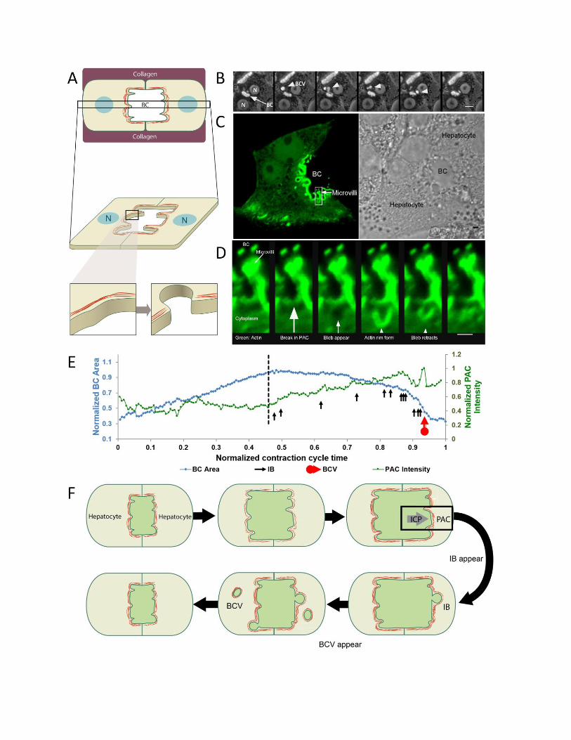
Figure 5: Inward bleb and BCV formation occurs following a break in the
pericanalicular actin cortex, as observed in collagen sandwich culture. (A) is a
schematic of the collagen sandwich culture system employed to monitor the inward
blebbing process in real-time. (B) shows a time lapse (at 7.5 min interval) progression of
BCV formation (white arrowhead) from a bile canaliculus (white arrow) in hepatocytes
cultured in the collagen sandwich culture system. The hepatocyte nucleus is represented
by ‘N’ and the bile canaliculus as ‘BC’. (C) shows a GFP-f-Tractin-transfected hepatocyte
(marker for actin, shown in green) forming a bile canaliculus with another non-transfected
hepatocyte (as shown in adjacent bright field image). (D) shows a high magnification time
lapse (at 26s interval) of the region highlighted by the square in (C). A local rupture in the
PAC is observed before a membrane intrusion into the hepatocyte cytoplasm occurs.
Following which, an actin rim forms along the membrane bleb and the bleb retracts back
towards the bile canalicular space. Scale bar in (B) = 10 µm, (C) = 2 µm and in (D) = 1
µm. (E) Changes in the area of a bile canaliculus and corresponding PAC intensity over
time. PAC starts to increase in intensity only after a maximum bile canalicular area is
reached (dashed black line). Like-wise, inward bleb (black arrows) and BCV formation
(red arrow) were observed to occur only at maximum bile canalicular area and when PAC
intensity started to increase. (F) Schematic of events that lead to formation of IB and BCV.
not peer-reviewed) is the author/funder. All rights reserved. No reuse allowed without permission. The copyright holder for this preprint (which was. http://dx.doi.org/10.1101/077792doi: bioRxiv preprint first posted online Sep. 28, 2016;

Figure 6: Canalicular pressure increase and perturbations to the pericanalicular
actin cortex affect formation of inward blebs (IB) and bile canaliculi-derived
vesicles (BCV). Frequency of both IB (A) and BCV (B) increase with increasing
canalicular pressure. Canalicular pressure was increased by using UDCA to stimulate
bile acid secretion. In the presence of increasing concentrations of cytochalasin D, an
not peer-reviewed) is the author/funder. All rights reserved. No reuse allowed without permission. The copyright holder for this preprint (which was. http://dx.doi.org/10.1101/077792doi: bioRxiv preprint first posted online Sep. 28, 2016;
inhibitor of actin polymerization, IB formation decreases (C) while that of BCV increases
then decreases (D). A similar trend is seen in the presence of blebbistatin, an inhibitor of
myosin activity as shown in (E) and (F). p < 0.01. *p < 0.05, **p < 0.01. N=40 for all
conditions analyzed.
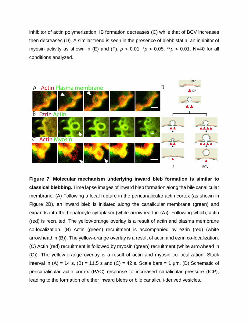
Figure 7: Molecular mechanism underlying inward bleb formation is similar to
classical blebbing. Time lapse images of inward bleb formation along the bile canalicular
membrane. (A) Following a local rupture in the pericanalicular actin cortex (as shown in
Figure 2B), an inward bleb is initiated along the canalicular membrane (green) and
expands into the hepatocyte cytoplasm (white arrowhead in (A)). Following which, actin
(red) is recruited. The yellow-orange overlay is a result of actin and plasma membrane
co-localization. (B) Actin (green) recruitment is accompanied by ezrin (red) (white
arrowhead in (B)). The yellow-orange overlay is a result of actin and ezrin co-localization.
(C) Actin (red) recruitment is followed by myosin (green) recruitment (white arrowhead in
(C)). The yellow-orange overlay is a result of actin and myosin co-localization. Stack
interval in (A) = 14 s, (B) = 11.5 s and (C) = 42 s. Scale bars = 1 µm. (D) Schematic of
pericanalicular actin cortex (PAC) response to increased canalicular pressure (ICP),
leading to the formation of either inward blebs or bile canaliculi-derived vesicles.
not peer-reviewed) is the author/funder. All rights reserved. No reuse allowed without permission. The copyright holder for this preprint (which was. http://dx.doi.org/10.1101/077792doi: bioRxiv preprint first posted online Sep. 28, 2016;

Figure 8: Bile canaliculi-derived vesicles (BCV) enables bile regurgitation during
increased canalicular pressure. (A) Using CLF to represent bile acids, CLF was not
cleared from the bile canalicular space under normal conditions (arrowhead in (A)) but
was rapidly cleared in the presence of UDCA (arrow in (A)). UDCA enables simulation of
not peer-reviewed) is the author/funder. All rights reserved. No reuse allowed without permission. The copyright holder for this preprint (which was. http://dx.doi.org/10.1101/077792doi: bioRxiv preprint first posted online Sep. 28, 2016;

increased canalicular pressure as it increases bile acid secretion. Scale bar = 20 µm. (B)
and (C) show quantification of CLF intensity within bile canaliculi under normal conditions
(B) and in the presence of BCV-stimulating UDCA (C). Each curve in (B) and (C)
represents CLF intensity in individual bile canaliculi normalized with respect to maximum
intensity during 30 min for each bile canaliculi. There were a few bile canaliculi which
show a drastic decrease in CLF intensity (black line) under normal conditions (B) but
those were bile canaliculi in which BCV formation was observed. 15 individual bile
canaliculi were analyzed in (B) and (C). (D) Montage showing release of bile canaliculi-
derived vesicle (red arrowhead) at cell edge (red line marked by blue arrow in first frame).
As can be seen, BCV travels toward the cell edge and merges with the cell membrane.
Scale bar = 10 µm. (E) Schematic of a proposed model of BCV-facilitated bile
regurgitation.
not peer-reviewed) is the author/funder. All rights reserved. No reuse allowed without permission. The copyright holder for this preprint (which was. http://dx.doi.org/10.1101/077792doi: bioRxiv preprint first posted online Sep. 28, 2016;
Top Related