


Languages
Pages
Legal


© SSER Ltd.

The Mammalian Liver
The liver is the largest gland in the body and the second largest organ after the skin
The liver is situated under the diaphragm on the right side of the abdominal cavity
Numerous metabolic reactions occur within the liver and it is an important organ of homeostasis

Blood Supply
The liver receives blood from two sources
The hepatic artery delivers oxygenated blood to the
liver
The hepatic portal vein delivers blood, rich in digested food molecules,
from the small intestine
Blood leaves the liver along the hepatic vein and
enters the vena cava

The Mammalian Liver
The liver is composed of a largenumber of lobules
Each lobule contains many vertical rows of liver cells (hepatocytes) arranged radially
around a central blood vessel calledthe central vein
Branches of the hepatic artery and hepatic portal vein supply blood to the capillaries
(sinusoids) of each lobule
Running between the lobules in the opposite direction to the blood, are fine ducts
(canaliculi), carrying bile from the liver cells towards the main bile duct

The Liver LobuleCentral vein of lobule
(to hepatic vein)
Plates of livercells (hepatocytes)
SinusoidCanaliculus
Branch of hepatic portal vein
Branch of hepatic artery
Bileduct
Network of canaliculi betweenliver cells
An enlarged portion of the liver lobule
provides further detail
Blood flows from branches of the hepatic portal vein and
hepatic artery along sinusoids (dilated capillaries) between
the liver cells

bloodflow
molecules enterliver cells
blood flowsinto central vein
bile from liver cells
flow ofbile
Part of Liver Lobule
Hepatocytes bear numerous microvilli at their surfaces in contact with the sinusoids, thereby increasing the surface area for facilitating the exchange of materials; numerous mitochondria within the cytoplasm reflects their high demand for ATP to provide for the numerous endergonic reactions
Branch of hepaticportal vein
Liver cells;Hepatocytes Sinusoid
PhagocyticKupffer cell
Central vein of lobule (to hepatic vein)
Branch of hepaticartery
Branch to bile duct Bile canaliculus
Fine channels, called canaliculi, collect bile from the liver cells and carry it towards the bile duct

Sinusoids Sinusoids are dilated capillaries in which the lining epithelial cells and basement membrane are discontinuous
Sinusoids have larger diameters than other capillaries with distinct gaps in their lining
The structure of the sinusoidal capillaries allows for the ready exchange of materials (including macromolecules) between
the blood and the liver cells
Epithelial liningcells
Basementmembrane

Rows of liver cells(hepatocytes)
Sinusoids
Central Vein

CarbohydrateMetabolism
Protein Metabolism
Lipid Metabolism
Haemoglobin and Hormone breakdown
and Detoxification
Storage of Vitamins and
Minerals
Bile Production

Carbohydrate Metabolism
The liver’s major role in the metabolism of carbohydrates is that of glucose homeostasis
Under the influence of the hormones insulin and glucagon (secreted by the Islets of Langerhans of the pancreas) and adrenaline from the adrenal
glands, blood glucose concentrations are regulated and adjusted to meet the metabolic
demands of the tissues
The digestion of polysaccharides and disaccharides in the gut yields the
monosaccharides glucose, fructose and galactose; these sugars are transported to
the liver along the hepatic portal vein

Carbohydrate Metabolism
In the liver, most of the fructose and galactose molecules are converted to glucose; the liver plays a significant role in the control
of blood glucose concentrationsin three major ways:
• Glycogenesis; activation of the liver enzymes that convert glucose into glycogen for storage
• Glycogenolysis; activation of the liver enzymes that convert glycogen into glucose when blood glucose levels fall
• Gluconeogenesis; activation of the liver enzymes that convert non-carbohydrates into glucose in response to low blood glucose concentrations

Glycogenolysis; the conversion
of stored glycogen into glucosewhen blood sugar levels fall
glucagon and adrenaline
Glycogenesis; the conversionof glucose into glycogen when
blood sugar levels rise
insulin

Gluconeogenesis is the conversion of non-carbohydrates, such as amino acids
and glycerol, into glucose by the liver
When the demand for glucose depletes the glycogen
stores, non-carbohydrate sources are converted by the
liver into glucose

Protein Metabolism
During digestion, proteins are hydrolysed into their constituent amino acids and transported to
the liver along the hepatic portal vein
Unlike glucose, excess amino acids cannot be stored in the liver; excess dietary amino acids undergo deamination and are also converted
into glucose and triglycerides
Transamination reactions occur in the liver; this involves the conversion of one amino acid
into another and is the process by whichnon-essential amino acids are synthesised

Protein Metabolism
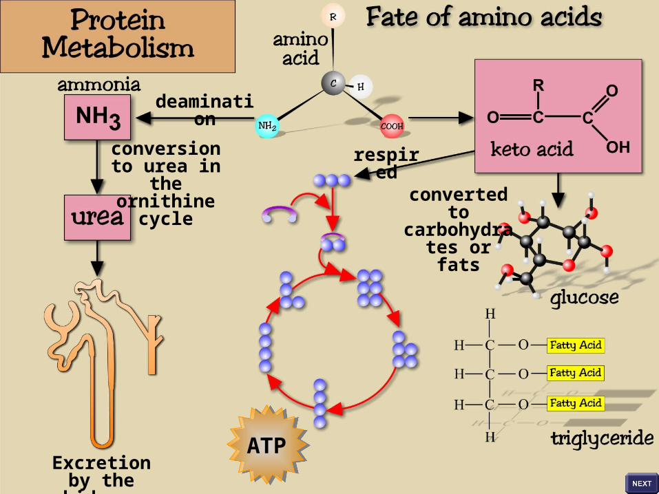
The fate of surplus amino acids withinthe liver cells involves:
• Deamination; the removal of the amino group from an amino acid, producing ammonia and a keto acid; the toxic ammonia is converted into urea, which is transported to the kidneys for excretion; the keto acid may enter the respiratory pathway to yield ATP or, may be used for the synthesis of glucose and fatty acids
• Gluconeogenesis; liver cells can convert amino acids into carbohydrate
• Lipogenesis; liver cells can convert amino acids into fats

Surplus amino acids cannot be stored and undergo deamination in the liver
The amino group of the amino acid, together
with a hydrogen atom, is removed to form ammonia and a keto acid
The highly toxic ammonia enters the ornithine cycle and is converted into urea
The keto acid either enters the respiratory
pathway and generates ATP, or it is converted into carbohydrates or
fats
The less toxic urea is excreted by the kidneys
deamination
conversion to urea in the
ornithine cycle
Excretion by the kidneys
respired
converted to carbohydrates
or fats
ATPATP

Transamination involves the transfer of an amino group from a donor amino acid to a recipient keto acid; the donor amino acid becomes a keto acid and the recipient
keto acid becomes an amino acid

All non-essential amino acids may be synthesised by transamination

Lipid Metabolism
The lipids are a diverse group of molecules and include cholesterol,
triglycerides and phospholipids
The liver synthesises, modifies, releases and eliminates lipids, playing a major role in
their homeostatic regulation
Surplus cholesterol and phospholipids are eliminated in the bile; the liver manufactures bile, which is stored in the gall bladder and
secreted into the duodenum of the gut

Lipid Metabolism
The roles of the liver in lipid metabolism include:
• Lipogenesis; the synthesis of triglycerides from glucose when glycogen stores are depleted; the resulting triglycerides can be stored or utilised in the production of cholesterol and phospholipids
• The synthesis of cholesterol and phospholipids
• The modification of cholesterol and triglycerides (combined with liver proteins) to producewater-soluble lipoproteins for transport toother body tissues
• The elimination of surplus cholesterol and phospholipids in the bile

Liver cells synthesise triglycerides from glucose or amino acids (lipogenesis)
when glycogen stores are full
The liver synthesises most of the cholesterol and
phospholipid found in the body and regulates their
concentrations in the blood
The resulting triglycerides can be stored or used to synthesise other lipids, such as cholesterol
and phospholipids
Excess cholesterol and phospholipid is removed in the bile and delivered to the
gut for elimination

The synthesis, release and elimination of cholesterol
and phospholipids is regulated by the liver
Surplus cholesterol and phospholipid is eliminated in the bile
Cholesterol and triglycerides are combined
with liver proteins to render them soluble for transport in the blood (lipoproteins)

‘Good’ and ‘Bad’ Cholesterol
Low density lipoproteins (LDLs) are loosely termed ‘bad cholesterol’ since excess LDLs remain in the
bloodstream and deposit cholesterol in and around the muscle fibres in arteries (forming fatty plaques); this
may lead to atherosclerosis (narrowing of the arteries)
LDLs attach to specific receptors on the surfaces of cells and are taken into the cells by
endocytosis where the cholesterol is released
When a cell’s cholesterol needs are met, the production of LDL receptors is shut down, and the
receptors already present are gradually removed; the lack of receptors raises plasma LDL levels, making it more likely that plaques will develop in the arteries

Fatty deposits begin to build up in the artery wall
Fatty deposits (plaques) build up in large quantities; calcium deposits harden the arteries;
blockage is extreme and blood flow is seriously affected

High density lipoproteins (HDLs) are associated with a decreased risk of atherosclerosis
HDLs remove excess cholesterol from body cells and transport it to the liver for elimination; accumulation of cholesterol in the blood is
prevented and the risk of fatty plaque formation in the arteries is reduced
‘Good’ and ‘Bad’ Cholesterol
Top Related