


Languages
Pages
Legal

1
2
Contents
Section 1 Learning Objectives for Phase 2 (4th & 5th Year) Page 2
Section 2 On-Line Resources Page 5
Section 3 Tutorial Material
Ophthalmoscopy Page 6
Eye Accident & Emergencies Page 8
Cataract Page 12
Uveitis & Floaters Page 17
Eye Banking and Corneal Transplantation in New Zealand Page 21
Pupil Abnormalities Page 24
Ocular Surface Inflammation and Allergy Page 28
Retinal Conditions & Signs of Disease Page 30
Oculopastics Overview Page 33
The Acute Red Eye Page 42
Glaucoma Page 48
Paediatric Ophthalmology Page 52
Dry Eye and Sjögren’s Syndrome Page 57
NB. These tutorial notes are to be read in conjunction with the 4th Year Handout and with the recommended reading selections, as listed in the 5th year program (University Dept of Ophthalmology, Auckland), from the textbook, “Ophthalmology. An illustrated colour text”, Batterbury and Bowling, 2nd Edition, Elsevier Churchill Livingston. A copy of the recommended reading from the 5th year program is included in Section 4 of this handout.
Section 4 Self-directed learning tool:
Ophthalmology Quiz Page 63 Quiz Answers Page 69
Recommended Reading Page 71
Clinical Scenarios – Links in the MBChB Portal (http://mbchb.auckland.ac.nz) Page 73
Section 5 Contact Details Page 75

3
Section 1. Learning Objectives Ophthalmology, University of Auckland
Ophthalmology (Phase 2, Years 4 and 5)
Expected learning outcomes:
Domain Acquisition and Application of Medical Knowledge
1 Review and reinforce pre-clinical knowledge applicable to eye disease
2 Develop an appreciation of key ocular symptoms and signs and the underlying pathological processes
3 Knowledge of symptoms, signs and main differential diagnoses of key ophthalmic presentations:
a. The acute red eye b. Sudden visual loss and visual impairment c. The squinting child d. Ocular trauma with visual impairment
4 Appreciation of the ocular manifestations of systemic disease, e.g. diabetes mellitus, hypertension, collagen diseases. Understand the pathology of diabetic retinopathy, its classification and prevention/treatment
5 Understand the causes, management and health burden of senile cataract
6 Understand the causes, management and health burden of age related macular degeneration
7 Understand the causes, management and health burden of chronic open angle glaucoma
8 Establish relevance of key elements of pathology, microbiology and immunology to eye disease
9 Basic ocular therapeutics
Domain Professional, Clinical and Research Skills
1 Take an appropriate ophthalmic history
2 Assessment of visual acuity (near, distance, colour)
3 Visual field assessment to confrontation / central field by Amsler grid
4 Examination of the anterior segment – conjunctivae, cornea, iris/pupil
5 Pupillary examination – near triad, response to light, consensual, swinging light test

4
Section 1. Learning Objectives Ophthalmology, University of Auckland
Domain Professional, Clinical and Research Skills cont.
6 Examination of the red reflex by ophthalmoscope
7 Examination of the posterior segment using the ophthalmoscope including assessment of optic disc, maculae and vasculature
8 Assess presence of squint, corneal reflexes, cover test and eye movements
9 Basic elements of anterior segment slit-lamp examination
Domain Hauora Maori
1 Develop awareness of the patient’s cultural background and usage of effective communication to facilitate appropriate ocular examination
2 Appreciation of epidemiology of different ocular conditions as well systemic diseases with ocular manifestations (e.g. diabetes) affecting Maori and Pacific Island population
Domain Population Health and Primary Health Care
1 Knowledge of important causes of blindness in New Zealand and worldwide
2 An appreciation of the impact of visual disability on the patient’s lifestyle, family and community
3 Awareness of the community agencies for the support of visually impaired (e.g. Royal New Zealand Foundation of the Blind, Glaucoma New Zealand)

Section 2. On-line Resources Ophthalmology, University of Auckland
5
On-Line Resources
The Ophthalmology Department’s website address is http://ophthalmology.auckland.ac.nz or it can be accessed via the University of Auckland’s homepage, under the School of Medicine.
Materials pertinent to the 5th year teaching program can be found by opening the Teaching page and following the sub-menus listed.
Teaching materials available on-line for use include:
1. CAL – computer assisted learning.
There are 4 subspecialty sections, each containing a series of clinical images, with questions to be answered. The correct responses are supplied on the answer pages. The standard of questions presented here corresponds to the level of questions set for the clinical exam paper, to be sat at the end of the Ophthalmology attachment.
2. "The Eyes Have It" is an interactive teaching program, courtesy of the
University of Michigan Kellogg Eye Center, about disorders of the eyes and visual system. In the Instructional Mode, you will see photographs or captioned streaming videos supplemented with text. In the Quiz Mode, you will have a chance to test your knowledge and be scored.
3. 5th Year Handout 2011 (PDF file) and Tutorial Presentations as given in the
Auckland rotation (PDF Files).
4. Clinical Skills Videos. These videos demonstrate the variety of skills to be acquired during the Ophthalmology attachment as listed in the expected learning outcomes.

6
Section 3. Tutorial Notes Ophthalmology, University of Auckland
Ophthalmoscopy Associate Professor Jennifer Craig, Dr Charlotte Jordan
Direct Ophthalmoscopy Indirect Ophthalmoscopy
Monocular
Erect image (upright)
High magnification (x 15)
Restricted field of view (7 - 8°)
Provides view to the arcades
Usually binocular
Inverted image, laterally reversed
Lower magnification (x2 – x3)
Increase field of view (approx. 30°)
Provides view to ora serrata
(with scleral indentation)
Heine Mini 3000 Ophthalmoscope
Examination:
Red Reflex External eye

7
Section 3. Tutorial Notes Ophthalmology, University of Auckland
WhaWhaWhattt’’’sss ttthhheee
cccupupup---tototo---dddiiisssccc
rararatttioioio???
Disc Vessels Macula
Shape Colour Margins Cupping
Colour Calibre Crossings
Colour Foveal Reflex Avascular
The normal fundus
Dii sc shape
coll ourmargii nscuppii ng
Vesselscoll our
calii bre crossings
Macull a
coll ourfoveal refll exavascull ar
The normal disc
Right disc Left disc
Peripapillary atropy
cup disc margin
Cup-to-disc ratio: 0.5
http://www.foto-web.com/simuladores/ Fundus examination simulator (click on picture)
(n.b. Download does not work via PC link!))

8
Section 3. Tutorial Notes Ophthalmology, University of Auckland
Eye accident and emergency Dr James McKelvie
Topics covered in this session:
1. Overview of history taking 2. Examination basics 3. Common conditions presenting to A&E 4. Sight threatening conditions 5. Life threatening conditions
1. The History
Specific considerations for ophthalmic history: • Trauma (High speed, metal) • Chemical (acid/alkaline) • Light (U.V./I.R) • No precipitating event
2. Examination basics
A systematic approach is essential so as not to miss important signs
Anatomical approach commonly used and easy to remember
Anatomical approach to exam • VA • Skin/Lids • Conjunctiva • Cornea • Anterior chamber • Lens • Vitreous • Macula/Retina • Optic nerve • Orbit/bone • Neurological
3. Common A&E problems (Case studies)
GP referral Grinding metal 2/7 ago
Eye red, irritated and sore Flourescein staining showing “scratches” on cornea
What important features must you ascertain from the history?
What do you examine first?
High velicity and no safety glasses are risk factors for penetrating injury and

9
Section 3. Tutorial Notes Ophthalmology, University of Auckland extraocular foreign body – need to exclude with careful examination

10
Section 3. Tutorial Notes Ophthalmology, University of Auckland
Common A&E problems: history of trauma and examination shows… Self referral Playing squash yesterday and hit in RE with ball. “Blurry” vision since VA R 6/48, 6/30ph
What important features must you ascertain from the history? What must you check on exam?
Common A&E problems: red eyes, lids and cornea
GP referral with red eyes 7/7 No improvement with chloramphenicol Gritty, itchy eyes, started in RE then LE 2/7 later. VA 6/6, 6/6
What important features must you ascertain from the history? What must you check on exam? How can you confirm the diagnosis? What treatment is indicated?
Common A&E problems: red eyes, lids and cornea
GP referral with L corneal ulcer that stains with flourescein VA L 6/18
What important features must you ascertain from the history? What should you check on exam before instilling flourescein and topical anesthetic? How can you confirm the diagnosis and what is your differential for corneal ulcers?
Chemical and thermal /UV injuries
Referral from Emergency Department with chemical injury to RE 2/7 ago.
What is the immediate management of chemical injuries that involve the eye? What is worse acid or alkali? Why is it important to assess the limbus?
Chemical and thermal /UV injuries
Grinding and welding a trailer at home 2/7 ago. LE sore and red from 10pm and now sensitive to bright light as well.
What is the diagnosis?
More red eyes, no precipitating event GP referral with viral conjunctivitis and no improvement with chloramphenicol Sore red eye for last 3/7. Happened out of the blue. Similar episode 12/12 ago but did not seek help VA L 6/18

11
Section 3. Tutorial Notes Ophthalmology, University of Auckland
What features in the history are you interested in? What examination findings are consistent with diagnosis? What investigations would you like to do (if any) and why?
4. Sight threatening problems (painful) GP referral with rapid onset (4/24) painful++ red eye cloudy cornea. VA 6/24
What are the essential features of the examination you must check? Why is the cornea hazy? Why is the pupil mid dilated and sluggish? Why is this a sight threatening problem that requires urgent management?
Sudden onset painless loss of vision
Optometry referral –patient woke with painless loss of vision in one eye VA 6/60 Differential diagnosis includes:
CRVO/BRVO • CRAO/BRAO • Retinal Detachment • Ischaemic optic neuropathy • Optic neuritis •
Intermittant loss of vision
GP calls with possible referral. 65 year old man with extensive history of left-sided headaches with general ache around his shoulders, mild weight loss and intermittent episodes of visual loss over past week.
Any additional questions for GP? What is the differential diagnosis? What investigations would you like on arrival? What are the expected results in this condition? What should you include in your exam and why? How can you confirm the diagnosis? What is the treatment and when should it be started?
5. Life threatening Orbital vs preseptal cellulitis
Referral from SSH. 5 year old boy presents with swollen red RE. Not able to check vision ?orbital cellulitis.
What is orbital cellulitis and how does it differ from preseptal cellulitis? How can you clinically differentiate these two conditions?

12
Section 3. Tutorial Notes Ophthalmology, University of Auckland
What investigations or imaging is required? Why is orbital cellulitis a potentially life threatening problem?
What is going on here?
GP referral with binocular diplopia for one month Patient complains of feeling hot at night but currently afebrile and FBC Normal
What is your differential diagnosis? What investigations would be helpful to confirm the diagnosis and plan management?
Ward call You are a house officer and have been asked by a nurse on the urology ward to see a man with double vision that started 2 hours ago and eyes look in “funny position”.
What is the abnormality? Do you need to do anything about it now or can you leave a message for the team in the morning? What is the next step in management?
Don’t forget…
Sight threatening Life threatening
Acute glaucoma Third nerve palsy with pupil involvement Giant cell arteritis Orbital cellulitis
Intra ocular foreign body

13
Section 3. Tutorial Notes Ophthalmology, University of Auckland
Cataract: past, present and future Professor Charles McGhee
Blindness from cataract: Globally, 25 million cataract blind Requires 36 million procedures to treat <6/12
Basic Lens Anatomy –
Transparent, biconvex structure •
Lies behind iris, suspended by zonules – loss of zonules leads to subluxated •lens
Approximately 10mm diameter, 4 mm thick •
Tends to opacity (cataract) with age
The eye has two principal focusing structures a) The cornea 2/3rd – approximately 40 dioptres b) The crystalline lens 1/3rd – approximately 20 dioptres c) If a lens cataract is removed the focussing power (20D) needs to be replaced d) Historically replaced by spectacles, in last 30 years mainly by intraocular
lenses
Cataract assessment: acuity, ophthalmoscope and slit lamp
Describing cataract anatomy: Anterior subcapsular Cortical Nuclear Posterior subcapsular
The global burden of cataract • 25 million blind (classified as 20/400) • Additional 2 million new cases per annum • 110 million severe visual impairment • 90% of blindness in developing world • Only 7 million procedures per annum

14
Section 3. Tutorial Notes Ophthalmology, University of Auckland
Cataract - multiple aetiologies • Congenital • Inherited • Age-related (the majority) • Metabolic – e.g. diabetes • Toxic – e.g. corticosteroids • Traumatic – e.g. irradiation • Secondary – e.g. ant. uveitis
Cataract aetiology in developing world:
repeated dehydrational crises
Cataract in NZ The Auckland Cataract Study • Provided a prospective snapshot of 500 patients with cataract, cataract
services, and state of the art surgical techniques in a public hospital service in the year 2000
• Also highlighted public cataract waiting list issues and access to appropriate government funded care in a major metropolitan area (now resolved)
• Results published in a series of scientific publications in the British Journal of Ophthalmology and Clinical and Experimental Ophthalmology
The Auckland Cataract Study: Systemic Disease The majority of those with significant cataract have General Health issues
Hypertension 25% Cerebral vascular disease 12% Diabetes Melitus 11% Ischaemic Heart disease 10% Rx Aspirin 42% Warfarin 6%
Auckland Cataract Study: commonly associated ocular disease Ocular co-morbidities in cataract patients -
Open-angle glaucoma 10% Diabetic retinopathy 6% Vein occlusion 2% Advanced AMD 1%

15
Section 3. Tutorial Notes Ophthalmology, University of Auckland
Cataract Surgery Ancient Techniques
Couching Current cataract extraction techniques
1. Intra-capsular – now mainly in developing world 2. Extra-capsular – some use in developed world 3. Phacoemulsification – most popular technique
Phacoemulsification Basics Use of high frequency ultrasound to emulsify cataract performed through a smaller incision (6mm verses traditional 10mm incision of ECCE procedures)
Phacoemulsification hand piece
• Piezoelectric crystal vibrates ultrasonically • Driven electrically • Frequency 25,000 – 60,000 Hz • Phaco “needle” is hollow (0.9mm diam) with central
aspiration port • Needle surrounded by soft irrigation sleeve with two ports which
maintains fluid in ant. chamber
Late 1990’s Intra-ocular lenses: small incision phacoemulsification and the evolution of foldable and injectable Intraocular lenses. Folding or injecting the 6mm diameter IOL enables the incision size to be reduced to 3mm width for no suture phaco with incisions moving from largely scleral to corneal based
Corneal Incision structure One and two step corneal incisions
• Quick • Single instrument • No suture • Enables topical anaesthetic approach • Popular
What’s new in IOL design? Considering -
PCO and square edge Correction of spherical aberration Multifocal / accommodative IOLs Coloured IOLs & macular protection Specialised IOLs for reconstruction

16
Section 3. Tutorial Notes Ophthalmology, University of Auckland
The Auckland Cataract Project Summary 1 In a public health system, a predominantly elderly, female population, frequently with significant systemic illness and co-existing ocular diseases, relatively advanced cataracts and poor visual acuity, presented for cataract surgery. The majority of subjects (97.5%) underwent small incision, phacoemulsification, local anaesthetic, day-case surgery. The remaining 2.5% underwent extra-capsular cataract surgery
Summary 2 Despite co-existing eye disease, almost 90% of patients achieved best spectacle corrected vision (BSCVA) of 6/12 [20/40] or better which meets the NZ & UK driver’s licence standard. Although approximately 5% of eyes sustained an adverse intra-operative event only 1.5% of eyes exhibited poorer post-operative BSCVA than anticipated

Section 3. Tutorial Notes Ophthalmology, University of Auckland
16

Section 3. Tutorial Notes Ophthalmology, University of Auckland
17
Uveitis & Floaters Dr. M. Pradhan, Dr T. Malik, Dr M. Donaldson
Introduction
• Inflammation of uveal tissue Involving iris, ciliary body, choroid
• Classification – Anterior – Intermediate – Posterior – Panuveitis
Uveitis Associations: • Idiopathic, • HLA B27 +ve
– Ankylosing spondylitis – Juvenile chronic arthritis – Psoriatic arthropathy – Reiter’s syndrome
• Sarcoidosis • Behcets • Inflammatory bowel disease • Collagen vascular disorders
SLE Polyarteritis nodosa Wegener’s
• Infection – Toxoplasma,Toxocara, Lyme – HSV, HZV, HIV, CMV – Tuberculosis, syphilis, leprosy – Candidiasis
• Trauma Sympathetic ophthalmia
Symptoms of Anterior Uveitis -
• Aching pain • Redness • Photophobia • Blurring of vision • Pain on accommodation

Section 3. Tutorial Notes Ophthalmology, University of Auckland
18
Signs of Anterior Uveitis - • Decreased acuity • Unilateral red eye • Circumciliary conjunctival injection • Small or irregular pupil • Inflammatory products in Anterior Chamber (AC) • Sequelae
Inflammatory Products: Cells – graded as +1 to +4 Protein (flare) - +1 to +4 Fibrin – Grade +4 flare Keratic precipitates
• Fine, non-granulomatous • Mutton-fat, granulomatous
Iris Nodules – Koeppe, Busacca Sequelae
• Posterior synechiae • Seclusio pupillae • Occlusio pupillae • Iris bombè • Sectoral iris atrophy - HZO • Low IOP - hypotony • Glaucoma • Cataract • Cystoid macular oedema • Neovascularisation
Glaucoma
• Inflammation of the trabecular meshwork • Steroid responsive glaucoma – in 20 %, due to topical treatment • Mechanical - ‘pupil block’
Symptoms of Posterior Uveitis
• Floaters • Blurred vision

Section 3. Tutorial Notes Ophthalmology, University of Auckland
19
Signs of Posterior Uveitis
• Inflammatory products
• Visible focus of chorioretinal inflammation
• Inflammatory consequences Macular oedema Vascular sheathing / occlusions Optic disc swelling
Management of uveitis Anterior:
• Topical steroids (intensive and early) • Cycloplegics (pain relief and prevent posterior synechiae) • Topical / oral Antivirals (aciclovir) – HSV, HZO • Local steroid injections (subconjunctival / sub tenons) • Systemic steroids
Posterior / panuveitis • Orbital floor / posterior subtenons Steroid injection • Systemic steroids • Antibiotics, anti-TB therapy • Antivirals – oral (HZO, CMV, HIV), intravitreal ganciclovir (CMV) • Immunosuppresion
Floaters Causes Posterior vitreous detachment - commonest Blood
– Neovascularisation eg diabetes – Torn peripheral retina
Inflammatory products – intermediate / posterior uveitis History... Onset Flashing lights Reduced vision Other symptoms e.g. field loss Other history - ocular e.g. myopia, surgery, trauma
- systemic e.g. diabetes

Section 3. Tutorial Notes Ophthalmology, University of Auckland
20
Retinal tear Floaters – haemorrhage / PVD Photopsiae / flashes – traction at edge of tear
Retinal detachment Secondary to PVD and retinal tear Floaters / flashes and loss of field ‘Curtain’ effect
Diabetic eye disease – neovascularisation at optic disc
Diabetes – preretinal and vitreous haemorrhage due to neovascularisation
Vitreous haemorrhage – blurred vision and floaters

Section 3. Tutorial Notes Ophthalmology, University of Auckland
21
Eye Banking and Corneal Transplantation in New Zealand Louise Moffatt, Manager, New Zealand National Eye Bank
Associate Professor Trevor Sherwin “The acquisition, evaluation & supply of high-quality corneal and other tissue to all New Zealanders needing a transplant to restore their sight ”
Tissues Supplied:
• Corneas [220 – 250 per annum] – Viable, unaltered, sight-restoring – Stored in warm organ culture (34OC), up to 21 days
• Sclera [140 – 160 per annum] – Structural, reconstruction – Stored refrigerated, up to 6 months
• Amniotic membrane [15 – 20 per annum] – Structural/biological, surface ‘bandage’ – Stored frozen (-80oC), up to 2 years
Tissue Donation: the process
Organ donation: heart, liver, kidneys, lungs, pancreas - 50 per year Tissue donation: corneas, skin, heart valves, bone – many 1000’s per year
Suitable donors identified – by transplant coordinators, medical staff, OR referral
from families Next-of kin contacted - information provided about donation
- possibility raised, no coercion - if consent given, process explained
Retrieval: - careful surgical procedure in hospital, mortuary or funeral home
- donor treated with dignity & respect - no delay to funeral arrangements - no visible difference, viewing can occur
• Driver’s licence is indication of wish only Importance of discussion with family/whanau

Section 3. Tutorial Notes Ophthalmology, University of Auckland
22
Donor requirements: Age: 10 – 85 years Time: within 24 hours
Medical contraindications:
- Death of unknown cause - Infectious disease: - Hepatitis B,C, HIV, meningitis - Systemic viral infection - CNS diseases, progressive dementia - Leukaemia, lymphoma - Previous eye disease or surgery, laser surgery - Various congenital disorders
Generally suitable:
- Cancers - Bacterial septicaemia - Heart disease, respiratory disease - Diabetes, arthritis - Vision problems – glasses/contacts usually ok
Eye Bank Operations
- Donor acquisition from Auckland area – within 24 hours - Links with donor sources: Mortuary, Hospitals, other transplant services - Donors selected by strict criteria for suitability - Long-term storage in 34oC culture system – up to 21 days - Extensive testing for infectious disease & contamination - Quality control / sterility of highest standard - Minimum endothelial cell count
Eye bank output:
- Supply 100% of all transplanted corneas in NZ - Schedule of planned, elective surgery: 6+ grafts per week - Reduction of surgical waiting lists
Corneal Transplantation:
Oldest form of transplant – early 1900’s
Most common transplant operation performed
USA 45,000 pa UK 9,000pa NZ 250 pa
Avascular - few rejection problems
- no systemic immunosuppression required - 12-18 months for optimal vision

Section 3. Tutorial Notes Ophthalmology, University of Auckland
23
Indications for Corneal Transplantation in New Zealand: Keratoconus - 47%
- Acquired abnormality – cornea protrudes, thin & distorted - Bilateral, progressive, occurs at young age (teens, twenties) - High prevalence in NZ – 50% of transplants - Does keratoconus progress more rapidly in NZ?
Keratopathy/oedema - 18%
- Painful epithelial blisters, scarring - Association with glaucoma & following cataract surgery
Corneal Dystrophies – 10%
- Intrinsic genetic disorders, or aging - Epithelial abrasion/erosion, endothelial cell loss & dysfunction
Viral/bacterial keratitis – 10%
- Inflammation – opacification – vascularisation - ulcers - Risk of rejection higher – blood vessels reduces graft tolerance
Trauma – 6%
- Perforation – physical/chemical injury - Heavy male preponderance
Section 3. Tutorial Notes Ophthalmology, University of Auckland
24
Pupil Abnormalities Dr Stephen Best, Dr Hussein Patel
Pathway of the papillary light reflex consists of:
Afferent visual pathway • retinal receptor cells • bipolar cells • ganglion cells • optic nerve and tract • Pretectal nucleus of the high midbrain • Edinger-Westphal nucleus Efferent visual pathway • Two neurone pathway via the • Oculomotor nerve • Constrictor muscle of the iris
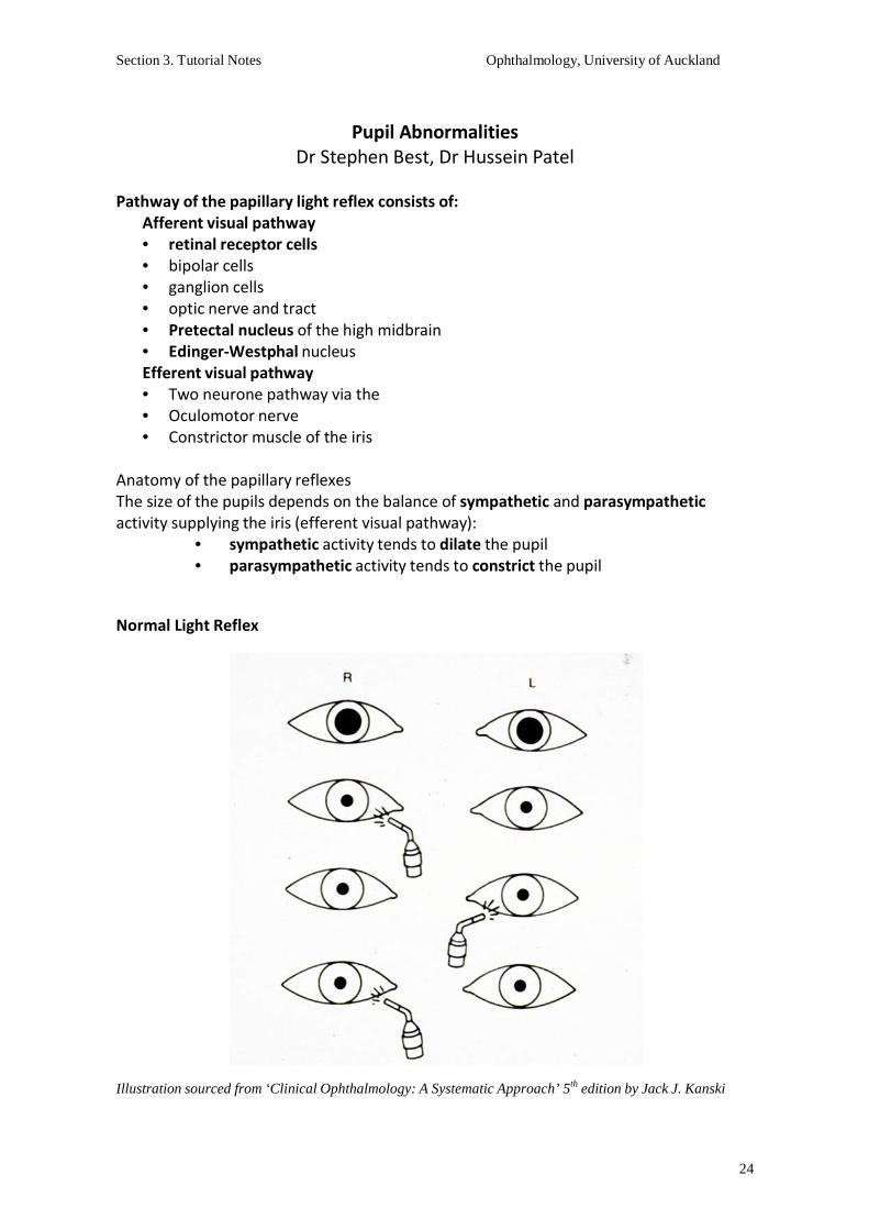
Anatomy of the papillary reflexes The size of the pupils depends on the balance of sympathetic and parasympathetic activity supplying the iris (efferent visual pathway):
• sympathetic activity tends to dilate the pupil • parasympathetic activity tends to constrict the pupil
Normal Light Reflex
Illustration sourced from ‘Clinical Ophthalmology: A Systematic Approach’ 5th edition by Jack J. Kanski

Section 3. Tutorial Notes Ophthalmology, University of Auckland
25
Examination of Pupils
• before dilating • size, symmetry • shape • near reflex • light reflex • RAPD (direct and indirect response)
Aniscoria Difference in pupil size between the eyes - may be physiological or pathological
Physiological anisocoria • normal variation in pupil size • 20% of individuals • Usually less than 1mm
Local Factors affecting pupil size
• Topical medications: – mydriatics/miotics/other agents
• Trauma: – traumatic mydriasis / sphincter rupture / surgical trauma / posterior
synechiae • Disease processes:
– uveitis / acute angle closure glaucoma • Systemic medications:
– narcotics = miosis Conditions with Pathological pupil size Abnormally small pupil:
• Horner’s Syndrome • Argyll Robertson pupil
Abnormally large pupil: • Adie’s Tonic Pupil • Pupil involved 3rd nerve palsy • Bilateral dilated pupils
Horner’s Syndrome
• Oculosympathetic paresis….interruption of the sympathetic supply along the three neuron pathway
• Miosis • Ptosis • Apparent enophthalmous • Cutaneous anhydrosis • Other features – transient hyperaemia/iris hypopigmentation in congenital cases • Diagnosis confirmed by topical cocaine test • Abnormal pupil fails to dilate whilst the normal pupil will dilate (loss of
noradrenaline at nerve junction )

Section 3. Tutorial Notes Ophthalmology, University of Auckland
26
• Other associated clinical signs and symptoms…. (headache / apical lung pathology / long tract neurology signs ) will determine appropriate investigations
Argyll Robertson Pupil
• Specific sign of neurosyphillis • Small and irregular pupils • Usually bilateral but asymmetric • Do not respond to light but near response normal (light-near dissociation)
Adie’s Pupil
Postganglionic parasympathetic denervation: • Causes: idiopathic, viral, diabetes, trauma • Glare / accommodative difficulties
Mydriasis • Light – near dissociation….slow constriction on prolonged near effort and slow re-
dilation to distance • Usually young females – 90% unilateral initially , but often becomes bilateral • Pupil becomes tonic with time….even miotic • If decreased tendon reflexes present = Adie’s syndrome
• Diagnosis confirmed by denervation hypersensitivity to weak cholinergic
(pilocarpine 0.1%)… abnormal pupil will constrict whilst normal pupil remains un- effected
• Aberrant re-innervation of pupillary sphincter muscle … contractions of part of the pupil margin (vermiform movement)
Causes of CN III Palsy
• Microvascular infarction – Occlusion vaso nevorum – Risks: diabetes, hypertension, artherosclerosis, hyperlipidaemia
• Compressive lesion – Aneurysm (usually post communicating artery) – Tumour
• Trauma What to look for if there is anisocoria?
• Make sure patient has not had any eye drops instilled • Check for prescription, over the counter ‘herbal’ and illicit drug use. • Any History of eye surgery ( Iatrogenic) • Check for signs such as ptosis, or ocular motility problems
Relative Afferent Pupillary Defect (RAPD)
• The presence of RAPD in the absence of gross ocular disease indicates a neurological lesion of the anterior visual pathway (afferent system)
• Detected using the ‘swinging flashlight test’ • Abnormal pupil respond to consensual light but not direct light

Section 3. Tutorial Notes Ophthalmology, University of Auckland
27
Causes of RAPD
• Optic nerve disorders • Chiasmal compromise • Optic tract lesions • Large retinal detachments or macular lesions, dense vitreous haemorrhage • RAPD not produced by cataract, refractive error, lesions posterior to the lateral
geniculate body, non-physiologic visual loss
Illustration sourced from ‘Clinical Ophthalmology: A Systematic Approach’ 5th edition by Jack J. Kanski

Section 3. Tutorial Notes Ophthalmology, University of Auckland
28
Ocular Surface Inflammation and Allergy Dr David Pendergrast
Papillae versus Follicles
Papillae Follicles
Chronic inflammation Acute inflammation
Allergy Viral
C/L, sutures, prosthesis Chlamydial
Cobblestones Toxic
Central vascularity Pale lesions
Surrounding vessels injected
Allergic Conjunctivitis:
acute to chronic
• Acute hay fever conjunctivitis • Seasonal allergic conjunctivitis • Perennial allergic conjunctivitis • Vernal keratoconjunctivitis • Atopic keratoconjunctivitis
Vernal Keratoconjunctivitis
• Age 9 to 19 • Boys > Girls • Warm dry climates • Symptoms: itching, mucus, photophobia • Signs: superior tarsal or limbal papillae • Pseudogerontoxon • Peripheral fibrovascular pannus • Shield ulcer
Shield Ulcer:
- Persistent Epithelial defect - Physical trauma from papillae, rubbing - Chemical trauma from inflammatory mediators - Mucous plaque formation

Section 3. Tutorial Notes Ophthalmology, University of Auckland
29
Atopic keratoconjunctivitis
• Adult onset • Symptoms: itch, photophobia, watering • Signs: redness • Fine papillary reaction • Periorbital atopic eczema • Microbial keratitis esp. opportunistic • Deep corneal vessels and scarring
Pathophysiology: Mast Cells
Mast cell degranulation in response to:
• Allergens and IgE • Physical trauma (rubbing) • UV exposure • Increased ocular surface temperature • Bacterial lipopolysacharides
Therapeutic options I: for milder disease
• Avoidance of allergens and rubbing • Cold compresses • Topical antihistamines: rapid onset • Systemic antihistamines: slower onset • Mast cell stabilisers: preventative use • Topical NSAIDs: Acular has some effect • Dual action agents: best current therapy
- Patanol Therapeutic options II: for sight threatening disease
• Topical corticosteroids Introduce at high frequency, tail off rapidly
• Topical cyclosporine 2% ointment • Systemic immunosuppression • Surgery:
Excision of papillae Superficial keratectomy
Review images in power-point presentation!

Section 3. Tutorial Notes Ophthalmology, University of Auckland
30
Signs of Retinal Disease Dr Tahira Malik, Dr Monika Pradhan
Blood supply of the retina • CRA - real artery • Atherosclerosis • AV crossings • Capillary-free zone –
foveal avascular zone Signs of Retinal Vascular Disease
• Vascular changes – arteriolosclerosis, AV crossing changes, venous tortuosity • Microaneurysms • Haemorrhages • Hard exudates • Cotton wool spots • Neovasclarisation
Vessel wall changes Vascular changes
Silver wiring of arterioles Venous dilatation and tortuosity Copper wiring of arterioles AV crossing changes Arteriolar attenuation AV crossing changes
Microaneurysms
• Tiny round red dots • In inner nuclear layer • Saccular outpouchings from vessels due to lack of pericytes
Retinal haemorrhages Appearance Localisation Aetiology
• Dot & blot haemorrhages – middle compact retinal layers – diabetic and hypertensive retinopathy
• Flame-shaped haemorrhages – superficial nerve fibre layer – vein occlusion • Pre-retinal haemorrhages – between posterior hyaloid face and retina – trauma,
Valsalva manouvre, neovascularisation

Section 3. Tutorial Notes Ophthalmology, University of Auckland
31
Retinal haemorrhages – Roth spots Caused by
• Endocarditis • Anaemia • Haematological
diseases
Hard exudates
• Lipid deposition • Outer plexiform layer • Abnormal vascular • permability • Circinate pattern • Macular star
Cotton wool spots
• Ischaemic infarction • Nerve fiber layer • Axoplasmic transport
defect • Diabetes • Hypertension • SLE • Leukemias
Neovascularisation hypoxia ⇒ vasoformative factors ⇒ neovascularisation
• in the retina NVE • on the optic disc NVD • on the iris NVI / rubeosis iridis • in the angle NVA
Causes: • Diabetes • Retinal vein occlusion • Retrolental fibroplasia • Sickle-cell disease • Inflammatory diseases
Section 3. Tutorial Notes Ophthalmology, University of Auckland
32
Occlusive Vascular Diseases Signs Venous occlusion:
• Dilated venules • Haemorrhages • Oedema
Retinal arteries occlusion:
• constricted arterioles • column interruption • retinal oedema • cherry red spot • visible emboli
Degeneration/dystrophy signs
• Drusen • Atrophy • Pigmentation • Scarring
- result of disease -iatrogenic – laser scars
Elevations of the Retina - by fluid – Retinal Detachment - by solid mass - tumours - Choroidal neovascularisation
Signs of Inflammation- Posterior uveitis
• Vitreous opacities, cells, snowballs • Choroiditis – deep yellow or grey lesions • Retinitis – white cloudy lesion with obscuration of retinal vessels • Vasculitis – periphlebitis (veins) or periarteritis – fluffy haziness around blood
column • Granlomas – choroidal or preretinal

Section 3. Tutorial Notes Ophthalmology, University of Auckland
33
Oculoplastics Overview Dr Neena Peters
Functions of the eyelids
1. Light protection and regulation 2. Keep cornea moist 3. Mechanical protection of globe
Lacrimal pump
EYELIDS (surface anatomy)
• Upper lid [UL] covers superior 2mm of cornea (10 – 2’o clock) • Lower Lid [LL] margin JUST touches inferior limbus • Distance between UL margin & central pupillary reflex is margin reflex distance
(MRD1) and normally, is 3.5 mm • Space between UL & LL is palpebral fissure height (PFH) and normally, it is 9 -12
mm. • A puncta (one in each lid) lies in close apposition to globe
To summarize- Normal parameters for lid position evaluation:
• A = Skin crease 10 mm in females 8-10 mm in males
• B = MRD1 – 3.5 – 4.0 mm • C = PFH – 10 -11mm • Inferior scleral show – Nil • Lagophthalmos (inability to close eyes) – Nil • Bell’s phenomenon (globe movement on eye closure) – Up and out
Eye Lids (Internal Anatomy)
EYELIDS (internal anatomy)
Orbital Septum
Orbital fat
Superioris (Aponeurosis)
Muller’s muscle
(Anterior lamellae)
-Skin - Orbicularis Oculi
(Posterior lamellae) -Tarsus (meibomian glands)
- Conjunctiva
Outward direction of lashes 9

Section 3. Tutorial Notes Ophthalmology, University of Auckland
34
Two halves – Lamellae
• Septum • Landmark – Grey Line
- Anatomical - Surgical
Orbital Septum
- thin sheet of fibrous tissue - natural barrier between eyelid and orbit
Muscles that open the eye
• Levator muscle
• Muller muscle
Preseptal cellulitis Orbital cellulitis
Eyelid: muscular and nervous control
Eyelid Retractors:
– Levator Aponeurosis (3rd CN. Parasym)
– Muller’s muscle (Sym) Eyelid Protractor:
– Orbicularis Oculi (VII N)
Abnormal Upper Eyelid Position
PTOSIS (droopy eyelid)
Levator Palpebrae superioris (LPS)
• LPS action – from extreme down gaze to extreme up gaze, by blocking the Frontalis
• Important while planning surgical management of ptosis • LPA - <4mm - poor
5 – 7mm – fair >8mm - good
Causes of Ptosis?
Congenital

Section 3. Tutorial Notes Ophthalmology, University of Auckland
35
Acquired
• Involutional • Neurogenic • Myogenic • Mechanical
Involutional (Aponeurotic dehiscence))
• the most common type of ptosis • Long term contact lens wear • Features:
Good LPS action High lid crease No lid lag on down gaze
• Frontalis overaction (s/o visual axis obstruction by ptotic lid) Neurogenic:
– Third Cranial nerve palsy Note:
• Total ptosis • Exotropia • Hypotropia • Mydriasis
Neurogenic:
– Horner’s syndrome Note:
• subtle ptosis (sympathetic innervation to Muller’s & smooth muscle of lower lid affected)
• elevation of LL (apparent enophthalmos) • miosis (sympathetic innervation to Pupil (dilators) affected)
Acquired Ptosis Neurogenic: - Horner’s syndrome
Acquired Ptosis
Neurogenic: -Marcus Gunn Jaw wink
24 25

Section 3. Tutorial Notes Ophthalmology, University of Auckland
36
Myogenic:
• Chronic Progressive External Ophthalmoplegia (CPEO) • Myasthenia gravis [diurnal variation] • Ptosis is one component of these syndromes
Mechanical:
• UL tumors BCC, SCC, Sebaceous Cell Ca Hemangioma Lacrimal gland masses
• Lid edema as post-trauma/ surgery
Congenital Ptosis Features:
• Fair to Poor LPS action • Faint/ Absent lid crease • Lid lag on down gaze • +/- poor upgaze
NB. Risk of amblyopia Ptosis Must assess
• Pupils • EOM, CN II • Levator functiom • Orbicularis power • Bell’s reflex • Evert UL
Abnormal Upper Eyelid Position
Lid Retraction Causes:
• Congenital • Acquired
- Thyroid Eye Disease - Midbrain lesions - Parinaud’s syndrome

Section 3. Tutorial Notes Ophthalmology, University of Auckland
37
Lower lid malpositions • 2 Lower lid retractors
• Less role in opening eyelid
• Pull lower lid inferiorly and
posteriorly in downgaze
• Structurally stabilises lid margin
Abnormal Lower Eyelid Position
Ectropion (Eyelid margin is away from the globe) Causes:
• Involutional • Cicatricial • Paralytic (VII CN palsy) • Mechanical
Prolonged ectropion leads to metaplastic changes in exposed conjunctiva
Involutional Due to laxity of canthal tendons (structural support of lids at medial & lateral ends) with age
Cicatricial e.g raumatic scar at cheek pulling the lower lid down
Paralytic (VII CN palsy) Facial nerve supplying Orbicularis Oculi is affected, as in Bell’s palsy, Parotid tumor excision, Acoustic Neuroma
Mechanical Lower eyelid edema causing mechanical ectropion
Abnormal Lower Eyelid Position
Entropion (Eyelid margin rolled in) Causes:
• Involutional • Trauma • Cicatricial

Section 3. Tutorial Notes Ophthalmology, University of Auckland
38
Entropion leads to trichiasis (misdirection of lashes), which may cause punctate epitheliopathy of cornea to frank corneal ulcers.
Cicatricial
Ocular Cicatricial Pemphigoid Stevens Johnson syndrome Trachoma (developing countries) Chemical injury
Contraction and thus, shortening of posterior lamella of the eyelid: which pulls and rolls in the eyelid margin along with eyelashes.
Common Eyelid Lesions Benign Eyelid Lesions
• Chalazion chronic, granulomatous inflammation of meibomian glands involving posterior eyelid lamellae Treatment: -warm compresses/ antibiotic ointment, if recent onset -Incision & Curettage in resistant cases
• Stye tender, acute inflammation of sebaceous glands of Zeiss or sweat glands of Moll at the base of eyelashes in anterior eyelid lamellae Treatment:
- warm compresses, antibiotic ointment - Expression of pus ± lash removal Keep a close watch, as in severe cases, stye may worsen to orbital cellulitis
• Xanthelasmas – yellowish, sessile plaques – S/o hypercholesterolaemia Treatment: - removal for cosmesis
Malignant Eyelid Lesions • Basal cell cancer - Commonest lid malignancy (90%) - Risk factors: Fair skin & UV exposure
Features: - Nodular/ulcerative with rolled edges - Loss of lashes - Telengiectatic blood vessels on surface Treatment: Excision with wide margins ± Cryotherapy

Section 3. Tutorial Notes Ophthalmology, University of Auckland
39
• Squamous cell cancer - Fast growing skin cancer Features: - Nodular
- Hyperkeratotic surface Treatment: Excision Biopsy with wide margins ± radiotherapy depending
upon the depth of involvement Orbit
Orbit Examination
• Exophthalmometer – axial/non-axial, look from above/below
• Optic nerve function • Pupils
• Extra-ocular movements, CN V • Lids – ptosis, retraction, masses, scleral show
• Palpate – masses, pulsatile • Lymph nodes
• Auscultate ?Bruits
Common Orbital disorders
Pre-septal cellulitis Aetiology: Trauma, Insect bite, Stye Features: - Inflamed, oedematous lids - Nil/ mild pain on eye movements - Visual acuity good - Intact Optic Nerve function
Orbital Cellulitis Aetiology:
Sinusitis (commoner in children) Trauma/ Tumor in adjoining sinuses
Features: - Proptosis - Inflamed pre-septal tissues - Painful/ limited eye movements - Drop in Visual acuity - Compromised Optic Nerve function

Section 3. Tutorial Notes Ophthalmology, University of Auckland
40
Thyroid Eye Disease
• Proptosis • Restrictive Myopathy • Optic Neuropathy
• Lid signs:
lid lag, lid retraction Lid swelling, lagophthalmos
• Ocular surface Inflammation esp. over horizontal recti muscles Dry eyes Exposure keratopathy Glaucoma
CT scan features in Thyroid eye disease
• Thick extra-ocular (recti) muscles • Maximum diameter of globe beyond lateral orbital rim • Tenting of optic nerve on right side (d)
Keep a close watch on:
• Corneal exposure – treat with lubricants/ taping eyelids shut • Optic nerve functions due to its possible compression by enlarged extra-ocular
muscles – might need urgent surgical intervention Orbital Fracture Classical Signs:
• Hypotropia • Enophthalmos • Restricted motility, esp. vertical gaze • Infra-orbital anesthesia

Section 3. Tutorial Notes Ophthalmology, University of Auckland
41
Lacirmal Drainage System & Common Disorders
Eyelid movement Lacrimal Pump
Acute dacryocystitis: Complete block Stagnant tears in sac Recurrent infections
Where is the block? Syringe & Probe
Dacryocystorhinostomy (DCR)
Congenital Naso-lacrimal Duct Obstruction • Symptoms start soon after birth - Watering ± discharge - Fluorescein dye disappearance delayed
Treatment Options in order of preference: a) Conservative [Lacrimal Massage ± Antibiotics] b) Probing & Syringing c) Intubation/ Balloon Dacryoplasty d) Dacryocystorhinostomy
Nearly 95% resolve conservatively by the end of one year of age
Section 3. Tutorial Notes Ophthalmology, University of Auckland
42
The Acute Red Eye Dr Shenton Chew, Assoc. Professor Jennifer Craig, Professor Charles McGhee
What is a red eye?
• Dilation of superficial ocular vessels – Conjunctiva – Episclera – Sclera
Systematic Approach to Diagnosis
1. History 2. Vision 3. Discharge 4. Appearance 5. Pupils
1. History
• Past ocular disease / symptoms • Decreased vision • Pain & severity • Photophobia • Ocular discharge • Associated systemic associations
2. Vision
• Snellen visual acuity • Pinhole
3. Discharge
• Serous / watery • Mucoid • Purulent • Mucopurulent
4. Appearance – view presentation
5. Pupils
• Miosis • Mid-dilated • Sluggish / no reaction to light
Section 3. Tutorial Notes Ophthalmology, University of Auckland
43
– Viral: • URTIs • Pre-auricular lymphadenopathy
Causes:
• Conjunctivitis • Subconjunctival haemorrhage • Keratitis • Episcleritis • Scleritis • Acute anterior uveitis • Acute angle closure crisis • Ocular trauma
Conjunctivitis
• Infective – Viral – Bacterial – Chlamydial
• Allergic – Seasonal (hayfever) / Perennial (dust mites) – Vernal – Atopic
History: Associated features
– Allergic: • Atopy/hayfever
• CL wear – Chlamydial:
• Urethritis (Reiters) Vision: Generally normal unless there is
– Excessive lid swelling – Excessive discharge – Corneal involvement
Discharge
• Viral: Watery • Allergic: Mucoid/watery • Bacterial: Purulent
• Chlamydial: Mucopurulent Pupils: Normal
Section 3. Tutorial Notes Ophthalmology, University of Auckland
44
Conjunctivitis: Management
• Swab and isolate responsible organism • Bacterial = Chloramphenicol • Viral = Supportive Rx (compresses, lubricants) • Chlamydia = Azithromycin/doxycycline
Keratitis History:
– Painful (foreign body sensation) – Photophobic – Tearing – Hx of CL wear/trauma
Vision: Decreased
– Especially if involves visual axis Discharge: Watery, purulent
– (depends on cause) Appearance:
– Circumcorneal injection – Corneal infiltrate/hazy cornea – Overlying epithelial defect
Pupils: Normal
Keratitis - Management
• Isolate responsible organism – Corneal scrape – Intensive treatment and close follow-up
Acute angle closure crisis
• in IOP due to obstruction of aqueous outflow by complete or partial closure of the angle by peripheral iris
• Incidence – 1/1000 in > 40 y.o.
• Female: Male – 4:1
Anatomical predisposition
• Short eye • Narrow angle • Large lens • OLDER HYPERMETROPE

Section 3. Tutorial Notes Ophthalmology, University of Auckland
45
History:
– Intense ocular pain & headache – Nausea & vomiting – Photophobia – Premonitory symptoms – Hypermetrope
Vision: Very blurred
– Secondary to corneal oedema
Discharge: None
Appearance: – Circumcorneal injection – Cloudy cornea – Optic nerve head swelling
• If prolonged attack Pupils: Mid-fixed, dilated
– Circumcorneal injection – Cloudy cornea – Optic nerve head swelling
• If prolonged attack Management:
• Reduce IOP (often starts > 50 mmHg) – Medical
• Topical: – Alpha-agonist, Beta-blockers, Mitotics (Pilocarpine)
• Systemic: – Carbonic anhydrase inhibitors (Diamox), Osmotics
(Mannitol) – Surgical • Peripheral iridotomy • Clear lens extraction/trabeculectomy
Acute Anterior uveitis/iritis Aetiology:
• Idiopathic • Ankylosing spondylitis • Reiter’s syndrome • Juvenile arthritis • Psoriatic arthropathy • Sarcoidosis

Section 3. Tutorial Notes Ophthalmology, University of Auckland
46
History:
– Moderate aching pain – Photophobia – Past history (esp if HLAB27) – Systemic symptoms
Vision: Blurred
Discharge: None
Appearance: – Circumcorneal injection – Clear cornea
Pupils: Miotic / sluggish response to light
Management: • Subdue inflammation
– Topical corticosteroids (g.predforte) • Prevent posterior synechiae
– Mydriatics (g.cyclopentolate) – Watch for elevated IOP – Topical ocular hypotensives (g.timolol)
Scleritis/Episceritis Scleritis Episceritis
• Relatively uncommon
• Severe boring pain
• Focal injection of deep scleral
vessels
• Associations
- Herpes Zoster Ophthalmicus
- Severe Rheumatoid Arthritis
• Can lead to blindness if untreated
- po.prednisone
• Relatively common
• Mild ocular discomfort
• Focal injection of episcleral vessels
• Generally no associations
• Usually requires no treatment
- g.lubricants/g.voltaren
Section 3. Tutorial Notes Ophthalmology, University of Auckland
47
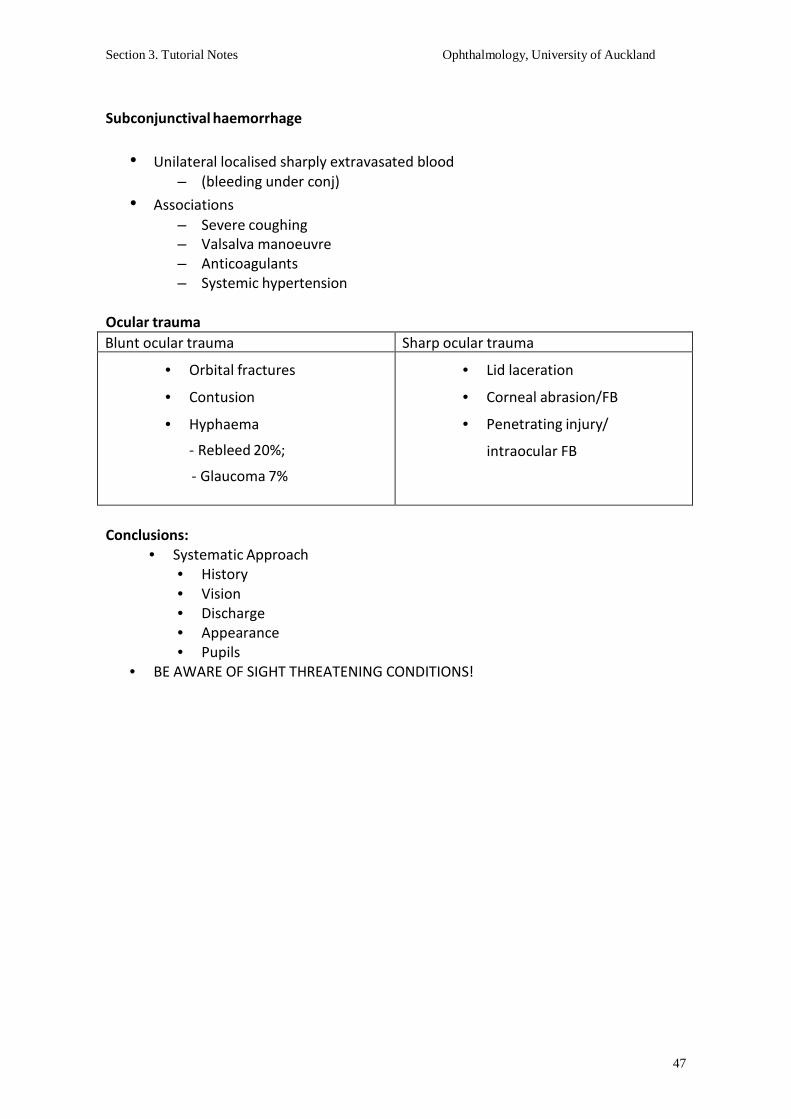
Subconjunctival haemorrhage
• Unilateral localised sharply extravasated blood – (bleeding under conj)
• Associations – Severe coughing – Valsalva manoeuvre – Anticoagulants – Systemic hypertension
Ocular trauma Blunt ocular trauma Sharp ocular trauma
• Orbital fractures
• Contusion
• Hyphaema
- Rebleed 20%;
- Glaucoma 7%
• Lid laceration
• Corneal abrasion/FB
• Penetrating injury/
intraocular FB
Conclusions:
• Systematic Approach • History • Vision • Discharge • Appearance • Pupils
• BE AWARE OF SIGHT THREATENING CONDITIONS!

Section 3. Tutorial Notes Ophthalmology, University of Auckland
48
Glaucoma Dr Justin Mora
Glaucoma is an optic neuropathy with a specific pattern of axonal loss which may be associated with elevated intraocular pressure and a typical pattern of visual field loss • A disease of the optic nerves • Two principal types
– Open angle glaucoma Primary – presumed angle predisposition Secondary – cells, inflammation
– Closed angle glaucoma
Primary – narrow anterior chamber angle Secondary – tumours, synechiae
Open Angle Glaucoma: It affects 2-3 % of people over 60 2nd leading cause of blindness in N.Z. In N.Z. 95 % of glaucoma of this type There are significant racial variations Risk factors: FHx, myopia, HT
Four Key Components to Glaucoma Assessment 1. Intraocular Pressure 2. Angle Assessment 3. Optic Nerve 4. Visual Fields
IOP Assessment
• Goldman tonometry is the “gold standard”
Normal aqueous flow (courtesy of JustAnswer.com)
• Applanates over 3.02 mm so tear meniscus pressure and corneal rigidity are balanced
• The inward pressure of the tonometer equals the IOP • Will vary with corneal abnormalities • “Normal IOP” is 6 - 22 mmHg • 95 % of normals fall within this range • Ocular hypertension > glaucoma • 25-30 % of glaucoma in N.Z. is normal pressure glaucoma • Proportion varies markedly with race
Students should understand the principles behind: Normal aqueous flow Gonioscopy

Section 3. Tutorial Notes Ophthalmology, University of Auckland
49
Closed Angle Glaucoma • Represents 5 % of glaucoma in N.Z. • Rapid onset with pain, redness, blurring and a mid-dilated pupil • Caused by a rapid elevation of pressure inside the eye • Treated with laser iridotomy
Optic nerve cupping • A normal optic nerve has 1,000,000 axons • Half can be lost before any visual loss occurs • Visual loss starts in the periphery and affects the central vision last
Histological changes Cup:disc Ratio Optic nerve imaging Visual Fields
Other causes for Visual Field loss - Infantile Glaucoma • Incidence: 1:10,000 (1:2500 - 1:20,000) • Usually bilateral, males > females • Usually sporadic: 10% inherited as AR with variable penetrance. 5% sib/child risk • Onset: 40% in utero, 50% <1 yr, 10% > 1 yr • Hazy corneas • Tearing • Photophobia • Buphthalmos
Glaucoma Treatment
• None • Medications – local, systemic • Laser - laser trabeculoplasty, iridotomy • Surgery:
Paediatric surgery Drainage surgery – trabeculectomy, tubes Cyclodestruction

Section 3. Tutorial Notes Ophthalmology, University of Auckland
50
Glaucoma Medications Increase Aqueous Outflow Through Trabecular Meshwork
Miotics: Pilocarpine Increase Uveoscleral Aqueous Outflow
Prostaglandin Analogue: Xalatan, Travatan, Lumigan Reduce Aqueous Production
a) B-Blockers: Timoptol, Betagan, Betoptic b) CAI Inhibitors: Diamox, Trusopt, Azopt c) Alpha Adrenergic Agonists: Iopidine, Alphagan
Reduce Vitreous Volume by Osmosis
Osmotic Agents: Mannitol, Glycerol Systemic side effects of Beta Blockers - • Bradycardia, heart block, asystole • Heart failure (may interact with others) • Shortness of breath and bronchospasm • Apnoea in infants • CNS - confusion, delerium, depression • Reduced exercise tolerance • Impotence and loss of libido • Masks symptoms of hypoglycaemia
Ophthalmic Medications – Questionnaire How many drops should be used per dose? Name 5 ways to reduce the systemic effect of drops Minimum time between instillation of two different drops to prevent major washout? What is the most likely reason a glaucoma drop doesn’t produce a good response? Name an eyedrop that is safe in pregnancy and lactation
Marijuana and Glaucoma Advocated initially in the 70’s Limited options for glaucoma treatment: miotics, epinephrine, acetozolamide Various studies have produced data from a total of 300 volunteer subjects Largest single study group was 40 people Inhaled marijuana lowers IOP in 60-65 % One smoke reduces IOP by 25 % Impressive results but……

Section 3. Tutorial Notes Ophthalmology, University of Auckland
51
(J.Pharm.Pharmacol 1981;33:40-1, Ophthalmology 1980;87:222-8, Pharmacological Reviews 1986;38:1-17, Arch Ophthalmol 1998;116:1433-7)
• Duration of effect only 3-4 hours • For a consistent response one would have to smoke:
– 8-9 / day – 3000 / year
(JAMA 1980; 244(22): 2498)
No green light for grass in glaucoma !
Diagnose with Care • Patients fear glaucoma • Treatment is for a lifetime • Treatment can carry significant morbidity, even mortality • If the signs are soft and the optic nerve still quite healthy, then watch for progression
before starting treatment Treat Aggressively - • The more nerve damage the lower the target pressure • Trial a drop and alter if poorly effective • If some effect but not enough add another drop • If control with drops is poor then surgery
Glaucoma Surgery to Improve Aqueous Drainage • Laser Surgery
Laser Trabeculoplasty, Laser Iridotomy • Paediatric Surgery
Goniotomy, Trabeculotomy • Filtration Surgery
Trabeculectomy • Aqueous Shunt Surgery
Molteno Implants Glaucoma Surgery to Reduce Aqueous Production • Cyclodestruction
Cyclocryotherapy External Laser Cyclophotocoagulation Endoscopic Laser Cyclophotocoagulation

Section 3. Tutorial Notes Ophthalmology, University of Auckland
52
Paediatric Ophthalmology and Strabismus Dr Justin Mora
Key issues in Paediatric Ophthalmology
• Assessing vision in children
• Assessing strabismus
• Types of strabismus
• Management of strabismus Nasolacrimal Duct Obstruction • Common, congenital, failure to canalize • Recurrent tearing and infections • 95 % resolve by 12/12. If not, unlikely to • Surgery to probe duct and open
Leukocoria (White Pupil) • Any opacity in the visual axis • Corneal e.g.: glaucoma, metabolic, trauma • Aqueous and vitreous e.g.: uveitis • Lens e.g.: cataract • Retinal e.g.: retinoblastoma, retinopathy of prematurity, retinal inflammatory disease
Retinoblastoma • Malignant. 1 in 20,000 • Mutation of tumour suppressor gene at 13q14.1 • 65 % sporadic, 25 % heritable, 10 % inherited with FHx • 1/3 bilateral • Rx gives high survival • Risk of other malignancies with heritable forms
Congenital Cataract • Occurs in about 1 in 2000 • 65% sporadic
20% inherited 15% systemic or ocular problems e.g.: Down’s, Peter’s
• Detected by absent red reflex • Surgery ideally performed by age 4-6 weeks • Vision corrected with contact lenses • Intraocular lens implants possible after age 6 months

Section 3. Tutorial Notes Ophthalmology, University of Auckland
53
Congenital Glaucoma • 1 in 10,000. Congenitally abnormal drainage angle • May be associated with systemic conditions • Photophobia, tearing, hazy corneas and buphthalmos (enlargement of the eye) • The management is generally surgical
Strabismus = squint = misaligned eyes
• Esotropia = ET = convergent squint
• Exotropia = XT = divergent squint
• Hypertropia = Eye is deviated up
• Hypotropia = Eye is deviated down
Normal Visual Development • At birth : VA = 3/60, no fixation, variable XT • VA = 6/12 by 6-12 months • Infants usually hyperopic • Eyes should be straight by 2 months with good fixation • Any strabismus at 3 months needs assessment
Measuring Visual Acuity • Infant: fix and follow, preferential looking tests, asymmetrical objection to occlusion,
fixation preference, optokinetic nystagmus • 2 yrs: Kay’s Pictures • 2 ½ yrs: Tumbling E’s • 3 yrs: Sheridan-Gardner • 4-5 yrs: Smelled Acuity
Amblyopia (Dull Eye) • Poorer development of the visual cortex due to a blurred visual input. Brain problem
(not an eye problem) • The younger the child the greater the risk but also a greater the likelihood of
successful treatment • System fixed and no treatment possible by 7-8 years

Section 3. Tutorial Notes Ophthalmology, University of Auckland
54
Causes of Amblyopia • Refractive
– anisometropia > astigmatism > hyperopia > myopia • Strabismus - treating amblyopia prior to surgery improves stability of outcome • Stimulus deprivation e.g.: cataract, overpatching
Amblyopia Treatment • Patching : Good eye is occluded (patched)
– part-time vs full-time occlusion – full time max 1 week per year of age – recent studies suggest 2 hrs = 6 hrs per day – compliance is the key
• Penalization : good eye is blurred with Atropine. Beware of cycloplegic toxicity: facial flushing, rapid heart rate, confusion, irritability, seizures
Management Issues
• Cycloplegic refraction is vital
– allow 40 mins for cycloplegia
• Strabismus is assessed with prism cover tests in 9 cardinal gaze positions depending on concerns
• Motility is assessed, versions and ductions
• The media and fundi are examined Assessing Strabismus • Corneal Light Reflex Test
Reflexes should be symmetrical just nasal to visual axis Reflex displaced temporally = Esotropia Reflex displaced nasally = Exotropia
• Cover Test – cover straight eye – if other eye moves it was deviated – if it moves in = exotropia / divergence – if it moves out = esotropia / convergence

Section 3. Tutorial Notes Ophthalmology, University of Auckland
55
Prism Cover Testing • Allows angle of deviation to be measured • Cover test performed with prism over deviating eye • Prism adjusted until any movement is negated • Performed at near and distance and in different gaze positions • Tables and experience used to calculate amount of surgery for deviation measured
Pseudoesotropia • Broad epicanthic folds • Medial sclera is buried with lateral gaze so the eyes look esotropic / convergent • Corneal light reflex and cover test confirms straight • The only “Strabismus” a child will “grow out of”
Infantile Esotropia • Onset from birth to 2 months of age • Due to poor fusion • Usually large angle, other motility issues: IOOA, DVD, latent nystagmus • Need to treat amblyopia before surgery • Surgery for fusion (stability) and 3D • Ideal time to operate is 6 - 12 months • Results poor if operate > 2 years • 50 % require further surgery
Refractive Esotropia • Onset 18 mths to 5 years • Due to hyperopia and accomodative response stimulating convergence • Many straighten with glasses alone, if given full hyperopic correction • Some with residual ET also require surgery
Intermittent Exotropia • Onset 2 - 5 years • Usually worse at distance • May close eye in bright light • 60% progress to constant XT, 35% stable, 15% improve • Surgery to preserve depth perception or for cosmesis • Control & proportion of time XT important
Section 3. Tutorial Notes Ophthalmology, University of Auckland
56
Superior Oblique Palsy
• Often congenital, may break down later in life. May be acquired. e.g.: trauma • SO underaction, IO overaction, ipsilateral hypertropia worse on contralateral gaze
and ipsilateral tilt • Surgery often IO weakening or SO tuck
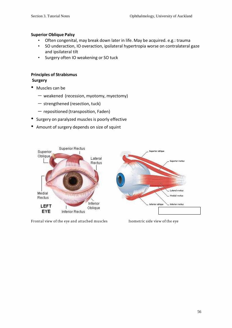
Principles of Strabismus Surgery
• Muscles can be
– weakened (recession, myotomy, myectomy)
– strengthened (resection, tuck)
– repositioned (transposition, Faden)
• Surgery on paralyzed muscles is poorly effective
• Amount of surgery depends on size of squint
www.e-sunbear.com
Frontal view of the eye and attached muscles Isometric side view of the eye
Section 3. Tutorial Notes Ophthalmology, University of Auckland
57
Dry Eye and Sjögren’s Syndrome Associate Professor Jennifer P. Craig
PhD MCOptom FAAO FBCLA
The Tear Film The tear film, which bathes the ocular surface, has a complex structure and composition and has a number of important functions:
• Optical • Mechanical • Nutritional • Defensive
Lacrimal functional unit
The lacrimal functional unit comprises the cornea, conjunctiva, eyelids, lacrimal gland, accessory glands and the connecting neural reflexes. Dysfunction of any component of the unit can alter the tear film quantity or quality and result in signs and symptoms of dry eye.
Dry Eye Definition
“Dry eye is a multifactorial disease of the tears and ocular surface that results in symptoms of discomfort, visual disturbance, and tear film instability with potential damage to the ocular surface. It is accompanied by increased osmolarity of the tear film and inflammation of the ocular surface” [2007 International Dry Eye Workshop (DEWS)]
Dry Eye
• Symptoms: dryness, grittiness, irritation, burning, ‘feeling of sand in the eye’ • Prevalence: between 8 and 13% of population affected. More common with
increasing age and most common in post-menopausal women • Environmental conditions and contact lens wear common exacerbating factors
Sjögren’s Syndrome • Chronic, systemic, inflammatory, auto-immune disorder characterised by lymphocytic
infiltration of the exocrine organs • Affects 0.1 - 4% of the population, no race predilection • More common in females; ratio of 9:1 • Onset typically in 30’s or 40’s • Characteristically presents with sicca symptoms:
- Xerophthalmia (dry eye) - Xerostomia (dry mouth) - Parotid gland enlargement - Extraglandular features e.g. arthralgia, RA, Raynaud’s phenomenon,
lymphoma

Section 3. Tutorial Notes Ophthalmology, University of Auckland
58
• Primary SS occurs in the absence of an underlying rheumatic disorder
1° SS = Dry Eye + Dry Mouth • Secondary SS is associated with an underlying rheumatic disease, such as SLE, RA, or
scleroderma. 2° SS = Dry Eye + Dry Mouth + Connective Tissue Disease
• Treatment typically centres around symptom management, but patients require monitoring for potential lymphoma development
• Morbidity is mainly associated with decreased exocrine organ function, and mortality with associated conditions such as SLE /RA.
• Patients with primary SS who do not develop a lymphoproliferative disorder have normal life expectancy
• Not fully understood
Pathophysiology of Sjögren’s Syndrome
• Believed that environmental or endogenous antigens can trigger a self-perpetuating inflammatory response in susceptible individuals
• Genetic associations • Sex hormones believed to play a role • Viruses may be responsible for triggering onset (e.g. HIV)
‘Vicious circle’
• Structural and functional changes in the lacrimal (and salivary) glands, result in reduced aqueous production and increased tear osmolarity. Tear hyperosmolarity, as a proinflammatory stimulus, induces an inflammatory cascade on the ocular surface causing an immune response in the ocular surface epithelium, and local cytokine and metalloproteinase production. The end result is damage to an epithelium, already vulnerable from poor tear film protection, and evidenced as epithelial erosions, surface irregularity and impaired vision.
Clinical findings
• History of ocular sicca symptoms (n.b. not exclusive to SS) • History of oral sicca symptoms (eating / speaking difficulties) • Dryness of other mucosal surfaces • Parotitis / visibly enlarged parotid glands • Extraglandular involvement
Symptoms related to extraglandular involvement
• Cutaneous symptoms (including dryness, pruritus, Raynaud’s) • Pulmonary symptoms (e.g. xerotrachea leading to dry cough or recurrent bronchitis) • GI symptoms due to pharyngeal/oesophageal dryness (causing difficulty swallowing,
reflux) • Cardiac symptoms (e.g. pericarditis) • Neurological symptoms (e.g. peripheral neuropathy, distal paresthesia, trigeminal /
facial nerve palsies) • Renal symptoms (secondary to interstitial nephritis) • Debilitating fatigue

Section 3. Tutorial Notes Ophthalmology, University of Auckland
59
Ocular examination (requires Ophthalmology referral) • Ocular redness (conjunctival hyperaemia, limbal redness) • Dullness of the corneal reflex (due to surface irregularity) • Blepharitis or meibomian gland dysfunction (eyelid disease) can also exist, affecting
the tear lipid layer. This requires treatment with lid hygiene and warm compresses. • Poor tear film stability (tear break up in <10 sec abnormal) • Mucous strands (filamentary keratitis) • Reduced tear production (reflex tearing) indicating lacrimal gland insufficiency
(Schirmer test) • Ocular surface staining
- Rose bengal or lissamine green will stain primarily dead/devitalised cells (and cells without mucous cover)
- Fluorescein will highlight areas of epithelial cell loss
Oral examination (ensure regular dental care) • Visibly reduced sublingual salivary pool • Tongue may stick to tongue depressor • Frequent dental caries. • Periodontal disease / tooth loss • Tendency to develop oral candidiasis. • ENT exam for bilateral parotid gland enlargement • Sialometry / sialochemistry
Joint examination • Arthritis may be a component of SS • Symmetric, polyarticular, inflammatory arthritis suggests underlying RA or a
connective-tissue disease such as SLE or scleroderma. • One third of patients with RA have Sjögren’s syndrome.
• Ig-related Amyloidosis • Bulimia • GVHD • Pancreatitis • Polymyositis • RA • Salivary Gland Tumors • Sarcoidosis • Scleroderma
Differential diagnoses
Section 3. Tutorial Notes Ophthalmology, University of Auckland
60
• RF – positive in most patients with SS • ANA • Anti-SSA/Ro
SS • Anti-SSB/La
– –
–
typically present in patients with SS found in ≈ 50% of patients (75% with 1° SS and 15% with 2°
present in 40-50% of patients with 1° SS
Immunological work-up includes: Work-up
n.b. titers of these antibodies do not reflect disease activity
Minor salivary gland biopsy • Biopsy of the minor salivary glands of the lower lip is the single most useful test to
confirm the diagnosis of SS. • A 1.5 to 2cm incision of normal-appearing mucosa allows for the harvesting of 5 or
more salivary gland lobules • Histopathological findings include focal lymphoid infiltration of minor salivary glands.
Classification Criteria American-European Consensus criteria for diagnosis of 1° SS: 4 of 6 positive responses to criteria below (must include no. 5 or 6)
1. Ocular symptoms (dry eye) 2. Oral symptoms (dry mouth) 3. Ocular signs (abnormal Schirmer without anaesthesia; < 5mm in 5 min / ocular
surface staining) 4. Oral signs (abnormal sialometry; unstimulated salivary flow <1.5 mL in 15 min) 5. Positive minor salivary gland biopsy 6. Positive anti–SSA or anti–SSB antibody results
• 2° SS is diagnosed in presence of:
- a connective tissue disease - symptoms of oral or ocular dryness, plus - criterion 3, 4 or 5 (ocular/oral signs or positive biopsy)
• For 1° SS, these criteria yield a sensitivity of 97.2% and a specificity of 48.6% • For 2° SS, the criteria yield a sensitivity of 64.7% and specificity of 97.2%
Treatment of dry eye: Level 1 • Level 1 (Mild symptoms, no corneal signs)
- Artificial tear supplements • tailor viscosity to symptom severity
• if instilled > 4 times per day, use non-preserved drops • modify environmental conditions • increase relative humidity • avoid windy conditions
- Avoid anticholinergic / antihistamine medications

Section 3. Tutorial Notes Ophthalmology, University of Auckland
61
Treatment of dry eye: Level 2 • Level 2 (Moderate-severe symptoms with tear film / visual signs, or mild ocular
surface staining) • Use Level 1 treatments plus:
- Unpreserved tears or gels ± ointments at night-time - Topical steroids (restricted course due to side effects risk) - Cyclosporine A (Restasis (US) / local pharmacy preparation) - Nutritional supplements (EFAs)
Treatment of dry eye: Level 3
• Level 3 (Severe symptoms with marked corneal changes or filamentary keratitis) • Use Level 2 treatments plus:
- Tetracyclines (low dose, long course) - Autologous / allogeneic serum tears - Punctal occlusion (to retain tears) - Acetylcysteine to break down mucous strands
Treatment of dry eye: Level 4
• Level 4 (Extremely severe symptoms with altered lifestyle, or severe corneal staining, erosions, or conjunctival scarring)
• Use Level 3 treatments plus: - Systemic anti-inflammatory therapy including acetylcysteine - Topical Vitamin A - Cautery for permanent punctal occlusion - Moisture chamber glasses
• Sip water frequently
Treatment of dry mouth
• Sugar-free lemon drops to stimulate saliva • Artificial saliva (variable patient tolerance) • Humidifiers for symptomatic relief • Regular dental visits
- fluoride treatment / special toothpaste • Topical anti-fungals as required for candidiasis
• Arthralgias
• Rheumatoid arthritis
Treatment of joint pain - NSAIDs - Hydroxycholoquine
- Likely to require other disease-modifying medications
(under direction of rheumatologist)
n.b. Cyclophosphamide used to treat serious systemic manifestations of SS has been associated with increased risk of lymphoma development

Section 3. Tutorial Notes Ophthalmology, University of Auckland
62
Multidisciplinary care of SS • Physician (provide overall perspective) • Rheumatologist (management of connective tissue disease) • Ophthalmologist (management of ocular surface manifestations) • ENT specialist (minor salivary gland / parotid biopsies) • Dentist (for oral prophylaxis and treatment) ± renal physician, chest physician, haematologist, oncologist, as necessary
Common SS medications • Cholinergic parasympathomimetics (muscarinic agonists)
(e.g. oral pilocarpine, cevimeline (US)) • Artificial tears • Artificial saliva agents • Disease modifying agents (e.g. hydroxycholoquine) • Immunosuppressants (e.g. cyclophosphamide) • Immunomodulators (e.g. cyclosporin A)
Complications of SS • Typically SS patients undergo outpatient reviews 3 – 6 monthly to check for
complications - Emergence of related disorders (e.g. SLE) - Parotid gland infections - Parotid tumours - Pseudolymphomas / lymphomas
(18x increased risk of NHL in SS)
Take home message • SS has a generally good prognosis • Prognosis more closely aligned with any associated disorders (e.g. SLE) • Patient education and support is essential
- Do not underestimate the impact SS, as a chronic condition, can have on daily life - Teach patient avoidance tactics and self-care strategies to help manage the
symptoms associated with their dry mucosal surfaces - Support groups: Sjogren’s Syndrome Society of NZ
www.sjogrensnewzealand.co.nz

63
Section 4. Self-directed learning Ophthalmology, University of Auckland
Ophthalmology Quiz
Please choose one answer for each question. There is only one correct answer to each question. To mark yourself add 1 point for every right answer, subtract one point for every wrong answer and give no points if you did not answer the question).
17-20 points - excellent
11-16 points - good
Less than 10 points – further study required!
Question 1 In regard to neoplasms of the eyelid (basal cell carcinoma, squamous cell carcinoma, melanoma). The most likely aetiological factor is:
A) Use of mascara B) A hereditary condition C) Recurrent herpes infection D) Sunlight exposure E) Recurrent local trauma
Question 2 A 60 year old male has noted painless decreasing vision in the right eye for the past year. Funduscopic examination reveals a darkly pigmented mass. These findings most strongly suggest a diagnosis of:
A) Diabetic retinopathy B) Retinoblastoma C) Melanoma D) Tay-Sachs disease E) Cytomegalovirus retinopathy
Question 3 A 56 year old female has noted increasingly frequent headaches for the past year. She recently had her vision checked, and her intraocular pressure was found to be 34mmHg. The most significant result of this condition, if not treated, is:
A) Cataract formation B) Conjunctivitis C) Hypertensive retinopathy D) Optic neuropathy E) Strabismus

64
Section 4. Self-directed learning Ophthalmology, University of Auckland
Question 4 A 42 year old male notes a progressively enlarging nodule on his right upper eyelid over the past month. This nodule is firm and painless. There is no ulceration. The conjunctiva and the cornea appear normal. Such a lesion can be characterized by all of the following findings EXCEPT one:
A) Resembles a sebaceous carcinoma on gross examination B) Prone to recurrence C) Infection of eccrine sweat glands D) Exhibits granulomatous inflammation E) Related to sun exposure
Question 5 A 3-year-old child, who had been born prematurely at 30 weeks gestation and then developed hyaline membrane disease at birth, is found to be visually impaired. Funduscopic examination reveals retinal detachment on the left. The most likely pathogenesis of this condition is:
A) Retinal damage by oxygen toxicity B) Hereditary hexosaminidase A deficiency C) Germinal matrix hemorrhage at birth D) Neonatal chlamydial infection E) Absence of tumor suppressor gene on chromosome 13
Question 6 A 70 year old female has noted the appearance of a "spot" discoloring the sclera of her right eye. Her physician notes a 0.3 cm raised yellowish-white lesion near the limbus. This lesion is LEAST likely to:
A) Appear more often in older persons B) Decrease visual acuity C) Increase in size very slowly over time D) Show elastosis of collagen histologically E) Have an association with sunlight exposure
Question 7 A 34-year-old female with a recent onset of right eye pain and irritation is noted to have an irregular dendritic corneal ulcer on slit lamp examination. This lesion is probably due to infection with:
A) Candida albicans B) Pseudomonas aeruginosa C) Hemophilus aegypticus D) Acanthamoeba E) Herpes simplex virus

65
Section 4. Self-directed learning Ophthalmology, University of Auckland
Question 8 A 17 year old female is 188 cm tall. She has long, thin fingers. Examination of her eyes reveals dislocated lenses. These findings are features of:
A) Marfan's syndrome B) Osteogenesis imperfecta C) Narrow angle glaucoma D) Aging E) Infection
Question 9 A 3 year old boy has lost vision in the right eye. Other children in his family have had a similar problem, with many losing vision in both eyes. A presumptive diagnosis of an intraocular tumor is made. Which of the following statements is LEAST appropriate for this condition:
A) Leukocoria is often noted on physical examination B) The patient may have inherited a faulty tumor suppressor gene C) The tumor is composed of small cells that form rosettes D) Dystrophic calcification is often seen histologically E) The tumor often spreads via scleral vascular channels
Question 10 A 41 year old female with a history of intravenous drug abuse presents with a 1 day history of severe headache and high fever. She has marked nuchal rigidity. You suspect that her intracranial cerebrospinal fluid pressure is increased. Which of the following funduscopic examination findings is most likely to be present:
A) Deepening of the optic cup with excavation B) Arteriolar narrowing C) Neovascularization of the optic nerve D) Elevation and swelling of the optic nerve head E) Flame-shaped haemorrhages
Question 11 An obese 50 year old male has a serum glucose of 9.9mmol/L and a hemoglobin A1C that is increased. He has decreased visual acuity. A funduscopic examination reveals a proliferative retinopathy. Which of the following findings is most characteristic of this process:
A) Capillary microaneurysms B) Dot and blot haemorrhages C) Neovascularization D) Flame-shaped hemorrhages E) Hard exudates

66
Section 4. Self-directed learning Ophthalmology, University of Auckland
Question 12 What is the most common cause of amblyopia?
A) Squint B) Trauma C) Uveitis D) Conjunctivitis E) Nuclear cataract
Question 13 A firm 0.4 cm nodule is noted on examination of the upper eyelid of a 41 year old male. Biopsy of this lesion demonstrates inflammation consisting of lymphocytes admixed with epithelioid cells and giant cells. The diagnosis is most likely to be a(an):
A) Pinguecula B) Hordeolum (stye) C) Pterygium D) Chalazion E) Actinic keratosis
Question 14 A 62 year old female has decreasing vision associated with deepening of the optic cup with excavation, as seen on funduscopic examination (atrophy of the optic nerve fibers). The disease that led to these findings could have been detected by screening for:
A) Elevated serum glucose B) Hypercholesterolemia C) Homocystinuria D) Increased intraocular pressure E) Elevated blood pressure
Question 15 An uncommon, but sight-threatening and difficult to treat, complication associated with poor contact lens hygiene affects the cornea of a 30 year old male. His eye has been painful and he has been photophobic for more than a month. The most likely diagnosis is:
A) Band keratopathy B) Acanthamoeba infection C) Pterygium D) Staphylococcus aureus infection E) Herpes simplex infection

67
Section 4. Self-directed learning Ophthalmology, University of Auckland
Question 16 A series of tissue samples for frozen section are sent to surgical pathology to make sure a lid lesion is completely excised. These samples are taken from the margin of a lesion of the medial lower eyelid of a 72 year old male. The lesion has been slowly enlarging for several years, and is now 0.4 cm in diameter, with slightly raised, pearly edges and an ulcerated center.
What is the most likely diagnosis?
A) Basal cell carcinoma B) Glioma C) Chalazion D) Squamous cell carcinoma E) Malignant melanoma
Question 17 Funduscopic examination of the right eye in a 50 year old male with 6/6 vision reveals a pigmented uveal mass. What is the most likely diagnosis?
A) Basal cell carcinoma B) Glioma C) Retinoblastoma D) Squamous cell carcinoma E) Malignant melanoma
Question 18 A 62 year old obese male is reviewed by a physician who notes decreased visual acuity and a non-healing ulcer on his left foot. Fasting blood glucose is 9.5mmol/L on two occasions. He is referred to an ophthalmologist, who on funduscopic examination observes a background retinopathy characterized by:
A) Neovascularization of the disc B) Vitreous hemorrhage C) Fibrous proliferation D) Dot and blot hemorrhage E) Papilloedema
68
Section 4. Self-directed learning Ophthalmology, University of Auckland
Question 19 A 78 year old male is bothered by chronic grittiness of the left eye. He has recurrent local redness in the eye. You find a slightly raised, pink, vascular, relatively smooth 0.3 x 0.6 cm lesion that encroaches from the conjunctiva onto the corneal surface of his left eye. The fundus examination is unremarkable. Which of the following conditions is most likely to explain these findings:
A) Hordeolum B) Primary acquired melanosis C) Pterygium D) Herpes simplex keratitis E) Pinguecula
Question 20
Which of the following eye drops has NO cycloplegic effect?
A) Atropine B) Cyclopentolate C) Midriacyl D) Homatropine E) Phenylephrine

69
Section 4. Self-directed learning Ophthalmology, University of Auckland
Ophthalmology Quiz
(Answers)
1. (D) Increasing sun exposure is strongly associated with development of malignant skin tumors.
2. (C) Melanomas produce melanin, which is pigmented (black), and are also a mass lesion.
3. (D) The increased intraocular pressure leads to optic nerve head atrophy.
4. (E) A chalazion involves the meibomian glands of the eyelid, can resemble sebaceous carcinoma and is a granulomatous inflammation that can recur. However, it is not associated with sunlight exposure.
5. (A) The child has retinopathy of prematurity, a complication of high dose oxygen therapy following birth, typically administered because of hyaline membrane disease.
6. (B) Pingueculae are located on the conjunctiva away from the cornea.
7. (E) HSV classically produces a dendritiform corneal lesion and would be the most likely causative factor.
8. (A) Marfan's syndrome is a connective tissue disorder associated with arachnodactyly and subluxation/dislocation of the crystalline lens.
9. (E) Retinoblatoma, is the most common childhood intraocular tumor. A retinoblastoma will invade into the optic nerve, whereas melanomas in adults tend to spread via scleral vascular channels. Histologically “rosettes” of cells are a pathognomonic feature.
10. (D) This patient has findings suggestive of acute bacterial meningitis that can lead to cerebral edema and increased intracranial pressure manifested as papilledema (disc swelling) on funduscopic examination.
11. (C) This is diabetic retinopathy. In proliferative retinopathy, fine new vessels grow upon the retina and into the vitreous and bleed easily, such that vitreal hemorrhages can
70
Section 4. Self-directed learning Ophthalmology, University of Auckland
obscure vision and contraction of fibrovascular and glial tissue can lead to traction retinal detachment. The other features mentioned may also be seen but new vessels are the defining element of proliferative retinopathy.
12. (A) A squinting eye (Strabismus) is the most common cause of amblyopia in children.
Treatment includes a full refractive assessment, provision of appropriate spectacles correction, patching of the better (non-amblyopic) eye, and surgery where necessary.
13. (D) This lesion is due to chronic inflammation of the meibomian glands and the histology suggests the key features of a chalazion.
14. (D) She has optic nerve atrophy from glaucoma, which is most often idiopathic primary open angle type.
15. (B) This protozoal infection is uncommon but responds poorly to antimicrobial therapy and is classically associated with poor contact lens care and swimming in contact lenses or exposing them to tap water.
16. (A) Basal cell carcinomas result from excessive sunlight exposure and are the most common tumour around the eyelids.
17. (E) The melanoma is typically pigmented (with melanin) and is the most common primary intraocular tumour of adulthood.
18. (D) This subject has diabetes mellitus with a diabetic background retinopathy that can be characterized by dot and blot hemorrhages, capillary microaneurysms, flame-shaped hemorrhages and hard exudates but not by cotton-wool spots or new vessel formation which are features of more advanced disease.
19. (C). A pterygium is similar to a pinguecula, but it encroaches onto the corneal surface. It is also related to aging and sun exposure. It is not premalignant.
20. (E) Phenylephrine is α mimetic. There is no sympathetic innervation of the ciliary muscle so it produces mild pupillary dilatation without cycloplegia – i.e. it does not cause loss of accommodation.

71
Section 4. Self-directed learning Ophthalmology, University of Auckland
Recommended Reading 4th year Handout – with particular reference to material included in the Appendix Section Vision Loss Clinical methods Visual Assessment Clinic (VAC) notes
2nd Edition 3rd Edition
Recommended reading as set in the 5th Year teaching program (Auckland), from the recommended textbook, “Ophthalmology. An illustrated colour text” by Batterbury and Bowling
ARMD (p58-9)
Conjunctiva (p34-7) Corneal inflammation (Keratitis) (p38-9) Corneal Dystrophies (p40-1) Correcting Refractive Errors (p84-5)
Epidemiology and functional implications of visual handicap (p90-1) Eyelids (p26-9)
Floaters (p110-1)
Glaucoma (p42-5)
Inherited Disease (p62-3) Iritis / Uveitis (p46-9)

72
Section 4. Self-directed learning Ophthalmology, University of Auckland
Lasers in Ophthalmology (p94-5) Orbital Disease (p32-3) Ocular Fundus – Retinal Vascular Disease (p52-57) Ocular pharmacology (p22-3), Ophthalmic Examination (p12), Ophthalmic surgery (p86), Optic disc (p62),
Pupils (p70),
Retinal dystrophies (p58),
Special Investigations (p14), Strabismus childhood (p66, 100), Strabismus adult (p68),
Tear secretion and drainage (p28), The Lens (p48), Trauma (p72, 74, 98)

73
Section 4. Self-directed learning Ophthalmology, University of Auckland
Clinical Scenarios – Links in the MBChB Portal (http://mbchb.auckland.ac.nz) Cataract Surgery Past Present Future Infant with an altered light reflex (Oph12) Gradual deterioration in visual acuity over time (Oph07) Type 2 diabetes mellitus (Endo11) 6 week check (Paed26) Family with a genetic disorder (MG02) Dry Eye & Sjogren's Syndrome Acute or chronic red eye (Oph01) Swollen and tender joints (Rh02) SLE / fatigue / pain and swelling in the hands (Rh03) Eye Accident & Emergency Acute or chronic red eye (Oph01) Acute trauma to the eye (Oph02) Child with red swelling around one eye (Oph10) Diplopia (Oph06) Pupil abnormality (Oph08) Sudden loss of vision and headache (Oph05) Sudden painless loss of vision (Oph04) Headache, morning stiffness and shoulder pain (Rh06) Eye Banking & Corneal Transplantation in New Zealand Gradual deterioration in visual acuity over time (Oph07) Glaucoma Gradual deterioration in visual acuity over time (Oph07) Sudden loss of vision and headache (Oph05) Watery eye in an infant (Oph03) Ocular Surface Inflammation and Allergy Watery eye in an infant (Oph03) Itching child (Derm01) Facial swelling and itchy rash (Derm04) Oculoplastics overview Child with red swelling around one eye (Oph10) Diplopia (Oph06) Gradual deterioration in visual acuity over time (Oph07) Watery eye in an infant (Oph03) Skin tumours (Derm03) Ophthalmoscopy Annual eye screen for patient with diabetes (Oph11) Infant with an altered light reflex (Oph12) Sudden loss of vision and headache (Oph05) Sudden painless loss of vision (Oph04) Sudden onset severe headache (ED07) Child or adolescent with a headache (Paed10) Space occupying lesion / progressive unilateral weakness (N11) Head trauma (ED09)

74
Section 4. Self-directed learning Ophthalmology, University of Auckland Paediatric Ophthalmology and Strabismus Diplopia (Oph06) Infant with an altered light reflex (Oph12) Infant with strabismus (Oph09) Pupil abnormality (Oph08) Watery eye in an infant (Oph03) Pupil Abnormalities Pupil abnormality (Oph08) Diplopia (Oph06) Infant with strabismus (Oph09) Sudden loss of vision and headache (Oph05) Altered level of consciousness in an adult (N04) Signs of Retinal Diseases Annual eye screen for patient with diabetes (Oph11) Gradual deterioration in visual acuity over time (Oph07) Sudden painless loss of vision (Oph04) The Acute Red Eye Acute or chronic red eye (Oph01) Acute trauma to the eye (Oph02) Child with red swelling around one eye (Oph10) Uveitis & Floaters Pupil abnormality (Oph08) Inflammatory low back pain (Rh07) Reactive arthritis (Rh01) Swollen and tender joints (Rh02)

75
Section 5. Contact Details Ophthalmology, University of Auckland
Contact Details Auckland: The Department of Ophthalmology 4th Floor, Building 504 Faculty of Medical & Health Sciences Grafton Campus University of Auckland Private Bag 92019 Auckland
Telephone No. 09 3737599
General Inquiries Maree McInerney ext 86991
Jane McGhee ext 86114 Selective / Electives Dr Trevor Sherwin ext 86466
Eye Clinic Ground Floor, Building 8 Greenlane Clinical Centre Greenlane West
Telephone No. 09 630 9990 ext 27600
Waikato: Mid North Island Cohort Waikato Clinical School Peter Rothwell Academic Centre Waikato Hospital Private Bag 3200 Hamilton
Inquiries Raewyn Wooderson, School Manager
Telephone No. 07 839 8750 Whangarei: Northland Health Campus Faculty of Medical and Health Sciences University of Auckland Whangarei Hospital P O Box 742 Whangarei, 0110
Inquiries Caroline Strydom, Manager
Telephone No. 09 430 4101 ext 3681 E-mail: [email protected]
Top Related