Zollinger Ellison Syndrome in a Patient with Multiple Endocrine Neoplasia...
5
Case Report Zollinger Ellison Syndrome in a Patient with Multiple Endocrine Neoplasia Type 1: A Classic Presentation Ishani Shah , 1 Neil Vyas, 2 and Kambiz S. Kadkhodayan 2 1 Department of Internal Medicine, Creighton University St. Joseph’s Hospital and Medical Center, Phoenix, AZ, USA 2 Department of Gastroenterology, Creighton University St. Joseph’s Hospital and Medical Center, Phoenix, AZ, USA Correspondence should be addressed to Ishani Shah; [email protected] Received 28 March 2019; Accepted 20 May 2019; Published 3 June 2019 Academic Editor: Tetsuo Hirata Copyright © 2019 Ishani Shah et al. is is an open access article distributed under the Creative Commons Attribution License, which permits unrestricted use, distribution, and reproduction in any medium, provided the original work is properly cited. Zollinger Ellison Syndrome (ZES) is characterized by a wide spectrum of conditions including severe gastroesophageal reflux disease, peptic ulcer disease, watery diarrhea, and weight loss. We present a case of a 60-year-old woman being evaluated for severe dyspepsia, vomiting, and chronic diarrhea, who was diagnosed to have ZES associated with a pancreatic neuroendocrine tumor, in the setting of multiple endocrine neoplasia (MEN) type 1. Although cases of ZES have been reported previously, we believe that our case is a classic presentation of ZES diagnosed on the basis of typical radiologic, endoscopic, and endosonographic features. 1. Introduction Zollinger Ellison Syndrome (ZES) is caused by ectopic hyper- secretion of gastrin from pancreatic or duodenal neuroen- docrine tumors (NETs), commonly referred to as gastrino- mas. Such high gastrin levels cause gastric acid overproduc- tion, leading to a typical presentation, usually consisting of peptic ulcer disease and severe diarrhea. We present a classic case of ZES in a patient with multiple endocrine neoplasia (MEN) type 1. 2. Case Presentation A 60-year-old woman presented to our hospital with severe nausea, vomiting, watery diarrhea, and burning epigastric pain for a duration of one week. Her epigastric pain was asso- ciated with severe acid reflux, which had been intermittently present for a duration of two years and was resistant to over- the-counter low-dose proton pump inhibitor (PPI) therapy. Her past medical history was negative for any evidence of gastrointestinal (GI) bleed. Interestingly, the patient had a daughter who had been diagnosed with multiple endocrine neoplasia (MEN) type 1 a year prior to presentation. On physical exam, she was afebrile with stable hemodynamics. Abdominal palpation revealed mild epigastric tenderness without any guarding or rigidity. Cardiopulmonary exam was within normal limits. Significant laboratory findings included WBC count of 15,000/microL, potassium of 3 mmol/L, magnesium of 0.7 mg/dL, and calcium of 11.8 mg/dL. Lipase level was within normal limits. Other pertinent laboratory values included fasting serum gastrin level of 1603 pg/mL (0- 180 pg/mL), chromogranin A level of 14600 ng/mL (0-100 ng/mL), prolactin hormone level of 21 ng/mL (2-29 ng/mL), and parathyroid hormone (PTH) level of 473 pg/mL (10- 65 pg/mL). She did not have any history of prior gastric surgeries, gastroparesis, or renal disease, to possibly explain her elevated gastrin level. An infectious workup for her diarrhea, including Clostridium difficile toxin and a stool PCR panel for common enteric pathogens, was negative. Subsequently, an extensive workup for evaluation of MEN was done, which revealed a unilateral parathyroid adenoma on neck imaging and diffuse stomach wall thickening along with pancreatic cystic lesions in body (1.2 cm) and tail (0.7 cm) on abdominal MRI (Figure 1). Testing for pituitary disease was negative. An esophagogastroduodenoscopy (EGD) was performed for further evaluation of her symptoms, which revealed severe reflux esophagitis, diffusely hypertrophic gastric rugae and multiple postbulbar ulcers in the duodenum (Figures 2(a), Hindawi Case Reports in Gastrointestinal Medicine Volume 2019, Article ID 9605769, 4 pages https://doi.org/10.1155/2019/9605769
Transcript of Zollinger Ellison Syndrome in a Patient with Multiple Endocrine Neoplasia...
Case ReportZollinger Ellison Syndrome in a Patient with Multiple EndocrineNeoplasia Type 1: A Classic Presentation
Ishani Shah ,1 Neil Vyas,2 and Kambiz S. Kadkhodayan2
1Department of Internal Medicine, Creighton University St. Joseph’s Hospital and Medical Center, Phoenix, AZ, USA2Department of Gastroenterology, Creighton University St. Joseph’s Hospital and Medical Center, Phoenix, AZ, USA
Correspondence should be addressed to Ishani Shah; [email protected]
Received 28 March 2019; Accepted 20 May 2019; Published 3 June 2019
Academic Editor: Tetsuo Hirata
Copyright © 2019 Ishani Shah et al. This is an open access article distributed under the Creative Commons Attribution License,which permits unrestricted use, distribution, and reproduction in any medium, provided the original work is properly cited.
Zollinger Ellison Syndrome (ZES) is characterized by a wide spectrum of conditions including severe gastroesophageal refluxdisease, peptic ulcer disease, watery diarrhea, and weight loss. We present a case of a 60-year-old woman being evaluated for severedyspepsia, vomiting, and chronic diarrhea, who was diagnosed to have ZES associated with a pancreatic neuroendocrine tumor,in the setting of multiple endocrine neoplasia (MEN) type 1. Although cases of ZES have been reported previously, we believe thatour case is a classic presentation of ZES diagnosed on the basis of typical radiologic, endoscopic, and endosonographic features.
1. Introduction
Zollinger Ellison Syndrome (ZES) is caused by ectopic hyper-secretion of gastrin from pancreatic or duodenal neuroen-docrine tumors (NETs), commonly referred to as gastrino-mas. Such high gastrin levels cause gastric acid overproduc-tion, leading to a typical presentation, usually consisting ofpeptic ulcer disease and severe diarrhea. We present a classiccase of ZES in a patient with multiple endocrine neoplasia(MEN) type 1.
2. Case Presentation
A 60-year-old woman presented to our hospital with severenausea, vomiting, watery diarrhea, and burning epigastricpain for a duration of one week. Her epigastric pain was asso-ciated with severe acid reflux, which had been intermittentlypresent for a duration of two years and was resistant to over-the-counter low-dose proton pump inhibitor (PPI) therapy.Her past medical history was negative for any evidence ofgastrointestinal (GI) bleed. Interestingly, the patient had adaughter who had been diagnosed with multiple endocrineneoplasia (MEN) type 1 a year prior to presentation. Onphysical exam, she was afebrile with stable hemodynamics.Abdominal palpation revealed mild epigastric tenderness
without any guarding or rigidity. Cardiopulmonary examwaswithin normal limits.
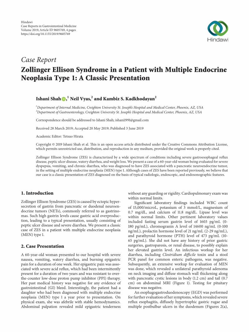
Significant laboratory findings included WBC countof 15,000/microL, potassium of 3 mmol/L, magnesium of0.7 mg/dL, and calcium of 11.8 mg/dL. Lipase level waswithin normal limits. Other pertinent laboratory valuesincluded fasting serum gastrin level of 1603 pg/mL (0-180 pg/mL), chromogranin A level of 14600 ng/mL (0-100ng/mL), prolactin hormone level of 21 ng/mL (2-29 ng/mL),and parathyroid hormone (PTH) level of 473 pg/mL (10-65 pg/mL). She did not have any history of prior gastricsurgeries, gastroparesis, or renal disease, to possibly explainher elevated gastrin level. An infectious workup for herdiarrhea, including Clostridium difficile toxin and a stoolPCR panel for common enteric pathogens, was negative.Subsequently, an extensive workup for evaluation of MENwas done, which revealed a unilateral parathyroid adenomaon neck imaging and diffuse stomach wall thickening alongwith pancreatic cystic lesions in body (1.2 cm) and tail (0.7cm) on abdominal MRI (Figure 1). Testing for pituitarydisease was negative.
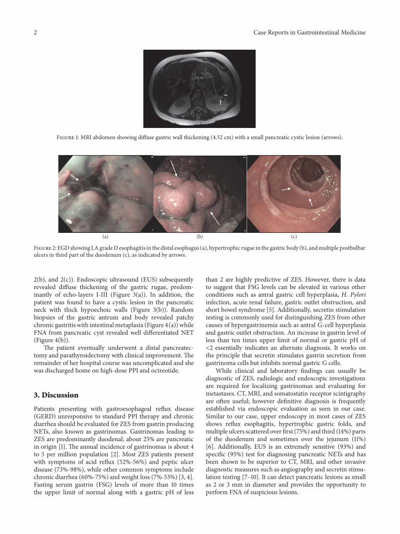
An esophagogastroduodenoscopy (EGD) was performedfor further evaluation of her symptoms,which revealed severereflux esophagitis, diffusely hypertrophic gastric rugae andmultiple postbulbar ulcers in the duodenum (Figures 2(a),
HindawiCase Reports in Gastrointestinal MedicineVolume 2019, Article ID 9605769, 4 pageshttps://doi.org/10.1155/2019/9605769
2 Case Reports in Gastrointestinal Medicine
Figure 1: MRI abdomen showing diffuse gastric wall thickening (4.52 cm) with a small pancreatic cystic lesion (arrows).
(a) (b) (c)
Figure 2: EGDshowingLAgradeDesophagitis in the distal esophagus (a), hypertrophic rugae in the gastric body (b), andmultiple postbulbarulcers in third part of the duodenum (c), as indicated by arrows.
2(b), and 2(c)). Endoscopic ultrasound (EUS) subsequentlyrevealed diffuse thickening of the gastric rugae, predom-inantly of echo-layers I-III (Figure 3(a)). In addition, thepatient was found to have a cystic lesion in the pancreaticneck with thick hypoechoic walls (Figure 3(b)). Randombiopsies of the gastric antrum and body revealed patchychronic gastritiswith intestinalmetaplasia (Figure 4(a)) whileFNA from pancreatic cyst revealed well differentiated NET(Figure 4(b)).
The patient eventually underwent a distal pancreatec-tomy and parathyroidectomy with clinical improvement. Theremainder of her hospital course was uncomplicated and shewas discharged home on high-dose PPI and octreotide.
3. Discussion
Patients presenting with gastroesophageal reflux disease(GERD) unresponsive to standard PPI therapy and chronicdiarrhea should be evaluated for ZES from gastrin producingNETs, also known as gastrinomas. Gastrinomas leading toZES are predominantly duodenal; about 25% are pancreaticin origin [1]. The annual incidence of gastrinomas is about 4to 5 per million population [2]. Most ZES patients presentwith symptoms of acid reflux (52%-56%) and peptic ulcerdisease (73%-98%), while other common symptoms includechronic diarrhea (60%-75%) and weight loss (7%-53%) [3, 4].Fasting serum gastrin (FSG) levels of more than 10 timesthe upper limit of normal along with a gastric pH of less
than 2 are highly predictive of ZES. However, there is datato suggest that FSG levels can be elevated in various otherconditions such as antral gastric cell hyperplasia, H. Pyloriinfection, acute renal failure, gastric outlet obstruction, andshort bowel syndrome [5]. Additionally, secretin stimulationtesting is commonly used for distinguishing ZES from othercauses of hypergastrinemia such as antral G-cell hyperplasiaand gastric outlet obstruction. An increase in gastrin level ofless than ten times upper limit of normal or gastric pH of<2 essentially indicates an alternate diagnosis. It works onthe principle that secretin stimulates gastrin secretion fromgastrinoma cells but inhibits normal gastric G cells.
While clinical and laboratory findings can usually bediagnostic of ZES, radiologic and endoscopic investigationsare required for localizing gastrinomas and evaluating formetastases. CT, MRI, and somatostatin receptor scintigraphyare often useful; however definitive diagnosis is frequentlyestablished via endoscopic evaluation as seen in our case.Similar to our case, upper endoscopy in most cases of ZESshows reflux esophagitis, hypertrophic gastric folds, andmultiple ulcers scattered over first (75%) and third (14%) partsof the duodenum and sometimes over the jejunum (11%)[6]. Additionally, EUS is an extremely sensitive (93%) andspecific (95%) test for diagnosing pancreatic NETs and hasbeen shown to be superior to CT, MRI, and other invasivediagnostic measures such as angiography and secretin stimu-lation testing [7–10]. It can detect pancreatic lesions as smallas 2 or 3 mm in diameter and provides the opportunity toperform FNA of suspicious lesions.

Case Reports in Gastrointestinal Medicine 3
(a) (b)
Figure 3: EUS showing hypertrophic gastric rugae (a) and neuroendocrine tumor in the pancreatic neck (b), respectively, as indicated byarrows.
(a) (b)
Figure 4: Gastric biopsy (a) showing patchy hypertrophic gastritis and intestinal metaplasia (green arrows) while pancreatic aspirate (b)showing neuroendocrine cells (area enclosed within circle).
Around 80% of gastrinomas are sporadic; however 20%are associated with MEN type 1, an autosomal dominantcondition associated with predisposition to tumors in thehyperparathyroidism glands, pancreas, and pituitary [11].About 40% of patients with MEN type 1 initially present withsymptoms of ZES [12]. Therefore, an extensive workup forMEN should be carried out in all patients with ZES. In manystudies, EUS has consistently been proven to be superior toother imaging modalities for diagnosing pancreatic NETs inpatients with a suspected or established diagnosis of MENtype 1 [8, 13, 14].
In conclusion, our case is a classic blend of clinical,radiologic, endoscopic, and endosonographic features of ZESin a patient with MEN type 1. It highlights the importance ofearly recognition of these findings for prompt diagnosis andappropriate treatment.
Consent
Informed consent was obtained from the patient for writingthis case report.
Conflicts of Interest
The authors declare that there are no conflicts of interestregarding the publication of this article.
Authors’ Contributions
Ishani Shah, MD, is responsible for writing and editing thecase report. Neil Vyas, MD, and Kambiz S. Kadkhodayan,MD, are responsible for editing the case report.
References
[1] J. I. Isenberg, J. H. Walsh, and M. I. Grossman, “Progressin gastroenterology, zollinger-ellison syndrome,”Gastroenterol-ogy, vol. 65, p. 140, 1973.
[2] K. Oberg, “Pancreatic endocrine tumors,” Seminars inOncology,vol. 37, no. 6, pp. 594–618, 2010.
[3] P. K. Roy, D. J. Venzon, H. Shojamanesh et al., “Zollinger-ellisonsyndrome. Clinical presentation in 261 patients,”Medicine, vol.79, no. 6, pp. 379–411, 2000.
[4] J. A. Norton, D. S. Foster, T. Ito, and R. T. Jensen, “Gastrinomas:medical or surgical treatment,” Endocrinology and MetabolismClinics, vol. 47, no. 3, pp. 577–601, 2018.
[5] M. J. Berna, K. M. Hoffmann, J. Serrano, F. Gibril, and R.T. Jensen, “Serum gastrin in Zollinger-Ellison syndrome: I.Prospective study of fasting serum gastrin in 309 patients fromthe national institutes of health and comparison with 2229 casesfrom the literature,”Medicine, vol. 85, no. 6, pp. 295–330, 2006.
[6] H. Tariq, M. U. Kamal, V. Vootla et al., “A rare cause ofabdominal pain andmass in an 18-year-old patient: a diagnosticdilemma,” Gastroenterology Research, vol. 11, no. 1, p. 75, 2018.

4 Case Reports in Gastrointestinal Medicine
[7] T. Rosch, C. J. Lightdale, J. F. Botet et al., “Localization ofpancreatic endocrine tumors by endoscopic ultrasonography,”The New England Journal of Medicine, vol. 326, no. 26, pp. 1721–1726, 1992.
[8] M. A. Anderson, S. Carpenter, N.W.Thompson, T. T. Nostrant,G. H. Elta, and J. M. Scheiman, “Endoscopic ultrasoundis highly accurate and directs management in patients withneuroendocrine tumors of the pancreas,” American Journal ofGastroenterology, vol. 95, no. 9, pp. 2271–2277, 2000.
[9] M. A. Khashab, E. Yong, A. M. Lennon et al., “EUS is stillsuperior to multidetector computerized tomography for detec-tion of pancreatic neuroendocrine tumors,” GastrointestinalEndoscopy, vol. 73, no. 4, pp. 691–696, 2011.
[10] P. D. James, A. V. Tsolakis, M. Zhang et al., “Incremental benefitof preoperative EUS for the detection of pancreatic neuroen-docrine tumors: A meta-analysis,” Gastrointestinal Endoscopy,vol. 81, no. 4, pp. 848–856, 2015.
[11] J. A. Norton, “Neuroendocrine tumors of the pancreas andduodenum,” Current Problems in Surgery, vol. 31, no. 2, pp. 89–156, 1994.
[12] F. Gibril, M. Schumann, A. Pace, and R. T. Jensen, “Multipleendocrine neoplasia type 1 and Zollinger-Ellison syndrome: aprospective study of 107 cases and comparison with 1009 casesfrom the literature,”Medicine, vol. 83, no. 1, pp. 43–83, 2004.
[13] S. J. Van Asselt, A. H. Brouwers, H. M. Van Dullemen et al.,“EUS is superior for detection of pancreatic lesions comparedwith standard imaging in patients with multiple endocrineneoplasia type 1,” Gastrointestinal Endoscopy, vol. 81, no. 1, pp.159–167, 2015.
[14] P. H. Kann, E. Balakina, D. Ivan et al., “Natural course of small,asymptomatic neuroendocrine pancreatic tumours in multipleendocrine neoplasia type 1: An endoscopic ultrasound imagingstudy,” Endocrine-Related Cancer, vol. 13, no. 4, pp. 1195–1202,2006.

Stem Cells International
Hindawiwww.hindawi.com Volume 2018
Hindawiwww.hindawi.com Volume 2018
MEDIATORSINFLAMMATION
of
EndocrinologyInternational Journal of
Hindawiwww.hindawi.com Volume 2018
Hindawiwww.hindawi.com Volume 2018
Disease Markers
Hindawiwww.hindawi.com Volume 2018
BioMed Research International
OncologyJournal of
Hindawiwww.hindawi.com Volume 2013
Hindawiwww.hindawi.com Volume 2018
Oxidative Medicine and Cellular Longevity
Hindawiwww.hindawi.com Volume 2018
PPAR Research
Hindawi Publishing Corporation http://www.hindawi.com Volume 2013Hindawiwww.hindawi.com
The Scientific World Journal
Volume 2018
Immunology ResearchHindawiwww.hindawi.com Volume 2018
Journal of
ObesityJournal of
Hindawiwww.hindawi.com Volume 2018
Hindawiwww.hindawi.com Volume 2018
Computational and Mathematical Methods in Medicine
Hindawiwww.hindawi.com Volume 2018
Behavioural Neurology
OphthalmologyJournal of
Hindawiwww.hindawi.com Volume 2018
Diabetes ResearchJournal of
Hindawiwww.hindawi.com Volume 2018
Hindawiwww.hindawi.com Volume 2018
Research and TreatmentAIDS
Hindawiwww.hindawi.com Volume 2018
Gastroenterology Research and Practice
Hindawiwww.hindawi.com Volume 2018
Parkinson’s Disease
Evidence-Based Complementary andAlternative Medicine
Volume 2018Hindawiwww.hindawi.com
Submit your manuscripts atwww.hindawi.com