Zika virus replication in the mosquito Culex ... - IAM - Zika... · 1Departamento de Entomologia,...
11
OPEN ORIGINAL ARTICLE Zika virus replication in the mosquito Culex quinquefasciatus in Brazil Duschinka RD Guedes 1, *, Marcelo HS Paiva 1,2, *, Mariana MA Donato 1 , Priscilla P Barbosa 1 , Larissa Krokovsky 1 , Sura W dos S Rocha 1 , Karina LA Saraiva 1 , Mônica M Crespo 1 , Tatiana MT Rezende 1 , Gabriel L Wallau 1 , Rosângela MR Barbosa 1 , Cláudia MF Oliveira 1 , Maria AV Melo-Santos 1 , Lindomar Pena 3 , Marli T Cordeiro 3 , Rafael F de O Franca 3 , André LS de Oliveira 4 , Christina A Peixoto 1 , Walter S Leal 5 and Constância FJ Ayres 1 Zika virus (ZIKV) is a flavivirus that has recently been associated with an increased incidence of neonatal microcephaly and other neurological disorders. The virus is primarily transmitted by mosquito bite, although other routes of infection have been implicated in some cases. The Aedes aegypti mosquito is considered to be the main vector to humans worldwide; however, there is evidence that other mosquito species, including Culex quinquefasciatus, transmit the virus. To test the potential of Cx. quinquefasciatus to transmit ZIKV, we experimentally compared the vector competence of laboratory-reared Ae. aegypti and Cx. quinquefasciatus. Interestingly, we were able to detect the presence of ZIKV in the midgut, salivary glands and saliva of artificially fed Cx. quinquefasciatus. In addition, we collected ZIKV-infected Cx. quinquefasciatus from urban areas with high microcephaly incidence in Recife, Brazil. Corroborating our experimental data from artificially fed mosquitoes, ZIKV was isolated from field-caught Cx. quinquefasciatus, and its genome was partially sequenced. Collectively, these findings indicate that there may be a wider range of ZIKV vectors than anticipated. Emerging Microbes & Infections (2017) 6, e69; doi:10.1038/emi.2017.59; published online 9 August 2017 Keywords: Aedes; Culex; microcephaly; vector competence; Zika INTRODUCTION Zika is classically considered a mild disease whose symptoms include fever, joint pain, rash and, in some cases, conjunctivitis. 1 However, the recent Zika outbreak in Brazil has been associated with an increased incidence of neonatal microcephaly and neurological disorders. 2,3 Zika virus (ZIKV) is a poorly understood, small, enveloped RNA virus with ssRNA (+) belonging to the family Flaviviridae. It was first isolated in April 1947 from a rhesus monkey and then, in January 1948, from the mosquito species Aedes africanus. 4 Subsequently, several ZIKV strains have been isolated from Aedes, Mansonia, Anopheles and Culex mosquitoes. 5 The first known Zika epidemic in an urban environment occurred in Micronesia in 2007, with ~73% of the human population on Yap Island becoming infected. 6 Intriguingly, although many Aedes mosquitoes were collected in the field and evaluated for virus detection, no samples were found to be positive for ZIKV. 6 In addition, it is important to highlight that Ae. aegypti is absent from most islands in the Micronesian archipelago and is rare on the islands where ZIKV is present. 6,7 There is a global consensus among scientists and health agencies that Ae. aegypti and Ae. albopictus are the main ZIKV vectors in urban areas (WHO, 2016). This belief is partly because vector competence experiments for ZIKV were conducted exclusively for species of this genus, mainly Ae. aegypti, until recently. 8,9 In fact, previous laboratory studies 8,10 suggested that Ae. aegypti is a ZIKV vector. Recently, high rates of dissemination and transmission of the ZIKV in Ae. aegypti have been observed under laboratory conditions. 11 Intriguingly, a few studies have demonstrated that Ae. aegypti and Ae. albopictus popula- tions have low rates of ZIKV transmission 12,13 or none, 14,15 but the role of other vectors in the spread of ZIKV has been overlooked. Thus, other mosquito species, such as Cx. quinquefasciatus, that coexists with Ae. aegypti in urban areas, could contribute to ZIKV transmission. 16 The literature is dichotomous regarding Culex vectors. Although there are recent reports showing that Culex is not a ZIKV vector, 17–22 a recent paper demonstrated that Cx. quinquefasciatus collected in urban areas in China were able to be infected with a local ZIKV strain and then transmit it to mice. 23 These controversial results are expected and could be due to differences in the combination of mosquito 1 Departamento de Entomologia, Instituto Aggeu Magalhães, Fundação Oswaldo Cruz (Fiocruz), Recife 50760-420, Brazil; 2 Núcleo de Ciências da Vida, Centro Acadêmico do Agreste, Universidade Federal de Pernambuco, Caruaru 55002-970, Brazil; 3 Laboratório de Virologia e Terapia Experimental, Instituto Aggeu Magalhães, Fundação Oswaldo Cruz (Fiocruz), Recife 50670-420, Brazil; 4 Núcleo de Estatística e Geoprocessamento, Instituto Aggeu Magalhães, Fundação Oswaldo Cruz (Fiocruz), Recife 50670-420, Brazil and 5 Department of Molecular and Cellular Biology, University of California-Davis, Davis, CA 95616, USA Correspondence: CFJ Ayres E-mail: tans@cpqam.fiocruz.br *These authors contributed equally to this work. Received 14 November 2016; revised 15 May 2017; accepted 4 June 2017 Emerging Microbes & Infections (2017) 6, e69; doi:10.1038/emi.2017.59 www.nature.com/emi
Transcript of Zika virus replication in the mosquito Culex ... - IAM - Zika... · 1Departamento de Entomologia,...

OPEN
ORIGINAL ARTICLE
Zika virus replication in the mosquito Culexquinquefasciatus in Brazil
Duschinka RD Guedes1,*, Marcelo HS Paiva1,2,*, Mariana MA Donato1, Priscilla P Barbosa1,Larissa Krokovsky1, Sura W dos S Rocha1, Karina LA Saraiva1, Mônica M Crespo1, Tatiana MT Rezende1,Gabriel L Wallau1, Rosângela MR Barbosa1, Cláudia MF Oliveira1, Maria AV Melo-Santos1, Lindomar Pena3,Marli T Cordeiro3, Rafael F de O Franca3, André LS de Oliveira4, Christina A Peixoto1, Walter S Leal5 andConstância FJ Ayres1
Zika virus (ZIKV) is a flavivirus that has recently been associated with an increased incidence of neonatal microcephaly and
other neurological disorders. The virus is primarily transmitted by mosquito bite, although other routes of infection have been
implicated in some cases. The Aedes aegypti mosquito is considered to be the main vector to humans worldwide; however, there
is evidence that other mosquito species, including Culex quinquefasciatus, transmit the virus. To test the potential of Cx.quinquefasciatus to transmit ZIKV, we experimentally compared the vector competence of laboratory-reared Ae. aegypti and Cx.quinquefasciatus. Interestingly, we were able to detect the presence of ZIKV in the midgut, salivary glands and saliva of
artificially fed Cx. quinquefasciatus. In addition, we collected ZIKV-infected Cx. quinquefasciatus from urban areas with high
microcephaly incidence in Recife, Brazil. Corroborating our experimental data from artificially fed mosquitoes, ZIKV was isolated
from field-caught Cx. quinquefasciatus, and its genome was partially sequenced. Collectively, these findings indicate that there
may be a wider range of ZIKV vectors than anticipated.
Emerging Microbes & Infections (2017) 6, e69; doi:10.1038/emi.2017.59; published online 9 August 2017
Keywords: Aedes; Culex; microcephaly; vector competence; Zika
INTRODUCTION
Zika is classically considered a mild disease whose symptoms includefever, joint pain, rash and, in some cases, conjunctivitis.1 However, therecent Zika outbreak in Brazil has been associated with an increasedincidence of neonatal microcephaly and neurological disorders.2,3 Zikavirus (ZIKV) is a poorly understood, small, enveloped RNA virus withssRNA (+) belonging to the family Flaviviridae. It was first isolated inApril 1947 from a rhesus monkey and then, in January 1948, from themosquito species Aedes africanus.4 Subsequently, several ZIKV strainshave been isolated from Aedes, Mansonia, Anopheles and Culexmosquitoes.5
The first known Zika epidemic in an urban environment occurredin Micronesia in 2007, with ~ 73% of the human population on YapIsland becoming infected.6 Intriguingly, although many Aedesmosquitoes were collected in the field and evaluated for virusdetection, no samples were found to be positive for ZIKV.6 Inaddition, it is important to highlight that Ae. aegypti is absent frommost islands in the Micronesian archipelago and is rare on the islandswhere ZIKV is present.6,7
There is a global consensus among scientists and health agenciesthat Ae. aegypti and Ae. albopictus are the main ZIKV vectors in urbanareas (WHO, 2016). This belief is partly because vector competenceexperiments for ZIKV were conducted exclusively for species of thisgenus, mainly Ae. aegypti, until recently.8,9 In fact, previous laboratorystudies8,10 suggested that Ae. aegypti is a ZIKV vector. Recently, highrates of dissemination and transmission of the ZIKV in Ae. aegyptihave been observed under laboratory conditions.11 Intriguingly, a fewstudies have demonstrated that Ae. aegypti and Ae. albopictus popula-tions have low rates of ZIKV transmission12,13 or none,14,15 but therole of other vectors in the spread of ZIKV has been overlooked. Thus,other mosquito species, such as Cx. quinquefasciatus, that coexists withAe. aegypti in urban areas, could contribute to ZIKV transmission.16
The literature is dichotomous regarding Culex vectors. Although thereare recent reports showing that Culex is not a ZIKV vector,17–22
a recent paper demonstrated that Cx. quinquefasciatus collected inurban areas in China were able to be infected with a local ZIKV strainand then transmit it to mice.23 These controversial results are expectedand could be due to differences in the combination of mosquito
1Departamento de Entomologia, Instituto Aggeu Magalhães, Fundação Oswaldo Cruz (Fiocruz), Recife 50760-420, Brazil; 2Núcleo de Ciências da Vida, Centro Acadêmico doAgreste, Universidade Federal de Pernambuco, Caruaru 55002-970, Brazil; 3Laboratório de Virologia e Terapia Experimental, Instituto Aggeu Magalhães, Fundação Oswaldo Cruz(Fiocruz), Recife 50670-420, Brazil; 4Núcleo de Estatística e Geoprocessamento, Instituto Aggeu Magalhães, Fundação Oswaldo Cruz (Fiocruz), Recife 50670-420, Brazil and5Department of Molecular and Cellular Biology, University of California-Davis, Davis, CA 95616, USA
Correspondence: CFJ AyresE-mail: [email protected]
*These authors contributed equally to this work.
Received 14 November 2016; revised 15 May 2017; accepted 4 June 2017
Emerging Microbes & Infections (2017) 6, e69; doi:10.1038/emi.2017.59www.nature.com/emi

and virus genotypes used in artificial blood-feeding assays, as previousstudies have also shown negative results for the main vectorAe. aegypti.13,15 Diagne et al.15 reported that Dakar and Kedougou(Senegal) populations of the yellow fever mosquito did not transmitsix different ZIKV strains, including the MR766 strain, which was firstisolated from a sentinel monkey in Uganda. In an evident contrast,Wegner-Lucarelli et al.24 found that Ae. aegypti from Poza Rica(Mexico) is a competent vector of the MR766 ZIKV strain. Theseare the first two studies that used the same strain of the virus vis-à-visthe same mosquito species. Therefore, it is reasonable to conclude thatthe genetic variability of mosquito species from different geographicalorigins might account for this apparent discrepancy in reported data.25
Here, we report data that support the idea that in Recife, northeastBrazil, Cx. quinquefasciatus, the southern house mosquito in tropicaland subtropical areas, is a potential ZIKV vector. We performedmosquito vector competence assays under laboratory conditions,comparing both Ae. aegypti and Cx. quinquefasciatus using differentvirus doses. ZIKV was detected in the salivary glands and in the salivaof artificially fed Cx. quinquefasciatus mosquitoes. In addition to theseresults, ZIKV was also detected in field-caught samples of Cx.quinquefasciatus, which had no signs of recent blood feeding. ZIKVwas isolated from these samples, and its genome was sequenced,providing the first partial genome of ZIKV obtained from Cx.quinquefasciatus mosquitoes. Collectively, our results suggest that thisspecies is likely a ZIKV vector in Brazil, which has several implicationsfor vector control strategies as well as for the understanding of theepidemiology of ZIKV.
MATERIALS AND METHODS
MosquitoesThe present study was conducted using two laboratory colonies ofmosquitoes and field-collected specimens of Ae. aegypti (F1–F2) fromthe Archipelago of Fernando de Noronha (FN), a district ofPernambuco State (PE). Cx. quinquefasciatus (formerly known asCx. pipiens quinquefasciatus) mosquitoes originated from eggs (rafts)collected in Peixinhos, a neighborhood in Recife, PE, Brazil, in 2009.The Ae. aegypti laboratory colony (RecLab) was established with~ 1000 specimens collected in Graças, a neighborhood in the RecifeMetropolitan Region, and has been maintained in the insectary ofAggeu Magalhães Institute (IAM)/FIOCRUZ since 1996 under stan-dard conditions: 26± 2 °C, 65%–85% relative humidity, 12/12 light/dark cycle. More information regarding the two laboratory colonieshas been described elsewhere.26,27 The mosquitoes were kept in theinsectary of the Department of Entomology IAM/FIOCRUZ understandard conditions described above. Larvae were maintained inplastic trays filled with potable water and were fed solely on cat food(Friskies), while adults were given access to a 10% sucrose solutionuntil they were administered defibrinated rabbit blood infectedwith ZIKV.
Virus strainExperimental infections of mosquitoes with ZIKV were conductedusing the ZIKV BRPE243/2015 strain derived from the serum of apatient with an acute maculopapular rash in Pernambuco State, Brazil,during the 2015 outbreak.28 This strain was named the ZIKV/H.sapiens/Brazil/PE243/2015 strain, according to the nomenclaturedescribed by Scheuermann,29 and was fully characterized (accessionnumber KX197192.1). Following isolation, the virus was passed onceon Ae. albopictus C6/36 cells. Viral stocks were then produced inVERO cells and stored at − 80 °C until use. Prior to oral infection, the
viral titer of the stock was calculated via plaque assay on VERO cellsand reached 106 plaque-forming units per milliliter (PFU/mL).
Artificial feedingTwo artificial-feeding assays were conducted using a viral stockconcentration of 106 PFU/mL as well as a 100-fold dilution from theviral stock in each experiment. Notably, in the first artificial-feedingassay, the frozen virus sample was mixed with defibrinated rabbitblood. In the second assay, ZIKV BRPE243/2015 was first grown inVERO cells at a multiplicity of infection of 0.5 for 4–5 days.Subsequently, the cell culture flasks were frozen at − 80 °C, thawedat 37 °C twice and then mixed with defibrinated rabbit blood in a 1:1proportion. Seven- to ten-day-old female mosquitoes were starved for18 h prior to artificial feeding. The mosquitoes were exposed to aninfectious blood meal for 90 min, as described in Salazar et al.,30 withminor modifications. Briefly, infectious blood was provided in amembrane-feeding device placed on each mosquito cage. The bloodfeeding was maintained at 37 °C by using heat packs during theprocess. Fully engorged mosquito females were cold anesthetized,transferred to a new cage and maintained in the infection room for15 days. Both assays included a control group fed on uninfectedcultured cells mixed with defibrinated rabbit blood.
RNA extraction and virus detectionFour to fifteen mosquitoes were dissected in order to collect themidguts and salivary glands at three, seven and 15 days post infection(dpi). To prevent hemolymph contamination, all midguts and salivaryglands were extensively washed with phosphate-buffered saline bufferimmediately after dissection and prior to tissue extraction. Tissueswere individually transferred to a 1.5-mL DNAse/RNAse-free micro-tube containing 300 μL of mosquito diluent31 and were stored at − 80 °C until further usage. Each tissue was ground with sterile micropestles,and RNA extraction was performed with 100 μL of the homogenate.The TRIzol method (Invitrogen, Waltham, MA, USA) was performedaccording to the manufacturer’s instructions with modifications asfollows. Tissue homogenate (100 μL) was mixed with 200 μL ofTRIzol, homogenized by vortexing for 15 s and incubated for 5 minat room temperature. Chloroform (100 μL) was added to the mixture,and homogenization was performed by shaking the tubes vigorouslyfor 15 s by hand. The mixture was then incubated at roomtemperature for 2–3 min. The samples were centrifuged at 12 000gfor 15 min at 4 °C. The aqueous phase of each sample was removedand transferred to a new tube containing 250 μL of 100% isopropanol.This mixture was incubated at room temperature for 10 min and thencentrifuged at 12000g for 10 min at 4 °C. The supernatant wasremoved, and the RNA pellet was washed with 300 μL of 75%ethanol. The samples were homogenized briefly and then centrifugedat 7500g for 5 min at 4 °C. The supernatant was discarded, and theRNA was then air-dried for 15 min. The RNA pellet was resuspendedin 30 μL of RNAse-free water. After RNA resuspension, the sampleswere treated with DNAse (Turbo DNase, Ambion, Foster City, CA,USA) according to the manufacturer’s protocol.Virus detection was performed by quantitative RT-PCR (RT-qPCR,
also known as real-time RT-PCR) in an ABI Prism 7500 SDS Real-Time system (Applied BioSystems, Foster City, CA, USA) using theQuantiNova Probe RT-PCR kit (Qiagen, Hilden, Germany). RT-qPCRwas performed in a 20-μL reaction volume containing 5 μL ofextracted RNA, 2× QuantiNova Probe RT-PCR Master Mix, 0.2 μLof the QuantiNova Probe RT Mix, 0.1 μL of the ROX Reference Dye,100 μM of each primer (stock) and 25 μM of the probe (stock). Theprimers, probe and PCR conditions were first described in Lanciotti
ZIKV in Cx. quinquefasciatus from BrazilDRD Guedes et al
2
Emerging Microbes & Infections

et al.,32 and each sample was tested in duplicate. RT-qPCR cyclingincluded a single cycle of reverse transcription for 15 min at 45 °C,followed by 5 min at 95 °C for reverse transcriptase inactivation andDNA polymerase activation, and then 45 cycles of 5 s at 95 °C and 45 sat 60 °C (annealing-extension step). The amount of viral RNA in eachsample was estimated by comparing the cycle threshold values (Ct) tothe standard curve for every RT-qPCR assay. The standard curveconsisted of different dilutions of previously titrated ZIKV BRPE243-/2015 RNA. Mosquitoes collected immediately after artificial feedingwere used as positive controls, while control mosquitoes fed onuninfected blood and RT-PCR reactions containing no RNArepresented negative controls. Fluorescence was analyzed at the endof the amplifications. Positive samples were used to calculate vectorcompetence parameters, such as the infection rate (IR), which is thenumber of positive midguts divided by the total number of midgutstested, and the proportion of infected salivary glands (SR), which is thenumber of positive salivary glands divided by the total number ofsalivary glands tested.
Collection of virus-infected mosquito salivaTo confirm whether the viral RNA detected by RT-qPCR within thesalivary glands was infective and could be released during blood-feeding meals, we assayed ZIKV from saliva samples. From the 8th to14th dpi, mosquitoes from each group were exposed to honey-soakedFlinders Technology Associates (FTA) Classic Cards (Whatman,Maidstone, UK) placed on the top of the cages to collect mosquitosaliva. Each FTA card was prepared in a sterilized Petri dish andsoaked in ~ 10 g of anti-bacterial honey (Manuka Honey Blend,Arataki Honey Ltd, Havelock North, Hastings, New Zealand) and1 mL of blue food dye (Soft Gel Mix) for 2 h. The blue food dye wasused to determine whether the mosquitoes had fed on the FTA cards.After 24 h of exposure, each card was placed in a 15-mL falcon tubeand stored at − 80 °C until further use. To extract the RNA, the cardswere placed individually in 2-mL microtubes with 600 μL of Ultra-PureDNase/RNase-Free Distilled Water (Invitrogen, Life Technologies,Grand Island, NY, USA). These eluted samples were kept on ice andvortexed five times for 10 s each. This process was repeated for 20 min.RNA was recovered from the FTA cards using the TRIzol method andwas used to detect ZIKV as previously described.
Transmission electron microscopySalivary glands from Cx. quinquefasciatus were dissected at 7 dpi andfixed for 2 h in a solution containing 2.5% glutaraldehyde and 4%paraformaldehyde in 0.1 M cacodylate buffer solution. After fixation,the samples were washed twice in the same buffer and post-fixed in asolution containing 1% osmium tetroxide, 2 mM calcium chlorideand 0.8% potassium ferricyanide in 0.1 M cacodylate buffer, pH 7.2,dehydrated in acetone as previously reported,33 and were thenembedded using a Fluka Epoxy Embedding kit (Fluka Chemie AG,Buchs, Switzerland). Polymerization was performed at 60 °C for 24 h.Ultrathin sections (70 nm) were placed on 300-mesh nickel grids andthen counterstained with 5% uranyl acetate and lead citrate, followedby examination using a transmission electron microscope (TecnaiSpirit Biotwin, FEI Company, Hillsboro, OR, USA).
Statistical analysisThe IR and SR were calculated for each species at different timepoints. Logistic regression, Χ2-test and Fisher’s Exact tests were used totest for differences in both the IR and SR within the two species. TheCochran–Mantel–Haenszel and log linear regression tests were alsoperformed to compare differences between the species. All statistical
tests were performed with R software (R DEVELOPMENT CORETEAM, 2012). GraphPad Prism software v.5.02 (GraphPad, San Diego,CA, USA) was used to plot graphics and to compare viral genomequantification values between the different time points, tissues andsamples using an unpaired t-test.
ZIKV detection and viral isolation in field-collected mosquitoesMosquito samples were collected in the metropolitan region of Recife,from February to May 2016, in two distinct types of locations: atpremises where zika cases were noted and at public Emergency CareUnits. Pernambuco Secretary of Health personnel carried out collec-tions at Emergency Care Units, and our own fieldwork team collectedmosquitoes at premises with Zika infections. Both sets of collectionswere performed using a battery-operated aspirator (Horst ArmadilhasLtd, São Paulo, Brazil). Samples were sent alive to the IAM where theywere anesthetized at 4 °C; morphologically identified; sorted byspecies, locality, sex and feeding status (engorged and not engorged);pooled (1–10 individuals/pool); and preserved at − 80 °C until beingassayed for RNA extraction and RT-qPCR as described above. Theminimum IR (MIR) for ZIKV in adults captured in the field wascalculated as (the number of positive pools for ZIKV/total number ofmosquitoes tested) × 1000.34
Positive samples were assayed for virus isolation in VERO cells asfollows. In a tissue culture tube (Techno Plastic Products AG,Trasadingen, Schaffhausen, Switzerland), 1 mL of a 5× 10⁵ cells/mLsuspension in Minimum Essential Media (MEM) was seeded for 24 hto form a monolayer. After that, the MEM medium was discarded,and 1 mL of the filter homogenate (100 μL of positive homogenate+900 μL of MEM medium) was inoculated in the cells. After a 1-hincubation for virus adsorption, 1 mL of fresh medium was added tothe tissue culture tubes, and they were then incubated at 37 °C in 5%CO2 atmosphere until the detection of cytopathic effects. After that,samples were frozen and thawed twice, followed by centrifugation for10 min at 2000 r/min. The supernatants were then transferred tocryotubes and stocked at − 80 °C until further usage.
Genome sequencingTwo positive field-collected samples of Cx. quinquefasciatus (Cx5 andCx17) identified previously by RT-qPCR were inoculated in VEROcells for virus replication to obtain ZIKV first passage. For genomesequencing, total RNA from each sample was extracted from theVERO cell cultures using TRIzol reagent as described above. A MiSeq(Illumina, San Diego, CA, USA) sequencing library was prepared witha Nextera XT Library Prep Kit (Illumina) using 2 ng of input cDNAfrom multiplex PCR following the manufacturer's instructions. Toobtain cDNA, RT-PCR reactions were carried out in a volume of25 μL using a SuperScript III Platinum One-Step RT-qPCR Kit(Invitrogen, Life Technologies) with modifications. Briefly, the cDNAfirst strand was reverse-transcribed from previously extracted RNA(minimum 10 ng for reaction) at 50 °C for 30 min, and randomhexamers (random sequence [d(N)6]) were used as the first-strandprimer. After reverse transcription, a PCR program was performed toamplify the whole ZIKV genome. Primers for ZIKV genomeamplification were described by the ZIBRA project.35 A total of 35cycles of 95 °C for 5 s and 65 °C for 15 min were performed on aVeriti Thermal Cycler (Invitrogen, Life Technologies). As described bythe ZIBRA protocol, two different sets of primers were designed for amultiplex PCR, and amplification reactions from each primer set werecarried out separately on single samples. After amplification, the PCRproducts were quantified using a Qubit dsDNA HS Assay Kit(Invitrogen, Life Technologies). A MiSeq Reagent Kit V3 of 150 cycles
ZIKV in Cx. quinquefasciatus from BrazilDRD Guedes et al
3
Emerging Microbes & Infections

Table
1IR
andproportionofinfectedSRofAe
desaegyptia
ndCu
lexqu
inqu
efasciatus
laboratory
coloniesafterartificialbloodfeedingwithZIKVAmericanstrain
(ZIKVBRPE243/2015)
Dose
Day
Aede
saegypti(RecLab)
Culexqu
inqu
efasciatus
Total
Amongspecies;
P-value
+−
+−
+−
N%
N%
N%
N%
N%
N%
10
6PFU
/mL
IR3
14
77.78
422.22
14
77.78
422.22
718
90.00
210.00
10
83.33
216.67
28
87.50
412.50
0.6196
15
743.75
956.25
738.89
11
61.11
14
41.18
20
58.82
1.0000
Total
39
72.22
15
27.78
17
56.67
13
43.33
P-value
0.0091
0.0256
P-value
Species
Day
0.2270
0.0002
SR
34
20.00
16
80.00
420.00
16
80.00
712
60.00
840.00
12
100.00
00.00
24
75.00
825.00
0.0100
15
637.50
10
62.50
527.78
13
72.22
11
32.35
23
67.65
0.7170
Total
22
59.46
15
40.54
17
56.67
13
43.33
P-value
0.0388
o0.0001
P-value
Species
Day
1.0000
o0.0001
10
4PFU
/mL
IR3
840.00
12
60.00
220.00
880.00
10
33.33
20
66.67
0.4190
79
45.00
11
55.00
936.00
16
64.00
18
40.00
27
60.00
0.5590
15
950.00
950.00
210.53
17
89.47
11
29.73
26
70.27
0.0128
Total
26
44.83
32
55.17
13
24.07
41
75.93
P-value
0.9443
0.1762
P-value
Species
Day
0.0353
0.6120
SR
311
55.00
945.00
110.00
990.00
12
40.00
18
60.00
0.0235
72
10.00
18
90.00
28.00
23
92.00
48.89
41
91.11
1.0000
15
422.22
14
77.78
00.00
19
100.00
410.81
33
89.19
0.0463
Total
17
29.31
41
70.69
35.56
51
94.44
ZIKV in Cx. quinquefasciatus from BrazilDRD Guedes et al
4
Emerging Microbes & Infections

was used to sequence both samples using a paired-end strategy, whichis expected to result in 75-bp reads separated by ~ 350 bp. Sequencingwas performed on the MiSeq (Illumina) platform at the TechnologicalPlatform Core at the IAM.
Reads quality check and mappingRaw reads were processed by Trimmomatic v 0.3536 for adapter andlow-quality read removal. FASTQC (http://www.bioinformatics.babra-ham.ac.uk/projects/fastqc/) was used to check Trimmomatic output,and Bowtie 237 was used to map the reads against the ZIKVBRPE243/2015 reference genome (KX197192).28
Spatial analysisWe georeferenced the points from which the mosquitoes werecollected using WGS-84 (World Geodesic System), a GPS receiverdevice that processed the data in QGIS software. We generated ageographical database and performed a kernel density analysis basedon the spatial distribution of reported cases of microcephaly registeredby the Health Department of Pernambuco. The illustrated map showsan overlay between the location of the mosquito sampling and thekernel density map of reported cases of microcephaly from August2015 to March 2016.
RESULTS
Vector competence assaysA total of 289 mosquitoes were examined for ZIKV infection byRT-qPCR. Among these mosquitoes, 130 were Ae. aegypti RecLab, 60were Ae. aegypti Fernando de Noronha (FN) and 99 were Cx.quinquefasciatus.Analysis suggests that, in general, all strains (Ae. aegypti RecLab, Ae.
aegypti FN and Cx. quinquefasciatus) are capable of transmitting ZIKVwhen artificially blood fed with ZIKV. Comparing the lab strains, atthe higher virus titer, the IR and SR determined for both species weresimilar (IR, P= 0.2270; SR, P= 1.0000; Table 1). We also observedthat at 7 dpi both species presented their highest IR and SR. When a104 PFU/mL viral dose was administered to these laboratory strains,lower IRs and SRs were observed for both species, apart from the IR ofAe. aegypti at 15 dpi and its SR, which increased in the shortestextrinsic incubation period (3 dpi). Overall, at 7 dpi, the two speciesdid not differ significantly for both the IR (P= 0.5590) and SR(P= 1.0000), with the exception of the SR at 106 PFU/mL (P= 0.0100;Table 1). Regarding the field-collected strain, Ae. aegypti FN, there wasno difference between the IR for the low and high viral doses, but thedifference for the SR was statistically significant (Table 2). We alsosampled mosquitoes at 11 dpi in the first trial, except for field-collected Ae. aegypti (FN), and, although no IR was detected, a 10%SR was observed at this particular dpi. Neither Cx. quinquefasciatusnor Ae. aegypti RecLab was ZIKV-positive at 11 dpi (data not shown).RT-qPCR was used to quantify ZIKV RNA load at 3, 7 and 15 dpi.
In general, viral RNA copies in Ae. aegypti RecLab varied considerablyin the midguts and salivary glands (Figures 1A–1D). Viral copies in thetarget organs (midguts and salivary glands) of both Ae. aegypti FN andCx. quinquefasciatus remained detectable at most of the studied timepoints (Figures 1A–1D). To confirm ZIKV-infective particles insalivary glands, two Ae. aegypti RecLab and two Cx. quinquefascia-tus-positive samples collected at 7 dpi were inoculated in VERO cellsfor 10 days. After that, RNA was extracted and RT-qPCR wasperformed; the Cts of the Ae. aegypti and Cx. quinquefasciatus-positivepools from their first passage in VERO cells when screened byRT-qPCR were 34.0. To evaluate ZIKV transmission in saliva forboth species, honey-soaked filter papers (FTA Classic Cards,T
able
1(Continued)
Dose
Day
Aede
saegypti(RecLab)
Culexqu
inqu
efasciatus
Total
Amongspecies;
P-value
+−
+−
+−
N%
N%
N%
N%
N%
N%
P-value
0.0056
0.4063
P-value
Species
Day
0.0024
0.0010
P-value
BetweenIR
0.0750
0.0014
BetweenSR
0.4702
o0.0001
Abbreviatio
ns:infectionrate,IR;prop
ortio
nof
infected
salivaryglan
d,SR;Zika
virus,
ZIKV.
‘N’represen
tsthenu
mbe
rof
analyzed
individu
als.
Statis
tical
analyses
werepe
rformed
usingtheR
package(R
DEVE
LOPMENTCO
RETE
AM,201
2).Th
esign
ificanc
elevelwas
setat
Po0.05.
ZIKV in Cx. quinquefasciatus from BrazilDRD Guedes et al
5
Emerging Microbes & Infections

Whatman) were placed on the top of each cage containing mosquitoesto feed upon from 8 to 14 dpi. At 9–12 dpi, ZIKV RNA was detectedin the saliva of both Ae. aegypti and Cx. quinquefasciatus species(Figure 2). When a high viral dose (106) was used, the amount of viralRNA copies expectorated during salivation in both Ae. aegypti and
Cx. quinquefasciatus were similar at all time points analyzed (P40.05).However, when the mosquitoes were challenged with a low viral dose(104), Ae. aegypti expectorated more RNA viral copies than Cx.quinquefasciatus (P= 0.0473). It is important to highlight that wehave no information regarding the number of mosquitoes that
Table 2 IR and SR of the Aedes aegypti field-caught (Fernando de Noronha—FN) colony after artificial blood feeding with ZIKV American
strain (ZIKV BRPE243/2015)
Aedes aegypti (FN) P-value
IR SR
Dose Day + − + − IR SR Between dose
N % N % N % N % IR SR
10 6 PFU/mL 3 1 10.00 9 90.00 1 10.00 9 90.00 0.2967 0.0064 0.3485 0.0018
7 4 40.00 6 60.00 6 60.00 4 40.00
15 4 40.00 6 60.00 0 0.00 10 100.00
Total 9 30.00 21 70.00 7 25.00 21 75.00
10 4 PFU/mL 3 2 20.00 8 80.00 1 10.00 9 90.00 0.4737 1.0000
7 0 0.00 10 100.00 0 0.00 10 100.00
15
Total 2 10.00 18 90.00 1 5.00 19 95.00
Abbreviations: infection rate, IR; proportion of infected salivary gland, SR; Zika virus, ZIKV.‘N’ represents the number of analyzed individuals. Statistical analyses were performed using the R package (R DEVELOPMENT CORE TEAM, 2012). The significance level was set at Po0.05.
Figure 1 Quantification of RNA viral copy numbers in the midguts and salivary glands of Ae. aegypti and Cx. quinquefasciatus mosquitoes experimentallyfed with blood containing ZIKV at 106 PFU/mL (A and B) and 104 PFU/mL (C and D). Green squares represent the Ae. aegypti (RecLab) population, blueinverted triangles represent the Ae. aegypti field population (FN) and red circles represent Cx. quinquefasciatus. Statistical analysis was performed usingGraphPad Prism software (GraphPad) by unpaired t-test (*Po0.05 and **Po0.01). Nonsignificant, NS; Zika virus, ZIKV.
ZIKV in Cx. quinquefasciatus from BrazilDRD Guedes et al
6
Emerging Microbes & Infections
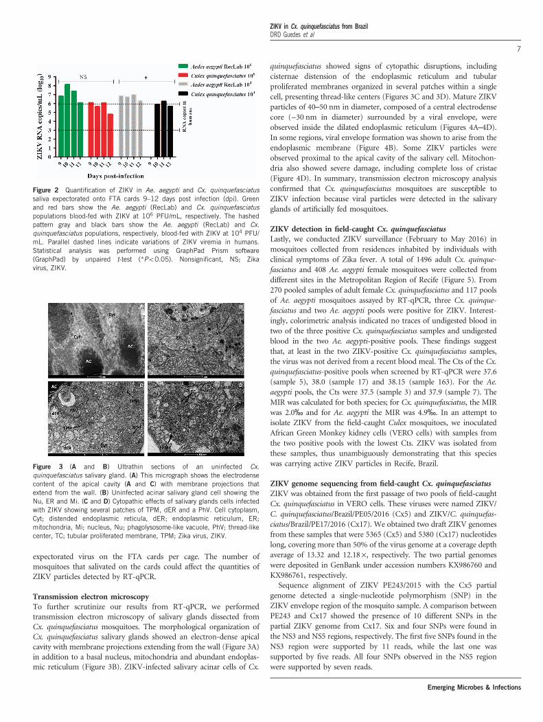
expectorated virus on the FTA cards per cage. The number ofmosquitoes that salivated on the cards could affect the quantities ofZIKV particles detected by RT-qPCR.
Transmission electron microscopyTo further scrutinize our results from RT-qPCR, we performedtransmission electron microscopy of salivary glands dissected fromCx. quinquefasciatus mosquitoes. The morphological organization ofCx. quinquefasciatus salivary glands showed an electron-dense apicalcavity with membrane projections extending from the wall (Figure 3A)in addition to a basal nucleus, mitochondria and abundant endoplas-mic reticulum (Figure 3B). ZIKV-infected salivary acinar cells of Cx.
quinquefasciatus showed signs of cytopathic disruptions, includingcisternae distension of the endoplasmic reticulum and tubularproliferated membranes organized in several patches within a singlecell, presenting thread-like centers (Figures 3C and 3D). Mature ZIKVparticles of 40–50 nm in diameter, composed of a central electrodensecore (~30 nm in diameter) surrounded by a viral envelope, wereobserved inside the dilated endoplasmic reticulum (Figures 4A–4D).In some regions, viral envelope formation was shown to arise from theendoplasmic membrane (Figure 4B). Some ZIKV particles wereobserved proximal to the apical cavity of the salivary cell. Mitochon-dria also showed severe damage, including complete loss of cristae(Figure 4D). In summary, transmission electron microscopy analysisconfirmed that Cx. quinquefasciatus mosquitoes are susceptible toZIKV infection because viral particles were detected in the salivaryglands of artificially fed mosquitoes.
ZIKV detection in field-caught Cx. quinquefasciatusLastly, we conducted ZIKV surveillance (February to May 2016) inmosquitoes collected from residences inhabited by individuals withclinical symptoms of Zika fever. A total of 1496 adult Cx. quinque-fasciatus and 408 Ae. aegypti female mosquitoes were collected fromdifferent sites in the Metropolitan Region of Recife (Figure 5). From270 pooled samples of adult female Cx. quinquefasciatus and 117 poolsof Ae. aegypti mosquitoes assayed by RT-qPCR, three Cx. quinque-fasciatus and two Ae. aegypti pools were positive for ZIKV. Interest-ingly, colorimetric analysis indicated no traces of undigested blood intwo of the three positive Cx. quinquefasciatus samples and undigestedblood in the two Ae. aegypti-positive pools. These findings suggestthat, at least in the two ZIKV-positive Cx. quinquefasciatus samples,the virus was not derived from a recent blood meal. The Cts of the Cx.quinquefasciatus-positive pools when screened by RT-qPCR were 37.6(sample 5), 38.0 (sample 17) and 38.15 (sample 163). For the Ae.aegypti pools, the Cts were 37.5 (sample 3) and 37.9 (sample 7). TheMIR was calculated for both species; for Cx. quinquefasciatus, the MIRwas 2.0‰ and for Ae. aegypti the MIR was 4.9‰. In an attempt toisolate ZIKV from the field-caught Culex mosquitoes, we inoculatedAfrican Green Monkey kidney cells (VERO cells) with samples fromthe two positive pools with the lowest Cts. ZIKV was isolated fromthese samples, thus unambiguously demonstrating that this specieswas carrying active ZIKV particles in Recife, Brazil.
ZIKV genome sequencing from field-caught Cx. quinquefasciatusZIKV was obtained from the first passage of two pools of field-caughtCx. quinquefasciatus in VERO cells. These viruses were named ZIKV/C. quinquefasciatus/Brazil/PE05/2016 (Cx5) and ZIKV/C. quinquefas-ciatus/Brazil/PE17/2016 (Cx17). We obtained two draft ZIKV genomesfrom these samples that were 5365 (Cx5) and 5380 (Cx17) nucleotideslong, covering more than 50% of the virus genome at a coverage depthaverage of 13.32 and 12.18 ×, respectively. The two partial genomeswere deposited in GenBank under accession numbers KX986760 andKX986761, respectively.Sequence alignment of ZIKV PE243/2015 with the Cx5 partial
genome detected a single-nucleotide polymorphism (SNP) in theZIKV envelope region of the mosquito sample. A comparison betweenPE243 and Cx17 showed the presence of 10 different SNPs in thepartial ZIKV genome from Cx17. Six and four SNPs were found inthe NS3 and NS5 regions, respectively. The first five SNPs found in theNS3 region were supported by 11 reads, while the last one wassupported by five reads. All four SNPs observed in the NS5 regionwere supported by seven reads.
Figure 2 Quantification of ZIKV in Ae. aegypti and Cx. quinquefasciatussaliva expectorated onto FTA cards 9–12 days post infection (dpi). Greenand red bars show the Ae. aegypti (RecLab) and Cx. quinquefasciatuspopulations blood-fed with ZIKV at 106 PFU/mL, respectively. The hashedpattern gray and black bars show the Ae. aegypti (RecLab) and Cx.quinquefasciatus populations, respectively, blood-fed with ZIKV at 104 PFU/mL. Parallel dashed lines indicate variations of ZIKV viremia in humans.Statistical analysis was performed using GraphPad Prism software(GraphPad) by unpaired t-test (*Po0.05). Nonsignificant, NS; Zikavirus, ZIKV.
Figure 3 (A and B) Ultrathin sections of an uninfected Cx.quinquefasciatus salivary gland. (A) This micrograph shows the electrodensecontent of the apical cavity (A and C) with membrane projections thatextend from the wall. (B) Uninfected acinar salivary gland cell showing theNu, ER and Mi. (C and D) Cytopathic effects of salivary glands cells infectedwith ZIKV showing several patches of TPM, dER and a PhV. Cell cytoplasm,Cyt; distended endoplasmic reticula, dER; endoplasmic reticulum, ER;mitochondria, Mi; nucleus, Nu; phagolysosome-like vacuole, PhV; thread-likecenter, TC; tubular proliferated membrane, TPM; Zika virus, ZIKV.
ZIKV in Cx. quinquefasciatus from BrazilDRD Guedes et al
7
Emerging Microbes & Infections

DISCUSSION
Our work has associated a second mosquito genus with the ZIKVtransmission cycle in northeast Brazil. We showed that Cx. quinque-fasciatus, also known as the southern house mosquito, which is themost common mosquito in urban areas in Brazil, is susceptible toinfection with ZIKV during experimental blood feeding; moreover, wefound that ZIKV has an active replication cycle in the salivary glandsand is subsequently released in the saliva. In addition, we were able todetect ZIKV circulating in wild Cx. quinquefasciatus collected fromRecife, and, for the first time, we sequenced a partial region of a ZIKVgenome (50%) obtained from mosquito samples in Brazil.Although Ae. aegypti is widely assumed to be the main ZIKV vector,
previous vector competence studies are controversial. In the presentstudy, a low dose of ZIKV (104 PFU/mL) was used for comparisonwith the higher doses used in previous studies.11,12,15 We found thatboth Ae. aegypti and Cx. quinquefasciatus can be experimentallyinfected by ZIKV even at low doses and can subsequently expectorateZIKV in their saliva.Cornet et al.10 concluded that part of a population of infected
mosquitoes may not be able to transmit the virus, and that, even ifthey do, transmission occurs during a particular timeframe. Thishypothesis differs from the idea that, once infected, a mosquito wouldtransmit virus for its entire life. This finding has implications forarbovirus surveillance programs as a time window for vector-borneZIKV transmission may exist. We found that after 11 dpi, most
samples were negative for ZIKV (apart from one positive salivarygland of Ae. aegypti fed 106 PFU/mL); thus, our maximum time pointanalysis was set to 15 dpi. However, Boorman and Porterfield8
reported that virus replication resumed at 15–20 dpi, and ZIKVremained present in Aedes mosquitoes for up to 60 days.To confirm that the virus detected in the salivary glands by RT-
qPCR was being released in saliva during consecutive blood meals, wefollowed the viral load from days 8 to 14 post infection using filterpaper cards. FTA cards were always placed on the top of the cages toavoid excreta contamination as noted in previous studies.38,39 Thisstrategy of viral RNA detection directly from FTA cards has beenemployed in previous studies for arbovirus surveillance.40,41 In thepresent study, we successfully detected ZIKV RNA copies in cardsfrom Ae. aegypti and Cx. quinquefasciatus mosquitoes. This resultdemonstrates that, in addition to being susceptible to ZIKV infectionand allowing virus replication in the salivary glands, both species arecapable of effectively transmitting ZIKV.RT-qPCR results were confirmed by transmission electron micro-
scopy. The general mature ZIKV morphology observed for the salivaryglands confirmed previous ultrastructural studies.42–44 In salivarygland cells, ZIKV replication causes cytopathic effects by 7 dpi, suchas tubular membrane proliferation, similar to results shown elsewherefor West Nile virus.45,46 The fact that we found salivary glands positivefor ZIKV when the midgut of the same mosquito was negativeindicates that mosquitoes may be clearing the viral infection in the
Figure 4 Mature ZIKV particles inside a Cx. quinquefasciatus salivary gland cell. (A) Numerous ZIKV particles (black arrows) within the dER. (B) Envelopeformation from the endoplasmic membrane (white arrow). (C) Enveloped virus particles with electrodense cores. (D) Viral particles accumulated proximal tothe acinar cavity (arrows); note the damaged mitochondria. Acinar cavity, AC; cell cytoplasm, Cyt; distended endoplasmic reticulum, dER; mitochondria, Mi;virion (s), Vi; Zika virus, ZIKV.
ZIKV in Cx. quinquefasciatus from BrazilDRD Guedes et al
8
Emerging Microbes & Infections

Figure 5 Kernel density map of reported cases of microcephaly versus a Point Map of the mosquito collection sites (with Culex samples positive andnegative for the presence of ZIKV). Zika virus, ZIKV.
ZIKV in Cx. quinquefasciatus from BrazilDRD Guedes et al
9
Emerging Microbes & Infections

midgut while virus replication continues in the salivary glands. Thisfinding has implications for the analytical methods employed in vectorcompetence studies.It is interesting to note that we were able not only to demonstrate
ZIKV infection in Cx. quinquefasciatus in laboratory assays but also infield-caught mosquitoes. Viruses isolated from field-caught Cx.quinquefasciatus were passed in VERO cells and sequenced, andmutations in the NS3 and NS5 regions were detected. These particularregions encompass an RNA helicase (NS3-Hel) and a putative RNA-dependent RNA polymerase (NS5-RdRp), which are key componentsfor genome replication and RNA synthesis.47 In addition, morenotably, these polymorphisms were not identified in the ZIKV straincirculating in Recife (PE243) that was obtained from human sera. Ourfindings are supported by Guo et al.,23 but we are cognizant ofprevious studies that were unable to confirm ZIKV replication in Cx.quinquefasciatus.17,18,21,24,48
Early studies have targeted only Aedes species, and only recentstudies have compared the natural IRs of different species (includingCulex). Notably, Diallo et al.5 observed a higher MIR for Cx. perfuscus(10× higher) than for Ae. aegypti. Positive Ae. aegypti field sampleshave always been reported at very low IRs, even in areas with highhuman ZIKV IRs, such as Malaysia.49 Indeed, in Micronesia6 andFrench Polynesia, ZIKV was not detected in wild-caught Aedes spp.mosquitoes during outbreaks. It is interesting that in all of these areas,Cx. quinquefasciatus is an abundant mosquito species that may havealso had an undetected role in ZIKV transmission. Indeed, Richardet al.13 demonstrated that two Aedes species from French Polynesia didnot show enough vector competence to transmit ZIKV and suggestedthat another mosquito species could be contributing to the spread ofthe virus in that region.Thus, our findings indicate that vector control strategies may need
to be re-examined since reducing Ae. aegypti populations may not leadto an overall reduction in ZIKV transmission if Culex populations arenot affected by Aedes-specific control measures. At the moment, thereis no broad ongoing program for Cx. quinquefasciatus control inBrazil, although Recife, Olinda and Jaboatão dos Guararapes, threemunicipalities in the Recife Metropolitan Region, have undertakenspecific control of Cx. quinquefasciatus to control lymphatic filariasis(LF) transmission locally,50 as this species is the only LF vector.Viral transmission via Cx. quinquefasciatus is not a new concept; in
North America, this species is the major vector of West Nile virus51
and it is also a known vector of Japanese encephalitis virus52 andequine encephalitis virus.53 Our data indicate that Cx. quinquefasciatusmosquitoes may be involved in ZIKV transmission in Recife. Thus,understanding the contributions of each species in the transmission ofZIKV is necessary to target each one properly. In conclusion,considering its high abundance in urban environments and itsanthropophilic behavior in Brazil,54–56 Cx. quinquefasciatus may be avector for ZIKV in this region.
ACKNOWLEDGEMENTS
This work was partially supported by the Fundação de Amparo à Pesquisa doEstado de Pernambuco (FACEPE; APQ-1608-2.13/15 and APQ-0085-2.13/16to CFJA; APQ-345-2.13/13 to MAVMS and APQ-0078-2.02/16 to GLW), theBrazilian National Council for Research and Development (CNPq; grant441100/2016-3) and the National Institute of Allergy and Infectious Diseases atthe National Institutes of Health (R01AI095514 and 1R21AI128931-01 toWSL). CFJA and CAP were supported by productivity fellowships from CNPq.We thank the insectary staff at the IAM for technical assistance, the Programfor Technological Development in Tools for Health (PDTIS-FIOCRUZ) forallowing us to use their facilities, the staff of the Pernambuco State Health
Department for sharing recent data on microcephaly and for assisting with
surveillance and the Platform of Statistic and Geographical Information
Systems (Núcleo de Estatística e Geoprocessamento, NEG) for helping us with
the GIS technologies and statistical analyses (George Tadeu Nunes Diniz).
1 Chang C, Ortiz K, Ansari A et al. The Zika outbreak of the 21st century. J Autoimmun2016; 68: 1–13.
2 Cordeiro MT, Pena LJ, Brito CA et al. Positive IgM for Zika virus in the cerebrospinalfluid of 30 neonates with microcephaly in Brazil. Lancet 2016; 387: 1811–1812.
3 Moron AF, Cavalheiro S, Milani H et al. Microcephaly associated with maternal Zikavirus infection. BJOG 2016; 123: 1265–1269.
4 Dick GW, Kitchen SF, Haddow AJ. Zika virus. I. Isolations and serological specificity.Trans R Soc Trop Med Hyg 1952; 46: 509–520.
5 Diallo D, Sall AA, Diagne CT et al. Zika virus emergence in mosquitoes in southeasternSenegal, 2011. PLoS ONE 2014; 9: e109442.
6 Duffy MR, Chen TH, Hancock WT et al. Zika virus outbreak on Yap Island, FederatedStates of Micronesia. N Engl J Med 2009; 360: 2536–2543.
7 Savage HM, Fritz CL, Rutstein D et al. Epidemic of dengue-4 virus in Yap State,Federated States of Micronesia, and implication of Aedes hensilli as anepidemic vector. Am J Trop Med Hyg 1998; 58: 519–524.
8 Boorman JP, Porterfield JS. A simple technique for infection of mosquitoes with viruses;transmission of Zika virus. Trans R Soc Trop Med Hyg 1956; 50: 238–242.
9 Li MI, Wong PS, Ng LC et al. Oral susceptibility of Singapore Aedes (Stegomyia) aegypti(Linnaeus) to Zika virus. PLoS Negl Trop Dis 2012; 6: e1792.
10 Cornet M, Robin Y, Adam C et al. Comparison between experimental transmission ofyellow fever and zika viruses in Aedes aegypti. Cah Orstom 1979; 17: 7.
11 Dutra HL, Rocha MN, Dias FB et al. Wolbachia blocks currently circulating Zikavirus isolates in Brazilian Aedes aegypti mosquitoes. Cell Host Microbe 2016; 19:771–774.
12 Chouin-Carneiro T, Vega-Rua A, Vazeille M et al. Differential susceptibilities of Aedesaegypti and Aedes albopictus from the Americas to Zika virus. PLoS Negl Trop Dis2016; 10: e0004543.
13 Richard V, Paoaafaite T, Cao-Lormeau VM. Vector competence of French PolynesianAedes aegypti and Aedes polynesiensis for Zika virus. PLoS Negl Trop Dis 2016; 10:e0005024.
14 Bearcroft WG. Zika virus infection experimentally induced in a human volunteer. TransR Soc Trop Med Hyg 1956; 50: 442–448.
15 Diagne CT, Diallo D, Faye O et al. Potential of selected Senegalese Aedesspp. mosquitoes (Diptera: Culicidae) to transmit Zika virus. BMC Infect Dis 2015; 15:492.
16 Ayres CF. Identification of Zika virus vectors and implications for control. Lancet InfectDis 2016; 16: 278–279.
17 Aliota MT, Peinado SA, Osorio JE et al. Culex pipiens and Aedes triseriatus mosquitosusceptibility to Zika virus. Emerg Infect Dis 2016; 22: 1857–1859.
18 Fernandes RS, Campos SS, Ferreira-de-Brito A et al. Culex quinquefasciatus from Riode Janeiro Is not competent to transmit the local Zika virus. PLoS Negl Trop Dis 2016;10: e0004993.
19 Hall-Mendelin S, Pyke AT, Moore PR et al. Assessment of local mosquito speciesincriminates Aedes aegypti as the potential vector of Zika virus in Australia. PLoS NeglTrop Dis 2016; 10: e0004959.
20 Huang YJ, Ayers VB, Lyons AC et al. Culex species mosquitoes and Zika virus. VectorBorne Zoonotic Dis 2016; 16: 673–676.
21 Amraoui F, Atyame-Nten C, Vega-Rua A et al. Culex mosquitoes are experimentallyunable to transmit Zika virus. Euro Surveill 2016; 21.
22 Ferreira-de-Brito A, Ribeiro IP, Miranda RM et al. First detection of natural infection ofAedes aegypti with Zika virus in Brazil and throughout South America. Mem InstOswaldo Cruz 2016; 111: 655–658.
23 Guo XX, Li CX, Deng YQ et al. Culex pipiens quinquefasciatus: a potential vector totransmit Zika virus. Emerg Microbes Infect 2016; 5: e102.
24 Weger-Lucarelli J, Ruckert C, Chotiwan N et al. Vector competence of Americanmosquitoes for three strains of Zika virus. PLoS Negl Trop Dis 2016; 10: e0005101.
25 Leal W. Zika mosquito vectors: the jury is still out [version 2; referees: 5 approved].F1000Research 2016; 5: 2546–2556.
26 Amorim LB, Helvecio E, de Oliveira CM et al. Susceptibility status of Culexquinquefasciatus (Diptera: Culicidae) populations to the chemical insecticide temephosin Pernambuco, Brazil. Pest Manag Sci 2013; 69: 1307–1314.
27 Araujo AP, Melo-Santos MA, Carlos SO et al. Evaluation of an experimental productbased on Bacillus thuringiensis sorovar. israelensis against Aedes aegypti larvae(Diptera: Culicidae). Biol Control 2017; 41: 339–347.
28 Donald CL, Brennan B, Cumberworth SL et al. Full genome sequence and sfRNAinterferon antagonist activity of Zika VIRUS from Recife, Brazil. PLoS Negl Trop Dis2016; 10: e0005048.
29 Scheuermann RH. Zika virus: designate standardized names. Nature 2016; 531: 173.30 Salazar MI, Richardson JH, Sanchez-Vargas I et al. Dengue virus type 2:
replication and tropisms in orally infected Aedes aegypti mosquitoes. BMC Microbiol2007; 7: 9.
31 Lambrechts L, Chevillon C, Albright RG et al. Genetic specificity and potential forlocal adaptation between dengue viruses and mosquito vectors. BMC Evol Biol 2009; 9:160.
ZIKV in Cx. quinquefasciatus from BrazilDRD Guedes et al
10
Emerging Microbes & Infections

32 Lanciotti RS, Kosoy OL, Laven JJ et al. Genetic and serologic properties of Zikavirus associated with an epidemic, Yap State, Micronesia, 2007. Emerg Infect Dis2008; 14: 1232–1239.
33 Donato MA, Ribeiro EL, Torres Dde O et al. Chronic treatment with Sildenafil has noeffect on folliculogenesis or fertility in C57BL/6 and C57BL/6 knockout for iNOS mice.Tissue Cell 2015; 47: 515–525.
34 Chow VT, Chan YC, Yong R et al. Monitoring of dengue viruses in field-caught Aedesaegypti and Aedes albopictus mosquitoes by a type-specific polymerase chain reactionand cycle sequencing. Am J Trop Med Hyg 1998; 58: 578–586.
35 Faria NR, Sabino EC, Nunes MR et al. Mobile real-time surveillance of Zika virusin Brazil. Genome Med 2016; 8: 97.
36 Bolger AM, Lohse M, Usadel B. Trimmomatic: a flexible trimmer for Illuminasequence data. Bioinformatics 2014; 30: 2114–2120.
37 Langmead B, Salzberg SL. Fast gapped-read alignment with Bowtie 2. Nat Methods2012; 9: 357–359.
38 Fontaine A, Jiolle D, Moltini-Conclois I et al. Excretion of dengue virus RNA by Aedesaegypti allows non-destructive monitoring of viral dissemination in individual mosqui-toes. Sci Rep 2016; 6: 24885.
39 Pilotte N, Zaky WI, Abrams BP et al. A novel xenomonitoring technique using mosquitoexcreta/feces for the detection of filarial parasites and malaria. PLoS Negl Trop Dis2016; 10: e0004641.
40 Flies EJ, Toi C, Weinstein P et al. Converting mosquito surveillance to arbovirussurveillance with honey-baited nucleic acid preservation cards. Vector Borne ZoonoticDis 2015; 15: 397–403.
41 Hall-Mendelin S, Ritchie SA, Johansen CA et al. Exploiting mosquito sugar feedingto detect mosquito-borne pathogens. Proc Natl Acad Sci USA 2010; 107:11255–11259.
42 Bell TM, Field EJ, Narang HK. Zika virus infection of the central nervous systemof mice. Arch Gesamte Virusforsch 1971; 35: 183–193.
43 Mlakar J, Korva M, Tul N et al. Zika virus associated with microcephaly. N Engl J Med2016; 374: 951–958.
44 Rosen L. Overwintering mechanisms of mosquito-borne arboviruses in temperateclimates. Am J Trop Med Hyg 1987; 37: 69S–76S.
45 Girard YA, Popov V, Wen J et al. Ultrastructural study of West Nile virus pathogenesis inCulex pipiens quinquefasciatus (Diptera: Culicidae). J Med Entomol 2005; 42:429–444.
46 Girard YA, Schneider BS, McGee CE et al. Salivary gland morphology and virustransmission during long-term cytopathologic West Nile virus infection in Culexmosquitoes. Am J Trop Med Hyg 2007; 76: 118–128.
47 Zhu Z, Chan JF, Tee KM et al. Comparative genomic analysis of pre-epidemic andepidemic Zika virus strains for virological factors potentially associated with the rapidlyexpanding epidemic. Emerg Microbes Infect 2016; 5: e22.
48 Boccolini D, Toma L, Di Luca M et al. Experimental investigation of the susceptibility ofItalian Culex pipiens mosquitoes to Zika virus infection. Euro Surveill 2016; 21:e30328.
49 Marchette NJ, Garcia R, Rudnick A. Isolation of Zika virus from Aedes aegyptimosquitoes in Malaysia. Am J Trop Med Hyg 1969; 18: 411–415.
50 Chalegre KD, Romao TP, Amorim LB et al. Detection of an allele conferring resistanceto Bacillus sphaericus binary toxin in Culex quinquefasciatus populations by molecularscreening. Appl Environ Microbiol 2009; 75: 1044–1049.
51 Nasci RS, White DJ, Stirling H et al. West Nile virus isolates from mosquitoes in NewYork and New Jersey, 1999. Emerg Infect Dis 2001; 7: 626–630.
52 Sucharit S, Surathin K, Shrestha SR. Vectors of Japanese encephalitis virus (JEV):species complexes of the vectors. Southeast Asian J Trop Med Public Health 1989; 20:611–621.
53 Wang Z, Zhang X, Li C et al. Vector competence of five common mosquito species in thePeople's Republic of China for Western equine encephalitis virus. Vector Borne ZoonoticDis 2012; 12: 605–608.
54 Nunes AT, Brito NF, Oliveira DS et al. Comparative proteome analysis reveals that bloodand sugar meals induce differential protein expression in Aedes aegypti female heads.Proteomics 2016; 16: 2582–2586.
55 Goertz GP, Fros JJ, Miesen P et al. Non-coding subgenomic flavivirus RNA is processedby the mosquito RNAi machinery and determines West Nile virus transmission by Culexpipiens mosquitoes. J Virol 2016; 90: 10145–10159.
56 Regis L, Silva-Filha MH, de Oliveira CM et al. Integrated control measures against Culexquinquefasciatus, the vector of filariasis in Recife. Mem Inst Oswaldo Cruz 1995; 90:115–119.
This work is licensed under a Creative Commons Attribution 4.0International License. The images or other third party material in this
article are included in the article’s Creative Commons license, unless indicated otherwisein the credit line; if thematerial is not includedunder theCreativeCommons license, userswill need toobtainpermission fromthe licenseholder to reproduce thematerial.Toviewacopy of this license, visit http://creativecommons.org/licenses/by/4.0/
r The Author(s) 2017
ZIKV in Cx. quinquefasciatus from BrazilDRD Guedes et al
11
Emerging Microbes & Infections