Zbtb20 modulates the sequential generation of neuronal layers · PDF fileZbtb20 modulates the...
15
RESEARCH Open Access Zbtb20 modulates the sequential generation of neuronal layers in developing cortex Anton B. Tonchev 1,2,3* , Tran Cong Tuoc 2,4 , Eva H. Rosenthal 1 , Michèle Studer 5,6 and Anastassia Stoykova 1,2,3* Abstract Background: During corticogenesis, genetic programs encoded in progenitor cells at different developmental stages and inherited in postmitotic neurons specify distinct layer and area identities. Transcription factor Zbtb20 has been shown to play a role for hippocampal development but whether it is implicated in mammalian neocortical morphogenesis remains unknown. Results: Here, we report that during embyogenesis transcription factor Zbtb20 has a dynamic spatio-temporal expression pattern in mitotic cortical progenitors through which it modulates the sequential generation of cortical neuronal layer identities. Zbtb20 knock out mice exhibited enhanced populations of early born L6-L4 neuronal subtypes and a dramatic reduction of the late born L3/L2 neurons. This defect was due to a temporal misbalance in the production of earlier versus later born neurons, leading to a progressive diminishing of the progenitor pool for the generation of L3-L2 neurons. Zbtb20 implements these temporal effects in part by binding to promoter of the orphan nuclear receptor CoupTF1/Nr2f1. In addition to its effects exerted in cortical progenitors, the postmitotic expression of Zbtb20 in L3/L2 neurons starting at birth may contribute to their proper differentiation and migration. Conclusions: Our findings reveal Zbtb20 as a novel temporal regulator for the generation of layer-specific neuronal identities. Keywords: Zbtb20, Development, Neocortex, Temporal identity, Transcription factor Background The mammalian neocortex (Ncx), in which neurons are arranged radially in six layers and tangentially in numer- ous functional domains, is a recent acquisition in brain evolution. During development, the majority of cortical glutamatergic neurons are generated by radial glial cells (RGCs) in the germinative ventricular (VZ) and subven- tricular (SVZ) zone of the dorsolateral pallium. Gener- ation of neuronal sets with a layer-specific identity depends on an intrinsically encoded genetic program and environmental cues acting during the S-phase of the mitotic cycle [1]. Neurons with different fates are pro- duced according to an “inside-first outside-last” sched- ule: first, lower layer (LL) neurons (L6/L5), followed by generation of the upper layer (UL) neurons (L4/L3/L2). During mouse development, the layer specific neuronal subtypes are generated throughout embryonic (E) stages E10.5 – E17.5 in partially overlapping time windows with a peak for generation at E11.5 for L1, E12.5 (L6), E13.5 (L5), E14.5 (L4) and E16.5-E17.5 (L3-L2) [2–5]. The birthdate of cortical neurons is directly also related to their projection identity. Thus, while the early born L6 and L5 neurons extend outside the brain thalamocor- tical (TCA) and corticospinal motor neuron (CSMN) projections, the late-born UL neurons make interhemi- spheric (callosal) projections inside the brain [6]. In- creasing recent evidence support the view that the precise temporal programs for production of LL and UL neuronal fates relies on intrinsic mechanisms in early and late progenitors, respectively, characterized by spe- cific combinatorial expression of TFs at distinct develop- mental time points [7–13]. For instance, suppression of the expression of Foxg1 at E10.5 is required to make a switch from generation of reelin-positive Cajal-Retzius * Correspondence: [email protected]; [email protected] 1 Molecular Developmental Neurobiology Laboratory, Max Planck Institute of Biophysical Chemistry, Am Fassberg, 37077 Gottingen, Germany Full list of author information is available at the end of the article © 2016 The Author(s). Open Access This article is distributed under the terms of the Creative Commons Attribution 4.0 International License (http://creativecommons.org/licenses/by/4.0/), which permits unrestricted use, distribution, and reproduction in any medium, provided you give appropriate credit to the original author(s) and the source, provide a link to the Creative Commons license, and indicate if changes were made. The Creative Commons Public Domain Dedication waiver (http://creativecommons.org/publicdomain/zero/1.0/) applies to the data made available in this article, unless otherwise stated. Tonchev et al. Molecular Brain (2016) 9:65 DOI 10.1186/s13041-016-0242-2
Transcript of Zbtb20 modulates the sequential generation of neuronal layers · PDF fileZbtb20 modulates the...

RESEARCH Open Access
Zbtb20 modulates the sequentialgeneration of neuronal layers in developingcortexAnton B. Tonchev1,2,3*, Tran Cong Tuoc2,4, Eva H. Rosenthal1, Michèle Studer5,6 and Anastassia Stoykova1,2,3*
Abstract
Background: During corticogenesis, genetic programs encoded in progenitor cells at different developmentalstages and inherited in postmitotic neurons specify distinct layer and area identities. Transcription factor Zbtb20 hasbeen shown to play a role for hippocampal development but whether it is implicated in mammalian neocorticalmorphogenesis remains unknown.
Results: Here, we report that during embyogenesis transcription factor Zbtb20 has a dynamic spatio-temporalexpression pattern in mitotic cortical progenitors through which it modulates the sequential generation of corticalneuronal layer identities. Zbtb20 knock out mice exhibited enhanced populations of early born L6-L4 neuronalsubtypes and a dramatic reduction of the late born L3/L2 neurons. This defect was due to a temporal misbalancein the production of earlier versus later born neurons, leading to a progressive diminishing of the progenitor poolfor the generation of L3-L2 neurons. Zbtb20 implements these temporal effects in part by binding to promoter ofthe orphan nuclear receptor CoupTF1/Nr2f1. In addition to its effects exerted in cortical progenitors, the postmitoticexpression of Zbtb20 in L3/L2 neurons starting at birth may contribute to their proper differentiation and migration.
Conclusions: Our findings reveal Zbtb20 as a novel temporal regulator for the generation of layer-specific neuronalidentities.
Keywords: Zbtb20, Development, Neocortex, Temporal identity, Transcription factor
BackgroundThe mammalian neocortex (Ncx), in which neurons arearranged radially in six layers and tangentially in numer-ous functional domains, is a recent acquisition in brainevolution. During development, the majority of corticalglutamatergic neurons are generated by radial glial cells(RGCs) in the germinative ventricular (VZ) and subven-tricular (SVZ) zone of the dorsolateral pallium. Gener-ation of neuronal sets with a layer-specific identitydepends on an intrinsically encoded genetic programand environmental cues acting during the S-phase of themitotic cycle [1]. Neurons with different fates are pro-duced according to an “inside-first outside-last” sched-ule: first, lower layer (LL) neurons (L6/L5), followed bygeneration of the upper layer (UL) neurons (L4/L3/L2).
During mouse development, the layer specific neuronalsubtypes are generated throughout embryonic (E) stagesE10.5 – E17.5 in partially overlapping time windowswith a peak for generation at E11.5 for L1, E12.5 (L6),E13.5 (L5), E14.5 (L4) and E16.5-E17.5 (L3-L2) [2–5].The birthdate of cortical neurons is directly also relatedto their projection identity. Thus, while the early bornL6 and L5 neurons extend outside the brain thalamocor-tical (TCA) and corticospinal motor neuron (CSMN)projections, the late-born UL neurons make interhemi-spheric (callosal) projections inside the brain [6]. In-creasing recent evidence support the view that theprecise temporal programs for production of LL and ULneuronal fates relies on intrinsic mechanisms in earlyand late progenitors, respectively, characterized by spe-cific combinatorial expression of TFs at distinct develop-mental time points [7–13]. For instance, suppression ofthe expression of Foxg1 at E10.5 is required to make aswitch from generation of reelin-positive Cajal-Retzius
* Correspondence: [email protected]; [email protected] Developmental Neurobiology Laboratory, Max Planck Institute ofBiophysical Chemistry, Am Fassberg, 37077 Gottingen, GermanyFull list of author information is available at the end of the article
© 2016 The Author(s). Open Access This article is distributed under the terms of the Creative Commons Attribution 4.0International License (http://creativecommons.org/licenses/by/4.0/), which permits unrestricted use, distribution, andreproduction in any medium, provided you give appropriate credit to the original author(s) and the source, provide a link tothe Creative Commons license, and indicate if changes were made. The Creative Commons Public Domain Dedication waiver(http://creativecommons.org/publicdomain/zero/1.0/) applies to the data made available in this article, unless otherwise stated.
Tonchev et al. Molecular Brain (2016) 9:65 DOI 10.1186/s13041-016-0242-2

cells, located in the marginal zone (MZ) of the cortex, tothe production of neuronal subsets located in corticalplate (CP) [14]. Furthermore, while Fezf2 and Otx1 ex-pression in apical VZ progenitors controls the fate speci-fication of the LL neurons [15, 16], the expression ofSvet1, Cux1 and Cux2 during later stages of neurogen-esis in SVZ progenitors seems to specify UL neuronalfate [17–20]. The expression of TFs in postmitotic CPneurons may regulate through feedback signalling mech-anism the progenitor progeny in the germinative zone[21] or the fate of the postmitotic neurons [7–13].Neurons with distinct morphology, connectivity, neuro-
transmitter usage and function are tangentially organizedin numerous functional domains, implicating that mecha-nisms of layer and area formation are interrelated. Accord-ing to the current view, cortical arealization is presaged byencoded positional information (“protomap”) throughgraded expression of sets of TFs along the anteroposteriorand mediolateral axis in the two germinative zones of theneocortex [5, 22]. Disruption of the graded expression ofsuch TFs in VZ/SVZ leads to severe defects in the arealsize and location in the Ncx [23–25]. As recently shown,affecting the intrinsic genetic mechanisms encoded by thegraded expression of TF Pax6 in cortical progenitors re-sults in an altered size of cortical somatosensory (SS) areaand in parallel alterations in the sensory thalamus involv-ing selective death of neurons in particular thalamic nu-clei. Consequently, a new type of “top-down plasticity”driven by competition-mediated axon elimination andneuronal apoptosis re-patterns the sensory thalamus [26].In a microarray screen aimed to find out genes with
graded expression in the developing cortex, we identifiedTF Zbtb20 as a gene showing caudal-high to rostral-lowexpression gradient in VZ of E16.5 cortex, and subse-quently maintains a restricted high expression in theadult hippocampus (Hi) [27]. The gene Zbtb20 (alsonamed DPZF [28], HOF [29] or ZNF288) encodes a TF,belonging to the POK-family of BTB zinc finger tran-scriptional repressors, implicated in developmental pro-cesses and cancer [30]. Zbtb20 has been localizedexclusively in immature post-mitotic neurons in Hi andmigrating granule cell precursors of DG [29]. Previousresearch, using both gain-of-function (GOF) [31–33]and loss-of-function (LOF) [34, 35] approaches, has de-scribed the important role of TF Zbtb20 in specifyingthe medial pallium, the anlage of the Hi formation.Recent studies have implicated Zbtb20 mutations in hu-
man neurodevelopmental syndromes associated with be-havioral abnormalities, including intellectual disability [36],Primrose syndrome [37], autism [38], and schizophrenia[39]. The reported alterations in brain morphology in thesedisorders suggest possible cortical involvement beyond thehippocampus. However, no data are presently available onthe involvement of Zbtb20 in neocortical morphogenesis.
In this study, we present first evidence that TF Zbtb20exerts a dynamic expression in the germinative zones ofthe cortex (pallium), marks specifically the uppermostL3-L2 cortical neurons and exerts a crucial control inthe timely generation of distinct neuronal fates through-out cortical neurogenesis.
ResultsDynamic Zbtb20 expression in telencephalic progenitorsBy E11.5 a gradient of Zbtb20-lacZ activity was evidentin the VZ of the lateral pallium (LP) of the heterozygousZbtb20lacZ/+ embryos (Additional file 1: Figure S1A,arrow). Between E12.5 and E13.5, Zbtb20 immunostain-ing confirmed a strong signal in both the ventral (VP)and lateral (LP) pallium (Fig. 1a, arrow). Double stainingfor Zbtb20 and the pallial progenitor marker Pax6 re-vealed a nearly complete co-expression at E13.5(Fig. 1b1-b6) in LP (101 out of 101 assayed Zbtb20+ cellsco-expressed Pax6 (100 %, n = 3). In VP, however, only40 % of the Zbtb20+ cells co-expressed Pax6 (59 out of148 Zbtb20+ cells, n = 3). Notably, co-labelling forZbtb20 and TF CoupTF1, which at this stage isexpressed by both pallial and subpallial progenitors,showed a complete co-expression in LP and VP (143 outof 143 Zbtb20+ cells in LP/VP (n = 3) co-expressedCoupTF1 (Fig. 1c1-c6).By E14.5-E15.5 the expression of Zbtb20 was spread
throughout the entire pallial VZ (Fig. 1d; Additional file1: Figure S1B, arrows). Co-staining for Zbtb20, RGCmarker Pax6 and the mitotic marker phosphorylatedvimentin (pVim) confirmed that TF Zbtb20 is expressedin dividing RGCs at the apical surface of VZ (Fig. 1e1-e4, arrows; 90 out of 93 assayed pVim+ cells on the ap-ical surface were co-labeled for Zbtb20, 97 %, n = 3).At early postnatal (P) stages (P4), Zbtb20 immuno-
signal was evident in the most superficial layers of LP/DP (Fig. 1f, arrow), where Zbtb20+ cells co-expressedSatb2 (Fig. 1g1-g3, arrows; 102 out of 129 assayedZbtb20+ cells were co-labeled for Satb2, 79 %, n = 3) andBrn2 (Additional file 2: Figure S2A1-A3, arrows; 111 outof 121 assayed Zbtb20+ cells were co-labeled for Brn2,93 %, n = 3), markers of UL neocortical neurons [40, 41].However, Zbtb20 did not co-localize with neither the L5marker Ctip2 (Additional file 2: Figure S2B1-B3) nor theL4 marker ROR [42] (Additional file 2: Figure S2C1-C3),suggesting that the Zbtb20 expression in UL neurons isrestricted to L2-L3 neurons. The expression pattern inULs was preserved at P8 but almost disappeared at P12(data not shown). In the early postnatal SVZ, Zbtb20also maintained a strong expression in Nestin + RGCs(Fig. 1h1-h3; 90 out of 92 assayed Zbtb20+ cells in SVZwere co-labeled for Nestin, 92 %, n = 3), suggesting apossible involvement in postnatal neurogenesis.
Tonchev et al. Molecular Brain (2016) 9:65 Page 2 of 15

Fig. 1 (See legend on next page.)
Tonchev et al. Molecular Brain (2016) 9:65 Page 3 of 15

In summary, beginning at E11.5 in VZ of VP/LP, theexpression of TF Zbtb20 expands into the VZ of the en-tire pallium at E14.5 and thereafter, suggesting that thetimed expression of Zbtb20 may be involved in gener-ation and/or specification of the UL neurons.
Disturbances in superficial neocortical layers in Zbtb20loss-of-functionCresyl violet (Nissl) staining of P10 coronal brain sec-tions revealed apparent defects in cortical layering inZbtb20lacZ/lacZ mice (Fig. 2a1-b2). While the LLs (L6,L5) appeared overrepresented in the Zbtb20lacZ/lacZ som-atosensory (SS) cortex, the ULs (L4-L2) seemed thinner(Fig. 2b1-b2). This impression was confirmed by NeuNimmunoassaying (Fig. 2b3-b4). In order to investigatethe UL disturbances in more detail, we performed im-munostaining for specific UL neuronal markers. Giventhe widespread expression of Zbtb20 in VZ of the entirepallium at E14.5, we first investigated the expression ofthe L4 marker ROR [43]. Intriguingly, in the mutant cor-tex, the thickness of L4 was greatly augmented (Fig. 2c1-c3). This notion was confirmed by another marker witha strong expression in L4, TF CoupTF1 (see below).Immunoassaying for the global (L2-L4) UL marker Cux1[18, 19], however, showed a reduction of UL neurons(Fig. 2d1-d3). Additional UL markers, Brn2 (L2, L3, andL5; [40, 41]) and Satb2 (L4-L2; [7, 8]) confirmed thestrongly diminished representation of UL subsets in theZbtb20lacZ/lacZ mice (Fig. 2h1-i3).The overall reduction of ULs, accompanied by the se-
lective expansion of L4, raised a question on the statusof the L2-L3 neurons in the mutant cortex. Therefore,we quantified the L3-L2 Cux1+/ROR− population(Fig. 2f2, g2, arrow), which was markedly depleted(Fig. 2g3), in contrast to the L4 Cux1+/ROR+ population,which was augmented (Fig. 2g4). In order to studywhether L3 or L2 neuronal subsets were specifically af-fected, we made use of the expression patterns of TFFoxP1, which is expressed in L6a and L5-L3 [44] and TFBrn2, which specifically marks L3 and L2 [40, 41].Double immunostaining for FoxP1 and Brn2 revealed an
ectopic expansion of FoxP1 expression into the normalposition of L2 in the mutant cortex, while cells specif-ically fated to L2 identity (Brn2+/FoxP1−, Fig. 2j1-j2,arrows) were almost completely missing (Fig. 2j3). Thedepletion of the ULs was not due to an enhancedapoptosis, as studied by the expression of activatedCaspase-3 (data not shown). To investigate whetherthe molecular boundary between L5 and L4 was pre-served, we applied Ctip2 (L5)/ROR (L4) double immu-nohistochemical (IHC) staining, and we found thatthese two subpopulations of cortical neurons wereproperly segregated (Fig. 2k1-k2).To sum up, these findings indicate that Zbtb20 defi-
ciency results in a significant diminishing of L3 and es-pecially L2 neuronal subsets as well as in an augmentedand ectopic presence of L4 neurons in UL position.
Enhanced deep layers and normal arealization inZbtb20lacZ/lacZ cortexTo study quantitatively the apparent enhancement ofboth L5 and L6 sets in the mutants (Fig. 2b1-b4), weperformed IHC staining and counting of cells in the SScortex on cross sections of both genotypes with anti-bodies for TFs FoxP2 [44] in L6 (Fig. 3a1-a4), Tbr1 [9]in L6 (Fig. 3b1-b4) and Ctip2 [15, 45] in L5 (Fig. 3c1-c4). Indeed, the results revealed a statistically signifi-cant increased number of both L6 and L5 neurons inthe mutant as compared with the control Ncx (Fig. 3a5,b5, c5). Furthermore, ISH staining of sagittal P4 brainsections indicated that the mutant cortex displayed en-hanced LL neuronal subsets, including Fezf2+ L5 [15,46] (Additional file 3: Figure S3A1-A4), Clim1+ L5 [46](Additional file 3: Figure S3B1-B4), and Id2+ L5 andId2+ L6 [47] (Additional file 3: Figure S3C1-C4). Inorder to confirm that the described layering abnormal-ities in the Zbtb20lacZ/lacZ mice are not restricted tothe SS cortex, we studied the patterning of the primarymotor cortex and, similarly to SS cortex, we found adecrease of the ULs and enhancement of the LLs(Additional file 4: Figure S4).
(See figure on previous page.)Fig. 1 Expression of TF Zbtb20 in the developing pallium. a Immunostaining for Zbtb20 at E13.5 demarcates a strong signal just ventral to thecortico-striatal sulcus (white line) in VP and a weaker gradient in LP (arrow) gradually disappearing toward DP. b1-b6 Double-labelling for Pax6(b1/b4) and Zbtb20 (b2/b5), and an overlay (b3/b6) in VZ of LP and VP at E13.5. c1-c6 Double-labelling for CoupTF1 (c1/c4) and Zbtb20 (c2/c5),and an overlay (c3/c6) in VZ of LP and VP at E13.5. The cortico-striatal sulcus is depicted by a white line. d Zbtb20 IHC on a cross brain sectionat stage E15.5 demonstrates strong immunosignal in germinative zones of both pallium and subpallim. e1-e4 Triple IHC with antibodies forphospho-vimentin (pVim, E1), Pax6 (e2) and Zbtb20 (e3), and an overlay (e4) in E16.5 DP. Arrows depict triple-positive cells at the apical surface,arrowheads depict a more basally located dividing cell. f IHC for Zbtb20 on coronal WT brain sections at P4. The arrow points to the Zbtb20expression in the uppermost neocortical layers. The frames depict the position of the micrographs shown in G1-G3. g1-g3 Double immunostainingfor Zbtb20 (g1, counterstained by DAPI) and Satb2 (g2), and an overlay (g3) on P4 brain cross sections. Arrows depict double-positive cells in theuppermost layers. h1-h3 Double immunostaining for Zbtb20 (H1, counterstained by DAPI) and Nestin (h2), and an overlay (h3) on P0 brain crosssections depicts almost complete co-staining in VZ/SVZ. Cgl, cingulate cortex; DP, dorsal pallium; LP, lateral pallium; Ncx, neocortex; VP, ventralpallium. Scale bars: a, 100 μm; b3/c3, 20 μm; d, 200 μm; e4, 10 μm; f, 200 μm; g3, 50 μm; b6/c6/h3, 10 μm
Tonchev et al. Molecular Brain (2016) 9:65 Page 4 of 15

Fig. 2 (See legend on next page.)
Tonchev et al. Molecular Brain (2016) 9:65 Page 5 of 15

In layer 5, the rostral limit of expression of TF Id2outlines the position of the border between the motor/somatosensory (M/SS) cortex, which is also marked bythe caudal limit of Id2 expression in L3-L2 [48]. InZbtb20lacZ/lacZ cortex, the M/SS boundary appeared ros-trally displaced into the M field (Additional file 3: FigureS3C1-C2, asterisk). We therefore examined othercortical area-specific markers, including Cadherin-8(Additional file 3: Figure S3D1-D2, asterisk), Serotonin(Additional file 3: Figure S3E1-E2, asterisk), Bhlhb5(Additional file 3: Figure S3 F1-F2, asterisk), ROR(Fig. 2c1-c2, asterisk, and data not shown), Cadherin-6and Lmo3 (data not shown). None of these markers ex-hibited shifts in the mutants along the antero-posterioraxis, so we concluded that the neocortical arealization in
Zbtb20lacZ/lacZ mice is grossly not affected. This notionis also supported by the preserved pattern of the gradedexpression of TFs Pax6, Emx2, Foxg1 and Lhx2 inZbtb20lacZ/lacZ embryonic pallium at E12.5 [35], a stageat which these TFs are known to have crucial roles inspecification of the intrinsic program of cortical arealiza-tion encoded in the progenitors [23].
ZBTB20 modulates the temporal onset for generation ofdistinct neuronal layer identitiesTo investigate whether Zbtb20 controls the switch togeneration of neurons with different layer identities, weperformed BrdU birthdating experiments at E12.5,E14.5 and E16.5 when predominantly LLs (L6, L5), L4or L3-L2 neurons are born, respectively (Fig. 4). Taking
(See figure on previous page.)Fig. 2 Upper layer defects in Zbtb20lacZ/lacZ mice. a1-b2 Cresyl violet staining of brain cross sections from P28 wild type (WT) and mutant cortex.(b1, b2) are images at high magnification from fields in the somatosensory cortex, labeled with * in A1/A2. (b3-b4) NeuN IHC at stage P4. c1-g4Evaluation of UL neuronal fate by using ROR and Cux1 antibodies as markers, and a statistical analysis. In (c1-c2) note the expansion of the RORsignal (arrows), while in (d1-d2), a slight reduction of the Cux1-positive band (arrows) in the mutant Ncx. (e1-g2) are higher magnifications of SScortex, labeled with the same markers. (c3,d3,g3,g4) represent graphs of statistical analysis of the number of positive cells/frame in SS cortex(*, P < 0.05, n = 3 per genotype). h1-i2 IHC with Brn2 and Satb2 antibodies as UL markers on cross sections from P4 brains, and a statistical analysisof the number of stained cells. Note the reduction of the ULs in the mutant, which was statistically confirmed (*, P < 0.05, n = 3 per genotype). j1-j3Double IHC with Brn2 and FoxP1 antibody to evaluate UL neurons with L2 identity (Brn2+/FoxP1−). Note the drastic diminishing of the number ofL2 neurons in the mutant Ncx (arrows), which is supported by a statistical analysis (*, P < 0.05, n = 3 per genotype). k1-k2 Double immunostainingfor ROR (L4) and Ctip2 (L5) reveals a preserved molecular border between the two layers in the mutant. Countings of positive cells were performedin frames sized 300 μm (h) × 100 μm (w) spanning the ULs of SS cortex. Scale bars: b4, 100 μm; d2, 500 μm; g2/j2, 50 μm; i2/k2, 100 μm
Fig. 3 Enhanced presentation of LL neurons in somatosensory cortex of Zbtb20lacZ/lacZ mutants. a1-b4 IHC with FoxP2 (a1 –a5) and Tbr1 (b1-b5)antibodies as L6 markers reveals an increased number of L6 neurons in the mutant compared with WT cortex. c1-c5 IHC with antibody Ctip2 asL5 marker shows an enhanced number of L5 neurons in the mutant. (a5,b5,c5) Graphs representing statistical evaluation of the results (*, P < 0.05,n = 3 per genotype). All stainings were performed at stage P4. Countings of positive cells were performed in frames sized 300 μm (h) × 100 μm(w) spanning the LLs of SS cortex. Scale bars: c2, 200 μm, c4, 100 μm
Tonchev et al. Molecular Brain (2016) 9:65 Page 6 of 15
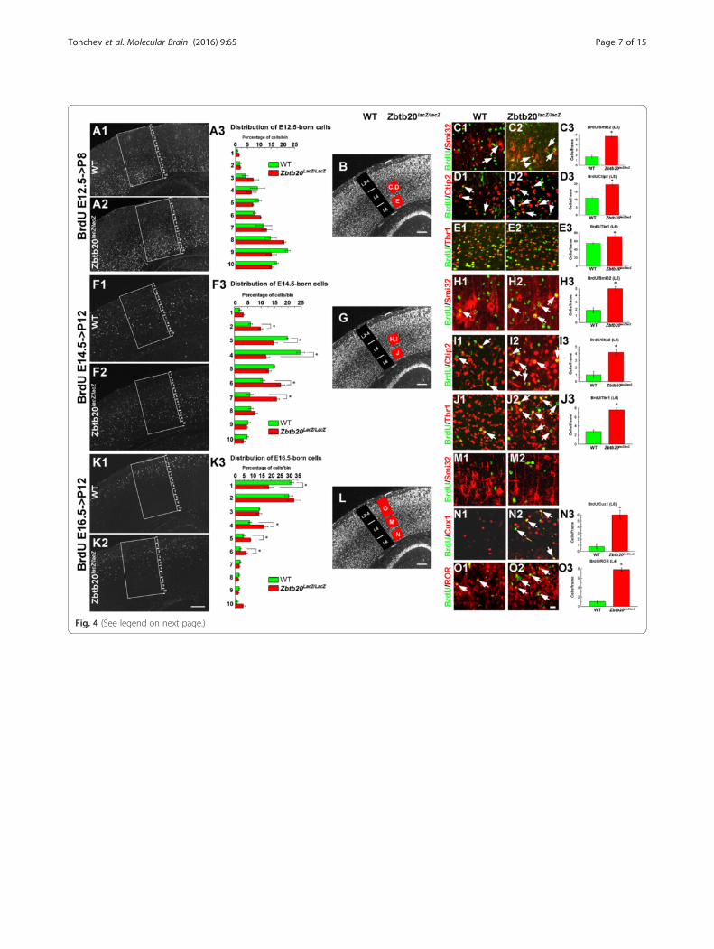
Fig. 4 (See legend on next page.)
Tonchev et al. Molecular Brain (2016) 9:65 Page 7 of 15

advantage of the fact the pattern of NeuN immuno-staining allowed distinguishing the location of LL andUL in the postnatal cortex (Fig. 2b3-b4), we calculatedthe percentage of BrdU+/NeuN+ cells located to eitherLLs (L5-L6) or ULs (L2-L4) out of the total BrdU+/NeuN+ cells in frames spanning the entire cortex(Additional file 5: Figure S5). Analysis of the position ofthe E12.5-born cells revealed no differences in theirlaminar location between WT and the mutant (Fig. 4a1-a3; Additional file 5: Figure S5A1-A3). In contrast,birthdating at E14.5, revealed a significantly larger pro-portion of BrdU+ cells in the deep position of theZbtb20lacZ/lacZ cortex, and on contrary, a smaller frac-tion of such cells was found in the superficial positionof the mutants (Additional file 5: Figure S5B1-B3; alsoFig. 4f1-f3). Similarly, BrdU pulse labelling at E16.5 (atthe peak of UL generation) showed a reduced distribu-tion of tagged cells at superficial position and signifi-cantly more deeply located BrdU+ cells in the mutantcortex (bins 4–6; Fig. 4k1-k3), that was also evident onBrdU/NeuN double-stained sections (Additional file 5:Figure S5C1-C3).Using BrdU co-staining with layer-specific neuronal
markers, for L5 (Neurofilament/Smi32 and Ctip2) andL6 (Tbr1), we quantitatively investigated the layer fate ofcells generated at stages E12.5, E14.5 or E16.5. The re-sults revealed that the mutant Ncx had significantly in-creased populations of L6 and L5 neurons born at E12.5(Fig. 4c1-e3), as well as at E14.5, the peak of generationof L4 neurons during normal corticogenesis (Fig. 4h1-j2). At E16.5 such expansion of L6/L5 fate identities was
no more evident (Fig. 4m1-m2). However, the cells bornat E16.5 in the mutant Ncx demonstrated a significantincrease of the co-labeling with the L4 marker ROR ascompared to the control Ncx (Fig. 4o1-o2, arrows, O3).These results suggest that the developmental window forgeneration of L6-L5-L4 neurons was expanded by atleast 2 days for each neuronal type, which will pro-foundly affects the progenitor pool size for L3-L2 neu-rons. In a further support of such a scenario were theresults after analysis of the fractions of Cux1+/ROR+
(L4) and Cux1+/ROR− (L3-L2) neuronal subsets born atstages E12.5, E14.5 or E16.5 in WT and mutant mice(Additional file 6: Figure S6). We found that at P12, theUL fractions born at E12.5 or E14.5 did not differ signifi-cantly between WT and mutant mice (Additional file 6:Figure S6A1-B3). However, changes were observed inthe E16.5-born UL neuronal fractions in Zbtb20 LOF ascompared to WT: the L4 fraction was larger, while theL3-L2 fraction was diminished (Additional file 6: FigureS6C1-C3).Together, these results strongly suggest that the timed
expression of transcriptional repressor Zbtb20 in corticalprogenitors (appearing in the entire pallium only afterE14.5) could control the transition from early- versuslate born neuronal layer identities.
Impaired late neurogenesis and neuronal migration inZbtb20-deficient cortexBy using 40 min BrdU-pulse labelling in vivo at E16.5(the peak of production of UL neurons) we found a sig-nificant reduction of the progenitor proliferation in the
(See figure on previous page.)Fig. 4 Laminar distribution and fate of neurons born at E12.5, E14.5 and E16.5 in WT and Zbtb20lacZ/lacZ neocortex. BrdU was injected at eachof the above indicated embryonic stages and the distribution of the BrdU-tagged cells was evaluated in the postnatal SS cortex. a1-e3Distribution and fate of cells born at E12.5 and investigated at P8. (a1-a3) Overview images of the localization of BrdU+ cells to bins 1–10and a statistical analysis showing no significant differences between WT and mutant in any of the bins. b NeuN IHC providing an overviewof the cortical layers and the position of frames where counting was done in c1-e3 (red squares with respective letters). (c1-e2) Cell fate ofE12.5-generated neurons in L5 (co-stained for Neurofilament-H/Smi32 and Ctip2) and L6 (co-labeled for Tbr1), and a statistical analysis(c3,d3,e3, *, P < 0.05, n = 3 per genotype). Double-labeled cells are depicted by arrows. f1-j3 Distribution and fate of cells born at E14.5 andinvestigated at P12. (f1-f3) Overview images of the localization of BrdU+ cells to bins 1–10 and a statistical analysis showing a significantincrease of the proportion of BrdU+ cells distributed in the lower bins (6–7), and a reduction of cells localized in upper bins (3–4) in themutant cortex as compared to WT. (g) NeuN IHC providing an overview of the cortical layers and the position of frames where counting wasdone in h1-j2 (red squares with respective letters). (h1-j3) Fate of E14.5-generated neurons with L5 identity (co-stained for Neurofilament-H/Smi32 and Ctip2) and L6 (co-labeled for Tbr1), and a statistical analysis (H3,I3,J3, *, P < 0.05, n = 3 per genotype). Double-labeled cells aredepicted by arrows. k1-o3 Distribution and fate of cells born on E16.5 in P12 SS cortex. (k1-k3) Overview images of the localization of BrdU+
cells to bins 1–10 and a statistical analysis showing a significant increase of the proportion of BrdU+ cells localized in bins 4–6 in mutant,while a reduction of the percentage of cells localized in uppermost position in bin 1 as compared to WT. (l) NeuN IHC providing an overviewof the cortical layers and the position of frames where counting was done in m1-o2 (red squares with respective letters). (m1-o3) Fate ofE16.5-generated neurons. (m1-m2) In L5, none of the BrdU+ cells in either WT or mutant co-expressed Neurofilament-H/Smi32. (n1-n3)Co-labelling of BrdU+ cells with UL marker Cux1 in L6 shows an increased number of retained in the LLs Cux1+ cells most of which expressBrdU (arrows). (n3) Statistical analysis (*, P < 0.05, n = 3 per genotype). (o1-o3) Double-labelling for BrdU and ROR demonstrates the stronglyincreased numbers of E16.5-born L4 ROR+ cells in the mutant compared to WT cortex (arrows). (n3) Statistical analysis (*, P < 0.05, n = 3 pergenotype). All countings of the laminar distribution of BrdU+ cells (A1/A2,F1/F2,K1/K2) were performed within frames sized 800 μm (h) ×400 μm (w) spanning the entire cortical thickness, subdivided into 10 equally-sized bins. BrdU/Ctip2 and BrdU/NF colabaling was evaluatedin 200 μm (h) × 200 μm (w) frames, while BrdU/Tbr1, BrdU/Cux1 and BrdU/ROR co-staining was evaluated within 300 μm (h) × 100 μm (w)frames, positioned as presented in panels B/G/L. b/g/l depict NeuN immunostaining of a WT cortex. Scale bars: k2/b/g/l, 200 μm, o2, 20 μm
Tonchev et al. Molecular Brain (2016) 9:65 Page 8 of 15

Zbtb20LOF cortex (Fig. 5a1-a3). Additionally, BrdU/Ki67double-labelling after a 24 h BrdU pulse (E15.5- > E16.5),indicated an increased progenitor exit from the mitoticcycle as measured by the percentage of the BrdU+/Ki67−
cells versus all BrdU+ cells (Fig. 5b1-b3). Consistent withthese data, we found at E16.5 in the mutant DP a reduc-tion of Tbr2+ IPs (Fig. 5c1-c3), the main neuronal sourcefor generation of neurons with an UL neural fate [49].Notably, we did not detect changes in the progenitor cellexit during early neurogenesis (time window of LL gen-eration) using a 24 h BrdU pulse at E12.5- > E13.5,followed by BrdU/Ki67 double-labelling (Fig. 5d1-d3).Our previous expression analysis of Zbtb20 in devel-
oping cortex at stage E18.5 suggested a migratory delayof NeuroD1+ neurons (Fig. 4h2 of [35]). At the samestage, we showed here a band of Id2+, Math2/Nex+ neu-rons in the pallial SVZ in Zbtb20 KO mice (Additionalfile 7: Figure S7, arrows), suggesting migratory abnor-malities of the lately-born neurons. Indeed, in the ULbirthdating experiments at E16.5, the mutant Ncx con-tained significantly more E16.5-born BrdU+/Cux1+ cellsin the L6 (Fig. 4n1-n3) as well as in the subcortical whitematter (data not shown). Notably, the retained Cux1+
cells were ROR− (Fig. 2e2/f2; arrowheads) indicating thata sub-population of correctly specified L3/L2 neuronsexhibits an impaired migration towards their finaldestination to the CP. Notably, the postmitotic ex-pression of Zbtb20 in cortical plate is confined to theCux1+/ROR− population.
Zbtb20 deficiency affects the expression of CoupTF1 indeveloping cortexAs noticed, the orphan nuclear receptor CoupTF1,whose strong expression is normally restricted to L4 inthe SS area [45, 50] was ectopically expressed as a thickband along the entire AP axis of the Zbtb20 mutant(Fig. 6a1-a4; arrows). Similar to the presented here ab-normalities of Ncx in Zbtb20 LOF, overexpression ofCoupTF1 in the pallial VZ promotes progenitor exitfrom mitotic cycle, inhibits the IPs production andcauses enhanced generation of early-born at the expenseof late generated neuronal fates [51]. At E12.5, bothCoupTF1 [50, 52] and Zbtb20 are expressed at the corti-costriatal border in faint DV gradients. Indeed, thedouble IHC at E12.5 showed a co-localization of Zbtb20and CoupTF1 in the VZ of the control animals (Fig. 1c1-c3), and significant enhancement of CoupTF1 expressionin Zbtb20lacZ/lacZ mice as revealed by both ISH(Fig. 6b1-b2) and IHC (Fig. 6C1-C2). Thus, the observedincreased generation of early-born neuronal fates (L6,L5)in Zbtb20 LOF might be mediated by modulation ofCoupTF1 expression.To study a possible regulation of CoupTF1 expression
by Zbtb20 at molecular level, we investigated whetherZbtb20 binds to the promoter of CoupTF1 [53] (Fig. 6d-d2, Additional file 8: Figure S8). In ChIP assays, weused neural stem cultures (NSC) from E15.5 corticesfrom WT and Zbtb20KO embryos and Zbtb20 antibody.We found that Zbtb20 binds with low affinities with
Fig. 5 Altered cell cycle parameters in Zbtb20lacZ/lacZ mice. a1-c3 Impaired cell cycle kinetics in DP of Zbtb20 mutant during late neurogenesisat stage E16.5. a1-a3 Decreased proliferation as measured by short - term (40 min) BrdU pulse labelling. Cells were evaluated in frames sized300 μm (h) × 200 μm (w) in DP. b1-b3 Co-immunostaining for BrdU and Ki67 after 24 h BrdU pulse labelling (E15.5- > E16.5) revealed an increasedpercentage of cells exiting the cell cycle in the DP of the mutant as compared with the WT cortex. Cells were evaluated in frames sized 300 μm (h)× 100 μm (w) in DP. c1-c3 Labelling with Tbr2 antibody indicates a depletion of intermediate progenitors in Zbtb20lacZ/lacZ DP. Cells were evaluatedin frames sized 300 μm (h) × 200 μm (w) in DP. Statistical analysis (a3,b3,c3) proves that the differences are significant (*, P < 0.05, n = 3 per genotype).d1-d3 Unaltered cell cycle exit kinetics for early (BrdU-tagged at E12.5) progenitors at E13.5. Cells were evaluated in frames sized 150 μm (h) × 75 μm(w) in DP. No change in the percentage of cells exiting the cell cycle in the mutant DP as calculated by BrdU/Ki67 co-staining after a 24 h(E12.5- > E13.5) BrdU pulse (P > 0.05, n = 3 per genotype). Scale bars: 20 μm
Tonchev et al. Molecular Brain (2016) 9:65 Page 9 of 15

fragments at locations of −172/-364 (ChIP_3) and +53/+256 (ChIP_5) of the CoupTF1 promoter and the firstintron (Additional file 8: Figure S8). Zbtb20 occupies theCoupTF1 promoter with highest affinity at the locationof −153/+49 (ChIP_4), which contains multiple DNAbinding motifs of Zbtb20 [33] (Fig. 6d1-d2, Additionalfile 8: Figure S8). These data suggests that TF Zbtb20 isa regulator of the expression of CoupTF1 most probablyacting as a repressor. In a further support, ISH withZbtb20 in situ probe on E15.5 brain sections from WTand CoupTF1−/− mice [51], showed a decreased mRNAsignal (Additional file 9: Figure S9; arrows) suggesting across regulatory loop between these two TFs.
DiscussionHere we identify TF Zbtb20 as essential regulator of thetimed sequential generation of distinct neuronal layeridentities in developing cortex. Our results suggest thatby executing a robust expression in the germinative zoneof the entire pallium after E14.5, the Zbtb20 imposescell-intrinsic temporal limits for generation of L6-L4 ver-sus L3-L2 neuronal fates.
Zbtb20 is a regulator of timed neurogenesis indeveloping neocortexDuring corticogenesis, the timed generation of layer-specific fates depends on intrinsic and extrinsic cuesacting at a given developmental stage [1]. This processincludes consecutive steps of repression of cell fatesgenerated during earlier stage. Here we showed that in alack of TF Zbtb20 in the cortical progenitors, neuronswith L6-L5-L4 identity continue to be produced hetero-chronicly, in an expanded by at least 2 days time
window, thus substantially shortening the time for pro-duction of L3 and especially of L2 neurons (Fig. 7). Weconsider that this temporal alteration of the schedule forlayer production together with the disclosed enhancedexit from mitosis of E16.5 progenitors are the main fac-tors leading to L6-L5-L4 enlargement in the Zbtb20LOF.Because of its dynamics of expression, Zbtb20 exerts dif-ferent roles at distinct developmental stages: at E14.5,the time at which the full extent of Zbtb20 expression inthe pallial VZ progenitors is achieved, this TF restrictsL6-L5 versus L4 fate, while at E16.5, the peak of UL gen-eration, Zbtb20 restricts L4 versus L3-L2 fate. Inaddition, in Zbtb20 LOF cohorts of progenitors leave thecell cycle prematurely, diminishing the progenitor poolthat remains for the latest-born neuronal types.Genes known to regulate the sequential transitions be-
tween cortical subtypes include CoupTF1 [51, 54, 55],FoxG1 [14, 56], Gli3 [57] and Brn2 [58]. The expressionof neither FoxG1 [35] nor Gli3 (this study, data notshown) were altered in Zbtb20lacZ/lacZ mice. Interest-ingly, both Zbtb20 and CoupTF1 exerted a similar ex-pression dynamics, starting in progenitors of VP, LP atE12.5 and progressively expanding at later stages in VZ/SVZ of the entire pallium, and even continue to be co-localized postnatally in UL neurons. As shown here,Zbtb20 binds the promoter of CoupTF1, and in Zbtb20LOF the expression of CoupTF1 was elevated as early asE12.5, indicating that Zbtb20 controls directly the ex-pression of CoupTF1. Notably, several aspects of theneocortical phenotype in Zbtb20KO mice are reminis-cent to abnormalities observed after in vivo overexpres-sion of CoupTF1 [51], including: (i) expansion of earlyborn neuronal sets, (ii) changed balance between early
Fig. 6 Regulation of Coup-TF1 expression by Zbtb20. a1-c2 Striking enhancement of the expression of TF CoupTF1 in Zbtb20lacZ/lacZ mutants.(a1-a4) Ectopic expansion of the CoupTF1 signal in the SS area at P4 as detected by ISH. Note the shift of a strong CoupTF1 signal in a wide bandin the ULs of the mutant (arrows in a2). (b1-c2) Ectopic expansion of CoupTF1 expression in mutant DP/MP at stage E12.5, detected either by ISH(b1-b2) or IHC (c1-c2). d1-d2 Zbtb20 binds to the CoupTF1 promoter. d1 The scheme depicts relative positions of fragments on the promoterand the first intron of CoupTF1 that are used in ChIP experiment. d2 ChIP analysis for the CoupTF1 promoter and first intron occupancy byZbtb20 in cortical neural stem cells from wild type (WT) and Zbtb20lacZ/lacZ cortices (as negative control). Scale bars: a2, 1 mm; a4/c2, 100 μm
Tonchev et al. Molecular Brain (2016) 9:65 Page 10 of 15

and late born neurons, (iii) enhanced cell cycle exit ofprogenitors and (iv) diminished production of IPs. Onthe contrary, in the pallium of CoupTF1 KO mice, theexpression of Zbtb20 was reduced, making possible theexistence of a feedback loop between Zbtb20 andCoupTF1. The lack of birthdating and cell cycle abnor-malities during L6-L5 neurogenesis in the mutantpallium during the early neurogenesis indicates thatZbtb20 most probably exerts effects on temporal specifi-cation of the LLs via the ectopic CoupTF1 expression.Acting at the border between the archi- and isocor-
tex, Zbtb20 repression seems to be important to in-hibit neocortical cell fate [33]. Upon overexpressionin progenitors of medial pallium, Zbtb20 directlybinds and represses the activity of genes that are spe-cifically expressed in LL neurons (Tbr1, FoxP2, Fezf2,Ctip2, Sox5) or ULs (e.g. Rorb, Satb2, Cux1/Cux2)[33]. However, in developing Ncx, Zbtb20 co-existswith, and thus might directly repress, only few ofthose TFs: Fezf2 and FoxP2 (in mitotic VZ precur-sors), Cux1/2 (in mitotic cells in VZ/SVZ as well asin postmitotic cells in ULs), and Satb2/Brn2 (in post-mitotic ULs). The enhancement of markers which donot co-exist in the same cells with Zbtb20, such asCtip2 (L6 and L6) and ROR (L4), can be explained by
action of Zbtb20 at the level of the progenitors ofthese neurons. The expansion of Ctip2+ L5 subsets inZbtb20 LOF of neurons most probably involves a de-repression of the Fezf2 gene in VZ/SVZ. Togetherwith the results in [33], our data suggest that TFZbtb20 acts as a modulator of the Fezf2/Ctip2/Tbr1/Satb2 network mostly in pallial VZ progenitors.The mutant phenotype of the latest-born Zbtb20+ L3-
L2 cells suggests also an involvement of Zbtb20 at thepostmitotic level, probably influencing their proper mi-gration towards the cortical plate. The involvement ofZbtb20 in the regulation of neuronal migration of late-born UL cells was supported by our findings indicating:(i) retention of Cux1+/ROR− (Fig. 2f2, arrowheads) andBhlhb5+ (Additional file 3: Figure S3 F2, inset) cellsbelow the ULs of the mutant at P12; (ii) their birth dateat E16.5 (Fig. 4n2); (iii) accumulation of postmitoticcells in the mutant SVZ at E18.5 (Additional file 7:Figure S7). Thus, the prenatal reduction of UL precur-sor pool is possibly exacerbated by a postnatal migra-tional deficit of L3-L2 neurons. Potential target genesfor this deficit are TFs Cux1 which is expressed in bothmitotic and postmitotic progenitors [19], and Brn2 [40]which was recently shown as a possible Zbtb20 down-stream target [59].
Fig. 7 Schematic summary of the observed effects of Zbtb20 on the temporal acquisition of identity by the cortical glutamatergic projectionneurons. a Arrangement of cortical pyramidal neurons in WT mice and their normal time windows of generation. b Layer defects in Zbtb20lacZ/lacZ
mice and the respective changes in the time windows for neuronal generation. Note the increase of L6, L5 and L4 neurons and the diminishingof L3 and L2 neurons. A few ROR+ (L4) cells can even be seen intermingled with L3/L2 cells. Some L3/L2 cells are retained in the subcorticalwhite matter and LLs
Tonchev et al. Molecular Brain (2016) 9:65 Page 11 of 15

ConclusionsTranscription factor Zbtb20 is expressed in the ventricu-lar zone of the developing neocortex in a dynamic spa-tiotemporal pattern. Loss of Zbtb20 in cortical radial gliacells leads to prolongation of the developmental timelimits for sequential production of early-born corticallayer identities (L6, L5 and L4). This dramaticallyshortens the time of production of later born (L3, L2)neuronal sets which are severely underrepresented.Mechanistically, the deficiency of Zbtb20 leads to de-creased proliferation and enhanced exit from mitoticcycle of the late cortical progenitors, an effect mediatedat least in part via modulation of the expression ofCoupTF1/Nr2f1.
MethodsAnimal experimentsAnimals were handled in accordance with the Ger-man Animal Protection Law, after an approval of theexperiments by the Niedersächsische Landesamt fürVerbraucherschutz und Lebesmittelsicherheit (LAVES)/Oldenburg, contract No 33.9-42502-04-11/0622 from07.12.2011. Experiments were completed prior to January1st 2013. All surgical procedures were performed underisoflurane/N2O anesthesia, and all efforts were made tominimize suffering. The Zbtb20 gene targeting and thegeneration of the transgenic mice have been previouslydescribed [35]. The Zbtb20 knock out (KO) mice lack thefunctionally important BTB/POZ domain of the proteinas well as the first of five zinc fingers, which were re-placed by a lacZ-neomycin cassette. Therefore, homozy-gous mutants will be referred to as Zbtb20lacZ/lacZ micewithin this study. The specificity of the deletion and thecomplete loss of Zbtb20 protein have been confirmed asdescribed [35].
ChIP analysisChIP analyses were performed as described previously[60] with some modifications. Cortical progenitors wereprepared from E15.5 WT and Zbtb20KO mice [35] andcultured as described [61]. ChIP experiments were per-formed using an EZ ChIP assay kit (Millipore), accordingto the supplier’s instructions with Zbtb20 antibody(HPA016815, Sigma). A genomic fragment of theTrim11 gene and a GFP antibody plus IgG were used asnegative DNA and antibody controls, respectively [60].Primer sequences are listed Additional file 10: Table S1.
Histological processingIsolated embryos or brains at defined stages werewashed in cold phosphate-buffered saline (PBS) andfixed in 4 % paraformaldehyde (PFA) overnight at 4 °C.Tissues were rinsed in PBS, and processed for standardcryo-embedding. Cryosections (16 μm thick) were
washed and blocked for 1 h in blocking solution con-taining a normal serum. Primary antibodies were incu-bated overnight at 4 °C in the blocking solution. Afterwashing, the sections were incubated with species-specific secondary antibodies from the Alexa series (Invi-trogen) in blocking solution for 2 h at room temperature(RT), washed again and mounted with Vectashieldmounting-medium (Vector Labs) containing DAPI. Weused the following primary antibodies/dilutions: mouseanti-β-galactosidase (1:200; Promega, Madison, WI), ratanti-BrdU (1:200; Abcam), mouse anti-BrdU (1:50; BDBioscience), goat anti-Brn2 (1:50, Santa Cruz Biotech,Santa Cruz, CA), rabbit anti-Caspase-3 (1:200; Cell Sig-nalling, Cambridge, UK), mouse anti-CoupTF1 (1:1000;Perseus Proteomics, Tokyo, Japan), rat anti-Ctip2 (1:200;Abcam), rabbit anti-Cux1 (1:250; Santa Cruz), rabbitanti-FoxP1 (1:500, Abcam), rabbit anti-FoxP2 (1:500,Abcam), rat anti-Ki67 (1:100; Dako), mouse anti-Nestin(1:100; Millipore, Billerica, MA), mouse anti-NeuN(1:100; Millipore), mouse anti-Neurofilament (1:100;Abcam), rabbit anti-Pax6 (1:300; Covance), mouse anti-Pax6 (1:100; Developmental Studies Hybridoma Bank,DHSB), mouse anti-phospho-Histone H3 (1:50; Cell Sig-nalling), mouse anti-phospho-Vimentin (1:300; MBL),mouse anti-ROR (1:100; Perseus Proteomics), mouseanti-Satb2 (1:200; Abcam), mouse anti-Sox2 (1:50; R&Dsystems), rabbit anti-Tbr1 (1:300; Abcam), rabbit anti-Tbr2 (1:200; Chemicon), rabbit anti-Zbtb20 (1:25–1:100,Sigma). The anti-BrdU antibodies were visualized afterpre-treatment of tissues in 2 N HCl at 37 °C for 30 min.The anti-Zbtb20 antibody was used after an antigen re-trieval by heating in a microwave (800 W, 3 times,5 min each) in a citrate buffer (pH 6.0).
In situ hybridizationWhole heads from E12.5 or whole brains from E18.5 orP4 mice were dissected in ice-cold DEPC-treated PBS,fixed in 4 % PFA/PBS for 3 h at 4 °C, washed in PBS,and incubated in 25 % sucrose overnight at 4 °C. Speci-mens were sectioned at 16 μm after embedding andfreezing in OCT cryomatrix (Leica Microsystems Nus-sloch GmbH, Wetzlar, Germany). Nonradioactive in situhybridization was done as described [27].
Image analysis and quantificationImages were captured with an Olympus BX60 micro-scope, a Leica DM6000 epifluorescent system or a laserconfocal microscope (Leica Sp5). For cell counts in sec-tions from wild type (WT) and homozygous brains, weblindly counted the positive cells within equally sizedframes (size of frames provided in the figure legends) oncoded cross sections of somatosensory cortex in WT andmutant mice (n ≥ 3 per genotype). For BrdU birthdatingexperiments, frames spanning the entire cortex on cross
Tonchev et al. Molecular Brain (2016) 9:65 Page 12 of 15

brain sections were divided into 10 equally-sized bins,the BrdU+ cells in every bin were counted and dividedby the total number of BrdU+ cells in all 10 bins. Thesize of the counting frames for BrdU/marker co-localization are provided in the figure legends.Laser confocal microscopy was used to verify co-
localization of multiple fluorescent signals. We per-formed Z sectioning at 0.5-1 μm intervals and opticalstacks of at least 10 images were used for analysis, bythe Leica Advanced Fluorescence software version 2.3.6.All images were processed with Adobe Photoshop (Ver-sion CS2) by overlaying the pictures, adjusting bright-ness, contrast and size.
Statistical analysisStatistical evaluation was performed by Student’s T-testor one-way ANOVA followed by Tukey-Kramer’s posthoc analysis. Statistical significance between control andexperimental condition was considered if p < 0.05. Dataare presented as means ± s.e.m.
Additional files
Additional file 1: Figure S1. Expression of TF Zbtb20 in developingpallium. (A) ß-gal staining of Zbtb20lacZ+/− embryos at E11.5. The arrowpoints to a ß-gal activity expanding from the subpallium into the VZ ofLP, while DP remains negative. (B) ISH on sagittal E14.5 brain sectionsreveals Zbtb20 expression in the entire pallial VZ (arrows). Expression inMZ is depicted by arrowheads. (C1-C2) ISH on cross E16.5 brain sectionsdemonstrates a strong Zbtb20 expression in the pallial VZ (asterisk),gradually decreasing in the SVZ (C2). Also evident is a positive signal inthe lateral migratory stream (arrow) as well as in MZ (arrowheads). Theimage in C2 corresponds to the boxed area in C1. BG, basal ganglia; DG,dentate gyrus, DP, dorsal pallium; Hi, hippocampus; LP, lateral pallium;Ncx, neocortex; Str, striatum; MP, medial pallium, VP, ventral pallium. Scalebars: A, 100 μm; B/C1 500 μm; C2, 100 μm. (PDF 451 kb)
Additional file 2: Figure S2. Expression of TF ZBTB20 in UL neurons inearly postnatal stage (P4) neocortex. (A1-A3) Double immunostaining forZbtb20 and Brn2, and an overlay on brain cross section. Arrows depictdouble-positive cells at uppermost position in the developing Ncx. (B1)Immunostaining for TF Ctip2 (counterstained for DAPI) on brain crosssection marks L5. The frame corresponds to the position of imagesshown in B2-B4. (B2-B4) Few L5 neurons express Zbtb20. Co-staining forZbtb20 and Ctip2, and an overlay on brain cross sections. Arrows depictZbtb20+ cells in L5 which do not exhibit co-labeling for Ctip2. (C1)Immunostaining for nuclear receptor ROR (counterstained for DAPI) onbrain cross section (the strong ROR immunosignal marks L4). The framecorresponds to the position of images shown in C2-C4. (C2-C4)Co-staining for Zbtb20 and ROR, and an overlay on brain cross sections.The Zbtb20+ neurons in the upper layers do not exhibit co-labeling forROR (arrows). Scale bars: A3, 50 μm; B1/C1, 100 μm; B4/C4, 20 μm.(PDF 161 kb)
Additional file 3: Figure S3. Cortical arealization in Zbtb20lacZ/lacZ mice.(A1-C4) ISH analysis on brain sagittal sections at matched levels revealingenhancement of L5 neuronal subsets, marked by Fezf2 (A1-A4), Clim1(B1-B4) and Id2 (C1-C4), in the Zbtb20lacZ/lacZ mutants. The pattern ofId2 expression confirms the increase of both L6 and L5 neurons,accompanied by a drastic decrease of the ULs in the Zbtb20KO cortex atP4. Asterisk in C1/C2 points to the presumptive border between themotor (M) and somatosensory (SS) area which appears rostrally displacedin the mutant. Images in A3/A4, B3/B4 and C3/C4 correspond to a fieldin SS cortex (indicated by a frame in A1,B1,C1). (D1-F2) However, ISH
staining for Cadherin 8 (D1-D2) and IHC labelling for Serotonin (E1-E2)and Bhlhb5 (F1-F2) on sagittal sections indicates a normal position of theM/SS border (asterisk). Scale bar: C2, 1 mm. (PDF 176 kb)
Additional file 4: Figure S4. Enhanced presence of lower layerneurons in primary motor cortex of Zbtb20lacZ/lacZ mutants. (A1-A2) NeuNIHC demonstrating an overview of the motor cortex (depicted on thescheme in the upper left corner) on cross brain sections. The enlargementof L6 and L5 and the thinning of L2-L4 is apparent. (B1-B3) Decreasednumbers of L2-L4 Cux1-immunostained neurons in Zbtb20-deficient cortex.(C1-D3) Immunostaining with FoxP1 (C1,C2) and Ctip2 (D1,D2) antibodiesrevealed in the Zbtb20 mutant an increased number of both FoxP2+ L6and Ctip2+ L5 neurons. (B3,C3,D3) Graphs representing statisticalevaluation of the results (*, P < 0.05, n = 3 per genotype). All stainingswere performed at stage P8. Countings were performed within framessized 400 μm (h) × 300 μm (w) for FoxP2, 300 μm (h) × 300 μm (w) forCtip2, 500 μm (h) × 300 μm (w) for Cux1. Scale bars: A2, 200 μm, B2/C2/D2, 100 μm. (PDF 183 kb)
Additional file 5: Figure S5. Distribution of neurons born at E12.5,E14.5 and E16.5 in WT and Zbtb20lacZ/lacZ mice across neocortical layers.BrdU was injected at each of the above pointed embryonic stages andthe neocortical layers were visualized by NeuN IHC. We then calculatedthe percentage of BrdU+/NeuN+ cells in L6, L5 and L2-L4 out of the totalBrdU+/NeuN+ cells in a frame 800 μm (h) × 200 μm (w) spanningthrough L2-L6. The image on the left side of the figure depicts NeuNimmunostaining of a WT cortex (identical with Fig. 4b,g,l) providing anoverview of the cortical layers. (A1-A3) BrdU (E12.5- > P8)/NeuN doubleIHC demonstrates the predominant distribution of BrdU+ cells in thedeep (L5-L6) layers. No significant differences between the WT andmutant mice were detected (A3, P > 0.05, n = 3 per genotype). (B1-B3)BrdU (E14.5- > P12)/NeuN double IHC depicts a larger proportion double-positive cells in deep (L5-L6) layers of the mutant cortex (arrows in B2),while a lower percentage in the superficial (L2-L4) layers (B3, *, P < 0.05,n = 3 per genotype). (C1-C3) BrdU (E16.5- > P12)/NeuN double IHC revealsa larger proportion double-positive cells in mutant deep (L5-L6) layers(arrows in C2), while a lower percentage in the superficial (L2-L4) layers(C3, *, P < 0.05, n = 3 per genotype). LL, lower neocortical layers; UL, upperneocortical layers. Scale bar: C2, 50 μm. (PDF 139 kb)
Additional file 6: Figure S6. Birth date analysis of specific subpopulationsof neurons within the superficial (L2-L4) neocortical layers of WT andZbtb20lacZ/lacZ mice. BrdU was injected at E12.5, E14.5 and E16.5 and BrdU IHCwas combined with Cux1 and ROR immunostaining to distinguish betweenL4 (Cux1+/ROR+) and L2-L3 (Cux1+/ROR−) neuronal subsets. The BrdU+/Cux1+/ROR+ and the BrdU+/Cux1+/ROR− neuronal subsets were calculated as apercentage of the BrdU+ cells within frames located in the upper layerswithin SS cortex. The same animals and corresponding frames were used asfor Fig. 4 and Additional file 5: Figure S5. (A1-A3) Analysis of UL phenotypesborn at stage E12.5. BrdU+/Cux1+/ROR+ cells are depicted by arrows, whilethe BrdU+/Cux1+/ROR− cells - by arrowheads. No significant differences inthe proportions of Cux1+/ROR+ (L4) and Cux1+/ROR− (L2-L3) cells, born atthis stage, were observed between the WT and the mutants (A3, P > 0.05, n= 3 per genotype). (B1-B3) Analysis of UL phenotypes born at stage E14.5.BrdU+/Cux1+/ROR+ cells are depicted by arrows, while the BrdU+/Cux1+/ROR− cells - by arrowheads. Similarly to stage E12.5, no significant differences inthe proportions of Cux1+/ROR+ and Cux1+/ROR− cells, born at E14.5, wereobserved between the WT and the mutants (A3, P > 0.05, n = 3 pergenotype). (C1-C3) Analysis of UL phenotypes born at stage E16.5.BrdU+/Cux1+/ROR+ cells are depicted by arrows, while the BrdU+/Cux1+/ROR−
cells - by arrowheads. Note that a significantly larger proportion of Cux1+/ROR+ (L4) cells are born at stage E16.5 in the mutant ULs than in the WT ULs (C3,*, P< 0.05, n= 3 per genotype). At the same time, in the Zbtb20 deficientcortex, a significantly smaller percentage of BrdU+ cells showed L2-L3 (Cux1+/ROR−) identity (C3, *, P< 0.05, n= 3 per genotype). Countings wereperformed within frames sized 300 μm (h) × 100 μm (w) spanning L2-L4.Scale bar: C2, 20 μm. (PDF 135 kb)
Additional file 7: Figure S7. Retention of postmitotic cells in ectopicpositions in Zbtb20 mutant cortex at E18.5. RNA ISH for TFs Id2 (A1-A2), Math2/NEX (B1-B2), and NeuroD1 (C1-D2) in DP (A1-C2) and pyriform cortex (D1-D2)of WT and mutant mice. Ectopic location of cells positive for the three TFs inthe mutant is depicted by arrows. Scale bar: 200 μm. (PDF 1980 kb)
Tonchev et al. Molecular Brain (2016) 9:65 Page 13 of 15

Additional file 8: Figure S8. Map of the CoupTF1 promoter andbinding sites of Zbtb20. The DNA sequence of the CoupTF1 promoter(Salas et al. [53]) contains multiple DNA binding motifs (motif_1, motif_2,motif_3) of Zbtb20 [33] . Note that Zbtb20 binds to the CoupTF1promoter with highest affinity at the DNA fragment ChIP_4 (as depictedin Fig. 6d1-d2), covering these motifs. (PDF 147 kb)
Additional file 9: Figure S9. Expression of Zbtb20 in CoupTF1−/− mice.ISH analysis at stage E15.5 using Zbtb20 in situ probe on cross sectionsfrom WT (A1) and CoupTF1−/− (A2) embryo brains. Note the markedreduction of the Zbtb20 ISH signal in VP of CoupTF1−/− mutants (arrowsin A2). Scale bar: 200 μm. (PDF 427 kb)
Additional file 10: Table S1. Primer sequences used in the study.(PDF 35 kb)
AbbreviationsCP, cortical plate; CSMN, corticospinal motor neurons; DP, dorsal pallium;GOF, gain-of-function; Hi, hippocampus; LL, lower layer; LOF, loss-of-function;LP, lateral pallium; MZ, marginal zone; Ncx, neocortex; pVim, phosphorylatedvimentin; RGCs, radial glial cells; SS, somatosensory; SVZ, subventricular zone;TCA, thalamocortical axons; TF, transcription factor; UL, upper layer; VP,ventral pallium; VZ, ventricular zone
AcknowledgementsWe are grateful to M. Daniel and S. Schlott for outstanding technicalassistance. We thank the following researchers for providing us with in situprobes: E.A. Grove, D.D.M. O’Leary, V. Tarabykin, F. Guillemot, M. Hibi, M. Lie, J.Liu, M. Price, T. Rabbitts, V. Tarabykin, S. Tole, and J. Rubenstein.
FundingThis work was supported by the Max Planck Society (AS), The DFG *Centerfor Nanoscale Microscopy and Molecular Physiology of the Brain (CNMPB)(AS, TCT), and the Alexander von Humboldt Foundation (ABT).
Authors’ contributionsABT and AS designed research, ABT and TCT performed research, EHRcontributed to generating Zbtb20KO mice; AS and ABT analyzed data; MSprovided embryo brains from CoupTF1KO mutant, AS and ABT wrote themanuscript. All authors read and approved the final manuscript.
Competing interestsThe authors declare that they have no competing interests.
Consent for publicationNot applicable.
Ethics approval and consent to participateNo experiments were done with human beings. Animal experiments havebeen approved by the Ethics Committee of Lower Saxony.
Author details1Molecular Developmental Neurobiology Laboratory, Max Planck Institute ofBiophysical Chemistry, Am Fassberg, 37077 Gottingen, Germany. 2Center forNanoscale Microscopy and Molecular Physiology of the Brain (CNMPB),37075 Göttingen, Germany. 3Department of Anatomy, Histology andEmbryology, Medical University-Varna, Varna, Bulgaria. 4MolecularNeurobiology Group, Institute of Neuroanatomy, University of GoettingenMedical Center, Goettingen, Germany. 5University Nice Sophia Antipolis, iBV,UMR 7277, F-06108 Nice, France. 6Inserm, iBV, U1091, F-06108 Nice, France.
Received: 11 January 2016 Accepted: 21 May 2016
References1. McConnell SK, Kaznowski CE. Cell cycle dependence of laminar
determination in developing neocortex. Science. 1991;254:282–5.2. Angevine Jr JB, Sidman RL. Autoradiographic study of cell migration during
histogenesis of cerebral cortex in the mouse. Nature. 1961;192:766–8.3. Takahashi T, Nowakowski RS, Caviness Jr VS. The mathematics of neocortical
neuronogenesis. Dev Neurosci. 1997;19:17–22.
4. Nowakowski RS, Caviness Jr VS, Takahashi T, Hayes NL. Population dynamicsduring cell proliferation and neuronogenesis in the developing murineneocortex. Results Probl Cell Differ. 2002;39:1–25.
5. Rakic P. Specification of cerebral cortical areas. Science. 1988;241:170–6.6. Kwan KY, Sestan N, Anton ES. Transcriptional co-regulation of neuronal
migration and laminar identity in the neocortex. Development. 2012;139:1535–46.
7. Alcamo EA, Chirivella L, Dautzenberg M, Dobreva G, Fariñas I, Grosschedl R,et al. Satb2 regulates callosal projection neuron identity in the developingcerebral cortex. Neuron. 2008;57:364–77.
8. Britanova O, de Juan Romero C, Cheung A, Kwan KY, Schwark M, Gyorgy A,et al. Satb2 is a postmitotic determinant for upper-layer neuronspecification in the neocortex. Neuron. 2008;57:378–92.
9. Hevner RF, Shi L, Justice N, Hsueh Y, Sheng M, Smiga S, et al. Tbr1 regulatesdifferentiation of the preplate and layer 6. Neuron. 2001;29:353–66.
10. Joshi PS, Molyneaux BJ, Feng L, Xie X, Macklis JD, Gan L. Bhlhb5 regulatesthe postmitotic acquisition of area identities in layers II-V of the developingneocortex. Neuron. 2008;60:258–72.
11. Kwan KY, Lam MM, Krsnik Z, Kawasawa YI, Lefebvre V, Sestan N. SOX5postmitotically regulates migration, postmigratory differentiation, andprojections of subplate and deep-layer neocortical neurons. Proc Natl AcadSci U S A. 2008;105:16021–6.
12. McKenna WL, Betancourt J, Larkin KA, Abrams B, Guo C, Rubenstein JL, et al.Tbr1 and Fezf2 regulate alternate corticofugal neuronal identities duringneocortical development. J Neurosci. 2011;31:549–64.
13. Srinivasan K, Leone DP, Bateson RK, Dobreva G, Kohwi Y, Kohwi-ShigematsuT, et al. A network of genetic repression and derepression specifiesprojection fates in the developing neocortex. Proc Natl Acad Sci U S A.2012;109:19071–8.
14. Hanashima C, Li SC, Shen L, Lai E, Fishell G. Foxg1 suppresses early corticalcell fate. Science. 2004;303:56–9.
15. Arlotta P, Molyneaux BJ, Chen J, Inoue J, Kominami R, Macklis JD. Neuronalsubtype-specific genes that control corticospinal motor neurondevelopment in vivo. Neuron. 2005;45:207–21.
16. Frantz GD, Weimann JM, Levin ME, McConnell SK. Otx1 and Otx2 definelayers and regions in developing cerebral cortex and cerebellum. J Neurosci.1994;14:5725–40.
17. Tarabykin V, Stoykova A, Usman N, Gruss P. Cortical upper layer neuronsderive from the subventricular zone as indicated by Svet1 gene expression.Development. 2001;128:1983–93.
18. Zimmer C, Tiveron MC, Bodmer R, Cremer H. Dynamics of Cux2 expressionsuggests that an early pool of SVZ precursors is fated to become uppercortical layer neurons. Cereb Cortex. 2004;14:1408–20.
19. Nieto M, Monuki ES, Tang H, Imitola J, Haubst N, Khoury SJ, et al. Expressionof Cux-1 and Cux-2 in the subventricular zone and upper layers II-IV of thecerebral cortex. J Comp Neurol. 2004;479:168–80.
20. Franco SJ, Gil-Sanz C, Martinez-Garay I, Espinosa A, Harkins-Perry SR, RamosC, et al. Fate-restricted neural progenitors in the mammalian cerebral cortex.Science. 2012;337:746–9.
21. Seuntjens E, Nityanandam A, Miquelajauregui A, Debruyn J, Stryjewska A,Goebbels S, et al. Sip1 regulates sequential fate decisions by feedbacksignaling from postmitotic neurons to progenitors. Nat Neurosci. 2009;12:1373–80.
22. Elsen GE, Hodge RD, Bedogni F, Daza RA, Nelson BR, Shiba N, et al. Theprotomap is propagated to cortical plate neurons through an Eomes-dependent intermediate map. Proc Natl Acad Sci U S A. 2013;110:4081–6.
23. Mallamaci A, Stoykova A. Gene networks controlling early cerebral cortexarealization. Eur J Neurosci. 2006;23:847–56.
24. O’Leary DD, Sahara S. Genetic regulation of arealization of the neocortex.Curr Opin Neurobiol. 2008;18:90–100.
25. Sur M, Rubenstein JL. Patterning and plasticity of the cerebral cortex.Science. 2005;310:805–10.
26. Zembrzycki A, Chou SJ, Ashery-Padan R, Stoykova A, O’Leary DD. Sensorycortex limits cortical maps and drives top-down plasticity in thalamocorticalcircuits. Nat Neurosci. 2013;16:1060–7.
27. Muhlfriedel S, Kirsch F, Gruss P, Chowdhury K, Stoykova A. Novel genesdifferentially expressed in cortical regions during late neurogenesis. Eur JNeurosci. 2007;26:33–50.
28. Zhang W, Mi J, Li N, Sui L, Wan T, Zhang J, et al. Identification andcharacterization of DPZF, a novel human BTB/POZ zinc finger protein sharinghomology to BCL-6. Biochem Biophys Res Commun. 2001;282:1067–73.
Tonchev et al. Molecular Brain (2016) 9:65 Page 14 of 15

29. Mitchelmore C, Kjaerulff KM, Pedersen HC, Nielsen JV, Rasmussen TE, FiskerMF, et al. Characterization of two novel nuclear BTB/POZ domain zinc fingerisoforms. Association with differentiation of hippocampal neurons,cerebellar granule cells, and macroglia. J Biol Chem. 2002;277:7598–609.
30. Kelly KF, Daniel JM. POZ for effect-POZ-ZF transcription factors in cancerand development. Trends Cell Biol. 2006;16:578–87.
31. Nielsen JV, Nielsen FH, Ismail R, Noraberg J, Jensen NA. Hippocampus-likecorticoneurogenesis induced by two isoforms of the BTB-zinc finger geneZbtb20 in mice. Development. 2007;134:1133–40.
32. Nielsen JV, Blom JB, Noraberg J, Jensen NA. Zbtb20-induced CA1 pyramidalneuron development and area enlargement in the cerebral midline cortexof mice. Cereb Cortex. 2010;20:1904–14.
33. Nielsen JV, Thomassen M, Møllgård K, Noraberg J, Jensen NA. Zbtb20defines a hippocampal neuronal identity through direct repression of genesthat control projection neuron development in the isocortex. Cereb Cortex.2014;24:1216–29.
34. Xie Z, Ma X, Ji W, Zhou G, Lu Y, Xiang Z, et al. Zbtb20 is essential for thespecification of CA1 field identity in the developing hippocampus. Proc NatlAcad Sci U S A. 2010;107:6510–5.
35. Rosenthal EH, Tonchev AB, Stoykova A, Chowdhury K. Regulation ofarchicortical arealization by the transcription factor Zbtb20. Hippocampus.2012;22:2144–56.
36. Vuillaume ML, Delrue MA, Naudion S, Toutain J, Fergelot P, Arveiler B, et al.Expanding the clinical phenotype at the 3q13.31 locus with a new case ofmicrodeletion and first characterization of the reciprocal duplication. MolGenet Metab. 2013;110:90–7.
37. Cordeddu V, Redeker B, Stellacci E, Jongejan A, Fragale A, Bradley TE, et al.Mutations in ZBTB20 cause Primrose syndrome. Nat Genet. 2014;46:815–7.
38. Rasmussen MB, Nielsen JV, Lourenco CM, Melo JB, Halgren C, Geraldi CV, et al.Neurodevelopmental disorders associated with dosage imbalance of ZBTB20correlate with the morbidity spectrum of ZBTB20 candidate target genes.J Med Genet. 2014;51:605–13.
39. Lowther C, Costain G, Melvin R, Stavropoulos DJ, Lionel AC, Marshall CR, et al.Adult expression of a 3q13.31 microdeletion. Mol Cytogenet. 2014;7:23.
40. McEvilly RJ, de Diaz MO, Schonemann MD, Hooshmand F, Rosenfeld MG.Transcriptional regulation of cortical neuron migration by POU domainfactors. Science. 2002;295:1528–32.
41. Sugitani Y, Nakai S, Minowa O, Nishi M, Jishage K, Kawano H, et al. Brn-1and Brn-2 share crucial roles in the production and positioning of mouseneocortical neurons. Genes Dev. 2002;16:1760–5.
42. Ino H. Immunohistochemical characterization of the orphan nuclearreceptor ROR alpha in the mouse nervous system. J Histochem Cytochem.2004;52:311–23.
43. Nakagawa Y, O’Leary DD. Dynamic patterned expression of orphan nuclearreceptor genes RORalpha and RORbeta in developing mouse forebrain. DevNeurosci. 2003;25:234–44.
44. Hisaoka T, Nakamura Y, Senba E, Morikawa Y. The forkhead transcriptionfactors, Foxp1 and Foxp2, identify different subpopulations of projectionneurons in the mouse cerebral cortex. Neuroscience. 2010;166:551–63.
45. Zhou C, Tsai SY, Tsai MJ. COUP-TFI: an intrinsic factor for earlyregionalization of the neocortex. Genes Dev. 2001;15:2054–9.
46. Bulchand S, Subramanian L, Tole S. Dynamic spatiotemporal expression ofLIM genes and cofactors in the embryonic and postnatal cerebral cortex.Dev Dyn. 2003;226:460–9.
47. Neuman T, Keen A, Zuber MX, Kristjansson GI, Gruss P, Nornes HO. Neuronalexpression of regulatory helix-loop-helix factor Id2 gene in mouse. Dev Biol.1993;160:186–95.
48. Rubenstein JL, Anderson S, Shi L, Miyashita-Lin E, Bulfone A, Hevner R.Genetic control of cortical regionalization and connectivity. Cereb Cortex.1999;9:524–32.
49. Gotz M, Huttner WB. The cell biology of neurogenesis. Nat Rev Mol Cell Biol.2005;6:777–88.
50. Liu Q, Dwyer ND, O’Leary DD. Differential expression of COUP-TFI, CHL1,and two novel genes in developing neocortex identified by differentialdisplay PCR. J Neurosci. 2000;20:7682–90.
51. Faedo A, Tomassy GS, Ruan Y, Teichmann H, Krauss S, Pleasure SJ, et al.COUP-TFI coordinates cortical patterning, neurogenesis, and laminar fateand modulates MAPK/ERK, AKT, and beta-catenin signaling. Cereb Cortex.2008;18:2117–31.
52. Qiu Y, Cooney AJ, Kuratani S, DeMayo FJ, Tsai SY, Tsai MJ. Spatiotemporalexpression patterns of chicken ovalbumin upstream promoter-transcription
factors in the developing mouse central nervous system: evidence for a rolein segmental patterning of the diencephalon. Proc Natl Acad Sci U S A.1994;91:4451–5.
53. Salas R, Petit FG, Pipaon C, Tsai MJ, Tsai SY. Induction of chicken ovalbuminupstream promoter-transcription factor I (COUP-TFI) gene expression ismediated by ETS factor binding sites. Eur J Biochem. 2002;269:317–25.
54. Naka H, Nakamura S, Shimazaki T, Okano H. Requirement for COUP-TFI andII in the temporal specification of neural stem cells in CNS development.Nat Neurosci. 2008;11:1014–23.
55. Tomassy GS, De Leonibus E, Jabaudon D, Lodato S, Alfano C, Mele A, et al.Area-specific temporal control of corticospinal motor neuron differentiationby COUP-TFI. Proc Natl Acad Sci U S A. 2010;107:3576–81.
56. Kumamoto T, Toma K, Gunadi, McKenna WL, Kasukawa T, Katzman S, et al.Foxg1 coordinates the switch from nonradially to radially migratingglutamatergic subtypes in the neocortex through spatiotemporalrepression. Cell Rep. 2013;3:931–45.
57. Wang H, Ge G, Uchida Y, Luu B, Ahn S. Gli3 is required for maintenance andfate specification of cortical progenitors. J Neurosci. 2011;31:6440–8.
58. Dominguez MH, Ayoub AE, Rakic P. POU-III transcription factors (Brn1, Brn2,and Oct6) influence neurogenesis, molecular identity, and migratorydestination of upper-layer cells of the cerebral cortex. Cereb Cortex. 2013;23:2632–43.
59. Nagao M, Ogata T, Sawada Y, Gotoh Y. Zbtb20 promotes astrocytogenesisduring neocortical development. Nat Commun. 2016;7:11102.
60. Tuoc TC, Stoykova A. Trim11 modulates the function of neurogenictranscription factor Pax6 through ubiquitin-proteosome system. Genes Dev.2008;22:1972–86.
61. Conti L, Pollard SM, Gorba T, Reitano E, Toselli M, Biella G, et al. Niche-independent symmetrical self-renewal of a mammalian tissue stem cell.PLoS Biol. 2005;3(9):e283.
• We accept pre-submission inquiries
• Our selector tool helps you to find the most relevant journal
• We provide round the clock customer support
• Convenient online submission
• Thorough peer review
• Inclusion in PubMed and all major indexing services
• Maximum visibility for your research
Submit your manuscript atwww.biomedcentral.com/submit
Submit your next manuscript to BioMed Central and we will help you at every step:
Tonchev et al. Molecular Brain (2016) 9:65 Page 15 of 15