YU ISSN 0042-8450 VOJNOSANITETSKI PREGLEDvma.mod.gov.rs/vsp-7-2017-1.pdf · Farmakološka...
100
YU ISSN 0042-8450 VOJNOSANITETSKI PREGLED ^asopis lekara i farmaceuta Vojske Srbije Military Medical and Pharmaceutical Journal of Serbia Vojnosanitetski pregled Vojnosanit Pregl 2017; July Vol. 74 (No. 7): p. 611–710. Vojnosanitetski Pregled 2017 July Vol. 74 (No. 7): p. 611–710.
Transcript of YU ISSN 0042-8450 VOJNOSANITETSKI PREGLEDvma.mod.gov.rs/vsp-7-2017-1.pdf · Farmakološka...
YU ISSN 0042-8450
VOJNOSANITETSKI PREGLED ^asopis lekara i farmaceuta Vojske Srbije Military Medical and Pharmaceutical Journal of Serbia
Vojnosanitetski pregled Vojnosanit Pregl 2017; July Vol. 74 (No. 7): p. 611–710.
Vo
jno
san
ite
tsk
i P
reg
led
20
17 J
uly
Vo
l. 7
4 (
No
. 7
): p
. 6
11–
710
.
vol. 74, br. 7, 2017 .
Štampa Vojna štamparija, Beograd, Resavska 40 b.
VOJNOSANITETSKI PREGLED
Prvi broj Vojnosanitetskog pregleda izašao je septembra meseca 1944. godine
Časopis nastavlja tradiciju Vojno-sanitetskog glasnika, koji je izlazio od 1930. do 1941. godine
IZDAVAČ Uprava za vojno zdravstvo MO Srbije
IZDAVAČKI SAVET
prof. dr sc. med. Boris Ajdinović prof. dr sc. farm. Mirjana Antunović
dr sc. med. Miroslav Broćić, puk. prof. dr sc. med. Dragan Dinčić, puk.
dr sc. med. Uglješa Jovičić, puk. (predsednik) prof. dr sc. med. Đoko Maksić, puk.
prof. dr Sonja Radaković prof. dr sc. med. Nenad Stepić, puk. prof. dr sc. med. Zoran Šegrt, puk.
prof. dr sc. med. Miroslav Vukosavljević, puk.
MEĐUNARODNI UREĐIVAČKI ODBOR
Assoc. Prof. Kiyoshi Ameno (Japan) Prof. Jovan Antonović (Sweden)
Prof. Rocco Bellantone (Italy) Prof. Thorsten Gehrke (Germany)
Prof. Hanoch Hod (Israel) Prof. Thomas John (USA)
Prof. Abu-Elmagd Kareem (USA) Prof. Hiroshi Kinoshita (Japan)
Prof. Celestino Pio Lombardi (Italy) Prof. Philippe Morel (Switzerland)
Prof. Kiyotaka Okuno (Japan) Prof. Mirjana Pavlović (USA) Prof. Hitoshi Shiozaki (Japan)
Prof. H. Ralph Schumacher (USA) Prof. Sadber Lale Tokgozoglu, (Turkey)
Assist. Prof. Tibor Tot (Sweden)
UREĐIVAČKI ODBOR
Glavni i odgovorni urednik prof. dr sc. pharm. Silva Dobrić
Urednici:
akademik Bela Balint prof. dr sc. stom. Zlata Brkić akademik Miodrag Čolić, brigadni general u penz. akademik Radoje Čolović prof. dr sc. med. Gordana Dedić prof. dr sc. med. Aleksandar Đurović, puk. prof. dr sc. med. Tihomir Ilić, ppuk. prof. dr sc. med. Borisav Janković prof. dr sc. med. Lidija Kandolf-Sekulović akademik Vladimir Kanjuh akademik Vladimir Kostić akademik Zoran Krivokapić doc. dr sc. med. Srđan Lazić, puk. prof. dr sc. med. Zvonko Magić prof. dr sc. med. Dragan Mikić, puk. prof. dr sc. med. Darko Mirković prof. dr sc. med. Branka Nikolić prof. dr sc. med. Slobodan Obradović, puk. akademik Miodrag Ostojić akademik Predrag Peško, FACS akademik Đorđe Radak prof. dr sc. med. Slavica Rađen prof. dr sc. med. Leposava Sekulović prof. dr sc. med. Slobodan Slavković prof. dr sc. med. Dušan Stefanović, puk. prof. dr sc. med. Dino Tarabar, puk. prof. dr sc. stom. Ljubomir Todorović prof. dr sc. med. Maja Šurbatović prof. dr sc. med. Slavica Vučinić prof. dr sc. med. Slavica Knežević-Ušaj
Tehnički sekretari Uređivačkog odbora:
dr sc. Aleksandra Gogić, prim. dr Snežana R. Janković
REDAKCIJA
Glavni menadžer časopisa: dr sc. Aleksandra Gogić
Stručni redaktori: mr sc. med. dr Sonja Andrić-Krivokuća, prim. dr Snežana R. Janković, dr Maja Marković Redaktor za srpski i engleski jezik: Dragana Mučibabić, prof.
Tehnički urednik: Aleksandar Veličković
Korektori: Ljiljana Milenović, Brana Savić
Kompjutersko-grafička obrada: Snežana Ćujić, Vesna Totić, Jelena Vasilj
Adresa redakcije: Vojnomedicinska akademija, Institut za naučne informacije, Crnotravska 17, poštanski fah 33–55, 11 040 Beograd, Srbija. Informacije o pretplati: Tel.: +381 11 3608 997. E-mail (redakcija): [email protected] Radove objavljene u „Vojnosanitetskom pregledu“ indeksiraju: Science Citation Index Expanded (SCIE), Journal Citation Reports/Science Edition, Index Medicus (Medline), Excerpta Medica (EMBASE), EBSCO, Biomedicina Serbica. Sadržaje objavljuju Giornale di Medicine Militare i Revista de Medicina Militara. Prikaze originalnih radova i izvoda iz sadržaja objavljuje International Review of the Armed Forces Medical Services. Časopis izlazi dvanaest puta godišnje. Pretplate: Žiro račun br. 840-314849-70 MO – Sredstva objedinjene naplate – VMA (za Vojnosanitetski pregled), poziv na broj 12274231295521415. Za pretplatu iz inostranstva obratiti se službi pretplate na tel. 3608 997. Godišnja pretplata: 5 000 dinara za građane Srbije, 10 000 dinara za ustanove iz Srbije i 150 € (u dinarskoj protivvrednosti na dan uplate) za pretplatnike iz inostranstva. Kopiju uplatnice dostaviti na gornju adresu.
vol. 74, No. 7, 2017
VOJNOSANITETSKI PREGLED The first issue of Vojnosanitetski pregled was published in September 1944
The Journal continues the tradition of Vojno-sanitetski glasnik which was published between 1930 and 1941
PUBLISHER Military Health Department, Ministry of Defence, Belgrade, Serbia
PUBLISHER’S ADVISORY BOARD
Prof. Boris Ajdinović, MD, PhD Assoc. Prof. Mirjana Antunović, BPharm, PhD
Col. Miroslav Broćić, MD, PhD Col. Prof. Dragan Dinčić, MD, PhD
Col. Uglješa Jovičić, MD, PhD (Chairman) Col. Prof. Đoko Maksić, MD, PhD Prof. Sonja Radaković, MD, PhD
Col. Assoc. Prof. Nenad Stepić, MD, PhD Col. Assoc. Prof. Zoran Šegrt, MD, PhD
Col. Prof. Miroslav Vukosavljević, MD, PhD
INTERNATIONAL EDITORIAL BOARD
Assoc. Prof. Kiyoshi Ameno (Japan) Prof. Jovan Antonović (Sweden)
Prof. Rocco Bellantone (Italy) Prof. Thorsten Gehrke (Germany)
Prof. Hanoch Hod (Israel) Prof. Abu-Elmagd Kareem (USA)
Prof. Thomas John (USA) Prof. Hiroshi Kinoshita (Japan)
Prof. Celestino Pio Lombardi (Italy) Prof. Philippe Morel (Switzerland)
Prof. Kiyotaka Okuno (Japan) Prof. Mirjana Pavlović (USA) Prof. Hitoshi Shiozaki (Japan)
Prof. H. Ralph Schumacher (USA) Prof. Sadber Lale Tokgozoglu (Turkey)
Assist. Prof. Tibor Tot (Sweden)
EDITORIAL BOARD Editor-in-chief Prof. Silva Dobrić, PhD
Co-editors:
Prof. Bela Balint, MD, PhD, FSASA Assoc. Prof. Zlata Brkić, DDM, PhD Prof. Gordana Dedić, MD, PhD Brigadier General (ret.) Prof. Miodrag Čolić, MD, PhD, FSASA Prof. Radoje Čolović, MD, PhD, FSASA Col. Assoc. Prof. Aleksandar Đurović, MD, PhD Lt. Col. Prof. Tihomir Ilić, MD, PhD Prof. Borisav Janković, MD, PhD Assoc. Prof. Lidija Kandolf-Sekulović, MD, PhD Prof. Vladimir Kanjuh, MD, PhD, FSASA Prof. Vladimir Kostić, MD, PhD, FSASA Prof. Zoran Krivokapić, MD, PhD, FSASA Col. Assist. Prof. Srđan Lazić, MD, PhD Prof. Zvonko Magić, MD, PhD Col. Assoc. Prof. Dragan Mikić, MD, PhD Prof. Darko Mirković, MD, PhD Prof. Branka Nikolić, MD, PhD Col. Assoc. Prof. Slobodan Obradović, MD, PhD Prof. Miodrag Ostojić, MD, PhD, FSASA Prof. Predrag Peško, MD, PhD, FSASA, FACS Prof. Đorđe Radak, MD, PhD, FSASA Assoc. Prof. Slavica Radjen, MD, PhD Assist. Prof. Leposava Sekulović, MD, PhD Col. Prof. Dušan Stefanović, MD, PhD Prof. Slobodan Slavković, MD, PhD Prof. Slavica Vučinić, MD, PhD Prof. Maja Šurbatović, MD, PhD Col. Prof. Dino Tarabar, MD, PhD Prof. Ljubomir Todorović, DDM, PhD Prof. Slavica Knežević-Ušaj, MD, PhD
Technical secretary Aleksandra Gogić, PhD; Snežana R. Janković, MD EDITORIAL OFFICE Main Journal Manager Aleksandra Gogić, PhD Editorial staff Sonja Andrić-Krivokuća, MD, MSc; Snežana R. Janković, MD; Maja Marković, MD; Dragana Mučibabić, BA Technical editor Aleksandar Veličković Proofreading Ljiljana Milenović, Brana Savić Technical editing Snežana Ćujić, Vesna Totić, Jelena Vasilj
Editorial Office: Military Medical Academy, Institute for Scientific Information, Crnotravska 17, PO Box 33–55, 11 040 Belgrade, Serbia. E-mail: [email protected]
Papers published in the Vojnosanitetski pregled are indexed in: Science Citation Index Expanded (SCIE), Journal Citation Reports/Science Edition, Index Medicus (Medline), Excerpta Medica (EMBASE), EBSCO, Biomedicina Serbica. Contents are published in Giornale di Medicine Militare and Revista de Medicina Militara. Reviews of original papers and abstracts of contents are published in International Review of the Armed Forces Medical Services.
The Journal is published monthly. Subscription: Giro Account No. 840-314849-70 Ministry of Defence – Total means of payment – VMA (for the Vojnosanitetski pregled), refer to number 12274231295521415. To subscribe from abroad phone to +381 11 3608 997. Subscription prices per year: individuals 5,000.00 RSD, institutions 10,000.00 RSD, and foreign subscribers 150 €.
Printed by: Vojna štamparija, Beograd, Resavska 40 b.
Vol. 74, No. 7 VOJNOSANITETSKI PREGLED Page DCXIII
CONTENTS / SADRŽAJ
ORIGINAL ARTICLES / ORIGINALNI RADOVI Nikola Riznić, Dragan R. Milovanović, Slavica Djukić Dejanović, Slobodan M. Janković, Dragan Ravanić, Dragana Ignjatović Ristić, Dušan Petrović, Mirjana Jovanović, Violeta Mladenović, Dejana Ružić Zečević, Vladimir Janjić Effects of antidepressants on serum concentrations of bone metabolism markers and major electrolytes in patients from routine psychiatric practice Dejstvo antidepresiva na serumske koncentracije markera metabolizma kosti i glavnih elektrolita kod bolesnika u rutinskoj psihijatrijskoj praksi .................................................................................................. 615 Sanja Šarac, Rade Milić, Mira Vasiljević, Momir Šarac Quality of life in patients with non-small cell lung cancer Kvalitet života bolesnika sa nesitnoćelijskim karcinomom pluća ............................................................... 625 Mirjana Bogavac, Ana Jakovljević, Zoran Stajić, Aleksandra Nikolić, Mirjana Milošević-Tošić, Jadranka Dejanović, Zagorka Lozanov-Crvenković Preeclampsia and level of oxidative stress in the first trimester of pregnancy Preeklampsija i nivo oksidativnog stresa u prvom trimestru trudnoće........................................................ 633 Predrag Marić, Mirko Jovanović, Novak Milović, Dušica Stamenković, Branko Košević, Predrag Aleksić, Snežana Cerović, Aleksandar Spasić, Dejan Simić, Jelena Rašković Complications of radical and partial nephrectomy for renal cell carcinoma up to 7 cm Komplikacije radikalne i parcijalne nefrektomije kod karcinoma bubrežnih ćelija manjih od 7 cm................. 639 Aleksandra Čolović, Olivera Jovičić, Radoje Stevanović, Mirjana Ivanović Oral health status in children with inherited dystrophic epidermolysis bullosa Stanje oralnog zdravlja dece obolele od nasledne distrofičke bulozne epidermolize.................................. 644
Silva Dobrić, Silvana Petrović, Jelena Kukić-Marković, Stevan Samardžić, Violeta Milutinović Pharmacological characterization of Cirsium ligulare Boiss. (Asteraceae) herb decoction Farmakološka karakterizacija dekokta herbe Cirsium ligulare Boiss. (Asteraceae) ................................... 652
Željka Tatomirović, Vesna Škuletić, Ivana Tufegdžić, Ljiljana Tomić, Jelena Džambas, Dino Tarabar The value of brush cytology and biopsy for the diagnosis of colorectal cancer Vrednost citologije tehnikom četkice i biopsije u dijagnozi kolorektalnog karcinoma .............................. 659
SHORT COMMUNICATIONS / KRATKA SAOPŠTENJA
Dragan Pucar, Zoran Janković, Zoran Baščarević, Srdjan Starčević, Milica Čizmić, Marija Radulović, Marija Šišić, Sanja Dugonjić, Ljiljana Jauković, Boris Ajdinović Combined bone scintigraphy with 99mTc-MDP and 99mTc-ciprofloxacin in differentiation of hip and knee prosthesis aseptic loosening and infection: A preliminary study Kombinovana scintigrafija kostiju sa 99mTc-MDP i 99mTc-ciprofloksacinom u razlikovanju aseptične nestabilnosti od infekcije periprotetskog tkiva zgloba kuka i kolena: preliminarna studija........................ 666
Marina Latković, Lazar Ranin, Nevenka Teodorović, Marko Andjelković Eradication of Helicobacter pylori in patients without gastric symptoms suffering from recurrent aphthous stomatitis: A pilot study Eradikacija Helicobacter pylori kod bolesnika bez gastričkih simptoma koji imaju rekurentni aftozni stomatitis: pilot studija ................................................................................................................................ 672
Page DCXIV VOJNOSANITETSKI PREGLED Vol. 74, No. 7
CURRENT TOPIC / AKTUELNA TEMA
Aleksandar Jevtić, Milena Todorović, Gordana Ostojić, Saša Vasilijić, Mirjana Pavlović, Bela Balint Autologous transfusions for elective surgery – from existing approaches to upcoming challenges Autologne transfuzije za elektivnu hirurgiju – od postojećih pristupa do predstojećih izazova ................. 676
IN FOCUS / U FOKUSU
Sanja Djajić International legal protection of medical personnel in warfare and peace missions Međunarodna pravna zaštita medicinskog osoblja u ratu i mirovnim misijama ......................................... 681
CASE REPORTS / KAZUISTIKA
Ksenija Bubnjević, Dušan Ugarković Aerobic physical exercise in the third trimester in pregnant woman with Hashimoto's thyroiditis – A case report Fizičko vežbanje aerobnog tipa tokom trećeg trimestra kod trudnice sa Hašimoto sindromom................. 687
Biljana Vuletić, Andjelka Stojković, Zoran Igrutinović, Lidija Stanković, Raša Medović, Katerina Dajić, Tanja Stojković, Marijana Janković, Sveta Janković, Ana Vujić Pneumococcal meningitis associated with glomerulonephritis: A case report Pneumokokni meningitis udružen sa glomerulonefritisom .................................................................................. 693
Silvija Stević-Carević, Branislava Glišić, Jelena Dedović Systemic lupus erythematosus and thymus persistens - A case report Sistemski eritemski lupus i perzistentni timus....................................................................................................... 699
Mirko D Jovanović, Predrag Marić, Vladimir Bančević, Radovan Milošević, Ivica Nikolić, Dejan Simić, Aleksandar Spasić, Novak Milović Kidney auto-transplantation due to upper and middle ureter defect after ureteroscopy injury Autotransplantacija bubrega zbog defekta gornjeg i srednjeg uretera nakon povrede pri ureteroskopiji.... 703
INSTRUCTIONS TO THE AUTHORS / UPUTSTVO AUTORIMA....................................................... 708
The Day of the Medical Service of the Serbian Army is marked on July 30, because on that date in 1839, for the first time, a decree was made to the appointment of the Head of the Service, thus establishing the legal framework of its existence. In recent years, members of the Serbian Military Medical Service have been regularly participating in peace missions under United Nations (UN) auspices (on the cover page of this issue of the Journal members of the Medical Service of the Serbian Army in UN peacekeeping missions in Congo and Central African Republic during 2014–2016 are presented). The safety of medical staff in war operations and peacekeeping missions is regulated by international laws. This is the subject of an article published in the section “In focus” (see pages 681–87).
Dan sanitetske službe Vojske Srbije obeležava se 30. jula jer je na taj dan 1839. godine, po prvi put donet Ukaz o postavljenju načelnika te službe, čime je ustanovljen pravni okvir njenog postojanja. Poslednjih godina pripadnici Sanitetske službe Vojske Srbije redovno učestvuju u mirovnim misijama pod pokroviteljstvom Ujedinjenih nacija (UN) (na koricama ovog broja časopisa nalaze se fotografije pripadnika naše Sanitetske službe koji su tokom 2014–2016. godine bili angažovani u mirovnim misijama u Kongu i Centralnoafričkoj Republici). Bezbednost medicinskog osoblja u ratnim operacijama i mirovnim misijama regulisana je međunarodnim pravnim aktima. O ovome se raspravlja u radu objavljenom u rubrici “U fokusu” (vidi str. 681–87).
Vojnosanit Pregl 2017; 74(7): 615–624. VOJNOSANITETSKI PREGLED Page 615
O R I G I N A L A R T I C L E S UDC: 616.89-085:615.214]:577.128 https://doi.org/10.2298/VSP150828084R
Effects of antidepressants on serum concentrations of bone metabolism markers and major electrolytes in patients from routine psychiatric practice
Dejstvo antidepresiva na serumske koncentracije markera metabolizma kosti i glavnih elektrolita kod bolesnika u rutinskoj psihijatrijskoj praksi
Nikola Riznić*, Dragan R. Milovanovi憇, Slavica Djukić Dejanovi憧,
Slobodan M. Jankovi憇, Dragan Ravani憧, Dragana Ignjatović Risti憧, Dušan Petrovi憧, Mirjana Jovanovi憧, Violeta Mladenović†║,
Dejana Ružić Zečević† and Vladimir Janji憧
Serbian Armed Forces, *Institute of Aviation Medicine, Belgrade, Serbia; University of Kragujevac, †Faculty of Medical Sciences, Kragujevac, Serbia; Clinical Center
“Kragujevac”, ‡Department of Clinical Pharmacology, §Clinic of Psychiatry, ║Clinic of Internal Medicine, Kragujevac, Serbia
Abstract Background/Aim. Data about effects of antidepressant on calcium, phosphorous and magnesium metabolisms are very scorce. The aim of this study was to investigate effects of an-tidepressants on serum concentration of bone metabolism markers and main electrolytes in patients from routine psy-chiatric practice. Methods. A prospective, before-and-after, time-series research included 9 males and 24 females, with average 53.3 ± 11.5 years-of-age, suffering from depression (n = 26) and neurotic disorders (n = 7), mostly taking selec-tive serotonin reuptake inhibitors. We measured analytes at baseline, and 4th, 6th and 12th weeks during the treatment and tested the parameter changes from baseline and the trends with appropriate statistics at p ≤ 0.05 significance level. Results. The age above 60 years was a significant factor for appearance of negative cumulative changes (in percent) of 25-hydroxyvitamin D – 25(OH)D concentrations from the base-line (OR = 11.4, 95% CI 1.2–113.1, p = 0.037). Serum con-centrations of calcium significantly correlated with sodium (rs
= 0.531, p < 0.001), with chloride (r = 0.496, p < 0.001), with magnesium (rs = 0.402, p < 0.001) and with osteocalcin (r =
0.240, p = 0.019). Significant correlations were among phos-phorous with chloride (r = -0.218, p = 0.035); magnesium with sodium (r = 0.295, p = 0.004) and with potassium, (r = 0.273, p = 0.009); osteocalcin with C-telopeptide (r = 0.760, p < 0.001) with sodium (r = 0.215, p = 0.039) and with chloride (r = 0.209, p = 0.041); sodium with chloride (r = 0.722, p < 0.001). There were no statistically significant changes between antidepressant treatment and changes of absolute serum con-centration of calcium, magnesium, phosphorous, 25(OH)D, osteocalcin, C-telopeptide, sodium, potassium and chloride. There were no statistically significant changes in frequency of disturbances in values of laboratory analytes (below/above lower/upper normal limits), too. Conclusion. Antidepressant treatment was not significantly associated with the changes in study analytes but some of them positively correlated with each other, suggesting the need for individual patient ap-proach and further research in the field of bone metabolism in patients with mental disorders. Key words: antidepressive agents; bone and bones; metabolism; electrolytes; risk assessment; clinical chemistry tests.
Apstrakt Uvod/Cilj. Nema dovoljno podataka o uticaju antidepresiva na metabolizam kalcijuma, fosfora i magnezijuma. Cilj rada bio je da se istraže efekti antidepresiva na serumsku koncen-traciju markera metabolizma kosti i glavnih elektrolita kod bolesnika u psihijatrijskoj praksi. Metode. U prospektivnu studiju, dizajna vremenske serije pre i posle, bio je uključeno 9 muškaraca i 24 žene, prosečne starosti 53,3 ± 11,5 godina,
koji su bolovali od depresije n = 26 i neurotičnih poremećaja n = 7 i koji su uglavnom lečeni selektivnim inhibitorima preuzimanja serotonina. Određivani su analiti na početku, u 4, 6. i 12. nedelje lečenja i analizirane promene parametara i trendovi u odnosu na početne vrednosti uz statističku znača-jnost od p ≤ 0,05. Rezultati. Utvrđeno je da starost iznad 60 godina predstavlja značajan faktor rizika od pojave negativnih kumulativnih promena (u procentima) u odnosu na bazalne koncentracije 25-hidroksivitamina D – 25(OH)D (OR = 11,4,
Correspondence to: Dragan Milovanović, University of Kragujevac, Faculty of Medical Sciences, Department of Pharmacology and Toxicology, Svetozara Markovića 69, 34 000 Kragujevac, Serbia. Phone/Fax: +381 343 06800. E-mail: [email protected]
Page 616 VOJNOSANITETSKI PREGLED Vol. 74, No 7
95% CI 1,2–113,1, p = 0,037). Koncentracije kalcijuma u se-rumu značajno su korelisale sa koncentracijama natrijuma (rs
= 0,531, p < 0,001), hlorida (r = 0,496, p < 0,001), magnezi-juma (rs = 0,402, p < 0.001 i osteokalcina r = 0,240, p = 0,019. Značajne korelacije nađene su između koncentracija fosfora i hlorida (r = -0,218, p = 0,035); magnezijuma i natrijuma (r = 0,295, p = 0,004) i kalijuma (r = 0,273, p = 0,009); osteokal-cina i C-telopeptida (r = 0,760, p < 0,001), natrijuma (r = 0,215, p = 0,039) i hlorida (r = 0,209, p = 0,041); natrijuma i hlorida (r = 0,722, p < 0,001). Nije utvrđena statistički znača-jna razlika između terapije antidepresivima i promena apso-lutnih vrednosti serumskih koncentracija kalcijuma, magnezi-juma, fosfora, 25 OH D, osteokalcina, C-telopeptida, natri-
juma, kalijuma i hlorida. Takođe nisu utvrđene statistički značajne razlike ni u učestalosti poremećaja i vrednostima la-boratorijskih analita (ispod/iznad donje/gornje granice refer-entnih vrednosti. Zaključak. Lečenje antidepresivima nije značajno povezano sa promenama studijskih analita ali neki od njih su međusobno bili u pozitivnoj korelaciji, što sugeriše potrebu individualnog pristupa bolesniku i daljih istraživanja u oblasti kosti metabolizma kod osoba sa mentalnim poreme-ćajima. Ključne reči: antidepresivi; kost; metabolizam; elektroliti; rizik, procena; hemija, klinička, testovi.
Introduction
The researches, conducted in recent years, have revea-led significant association between depression and fracture risk, mostly based on the presence of prior osteoporosis and often with increased prevalence of vitamin D deficiency in depressive patients 1–3. Although the researches are still de-bating about the exact causes and mechanisms of these events, many of them suggested causal contribution of anti-depressant drugs. Clinical studies, primarily of observational design, and subsequent meta-analyses reported evidence of significant association between antidepressant use, bone loss and fracture risk 4. Experimental studies confirmed the pre-sence of the signaling molecules in the bone tissue that, after selective targeting by different antidepressants, measurable changes in bone density were induced 5, 6. Additional path-ways, such as drug-induced disturbances of vitamin D meta-bolism, could also play the role in bone loss presumably cau-sed by antidepressant medicines 7.
On the other side, disturbances of electrolyte homeosta-sis, particularly bone minerals, was almost outside the scope of researchers in the field. It is a surprising detail, taking into account crucial role of calcium and phosphate in bone mine-ralization as well as of magnesium in their regulation. In fact, osteomalacia, a syndrome of bone mineral depletion, repre-sents the underlying risk for development of bone fractures, too 8. Dietary habits, which directly or indirectly influence the electrolyte content in the body, such as low calcium inta-ke and high-salt nutrition, are the recognized contributing factors for development of osteoporotic fractures 9.
The published studies, which focused on the effects of antidepressant on calcium, phosphorous and magnesium me-tabolisms (mainly measuring their serum levels), are very scarce, methodologiclly modest and sometimes with contro-versial results 10, 11. Recent finding that the use of an antipsychotic with antagonistic action on serotonergic recep-tors could be associated with significant hypocalcaemia rai-ses a possibility of detecting the influence of antidepressants on homeostasis of bone minerals, at least for some such drugs and in some patients 12. Indeed, novel basic research has found that antidepressants induced expression of mRNA of intestinal calcium transporter, which stimulated calcium
absorption 13. This or similar biological effects of antidepres-sant drugs could be, in fact, protective for bone health, which might partially explain the results of some basic and clinical studies that provided evidences against the above-mentioned, harmful associations.
It seems that true nature of antidepressant effects on bone metabolism is a rather complex matter, influenced by many factors including different biological pathways. There-fore, the primary aim of our study was to investigate the ef-fects of antidepressants on serum concentration of calcium proposing its depletion to be one of the possible pathogenetic arms of the observed association between treated depression and fracture risk. We focused on early changes in patients from routine psychiatric practice, during the period of three months, hypothesizing that the altering of the treatment pro-tocol (e.g. dose change) was the trigger for disturbances of calcium homeostatic axis. Serum levels of other bone mine-rals, their regulator, markers of bone turnover and main electrolytes were secondary outcomes because we believed that they are the proxy measure for both calcium and related bone metabolism processes, which primarily depend on physiologically-maintained mineralization cycles.
Methods
Study design
The clinical study was a before-and-after, time-series trial with prospective data collection aiming to assess the pa-rameters of physical health from the patients with mental di-sorder who take antidepressants, according to the designs of previous similar studies 14, 15. We performed the study in Cli-nical Center “Kragujevac”, in Kragujevac, Serbia, at its de-partments (Psychiatric, Clinical Pharmacology, Clinical Biochemistry, Internal Medicine) during years 2013 and 2014. We included both the hospitalized subjects and outpa-tients, at the setting of everyday psychiatric practice. Study visit were conducted at baseline and then after 4, 6 and 12 weeks according to experimental data about the temporal changes of calcium homeostasis tracers after the initiation of the disturbing factor 16. Institutional Ethics Committee ap-proved the study and the patients gave voluntary written in-formed consent to participate in the research.
Riznić N, et al. Vojnosanit Pregl 2017; 74(7): 615–624.
Vol. 74, No 7 VOJNOSANITETSKI PREGLED Page 617
Study population and drug treatment
The study participants were adult patients of both gen-ders, 35–85 years old, suffering from mental disorder, which was an indication for starting the antidepressant treatment. The majority of patients had some type of depressive disor-der and other neurotic illness, represented as the first episo-de, chronic stable disease or relapsing episode. In a few study subjects, mental disorders appeared in comorbid pat-tern, giving the mixed picture of depressive, neurotic and, exceptionally psychotic symptoms, which were the reasons for use of anxiolytics and antipsychotics, too. In all cases of relapsing mental disease, there was a long period of previous clinical stability without antidepressant treatment (three months or more). Psychiatrist screened the patients about the eligibility for study enrollment but he or she made decision about introduction of antidepressant and other psychotropic drugs according to clinical judgment only, independently of the patient’s participation in the study. Exclusion criteria were the age outside defined range, pregnancy or lactation and any documented or clinically obvious condition or disea-se at baseline, which indicated the presence of preexisting, active disorder of bone and mineral homeostasis (e.g. fractu-re, infection, pancreatitis, rhabdomyolysis, acid-base or electrolyte disorder). Depressive disorders represented the leading mental illness in our study subjects (n = 26, 79%), Some patients suffered from neurotic disorders (n = 7; 21%; either as single clinical entity or as comorbid with another mental illness) which were the reasons for antidepressant use. Consequently, all patients took an antidepressant and many of them an anxiolytic or a hypnotic agent, too. Antide-pressants used were: escitalopram (n = 16; 48%), sertraline (n = 7; 21%), paroxetine (n = 4; 12%), venlafaxine (n = 4; 12%), mirtazapine (n = 3; 9%), trazodone (n = 2; 6%), fluoxetine (n = 1; 3%), maprotiline (n = 1; 3%). There were 11 (33.3%) antidepressant-naïve patients taking the drug for the first time. Other 22 (66.7%) study subjects had chronic mental illness (the mean duration of 5.6 years, standard devi-ation 3.8 years) suffering from the relapsing episode. However, those subjects had prior antidepressant-free period of at least six months before enrollment in the study. Rare subjects with depressive episodes had psychotic symptoms, which were the reasons for prescribing adjunctive antipsychotic drug. Therefore, some patients used combina-tion therapy (in five cases two antidepressants, from different pharmacological classes) in order to augment clinical res-ponse. Besides the other psychotropic drugs, the most frequently used were alprazolam [in 8 (24%) patients], dia-zepam [8 (24%)], zolpidem [7 (21%)], bromazepam [5, (15%)] and risperidone [5 (15%)].
The number of identified factors per patient, which were initially recognized to bear risk for osteoporosis and consequent bone fractures, ranged from 0 to 7, with the me-dian of 4. The leading risks for our study subjects were fema-le gender, older age, smoking and postmenopause. An inter-nal medicine specialist performed baseline general physical examination of the study subjects. The internist excluded si-gnificant symptoms and signs of somatic disorders which
could bear additional risk for bone homeostasis disturbances and which required further diagnostics. However, during the study conduct it was revealed that near a half of the subjects had low baseline serum levels of vitamin D, below the deficiency threshold, which itself, represented additional risk factor for bone loss.
Study procedures and biochemical analyses
Patients have been carefully examined and medical records have been retrieved for identification of the array of basal risk factors for osteoporosis, osteomalacia and calcium disturbance and other key parameters 9. At the study visits, a blood samples (20 mL) were taken, serum was separated using centrifugation and stored at 25°C. Shortly after completion of active study phase, clinical biochemist performed serum sample analysis using Beckman Coulter UniCel DxC 800 Synchron Clinical System (Beckman Coulter, Inc., Brea, USA) for calcium (total calcium), magnesium, phosphorous (inorganic phosphorous), so-dium, potassium, chloride and albumin. Cobas e411 chemical analyzer (Roche Diagnostics GmbH, Mannheim, Germany) ser-ved as the platform for total 25-hydroxyvitamin D (25(OH)D: 25-hydroxyvitamin D3 and 25-hydroxyvitamin D2), osteocalcin (N-MID osteocalcin) and C-telopeptide [-isomerized C-terminal telopeptides -CTx)] measurements. Endocrinologist explored bone mineral density (BMD) of the hip and lumbar spine (L1-L4) using dual-energy X-ray absorptiometry (DXA) equipment (Hologic Discovery W, Hologic, Inc. Bedford, USA). Details about exact chemical reactions and methods of measurements are described in the manufacturers’ product manuals.
Statistical analysis
Sample size calculation was powered with and er-ror 0.05 and 0.2, respectively, considering the decrease of 5% of serum calcemia (primary outcome variable) in final blood sample from baseline calcemia of 2.40 mmol/L (sta-ndard deviation 0.16 mmol/L) as statistically significant, for paired, two-tailed, one-sample analysis. We based calculati-on on previous research in the topic but concerning antipsychotic treatment as we consider available data of primary endpoint from studies, which investigated antidep-ressants in the similar setting to be scarce and unreliable 17. The initial computed sample size was increased for a half, in order to counteract possible non-parametric distribution and missing data, giving the study group of at least 25 subjects. Statistical methods, used to analyze collected data were: desc-riptions, Student’s t-test, Wilcoxon signed-rank test, correlati-on Pearson’s r, Spearman’s rs), linear regression, analysis of variance, Friedman’s test, χ2 test, McNemar's test and binary logistic regression, as appropriate. The repeated-measures and paired-sample analysis was used where appropriate, too. The sum of changes from the baseline served as useful outcome variable for logistic regression in order to aggregate small fluc-tuations of calcemia, based on previous report from another in-terventional research 18. A significance threshold was determi-ned at probability of null hypothesis of 5% or less for all statis-tical calculations, with two-tailed approach.
Riznić N, et al. Vojnosanit Pregl 2017; 74(7): 615–624.
Page 618 VOJNOSANITETSKI PREGLED Vol. 74, No 7
Table 1 Characteristics of study subjects
Variable Value 2; df; p 2 = 6.8; df = 1; p = 0.090 Gender, n (%)
male 9 (27) female 24 (73)
Age (years), ґ ± SD (min–max) 53.2 ± 11.5 (35–74) n.a. ≤ 60 years, n (%) 21 (64) > 60 years, n (%)
p > 0.05 12 (36)
Body mass index, (kg/m2), ґ ± SD (min–max) 25.0 ± 5.0 (17.3–35.9) n.a. Inhabitance, n (%)
urban 16 (48) rural 17 (52)
p > 0.05
2 = 6.5, df = 2, p = 0.038 Education, n (%) elementary 11 (33) high school 17 (52) faculty 5 (15)
Life style, n (%) moderate 15 (45) comfortable 18 (55)
p > 0.05
2 = 8.8, df = 1, p = 0.003 Working environment, n (%) sedentary 8 (24) manual 25 (76)
Smoking, n (%) non-smokers 16 (48) p > 0.05 smokers 17 (52) 1cigarettes per day2, ґ (min–max) 20 (10–40) 1years of smoking2, ґ (min–max) 16 (7–50)
2 = 16; df = 1; p < 0.001 Coffee drinking, n (%) non-drinkers 5 (15) drinkers 28 (85) cups per day3 for those who drink coffee,
ґ (min–max) 2 (1–7)
2 = 13.4; df = 1; p < 0.001 Exercise, n (%) unwilling 6 (18) active 27 (82) hours per day, ґ (min–max) 4 (1–10)
2 = 25.5; df = 1; p < 0.001 Nutrition, n (%) inadequate 2 (6) satisfactory 31 (94)
Fracture risks, additional n.a. postmenopause females only, n (%) 14 (58) t-score, ґ (min–max) -0.9 ± 1.7 (-4.4 to 1.9)
Results
Characteristics of study patients
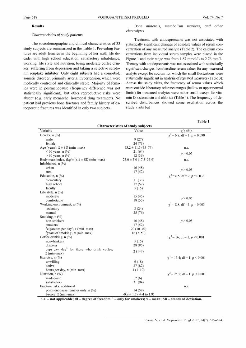
The sociodemographic and clinical characteristics of 33 study subjects are summarized in the Table 1. Prevailing fea-tures are adult females in the beginning of her sixth life de-cade, with high school education, satisfactory inhabitance, working, life style and nutrition, being moderate coffee drin-ker, suffering from depression and taking a selective seroto-nin reuptake inhibitor. Only eight subjects had a comorbid, somatic disorder, primarily arterial hypertension, which were medically controlled and clinically stable. Majority of fema-les were in postmenopause (frequency difference was not statistically significant), but other reproductive risks were absent (e.g. early menarche, hormonal drug treatment). No patient had previous bone fractures and family history of os-teoporotic fractures was identified in only two subjects.
Bone minerals, metabolism markers, and other electrolytes
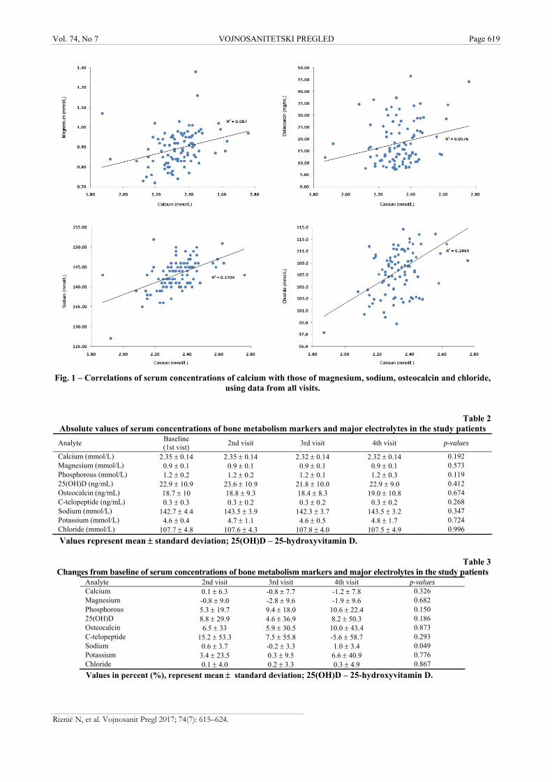
Treatment with antidepressants was not associated with statistically significant changes of absolute values of serum con-centration of any measured analyte (Table 2). The calcium con-centrations from individual serum samples were placed in the Figure 1 and their range was from 1.87 mmol/L to 2.76 mm/L. Therapy with antidepressants was not associated with statistically significant changes from baseline serum values for any measured analyte except for sodium for which the small fluctuations were statistically significant in analysis of repeated measures (Table 3). Across the study visits, the frequency of serum values which were outside laboratory reference ranges (bellow or upper normal limits) for measured analytes were rather small, except for vita-min D, osteocalcin and chloride (Table 4). The frequency of de-scribed disturbances showed some oscillation across the study visits but
n.a. – not applicable; df – degree of freedom. 1 – only for smokers; ґ – mean; SD – standard deviation.
Riznić N, et al. Vojnosanit Pregl 2017; 74(7): 615–624.
Vol. 74, No 7 VOJNOSANITETSKI PREGLED Page 619
Riznić N, et al. Vojnosanit Pregl 2017; 74(7): 615–624.
Fig. 1 – Correlations of serum concentrations of calcium with those of magnesium, sodium, osteocalcin and chloride,
using data from all visits.
Table 2
Absolute values of serum concentrations of bone metabolism markers and major electrolytes in the study patients
Analyte Baseline (1st vist)
2nd visit 3rd visit 4th visit p-values
Calcium (mmol/L) 2.35 0.14 2.35 0.14 2.32 0.14 2.32 0.14 0.192 Magnesium (mmol/L) 0.9 0.1 0.9 0.1 0.9 0.1 0.9 0.1 0.573 Phosphorous (mmol/L) 1.2 0.2 1.2 0.2 1.2 0.1 1.2 0.3 0.119 25(OH)D (ng/mL) 22.9 10.9 23.6 10.9 21.8 10.0 22.9 9.0 0.412 Osteocalcin (ng/mL) 18.7 10 18.8 9.3 18.4 8.3 19.0 10.8 0.674 C-telopeptide (ng/mL) 0.3 0.3 0.3 0.2 0.3 0.2 0.3 0.2 0.268 Sodium (mmol/L) 142.7 4.4 143.5 3.9 142.3 3.7 143.5 3.2 0.347 Potassium (mmol/L) 4.6 0.4 4.7 1.1 4.6 0.5 4.8 1.7 0.724 Chloride (mmol/L) 107.7 4.8 107.6 4.3 107.8 4.0 107.5 4.9 0.996
Values represent mean standard deviation; 25(OH)D – 25-hydroxyvitamin D.
Table 3 Changes from baseline of serum concentrations of bone metabolism markers and major electrolytes in the study patients
Analyte 2nd visit 3rd visit 4th visit p-values Calcium 0.1 6.3 -0.8 7.7 -1.2 7.8 0.326 Magnesium -0.8 9.0 -2.8 9.6 -1.9 9.6 0.682 Phosphorous 5.3 19.7 9.4 18.0 10.6 22.4 0.150 25(OH)D 8.8 29.9 4.6 36.9 8.2 50.3 0.186 Osteocalcin 6.5 33 5.9 30.5 10.0 43.4 0.873 C-telopeptide 15.2 53.3 7.5 55.8 -5.6 58.7 0.293 Sodium 0.6 3.7 -0.2 3.3 1.0 3.4 0.049 Potassium 3.4 23.5 0.3 9.5 6.6 40.9 0.776 Chloride 0.1 4.0 0.2 3.3 0.3 4.9 0.867
Values in percent (%), represent mean standard deviation; 25(OH)D – 25-hydroxyvitamin D.
Page 620 VOJNOSANITETSKI PREGLED Vol. 74, No 7
Table 4 The frequency of patients’ serum samples in which laboratory values of study analytes were below lower normal
limits (LNL) or above upper normal limits (UNL) Analyte Below LNL p-values* Above UNL p-values** Calcium 7.7–19.2 0.543 0–3.8 0.392 Magnesium none n.a. 0–3.8 0.392 Phosphorous 0–3.8 0.392 none n.a. 25(OH)D 40.9–50.0 0.860 0–11.5 0.494 Osteocalcin 26.9–33.3 0.934 n.a. n.a. C-telopeptide n.a. n.a. none n.a. Sodium 0–7.7 0.494 3.8–15.4 0.585 Potassium 0–5.0 0.392 0–5.0 0.572 Chloride 0–4.8 0.572 66.7–73.1 0.927 Values represent range (min–max) of % across the visits; n.a.– not applicable; p – probability of change in frequency across the study visits for below LNL and above UNL.
Table 5 Correlation between serum concentrations of study variables, which includes data from all blood samples
Variable Calcium A B C D E F G H A – Calcium, adj.1 rs = 0.848* p < 0.001 B – Vitamin D r = 0.118 rs = -0.043 p = 0.265 p = 0.683 C – Phosphorous rs = -0.075 rs = -0.09 r = 0.046 p = 0.473 p = 0.388 p = 0.668 D – Osteocalcin r = 0.240* rs = 0.203* r = 0.021 r = 0.139 p = 0.019 p = 0.047 p = 0.838 p = 0.181 E – Magnesium rs = 0.402* rs = 0.207* r = 0.087 r = 0.020 r = 0.106 p < 0.001 p = 0.045 p = 0.412 p = 0.849 p = 0.310
such changes were not statistically significant during antide-pressant treatment for any analyte.
The risk factors, examined by means of univariate binary logistic regressions analyses were: female gender, age, age > 60 years, sedentary work, body mass index, obesity, smoking, coffee drinking 4 cups per day, exercise 4 h per day, unhealthy dietary habits (inadequate intake of food reach in calcium and fish product), family history of bone fractures until 75 years of life, postmenopause, dual-energy x-ray absorptiometry (DXA) T-score, number of risk factors for each patient and individual antidepressant drugs. However, none of the examined factors was significantly as-sociated with negative, cumulative changes from baseline se-rum concentrations of calcium, magnesium, phosphorous, osteocalcin, C-telopeptide, sodium, potassium and chloride. The age of 60 or more was significant risk factor for appea-rance of negative cumulative changes of vitamin D concen-trations from baseline (OR = 11.4, 95% CI 1.2–113.1, p = 0.037) but the other examined factors were not.
A separate analysis was done for calcium values adjusted for serum concentrations of albumin in the respective blood samples. The mean albumin concentration in all serum sam-
ples was 43.7 g/L with standard deviation of 3 g/L (range 35–50 g/L). In general, such results, in essence, follow those rela-ted to the uncorrected calcium values. Therefore, the details of this part of analysis are omitted for the sake of clarity.
Correlation analysis
Across the study days, statistically significant trend was not observed with serum concentrations of calcium (rs = -0.083, p = 0.426), magnesium (rs = -0.062, p = 0.554), vita-min D (r = 0.001, p = 0.993), osteocalcin (rs = 0.024, p = 0.817), C-telopeptide (rs = -0.225, p = 0.059), sodium (r = 0.041, p = 0.697), potassium (rs = -0.034, p = 0.786) and chloride (r = -0.033, p = 0.747). On the other side, during the study course, fluctuations of serum concentrations of several parameters correlated with each other, in the same direction for almost all pairs (Table 5). Taking into account the strength of association, calcium serum concentrations significantly correlated firstly with sodium and chloride con-centrations, then with magnesium concentrations and finally with osteocalcin concentrations (Figure 1). Calcium albu-min-adjusted values, in general, showed almost identical pat-
F – C-telopeptide rs = -0.014 rs = 0.042 r = 0.161 r = 0.106 r = 0.760* r = -0.058 p = 0.890 p = 0.686 p = 0.123 p = 0.311 p < 0.001 p = 0.581 rs = 0.531* rs = 0.418* r = -0.016 r = -0.069 r = 0.215* r = 0.295* rs = -0.096 G – Sodium p < 0.001 p < 0.001 p = 0.884 p = 0.514 p = 0.039 p = 0.004 p = 0.360
rs = 0.153 r = 0.202 r = 0.029 r = -0.002 r = 0.273* rs = -0.002 r = 0.124 H – Potassium r = -0.197 p = 0.059 p = 0.142 p = 0.058 p = 0.787 p = 0.987 p = 0.009 p = 0.983 p = 0.238
rs = 0.380* rs = 0.035 r = -0.218* r = 0.209* r = -0.050 rs = -0.002 r = 0.722* rs = 0.114 I – Chloride r = 0.496* p < 0.001 p < 0.001 p = 0.738 p = 0.035 p = 0.041 p = 0.629 p = 0.982 p < 0.001 p = 0.276
1adjusted for serum albumin concentration in the same blood sample; *considered statistically significant.
Riznić N, et al. Vojnosanit Pregl 2017; 74(7): 615–624.
Vol. 74, No 7 VOJNOSANITETSKI PREGLED Page 621
tern of correlations with the other parameters as uncorrected calcium concentrations. Concentrations of osteocalcin and C-telopeptide showed strong, significant correlation with each other, too. Interestingly, sodium, chloride and, in one case potassium, correlated significantly with some parameters of bone homeostasis like osteocalcin, magnesium and phospho-rous. On the other side, no parameter was statistically corre-lated with concentrations of vitamin D in serum samples.
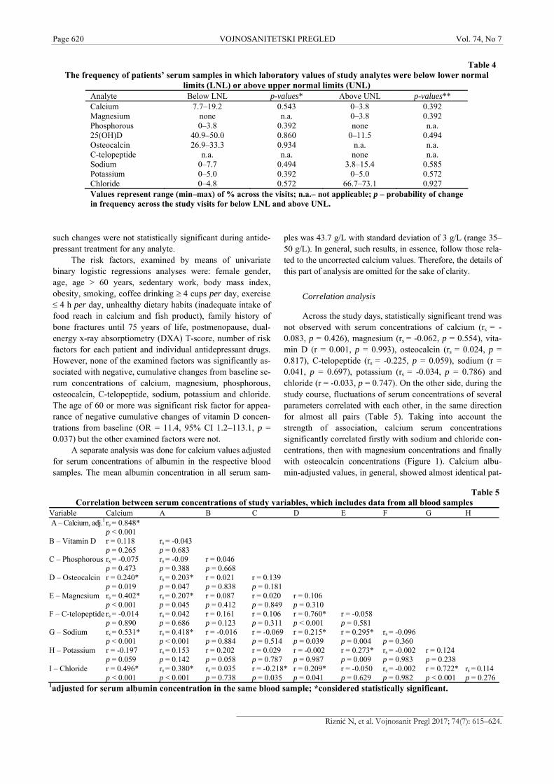
Correlation analysis, with the data which were clustered according to the visit samples, showed dynamic relationship between the study variables during the antidepressant treat-ment course. Correlations between calcium and magnesium serum concentrations were statistically significant in blood samples from the visit 1 (rs = 0.389, p = 0.050), the visit 2 (r = 0.449, p = 0.036) and the visit 3 (r = 0.497, p = 0.015) but not from the visit 4 (rs = 0.075, p = 0.747). Correlations between calcium and osteocalcin serum concentrations were
statistically significant in blood samples only from the visit 4 (rs = 0.496, p = 0.022), between calcium and sodium, from the visit 1 (r = 0.705, p < 0.001) and the visit 3 (r = 0.734, p < 0.001) (Figure 2) and between calcium and chloride, from the visit 1 (r = 0.601, p = 0.001) and visit 3 (r = 0.563, p = 0.003). Albumin-adjusted values of calcium, as in the case of aggrega-te data, showed comparable pattern of correlations with other parameters like that of uncorrected calcium concentrations when analysis included only data from particular visits.
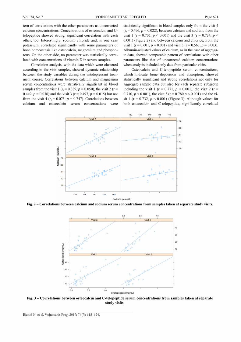
Osteocalcin and C-telopeptide serum concentrations, which indicate bone deposition and absorption, showed statistically significant and strong correlations not only for aggregate sample data but also for each separate subgroup including the visit 1 (r = 0.771, p < 0.001), the visit 2 (r = 0.710, p < 0.001), the visit 3 (r = 0.780 p < 0.001) and the vi-sit 4 (r = 0.732, p < 0.001) (Figure 3). Although values for both osteocalcin and C-telopeptide, significantly correlated
Fig. 2 – Correlations between calcium and sodium serum concentrations from samples taken at separate study visits.
Fig. 3 – Correlations between osteocalcin and C-telopeptide serum concentrations from samples taken at separate
study visits.
Riznić N, et al. Vojnosanit Pregl 2017; 74(7): 615–624.
Page 622 VOJNOSANITETSKI PREGLED Vol. 74, No 7
with sodium concentrations when aggregated data have been analyzed (blood samples from all visits), such correlation was not observed when data sets have been divided into the four visit subgroups (p > 0.05). Similarly, weak but statistically significant correlations between the aggregated values of magnesium and sodium, magnesium and potassi-um, together with phosphorous and chloride were not con-firmed throughout analysis of separate visits. Significant cor-relations have been observed only for magnesium and potas-sium values from the visit 1 (rs = 0.595, p = 0.002) as well as for phosphorous and chloride values from the visit 4 (r = -0.526, p = 0.017).
Discussion
The results of our study showed that antidepressant tre-atment did not significantly change the values of primary study variable, serum concentrations of calcium, during three months in the setting of routine psychiatric practice. In addi-tion, other bone minerals, vitamin D and markers of bone deposition and absorption were unaffected, too. However, changes of some parameters connected with bone metabo-lism positively correlated with each other during the study, suggesting existence of active bone turnover in our patients. In addition, older age of our study subjects was significantly associated with vitamin D depletion. Therefore, the evidence presented in our study favored importance of underlying pa-tient’s characteristics relative to antidepressant medication for bone metabolism. Final result of the interplay of different risk and protective factors could be either bone health or bo-ne disease in the subject who suffer from a mental disorder which needs particular antidepressant drug.
Previous studies reported significant association among antidepressants, osteoporosis and bone fractures 4, 19, 20 but some recent clinical studies found that pharmacotherapy of depression either suppressed or unaffected markers of bone absorption 11, 21. In addition, results of laboratory studies told us that antidepressants were capable to induce bone formati-on in experimental animals 6, 22. Even in the studies with “po-sitive” findings, including the most recent ones, the risk for fracture that could be attributed to antidepressants was rather small and very variable between different societies, and might diminish during the treatment course 4, 19, 23. All these experiences are in well agreement with the findings presen-ted in our study which contributed to the knowledge about relative safety of antidepressant for calcium homeostasis, at least in short term, the topic rarely aimed in previous studies as the primary research focus.
It was well established that many cases of bone fractu-res in patients with depression were, in fact, caused by falls, particularly in older people, and the importance of confoun-ders was pointed out 24. Furthermore, deficiency of vitamin D might not be associated with presence of depression in some populations of adults, so treatment with vitamin D would unlikely improve depressive symptoms, and in the same time different antidepressants might variously influen-ce metabolic pathways of the vitamin 7, 25, 26. Therefore, pre-diction of the risk for disturbances of bone metabolism and
consequent fractures in a patient taking a depressant seems to be much more complex task than simple connection with the drug class. Our study indicates that, if antidepressants do inc-rease fracture risk, disturbances of calcium and, possible, ot-her bone minerals seem to play no or little role in this pro-cess. In this regard, further research in the field, focused on well-characterized underlying risks and, particularly, on their interactions either with each other as well as with individual antidepressants is needed.
Statistically significant, moderate-to-strong and rather consistent link between serum concentration of bone mine-rals and major electrolytes, during the study visits, is very in-teresting and, in some regard, novel finding, which our study presents. It is reasonably assumed that maintaining physiological electrolyte homeostasis represent important health issue for the patients who take antidepressants. Excessive sodium dietary intake is a recognized risk factor for development of osteoporosis 27. In addition, hypomagnesaemia could be associated with hypokalemia, due to increased calciuria and consequent potassium tissue redistribution, sometimes requiring potassium and magnesi-um supplementation 28. However, as far as we know, pros-pective clinical studies, which examined relationships between serum levels of bone minerals and sodium, chloride and potassium during the antidepressant treatment and their consequences on major clinical outcomes were not conduc-ted previously. For example, the newest systematic evidence strongly put in question widespread, indiscriminate use of dietary calcium supplements as they had minimal (if at all) impact on bone mineral density and fracture risk 29,30. There-fore, individual approach to patients and future, focused re-searches are necessary in order to provide more data about both risks and preventive and treatment strategies which ha-ve the most relevant to everyday clinical practice.
Frequency of important disturbances of serum parame-ters in our patients (mostly below the lower normal limits) did not change significantly from baseline to the end of the study. Antidepressant-induced hyponatremia is a well-recognized clinical entity, with syndrome of inappropriate secretion of antidiuretic hormone being major mechanism 29. In our study, we detected minimal, but statistically signifi-cant relative changes of sodium from baseline serum levels. There are case-reports directly connecting use of some anti-depressants with disturbances of potassium and chloride se-rum levels, sometimes with concomitant depletions of other major serum parameters 30–32. Therefore, the possibility that antidepressants in an individual patient induced subtle fluc-tuations of some parameters within the reference range, which further disturbed homeostasis of other counterparts, could not be excluded in our research. It remains unknown which biological mechanisms connect the observed effects in our study and what are consequences for bone health, so fur-ther focused research is necessary.
Our results have to be considered taking into account several limitations of the study such as relatively small sam-ple-size, patients of heterogeneous characteristics, limited time-period, absence of control group and lack of informati-on about some serum markers important for bone metabo-
Riznić N, et al. Vojnosanit Pregl 2017; 74(7): 615–624.
Vol. 74, No 7 VOJNOSANITETSKI PREGLED Page 623
Riznić N, et al. Vojnosanit Pregl 2017; 74(7): 615–624.
lism. However, we believe that improvement of these met-hodological shortcomings would not significantly change the main finding in our study, that antidepressants, as a drug-class, do not cause per se disturbances of serum calcium and, likely, bone minerals during short-to-medium time period. For example, our sample-size was sufficiently powered to detected small but notable fluctuations of primary variable and the researchers have already used self-controlled design (without control-group) as a valid approach in order to examine the somatic risks in the patients who has taken the psychotropic drugs 15. Due to logistic constraints, the serum level of parathyroid hormone was not measured, but it has been reported that, if its baseline levels were increased in pa-tients with depression the antidepressants rather normalized than disturbed them; alternatively, finding of increased parathyroid hormone could be confounded with other regula-tors, as the vitamin D 11, 33.
Many patients in our study took benzodiazepines and some antipsychotics but, as we are aware, no protective ef-fects of these drugs on bone metabolism were reported. In-stead, sedatives increase fracture risk mainly due to gait dis-turbances and antipsychotics induce hyperprolactinemia and hypogonadism predisposing patients to osteoporosis. In fact, the fracture risk in people taking antipsychotic drugs had much more magnitude than in patients taking antidepressants 34–36; moreover, they could significantly disturb calcemia in the notable number of patients, manly toward low levels 17. The-refore, if anxiolytics and antipsychotics used by patients in our study did influence bone mineral homeostasis and their serum levels, the aggregate resultant would either facilitate or counteract presumed action of antidepressants. In both ca-ses the oscillation of their serum calcemia would escape the
pre-defined level (change of 5% from baseline), the event which had to be powerfully detected within our sample size. In addition, the mean serum levels of other bone minerals, markers of bone turnover and vitamin D remained within re-ference ranges and frequencies of their baseline disturbances did not change significantly throughout our study course. It decreases possibility of major influence of factors other than antidepressants for study outcomes followed in the patients, who were enrolled in our research.
Conclusion
Results of our study demonstrated that antidepressants do not disturb significantly serum levels of calcium and other bone minerals and vitamin D homeostasis during acute trea-tment phase within the settings of routine psychiatric practi-ce. Further research should prospectively examine the effects of long-lasting treatment course, beyond three month of therapy of individual antidepressant drugs in different sub-groups of patients with particular risk factors for disorders of bone and electrolyte metabolisms.
Acknowledgements
Dragan R. Milovanović and Slobodan M. Jankovic thank to the Ministry of Education, Science and Technologi-cal Development of the Republic of Serbia on partial support of their research by grant N°175007.
Declaration of interest
There is no conflict of interest for any author.
R E F E R E N C E S
1. Petronijević M, Petronijević N, Ivković M, Stefanović D, Radonjić N, Glisić B, et al. Low bone mineral density and high bone me-tabolism turnover in premenopausal women with unipolar depression. Bone 2008; 42(3): 582−90.
2. Wu Q, Liu J, Gallegos-Orozco JF, Hentz JG. Depression, fracture risk, and bone loss: A meta-analysis of cohort studies. Osteo-poros Int 2010; 21(10): 1627−35.
3. Anglin RE, Samaan Z, Walter SD, McDonald SD. Vitamin D deficiency and depression in adults: Systematic review and meta-analysis. Br J Psychiatry 2013; 202: 100−7.
4. Prieto-Alhambra D, Petri H, Goldenberg JS, Khong TP, Klungel OH, Robinson NJ, et al. Excess risk of hip fractures attributable to the use of antidepressants in five European countries and the USA. Osteoporos Int 2014; 25(3): 847−55.
5. Collet C, Schiltz C, Geoffroy V, Maroteaux L, Launay JM, Verne-joul MC. The serotonin 5-HT2B receptor controls bone mass via osteoblast recruitment and proliferation. FASEB J 2008; 22(2): 418−27.
6. Mortazavi SH, Khojasteh A, Vaziri H, Khoshzaban A, Roudsari MV, Razavi SH. The effect of fluoxetine on bone regenera-tion in rat calvarial bone defects. Oral Surg Oral Med Oral Pathol Oral Radiol Endod 2009; 108(1): 22−7.
7. Oude Voshaar RC, Derks WJ, Comijs HC, Schoevers RA, de Borst MH, Marijnissen RM. Antidepressants differentially related to 1, 25-(OH)2 vitamin D3 and 25-(OH) vitamin D3 in late-life depression. Transl Psychiatry 2014; 4: e383.
8. Russell LA. Osteoporosis and osteomalacia. Rheum Dis Clin North Am 2010; 36(4): 665−80.
9. National Osteoporosis Foundation. Clinician's Guide to preven-tion and treatment of osteoporosis. Washington, DC: Na-tional Osteoporosis Foundation; 2010.
10. Perry PJ, Garvey MJ, Dunner DL, Rush AJ, Kyhl J. A report of trazodone-associated laboratory abnormalities. Ther Drug Monit 1990; 12(6): 517−9.
11. Aydin H, Mutlu N, Akbas NB. Treatment of a major depres-sion episode suppresses markers of bone turnover in preme-nopausal women. J Psychiatr Res 2011; 45(10): 1316−20.
12. Milovanovic DR, Janjic V, Zornic N, Dejanovic SD, Jankovic SM. Risperidone-associated hypocalcemia. Am J Psychiatry 2010; 167(12): 1533−4.
13. Charoenphandhu N, Teerapornpuntakit J, Lapmanee S, Krishnamra N, Charoenphandhu J. Duodenal calcium transporter mRNA expression in stressed male rats treated with diazepam, fluo-xetine, reboxetine, or venlafaxine. Mol Cell Biochem 2012; 369(1−2): 87−94.
14. Keks NA, Hope J. Long-term management of people with psychotic disorders in the community. Aust Prescr 2007; 30(2): 44−6.
15. Douglas IJ, Smeeth L. Exposure to antipsychotics and risk of stroke: Self controlled case series study. BMJ 2008; 337(2): a1227.
Page 624 VOJNOSANITETSKI PREGLED Vol. 74, No 7
16. Morgan JL, Skulan JL, Gordon GW, Romaniello SJ, Smith SM, Anbar AD. Rapidly assessing changes in bone mineral balance using natural stable calcium isotopes. Proc Natl Acad Sci U.S.A. 2012; 109(25): 9989−94.
17. Milovanovic DR, Stanojevic-Pirkovic M, Zivancevic-Simonovic S, Ma-tovic M, Djukic-Dejanovic S, Jankovic SM, et al. Parameters of calcium metabolism fluctuated during initiation or changing of antipsychotic drugs. Psychiatry Investig 2016; 13(1): 89−101.
18. Benrimoj SI, Langford JH, Christian J, Charlesworth A, Steans A. Efficacy and Tolerability of the Anti-inflammatory Throat Lozenge Flurbiprofen 8.75mg in the Treatment of Sore Throat. Clin Drug Investig 2001; 21(3): 183−93.
19. Wu Q, Qu W, Crowell MD, Hentz JG, Frey KA. Tricyclic anti-depressant use and risk of fractures: A meta-analysis of co-hort and case-control studies. J Bone Miner Res 2013; 28(4): 753−63.
20. Moura C, Bernatsky S, Abrahamowicz M, Papaioannou A, Bessette L, Adachi J, et al. Antidepressant use and 10-year incident fracture risk: Tthe population-based Canadian Multicentre Osteoporosis Study (CaMoS). Osteoporos Int 2014; 25(5): 1473−81.
21. Diem SJ, Joffe H, Larson JC, Tsai JN, Guthrie KA, LaCroix AZ, et al. Effects of escitalopram on markers of bone turnover: A randomized clinical trial. J Clin Endocrinol Metab 2014; 99(9): E1732−7.
22. Battaglino R, Vokes M, Schulze-Späte U, Sharma A, Graves D, Kohler T, et al. Fluoxetine treatment increases trabecular bone formation in mice. J Cell Biochem 2007; 100(6): 1387−94.
23. Lanteigne A, Sheu Y, Stürmer T, Pate V, Azrael D, Swanson SA, et al. Serotonin-norepinephrine reuptake inhibitor and selec-tive serotonin reuptake inhibitor use and risk of fractures: A new-user cohort study among US adults aged 50 years and older. CNS Drugs 2015; 29(3): 245−52.
24. Iaboni A, Flint AJ. The complex interplay of depression and falls in older adults: A clinical review. Am J Geriatr Psychiatry 2013; 21(5): 484−92.
25. Zhao G, Ford ES, Li C, Balluz LS. No associations between serum concentrations of 25-hydroxyvitamin D and parathy-roid hormone and depression among US adults. Br J Nutr 2010; 104(11): 1696−702.
26. Li G, Mbuagbaw L, Samaan Z, Falavigna M, Zhang S, Adachi JD, et al. Efficacy of vitamin D supplementation in depression in
adults: A systematic review. J Clin Endocrinol Metab 2014; 99(3): 757−67.
27. Teucher B, Dainty JR, Spinks CA, Majsak-Newman G, Berry DJ, Hoogewerff JA, et al. Sodium and bone health: Impact of mod-erately high and low salt intakes on calcium metabolism in postmenopausal women. J Bone Miner Res 2008; 23(9): 1477−85.
28. Seifter JL. Potassium disorders. In: Goldman L, Ausiello D, edi-tors. Cecil medicine. 23rd ed. Philadelphia, PA: WB Saunders CO; 2008. p. 839−46.
29. Tai V, Leung W, Grey A, Reid IR, Bolland MJ. Calcium intake and bone mineral density: systematic review and meta-analysis. BMJ 2015; 351: h4183.
30. Bolland MJ, Leung W, Tai V, Bastin S, Gamble GD, Grey A, et al. Calcium intake and risk of fracture: systematic review. BMJ 2015; 351: h4580.
31. de Picker L, van den Eede F, Dumont G, Moorkens G, Sabbe DG. Antidepressants and the risk of hyponatremia: A class-by-class review of literature. Psychosomatics 2014; 55(6): 536−47.
32. Ishii T, Ohtake T, Yasu T, Kadotani Y, Hayashi S, Oka M, et al. A rare case of combined syndrome of inappropriate antidiu-retic hormone secretion and Fanconi syndrome in an elderly woman. Am J Kidney Dis 2006; 48(1): 155−8.
33. Jagsch C, Marksteiner J, Seiringer E, Windhager F. Successful mir-tazapine treatment of an 81-year-old patient with syndrome of inappropriate antidiuretic hormone secretion. Pharma-copsychiatry 2007; 40(3): 129−31.
34. Izgi C, Erdem G, Mansuroglu D, Kurtoglu N, Kara M, Gunesdogdu F. Severe hypokalemia probably associated with sertraline use. Ann Pharmacother 2014; 48(2): 297−300.
35. May HT, Bair TL, Lappé DL, Anderson JL, Horne BD, Carlquist JF, et al. Association of vitamin D levels with incident de-pression among a general cardiovascular population. Am Heart J 2010; 159(6): 1037−43.
36. Wu C, Chang C, Tsai Y, Huang Y, Tsai H. Antipsychotic treat-ment and the risk of hip fracture in subjects with schizophre-nia: A 10-year population-based case-control study. J Clin Psychiatry 2015; 76(9): 1216−23.
Received on August 4, 2015. Revised on February 4, 2016.
Accepted on February 22, 2016. Online First April, 2016.
Riznić N, et al. Vojnosanit Pregl 2017; 74(7): 615–624.
Vojnosanit Pregl 2017; 74(7): 625–632. VOJNOSANITETSKI PREGLED Page 625
O R I G I N A L A R T I C L E UDC: 616.24-006-085 https://doi.org/10.2298/VSP160125117S
Quality of life in patients with non-small cell lung cancer
Kvalitet života bolesnika sa nesitnoćelijskim karcinomom pluća
Sanja Šarac*†, Rade Milić*†, Mira Vasiljević*, Momir Šarac†‡
Military Medical Academy, *Clinic for Pulmonology, ‡Clinic for Vascular and Endovascular Surgery, Belgrade, Serbia; University of Defence,
†Faculty of Medicine of the Military Medical Academy, Belgrade, Serbia
Abstract Background/Aim. As lung cancer is considered the greatest contributor to death among all cancer types any help might be valuable in the assessment of treatment effects. The aim of this study was for assess the quality of life (QoL) in patients with non-small cell lung cancer (NSCLC) treated with gem-citabine-cisplatin regimen as the first line of chemotherapy. Methods. The QoL was assessed using certified Serbian translations of the European Organization for Research and Treatment of Cancer Quality Life Questionnaire Core 30 (EORTC QLQ-C30) and Lung Cancer Module (QLQ-LC13) – version 3. The questionnaire was used before starting treatment and after the completion of the 2nd and the 4th cy-cle of chemotherapy. The questionnaire scales and single items were compared in order to assess the impact of treat-ment on the QoL. Results. A total of 60 patients started and 51 completed all questionnaires. There were no changes in the global health status score between the baseline, the 2nd and the 4th cycle of chemotherapy (42.78 ± 15.76, 45.56 ± 17.59, 48.20 ± 19.24, respectively; p = 0.1). Social function score, symptom scores: nausea and vomiting, pain, appetite loss, constipation, diarrhea and financial difficulties score dif-fered significantly among chemotherapy cycles, indicating improved or worsened the QoL. In the lung cancer symptom score a significant difference between measurements was ob-served in cough, alopecia, chest pain and in using analgesics. Conclusion. Monitoring of changes in the QoL among pa-tients with locally advanced and metastatic NSCLC showed that chemotherapy did not decrease the global health status but led to significant changes in the social and financial func-tioning of patients. Some symptoms associated with the dis-ease reduced in the intensity but some new occurred as a re-sult of chemotherapy. Using questionnaires to assess the QoL helped in easier identification of adverse effects and specific problems for adequate treatment. Key words: quality of life; carcinoma, non-small-cell lung; antineoplastic combined chemotherapy protocols; surveys and questionnaires.
Apstrakt Uvod/Cilj. S obzirom na činjenicu da je karcinom pluća naj-smrtonosniji među svim karcinomima, dragocena je svaka pomoć u proceni efekta lečenja. Cilj ove studije bio je da se proceni kvalitet života (quality of life – QoL) obolelih od nesit-noćelijskog karcinoma pluća (NSCLC) koji su lečeni prvom linijom hemioterapije po protokolu gemcitabin-cisplatin. Metode. QoL procenjivan je primenom sertifikovane srpske verzije upitnika Evropske organizacije za istraživanje i lečenje karcinoma (EORTC QLQ-C30) i dodatka koji se odnosi na karcinom pluća (EORTC QLQ-LC13) – verzija 3. Bolesnici su ispunjavali upitnik pre započinjanja lečenja i nakon kom-pletiranja drugog i četvrtog ciklusa hemioterapije. Rezultati su poređeni kako bi se procenio uticaj lečenja na kvalitet života bolesnika. Rezultati. Ukupno 60 bolesnika bilo je uključeno u istraživanje, a 51 je popunio sve upitnike. Nije bilo statistič-ki značajnih promena ukupnog QoL između vremena pre po-četka lečenja, nakon drugog i nakon četvrtog ciklusa hemiote-rapije (42,78 ± 15,76, 45,56 ± 17,59, 48,20 ± 19,24; p = 0,1). U socijalnom funkcionisanju, simptomatskim skalama (muč-nina i povraćanje, bol, gubitak apetita, proliv, zatvor) i finan-sijskim teškoćama nađene su statistički značajne razlike pre lečenja i između ciklusa, ukazujući na poboljšanje ili pogorša-nje QoL. Dodatni simptom skor za karcinom pluća pokazao je značajne razlike za kašalj, gubitak kose, bol u grudima i upotrebu analgetika. Zaključak. Praćenje promena QoL bo-lesnika sa lokalno uznapredovalim i metastatskim NSCLC pokazalo je da primena hemioterapije ne narušava ukupni QoL, ali da dovodi do značajnih promena u socijalnom i fi-nansijskom funkcionisanju bolesnika. Smanjuje se intenzitet pojedinih simptoma povezanih sa bolešću, ali se kao posledi-ca primene hemioterapije javljaju novi simptomi. Korišćenje upitnika za procenu QoL pomaže u lakšem prepoznavanju neželjenih efekata i specifičnih problema omogućavajući ade-kvatno lečenje. Ključne reči: kvalitet života; pluća, nesitnoćelijski karcinom; lečenje kombinovanjem antineoplastika, protokoli; ankete i upitnici.
Correspondence to: Sanja Šarac, Military Medical Academy, Clinic for Pulmonology, Crnotravska 17, 11 000 Belgrade, Serbia. E-mail: [email protected]
Page 626 VOJNOSANITETSKI PREGLED Vol. 74, No 7
Introduction
Lung cancer (small-cell and non-small cell) has been the second most frequent malignancy in the world population for the last ten years. Among men the most common is pros-tate cancer, while among women breast cancer. Lung cancer includes about 13% of all newly diagnosed malignancies 1. It is responsible for 19.4% of all deaths from malignancies and the most common cause of death from malignancy in female and male population 2. Each year more people die from lung cancer than from breast, prostate and colon cancer together 3.
Lung cancer is usually diagnosed in the elderly popula-tion. Two-thirds of patients with this malignancy are older than 65 years, 70 years is the average age. The disease is very rare in people younger than 45 years, less than 2% 3. Non-small cell lung cancer (NSCLC) includes adenocarci-noma, squamous cell carcinoma and “not otherwise specifi-ed” histopathological type accounts for 85% of all lung can-cer cases 4. Lung cancer retains its status as the leading cause of cancer death (26.1%) in Europe 1.
The majority of patients at the time of diagnosis is in the advanced stage of the disease. The treatment strategy for NSCLC depends on the disease stage. In the early stages the treatment of choice is surgical intervention, in locally advan-ced disease the therapy of choice is a combination of radiotherapy and chemotherapy, and chemotherapy alone is an option for patients with metastatic disease 5.
A growing consensus among healthcare providers and researches is that treatment efficiency should be judged not only by its effects on surviving time but also by the quality of life (QoL).
The QoL is defined as a multidimensional construct that encompasses social, physical, cognitive, and psychological domains 6, 7. QoL assessment is an important indicator of tre-atment success with the traditional indicators of tumor res-ponse, progression-free survival and surviving time 8. There are different instruments for the evaluation of QoL and some of them are specifically designed for patients with lung can-cer. The European Organization for Research and Treatment of Cancer (EORTC) – Lung Cancer (LC)-13 questionnaire, a list of symptoms that is used together with the core C-30 questionnaire, is the most commonly used in studies worldwide. The Lung Cancer Symptom Scale (LCSS) is a list of 9 organ-specific symptoms, assessed by the patients and 6 symptoms which were evaluated by an outside obser-ver. There are no items associated with the evaluation of toxicity of treatment. The Functional Assessment of Cancer Therapy – Lung (FACT-L) questionnaire consists of 41 items, includes the general health status and organ-specific symptoms 9.
The aim of this study was assessment of the QoL in pa-tients with NSCLC treated with gemcitabine-cisplatin regi-men as the first line of chemotherapy.
Methods
This prospective follow-up study included 60 patients with histopathologically confirmed NSCLC in stage IIIb and
IV (according to the TNM classification of malignant tu-mors) 10.
In our study QoL was measured using standard questionnaires: the 30-item EORTC Quality of Life Questionnaire (EORTC QLQ-C30) and its lung cancer supplementary questionnaire – (EORTC QLQ-LC13). Inspite of the recommendation, QoL assessments have not been in-corporated in clinical practice yet 11, 12.
The questionnaires in Serbian language, used in the study, had been approved and certified by the EORTC 13.
The patients were treated with gemcitabine-cisplatin re-gimen as the first line chemotherapy.
Inclusion criteria were as follows: age between 18 and 75 years, general condition of the patient-performance status of 0 and 1 according the scale Eastern Cooperative Oncology Group (ECOG) 14, satisfactory haematological status (num-ber of leukocytes 3.5 109/L, the platelet count 100 109/L and hemoglobin 100 g/L), satisfactory liver and kidney function (creatinine, urea, bilirubin, transaminases within normal range), sufficient cardiac function without ac-tive arrhythmia, signs and symptoms of congestive heart fai-lure.
Exclusion criteria were: pregnancy, previously applied chemotherapy or radiotherapy, estimated survival less than three months, the presence of metastases in the central ner-vous system, the simultaneous presence of other malignant disease or systemic connective tissue disease, patients with adenocarcinoma with activating mutation of epidermal growth factor receptor (EGFR) gene, they were treated with tyrosine kinase inhibitors as the first line therapy 15.
The questionnaire and its purpose were explained to each patient in individual interviews and it was self-completed by each patient. It is necessary to avoid any in-volvement by health professionals. The patients were infor-med on the confidentiality of all data obtained and their right not to respond either partially or totally.
The patients personally completed the EORTC QLQ-C30 and QLQ-LC13 (version 3.0). The QLQ-C30 consists of multi-item scales and single-item measures. There are 5 fun-ctional scales, 3 symptom scales, a global health status/QoL scale, and 6 single items. Multi-item scales include a diffe-rent set of items. A specific item occurs in only one scale.
All measurements ranged from 0 to 100 due to easier comparison. High scores on the global health status and functional scales indicate a high level of functioning – good QoL, while on the symptom scales low scores represent less intense symptom experience and consequently a higher QoL 14. The QLQ-LC13 is intended for use among lung can-cer patients varying in disease stage and treatment modality (surgery, chemotherapy and radiotherapy) and consisting of 13 items. It should always be complemented by the QLQ-C30. It consists of questions for assessing lung cancer-associated symptoms (cough, hemoptysis, dyspnea and site specific pain), side effects of the therapy (sore mouth, dysphagia, peripheral neuropathy and alopecia) and use of pain medication 16.
A total of 60 patients started, but 51 completed all three questionnaires. The patients filled questionnaires before sta-
Šarac S, et al. Vojnosanit Pregl 2017; 74(7): 625–632.
Vol. 74, No 7 VOJNOSANITETSKI PREGLED Page 627
Table 1 Basic characteristics of patients with non-small cell lung cancer (NSCLC)
Patient’s characteristics Patients Demographic
age (year), ґ ± SD 62.9 ± 8.1 gender, n (%)
male 45 (75) female 15 (25)
Clinical HP*, n (%) 30 (50)
adenocarcinoma 30 (50) squamous cell
Stage**, n (%) IIIb 35 (58.3) IV 25 (41.7)
PS ECOG*** 0 17 (28.3) 1 43 (71.7)
*Histological type (HP) of NSCLC World Health Organisation – WHO histological classification of tumors of the lung) 18;
**Disease stage (7th Edition of the tumor, node, metastasis (TNM) classification of malignant tumors) 12;
***Performance status for the Eastern Cooperative Oncology Group (PS ECOG) 14. ґ – mean; SD – standard deviation.
Table 2
Response to chemotherapy in patients with non-small cell lung cancer (NSCLC) according to Response Evaluation Criteria in Solid tumors (RECIST) 1.1 18
Patients, n (%) Chemotherapy cycle PR SD PD
After 2nd 32 (53.3) 20 (33.3) 8 (13.3) After 4th 19 (37.3) 24 (47.1) 8 (15.7)
PR – partial response; SD – stable disease; PD – progression of disease. Note: no one patient had complete response.
rting the treatment, and after completing 2th and 4th cycle of chemotherapy. There was a 21-day interval between the cycles. Nine patients did not complete all the questionnaires. They were excluded during the study because of the progres-sion of the disease after two cycles of chemotherapy and then chemotherapy regimen was changed. Unfortunately, one pa-tient died after the second cycle of chemotherapy. Monito-ring took four months for each patient. Tumor response was evaluated by the Response Evaluation Criteria in Solid Tu-mors (RECIST 1.1) 17, 18.
Statistical analysis
Data are presented as counts (%) or the mean ± sta-ndard deviation, depending on their type. The linear mixed model was used to assess differences between three measu-rements (baseline, second and fourth month). The linear mixed model was used to analyse changes in all scales. It has flexibility to model time effect and, the most important, it can handle missing data. Post hoc test with Bonferroni cor-rection was used to assess significant differences between each measurement. All p values less than 0.05 were conside-red significant. All data were analyzed using SPSS 20.0 (IBM Corp.) statistical software. Our study has a number of outcomes. That is the reason for not performing multivariate analysis.
Results
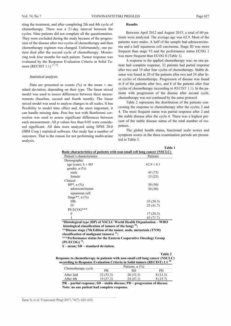
Between April 2012 and August 2015, a total of 60 pa-tients were analyzed. The average age was 62.9. Most of the patients were males. A half of the sample had adenocarcino-ma and a half squamous cell carcinoma. Stage III was more frequent than stage VI and the performance status ECOG 1 was more frequent than ECOG 0 (Table 1).
A response to the applied chemotherapy was: no one pa-tient had complete response, 32 patients had partial response after two and 19 after four cycles of chemotherapy. Stable di-sease was found in 20 of the patients after two and 24 after fo-ur cycles of chemotherapy. Progression of disease was found in 8 of the patients after two, and 8 of the patients after four cycles of chemotherapy (according to RECIST 1.1). In the pa-tients with progression of the disease after second cycle, chemotherapy was not continued by the same protocol.
Table 2 represents the distribution of the patients con-cerning the response to chemotherapy after the cycles 2 and 4. The most frequent status was partial response after 2 and the stable disease after the cycle 4. There was a highest per-cent of the stable disease status of the total number of res-ponses.
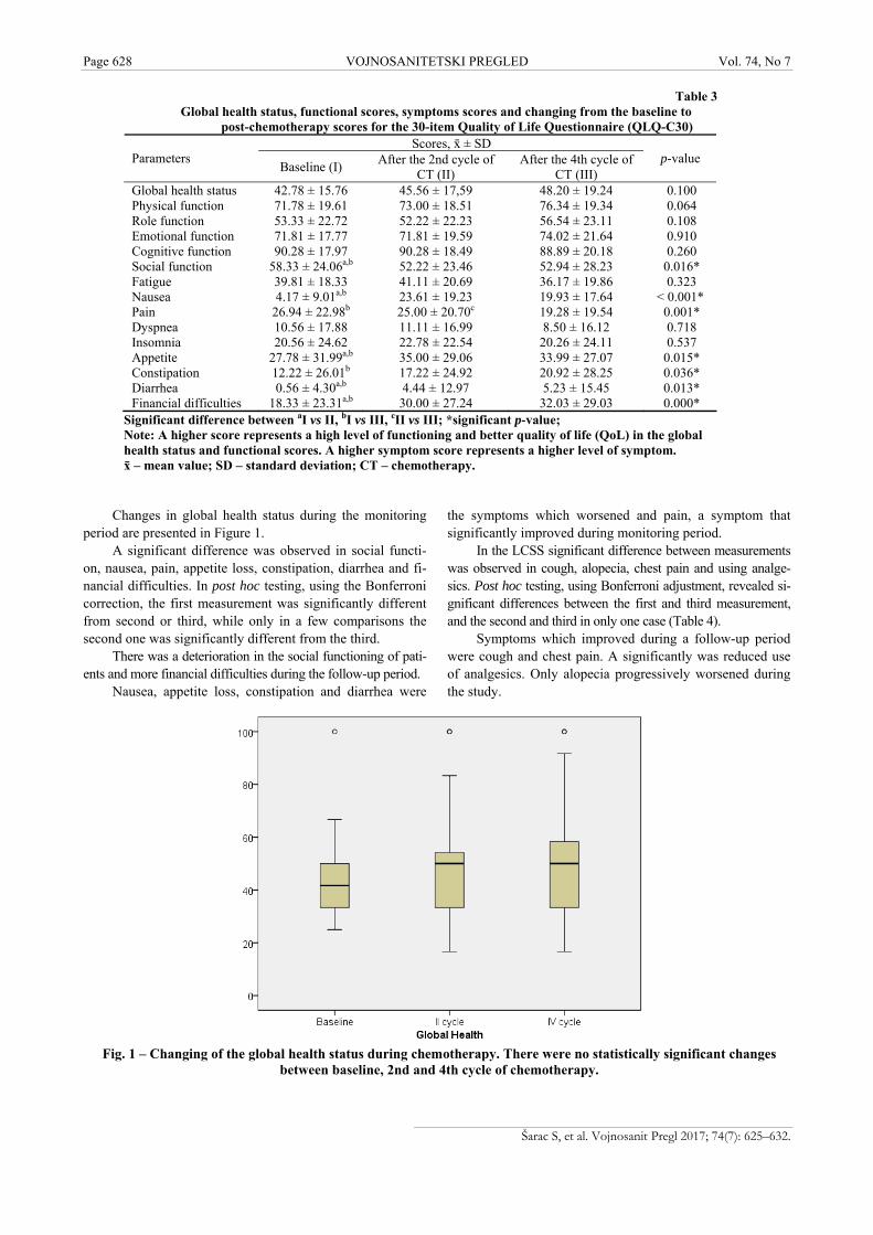
The global health status, functional scale scores and symptom scores in the three examination periods are presen-ted in Table 3.
Šarac S, et al. Vojnosanit Pregl 2017; 74(7): 625–632.
Page 628 VOJNOSANITETSKI PREGLED Vol. 74, No 7
Table 3 Global health status, functional scores, symptoms scores and changing from the baseline to
post-chemotherapy scores for the 30-item Quality of Life Questionnaire (QLQ-C30) Scores, ґ ± SD
Parameters Baseline (I)
After the 2nd cycle of CT (II)
After the 4th cycle of CT (III)
p-value
Global health status 42.78 ± 15.76 45.56 ± 17,59 48.20 ± 19.24 0.100 Physical function 71.78 ± 19.61 73.00 ± 18.51 76.34 ± 19.34 0.064 Role function 53.33 ± 22.72 52.22 ± 22.23 56.54 ± 23.11 0.108 Emotional function 71.81 ± 17.77 71.81 ± 19.59 74.02 ± 21.64 0.910 Cognitive function 90.28 ± 17.97 90.28 ± 18.49 88.89 ± 20.18 0.260 Social function 58.33 ± 24.06a,b 52.22 ± 23.46 52.94 ± 28.23 0.016* Fatigue 39.81 ± 18.33 41.11 ± 20.69 36.17 ± 19.86 0.323 Nausea 4.17 ± 9.01a,b 23.61 ± 19.23 19.93 ± 17.64 < 0.001* Pain 26.94 ± 22.98b 25.00 ± 20.70c 19.28 ± 19.54 0.001* Dyspnea 10.56 ± 17.88 11.11 ± 16.99 8.50 ± 16.12 0.718 Insomnia 20.56 ± 24.62 22.78 ± 22.54 20.26 ± 24.11 0.537 Appetite 27.78 ± 31.99a,b 35.00 ± 29.06 33.99 ± 27.07 0.015* Constipation 12.22 ± 26.01b 17.22 ± 24.92 20.92 ± 28.25 0.036* Diarrhea 0.56 ± 4.30a,b 4.44 ± 12.97 5.23 ± 15.45 0.013* Financial difficulties 18.33 ± 23.31a,b 30.00 ± 27.24 32.03 ± 29.03 0.000*
Significant difference between aI vs II, bI vs III, cII vs III; *significant p-value; Note: A higher score represents a high level of functioning and better quality of life (QoL) in the global health status and functional scores. A higher symptom score represents a higher level of symptom. ґ – mean value; SD – standard deviation; CT – chemotherapy.
Fig. 1 – Changing of the global health status during chemotherapy. There were no statistically significant changes
between baseline, 2nd and 4th cycle of chemotherapy.
Changes in global health status during the monitoring period are presented in Figure 1.
A significant difference was observed in social functi-on, nausea, pain, appetite loss, constipation, diarrhea and fi-nancial difficulties. In post hoc testing, using the Bonferroni correction, the first measurement was significantly different from second or third, while only in a few comparisons the second one was significantly different from the third.
There was a deterioration in the social functioning of pati-ents and more financial difficulties during the follow-up period.
Nausea, appetite loss, constipation and diarrhea were
the symptoms which worsened and pain, a symptom that significantly improved during monitoring period.
In the LCSS significant difference between measurements was observed in cough, alopecia, chest pain and using analge-sics. Post hoc testing, using Bonferroni adjustment, revealed si-gnificant differences between the first and third measurement, and the second and third in only one case (Table 4).
Symptoms which improved during a follow-up period were cough and chest pain. A significantly was reduced use of analgesics. Only alopecia progressively worsened during the study.
Šarac S, et al. Vojnosanit Pregl 2017; 74(7): 625–632.
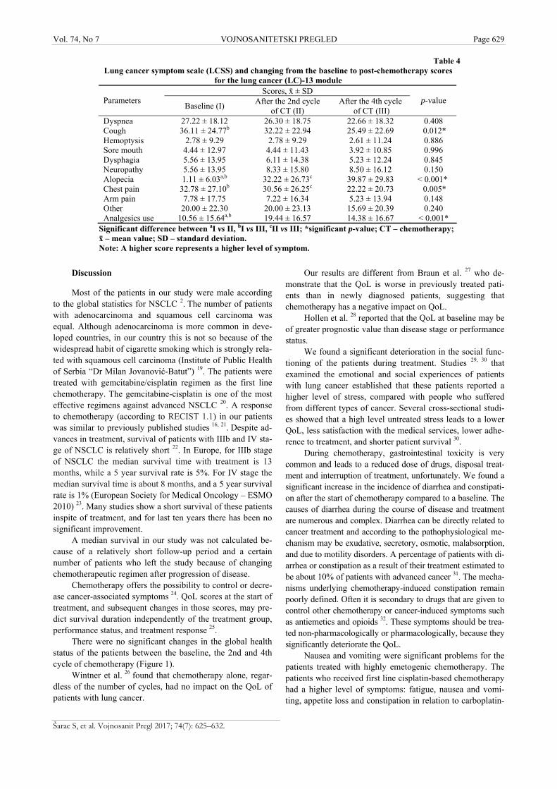
Vol. 74, No 7 VOJNOSANITETSKI PREGLED Page 629
Table 4 Lung cancer symptom scale (LCSS) and changing from the baseline to post-chemotherapy scores
for the lung cancer (LC)-13 module Scores, ґ ± SD
Parameters Baseline (I)
After the 2nd cycle of CT (II)
After the 4th cycle of CT (III)
p-value
Dyspnea 27.22 ± 18.12 26.30 ± 18.75 22.66 ± 18.32 0.408 Cough 36.11 ± 24.77b 32.22 ± 22.94 25.49 ± 22.69 0.012* Hemoptysis 2.78 ± 9.29 2.78 ± 9.29 2.61 ± 11.24 0.886 Sore mouth 4.44 ± 12.97 4.44 ± 11.43 3.92 ± 10.85 0.996 Dysphagia 5.56 ± 13.95 6.11 ± 14.38 5.23 ± 12.24 0.845 Neuropathy 5.56 ± 13.95 8.33 ± 15.80 8.50 ± 16.12 0.150 Alopecia 1.11 ± 6.03a,b 32.22 ± 26.73c 39.87 ± 29.83 < 0.001* Chest pain 32.78 ± 27.10b 30.56 ± 26.25c 22.22 ± 20.73 0.005* Arm pain 7.78 ± 17.75 7.22 ± 16.34 5.23 ± 13.94 0.148 Other 20.00 ± 22.30 20.00 ± 23.13 15.69 ± 20.39 0.240 Analgesics use 10.56 ± 15.64a,b 19.44 ± 16.57 14.38 ± 16.67 < 0.001*
Significant difference between aI vs II, bI vs III, cII vs III; *significant p-value; CT – chemotherapy; ґ – mean value; SD – standard deviation. Note: A higher score represents a higher level of symptom.
Discussion
Most of the patients in our study were male according to the global statistics for NSCLC 2. The number of patients with adenocarcinoma and squamous cell carcinoma was equal. Although adenocarcinoma is more common in deve-loped countries, in our country this is not so because of the widespread habit of cigarette smoking which is strongly rela-ted with squamous cell carcinoma (Institute of Public Health of Serbia “Dr Milan Jovanović-Batut”) 19. The patients were treated with gemcitabine/cisplatin regimen as the first line chemotherapy. The gemcitabine-cisplatin is one of the most effective regimens against advanced NSCLC 20. A response to chemotherapy (according to RECIST 1.1) in our patients was similar to previously published studies 16, 21. Despite ad-vances in treatment, survival of patients with IIIb and IV sta-ge of NSCLC is relatively short 22. In Europe, for IIIb stage of NSCLC the median survival time with treatment is 13 months, while a 5 year survival rate is 5%. For IV stage the median survival time is about 8 months, and a 5 year survival rate is 1% (European Society for Medical Oncology – ESMO 2010) 23. Many studies show a short survival of these patients inspite of treatment, and for last ten years there has been no significant improvement.
A median survival in our study was not calculated be-cause of a relatively short follow-up period and a certain number of patients who left the study because of changing chemotherapeutic regimen after progression of disease.
Chemotherapy offers the possibility to control or decre-ase cancer-associated symptoms 24. QoL scores at the start of treatment, and subsequent changes in those scores, may pre-dict survival duration independently of the treatment group, performance status, and treatment response 25.
There were no significant changes in the global health status of the patients between the baseline, the 2nd and 4th
cycle of chemotherapy (Figure 1). Wintner et al. 26 found that chemotherapy alone, regar-
dless of the number of cycles, had no impact on the QoL of patients with lung cancer.
Our results are different from Braun et al. 27 who de-monstrate that the QoL is worse in previously treated pati-ents than in newly diagnosed patients, suggesting that chemotherapy has a negative impact on QoL.
Hollen et al. 28 reported that the QoL at baseline may be of greater prognostic value than disease stage or performance status.
We found a significant deterioration in the social func-tioning of the patients during treatment. Studies 29, 30 that examined the emotional and social experiences of patients with lung cancer established that these patients reported a higher level of stress, compared with people who suffered from different types of cancer. Several cross-sectional studi-es showed that a high level untreated stress leads to a lower QoL, less satisfaction with the medical services, lower adhe-rence to treatment, and shorter patient survival 30.
During chemotherapy, gastrointestinal toxicity is very common and leads to a reduced dose of drugs, disposal treat-ment and interruption of treatment, unfortunately. We found a significant increase in the incidence of diarrhea and constipati-on after the start of chemotherapy compared to a baseline. The causes of diarrhea during the course of disease and treatment are numerous and complex. Diarrhea can be directly related to cancer treatment and according to the pathophysiological me-chanism may be exudative, secretory, osmotic, malabsorption, and due to motility disorders. A percentage of patients with di-arrhea or constipation as a result of their treatment estimated to be about 10% of patients with advanced cancer 31. The mecha-nisms underlying chemotherapy-induced constipation remain poorly defined. Often it is secondary to drugs that are given to control other chemotherapy or cancer-induced symptoms such as antiemetics and opioids 32. These symptoms should be trea-ted non-pharmacologically or pharmacologically, because they significantly deteriorate the QoL.
Nausea and vomiting were significant problems for the patients treated with highly emetogenic chemotherapy. The patients who received first line cisplatin-based chemotherapy had a higher level of symptoms: fatigue, nausea and vomi-ting, appetite loss and constipation in relation to carboplatin-
Šarac S, et al. Vojnosanit Pregl 2017; 74(7): 625–632.
Page 630 VOJNOSANITETSKI PREGLED Vol. 74, No 7
Šarac S, et al. Vojnosanit Pregl 2017; 74(7): 625–632.
based chemotherapy. Our results, showing a significant inc-rease in the level of of nausea and vomiting compared with the baseline agree with the results of other studies 33, 34.. Early detection and control of these symptoms is very important part of treatment to avoid development of anticipatory nau-sea and vomiting 35.
The loss of appetite is typically present in 15–25% of all cancer patients at diagnosis and may also occur as a side effect of treatment. It can be exacerbated by chemotherapy and radia-tion therapy side effects such as taste and smell changes, nausea, and vomiting 36. Xara et al. 37 report that a number of lung can-cer related symptoms such as the loss of appetite were associa-ted with worse QoL among 56 patients with NSCLC. Increased appetite loss is associated with shorter survival 28. Our study show that in the second measuring these symptoms were most expressed. Better scores at the third measuring were the result of timely application of symptomatic therapy.
The study showed a significant increase in financial dif-ficulties in the second and especially in the third measuring. Patients during the course of the disease in most cases are unable to work and spend their financial resources to the inc-reased cost of living due to the disease. The results of our study are consistent with those from a large database study by Buzaglo et al. 38 which reported that lung cancer patients had the highest rate (> 8%) of serious financial consequences and personal bankruptcy in relation to all other malignancies.
Symptoms associated with lung cancer which require pal-liative treatment may arise from the primary tumor (dyspnea, hemoptysis, pain, fatigue, etc.), symptoms of the regional spread of disease (pleural effusion, superior vena cava syndrome), and symptoms of distant metastasis (liver, brain, bone, etc). These symptoms may have significant negative effects on the QoL.
Approximately 65% of people with lung cancer have a chronic cough. Cough in lung cancer is a distressing symptom with a significant impact on the QoL, and there is no effective therapy. Persistent cough can interfere with speech, eating, and sleeping, thus impacting the QoL 39. During our research we fo-und a reduction in the intensity of cough compared with baseline and it is consistent with Park et al. 40 who reported that cough tends to improve during chemotherapy.
Alopecia is a very common side effect of antineoplastic drugs. The patients in our study had significant hair loss after the 2nd and even more evident after the 4th cycle of chemotherapy. Studies reported increased occurrence of alopecia after the 1st
cycle of chemotherapy, a result that indicates low QoL. Accor-ding to Can et al. 41, hair loss is the most devastating effect and can directly affect social and emotional aspects of the QoL of female patients undergoing chemotherapy.
Chemotherapy-induced hair loss is considered to be one of the most traumatic factors in cancer patient care. Hair loss can negatively impact individual perceptions of appearance, body image, sexuality, and self-esteem, as well as deprive patients of their privacy, because this treatment-related outcome is readily associated with having cancer by the lay public. About 47% of female cancer patients consider hair loss to be the most trauma-tic aspect of chemotherapy. Motivation for a comprehensive support program has the potential to improve psychological sta-tus of patients with hair loss during their cancer therapy 42.
Pain is one of the several symptoms of cancer that crea-te a poor QoL because pain affects physical functions and has an emotional impact. For cancer patients, pain and symptom control are the best predictors of overall QoL sco-res because the effects of unrelieved pain and poorly mana-ged symptoms interfere with the activities of daily living, mood, mobility, and independence. It is also the most com-mon cause of disability and is associated with depression, anxiety, and sleep disturbances 43.
A reduction of pain is one of the most important goals in the treatment of cancer patients. In this study we found that pain was significantly lower after starting and during chemotherapy compared to the time before the treatment. Several studies such as that of Herndon et al. 44 showed that pain is the principal prognostic factor in advanced NSCLC.
During our study, the level of pain decreased due to an-tineoplastic therapy and use of analgesic. Successful treat-ment of pain includes the following: assessment of cancer pain, a review of specific cancer pain syndromes, general principles of cancer pain management, an overview of risk management in patients treated with opioids, prevention and management of opioid side effects, the clinical use of non-opioid analgesics (including nonsteroidal anti-inflammatory drugs and adjuvant analgesics), non-pharmacologic methods of cancer pain management 45.
Opioids are widely used for treatment of pain in pati-ents with cancer because of their safety, multiple routes of administration, ease of titration, reliability, and effectiveness for all types of pain (somatic, visceral, neuropathic) 46.
We found no significant changes in the scores for dyspnea, hemoptysis, sore mouth, dysphagia and neuropathy. Literature data show a low incidence of the aforementioned symptoms when cisplatin are used in combination with gem-citabine, a factor that should be considered in the choice of drug therapy 47.
Recent studies suggest that among patients with NSCLC more lung cancer related symptoms may adversely affect both a response to the treatment and the overall survi-val. Cancer treatment may positively and negatively affect the QoL. Tumor response may have a positive influence on survival and QoL, but adverse effects of treatment may have a negative effect on these parameters 48, 49.
Conclusion
The influence of treatment on the QoL is ever more im-portant when considering treatment options for patients. In this study monitoring of changes in the QoL among patients with locally advanced and metastatic NSCLC show that chemotherapy does not decrease the global health status, but leads to significant changes in social and financial functio-ning of patients. Some symptoms associated with the disease reduce their intensity but some new occur as the result of chemotherapy. Using questionnaires to assess the QoL du-ring treatment helps in identifying changes of the QoL, ad-verse effects of therapy and specific problems for adequate treatment. Palliative treatment should not deteriorate the QoL.
Vol. 74, No 7 VOJNOSANITETSKI PREGLED Page 631
R E F E R E N C E S
1. Ferlay J, Soerjomataram I, Ervik M, Dikshit R, Eser S, Mathers C, et al. GLOBOCAN 2012 v1.0, Cancer Incidence and Mortality Worldwide: IARC CancerBase No. 11 [Internet]. Lyon, France: International Agency for Research on Cancer; 2013. Available from: http://globocan.iarc.fr, accessed on day/month/year.
2. Torre LA, Bray F, Siegel RL, Ferlay J, Lortet-Tieulent J, Jemal A. Global cancer statistics, 2012. CA Cancer J Clin 2015; 65(2): 87−108.
3. Bray F, Ren J, Masuyer E, Ferlay J. Global estimates of cancer prevalence for 27 sites in the adult population in 2008. Int J Cancer 2013; 132(5): 1133−45.
4. Siegel RL, Miller KD, Jemal A. Cancer Statistics. CA Cancer J Clin 2015; 65(1): 5−29.
5. Petrosyan F, Daw H, Haddad A, Spiro T. Targeted therapy for lung cancer. Anticancer Drugs 2012; 23(10): 1016−21.
6. Pearman T. Psychosocial factors in lung cancer: Quality of life, economic impact, and survivorship implications. J Psychosoc Oncol 2008; 26(1): 69−80.
7. Sarna L, Padilla G, Holmes C, Tashkin D, Brecht ML, Evangelista L. Quality of life of long-term survivors of non-small-cell lung cancer. J Clin Oncol 2002; 20(13): 2920−9.
8. Aaronson NK, Bullinger M, Ahmedzai S. A Modular approach to quality-of-life assessment in cancer clinical trials. Recent Re-sults Cancer Res 1988; 111: 231−49.
9. Hollen PJ, Gralla RJ. Comparison of instruments for measuring quality of life in patients with lung cancer. Semin Oncol 1996; 23(5): 31−40.
10. Goldstraw P, Crowley J, Chansky K, Giroux DJ, Groome PA, Rami-Porta R, et al. The IASCL Lung Cancer Staging Project: Pro-posals for the revision of the TNM groupings in the forthcom-ing (seventh) edition of the TNM classification of malignant tumors. J Thorac Oncol 2007; 2(8): 706−14.
11. Engelen V, Detmar S, Koopman H, Maurice-Stam H, Caron H, Hoo-gerbrugge P, et al. Reporting health-related quality of life scores to physicians during routine follow-up visits of pediatric on-cology patients: Is it effective. Pediatr Blood Cancer 2012; 58(5): 766−74.
12. Gridelli C, Perrone F, Nelli F, Ramponi S, de Marinis F. Quality of life in lung cancer patients. Ann Oncol 2001; 12(3): S21−5.
13. Aaronson NK, Ahmedzai S, Bergman B, Bullinger M, Cull A, Duez NJ, et al. The European Organisation for Research and Treatment of Cancer QLQ-C30: A quality-of-life instrument for use in international clinical trials in oncology. J Natl Cancer Inst 1993; 85(5): 365−76.
14. Oken MM, Creech RH, Tormey DC, Horton J, Davis TE, McFadden ET, et al. Toxicity and response criteria of the Eastern Coop-erative Oncology Group. Am J Clin Oncol 1983; 5(6): 649−55.
15. Gazdar A. Activating and resistance mutations of EGFR in non-small-cell lung cancer: role in clinical response to EGFR tyrosine kinase inhibitors. Oncogene 2009; 28(1): S24−31.
16. Bergman B, Aaronson NK, Ahmedzai S, Kaasa S, Sullivan M. The EORTC QLQ-LC13: a modular supplement to the EORTC Core Quality of Life Questionnaire (QLQ-C30) for use in lung cancer clinical trials. EORTC Study Group on Quality of Life. Eur J Cancer 1994; 30A(5): 635−42.
17. Eisenhauer E, Verweij J, Therasse P. Response assessment in solid tumours (RECIST): Version 1.1 and Supporting Papers. Eur J Cancer 2009; 45(2): 225−310.
18. Travis WD, Brambilla E, Nicholson AG, Yatabe Y, Austin J, Beasley MB, et al. The 2015 World Health Organization Classification of Lung Tumors: Impact of Genetic, Clinical and Radiologic
Advances Since the 2004 Classification. J Thorac Oncol 2015; 10(9): 1243−60.
19. Department for Prevention and Control of Noncommunicable Diseases. The incidence and mortality in central Serbia 2012. Belgrade: Institute of Public Health of Serbia “Dr Milan Jovanovic-Batut”; 2014. (Serbian)
20. le Chevalier T, Scagliotti G, Natale R, Danson S, Rosell R, Stahel R, et al. Efficacy of gemcitabine plus platinum chemotherapy compared with other platinum containing regimens in ad-vanced non-small-cell lung cancer: A meta-analysis of survival outcomes. Lung Cancer 2005; 47(1): 69−80.
21. Li L, Schaid DJ, Fridley BL, Kalari KR, Jenkins GD, Abo RP, et al. Gemcitabine metabolic pathway genetic polymorphisms and response in patients with non-small cell lung cancer. Pharmacogenet Genomics 2012; 22(2): 105−16.
22. Rajappa S, Gundeti S, Talluri MR, Digumarti R. Chemotherapy for advanced lung cancer: A 5-year experience. Indian J Cancer 2008; 45(1): 20−6.
23. D'Addario G, Früh M, Reck M, Baumann P, Klepetko W, Felip E, et al. Metastatic non-small-cell lung cancer: ESMO Clinical Practice Guidelines for diagnosis, treatment and follow-up. Ann Oncol 2010; 21 Suppl 5: v116−9.
24. Hickish TF, Smith IE, O'Brien ME, Ashley S, Middleton G. Clini-cal benefit from palliative chemotherapy in non-small-cell lung cancer extends to the elderly and those with poor prognostic factors. Br J Cancer 1998; 78(1): 28−33.
25. Higginson IJ, Carr AJ. Measuring quality of life: Using quality of life measures in the clinical setting. BMJ 2001; 322(7297): 1297−300.
26. Wintner LM, Giesinger JM, Zabernigg A, Sztankay M, Meraner V, Pall G, et al. Quality of life during chemotherapy in lung can-cer patients: Results across different treatment lines. Br J Can-cer 2013; 109(9): 2301−8.
27. Braun DP, Gupta D, Staren ED. Quality of life assessment as a predictor of survival in non-small cell lung cancer. BMC Can-cer 2011; 11: 353.
28. Hollen PJ, Gralla RJ, Kris MG, Cox C, Belani CP, Grunberg SM, et al. A dilemma in analysis: Issues in the serial measurement of quality of life in patients with advanced lung cancer. Lung Cancer 1997; 18(2): 119−36.
29. Zabora J, Brintzenhofeszoc K, Curbow B, Hooker C, Piantadosi S. The prevalence of psychological distress by cancer site. Psy-chooncology 2001; 10(1): 19−28.
30. Kennard BD, Stewart SM, Olvera R, Bawdon RE, Hailin AO, Lewis CP, et al. Nonadherence in adolescent oncology patients: Pre-liminary data on psychological risk factors and relationships to outcome. J Clin Psychol Med Settings 2004; 11(1): 31−9.
31. Gibson RJ, Keefe DM. Cancer chemotherapy-induced diarrhoea and constipation: mechanisms of damage and prevention strategies. Support Care Cancer 2006; 14(9): 890−900.
32. Portenoy RK. Constipation in the cancer patient: causes and management. Med Clin North Am 1987; 71(2): 303−11.
33. Hotta K, Matsuo K, Ueoka H, Kiura K, Tabata M, Tanimoto M. Meta-analysis of randomized clinical trials comparing Cisplatin to Carboplatin in patients with advanced non-small-cell lung cancer. J Clin Oncol 2004; 22(19): 3852−9.
34. Jiang J, Liang X, Zhou X, Huang R, Chu Z. A meta-analysis of randomized controlled trials comparing carboplatin-based to cisplatin-based chemotherapy in advanced non-small cell lung cancer. Lung Cancer 2007; 57(3): 348−58.
35. Roscoe JA, Morrow GR, Aapro MS, Molassiotis A, Olver I. Antici-patory Nausea and Vomiting. Support Care Cancer 2011; 19(10): 1533−8.
Šarac S, et al. Vojnosanit Pregl 2017; 74(7): 625–632.
Page 632 VOJNOSANITETSKI PREGLED Vol. 74, No 7
36. Scott HR, McMillan DC, Forrest LM, Brown DJ, McArdle CS, Mil-roy R. The systemic inflammatory response, weight loss, per-formance status and survival in patients with inoperable non-small cell lung cancer. Br J Cancer 2002; 87(3): 264−7.
37. Xara S, Amaral TF, Parente B. Undernutrition and quality of life in non-small cell lung cancer patients. Rev Port Pneumol 2011; 17(4): 153−8. (Portuguese)
38. Buzaglo J, Gayer C, Mallick R, Charap E, Ferris A, Golant M, et al. Understanding the experience of living with non-small-cell lung cancer (NSCLC): A qualitative study. J Community Sup-port Oncol 2014; 12(1): 6−12.
39. Harle AS, Blackhall FH, Smith JA, Molassiotis A. Understanding cough and its management in lung cancer. Curr Opin Support Palliat Care 2012; 6(2): 153−62.
40. Park S, Kim IR, Baek KK, Lee SJ, Chang WJ, Maeng CH, et al. Prospective analysis of quality of life in elderly patients treated with adjuvant chemotherapy for non-small-cell lung cancer. Ann Oncol 2013; 24(6): 1630−9.
41. Can G, Demir M, Erol O, Aydiner A. A comparison of men and women's experiences of chemotherapy-induced alopecia. Eur J Oncol Nurs 2013; 17(3): 255−60.
42. Shin H, Jo SJ, Kim do H, Kwon O, Myung SK. Efficacy of inter-ventions for prevention of chemotherapy-induced alopecia: A systematic review and meta-analysis. Int J Cancer 2015; 136(5): 442−54.
43. Green CR, Hart-Johnson T, Loeffler DR. Cancer-related chronic pain: Examining quality of life in diverse cancer survivors. Cancer 2011; 117(9): 1994−2003.
44. Herndon JE 2nd, Fleishman S, Kornblith AB, Kosty M, Green MR, Holland J. Is quality of life predictive of the survival of patients
with advanced nonsmall cell lung carcinoma? Cancer 1999; 85(2): 333−40.
45. Afsharimani B, Kindl K, Good P, Hardy J. Pharmacological op-tions for the management of refractory cancer pain-what is the evidence? Support Care Cancer 2015; 23(5): 1473−81.
46. Riley J, Branford R, Droney J, Gretton S, Sato H, Kennett A, et al. Morphine or oxycodone for cancer-related pain? A random-ized, open-label, controlled trial. J Pain Symptom Manage 2015; 49(2): 161−72.
47. Ohe Y, Ohashi Y, Kubota K, Tamura T, Nakagawa K, Negoro S. Randomized phase III study of cisplatin plus irinotecan versus carboplatin plus paclitaxel, cisplatin plus gemcitabine, and cis-platin plus vinorelbine for advanced non-small cell lung can-cer: Four-Arm Cooperative Study in Japan. Ann Oncol 2007; 18(2): 317−23.
48. Wang XS, Shi Q, Lu C, Basch EM, Johnson VE, Mendoza TR, et al. Prognostic value of symptom burden for overall survival in patients receiving chemotherapy for advanced nonsmall cell lung cancer. Cancer 2010; 116(1): 137−45.
49. Lemonnier I, Guillemin F, Arveux P, Clément-Duchêne C, Velten M, Woronoff-Lemsi MC, et al. Quality of life after the initial treat-ments of non-small cell lung cancer: a persistent predictor for patients' survival. Health Qual Life Outcomes 2014; 12: 73.
Received on January 25, 2016. Revised on March 13, 2016.
Accepted on March 16, 2016. Online First May, 2016.
Šarac S, et al. Vojnosanit Pregl 2017; 74(7): 625–632.
Vojnosanit Pregl 2017; 74(7): 633–638. VOJNOSANITETSKI PREGLED Page 633
O R I G I N A L A R T I C L E UDC: 577.1::618.3-06 https://doi.org/10.2298/VSP150517251B
Preeclampsia and level of oxidative stress in the first trimester of pregnancy
Preeklampsija i nivo oksidativnog stresa u prvom trimestru trudnoće
Mirjana Bogavac*†, Ana Jakovljevi憇, Zoran Stajić§, Aleksandra Nikolić†║, Mirjana Milošević-Toši憶, Jadranka Dejanović†**, Zagorka Lozanov-Crvenkovi憶
Clinical Centre of Vojvodina, *Department of Obstetrics and Gynecology, ‡Centre for Laboratory Medicine, ║Emergency Centre, Novi Sad, Serbia; University of Novi Sad,
†Faculty of Medicine, ¶Faculty of Science, Novi Sad, Serbia; Institute for Health Protection of the Ministry of Interior, Department of Internal Medicine, §Division of
Cardiology, Belgrade, Serbia; Institute for Cardiovascular Diseases, **Clinic of Cardiology, Sremska Kamenica, Serbia
Abstract Background/Aim. Preeclampsia (PE) is a multisystemic syndrome that complicates 5–8% of all pregnancies. The aim of this study was to evaluate the biochemical parame-ters of oxidative stress in the first trimester of pregnancy in patients with preeclampsia, with the purpose of comparing the level of oxidative stress with normal pregnancy. Met-hods. The study was conducted as a prospective study. It included totally 107 pregnant women divided into two gro-ups. In the study group (n = 33) there were women who developed preeclampsia in the current pregnancy. The con-trol group (n = 74) included healthy pregnant women. Blo-od samples were taken between 11th and 14th weeks of ges-tation, and the values of superoxide dismutase (SOD), glu-tathione peroxidase (GSH-Px) and total antioxidant status (TAS) were determined in serum by enzymatic colorimetric methods. Results. The values of SOD and GHS-Px were statistically higher in the study group, while the values of TAS were statistically higher in the control group. The level of TAS inversely correlated with GSH-Px and SOD, but there is no statistically significant correlation between GSH-Px and SOD in the study group. Conclusion. The results of this study suggest a higher level of oxidative stress in the first trimester of pregnancy with preeclampsia, which may indicate that the initiation and development of pathophysiological processes underlying preeclampsia start much earlier than the clinical syndrome exhibit. Keywords: pregnancy complications; pre-eclampsia; oxidative stress; superoxide dismutase; glutathione peroxidase; sensitivity and specificity.
Apstrakt Uvod/Cilj. Preeklampsija (PE) je multisistemski sindrom koji komplikuje 5–8% svih trudnoća. Cilj ove studije bio je procena biohemijskih pokazatelja oksidativnog stresa u pr-vom trimestru trudnoće kod bolesnice koje su u toku aktu-elne trudnoće razvile preeklampsiju u poređenju sa stepe-nom oksidativnog stresa kod trudnica sa fiziološkom trud-noćom. Metode. Istraživanje je sprovedeno kao prospekti-vna studija i uključila je ukupno 107 trudnica podeljenih u ispitivanu i kontrolnu grupu. Ispitivanu grupu (n = 33) čini-le su trudnice koje su u toku aktuelne trudnoće razvile pree-klampsiju, dok su kontrolnu grupu (n = 74) činile trudnice sa fiziološkom trudnoćom. Uzorci krvi uzimani su svim trudnicama između 11. i 14. nedelje gestacije i vrednosti su-peroksid dismutaze (SOD), glutation peroksidaze (GSH-Px) i totalnog antioksidativnog statusa (TAS) određivani su en-zimskim kolorimetrijskim metodama. Rezultati. Vrednosti SOD i GSH-Px bile su statistički značajno više u ispitivanoj grupi dok su vrednosti TAS bili statistički značajno više u kontrolnoj grupi. Utvrđena je statistički značajna inverzna korelacija TAS sa SOD i GSH-Px, dok između SOD i GSH-Px nije bilo statistički značajne povezanosti. Zaklju-čak. Rezultati ove studije ukazuju na viši stepen oksidativ-nog stresa u prvom trimestru trudnoće kod trudnica sa pre-eklampsijom, što sugeriše da patofiziološki mehanizmi koji čine osnovu preeklampsije započinju znatno pre, nego se klinički sindrom ispolji. Ključne reči: trudnoća, komplikacije; preeklampsija; stres, oksidativni; peroksid dismutaza; glutation peroksidaza; osetljivost i specifičnost.
Correspondence to: Mirjana Bogavac, Clinical Centre of Vojvodina, Department of Obstetrics and Gynecology, 21 000 Novi Sad, Serbia. E-mail: [email protected]
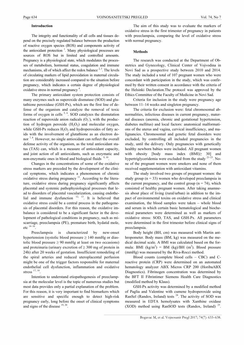
Page 634 VOJNOSANITETSKI PREGLED Vol. 74, No 7
Introduction
The integrity and functionality of all cells and tissues de-pend on the precisely regulated balance between the production of reactive oxygen species (ROS) and components activity of the antioxidant protection 1. Many physiological processes are sources of ROS but in limited and controlled amounts. Pregnancy is a physiological state, which modulates the proces-ses of metabolism, hormonal status, coagulation and immune mechanisms, all of which affect the redox balance 2, 3. The levels of circulating markers of lipid peroxidation in maternal circula-tion are considerably increased compared to the situation before pregnancy, which indicates a certain degree of physiological oxidative stress in normal pregnancy 4.
The primary antioxidant system protection consists of many enzymes such as superoxide dismutase (SOD) and glu-tathione peroxidase (GSH-Px), which are the first line of de-fense of the organism and catalyze the removal of toxic forms of oxygen in cells 5, 6. SOD catalyzes the dismutation reaction of superoxide anion radicals (O2
-), with the produc-tion of hydrogen peroxide (H2O2) and molecular oxygen, while GSH-Px reduces H2O2 and hydroperoxides of fatty ac-ids with the involvement of glutathione as an electron do-nor 7, 8. However, no single antioxidant can reflect the overall defense activity of the organism, as the total antioxidant sta-tus (TAS) can, which is a measure of antioxidant capacity, and joint action of all antioxidants, such as enzymatic and non-enzymatic ones in blood and biological fluids 9, 10.
Changes in the concentrations of some of the oxidative stress markers are preceded by the development of the clini-cal symptoms, which indicates a phenomenon of chronic oxidative stress during pregnancy 11. According to the litera-ture, oxidative stress during pregnancy significantly affects placental and systemic pathophysiological processes that le-ad to disorders of placental vascularization, causing endothe-lial and immune dysfunction 12, 13. It is believed that oxidative stress could be a central process in the pathogene-sis of placental disorders. For this reason, the oxidative im-balance is considered to be a significant factor in the deve-lopment of pathological conditions in pregnancy, such as mi-scarriage, preeclampsia (PE), premature birth, hydatid mole, etc 14–16.
Preeclampsia is characterized by new-onset hypertension (systolic blood pressure ≥ 140 mmHg or dias-tolic blood pressure ≥ 90 mmHg at least on two occasions) and proteinuria (urinary excretion of ≥ 300 mg of protein in 24h) after 20 weeks of gestation. Insufficient remodeling of the spiral arteries and reduced uteroplacental perfusion might be one of the trigger factors responsible for maternal endothelial cell dysfunction, inflammation and oxidative stress 17, 18.
Intention to understand etiopathogenesis of preeclamp-sia at the molecular level is the topic of numerous studies but most data provides only a partial explanation of the problem. For this reason, it is very important to find biomarkers which are sensitive and specific enough to detect high-risk pregnancy early, long before the onset of clinical symptoms and signs of the disease 19, 20.
The aim of this study was to evaluate the markers of oxidative stress in the first trimester of pregnancy in patients with preeclampsia, comparing the level of oxidative stress with normal pregnancy.
Methods
The research was conducted at the Department of Ob-stetrics and Gynecology, Clinical Center of Vojvodina in Novi Sad as a prospective study between 2010 and 2014. The study included a total of 107 pregnant women who were concordant with participation in the study, which was confir-med by their written consent in accordance with the criteria of the Helsinki Declaration.The protocol was approved by the Ethics Committee of the Faculty of Medicine in Novi Sad.
Criteria for inclusion in the study were pregnancy age between 11–14 weeks and singleton pregnancy.
The criteria for exclusion were: fetal chromosomal ab-normalities, infectious diseases in current pregnancy, mater-nal diseases (anemia, chronic and gestational hypertension, diabetes mellitus) and local factors: anatomical malformati-ons of the uterus and vagina, cervical insufficiency, and ma-lignancies. Chromosomal and genetic fetal disorders were excluded, by controlling all included pregnancies in the study, until the delivery. Only pregnancies with genetically healthy newborn babies were included. All pregnant women with obesity [body mass index (BMI)≥ 30] and hypertriglyceridemia were excluded from the study 21–23. No-ne of the pregnant women were smokers and none of them received supplementation with antioxidant vitamins.
The study involved two groups of pregnant women: the study group (n = 33) women who developed preeclampsia in the current pregnancy, and the control group (n = 74), which consisted of healthy pregnant women. After taking anamne-sis about place of living (rural/urban) in addition to the im-pact of environmental toxins on oxidative stress and clinical examination, the blood samples were taken – whole blood and serum in which certain basic hematological and bioche-mical parameters were determined as well as markers of oxidative stress: SOD, TAS, and GSH-Px. All parameters were determined in the first trimester before clinical signs of preeclampsia.
Body height (BH, cm) was measured with Martin ant-hropometer. Body mass (BM, kg) was measured on the me-dical decimal scale. A BMI was calculated based on the for-mula: BMI (kg/m2) = BM (kg)/BH (m2). Blood pressure (mmHg) was measured by the Riva-Rocci method.
Blood counts (complete blood cells – CBC) and C-reactive protein (CRP) were determined on an automated hematology analyzer ABX Micros CRP 200 (HoribaABX Diagnostics). Fibrinogen concentration was determined by the BFT II Fibrintimer Siemens Health Care Diagnostics (modified method by Klaus).
GSH-Px activity was determined by a modified method of Paglia and Valentine with cumene hydroperoxide using RanSel (Randox, Ireland) tests 24. The activity of SOD was measured in EDTA hemolysates with Xanthine oxidase (XOD) method using RanSOD tests (Randox, Ireland) 25.
Bogavac M, et al. Vojnosanit Pregl 2017; 74(7): 633–638.
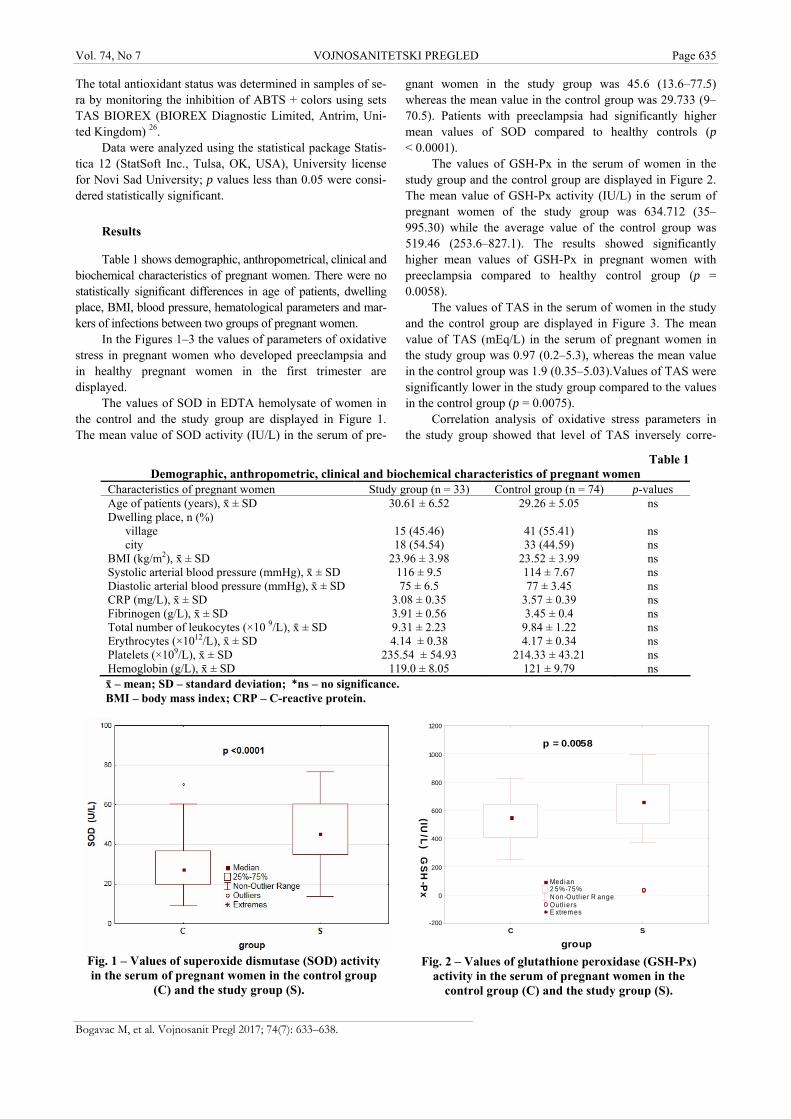
Vol. 74, No 7 VOJNOSANITETSKI PREGLED Page 635
The total antioxidant status was determined in samples of se-ra by monitoring the inhibition of ABTS + colors using sets TAS BIOREX (BIOREX Diagnostic Limited, Antrim, Uni-ted Kingdom) 26.
Data were analyzed using the statistical package Statis-tica 12 (StatSoft Inc., Tulsa, OK, USA), University license for Novi Sad University; p values less than 0.05 were consi-dered statistically significant.
Results
Table 1 shows demographic, anthropometrical, clinical and biochemical characteristics of pregnant women. There were no statistically significant differences in age of patients, dwelling place, BMI, blood pressure, hematological parameters and mar-kers of infections between two groups of pregnant women.
In the Figures 1–3 the values of parameters of oxidative stress in pregnant women who developed preeclampsia and in healthy pregnant women in the first trimester are displayed.
The values of SOD in EDTA hemolysate of women in the control and the study group are displayed in Figure 1. The mean value of SOD activity (IU/L) in the serum of pre-
gnant women in the study group was 45.6 (13.6–77.5) whereas the mean value in the control group was 29.733 (9–70.5). Patients with preeclampsia had significantly higher mean values of SOD compared to healthy controls (p < 0.0001).
The values of GSH-Px in the serum of women in the study group and the control group are displayed in Figure 2. The mean value of GSH-Px activity (IU/L) in the serum of pregnant women of the study group was 634.712 (35–995.30) while the average value of the control group was 519.46 (253.6–827.1). The results showed significantly higher mean values of GSH-Px in pregnant women with preeclampsia compared to healthy control group (p = 0.0058).
The values of TAS in the serum of women in the study and the control group are displayed in Figure 3. The mean value of TAS (mEq/L) in the serum of pregnant women in the study group was 0.97 (0.2–5.3), whereas the mean value in the control group was 1.9 (0.35–5.03).Values of TAS were significantly lower in the study group compared to the values in the control group (p = 0.0075).
Correlation analysis of oxidative stress parameters in the study group showed that level of TAS inversely corre-
Table 1 Demographic, anthropometric, clinical and biochemical characteristics of pregnant women
Characteristics of pregnant women Study group (n = 33) Control group (n = 74) p-values Age of patients (years), ґ ± SD 30.61 ± 6.52 29.26 ± 5.05 ns Dwelling place, n (%)
village 15 (45.46) 41 (55.41) ns city 18 (54.54) 33 (44.59) ns
BMI (kg/m2), ґ ± SD 23.96 ± 3.98 23.52 ± 3.99 ns Systolic arterial blood pressure (mmHg), ґ ± SD 116 ± 9.5 114 ± 7.67 ns Diastolic arterial blood pressure (mmHg), ґ ± SD 75 ± 6.5 77 ± 3.45 ns CRP (mg/L), ґ ± SD 3.08 ± 0.35 3.57 ± 0.39 ns Fibrinogen (g/L), ґ ± SD 3.91 ± 0.56 3.45 ± 0.4 ns Total number of leukocytes (×10 9/L), ґ ± SD 9.31 ± 2.23 9.84 ± 1.22 ns Erythrocytes (×1012/L), ґ ± SD 4.14 ± 0.38 4.17 ± 0.34 ns Platelets (×109/L), ґ ± SD 235.54 ± 54.93 214.33 ± 43.21 ns Hemoglobin (g/L), ґ ± SD 119.0 ± 8.05 121 ± 9.79 ns ґ – mean; SD – standard deviation; *ns – no significance. BMI – body mass index; CRP – C-reactive protein.
Fig. 1 – Values of superoxide dismutase (SOD) activity in the serum of pregnant women in the control group
(C) and the study group (S).
C S
group
-200
0
200
400
600
800
1000
1200
GS
H-P
x
(IU/L
)
p = 0.0058
Median 2 5%-75% N on-Outl ie r R ange Outl iers E xtrem es
Fig. 2 – Values of glutathione peroxidase (GSH-Px)
activity in the serum of pregnant women in the control group (C) and the study group (S).
Bogavac M, et al. Vojnosanit Pregl 2017; 74(7): 633–638.
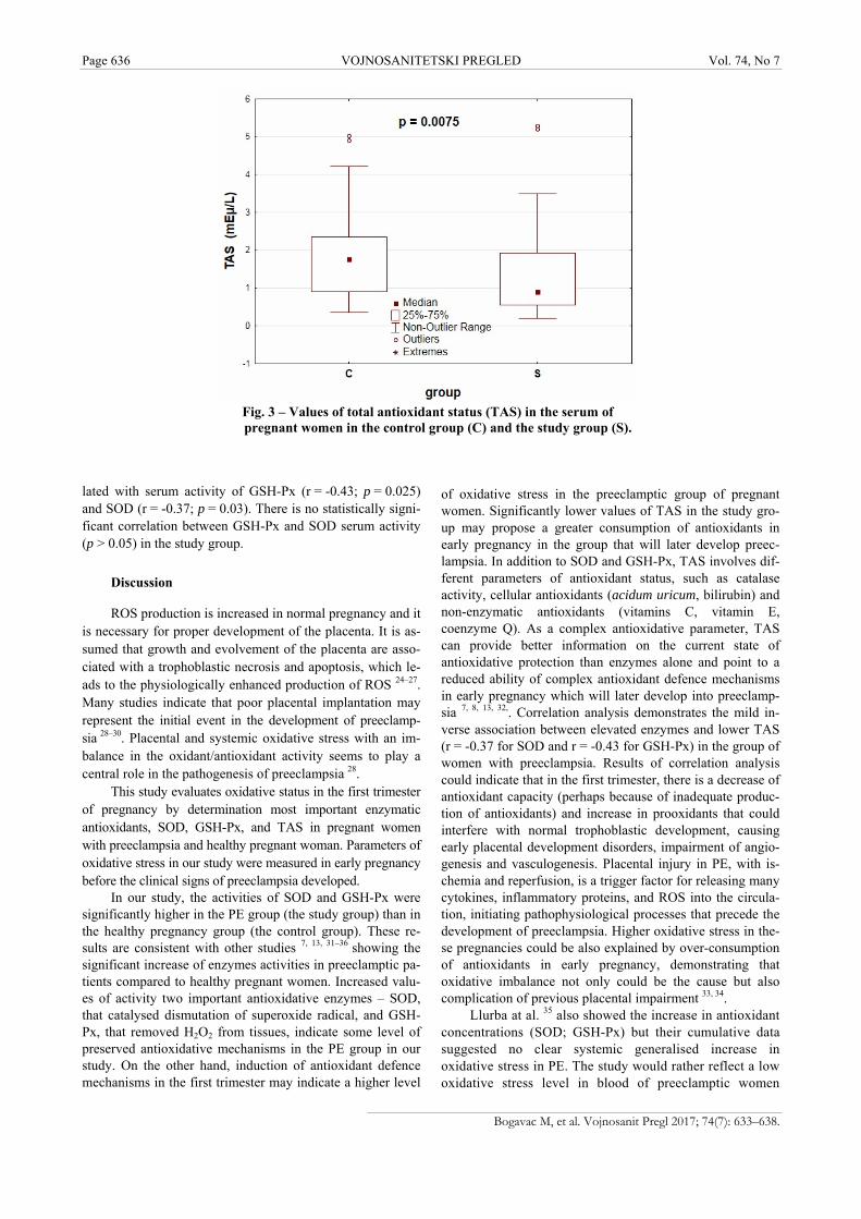
Page 636 VOJNOSANITETSKI PREGLED Vol. 74, No 7
Fig. 3 – Values of total antioxidant status (TAS) in the serum of pregnant women in the control group (C) and the study group (S).
lated with serum activity of GSH-Px (r = -0.43; p = 0.025) and SOD (r = -0.37; p = 0.03). There is no statistically signi-ficant correlation between GSH-Px and SOD serum activity (p > 0.05) in the study group.
Discussion
ROS production is increased in normal pregnancy and it is necessary for proper development of the placenta. It is as-sumed that growth and evolvement of the placenta are asso-ciated with a trophoblastic necrosis and apoptosis, which le-ads to the physiologically enhanced production of ROS 24–27. Many studies indicate that poor placental implantation may represent the initial event in the development of preeclamp-sia 28–30. Placental and systemic oxidative stress with an im-balance in the oxidant/antioxidant activity seems to play a central role in the pathogenesis of preeclampsia 28.
This study evaluates oxidative status in the first trimester of pregnancy by determination most important enzymatic antioxidants, SOD, GSH-Px, and TAS in pregnant women with preeclampsia and healthy pregnant woman. Parameters of oxidative stress in our study were measured in early pregnancy before the clinical signs of preeclampsia developed.
In our study, the activities of SOD and GSH-Px were significantly higher in the PE group (the study group) than in the healthy pregnancy group (the control group). These re-sults are consistent with other studies 7, 13, 31–36 showing the significant increase of enzymes activities in preeclamptic pa-tients compared to healthy pregnant women. Increased valu-es of activity two important antioxidative enzymes – SOD, that catalysed dismutation of superoxide radical, and GSH-Px, that removed H2O2 from tissues, indicate some level of preserved antioxidative mechanisms in the PE group in our study. On the other hand, induction of antioxidant defence mechanisms in the first trimester may indicate a higher level
of oxidative stress in the preeclamptic group of pregnant women. Significantly lower values of TAS in the study gro-up may propose a greater consumption of antioxidants in early pregnancy in the group that will later develop preec-lampsia. In addition to SOD and GSH-Px, TAS involves dif-ferent parameters of antioxidant status, such as catalase activity, cellular antioxidants (acidum uricum, bilirubin) and non-enzymatic antioxidants (vitamins C, vitamin E, coenzyme Q). As a complex antioxidative parameter, TAS can provide better information on the current state of antioxidative protection than enzymes alone and point to a reduced ability of complex antioxidant defence mechanisms in early pregnancy which will later develop into preeclamp-sia 7, 8, 13, 32,. Correlation analysis demonstrates the mild in-verse association between elevated enzymes and lower TAS (r = -0.37 for SOD and r = -0.43 for GSH-Px) in the group of women with preeclampsia. Results of correlation analysis could indicate that in the first trimester, there is a decrease of antioxidant capacity (perhaps because of inadequate produc-tion of antioxidants) and increase in prooxidants that could interfere with normal trophoblastic development, causing early placental development disorders, impairment of angio-genesis and vasculogenesis. Placental injury in PE, with is-chemia and reperfusion, is a trigger factor for releasing many cytokines, inflammatory proteins, and ROS into the circula-tion, initiating pathophysiological processes that precede the development of preeclampsia. Higher oxidative stress in the-se pregnancies could be also explained by over-consumption of antioxidants in early pregnancy, demonstrating that oxidative imbalance not only could be the cause but also complication of previous placental impairment 33, 34.
Llurba at al. 35 also showed the increase in antioxidant concentrations (SOD; GSH-Px) but their cumulative data suggested no clear systemic generalised increase in oxidative stress in PE. The study would rather reflect a low oxidative stress level in blood of preeclamptic women
Bogavac M, et al. Vojnosanit Pregl 2017; 74(7): 633–638.
Vol. 74, No 7 VOJNOSANITETSKI PREGLED Page 637
Bogavac M, et al. Vojnosanit Pregl 2017; 74(7): 633–638.
which does not represent a pathogenetically relevant pro-cess contributing to preeclampsia. Results of other investi-gators show the strong association between oxidative stress and preeclampsia, by significantly reducing the incidence of preeclampsia with multivitamin supplementation in early pregnancy 36, 37.
However, a limited number of samples in our study, the heterogeneity of disease-induced preeclampsia and the fact that we have only studied some antioxidative parameters in early pregnancy and not a wide spectrum of oxidation pro-ducts were likely the reasons why the complete pathophysiologic role of oxidative stress cannot be elucida-ted in our study. But the results of our study indicate that SOD, GSH-Px, and TAS could be included in the diagnostic algorithm for early detection of preeclampsia.
Conclusion
The results of this study suggest a higher level of oxidative stress in the first trimester of pregnancy with pre-eclampsia, which may indicate that the initiation and deve-lopment of pathophysiological processes underlying preec-lampsia start much earlier than the clinical syndrome exhibit.
Acknowledgment
The work of Z. Lozanov-Crvenković was supported by the grant No.174019 of the Ministry of Education, Sci-ence and Technological Development of the Republic of Serbia.
R E F E R E N C E S
1. Kambayashi Y, Tero-Kubota S, Yamamoto Y, Kato M, Nakano M, Yagi K, et al. Formation of superoxide anion during ferrous ion-induced decomposition of linoleic acid hydroperoxide un-der aerobic conditions. J Biochem 2003; 134(6): 903−9.
2. Gupta S, Malhotra N, Sharma D, Chandra A, Agarwal A. Oxida-tive stress and role in female infertility and assisted reproduc-tion: clinical implications. Int J Fertil Steril 2009; 2: 147−64.
3. Kashinakunti SV, Sunitha H, Gurupadappa K, Shankarprasad DS, Suryaprakash G, Ingin JB. Lipid Peroxidation and Antioxidant Status in Preeclampsia. Al Ameen J Med Sci 2010; 3(1): 38−41.
4. Djukic MM. Oxidative stress-free radicals, prooxidants and an-tioxidants. Belgrade: Mono i Manjana; 2008. (Serbian)
5. Halliwell B, Gutteridge JM. Free radicals in biology and medicine. 3rd ed. New York: Oxford University Press Inc; 1999.
6. Bogavac M, Lakic N, Simin N, Nikolic A, Sudji J, Bozin B. Bacte-rial vaginosis and biomarkers of oxidative stress in amniotic fluid. J Matern Fetal Neonatal Med 2012; 25(7): 1050−4.
7. Ruder EH, Hartman TJ, Goldman MB. Impact of oxidative stress on female fertility. Curr Opin Obstet Gynecol 2009; 21(3): 219−22.
8. Al-Gubory KH, Fowler PA, Garrel C. The roles of cellular reac-tive oxygen species, oxidative stress and antioxidants in preg-nancy outcomes. Int J Biochem Cell Biol 2010; 42(10): 1634−50.
9. Koruk M, Taysi S, Savas CM, Yilmaz O, Akcay F, Karakok M. Oxidative stress and enzymatic antioxidant status in patients with nonalcoholic steatohepatitis. Ann Clin Lab Sci 2004; 34(1): 57−62.
10. Suresh DR, Annam V, Pratibha K, Prasad BV. Total antioxidant capacity: A novel early biochemical marker of oxidative stress in HIV infected individuals. J Biomed Sci 2009; 16(1): 61.
11. Jauniaux E, Watson AL, Hempstock J, Bao YP, Skepper JN, Burton GJ. Onset of maternal arterial blood flow and placental oxida-tive stress. A possible factor in human early pregnancy failure. Am J Pathol 2000; 157(6): 2111−22.
12. Perrone S, Longini M, Bellieni CV, Centini G, Kenanidis A, De Mar-co L, et al. Early oxidative stress in amniotic fluid of pregnan-cies with Down syndrome. Clin Biochem 2007; 40(3−4): 177−80.
13. Chamy VM, Lepe J, Catalán A, Retamal D, Escobar JA, Madrid EM. Oxidative stress is closely related to clinical severity of pre-eclampsia. Biol Res 2006; 39(2): 229−36.
14. Palmieri B, Sblendorio V. Oxidative stress tests: Overview on re-liability and use. Part II. Eur Rev Med Pharmacol Sci 2007; 11(6): 383−99.
15. Gupta S, Agarwal A, Banerjee J, Alvarez JG. The role of oxidative stress in spontaneous abortion and recurrent pregnancy loss: A systematic review. Obstet Gynecol Surv 2007; 62(5): 335−47; quiz 353−4.
16. Jakovljevic B, Novakov-Mikic A, Brkic S, Bogavac M, Tomic S, Miler V. Lipid peroxidation in the first trimester of pregnancy J Ma-tern Fetal Neonatal Med 2012; 25(8): 1316−8.
17. Hutcheon JA, Lisonkova S, Joseph KS. Epidemiology of pre-eclampsia and the other hypertensive disorders of pregnancy. Best Pract Res Clin Obstet Gynaecol 2011; 25(4): 391−403.
18. Ferrazzani S, Luciano R, Garofalo S, Andrea VD, Carolis SD, Ca-rolis MP, et al. Neonatal outcome in hypertensive disorders of pregnancy. Early Hum Dev 2011; 87(6) 445−9.
19. Gaillard R, Arends LR, Steegers EA, Hofman A, Jaddoe VW. Sec-ond- and third-trimester placental hemodynamics and the risks of pregnancy complications: The Generation R Study. Am J Epidemiol 2013; 177(8): 743−54.
20. Wallis AB, Saftlas AF, Hsia J, Atrash HK. Secular trends in the rates of preeclampsia, eclampsia, and gestational hypertension, United States, 1987-2004. Am J Hypertens 2008; 21(5): 521−6.
21. Lei Q, Lv LJ, Zhang BY, Wen JY, Liu GC, Lin XH, et al. Ante-partum and post-partum markers of metabolic syndrome in pre-eclampsia. J Hum Hypertens 2011; 25(1): 11−7.
22. Ministry of Health of the Republic Serbia. Guideline for Diag-nostics and Treatment of lipid Disorders. Available from: http://www.azus.gov.rs/wp-content/uploads/2011/04/ (Serbian)
23. Ministry of Health of the Republic of Serbia. National Guide-line of good Clinical Practice – Diabetes Mellius. Available from: http//www.minzdravlja.info/downloads/2008/Sa%20Zdravlja/dokumenta/Vodici/diabetes.pdf (Serbian)
24. Paglia DE, Valentine WN. Studies on the quantitative and quali-tative characterization of erythrocyte glutathione peroxidase. J Lab Clin Med 1967; 70(1): 158−69.
25. Arthur JR, Boyne R. Superoxide dismutase and glutathione per-oxidase activities in neutrophils from selenium deficient and copper deficient cattle. Life Sci 1985; 36(16): 1569−75.
26. Erel O. A novel automated direct measurement method for to-tal antioxidant capacity using a new generation, more stable ABTS radical cation. Clin Biochem 2004; 37(4): 277−85.
27. Yang X, Guo L, Li H, Chen X, Tong X. Analysis of the original causes of placental oxidative stress in normal pregnancy and pre-eclampsia: a hypothesis. J Matern Fetal Neonatal Med 2012; 25(7): 884−8.
Page 638 VOJNOSANITETSKI PREGLED Vol. 74, No 7
28. George EM, Granger JP. Endothelin: key mediator of hyperten-sion in preeclampsia. Am J Hypertens 2011; 24(9): 964−9.
29. Rosta K, Molvarec A, Enzsöly A, Nagy B, Rónai Z, Fekete A, et al. Association of extracellular superoxide dismutase (SOD3) Ala40Thr gene polymorphism with pre-eclampsia complicated by severe fetal growth restriction. Eur J Obstet Gynecol Re-prod Biol 2009; 142(2): 134−8.
30. Sharma JB, Sharma A, Bahadur A, Vimala N, Satyam A, Mittal S. Oxidative stress markers and antioxidant levels in normal pregnancy and pre-eclampsia. Int J Gynaecol Obstet 2006; 94(1): 23−7.
31. Wruck CJ, Huppertz B, Bose P, Brandenburg L, Pufe T, Kadyrov M. Role of a fetal defence mechanism against oxidative stress in the aetiology of preeclampsia. Histopathology 2009; 55(1): 102−6.
32. Genc H, Uzun H, Benian A, Simsek G, Gelisgen R, Madazli R, et al. Evaluation of oxidative stress markers in first trimester for assessment of preeclampsia risk. Arch Gynecol Obstet 2011; 284(6): 1367−73.
33. Hansson SR, Nääv Å, Erlandsson L. Oxidative stress in pree-clampsia and the role of free fetal hemoglobin. Front Physiol 2015; 5: 516.
34. Lissette C, Prada CE, Medina C, Lopez M. Endothelial dysfunc-tion and preeclampsia: Role of oxidative stress. Front Physiol 2014; 5: 372.
35. Llurba E, Gratacós E, Martín-Gallán P, Cabero L, Dominguez C. A comprehensive study of oxidative stress and antioxidant status in preeclampsia and normal pregnancy. Free Radic Biol Med 2004; 37(4): 557−70.
36. Cunninghm FG, Lenevo KJ, Bloom SL, Hauth JC, Rouse DJ, Spong CY. Pregnancy hypertension (Chapter 34). In: Cunninghm FG, Lenevo KJ, Bloom SL, Hauth JC, Rouse DJ, Spong CY, editors. Williams Obstretics. 23rd ed. New York: McGraw-Hill; 2010. p. 706−56.
37. Vanderlelie J, Scott R, Shibl R, Lewkowicz J, Perkins A, Scuffham PA. First trimester multivitamin/mineral use is associated with reduced risk of pre-eclampsia among overweight and obese women. Matern Child Nutr 2014; doi: 10.1111/mcn.12133. (In Press).
Received on May 17, 2015. Revised on December 20, 2015.
Accepted on December 24, 2015. Online First September, 2016.
Bogavac M, et al. Vojnosanit Pregl 2017; 74(7): 633–638.
Vojnosanit Pregl 2017; 74(7): 639–643. VOJNOSANITETSKI PREGLED Page 639
Correspondence to: Predrag Marić, Military Medical Academy, Clinic of Urology, Crnotravska 17, 11 000 Belgrade, Serbia. E-mail: [email protected]
O R I G I N A L A R T I C L E UDC: 616.61-006-089-06 https://doi.org/10.2298/VSP151017254M
Complications of radical and partial nephrectomy for renal cell carcinoma up to 7 cm
Komplikacije radikalne i parcijalne nefrektomije kod karcinoma bubrežnih ćelija manjih od 7 cm
Predrag Marić*, Mirko Jovanović*, Novak Milović*†, Dušica Stamenkovi憇,
Branko Košević*†, Predrag Aleksić*†, Snežana Cerovi憧, Aleksandar Spasić*, Dejan Simić*, Jelena Rašković║
Military Medical Academy, *Clinic of Urology, ‡Clinic of Anesthesiology and Intensive Care, §Institute for Pathology and Forensic Medicine, Belgrade, Serbia; University of
Defence, †Faculty of Medicine of the Military Medical Academy, Belgrade, Serbia; University of Belgrade, School of Economics, ║Department of Statistics, Belgrade, Serbia
Abstract Background/Aim. Renal cell carcinoma (RCC) is the third most frequent urological carcinoma. Radical nephrectomy (RN) is considered as the gold standard in the treatment of localized RCC, but recently the use of minimally invasive techniques are more frequently used. The aim of this study is to determine is there a difference in the incidence of compli-cations in the group of patients treated by RN and partial nephrectomy (PN) for renal cell carcinoma up to 7 cm. Me-thods. The retrospective study included the analysis of the medical history of patients surgically treated in the six years period. The inclusion criteria were RCC size up to 7 cm and no detectable metastasis. The exclusion criterion was the presence of a bilateral tumor. Intraoperative and early post-operative complications were followed-up. The Clavien-Dindo grade system was used for classification of surgical complications. Results. In six years period 481 (76.35%) rad-ical transperitoneal nephrectomies and 149 (23.65%) partial nephrectomies were performed. Bilateral RCCs were verified
in 2.06% (13/630), an initial metastatic disease in 15.8% (100/630) and lymph node involvement in 7.14% (45/630) of the patients and their data were not included in analysis. Therefore, data from 120 patients with RN and 97 patients with PN who fulfill inclusion criteria were analyzed. Compli-cations were recorded in 29.5% (64/217) of patients. Signifi-cantly less patients had complications in the RN group [22.5 % (27/120)] compared to the PN group [38.1% (37/97)] (p = 0.006). These complications were mostly grade I and II. Complications grade III and IV were only present in the group of patients treated by PN. Conclusion. Based on our data in selected patients with renal cell carcinoma in stage T1, PN is a proper and safe choice. The patient must be involved in making the definitive decision of modalities of surgical treatment. Keywords: kidney neoplasms; urologic surgical procedures; intraoperative complications; postoperative period; postoperative complications.
Apstrakt Uvod/Cilj. Karcinom renalnih ćelija (real cell carcinoma – RCC) treći je najčešći urološki karcinom. Radikalna nefre-ktomija (RN) smatra se zlatnim standardom u terapiji loka-lizovanog RCC, ali u novije vreme sve češće se koriste mi-nimalno invazivne tehnike. Cilj ove studije bio je da se de-finiše razlika u incidenci intraoperativnih i postoperativnih komplikacija kod bolesika podvrgnutih RN ili parcijalnoj nefrektomiji (PN) zbog RCC veličine do 7 cm. Metode. Retrospektivno su analizirani podaci iz medicinske doku-mentacije bolesnika operisanih u šestogodišnjem vremen-skom periodu. Kriterijumi za uključivanje u studiju bili su
tumor bubrega veličine do 7 cm i odsustvo metastaza. Kri-terijum za isključivanje iz studije bilo je prisustvo bilateral-nog tumora. Praćene su intraoperativne i rane postoperati-vne komplikacije. Clavien-Dindo sistem klasifikacije ko-rišćen je za klasifikaciju komplikacija. Rezultati. U perio-du od šest godina urađeno je 481 (76.35%) RN i 149 (23.65%) PN. Bilateralni tumori urađeni su kod 2.06% (13/630) bolesnika, inicijalna metastatska bolest kod 15.87% (100/630) i zahvaćenost limfnih čvorova kod 7.14% (45/630) bolesnika i njihovi podaci nisu uzeti u analizu. Analizom je obuhvaćeno 120 bolesnika sa RN i 97 sa PN koji su ispunjavali kriterijume za uključenje u studi-ju. Komplikacije su zabeležene kod ukupno 29.5%
Page 640 VOJNOSANITETSKI PREGLED Vol. 74, No 7
Marić P, et al. Vojnosanit Pregl 2017; 74(7): 639–643.
Table 1The Clavien-Dindo Classification of Surgical Complications 15, 16
Complication grade
Definition Severity
I Any deviation from the normal postoperative course without the need for pharmacological treatment or surgical, endoscopic and radiological interventions. Surgical site infections.
minor
II Requiring pharmacological treatment with drugs other than such allowed for grade I complica-tions. Blood transfusions and total parenteral nutrition are also included.
minor
III Requiring surgical, endoscopic or radiological intervention.
major
IV Life-threatening complication. major
V Death major
(64/217) bolesnika. Značajno niži broj bolesnika imao je komplikacije u RN grupi [22.5 % (27/120)] u poređenju sa PN grupom [38.1% (37/97)] (p = 0,006). Komplikacije su uglavnom bile gradusa I i II. Komplikacije gradusa III i IV zabeležene su jedino u PN grupi. Zaključak. Na osnovu analize podataka naših bolesnika, PN je dobar i siguran iz-bor kod RCC veličine do 7 cm, T1 stadijuma. Bolesnik
mora biti uključen u donošenje odluke o hirurškom moda-litetu koji će biti primenjen. Ključne reči: bubreg, neoplazme; hirurgija, urološka, procedure; intraoperativne komplikacije; postoperativni period; postoperativne komplikacije.
Introduction
Renal cell carcinoma (RCC) is the third most frequent uro-logical carcinoma 1. Radical nephrectomy was for decades con-sidered as the golden standard in the curative treatment of loca-lized renal cell carcinoma (RCC) 2–4. Recently preservation surgery of renal parenchyma and the use of minimally invasive techniques are more frequently used in the treatment of RCC.
Partial nephrectomy (PN) is a surgical resection of the tu-mor in total with a remained part of normal surrounding renal parenchyma 5. Today PN is performed not only for classic indi-cations as a tumor of the solitary kidney and bilateral tumors but also in patients with a normal contralateral kidney 6, 7. The intro-duction of modern diagnostic techniques, as the use of abdo-minal ultrasound and computer tomography scans led to an increased number of newly diagnosed incidental renal tu-mors, which are smaller and asymptomatic, with a better prognosis than symptomatic tumors of the same size and cli-nical stage 8–10.
Open PN is accepted as a golden standard, mainly in the treatment of younger patients with incidentally diagnosed renal tumors smaller than 4 cm 5. Based on literature data, this surgi-cal technique has excellent results, similar to RN and generally is proposed as an option in the treatment of patients with stage T1 11, 12. The patients have an overall better survival rate and preservation of renal function compared to patients treated by RN 13. The benefit of renal function preservation must exceed than intra and postoperative complications in RN 14.
The aim of this study was to determine the incidence of complications in the group of patients with RCC up to 7 cm treated by radical and partial nephrectomy.
Methods
This retrospective study included the analysis of the medical history of patients surgically treated in the period of 2006 until the end of 2012. The inclusion criteria were renal tu-mor size up to 7 cm and no detectable metastasis. The size of the tumor and the absence of metastasis were determined by multis-lice computer tomography (MSCT). The exclusion criterion for this study was the presence of a bilateral tumor.
Intraoperative and early postoperative complications were followed up. The Clavien-Dindo grade system was used for classification of surgical complications (Table 1) 15, 16. Acute re-nal failure was defined as an increase of creatinine level more than 50% of the preoperative level or need for hemodialysis 17.
The data are presented as median (range) or mean ± SD. Pearson's χ² and Likelihood ratio χ² -test, were used for stati-stical analysis. For intergroup comparisons, Crosstabs statisti-cal procedure was used to evaluate differences between gro-ups. P < 0.05 was considered statistically significant.
Results
In this retrospective study we analyzed data of 630 pati-ents diagnosed with RCC and underwent surgical treatment in the period from 2006 till the end of 2012, 356 patients were in stage T1. In total 481 (76.35%) patients radical transperitoneal nephrectomy and 149 (23.65%) patients PN were perfor-med.The initial metastatic disease was verified in 15.87% (100/630) lymph node involvement in 7.14% (45/630) of the patients' bilateral tumors in 2.06 % (13/630) and their data were not included in the analysis of patients.
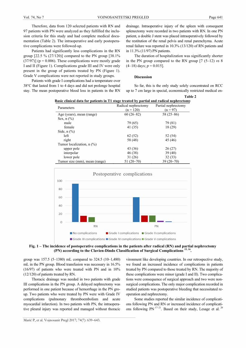
Vol. 74, No 7 VOJNOSANITETSKI PREGLED Page 641
Marić P, et al. Vojnosanit Pregl 2017; 74(7): 639–643.
Table 2 Basic clinical data for patients in T1 stage treated by partial and radical nephrectomy
Parameters Radical nephrectomy
(n = 120) Partial nephrectomy
(n = 97) Age (years), mean (range) 60 (26–82) 58 (25–86) Sex, n (%)
male female
79 (65) 41 (35)
79 (81) 18 (29)
Side, n (%) left right
62 (52) 58 (48)
52 (54) 45 (46)
Tumor localization, n (%) upper pole interpolar lower pole
43 (36) 46 (38) 31 (26)
26 (27) 39 (40) 32 (33)
Tumor size (mm), mean (range) 51 (20–70) 39 (20–70)
Fig. 1 – The incidence of postoperative complications in the patients after radical (RN) and partial nephrectomy
(PN) according to the Clavien-Dindo Classification of Surgical Complications 15, 16.
Therefore, data from 120 selected patients with RN and 97 patients with PN were analyzed as they fulfilled the inclu-sion criteria for this study and had complete medical docu-mentation (Table 2). The intraoperative and early postopera-tive complications were followed-up.
Patients had significantly less complications in the RN group [22.5 % (27/120)] compared to the PN group [38.1% (37/97)] (p = 0.006). These complications were mostly grade I and II (Figure 1). Complications grade III and IV were only present in the group of patients treated by PN (Figure 1). Grade V complications were not reported in study groups.
Patients with grade I complications had a temperature over 38°C that lasted from 1 to 4 days and did not prolonge hospital stay. The mean postoperative blood loss in patients in the RN
group was 157.5 (5–1380) mL compared to 324.5 (10–1,480) mL in the PN group. Blood transfusion was necessary in 16.5% (16/97) of patients who were treated with PN and in 10% (12/120) of patients treated by RN.
Thoracic drainage was needed in two patients with grade III complications in the PN group. A delayed nephrectomy was performed in one patient because of hemorrhage in the PN gro-up. Two patients who were treated by PN were with Grade IV complications (pulmonary thromboembolism and acute myocardial infarction). In two patients with PN, the intraopera-tive pleural injury was reported and managed without thoracic
drainage. Intraoperative injury of the spleen with consequent splenectomy were recorded in two patients with RN. In one PN patient, a double J stent was placed intraoperatively followed by the restitution of the renal pelvis and renal parenchyma. Acute renal failure was reported in 10.3% (13/120) of RN patients and in 11.3% (11/97) PN patients.
The duration of hospitalization was significantly shorter in the PN group compared to the RN group [7 (5–12) vs 8 (4–18) days; p = 0.015].
Discussion
So far, this is the only study solely concentrated on RCC up to 7 cm large in special, economically restricted medical en-
vironment like developing countries. In our retrospective study, we found an increased incidence of complications in patients treated by PN compared to those treated by RN. The majority of these complications were minor (grade I and II). Two complica-tions were consequence of surgical approach and two were non-surgical complications. The only major complication recorded in studied patients was postoperative bleeding that necessitated re-operation and nephrectomy.
Some studies reported the similar incidence of complicati-ons following PN and RN or increased incidence of complicati-ons following PN 17–21. Based on their study, Lesage et al. 20
Page 642 VOJNOSANITETSKI PREGLED Vol. 74, No 7
Marić P, et al. Vojnosanit Pregl 2017; 74(7): 639–643.
comparing intraoperative complications found that the inci-dence of complications was increased in the PN group, but this increase was not statistically significant. The result of a phase III randomized trial comparing PN and RN in the trea-tment of renal masses up to 5 cm, has shown a slightly hig-her incidence of intraoperative complications in the PN gro-up, primarily considering bleeding and development of a urinary fistula. They concluded that PN can be used safely, with just slightly more complications compared to RN 21.
The risk of urinary fistula in published studies ranges from 1.4% to 17.4% 22. In our analyzed group of patients, no urinary fistulas were recorded. This can be explained by adequate selection of patients and surgical technique. In one case when there was reasonable doubt of possible postopera-tive urinary fistula development due to insufficient closure of the calicial system of the kidney a double J stent was intro-duced intraoperatively.
The incidence of blood transfusion was higher in the PN group (16.49%), compared to the RN group where it was necessary for only 10% of patients. Shvarts et al. 23 data showed that 18% of patients treated by RN and up to 30% of patients treated by PN needed a blood transfusion which is higher than in our study.
The incidence of re-operations following PN was 1.03%, without reopeartions in the RN group. Literature data shows
that reoperation rate is very low after RN (0–3.1%) 22–24. Lau et al. 18, found that one out of 164 patients in the PN group had undergone nephrectomy after preservation surgery due to pos-toperative bleeding. In three patients with urinary fistula, only one patient underwent nephrectomy because of a perirenal ab-scess. In the RN group, there was no need for reopeartions just like in our study 18.
There is a higher incidence of complications in patients treated by PN. The majority of these complications are minor and are presented as elevated body temperature and blood loss that needed blood transfusions. In both groups, compli-cations were solved in a conservative manor. Only in one pati-ent, a reoperation was necessary due to postoperative blee-ding.
Conclusion
Based on our data in selected patients with RCC up to 7 cm (stage T1), PN can be a proper and safe choice. If the urologist has no experience in performing PN and there is a possibility for complications the patient should be referred to a specialized center. A patient must be involved in making the definitive decision of modalities of surgical treatment. However, multicenter studies are needed for justification of our experience.
R E F E R E N C E S
1. Jemal A, Siegel R, Xu J, Ward E. Cancer statistic 2010. CA Can-cer J Clin 2010; 60(5): 277−300.
2. Van Poppel H, Da Pozzo L, Albrecht W, Matveev V, Bono A, Borkowski A,et al. A prospective, randomised EORTC inter-group phase 3 study comparing the oncologic outcome of elective nephron-sparing surgery and radical nephrectomy for low-stage renal cell carcinoma. Eur Urol 2011; 59(4): 543−52.
3. Robson CJ. Radical nephrectomy for renal cell carcinoma. J Urol 1963; 89: 37−41.
4. Ljunberg B, Almdari FI, Holmberg G, Granfors T, Duchek M. Radi-cal nephrectomy is stil preferable in the treatment of localized renal cell carcinoma. Eur Urol 1998; 33(1): 79−85.
5. Cozar JM, Tallada M. Open partial nephrectomy in renal can-cer: A feasibile gold standard technique in all hospitals. Adv Urol 2008; 916463.
6. Shuch B, Lam JS, Belldegrun AS. Open partial nephrectomy for the treatment of renal cell carcinoma. Curr Urol Rep 2006; 7(1): 31−8.
7. Novick AC, Campbell SC. Tumores renales. In: Walsh PC, Retik AB, Vaughan ED, Wein AJ, editors. Campabell Urology. Bue-nos Aires, Spain: Editorial Panamericana; 2005. p. 2911−79.
8. Jayson M, Sanders H. Increased incidence of serendipitously dis-covered renal cell carcinoma.Urology 1998; 51(2): 203−5.
9. Fergany AF, Hafez KS, Novick AC. Long-term results of nephron sparing surgery for localized renal cell carcinoma: 10-year follow up. J Urol 2000; 163(2): 442−5.
10. Moll V, Becht E, Ziegler M. Kidney preserving surgery in renal cell tumors: Indications, technicues and results in 152 patinets. J Urol. 1993; 150(2 Pt 1): 319−23.
11. Ljungberg B, Cowan NC, Hanbury DC, Hora M, Kuczyk MA, Merseburger AS, et al. EAU guidelines on renal cell carcinoma: the 2010 update. Eur Urol 2010; 58(3): 398−406.
12. Peycelon M, Hupertan V, Comperat E, Renard-Penna R, Vaessen C, Conort P, et al. Long-term outcomes after nephron sparing sur-gery for renal cell carcinoma larger than 4 cm. J Urol 2009; 181(1): 35−41.
13. Huang WC, Levey AS, Serio AM, Snyder M, Vickers AJ, Raj GV, et al. Chronic kidney disease after nephrectomy in patients with renal cortical tumours: a retrospective cohort study. Lan-cet Oncol 2006; 7(9): 735−40.
14. Permpongkosol S, Link RE, Su LM, Romero FR, Bagga HS, Pav-lovich CP, et al. Complications of 2,775 urological laparoscopic procedures: 1993 to 2005. J Urol 2007; 177(2): 580−5.
15. Xu H, Ding Q, Jiang HW. Fewer complications after laparo-scopic nephrectomy as compared to the open procedure with the modified Clavien classification system-a retrospective anal-ysis from southern China. World J Surg Oncol 2014; 12: 242.
16. Yang CM, Chung HJ, Huang YH, Lin TP, Lin AT, Chen KK. Standardized analysis of laparoscopic and robotic-assisted par-tial nephrectomy complications with Clavien classification. J Chin Med Assoc 2014; 77(12): 637−41.
17. Stephenson AJ, Hakimi AA, Snyder ME, Russo P. Complications of radical and partial nephrectomy in a large contemporary co-hort. J Urol 2004; 171(1): 130−4.
18. Lau WK, Blute ML, Weaver AL, Torres VE, Zincke H. Matched comparison of radical nephrectomy vs nephron-sparing sur-gery in patients with unilateral renal cell carcinoma and a normal contralateral kidney. Mayo Clin Proc 2000; 75(12): 1236−42.
19. Corman JM, Penson DF, Hur K, Khuri SF, Daley J, Henderson W, et al. Comparison of complications after radical and partial nephrectomy: results from the National Veterans Administra-tion Surgical Quality Improvement Program. BJU Int 2000; 86(7): 782−9.
Vol. 74, No 7 VOJNOSANITETSKI PREGLED Page 643
Marić P, et al. Vojnosanit Pregl 2017; 74(7): 639–643.
20. Lesage K, Joniau S, Fransis K, Van Poppel H. Comparison be-tween open partial and radical nephrectomy for renal tumours: perioperative outcome and health-related quality of life. Eur Urol 2007; 51(3): 614−20.
21. Van Poppel H, Da Pozzo L, Albrecht W, Matveev V, Bono A, Borkowski A, et al. A prospective, randomised EORTC inter-group phase 3 study comparing the oncologic outcome of elective nephron-sparing surgery and radical nephrectomy for low-stage renal cell carcinoma. Eur Urol 2011; 59(4): 543−52.
22. Uzzo RG, Novick AC. Nephron sparing surgery for renal tu-mors: indications, techniques and outcomes. J Urol 2001; 166(1): 6−18.
23. Shvarts O, Tsui KH, Smith RB, Kernion JB, Belldegrun A. Blood loss and the need for transfusion in patients who undergo par-tial or radical nephrectomy for renal cell carcinoma. J Urol 2000; 164(4): 1160−3.
24. Campbell SC, Novick AC, Streem SB, Klein E, Licht M. Complica-tions of nephron sparing surgery for renal tumors. J Urol 1994; 151(5): 1177−80.
Received on October 17, 2015. Revised on December 27, 2015.
Accepted on January 27, 2016. Online First September, 2016.
Page 644 VOJNOSANITETSKI PREGLED Vojnosanit Pregl 2017; 74(7): 644–651.
Correspondence to: Aleksandra Čolović, University of Belgrade, Faculty of Dentistry, Clinic for Pediatric and Preventive Dentistry, Dr Subotića 11, 11 000 Belgrade, Serbia. E-mail: [email protected]
O R I G I N A L A R T I C L E UDC: 616-053.2:616-018.2-056.7]:616.31-053.2 https://doi.org/10.2298/VSP151023159C
Oral health status in children with inherited dystrophic epidermolysis bullosa
Stanje oralnog zdravlja dece obolele od nasledne distrofičke bulozne epidermolize
Aleksandra Čolović*, Olivera Jovičić*, Radoje Stevanović†, Mirjana Ivanović*
University of Belgrade, Faculty of Dentistry, *Clinic for Pediatric and Preventive Dentistry, Belgrade, Serbia; University of Travnik, Faculty of Pharmacy and Health,
†Department of Dentistry, Travnik, Bosnia and Herzegovina
Abstract Background/Aim. Epidermolysis bullosa is a group of rare, genetic connective tissue diseases that cause blisters in the skin and mucosal membranes. The aim of this study was to evaluate the oral health status of patients with epidermolysis bullosa dystrophica and level of knowledge and opinion of parents about the implementation of preventive measaures and quality of dental care of affected children. Methods. This study included a group of 17 patients from Serbia suffering from dystrophic epidermolysis bullosa and matched control group. Dental caries status was assessed using the Klein-Palmer index. Oral hygiene status was verified with oral hygiene indices, simplified plaque index, and calculus index as described by Green and Vermillion. The gingiva was assessed as healthy or inflamed (gingivitis) on the basis of any changes in color, shape, size and surface texture. The condition of oral mucosa has been registered on the basis of inspection of the lips, tongue, a floor of the mouth, mouth vestibule and palate. The level of knowledge and the impressions of parents about the application of preventive measures were investigated through two
questionnaires specifically designed for this study. Results. In both dentitions, there was the highest percentage of cari-es teeth. In primary dentition average value of the modified plaque index was 1.4 ± 1.14 and modified calculus was 0.7 ± 1. On permanent teeth average plaque index was 2 ± 0.4, and average calculus 1.6 ± 0.6. Statistically, significant hig-her values were found in permanent dentition in percentage distribution of decayed, missing, filled teeth and also for plaque and calculus indices between affected children and the control group. Most common findings on mucosa were microstomia (76.5%) and ankyloglossia (88.2%). Conclusion. The absence of protocol between the treating physician and the dentist and not sufficiently informed pa-rents are leading to inadequate dental care. The implementation of preventive measures is of most importance to decrease the risk of severe complications that are difficult to be managed. Keywords: epidermolysis bullosa; child; oral health; preventive dentistry; surveys and questionnaires; dental caries; dental plaque index; periodontal diseases.
Apstrakt Uvod/Cilj. Bulozna epidermoliza je grupa retkih, genetski predisponiranih bolesti vezivnog tkiva koja se karakteriše formiranjem bula (mehurova) u koži i mukoznim mem-branama. Cilj rada bio je da se ispita stanje oralnog zdravlja dece obolele od distrofične bulozne epidermolize, kao i nivo znanja i mišljenje roditelja o primeni preventivnih mera i kvalitetu stomatološke zaštite obolele dece. Metode. U studiju je bilo uključeno 17 bolesnika sa distrofičnom bu-loznom epidermolizom iz Srbije i odgovarajuća kontrolna grupa. Stanje zuba ustanovljeno je Klein Palmerovim indek-som karijesnih, ekstrakovanih plombiranih zuba – KEP indeksom. Stanje oralne higijene analizirano je indeksima oralne higijene (pojednostavljeni plak indeks i indeks zub-nog kamenca po Green-Vermillionu). Stanje gingive je na
osnovu inspekcije ocenjivano kao zdrava gingiva ili gingivi-tis, u zavisnosti od toga da li su joj bili promenjeni boja, ob-lik, veličina ili struktura. Stanje oralne sluzokože evidenti-rano je na osnovu inspekcije usana, jezika, poda usne duplje, vestibuluma, plika i frenuluma, kao i mekog i tvrdog nepca. Nivo znanja i utisci roditelja o primeni preventivnih mera i kvalitetu stomatološke zaštite dece obolele od bolozne epi-dermolize, ispitivani su kroz dva upitnika specijalno diza-jnirana za potrebe ove studije. Rezultati. U obe denticije bilo je najviše karijesno obolelih zuba. Za mlečnu denticiju prosečan modifikovani plak indeks iznosio je 1,4 ± 1,14, a modifikovani kalkulus indeks 0,7 ± 1. U stalnoj denticiji vrednosti prosečnog plak indesa i kalkulus indeksa redom su bile 2 ± 0,4 i 1,6 ± 0,6. Utvrđena je statistički značajna razlika u procentualnoj zastupljenosti karijesnih, ekstrahovanih i plombiranih zuba stalne denticije između
Vol. 74, No 7 VOJNOSANITETSKI PREGLED Page 645
Čolović A, et al. Vojnosanit Pregl 2017; 74(7): 644–651.
dece obolele od bulozne epidermolize i kontrolne grupe, kao i za vrednosti plak i kalkulus indeksa. Najčešće promene oralne sluzokože bile su mikrostomija (76,5%) i ankiloglosija (88,2%). Zaključak. Odsustvo protokolarne saradnje iz-među ordinirajućeg lekara i stomatologa, kao i nedovoljna informisanost roditelja, glavni su razlozi lošeg stanja oralnog zdravlja. Primena preventivnih mera je od najvećeg značaja
kako bi se sprečio nastanak komplikacija koje je teško sanirati kod ove grupe bolesnika. Ključne reči: epidermoliza, bulozna; deca; usta, zdravlje; stomatologija, preventivna; ankete i upitnici; zub, karijes; zub, indeks plaka; periodontalne bolesti.
Introduction
Epidermolysis bullosa (EB) hereditaria is a multisystem disease that is characterized by extreme fragility of the skin and mucous membranes, on which blisters and erosions appear spontaneously or following minor trauma 1. It is a rare disease, with a frequency of 1 : 17,000, and is transmitted through au-tosomal dominant and recessive inheritance 2, 3. Inherited EB is classified into four major types: simplex (EBS), junctional (JEB), dystrophic (DEB) and Kindler Syndrome 4–7. DEB has two subtypes: dominant DEB (DDEB) and recessive DEB (RDEB). The worst type of EB is RDEB with the frequency of 1 : 1,000,000. Healing in patients with DEB is followed by scarring 4. Diagnosis is made after birth clinically using accompanying laboratory analyses 8.
There are numerous bodily systems and oral manifesta-tions. Besides blisters and erosions on skin and mucosa, ot-her extracutaneous involvements can be found on the gastro-intestinal and in the genitourinary tract as well as within the cardiovascular and musculoskeletal system. The most diffi-cult complication is squamous cell cancer 6, 9.
Common oral symptoms include blisters and erosions on mucosa, microstomia, ankyloglossia, loss of the vestibular spa-ce, absence of lingual papillae and palatal folds, enamel hypoplasia and early appearance of caries on both deciduous and permanent teeth 1, 6, 10–12. Squamous cell carcinoma found on intraoral sites, mostly developing where tissue has experienced chronic ulcerations and repeated epithelialization. The carcino-mas grow rapidly and give off metastases very early 13.
The aim of this study was to determine the oral health status of DDEB patients by performing the following: dental caries assessments, oral hygiene assessments and gingival health assessments. Further assessments were made regar-ding the level of knowledge and opinions of parents about the implementation of preventive measures and also on the quality of dental care of EB children.
Methods
This study included a group of 17 patients from Serbia (9 males and 8 females) suffering from DEB, aged 1-21. Two of these patients were adults with DDEB and the remai-ning 15 were children with RDEB. All patients were examined at the Clinic for Pediatric and Preventive Dentistry, Faculty of Dental Medicine, the University of Belgrade. Healthy controls were correspondingly matched individually by sex and age (± 6 months) in consideration of the fact that this is a rare disease with severe symptoms and in order to obtain results that were as precise as possible.
An oral health status examination was conducted using ar-tificial lighting, dental mirrors, and probes in accordance with criteria listed by the World Health Organization (WHO) 14.
Dental caries statuses were assessed using the Klein–Palmer index for both primary (dmft1) and permanent dentition (DMFT) 15. The condition of oral mucosa was eva-luated on the basis of an inspection of the lips, tongue, a flo-or of the mouth, oral vestibule and the frenulum as well as the soft and hard palates. The mouth opening capacity was measured as the distance between the lower and upper lips vermilion lines when the patient opens the mouth as widely as possible 15. The gingiva was assessed as healthy or infla-med (gingivitis) on the basis of any changes in color, shape, size and surface texture.
For the oral hygiene status in cases of primary dentition, a modified index was implemented (dental plaque index and cal-culus index) 16. The presence of plaque and calculus was evalua-ted for the buccal surfaces of six representative teeth (the four second primary molars, the upper right central incisor and the lower left central incisor). For permanent dentition, a simplified plaque index and calculus index as described by Green and Vermillion 17 was applied (Oral Hygiene Index – OHI-S).
The level of knowledge and the impressions of parents about preventive measures in dentistry and the quality of dental care of EB children were investigated through two questionnaires specifically designed for this study (Tables 1 and 2).
Descriptions of categorical data were done using abso-lute and relative numbers (percentages), and for numerical data by arithmetic means and standard deviation. Frequencies were compared using the χ2 test (if numerical conditions were not met by Fisher exact test) whilst numeri-cal data comparison was made with the t-test and the Mann-Whitney U test (with and without normal distribution respectively). All applied statistical methods were considered significant if p ≤ 0.05. Statistical analyses were done in IBM SPSS Statistics version 21.0 software (IBM, USA).
Results
Oral health status
Dental caries status
Among the twelve children with deciduous dentition, ten (83.3%) had affected teeth and two children (16.7%) had all caries-free teeth. From a total of 165 teeth, 95 (57.6%) were healthy and those listed with caries or as extractions or
1The decyed, missing, filled teeth index (DMFT) when
written in lowercase letters-dmft, is a variation that is applied to the primary dentition.
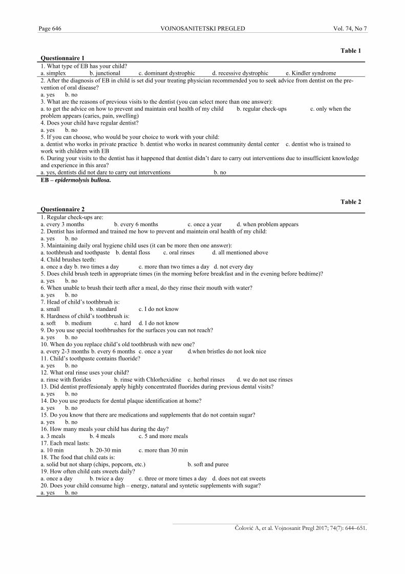
Page 646 VOJNOSANITETSKI PREGLED Vol. 74, No 7
Čolović A, et al. Vojnosanit Pregl 2017; 74(7): 644–651.
Table 1
Questionnaire 1 1. What type of EB has your child? a. simplex b. junctional c. dominant dystrophic d. recessive dystrophic e. Kindler syndrome 2. After the diagnosis of EB in child is set did your treating physician recommended you to seek advice from dentist on the pre-vention of oral disease? a. yes b. no 3. What are the reasons of previous visits to the dentist (you can select more than one answer): a. to get the advice on how to prevent and maintain oral health of my child b. regular check-ups c. only when the problem appears (caries, pain, swelling) 4. Does your child have regular dentist? a. yes b. no 5. If you can choose, who would be your choice to work with your child: a. dentist who works in private practice b. dentist who works in nearest community dental center c. dentist who is trained to work with children with EB 6. During your visits to the dentist has it happened that dentist didn’t dare to carry out interventions due to insufficient knowledge and experience in this area? a. yes, dentists did not dare to carry out interventions b. no EB – epidermolysis bullosa.
Table 2 Questionnaire 2 1. Regular check-ups are: a. every 3 months b. every 6 months c. once a year d. when problem appears 2. Dentist has informed and trained me how to prevent and maintein oral health of my child: a. yes b. no 3. Maintaining daily oral hygiene child uses (it can be more then one answer): a. toothbrush and toothpaste b. dental floss c. oral rinses d. all mentioned above 4. Child brushes teeth: a. once a day b. two times a day c. more than two times a day d. not every day 5. Does child brush teeth in appropriate times (in the morning before breakfast and in the evening before bedtime)? a. yes b. no 6. When unable to brush their teeth after a meal, do they rinse their mouth with water? a. yes b. no 7. Head of child’s toothbrush is: a. small b. standard c. I do not know 8. Hardness of child’s toothbrush is: a. soft b. medium c. hard d. I do not know 9. Do you use special toothbrushes for the surfaces you can not reach? a. yes b. no 10. When do you replace child’s old toothbrush with new one? a. every 2-3 months b. every 6 months c. once a year d.when bristles do not look nice 11. Child’s toothpaste contains fluoride? a. yes b. no 12. What oral rinse uses your child? a. rinse with florides b. rinse with Chlorhexidine c. herbal rinses d. we do not use rinses 13. Did dentist proffesionaly apply highly concentrated fluorides during previous dental visits? a. yes b. no 14. Do you use products for dental plaque identification at home? a. yes b. no 15. Do you know that there are medications and supplements that do not contain sugar? a. yes b. no 16. How many meals your child has during the day? a. 3 meals b. 4 meals c. 5 and more meals 17. Each meal lasts: a. 10 min b. 20-30 min c. more than 30 min 18. The food that child eats is: a. solid but not sharp (chips, popcorn, etc.) b. soft and puree 19. How often child eats sweets daily? a. once a day b. twice a day c. three or more times a day d. does not eat sweets 20. Does your child consume high – energy, natural and syntetic supplements with sugar? a. yes b. no
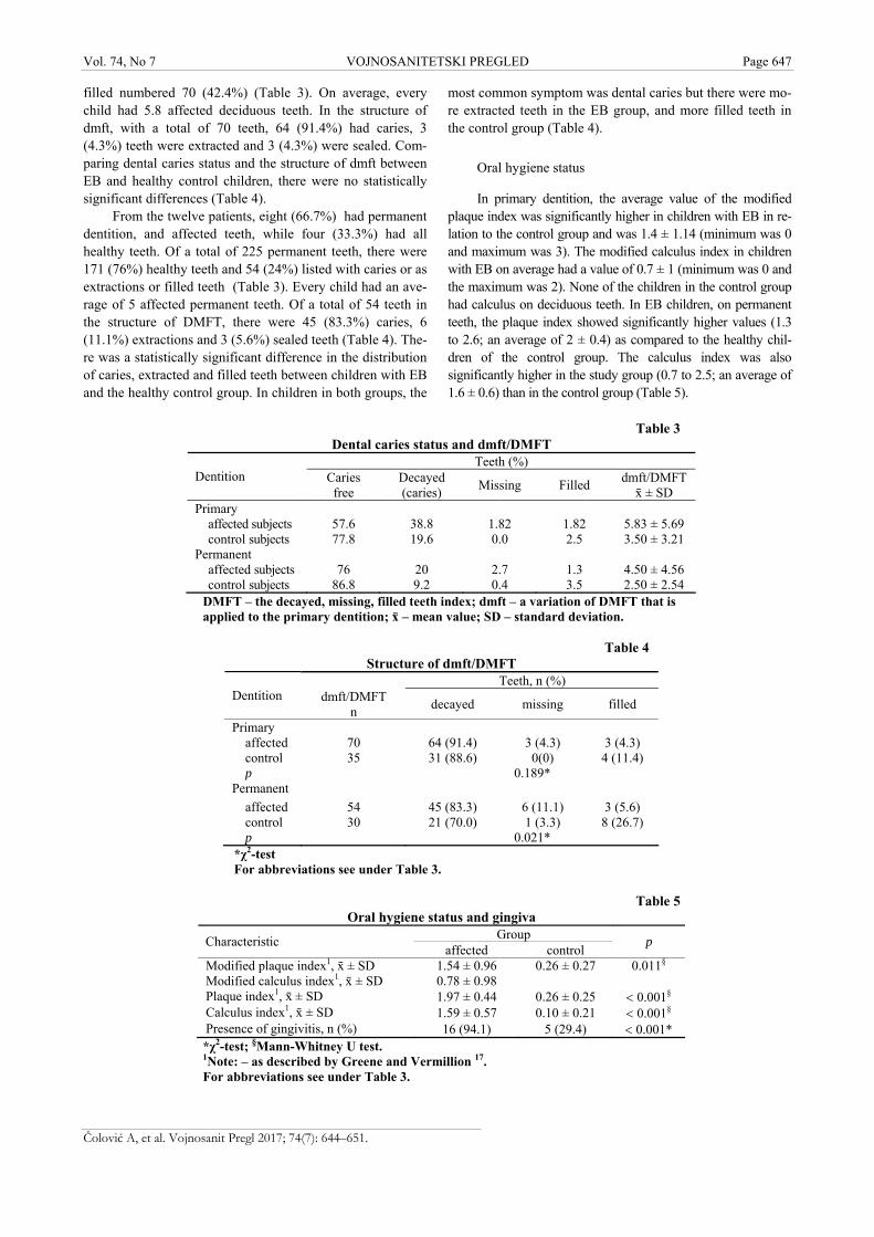
Vol. 74, No 7 VOJNOSANITETSKI PREGLED Page 647
Čolović A, et al. Vojnosanit Pregl 2017; 74(7): 644–651.
Table 3 Dental caries status and dmft/DMFT
Teeth (%) Dentition Caries
free Decayed (caries)
Missing Filled dmft/DMFT ґ ± SD
Primary affected subjects 57.6 38.8 1.82 1.82 5.83 ± 5.69 control subjects 77.8 19.6 0.0 2.5 3.50 ± 3.21
Permanent affected subjects 76 20 2.7 1.3 4.50 ± 4.56 control subjects 86.8 9.2 0.4 3.5 2.50 ± 2.54
DMFT – the decayed, missing, filled teeth index; dmft – a variation of DMFT that is applied to the primary dentition; ґ – mean value; SD – standard deviation.
Table 4
Structure of dmft/DMFT Teeth, n (%)
Dentition dmft/DMFT n
decayed missing filled
Primary affected 70 64 (91.4) 3 (4.3) 3 (4.3) control 35 31 (88.6) 0(0) 4 (11.4) p 0.189*
Permanent
affected 54 45 (83.3) 6 (11.1) 3 (5.6) control 30 21 (70.0) 1 (3.3) 8 (26.7) p 0.021*
*χ2-test For abbreviations see under Table 3.
Table 5
Oral hygiene status and gingiva Group Characteristic
affected control p
Modified plaque index1, ґ ± SD 1.54 ± 0.96 0.26 ± 0.27 0.011§ Modified calculus index1, ґ ± SD 0.78 ± 0.98 Plaque index1, ґ ± SD 1.97 ± 0.44 0.26 ± 0.25 0.001§ Calculus index1, ґ ± SD 1.59 ± 0.57 0.10 ± 0.21 0.001§ Presence of gingivitis, n (%) 16 (94.1) 5 (29.4) 0.001* *χ2-test; §Mann-Whitney U test. 1Note: – as described by Greene and Vermillion 17. For abbreviations see under Table 3.
filled numbered 70 (42.4%) (Table 3). On average, every child had 5.8 affected deciduous teeth. In the structure of dmft, with a total of 70 teeth, 64 (91.4%) had caries, 3 (4.3%) teeth were extracted and 3 (4.3%) were sealed. Com-paring dental caries status and the structure of dmft between EB and healthy control children, there were no statistically significant differences (Table 4).
From the twelve patients, eight (66.7%) had permanent dentition, and affected teeth, while four (33.3%) had all healthy teeth. Of a total of 225 permanent teeth, there were 171 (76%) healthy teeth and 54 (24%) listed with caries or as extractions or filled teeth (Table 3). Every child had an ave-rage of 5 affected permanent teeth. Of a total of 54 teeth in the structure of DMFT, there were 45 (83.3%) caries, 6 (11.1%) extractions and 3 (5.6%) sealed teeth (Table 4). The-re was a statistically significant difference in the distribution of caries, extracted and filled teeth between children with EB and the healthy control group. In children in both groups, the
most common symptom was dental caries but there were mo-re extracted teeth in the EB group, and more filled teeth in the control group (Table 4).
Oral hygiene status
In primary dentition, the average value of the modified plaque index was significantly higher in children with EB in re-lation to the control group and was 1.4 ± 1.14 (minimum was 0 and maximum was 3). The modified calculus index in children with EB on average had a value of 0.7 ± 1 (minimum was 0 and the maximum was 2). None of the children in the control group had calculus on deciduous teeth. In EB children, on permanent teeth, the plaque index showed significantly higher values (1.3 to 2.6; an average of 2 ± 0.4) as compared to the healthy chil-dren of the control group. The calculus index was also significantly higher in the study group (0.7 to 2.5; an average of 1.6 ± 0.6) than in the control group (Table 5).
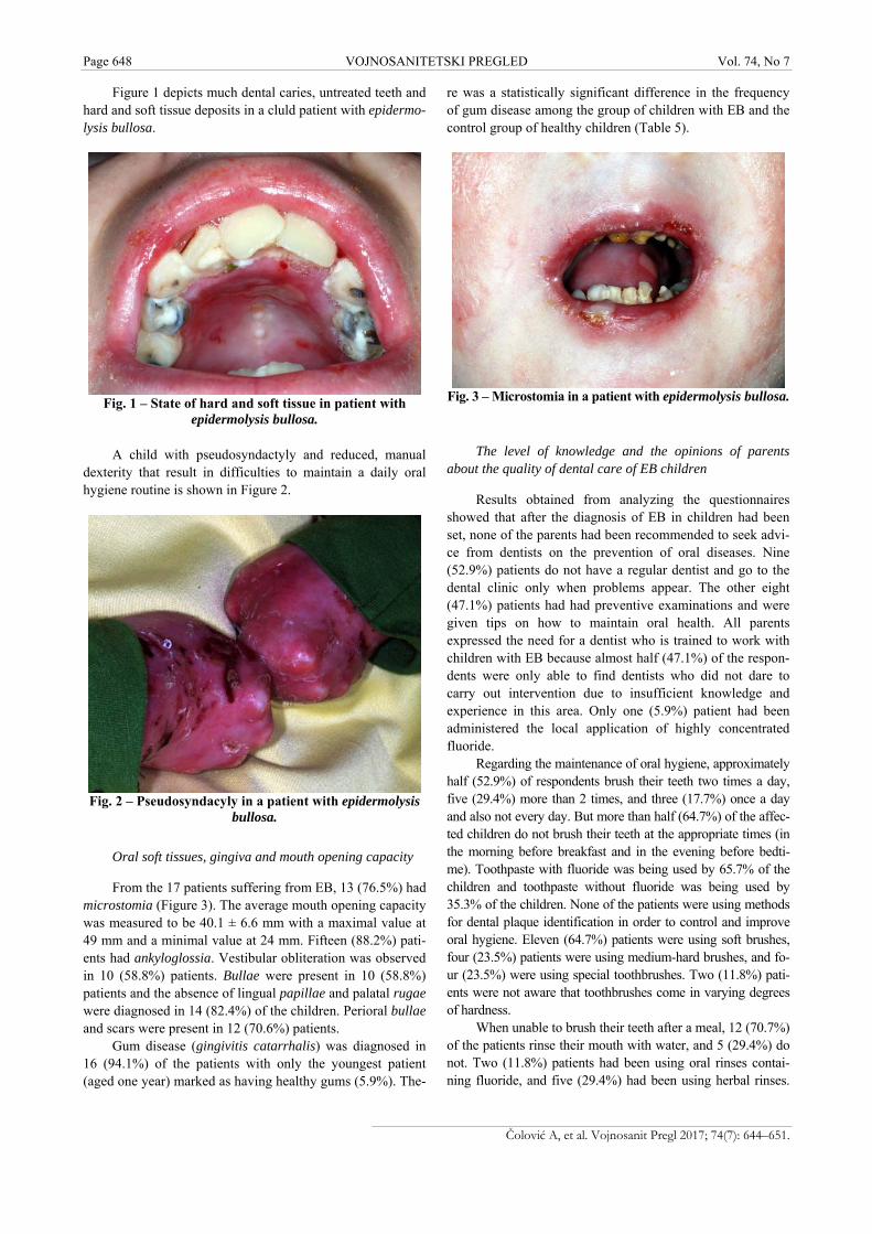
Page 648 VOJNOSANITETSKI PREGLED Vol. 74, No 7
Čolović A, et al. Vojnosanit Pregl 2017; 74(7): 644–651.
Figure 1 depicts much dental caries, untreated teeth and hard and soft tissue deposits in a cluld patient with epidermo-lysis bullosa.
Fig. 1 – State of hard and soft tissue in patient with
epidermolysis bullosa.
A child with pseudosyndactyly and reduced, manual dexterity that result in difficulties to maintain a daily oral hygiene routine is shown in Figure 2.
Fig. 2 – Pseudosyndacyly in a patient with epidermolysis
bullosa.
Oral soft tissues, gingiva and mouth opening capacity
From the 17 patients suffering from EB, 13 (76.5%) had microstomia (Figure 3). The average mouth opening capacity was measured to be 40.1 ± 6.6 mm with a maximal value at 49 mm and a minimal value at 24 mm. Fifteen (88.2%) pati-ents had ankyloglossia. Vestibular obliteration was observed in 10 (58.8%) patients. Bullae were present in 10 (58.8%) patients and the absence of lingual papillae and palatal rugae were diagnosed in 14 (82.4%) of the children. Perioral bullae and scars were present in 12 (70.6%) patients.
Gum disease (gingivitis catarrhalis) was diagnosed in 16 (94.1%) of the patients with only the youngest patient (aged one year) marked as having healthy gums (5.9%). The-
re was a statistically significant difference in the frequency of gum disease among the group of children with EB and the control group of healthy children (Table 5).
Fig. 3 – Microstomia in a patient with epidermolysis bullosa.
The level of knowledge and the opinions of parents about the quality of dental care of EB children
Results obtained from analyzing the questionnaires showed that after the diagnosis of EB in children had been set, none of the parents had been recommended to seek advi-ce from dentists on the prevention of oral diseases. Nine (52.9%) patients do not have a regular dentist and go to the dental clinic only when problems appear. The other eight (47.1%) patients had had preventive examinations and were given tips on how to maintain oral health. All parents expressed the need for a dentist who is trained to work with children with EB because almost half (47.1%) of the respon-dents were only able to find dentists who did not dare to carry out intervention due to insufficient knowledge and experience in this area. Only one (5.9%) patient had been administered the local application of highly concentrated fluoride.
Regarding the maintenance of oral hygiene, approximately half (52.9%) of respondents brush their teeth two times a day, five (29.4%) more than 2 times, and three (17.7%) once a day and also not every day. But more than half (64.7%) of the affec-ted children do not brush their teeth at the appropriate times (in the morning before breakfast and in the evening before bedti-me). Toothpaste with fluoride was being used by 65.7% of the children and toothpaste without fluoride was being used by 35.3% of the children. None of the patients were using methods for dental plaque identification in order to control and improve oral hygiene. Eleven (64.7%) patients were using soft brushes, four (23.5%) patients were using medium-hard brushes, and fo-ur (23.5%) were using special toothbrushes. Two (11.8%) pati-ents were not aware that toothbrushes come in varying degrees of hardness.
When unable to brush their teeth after a meal, 12 (70.7%) of the patients rinse their mouth with water, and 5 (29.4%) do not. Two (11.8%) patients had been using oral rinses contai-ning fluoride, and five (29.4%) had been using herbal rinses.
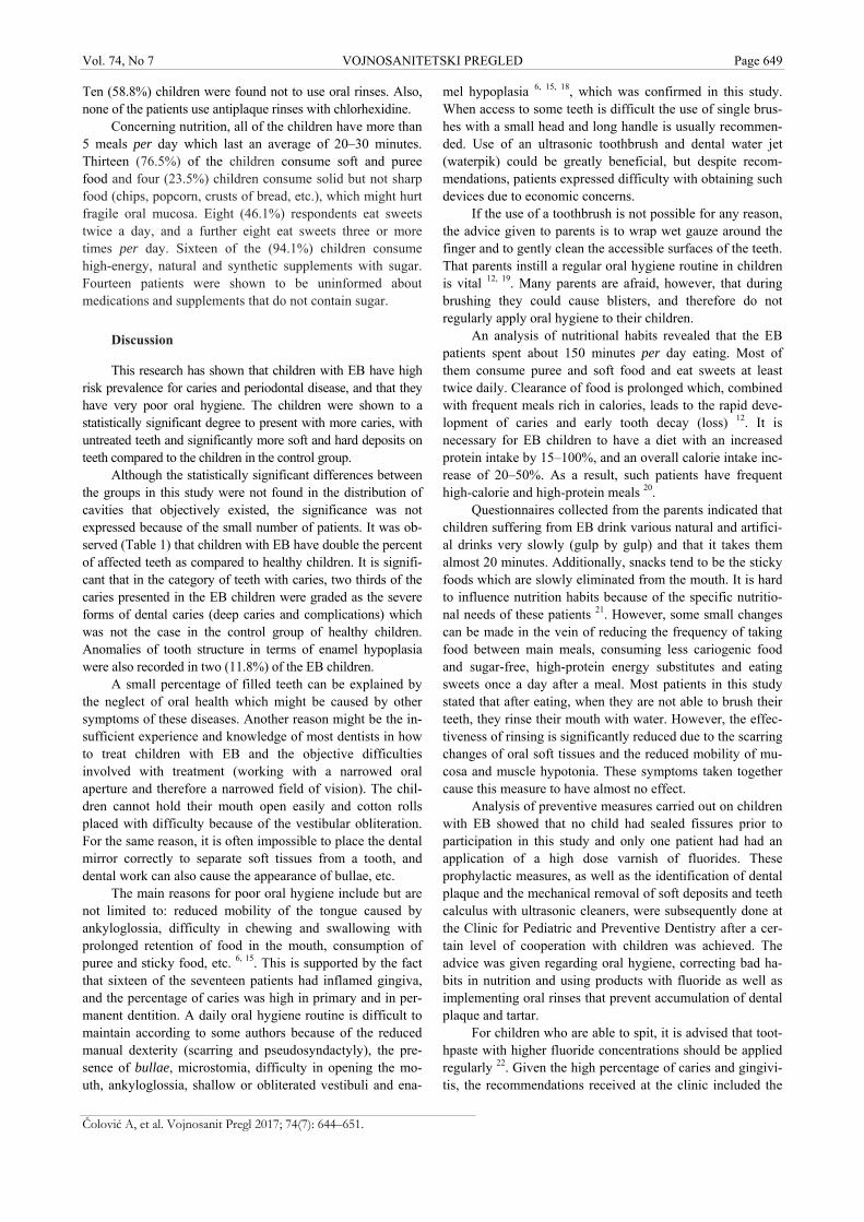
Vol. 74, No 7 VOJNOSANITETSKI PREGLED Page 649
Čolović A, et al. Vojnosanit Pregl 2017; 74(7): 644–651.
Ten (58.8%) children were found not to use oral rinses. Also, none of the patients use antiplaque rinses with chlorhexidine.
Concerning nutrition, all of the children have more than 5 meals per day which last an average of 20–30 minutes. Thirteen (76.5%) of the children consume soft and puree food and four (23.5%) children consume solid but not sharp food (chips, popcorn, crusts of bread, etc.), which might hurt fragile oral mucosa. Eight (46.1%) respondents eat sweets twice a day, and a further eight eat sweets three or more times per day. Sixteen of the (94.1%) children consume high-energy, natural and synthetic supplements with sugar. Fourteen patients were shown to be uninformed about medications and supplements that do not contain sugar.
Discussion
This research has shown that children with EB have high risk prevalence for caries and periodontal disease, and that they have very poor oral hygiene. The children were shown to a statistically significant degree to present with more caries, with untreated teeth and significantly more soft and hard deposits on teeth compared to the children in the control group.
Although the statistically significant differences between the groups in this study were not found in the distribution of cavities that objectively existed, the significance was not expressed because of the small number of patients. It was ob-served (Table 1) that children with EB have double the percent of affected teeth as compared to healthy children. It is signifi-cant that in the category of teeth with caries, two thirds of the caries presented in the EB children were graded as the severe forms of dental caries (deep caries and complications) which was not the case in the control group of healthy children. Anomalies of tooth structure in terms of enamel hypoplasia were also recorded in two (11.8%) of the EB children.
A small percentage of filled teeth can be explained by the neglect of oral health which might be caused by other symptoms of these diseases. Another reason might be the in-sufficient experience and knowledge of most dentists in how to treat children with EB and the objective difficulties involved with treatment (working with a narrowed oral aperture and therefore a narrowed field of vision). The chil-dren cannot hold their mouth open easily and cotton rolls placed with difficulty because of the vestibular obliteration. For the same reason, it is often impossible to place the dental mirror correctly to separate soft tissues from a tooth, and dental work can also cause the appearance of bullae, etc.
The main reasons for poor oral hygiene include but are not limited to: reduced mobility of the tongue caused by ankyloglossia, difficulty in chewing and swallowing with prolonged retention of food in the mouth, consumption of puree and sticky food, etc. 6, 15. This is supported by the fact that sixteen of the seventeen patients had inflamed gingiva, and the percentage of caries was high in primary and in per-manent dentition. A daily oral hygiene routine is difficult to maintain according to some authors because of the reduced manual dexterity (scarring and pseudosyndactyly), the pre-sence of bullae, microstomia, difficulty in opening the mo-uth, ankyloglossia, shallow or obliterated vestibuli and ena-
mel hypoplasia 6, 15, 18, which was confirmed in this study. When access to some teeth is difficult the use of single brus-hes with a small head and long handle is usually recommen-ded. Use of an ultrasonic toothbrush and dental water jet (waterpik) could be greatly beneficial, but despite recom-mendations, patients expressed difficulty with obtaining such devices due to economic concerns.
If the use of a toothbrush is not possible for any reason, the advice given to parents is to wrap wet gauze around the finger and to gently clean the accessible surfaces of the teeth. That parents instill a regular oral hygiene routine in children is vital 12, 19. Many parents are afraid, however, that during brushing they could cause blisters, and therefore do not regularly apply oral hygiene to their children.
An analysis of nutritional habits revealed that the EB patients spent about 150 minutes per day eating. Most of them consume puree and soft food and eat sweets at least twice daily. Clearance of food is prolonged which, combined with frequent meals rich in calories, leads to the rapid deve-lopment of caries and early tooth decay (loss) 12. It is necessary for EB children to have a diet with an increased protein intake by 15–100%, and an overall calorie intake inc-rease of 20–50%. As a result, such patients have frequent high-calorie and high-protein meals 20.
Questionnaires collected from the parents indicated that children suffering from EB drink various natural and artifici-al drinks very slowly (gulp by gulp) and that it takes them almost 20 minutes. Additionally, snacks tend to be the sticky foods which are slowly eliminated from the mouth. It is hard to influence nutrition habits because of the specific nutritio-nal needs of these patients 21. However, some small changes can be made in the vein of reducing the frequency of taking food between main meals, consuming less cariogenic food and sugar-free, high-protein energy substitutes and eating sweets once a day after a meal. Most patients in this study stated that after eating, when they are not able to brush their teeth, they rinse their mouth with water. However, the effec-tiveness of rinsing is significantly reduced due to the scarring changes of oral soft tissues and the reduced mobility of mu-cosa and muscle hypotonia. These symptoms taken together cause this measure to have almost no effect.
Analysis of preventive measures carried out on children with EB showed that no child had sealed fissures prior to participation in this study and only one patient had had an application of a high dose varnish of fluorides. These prophylactic measures, as well as the identification of dental plaque and the mechanical removal of soft deposits and teeth calculus with ultrasonic cleaners, were subsequently done at the Clinic for Pediatric and Preventive Dentistry after a cer-tain level of cooperation with children was achieved. The advice was given regarding oral hygiene, correcting bad ha-bits in nutrition and using products with fluoride as well as implementing oral rinses that prevent accumulation of dental plaque and tartar.
For children who are able to spit, it is advised that toot-hpaste with higher fluoride concentrations should be applied regularly 22. Given the high percentage of caries and gingivi-tis, the recommendations received at the clinic included the
Page 650 VOJNOSANITETSKI PREGLED Vol. 74, No 7
Čolović A, et al. Vojnosanit Pregl 2017; 74(7): 644–651.
daily use of a solution of chlorhexidine (0.05%) and fluoride (223 ppmF). Excellent results have shown to be achieved using higher concentrations of chlorhexidine for a period of one week every 2–3 months 19. It should be noted that prior to this study none of the respondents did not use chlorhexidine, which is one of the most effective agents for chemical control of dental plaque. When a child does not know how to spit out, parents were advised to apply rinse to a gauze or cotton swab and coat the teeth and gums. Children with EB should use solutions without alcohol and menthol as rinses can irritate the oral mucosa 21. Dental visits were sche-duled for every 3 months. Although all patients received in-formation about basic and additional measures to maintain and improve oral health, few have succeeded to implement them in a daily routine. Because of that, it is necessary to ha-ve constant monitoring and motivation of child and parent; otherwise, there is an inevitable neglect of oral health care.
This study showed that dental therapy interventions can be conducted in children with EB. On their first visits to the clinic, the parents wanted restorations to be done rather than preventive measures to be performed on their children. The reason for that is that they did not come on time to regular check-ups in the past and many of the children already had severe dental problems. Dental treatment was difficult beca-use of microstomia, shallow vestibuli and the appearance of bullae at the slightest application of pressure on the oral mu-cosa. Bullae were perforated with thin sterile needles of syringes, to avoid the formation of scar tissue. With cautious work "in the air" and with frequent interruptions as well as the application of local anesthesia almost all conservative
treatments were made and tooth extractions completed. It should be noted that the indications for endodontic treatment in children with EB are limited to the front teeth (due to mic-rostomia) and that for the more complicated tooth extraction it is necessary to use general anesthesia which requires spe-cial conditions and additional trained staff.
Conclusion
Children with EB account for a group of high-risk pa-tients for developing caries and periodontal disease. Of all patients with a medical risk, the toughest dental work is with this category of patients due to the sensitivity of the oral mucosa, so only a small number of dentists is available to these patients even in the performance of simpler pre-ventive dental procedures. The parents of children are not sufficiently informed about the importance of the early ap-plication of preventive measures to preserve oral health and they find that their children do not have adequate dental ca-re. In order to alleviate this problem there must be a proper cooperation protocol between the treating physician and the dentist. After an EB diagnosis is made, the child and pa-rents need to be in contact with a qualified dentist who will educate them and take preventive measures to preserve oral health as light and non-aggressive as possible: the treat-ment of choice for all patients at risk, especially for chil-dren with EB. It is necessary even within the specialization of pediatric and preventive dentistry to organize subspecialty training for dentists who are to work with pa-tients affected with EB.
R E F E R E N C E S
1. Lanschuetzer CM. Definition. In: Fine JD, Hintner H, editors. Li-fe with Epidermolysis Bullosa (EB): Etiology, Diagnosis, Multidisciplinary Care and Therapy.Wien: Springer Verlag; 2009. p. 3−5.
2. National organization for rare disorders. Rare diseases. In: Associa-tion for epidermolysis bullosa “DEBRA”, 1st. Beograd. 2014. p. 103.
3. Lanschuetzer CM, Fine JD. Classification and molecular basis of hereditary epidermolysis bullosa. In: Fine JD, Hintner H, edi-tors. Life with Epidermolysis Bullosa (EB): Etiology, Diagno-sis, Multidisciplinary Care and Therapy. Wien: Springer Verlag; 2009. p. 6−23.
4. Fine JD, Eady RA, Bauer EA, Bauer JW, Bruckner-Tuderman L, Heagerty A, et al. The classification of inherited epidermolysis bullosa (EB): Report of the Third International Consensus Meeting on Diagnosis and Classification of EB. J Am Acad Dermatol 2008; 58(6): 931−50.
5. Wright JT, Fine JD, Johnson L. Dental caries risk in hereditary epidermolysis bullosa. Pediatr Dent 1994; 16(6): 427−32.
6. Fine J, Mellerio JE. Extracutaneous manifestations and compli-cations of inherited epidermolysis bullosa: Part II. Other or-gans. J Am Acad Dermatol 2009; 61(3): 387−402; quiz 403−4.
7. Ashton GH. Kindler syndrome. Clin Exp Dermatol 2004; 29(2): 116−21.
8. Intong L, Murrell F. Inherited epidermolysis bullosa: New diag-nostic criteria and classification. Clin Dermatol 2012; 30(1): 70−7.
9. Fine JD, Mellerio JE. Extracutaneous manifestations and com-plications of inherited epidermolysis bullosa: Part I. Epithelial associated tissues. J Am Acad Dermatol 2009; 61(3): 367−384.
10. Wright JT, Fine JD, Johnson L. Hereditary epidermolysis bullosa: Oral manifestations and dental management. Pediatr Dent 1993; 15(4): 242−8.
11. Krämer S, Serrano MC, Zillmann G, Gálvez P, Araya I, Yanine N, et al. Oral Health Care for Patients with Epidermolysis Bullo-sa: Best Clinical Practice Guidelines. Int J Paediatr Dent 2012; 22(Suppl1): 1−35.
12. Azrak B, Kaevel K, Hofmann L, Gleissner C, Willershausen B. Dystrophic epidermolysis bullosa: oral findings and problems. Spec Care Dentist 2006; 26(3): 111−5.
13. Martin L, Elke N. Intraoral disease. In: Fine JD, Hintner H, edi-tors. Life with Epidermolysis Bullosa (EB) Etiology, Diagno-sis, Multidisciplinary Care and Therapy. Wien: Springer-Verlag; 2009. p. 150−66.
14. World Health Organization. Oral health surveys. Basic methods. 4th ed. Geneva: World Health Organization; 1999.
15. Wright JT, Fine JD, Johnson LB. Oral soft tissues in hereditary epidermolysis bullosa. Oral Surg Oral Med Oral Pathol 1991; 71(4): 440−6.
16. Dwivedi S, Mittal M, Vashisth P, Jaiswal D, Arora S. Oral Hygiene Pattern observed in Primary School Children as Re-ported by Their Mother: A Longitudinal Study. World J Dent 2012; 3(4): 308−12.
17. Greene J, Vermillion J. The simplified oral hygiene index. J Am Dent Assoc 1964; 68: 7−13.

Vol. 74, No 7 VOJNOSANITETSKI PREGLED Page 651
Čolović A, et al. Vojnosanit Pregl 2017; 74(7): 644–651.
18. Siqueira MA, de Souza Silva J, Silva FW, Díaz-Serrano KV, Freitas AC, Queiroz AM. Dental treatment in a patient with epidermolysis bullosa. Spec Care Dentist 2008; 28(3): 92−5.
19. EB Handbook Austria. Available from: http://www.ebhandbuch.org/fileadmin/media_data/5_EBHandbuch/7_pdf_s_ENGLISCH/Teeth_DDEB.pdf
20. Tješić-Drinković D. The role of nutrition in epidermolysis bullosa. The second regional symposium on hereditary epidermolysis bullosa. The satellite symposium of the 5th congress of Croatian dermatovenerologist with international participation; Zagreb; 2014 May 7; 2014
21. Wright T. Epidermolysis bullosa and Oral Health: Problems and solutions. Available from: http://ebnurse. org/assets/files/Dental_Care_by_Tim_Wright_DDS.pdf
22. Dental health foundation. Available from: http://www.dentalhealth.ie/dentalhealth/teeth/fluoridetoothpastes.htm
Received on October 23, 2015. Revised on December 22, 2015.
Accepted on January 8, 2016. Online First July, 2016.
Vojnosanit Pregl 2017; 74(7): 652–658. VOJNOSANITETSKI PREGLED Page 652
Correspondence to: Silva Dobrić, Military Medical Academy, Institute for Scientific Information, Crnotravska 17, 11 000 Belgrade, Serbia. Phone/Fax: + 381 11 26 69 689. E-mail: silva.dobrić@gmail.com
O R I G I N A L A R T I C L E UDC: 615.03::[615.322::615.45 https://doi.org/10.2298/VSP160422086D
Pharmacological characterization of Cirsium ligulare Boiss. (Asteraceae) herb decoction
Farmakološka karakterizacija dekokta herbe Cirsium ligulare Boiss. (Asteraceae)
Silva Dobrić*†, Silvana Petrović‡, Jelena Kukić-Marković‡, Stevan Samardžić‡,
Violeta Milutinović‡
Military Medical Academy, *Institute for Scientific Information, Belgrade, Serbia; University of Defence, †Faculty of Medicine of the Military Medical Academy, Belgrade, Serbia; University of Belgrade, Faculty of Pharmacy, ‡Department of Pharmacognosy,
Belgrade, Serbia
Abstract Background/Aim. Data on phytochemical and pharmacolo-gical investigations of genus Cirsium Mill. (Asteraceae) are scar-ce. Some data suggest that decoctions or infusions prepared from these plants are used in folk medicine as tonics, particularly in inflammatory, liver and stomach diseases. So far there have been no pharmacological investigations related to Cirsium ligulare (C. ligulare) Boiss. Accordingly, the aim of this study was to estimate antioxidative, anti-inflammatory and gas-troprotective activities of aqueous extracts of C. ligulare herb prepared as 5% and 10% decoctions. Methods. Antioxidative activity was determined using the method of 2,2-diphenyl-1-picrylhydrazyl (DPPH) radical scavenging. Investigations of anti-inflammatory (a model of systemic inflammatory response induced by endotoxin of Escherichia coli and carrageenan-induced rat paw oedema model for local inflammatory respon-se), as well as gastroprotective effects (a model of stress-ulcer induced by absolute ethanol), were conducted in adult female Wistar rats that were given the aqueous extracts of C. ligulare herb per os. Indomethacin and ranitidine were used as reference drugs for evaluation of local anti-inflammatory and gastropro-tective effects, respectively. Results. The results demonstrated that aqueous extracts of C. ligulare herb produced strong antioxidative activity, diminished body weight loss induced by endotoxin, significantly reduced carrageenan-induced paw oedema, and prevented the ulcerogenic action of absolute ethanol. Both anti-inflammatory and gastroprotective activi-ties of the extract tested were comparable to those of the re-ference drugs. Conclusion. Presented results justify the tradi-tional use of C. ligulare herb decoctions and further phytochemical and pharmacological investigations are warranted. Key words: cirsium; asteraceae; pharmacologic actions; herbal medicine; rats, wistar.
Apstrakt Uvod/Cilj. Biljke roda Cirsium su hemijski i farmakološki nedovoljno ispitane. Tradicionalno se dekokti ili infuzi ovih biljaka koriste kao tonici, naročito kod zapaljenjskih procesa, kao i kod bolesti jetre i želuca. Vrsta Cirsium ligulare (C. ligulare) Boiss. do sada nije farmakološki ispitivana. Stoga je cilj ovog istraživanja bio da se proceni antioksidantna, an-tiinflamatorna i gastroprotektivna aktivnost vodenog ek-strakta herbe C. ligulare izrađenog kao 5% i 10% dekokt. Metode. Ispitivanje antioksidantne aktivnosti vršeno je tes-tom neutralizacije 2,2-diphenyl-1-picrylhydrazyl (DPPH) radikala. Ispitivanje antiinflamatornog (model sistemskog in-flamatornog odgovora indukovan endotoksinom bakterije Escherichia coli i karageninom-indukovani edem šape pacova kao model lokalne inflamcije), kao i gastroprotektivnog efekta (model ulkusa indukovanog apsolutim etanolom), sprovedeno je na odraslim ženkama pacova Wistar soja, ko-jima su per os dati vodeni ekstrakti herbe C. ligulare. Kao ref-erentni lekovi prilikom ispitivanja lokalnog antiinflamator-nog i gastroprotektivnog delovanja, primenjeni su indo-metacin i ranitidin, redom. Rezultati. Rezultati pokazuju da su ispitivani vodeni ekstrakti herbe C. ligulare ispoljili jaku antioksidantnu aktivnost, smanjili gubitak telesne mase in-dukovan endotoksinom, značajno redukovali karageninom indukovani edem šape pacova i spečili ulcerogeni efekat ap-solutnog etanola. Antiinflamatorna i gastroprotektivna ak-tivnost ispitivanih ekstrakata bila je uporediva sa aktivnošću referentnih lekova. Zaključak. Dobijeni rezultati u značajnoj meri opravdavaju tradicionalnu primenu dekokta herbe C. lig-ulare, a planiraju se i dodatna hemijska i farmakološka ispiti-vanja ove droge. Ključne reči: cirsium; asteraceae; farmakološka dejstva; medicina, biljna; pacovi, wistar.
Vol. 74, No 7 VOJNOSANITETSKI PREGLED Page 653
Dobrić S, et al. Vojnosanit Pregl 2017; 74(7): 652–658.
Introduction
Genus Cirsium Mill. (Asteraceae) includes about 120 spe-cies widely distributed in Europe, North Africa, West and North Asia, as well as North and Central America 1. According to avai-lable data, these species contain flavonoids 2–4, phenolic acids, essential oils, sterols, triterpenes, as well as alkaloids, polyacetylenes, hydrocarbons, aliphatic aldehydes and guaiano-lides–sesquiterpene lactones 5, 6. Traditionally, they are used as tonics, antiphlogistics, diuretics, diaphoretics, adstringents and venoactive remedies 6, 7, as well as anti-hemorrhagic, diuretic and anticancer agents 4, against a cough and peptic ulcers 8.
Cirsium ligulare (C. ligulare) Boiss. is one of 22 Cirsium species present in the Serbian flora 9. In folk medicine of Serbia and Montenegro, according to our ethnomedicinal experience, decoctions or infusions prepared from its herb are used as tonics, particularly in inflammatory, liver and stomach complaints. However, data on C. ligulare research are scarce. Chlorogenic acid and flavonoids (apigenin, apigenin 7-glucoside, kaempferol 3-glucoside and kaempferol 3-rhamnoglucoside) were identified in methanolic extracts of C. ligulare leaves and inflorescences 2. In the essential oil of its inflorescence, beside predominant alip-hatic hydrocarbons, small amounts of thymol (2.4%), eugenol (0.6%) and carvacrol (0.3%) were present 5. To the best of our knowledge, no pharmacological investigations of this species have been conducted.
Thus, the aim of this study was to estimate antioxidative, anti-inflammatory and gastroprotective activi-ties of aqueous extracts of C. ligulare herb, prepared in the traditional manner as decoctions.
Methods
Chemicals
Dimethylsulfoxide (DMSO), 2,2-diphenyl-1-picrylhydrazyl, lipopolysaccharides (LPS) from Escherichia coli 0128:B8 (Cat. No L3880) and indomethacin were obtained from Sig-ma Chemical Co. (St. Louis, USA); absolute ethanol (96%, v/v) from Merck (Darmstadt, Germany); L-ascorbic acid from Lachema (Neratovice, Czech Republic); rutin from Roth (Karlsruhe, Germany), and ranitidine from Zdravlje-Actavis Company (Leskovac, Serbia).
Plant material
The herb of C. ligulare was collected in summer 2004 from natural habitat in the river Lim valley, close to its spring from Plavsko lake (North-Eastern region of Montenegro). Once harvested, the plant material was dried at room temperature. It was identified by Prof. P. Marin, PhD (Institute of Botany, Faculty of Biology, University of Belgrade, Serbia).
Preparation of decoctions
Prior to extraction, the plant material was reduced to a coarse powder. Decoctions were prepared by boiling 5.0 and 10.0 g of powdered material in 100 mL of distilled water for
5 min to prepare 5 and 10% (w/v) decoctions. After 10 min, the extracts were allowed to cool at room temperature and filtered. Extracted plant material was washed with distilled water and the volume of the prepared decoctions was adjus-ted to 100 mL using these washings. Decoctions were each time prepared ex tempore.
Animals
Adult female Wistar rats, weighing 200–250 g, were used in the experiments in which anti-inflammatory and anti-ulcer activities of decoctions of C. ligulare were tested. Be-fore the experiments, the animals were housed in groups of 5 to a plastic cage in standard laboratory condition (room tem-perature of 22 ± 1 ºC, 30% humidity, 12/12 h light/dark cycle) with free access to food and tap water. Animal studies were conducted in accordance with the internationally accep-ted principles for laboratory animal use and care in the Euro-pean Community (EEC Directive of 1986; 86/609/EEC) adopted by Ethical Committee of Military Medical Academy, Belgrade.
Antioxidant activity (DPPH radical assay)
Four milliliters of 5% decoction of the drug, prepared as described above, was mixed with 1 mL of 0.5 mM DPPH in methanol, and final volume was adjusted up to 5 mL. The mixture was vigorously shaken and left 30 min in dark. Ab-sorbance was measured at 517 using methanol as blank. One milliliter of 0.5 mM DPPH diluted with 4 mL of methanol was used as a control. Scavenging (SC) of DPPH radical was calculated using the equation:
SC(%)=100×(A0-As)/A0 (1) A0 - the absorbance of the control (containing all rea-
gents except the test compound); As - the absorbance of the tested sample.
The SC50 value represents the concentration of the sam-ple that caused 50% of DPPH radical scavenging. The relati-ve activity of the samples was compared to those of L-ascorbic acid and rutin 10.
Anti-inflammatory activity
Lipopolysaccharide (LPS)-induced systemic inflammatory response
A systemic inflammatory reaction was induced by s.c. injection of LPS given in a dose of 5 mg/kg. This dose was chosen as a non-lethal one, but sufficient for producing a systemic inflammatory response, on the basis of previously performed experiments in which the LD50 of LPS for this species and this route of administration had been established (approximately 20 mg/kg).
Before the experiments, the animals were divided into six groups of 15 rats in each, and their body mass, as well as the average consumption of food and water were determined. After that, they were given following treatments:
Group 1 – intact controls drinking tap water; Group 2 – animals drinking 5% decoction of C. ligulare herb Group 3 –
Page 654 VOJNOSANITETSKI PREGLED Vol. 74, No 7
Dobrić S, et al. Vojnosanit Pregl 2017; 74(7): 652–658.
animals drinking 10% decoction of C. ligulare herb; Group 4 – animals injected by 5 mg/kg s.c. of LPS and drinking tap water; Group 5 – animals injected by 5 mg/kg s.c. of LPS and drinking 5% decoction of C. ligulare herb; Group 6 – animals injected by 5 mg/kg s.c. of LPS and drinking 10% decoction of C. ligulare herb.
The experiment lasted 30 days after injecting LPS and, during this period, tap water or extracts (5 and 10% decocti-ons of C. ligulare herb) were given to animals ad libitum. The body mass and consumption of food and water or the extract were measured each day, always at the same time (9 a.m.).
Carrageenan-induced rat paw oedema test
The carrageenan-induced rat paw oedema was used as an experimental model of local inflammatory reaction as re-ported earlier 11.
The experimental groups consisted of 6–8 animals each. In order to estimate an anti-inflammatory activity of an aqueous extract of C. ligulare herb, the animals were gi-ven 10% decoction of C. ligulare herb instead tapping water for seven days. On the day 8 of the experiment, a lo-cal inflammatory reaction was induced by the injection of 0.1 mL of carrageenan-saline solution (0.5%, w/v) into the plantar surface of the right hind paw of the rat. A saline was injected in a volume of 0.1 mL into the plantar surface of the left hind paw that served as the control (non-inflamed) paw. The animals were sacrificed 3 h after the carrageenan and saline injections and paws were cut off for weighing. The difference in weight between right and left paw (in mg) served as an indicator of the inflammatory res-ponse intensity (i.e. extent of the oedema). The control animals, instead of decoction, were drinking tap water for seven days before inducing paw oedema. The positive con-trol group consisted of the animals treated with the well-known non-steroidal anti-inflammatory drug (NSAID) in-domethacin, dissolved in DMSO and given in a dose of 2 mg/kg p.o. 60 min prior to injection of carrageenan soluti-on. This dose of indomethacin reduced the carrageenan-induced rat paw oedema by approximately 50%, as had shown in previously performed pilot-experiment.
Effects of the treatments applied were estimated regarding the differences in rat paw oedema extent, i.e. intensity of inflammatory response, between the experimental groups. The difference in weight between right and left paw, active drug (de-coct or indomethacin)-treated versus tap water (vehicle)-treated rats (the control group), served as an indicator of the anti-inflammatory activity of drugs tested (the extract and indomet-hacin). The inflammatory response was calculated using the equation:
e Inflammatory response (%) = c 100 (2)
e – the difference in the paw weight in the active drug/treatment groups; c – difference in the paw weight in the control groups.
Gastroprotective activity In order to study the anti-ulcer activity of decoction of C.
ligulare herb an experimental model of acute gastric mucosal damage induced by absolute ethanol (1 mL/rat p.o.) was used.
The experimental groups consisted of 6-8 animals each. For seven days the animals were given 10% decoction of C. ligulare herb instead of tap water, while the control animals were given tap water all the time. On the day 8 after the be-ginning of the experiment the animals were given 1 mL of absolute ethanol through the gastric tube (the animals were starved at least 18 h before giving ethanol). Animals in the positive control group were treated by a single dose of raniti-dine (20 mg/kg p.o.), the well-known anti-ulcer drug, given 60 min prior the absolute ethanol. This dose of ranitidine was chosen as a proven anti-ulcer dose on the basis of previously performed pilot-experiment.
The animals were sacrificed 1 h after ethanol administra-tion, their stomachs removed and opened along the great cur-vature. Lesions were examined under an illuminated magnifier (3×). The intensity of gastric lesions was assessed in accordan-ce with a modified scoring system of Adami et al. 12.
Results
Antioxidative activity
Antioxidative activity of 5% decoction of C. ligulare herb was determined using the method of DPPH radical sca-venging, and the results are presented in Table 1.The anti-DPPH activity of investigated decoction was comparable with the activity of vitamin C and rutin.
Table 1 Antioxidative activity of Cirsium ligulare
herb decoction Sample tested SC50 5% decoction of the C. ligulare herb 8.44 ± 1.17L-ascorbic acid 4.09 ± 0.08Rutin 5.75 ± 0.18
SC50 – concentration of the sample that caused 50% of 2,2-difenil-1-picrylhydrazyl (DPHH) radical scavenging.
Anti-inflammatory activity
Results of LPS-induced systemic inflammatory response experiment are presented in Table 2. In our experimental condi-tions, main changes were caused by LPS dose of 5 mg/kg s.c. related to the changes in body mass of treated animals. Namely, LPS led to the significant decrease of body mass gain in rats compared to the intact controls, particularly in the first two weeks after its injection. Reduced body mass in these animals was maintained during the whole period of observation. The usage of decoctions prepared from C. ligulare herb in both con-centrations alleviated body mass decrease just within the first two weeks of the experiment when the body mass decrease in-duced by LPS was the most pronounced.
In the model of carrageenan-induced rat paw oedema, the 10% decoction of C. ligulare herb also produced a signi-ficant anti-inflammatory effect (Table 3). Results of this

Vol. 74, No 7 VOJNOSANITETSKI PREGLED Page 655
Dobrić S, et al. Vojnosanit Pregl 2017; 74(7): 652–658.
Table 2
Influence of Cirsium ligulare herb decoctions on body mass changes (% of basal values) in rats with lipopolysaccharide induced systemic inflammatory response
Days Treatment 7 14 21 28
Group 1 16.81 ± 1.08 32.74 ± 1.95 40.7 ± 2.07 53.1 ± 2.21 Group 2 14.83 ± 1.42 23.81 ± 1.89 36.74 ± 1.85 49.1 ± 1.97 Group 3 12.56 ± 1.15 22.76 ± 1.72 32.56 ± 2.31 47.51 ± 2.18 Group 4 - 0.62 ± 0.9a, b 6.26 ± 0.58a, b 21.52 ± 1.46a 27.8 ± 1.79a, b Group 5 2.12 ± 0.24a, b 13.34 ± 1.4a, c 25.56 ± 1.67a 31.8 ± 1.23a, b Group 6 2.23 ± 0.21a, b 14.65± 1.42a,c 27.25 ± 2.1a 31.5 ± 1.17a,b
Group 1 – intact controls given tap water; Group 2 – animals given 5% decoction of C. ligulare herb; Group 3 – ani-mals given 10% decoction of C. ligulare herb; Group 4 – animals injected by 5 mg/kg sc of lipopolysaccharide (LPS) and given tap water; Group 5 – animals injected by 5 mg/kg sc of LPS and given 5% decoction of C. ligulare herb; Group 6 - animals injected by 5 mg/kg sc of LPS and given 10% decoction of C. ligulare herb ap < 0.05 vs Group 1; bp < 0.05 vs Groups 2 and 3; cp < 0.05 vs Group 4.
Table 3 Influence of Cirsium ligulare herb decoction on the carrageenan-
induced paw oedema (local inflammatory response) in rats Treatment Inflammatory response (%) None (the control group) 100.00 ± 25.98 10% decoction of C. ligulare herb 70.05 ± 9.02a Indomethacin* (2 mg/kg p.o.) 59.15 ± 7.09b
*Reference drug; ap < 0.05; bp < 0.01 vs the control group.
Table 4
Influence of Cirsium ligulare herb decoction on the ethanol-induced acute stress-ulcer in rats Treatment Intensity of gastric lesions** None (the control group) 5.90 ± 1.14 10% decoction of C. ligulare herb 2.26 ± 1.10a Ranitidine* (20 mg/kg p.o.) 1.77 ± 1.08a
*Reference drug; ap < 0.01 vs the control group **0 = no lesions; 0.5 = slight hyperaemia or 5 petechiae; 1 = 5 erosions 5 mm in lenght; 1.5 = 5 ero-sions 5 mm in lenght and many petechiae; 2 = 6–10 erosions 5 mm in lenght; 2.5 = 1–5 erosions > 5 mm in lenght; 3 = 5–10 erosions > 5 mm in lenght; 3.5 = >10 erosions > 5 mm in lenght; 4 = 1–3 erosions 5 mm in lenght and 0.5-1 mm in width; 4.5 = 4–5 erosions 5 mm in lenght and 0.5–1 mm in width; 5 = 1–3 ero-sions > 5 mm in lenght and 0.5-1 mm in width; 6 = 4 or 5 grade 5 lesions; 7 = 6 grade 5 lesions; 8 = com-plete lesion of the mucosa with haemorrhage (according to Adami et al. 12).
experiment clearly demonstrate that 7-day drinking of the 10% decoction of C. ligulare herb produced significant anti-inflammatory effect comparable to that of indomethacin, a strong NSAID, given in a dose of 2 mg/kg which corresponded to its ED50 (the dose that reduced the carrageenan-induced rat paw oedema by approximately 50%). However, post-mortem examination of the stomach of treated animals, revealed no changes in gastric mucosa in animals drinking the 10% decocti-on of C. ligulare herb, while those given indomethacin had so-me gastric lesions, mostly hyperaemia and petechiae.
Gastroprotective activity
The results of the present study demonstrated that 7-day use of the 10% decoction of C. ligulare herb significantly re-
duced the ulcerogenic effect of absolute ethanol in rats. More-over, this effect was comparable to that achieved by a single dose of ranitidine, a well-known anti-ulcer drug (Table 4).
Discussion
Antioxidative activity The method of DPPH radical neutralization is often used for
preliminary estimation of the antioxidative potential of various compounds because it is simple and provides a screening of antioxidative activities of a number of samples in a very short pe-riod of time. Each substance with SC50 value ≤ 50 µg/mL is con-sidered as a very strong antioxidant. The values of SC50 between 50–100 µg/mL, also imply satisfactory antioxidative activity 13, 14.
Page 656 VOJNOSANITETSKI PREGLED Vol. 74, No 7
Dobrić S, et al. Vojnosanit Pregl 2017; 74(7): 652–658.
Our preliminary chemical investigations on C. ligulare herb decoction have shown the presence of flavonoids (e.g. rutin and isoquercitrin) and phenolic acids (e.g. chlorogenic and rosmarinic acid) (unpublished data). This is partially in line with results presented earlier by Kozyra et al. 2. Since many flavonoids and phenolic acids are proven to be strong anti-DPPH scavengers 15, 16, presence of the above mentioned compounds could partly explain the obtained strong DPPH scavenging activity (Table 1).
Anti-inflammatory activity For studying an anti-inflammatory activity of aqueous
extracts of C. ligulare herb, two experimental models of inflammatory reaction were used. The first one was based on producing a systemic inflammatory response by the use of LPS, a major component of the outer membrane of Gram-negative bacteria, which acts as the typical endotoxin. LPS binds the CD14/TLR4/MD2 receptor complex which acts as a promoter of the proinflammatory cytokines secretion in many cells, particularly in macrophages. In animals non-lethal doses of LPS induce multisystemic responses that are manifested as changes in secretion of pituitary-hypophyse-adrenal axis hormones, changes in body temperature and body mass, changes in normal sleeping cycle, etc. 17–20. As expected, LPS led to the significant decrease of body mass gain in rats during the whole period of the experiment. Tho-ugh both decoctions alleviated body mass decrease within the first two weeks of the experiment, in the last two weeks everyday-drinking of the aqueous extract of the C. ligulare herb failed to influence the body mass gain in animals with LPS-induced inflammatory reaction. On the other hand, the extracts themselves did not significantly influence the body mass gain in intact animals. Contrary to the changes in body mass, there were no changes in consumption of food and drink in any of the experimental groups during the experiment. This finding implies an absence of anorectic ef-fect of LPS and suggests that decrease of body mass gain in animals given LPS might be the consequence of its harmful effect on utilization of nutritive substances. It has been shown that LPS causes gut mucosal injury 19 that might be the reason of poor intestinal absorption of nutritive substan-ces. If this assumption is correct it could mean that the aqueous extracts of C. ligulare herb tested may at least partly reduce this injury and, consequently, improve absorption of nutritive components from food.
The model of carrageenan-induced rat paw oedema is often used for estimating the anti-inflammatory activity of various substances. It is known that injection of carrageenan in rat paw produces an acute local inflammatory response consisting of two phases. During the first hour after injection of carrageenan (the early phase) many vasoactive substances including bradykinins, prostaglandins, histamine, and 5-hydroxytryptamin, are released. The second phase is charac-terized by neutrophil infiltration, as well as by the additional production of prostaglandins 21–23.
Since the production of prostaglandins is the key factor involved in both phases of carrageenan-induced inflammati-
on it might be proposed that the anti-inflammatory effect of the decoction of C. ligulare herb may be the consequence of the inhibition of synthesis and/or release of these arachidonic acid metabolites. Currently, no data about such an effect of the extract exist. However, during the second phase of carra-geenan-induced acute inflammation induced, a great amount of free radical species are produced by activated polymorphonuclear cells additionally damaging the inflamed tissue 24. Several investigations have shown that many active constituents from medicinal plants with strong antioxidative activity, such as flavonoids and phenolic acids, act as anti-inflammatory agents due to the capability to prevent neut-rophil infiltration and neutralize free radical species in an in-flamed area 24, 25. Regarding the high DPPH-scavenging capacity of the C. ligulare herb decoct, it could be hypothesized that its strong anti-inflammatory effect in the model of carrageenan-induced acute inflammation is a consequence of its antiradical activity.
Moreover, observed anti-inflammatory effect could be substantiated by results of previous in vivo investigations which showed that orally administered rosmarinic and chlo-rogenic acids, phenolics preliminary identified in investiga-ted C. ligulare decoctions, reduce oedema in the same ani-mal model of inflammation 26, 27.
Gastroprotective activity Absolute ethanol is noxious for the stomach and is often
used as a model substance for induction of gastric lesions. It affects the gastric mucosa topically and disrupts its barrier causing significant microvascular changes including rapid and strong vasoconstriction accompanied by rapid and vigo-rous arterial dilation. As a consequence, ischemia followed by reperfusion occurs with formation of oxygen free radicals which additionally disturb gastric mucosa barrier 28–30. It has been demonstrated that oxygen-derived free radicals are directly involved in this process because their removing sti-mulates healing of gastric lesions induced by ethanol 31.
As in the case of acute inflammatory response, substances with antioxidant properties (e.g. phenolic compounds like flavo-noids and phenolic acids), may produce protective effects aga-inst the ulcerogenic action of absolute ethanol 32–36. Since the de-coction of C. ligulare herb, tested in this study, showed high free-radical scavenging activity it could be suggested that not only its anti-inflammatory effect but also the gastroprotective one, might just in part be the consequence of this anti-radical activity of phenolic compounds, the main active substances of the extract, as responsible for producing such effects 37.
Conclusion
The results of the present study clearly suggested that the aqueous extracts of C. ligulare herb produced very strong DPPH-radical scavenging activity, as well as significant anti-inflammatory and gastroprotective effects in experimental models of acute inflammation and acute stress-ulcer, respectively, in the rat. Both anti-inflammatory and anti-ulcer effects of the extract tested were close to those of the
Vol. 74, No 7 VOJNOSANITETSKI PREGLED Page 657
Dobrić S, et al. Vojnosanit Pregl 2017; 74(7): 652–658.
reference drugs, indomethacin and ranitidine, respectively, and might be a consequence of its high antioxidant activity.
These results justify the traditional use of C. ligulare herb decoctions, but further phytochemical and pharmacolo-gical investigations are warranted.
Aknowledgements
This research was supported by the Ministry of Educa-tion, Science and Technological Development of the Repub-lic of Serbia (Grant No. 173021).
R E F E R E N C E S
1. Klinkhamer PGL, de Jong TJ. Cirsium vulgare. In: Halevy AH, editor. CRC Handbook of flowering. Boca Raton: CRC Press; 1989; VI: 228−33.
2. Kozyra M, Skalicka-Wozniak K. Quantitative analiysis of flavon-oids and phenolic acids from inflorescence and aerial parts of selected Cirsium species using ASE method. Acta Pol Pharm 2014; 5(71): 877−81.
3. Zhang Z, Jia P, Zhang X, Zhang Q, Yang H, Shi H, et al. LC-MS/MS determination and pharmacokinetic study of seven flavonoids in rat plasma after oral administration of Cirsium japonicum DC. extract. J Ethnopharmacol 2014; 158(Pt A): 66−75.
4. Nalewajko-Sieliwoniuk E, Malejko J, Mozolewska M, Wołynieca E, Nazaruk J. Determination of polyphenolic compounds in Cir-sium palustre (L.) extracts by high performance liquid chroma-tography with chemiluminescence detection. Talanta 2015; 133: 38−44.
5. Kozyra M, Mardarowicz M, Kochmańska J. Chemical composition and variability of the volatile components from inflorescences of Cirsium species. Nat Prod Res 2015; 29(20): 1942−4.
6. Hänsel R, Keller K, Rimpler H, Scheider G. Hagers Handbuch der Pharmazeutischen Praxis. Berlin, New York: Springer-Verlag; 1973.
7. Nazaruk J. Antioxidant activity and total phenolic content in Cirsium five species from north-east region of Poland. Fi-toterapia 2008; 79(3): 194–6.
8. Orhan DD, Ergun F, Yeşilada E, Tsuchiya K, Takaishi Y, Kawazoe K. Antioxidant activity of two flavonol glycosides from Cir-sium hypoleucum dc. through bioassay guided fractionation. Turkish J Pharm Sci 2007; 4(1): 1−14.
9. Josifović M. The Flora of the Republic of Serbia. Belgrade: Ser-bian Academy of Sciences and Arts; 1975. (Serbian)
10. Kukić J, Petrović S, Niketić M. Antioxidant activity of four en-demic Stachys taxa. Biol Pharm Bull 2006; 29(4): 725−9.
11. Petrović SD, Dobrić S, Bokonjić D, Niketić M, Garcia-Piñeres A, Merfort I. Evaluation of Tanacetum larvatum for an anti-inflammatory activity and for the protection against indo-methacin-induced ulcerogenesis in rats. J Ethnopharmacol 2003; 87(1): 109−13.
12. Adami E, Marazzi-Uberti E, Turba C. Pharmacological research on gefarnate, a new synthetic isoprenoid with an anti-ulcer ac-tion. Arch Int Pharmacodyn Ther 1964; 147: 113−45.
13. Cuendet M, Hostettmann K, Potterat O, Dyatmiko W. Iridoid gluco-sides with free radical scavenging properties from Fagraea blumei. Helv Chim Acta 1997; 80(4): 1144−52.
14. Cheel J, Theoduloz C, Rodriguez J, Schmeda-Hirschmann G. Free radical scavengers and antioxidants from Lemongrass (Cym-bopogon citratus (DC.) Stapf.). J Agric Food Chem 2005; 53(7): 2511−7.
15. yxc Kim DO, Lee KW, Lee HJ, Lee CY. Vitamin C equivalent an-tioxidant capacity (VCEAC) of phenolic phytochemicals. J Ag-ric Food Chem 2002; 50(13): 3713−7.
16. Sroka Z, Cisowski W. Hydrogen peroxide scavenging, antioxi-dant and anti-radical activity of some phenolic acids. Food Chem Toxicol 2003; 41(6): 753−8.
17. Dogan MD, Ataoglu H, Akarsu ES. Effects of different sero-types of Escherichia coli lipopolysaccharides on body tempera-ture in rats. Life Sci 2000; 67(19): 2319−29.
18. Dogan MD, Ataoglu H, Akarsu ES. Characterization of hypo-thermic component of LPS-induced dual thermoregulatory re-sponse in rats. Pharm Biochem Behav 2002; 72(1−2): 143−50.
19. Sukhotnik I, Mogilner J, Krausz MM, Lurie M, Hirsh M, Coran AG, et al. Oral arginine reduces gut mucosal injury caused by lipopolysaccharide endotoxemia in rat. J Surg Res 2004; 122(2): 256−62.
20. Mackenzie ML, Bedard N, Wing SS, Baracos VE. A proinflamma-tory tumor that activates protein degradation sensitizes rats to catabolic effects of endotoxin. Am J Physiol Endocrinol Me-tab 2005; 289(4): E527−33.
21. Di Rosa M, Giroud JP, Willoughby DA. Studies of the mediators of the acute inflammatory response induced in rats in different sites by carrageenan and turpentine. J Path 1971; 104(1): 15−29.
22. Dawson J, Sedgwick AD, Edwards JC, Lees PA. Comparative study of the cellular, exudative and histological responses to carrageenan, dextran and zymosan in the mouse. Int J Tissue React 1991; 13(4): 171−85.
23. Boughtin-Smith NK, Deakin AM, Follenfant RL, Whittle BJ, Coar-land LG. Role of oxygen radicals and arachidonic acid metabo-lites in the reverse passive Arthus reaction and carrageenin paw oedema in the rat. Br J Pharmacol 1999; 110(2): 896−902.
24. Halliwell B. Drug antioxidant effects. A basis for dug selection. Drugs 1991; 42(4): 569−605.
25. Maleki N, Garjani A, Nazemiyeh H, Nilfouroushan N, Eftekhar SA, Allameh Z, et al. Potent anti-inflammatory activities of hy-droalcoholic extract from aerial parts of Stachys inflata in rats. J Ethnopharmacol 2001; 75(2-3): 213−8.
26. Dos Santos MD, Almeida MC, Lopes NP, de Souza GE. Evalua-tion of the anti-inflammatory, analgesic and antipyretic activi-ties of the natural polyphenol chlorogenic acid. Biol Pharm Bull 2006; 29(11): 2236−40.
27. Rocha J, Eduardo-Figueira M, Barateiro A, Fernandes A, Brites D, Bronze R, et al. Anti-inflammatory effect of rosmarinic acid and an extract of Rosmarinus officinalis in rat models of local and systemic inflammation. Basic Clin Pharmacol Toxicol 2015; 116(5): 398−413.
28. Oates PJ, Hakkinem JP. Studies on the mechanism of ethanol-induced gastric damage in rats. Gastroenterology 1988; 94(1): 10−21.
29. Smith SM, Kvietys PR. Gastric ulcers. Role of oxygen radicals. Crit Care Med 1988; 16(9): 892−8.
30. Terano A, Hiraishi H, Ota S, Shi TJ, Sugimoto T. Role of superox-ide and hydroxyl radicals in rat gastric mucosal injury induced by ethanol. Gastroenterol Jpn 1989; 24(5): 488−93.
31. Salim AS. Removing oxygen-derived free radicals stimulates healing of ethanol-induced erosive gastritis in the rat. Diges-tion 1990; 47(1): 24−8.
32. Alarcon de la Lastra C, Martin MJ, Marhuenda E. Gastric antiul-cer activity of sylamarin, a lipoxygenase inhibitor, in rats. J Pharm Pharmacol 1992; 44(11): 929−31.
33. Alarcon de la Lastra C, Martin MJ, Motilva V. Antiulcer and gas-troprotective effect of quercetin: A gross and histology study. Pharmacology 1994; 48(1): 56−62.
34. Al-Harbi MM, Quershi S, Raza M, Ahmed MM, Afzal M, Shah AH. Evaluation of Caralluma tuberculata pretreatment for the protection of rat gastric mucosa against toxic damage. Toxicol Appl Pharmacol 1994; 128(1): 1−8.
Page 658 VOJNOSANITETSKI PREGLED Vol. 74, No 7
Dobrić S, et al. Vojnosanit Pregl 2017; 74(7): 658–664.
35. Martin MJ, Marhuenda E, Perez-Guerrero C, Franco JM. Antiulcer effect of naringin on gastric lesions induced by ethanol in rats. Pharmacology 1994; 49(3): 144−50.
36. la Casa C, Villegas I, Alarcón de la Lastra C, Motilva V, Martín Ca-lero MJ. Evidence for protective and antioxidant properties of rutin, a natural flavone, against ethanol induced gastric lesions. J Ethnopharmacol 2000; 71(1−2): 45−53.
37. Cristians S, Bye R, Navarrete A, Mata R. Gastroprotective effect of Hintonia latiflora and Hintonia standleyana aqueous ex-
tracts and compounds. J Ethnopharmacol 2013; 145(2): 530−5.
Received on April 22, 2016. Revised on April 26, 2016.
Accepted on April 27, 2016. Online First May, 2016.
Vojnosanit Pregl 2017; 74(7): 659–665. VOJNOSANITETSKI PREGLED Page 659
Correspondence to: Željka Tatomirović, Military Medical Academy, Institute of Pathology and Forensic Medicine, Crnotravska 17, 11 000 Belgrade, Serbia. E-mail: [email protected]
O R I G I N A L A R T I C L E UDC: 616.348/.351-076 https://doi.org/10.2298/VSP160112115T
The value of brush cytology and biopsy for the diagnosis of colorectal cancer
Vrednost citologije tehnikom četkice i biopsije u dijagnozi kolorektalnog karcinoma
Željka Tatomirović*†, Vesna Škuletić*†, Ivana Tufegdžić*†, Ljiljana Tomić*,
Jelena Džambas*, Dino Tarabar†‡
Military Medical Academy, *Institute of Pathology and Forensic Medicine, ‡Clinic for Gastroenterology and Hepatology, Belgrade, Serbia; University of Defence, †Faculty of
Medicine of the Military Medical Academy, Belgrade, Serbia
Abstract Background/Aim. Although it is well-known the high sen-sitivity of brush cytology for the diagnosis of colorectal ade-nocarcinoma, this kind of diagnostics is not routinely used, and for the past years it has even been declining. The purpose of this study was to evaluate the value of brush cytology for the diagnosis of colorectal carcinoma, by comparison the re-sults of brush cytology and biopsy, and then the results of both diagnostic methods with the final patohistological diag-nosis of colorectal resection. Methods. This retrospective study included 173 patients with brush cytology of colorectal region during colonoscopy. In 166 patients concomitant bi-opsy specimens were obtained, and in 116 of them resection of the intestine as well. A total of the 106 patients underwent to all three diagnostic procedures. Results. Out of 166 pa-tients who went through both brush cytology and biopsy, the congruent diagnosis was made in 129 (77.7%) patients: in 109 (65.7%) adenocarcinoma was diagnosed, which was con-firmed after the resection of the intestine in 75 of the pa-tients, and in 14 (8.4%) benign lesion, so there was no need for resection of the intestine. In 6 (3.6%) of the patients, both cytology and biopsy were negative, but the resected specimen was malignant. In 10 of the patients with malignant cytology
in whom biopsy was not done, resection of the intestine con-firmed malignancy. The sensitivity of detecting malignancy by brush cytology and biopsy were 87.9% and 78.3%, respec-tively (but this difference was not statistically significant, p = 0.083). Both methods had specificity and positive predictive values 100%. Negative predictive values for cytology and bi-opsy were 50% and 37.8%, respectively. The accuracy of cy-tology and biopsy was 89.2% and 80.8%, respectively. The combination of the results of brush cytology and biopsy in-creased the sensitivity of preoperative diagnostics to 94.8% which was significantly higher than sensitivity of biopsy (p < 0.001), but not than sensitivity of cytology (p = 0.102). Conclusion. Brush cytology could be a routine method, along with biopsy, in the diagnosis of colorectal malignancy. Both methods have comparable both sensitivity and accuracy, and its combination increases sensitivity of preoperative diag-nostics of colorectal adenocarcinoma, which gives opportu-nity to better estimation of further diagnostic and therapeutic approach. Key words: colorectal neoplasms; adenocarcinoma; diagnosis; biopsy; cytological techniques; sensitivity and specificity.
Apstrakt Uvod/Cilj. Iako je dobro poznat visok senzitivitet citolo-gije tehnikom četkice u dijagnozi kolerektalnog karcinoma, ova vrsta dijagnostike nije u rutinskoj upotrebi, a poslednjih godina je čak i u padu. Cilj ove studije bila je procena vred-nosti citologije tehnikom četkice u dijagnozi kolorektalnog karcinoma, poređenjem rezultata ove citološke tehnike i bi-opsije, a zatim rezultata obe ove dijagnostičke metode sa konačnom patohistološkom dijagnozom resektata creva. Metode. U ovu retrospektivnu studiju bila su uključena 173
bolesnika sa citološkim uzorcima lezije kolorektalne regije, uzetim tokom kolonoskopije. Kod 166 bolesnika urađena je i biopsija, a kod 116 i reseksija creva. Ukupno 106 bolesnika imalo je sve tri dijagnostičke procedure. Rezultati. Od 166 bolesnika koji su imali i citološke i biopsijske uzorke, dijag-noze su se slagale kod njih 129 (77,7%): kod 109 (65,7%) di-jagnostikovan je adenokarcinom, što je potvrđeno na resek-tatu creva kod 75 bolesnika, a kod 14 (8,4%) dijagnostik-ovana je benigna lezija, tako da nije bilo potrebe za resekci-jom creva. Kod 6 (3,6%) bolesnika, i citologija i biopsija su bile negativne, ali je resektat bio maligan. Kod 10 bolesnika
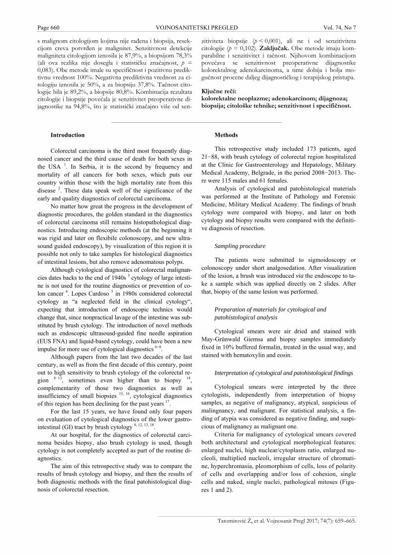
Page 660 VOJNOSANITETSKI PREGLED Vol. 74, No 7
Tatomirović Ž, et al. Vojnosanit Pregl 2017; 74(7): 659–665.
s malignom citologijom kojima nije rađena i biopsija, resek-cijom creva potvrđen je malignitet. Senzitivnost detekcije maligniteta citologijom iznosila je 87,9%, a biopsijom 78,3% (ali ova rezlika nije dosegla i statističku značajnost, p = 0,083). Obe metode imale su specifičnost i pozitivnu predik-tivnu vrednost 100%. Negativna prediktivna vrednost za ci-tologiju iznosila je 50%, a za biopsiju 37,8%. Tačnost cito-logije bila je 89,2%, a biopsije 80,8%. Kombinacija rezultata citologije i biopsije povećala je senzitivitet preoperativne di-jagnostike na 94,8%, što je statistički značajno više od sen-
zitiviteta biopsije (p < 0,001), ali ne i od senzitiviteta citologije (p = 0,102). Zaključak. Obe metode imaju kom-parabilne i senzitivitet i tačnost. Njihovom kombinacijom povećava se senzitivnost preoperativne dijagnostike kolorektalnog adenokarcinoma, a time dobija i bolja mo-gućnost procene daljeg dijagnostičkog i terapijskog pristupa. Ključne reči: kolorektalne neoplazme; adenokarcinom; dijagnoza; biopsija; citološke tehnike; senzitivnost i specifičnost.
Introduction
Colorectal carcinoma is the third most frequently diag-nosed cancer and the third cause of death for both sexes in the USA 1. In Serbia, it is the second by frequency and mortality of all cancers for both sexes, which puts our country within those with the high mortality rate from this disease 2. These data speak well of the significance of the early and quality diagnostics of colorectal carcinoma.
No matter how great the progress in the development of diagnostic procedures, the golden standard in the diagnostics of colorectal carcinoma still remains histopathological diag-nostics. Introducing endoscopic methods (at the beginning it was rigid and later on flexibile colonoscopy, and new ultra-sound guided endoscopy), by visualization of this region it is possible not only to take samples for histological diagnostics of intestinal lesions, but also remove adenomatous polyps.
Although cytological diagnostics of colorectal malignan-cies dates backs to the end of 1940s 3 cytology of large intesti-ne is not used for the routine diagnostics or prevention of co-lon cancer 4. Lopes Cardoso 5 in 1980s considered colorectal cytology as “a neglected field in the clinical cytology“, expecting that introduction of endoscopic technics would change that, since nonpractical lavage of the intestine was sub-stituted by brush cytology. The introduction of novel methods such as endoscopic ultrasousd-guided fine needle aspiration (EUS FNA) and liquid-based cytology, could have been a new impulse for more use of cytological diagnostics 6−8.
Although papers from the last two decades of the last century, as well as from the first decade of this century, point out to high sensitivity to brush cytology of the colorectal re-gion 8−13, sometimes even higher than to biopsy 14, complementarity of those two diagnostics as well as insufficiency of small biopsies 15, 16, cytological diagnostics of this region has been declining for the past years 17.
For the last 15 years, we have found only four papers on evaluation of cytological diagnostics of the lower gastro-intestinal (GI) tract by brush cytology 8, 12, 13, 18.
At our hospital, for the diagnostics of colorectal carci-noma besides biopsy, also brush cytology is used, though cytology is not completely accepted as part of the routine di-agnostics.
The aim of this retrospective study was to compare the results of brush cytology and biopsy, and then the results of both diagnostic methods with the final patohistological diag-nosis of colorectal resection.
Methods
This retrospective study included 173 patients, aged 21−88, with brush cytology of colorectal region hospitalized at the Clinic for Gastroenterology and Hepatology, Military Medical Academy, Belgrade, in the period 2008−2013. The-re were 115 males and 61 females.
Analysis of cytological and patohistological materials was performed at the Institute of Pathology and Forensic Medicine, Military Medical Academy. The findings of brush cytology were compared with biopsy, and later on both cytology and biopsy results were compared with the definiti-ve diagnosis of resection.
Sampling procedure
The patients were submitted to sigmoidoscopy or colonoscopy under short analgosedation. After visualization of the lesion, a brush was introduced via the endoscope to ta-ke a sample which was applied directly on 2 slides. After that, biopsy of the same lesion was performed.
Preparation of materials for cytological and patohistological analysis
Cytological smears were air dried and stained with May-Grünwald Giemsa and biopsy samples immediately fixed in 10% buffered formalin, treated in the usual way, and stained with hematoxylin and eosin.
Interpretation of cytological and patohistological findings
Cytological smears were interpreted by the three cytologists, independently from interpretation of biopsy samples, as negative of malignancy, atypical, suspicious of malingnancy, and malignant. For statistical analysis, a fin-ding of atypia was considered as negative finding, and suspi-cious of malignancy as malignant one.
Criteria for malignancy of cytological smears covered both architectural and cytological morphological features: enlarged nuclei, high nuclear/cytoplasm ratio, enlarged nu-cleoli, multiplied nucleoli, irregular structure of chromati-ne, hyperchromasia, pleomorphism of cells, loss of polarity of cells and overlapping and/or loss of cohesion, single cells and naked, single nuclei, pathological mitoses (Figu-res 1 and 2).
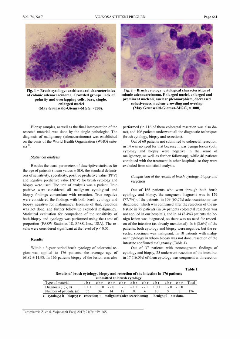
Vol. 74, No 7 VOJNOSANITETSKI PREGLED Page 661
Tatomirović Ž, et al. Vojnosanit Pregl 2017; 74(7): 659–665.
Fig. 1 − Brush cytology: architectural characteristics of colonic adenocarcinoma. Crowded groups, lack of
polarity and overlapping cells, bare, single, enlarged nuclei
(May Grunwald-Giemsa-MGG, ×200).
Fig. 2 − Brush cytology: cytological characteristics of
colonic adenocarcinoma. Enlarged nuclei, enlarged and prominent nucleoli, nuclear pleomorphism, decreased
cohesiveness, nuclear crowding and overlap (May Grunwald-Giemsa-MGG, ×1000)
Table 1 Results of brush cytology, biopsy and resection of the intestine in 176 patients
submitted to brush cytology Type of material c b r c b r c b r c b r c b r c b r c b r c b r c b r Total Diagnosis (+, -, 0) + + + + + 0 - - 0 + - + - + + - - + + 0 + + - 0 - + 0 Number of patients, (n) 75 34 14 17 8 6 10 9 3 176
c – cytology; b – biopsy; r – resection; + – malignant (adenocarcinoma); - – benign; 0 – not done.
Biopsy samples, as well as the final interpretation of the resected material, was done by the single pathologist. The diagnosis of malignancy (adenocarcinoma) was established on the basis of the World Health Organization (WHO) crite-ria 19.
Statistical analysis
Besides the usual parameters of descriptive statistics for the age of patients (mean values ± SD), the standard definiti-ons of sensitivity, specificity, positive predictive value (PPV) and negative predictive value (NPV) for brush cytology and biopsy were used. The unit of analysis was a patient. True positive were considered all malignant cytological and biopsy findings concordant with resection. True negative were considered the findings with both brush cytology and biopsy negative for malignancy. Because of that, resection was not done, and further follow up excluded malignancy. Statistical evaluation for comparison of the sensitivity of both biopsy and cytology was performed using the t-test of proportion (PASW Statistics 18, SPSS, Inc., USA). The re-sults were considered significant at the level of p < 0.05.
Results
Within a 3-year period brush cytology of colorectal re-gion was applied to 176 patients, the average age of 68.82 ± 11.98. In 166 patients biopsy of the lesion was also
performed (in 116 of them colorectal resection was also do-ne), and 106 patients underwent all the diagnostic techniques (brush cytology, biopsy and resection).
Out of 60 patients not submitted to colorectal resection, in 14 was no need for that because it was benign lesion (both cytology and biopsy were negative in the sense of malignancy, as well as further follow-up), while 46 patients continued with the treatment in other hospitals, so they were excluded from statistical analysis.
Comparison of the results of brush cytology, biopsy and resection
Out of 166 patients who went through both brush cytology and biopsy, the congruent diagnosis was in 129 (77.7%) of the patients: in 109 (65.7%) adenocarcinoma was diagnosed, which was confirmed after the resection of the in-testine in 75 patients (in 34 patients colorectal resection was not applied in our hospital), and in 14 (8.4%) patients the be-nign lesion was diagnosed, so there was no need for resecti-on of the intestine (as already mentioned). In 6 (3.6%) of the patients, both cytology and biopsy were negative, but the re-sected specimen was malignant. In 10 patients with malig-nant cytology in whom biopsy was not done, resection of the intestine confirmed malignancy (Table 1).
Out of 37 patients with noncongruent findings of cytology and biopsy, 25 underwent resection of the intestine: in 17 (16.0%) of them cytology was congruent with resection
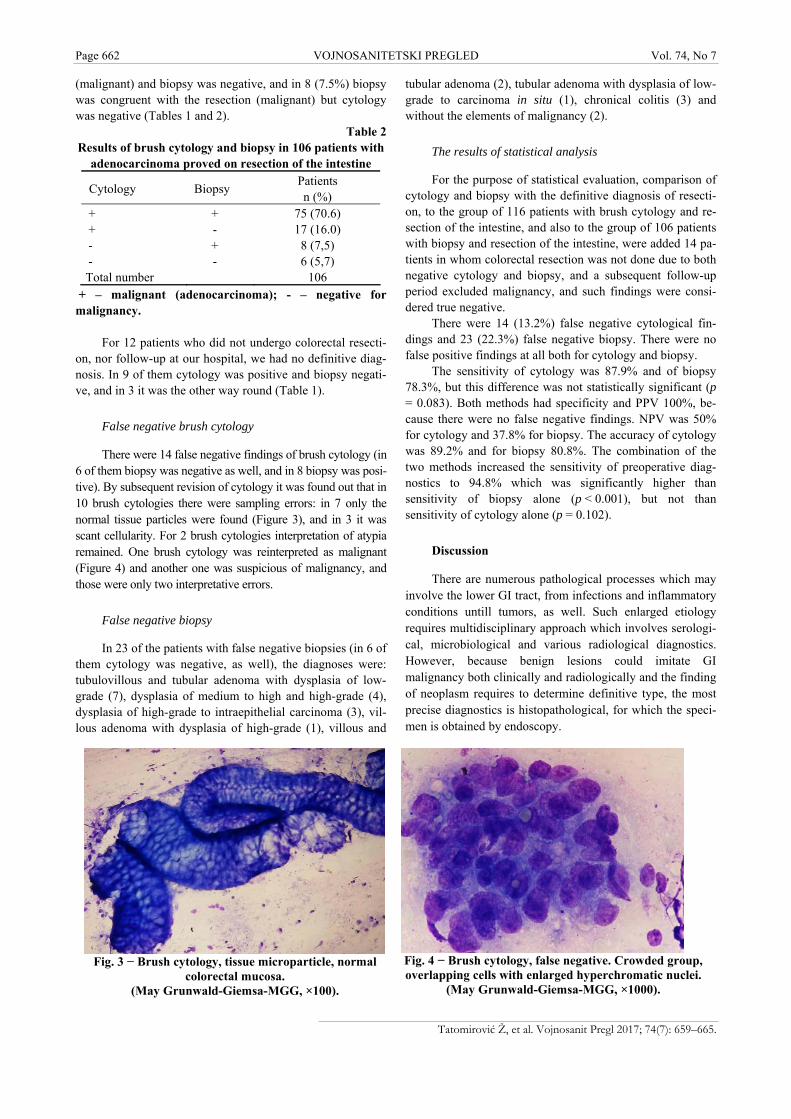
Page 662 VOJNOSANITETSKI PREGLED Vol. 74, No 7
Tatomirović Ž, et al. Vojnosanit Pregl 2017; 74(7): 659–665.
Fig. 3 − Brush cytology, tissue microparticle, normal
colorectal mucosa. (May Grunwald-Giemsa-MGG, ×100).
Fig. 4 − Brush cytology, false negative. Crowded group, overlapping cells with enlarged hyperchromatic nuclei.
(May Grunwald-Giemsa-MGG, ×1000).
(malignant) and biopsy was negative, and in 8 (7.5%) biopsy was congruent with the resection (malignant) but cytology was negative (Tables 1 and 2).
Table 2 Results of brush cytology and biopsy in 106 patients with
adenocarcinoma proved on resection of the intestine
Cytology Biopsy Patients n (%)
+ + 75 (70.6) + - 17 (16.0) - + 8 (7,5) - - 6 (5,7)
Total number 106 + – malignant (adenocarcinoma); - – negative for malignancy.
For 12 patients who did not undergo colorectal resecti-on, nor follow-up at our hospital, we had no definitive diag-nosis. In 9 of them cytology was positive and biopsy negati-ve, and in 3 it was the other way round (Table 1).
False negative brush cytology
There were 14 false negative findings of brush cytology (in 6 of them biopsy was negative as well, and in 8 biopsy was posi-tive). By subsequent revision of cytology it was found out that in 10 brush cytologies there were sampling errors: in 7 only the normal tissue particles were found (Figure 3), and in 3 it was scant cellularity. For 2 brush cytologies interpretation of atypia remained. One brush cytology was reinterpreted as malignant (Figure 4) and another one was suspicious of malignancy, and those were only two interpretative errors.
False negative biopsy
In 23 of the patients with false negative biopsies (in 6 of them cytology was negative, as well), the diagnoses were: tubulovillous and tubular adenoma with dysplasia of low-grade (7), dysplasia of medium to high and high-grade (4), dysplasia of high-grade to intraepithelial carcinoma (3), vil-lous adenoma with dysplasia of high-grade (1), villous and
tubular adenoma (2), tubular adenoma with dysplasia of low-grade to carcinoma in situ (1), chronical colitis (3) and without the elements of malignancy (2).
The results of statistical analysis
For the purpose of statistical evaluation, comparison of cytology and biopsy with the definitive diagnosis of resecti-on, to the group of 116 patients with brush cytology and re-section of the intestine, and also to the group of 106 patients with biopsy and resection of the intestine, were added 14 pa-tients in whom colorectal resection was not done due to both negative cytology and biopsy, and a subsequent follow-up period excluded malignancy, and such findings were consi-dered true negative.
There were 14 (13.2%) false negative cytological fin-dings and 23 (22.3%) false negative biopsy. There were no false positive findings at all both for cytology and biopsy.
The sensitivity of cytology was 87.9% and of biopsy 78.3%, but this difference was not statistically significant (p = 0.083). Both methods had specificity and PPV 100%, be-cause there were no false negative findings. NPV was 50% for cytology and 37.8% for biopsy. The accuracy of cytology was 89.2% and for biopsy 80.8%. The combination of the two methods increased the sensitivity of preoperative diag-nostics to 94.8% which was significantly higher than sensitivity of biopsy alone (p < 0.001), but not than sensitivity of cytology alone (p = 0.102).
Discussion
There are numerous pathological processes which may involve the lower GI tract, from infections and inflammatory conditions untill tumors, as well. Such enlarged etiology requires multidisciplinary approach which involves serologi-cal, microbiological and various radiological diagnostics. However, because benign lesions could imitate GI malignancy both clinically and radiologically and the finding of neoplasm requires to determine definitive type, the most precise diagnostics is histopathological, for which the speci-men is obtained by endoscopy.
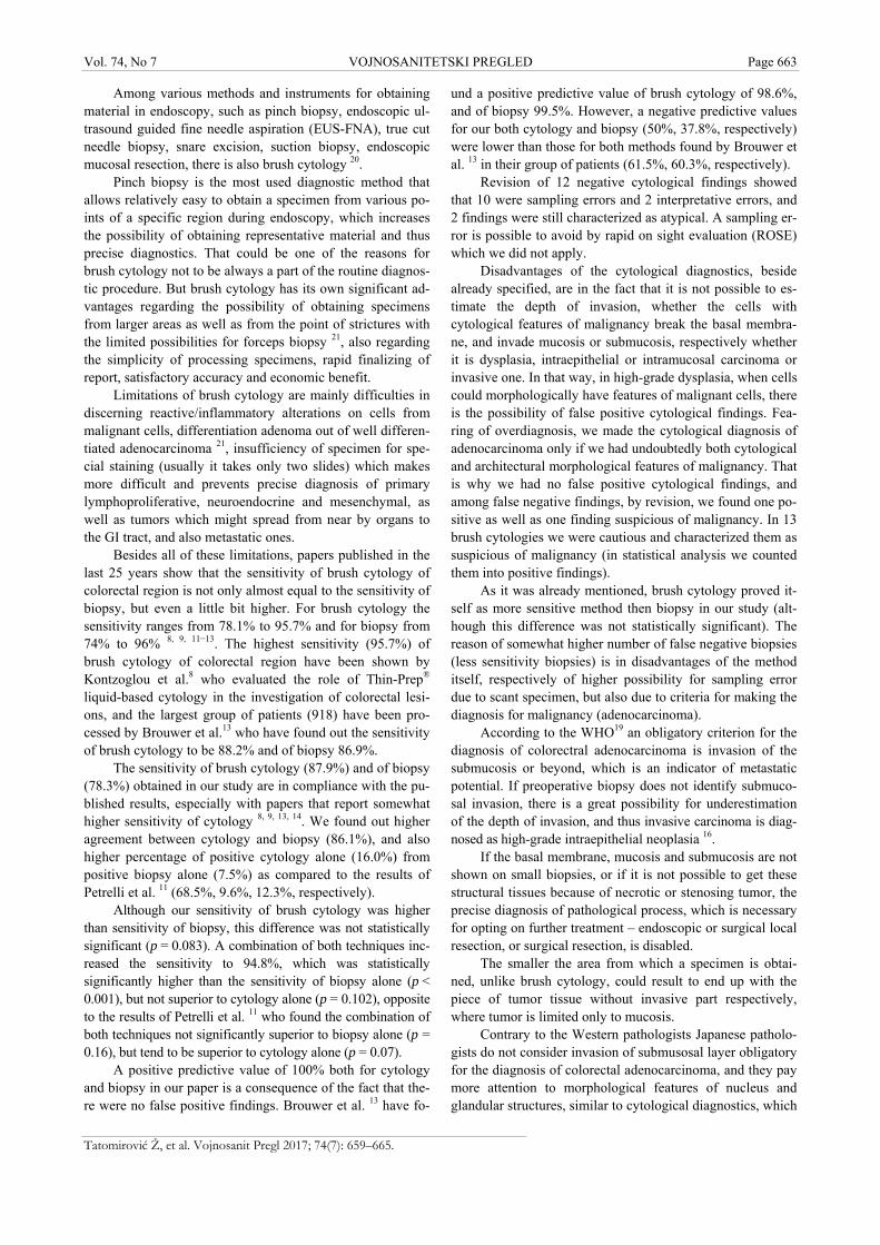
Vol. 74, No 7 VOJNOSANITETSKI PREGLED Page 663
Tatomirović Ž, et al. Vojnosanit Pregl 2017; 74(7): 659–665.
Among various methods and instruments for obtaining material in endoscopy, such as pinch biopsy, endoscopic ul-trasound guided fine needle aspiration (EUS-FNA), true cut needle biopsy, snare excision, suction biopsy, endoscopic mucosal resection, there is also brush cytology 20.
Pinch biopsy is the most used diagnostic method that allows relatively easy to obtain a specimen from various po-ints of a specific region during endoscopy, which increases the possibility of obtaining representative material and thus precise diagnostics. That could be one of the reasons for brush cytology not to be always a part of the routine diagnos-tic procedure. But brush cytology has its own significant ad-vantages regarding the possibility of obtaining specimens from larger areas as well as from the point of strictures with the limited possibilities for forceps biopsy 21, also regarding the simplicity of processing specimens, rapid finalizing of report, satisfactory accuracy and economic benefit.
Limitations of brush cytology are mainly difficulties in discerning reactive/inflammatory alterations on cells from malignant cells, differentiation adenoma out of well differen-tiated adenocarcinoma 21, insufficiency of specimen for spe-cial staining (usually it takes only two slides) which makes more difficult and prevents precise diagnosis of primary lymphoproliferative, neuroendocrine and mesenchymal, as well as tumors which might spread from near by organs to the GI tract, and also metastatic ones.
Besides all of these limitations, papers published in the last 25 years show that the sensitivity of brush cytology of colorectal region is not only almost equal to the sensitivity of biopsy, but even a little bit higher. For brush cytology the sensitivity ranges from 78.1% to 95.7% and for biopsy from 74% to 96% 8, 9, 11−13. The highest sensitivity (95.7%) of brush cytology of colorectal region have been shown by Kontzoglou et al.8 who evaluated the role of Thin-Prep® liquid-based cytology in the investigation of colorectal lesi-ons, and the largest group of patients (918) have been pro-cessed by Brouwer et al.13 who have found out the sensitivity of brush cytology to be 88.2% and of biopsy 86.9%.
The sensitivity of brush cytology (87.9%) and of biopsy (78.3%) obtained in our study are in compliance with the pu-blished results, especially with papers that report somewhat higher sensitivity of cytology 8, 9, 13, 14. We found out higher agreement between cytology and biopsy (86.1%), and also higher percentage of positive cytology alone (16.0%) from positive biopsy alone (7.5%) as compared to the results of Petrelli et al. 11 (68.5%, 9.6%, 12.3%, respectively).
Although our sensitivity of brush cytology was higher than sensitivity of biopsy, this difference was not statistically significant (p = 0.083). A combination of both techniques inc-reased the sensitivity to 94.8%, which was statistically significantly higher than the sensitivity of biopsy alone (p < 0.001), but not superior to cytology alone (p = 0.102), opposite to the results of Petrelli et al. 11 who found the combination of both techniques not significantly superior to biopsy alone (p = 0.16), but tend to be superior to cytology alone (p = 0.07).
A positive predictive value of 100% both for cytology and biopsy in our paper is a consequence of the fact that the-re were no false positive findings. Brouwer et al. 13 have fo-
und a positive predictive value of brush cytology of 98.6%, and of biopsy 99.5%. However, a negative predictive values for our both cytology and biopsy (50%, 37.8%, respectively) were lower than those for both methods found by Brouwer et al. 13 in their group of patients (61.5%, 60.3%, respectively).
Revision of 12 negative cytological findings showed that 10 were sampling errors and 2 interpretative errors, and 2 findings were still characterized as atypical. A sampling er-ror is possible to avoid by rapid on sight evaluation (ROSE) which we did not apply.
Disadvantages of the cytological diagnostics, beside already specified, are in the fact that it is not possible to es-timate the depth of invasion, whether the cells with cytological features of malignancy break the basal membra-ne, and invade mucosis or submucosis, respectively whether it is dysplasia, intraepithelial or intramucosal carcinoma or invasive one. In that way, in high-grade dysplasia, when cells could morphologically have features of malignant cells, there is the possibility of false positive cytological findings. Fea-ring of overdiagnosis, we made the cytological diagnosis of adenocarcinoma only if we had undoubtedly both cytological and architectural morphological features of malignancy. That is why we had no false positive cytological findings, and among false negative findings, by revision, we found one po-sitive as well as one finding suspicious of malignancy. In 13 brush cytologies we were cautious and characterized them as suspicious of malignancy (in statistical analysis we counted them into positive findings).
As it was already mentioned, brush cytology proved it-self as more sensitive method then biopsy in our study (alt-hough this difference was not statistically significant). The reason of somewhat higher number of false negative biopsies (less sensitivity biopsies) is in disadvantages of the method itself, respectively of higher possibility for sampling error due to scant specimen, but also due to criteria for making the diagnosis for malignancy (adenocarcinoma).
According to the WHO19 an obligatory criterion for the diagnosis of colorectral adenocarcinoma is invasion of the submucosis or beyond, which is an indicator of metastatic potential. If preoperative biopsy does not identify submuco-sal invasion, there is a great possibility for underestimation of the depth of invasion, and thus invasive carcinoma is diag-nosed as high-grade intraepithelial neoplasia 16.
If the basal membrane, mucosis and submucosis are not shown on small biopsies, or if it is not possible to get these structural tissues because of necrotic or stenosing tumor, the precise diagnosis of pathological process, which is necessary for opting on further treatment – endoscopic or surgical local resection, or surgical resection, is disabled.
The smaller the area from which a specimen is obtai-ned, unlike brush cytology, could result to end up with the piece of tumor tissue without invasive part respectively, where tumor is limited only to mucosis.
Contrary to the Western pathologists Japanese patholo-gists do not consider invasion of submusosal layer obligatory for the diagnosis of colorectal adenocarcinoma, and they pay more attention to morphological features of nucleus and glandular structures, similar to cytological diagnostics, which
Page 664 VOJNOSANITETSKI PREGLED Vol. 74, No 7
Tatomirović Ž, et al. Vojnosanit Pregl 2017; 74(7): 659–665.
gives high sensitivity to biopsy, but could lessen specificity, leading into more radical approach than it is necessary 22. These differences in diagnostic approach have been coordi-nated by the Vienna classification of GI epithelial neoplasia in which an intramucosal carcinoma has been accepted as the earliest form of invasive carcinoma 23. In the revised Vienna classification it is recommended to qualify a biopsy finding of intramucosal carcinoma as “at least 24. However, the esti-mation of efficiency of the revised Vienna classification for biopsy diagnostics of colorectal epithelial neoplasa has shown its high positive predictive value, and low sensitivity in the diagnostics of colorectal carcinoma, if pathologists use the invasion of submucosis or deeper invasion as an obligatory criterion for the diagnosis of carcinoma 25.
Analysis of our 23 false negative biopsies showed a sampling error. In 14 biopsies adenoma was diagnosed, dysplasia of low to medium-grade, chronic inflammation, while in 9 biopsies with high-grade dysplasia to intraepithe-lial carcinoma, invasion of muscular mucosa and deeper co-uld not be confirmed.
Having considered the advantages and disadvantages of both methods for sampling specimens, we find that in esti-mation of further diagnostic and therapeutic approach to co-lorectal lesions, brush cytology could be of great help when it is undoubtedly malignant, in cases when simultaneously biopsy could not estimate whether there is an invasion of submucosis. Brush cytology could reduce the need for rebiopsy, especially if there is a clear-cut clinical malignancy picture. In our 10 patients with a clear-cut local finding of carcinoma, only brush cytology was done.
Conclusion
We think that brush cytology could be the routine met-hod along with biopsy in the diagnosis of colorectal malignancy. Both methods have comparable both sensitivity and accuracy, and their combination increases sensitivity of the preoperative diagnostics of colorectal adenocarcinoma providing better estimation of further diagnostic and thera-peutic approach.
R E F E R E N C E S
1. Ferlay J, Soerjomataram I, Ervik M, Dikshit R, Eser S, Mathers C, et al. GLOBOCAN 2012 v1.0, Cancer Incidence and Mortality Worldwide: IARC CancerBase No 11. Lyon, France: Interna-tional Agency for Research on Cancer; 2013.
2. Vlajinac H, Sipetić-Grujicić S, Janković S, Marinković J, Kocev N, Marković-Denić L, et al. Burden of cancer in Serbia. Croat Med J 2006; 47(1): 134−41.
3. Wisseman CL Jr, Lemon HM, Lawrence KB. Cytologic diagnosis of cancer of the descending colon and rectum. Surg Gynecol Obstet 1949; 89(1): 24−30.
4. Schmitt F, Oliveira MH. Oesophagus and gastrointestinal tract. In: Gray W, Kocjan G, editors. Diagnostic cytopathology. Lon-don: Churchill Livingstone Elsevier; 2010. p. 279−81.
5. Cardoso L. Cytology of colorectal carcinoma In: Welvaart K, Blumgart LH, Kreuning J, editors. Colorectal Cancer. Hague: Leiden University Press; 1980. p. 43−52.
6. Vander Noot MR 3rd, Eloubeidi MA, Chen VK, Eltoum I, Jhala D, Jhala N, et al. Diagnosis of gastrointestinal tract lesions by en-doscopicultrasound-guided fine-needle aspiration biopsy. Can-cer 2004; 102(3): 157−63.
7. Fernández-Esparrach G, Alberghina N, Subtil JC, Vázquez-Sequeiros E, Florio V, Zozaya F, et al. Endoscopic ultrasound-guided fine needle aspiration is highly accurate for the diagnosis of perirec-tal recurrence of colorectal cancer. Dis Colon Rectum 2015; 58(5): 469−73.
8. Kontzoglou K, Moulakakis KG, Alexiou D, Safioleas MC, Nikiteas N, Giahnaki A, et al. The role of liquid-based cytology in the investigation of colorectal lesions: A cytohistopathological cor-relation and evaluation of diagnostic accuracy. Langenbecks Arch Surg 2007; 392(2): 189−95.
9. Farouk R, Dodds J, MacDonald AW, Young W, Duthie GS, Lee PW, et al. Feasibility study for use of brush cytology as a com-plementary method for diagnosis of rectal cancer. Dis Colon Rectum 1997; 40(5): 609−13.
10. Halpern M, Gal R, Rath-Wolfson L, Koren R, Weil R, Avni A. Brush cytology and biopsy in the diagnosis of colorectal can-cer. A comparison. Acta Cytol 1997; 41(3): 628−32.
11. Petrelli NJ, Létourneau R, Weber T, Nava ME, Rodriguez-Bigas M. Accuracy of biopsy and cytology for the preoperative diagnosis of colorectal adenocarcinoma. J Surg Oncol 1999; 71(1): 46−9.
12. Geramizadeh B, Hooshmand F, Kumar PV. Brush Cytology of Co-lorectal Malignancies. Acta Cytol 2003; 47(3): 431−4.
13. Brouwer R, MacDonald A, Matthews R, Gunn J, Monson JR, Hartley JE. Brush cytology for the diagnosis of colorectal cancer. Dis Colon Rectum 2009; 52(4): 598−601.
14. Ehya H, O'Hara BJ. Brush cytology in the diagnosis of colonic neoplasms. Cancer 1990; 66(7): 1563−7.
15. MacDonald AW, Tayyab M, Arsalani-Zadeh R, Hartley JE, Monson JR. Intramucosal carcinoma on biopsy reliably predicts inva-sive colorectal cancer. Ann Surg Oncol 2009; 16(12): 3267−70.
16. Wei XB, Gao XH, Wang H, Fu CG, Zheng WQ, Zheng JM, et al. More advanced or aggressive colorectal cancer is associated with a higher incidence of "high-grade intraepithelial neopla-sia" on biopsy-based pathological examination. Tech Colo-proctol 2012; 16(4): 277−83.
17. Kini SR. Alimentary tract: Esophagus, gastrointestinal, gall bladder and extrahepatic biliary tract (Exfoliative and aspira-tion cytology). In: Kini SR, editor. Color atlas of differential di-agnosis in exfoliative and aspiration cytopathology. Baltimore: Lippincott Williams&Wilkins; 2011. p. 305−7.
18. Yu GH, Nayar R, Furth EE. Adenocarcinoma in colonic brush-ing cytology: High-grade dysplasia as a diagnostic pitfall. Diagn Cytopathol 2001; 24(5): 364−8.
19. Hamilton SR, Aaltonen LA. World Health Organisation classifi-cation of tumors: Pathology and genetics of tumours of the di-gestive system. Lyon: IARC Press; 2000.
20. Technology Assessment Committee, Barkun A, Liu J, Carpenter S, Chotiprasidhi P, Chuttani R, et al. Update on endoscopic tissue sampling devices. Gastrointest Endosc 2006; 63(6): 741−5.
21. Conrad R, Castelino-Prabhu S, Cobb C, Raza A. Role of cytopa-thology in the diagnosis and management of gastrointestinal tract cancers. J Gastrointest Oncol 2012; 3(3): 285−98.
22. Schlemper RJ, Itabashi M, Kato Y, Lewin KJ, Riddell RH, Shimoda T, et al. Differences in the diagnostic criteria used by Japanese and Western pathologists to diagnose colorectal carcinoma. Cancer 1998; 82(1): 60−9.
Vol. 74, No 7 VOJNOSANITETSKI PREGLED Page 665
Tatomirović Ž, et al. Vojnosanit Pregl 2017; 74(7): 659–665.
23. Schlemper RJ, Riddell RH, Kato Y, Borchard F, Cooper HS, Dawsey SM, et al. The Vienna classification of gastrointestinal epithe-lial neoplasia. Gut 2000; 47(2): 251−5.
24. Dixon MF. Gastrointestinal epithelial neoplasia: Vienna revis-ited. Gut 2002; 51(1): 130−1.
25. Tominaga K, Fujinuma S, Endo T, Saida Y, Takahashi K, Maetani I. Efficacy of the revised Vienna Classification for diagnosing
colorectal epithelial neoplasias. World J Gastroenterol 2009; 15(19): 2351−6.
Received on January 12, 2016. Revised on March 6, 2016.
Accepted on March 7, 2016. Online First May, 2016.
Page 666 VOJNOSANITETSKI PREGLED Vojnosanit Pregl 2017; 74(7): 666–671.
S H O R T C O M M U N I C A T I O N S UDC: 539.1:61]::[617.3:617.581/.583
https://doi.org/10.2298/VSP151211150P
Combined bone scintigraphy with 99mTc-MDP and 99mTc-ciprofloxacin in differentiation of hip and knee prosthesis aseptic loosening and infection: A preliminary study
Kombinovana scintigrafija kostiju sa 99mTc-MDP i 99mTc-ciprofloksacinom u razlikovanju aseptične nestabilnosti od infekcije periprotetskog tkiva zgloba
kuka i kolena: preliminarna studija
Dragan Pucar*, Zoran Janković*†, Zoran Baščarević‡, Srdjan Starčevi憧,
Milica Čizmić†||, Marija Radulović*, Marija Šišić*, Sanja Dugonjić*, Ljiljana Jauković*†, Boris Ajdinović*†
Military Medical Academy, *Institute of Nuclear Medicine, §Clinic for Orthopedic Surgery and Traumatology, ||Clinic of Endocrinology, Belgrade, Serbia; University of
Defence, †Faculty of Medicine of the Military Medical Academy, Belgrade, Serbia; University of Belgrade, Faculty of Medicine ‡Institute for Orthopedic Surgery “Banjica”,
Belgrade, Serbia
Abstract Background/Aim. Although the number of new primary implantation of hip and knee prostheses every year increa-ses, the rate of failed arthroplasty is nearly the same. The main question is whether it is an aseptic instability or instability caused by infection. The aim of this preliminary study was an attempt with combined 99mTc-ciprofloxacin and 99mTc-methylene diphosphonate (MDP) bone scintigraphy to improve diagnostic accuracy in the differen-tiation of hip and knee prosthesis aseptic loosening and pe-riprosthetic joint infection. Methods. Inclusion criteria of patients for this study were based on suspected periprosthe-tic joint infection: painful prosthetic joint, restricted joint movements and increased value of erythrocyte sedimentati-on rate or levels of C-reactive protein. We examined 20 pa-tients with implanted 14 hip and 6 knee prosthesis. All pati-ents also underwent plain radiography of suspected joint. In all patients, three-phase 99mTc-MDP bone scintigraphy was performed. Three to five days after the bone scan, we per-formed scintigraphy using 99mTc-ciprofloxacin with the cal-culation of accumulation index. Periprosthetic joint infecti-on was confirmed on the basis of microbiological findings.
Results. Periprosthetic joint infection was confirmed in fo-urteen of twenty observed joints, in five of them the aseptic loosening was present and in one patient’s symptoms were not related to the prosthesis (poor biomechanics of prosthe-tic joints caused by weaknesses of muscle). Estimated sensitivity/specificity for 99mTc-MDP bone scintigraphy alone were 100/17%; for 99mTc-ciprofloxacin scintigraphy were 85,7/100%. Sensitivity and specificity were 92,3% and 83,3%, respectively for results obtained with combined as-sessment by both methods. Our study confirmed the high negative predictive value of 99mTc-MDP bone scan. The negative result of bone scan virtually excludes the possibility of periprosthetic infection. On the other hand, positive fin-dings of 99mTc-MDP scintigraphy cannot with certainty con-firm the infection. Conclusion. Combined 99mTc-MDP scintigraphy with 99mTc-ciprofloxacin scintigraphy significantly increases the ability of differentiation of aseptic loosening from periprosthetic joint infection. Keywords: knee prosthesis; hip prosthesis; infection; diagnosis, differential; radionuclide imaging; technetium tc 99m medronate; technetium tc 99m ciprofloxacin.
Apstrakt Uvod/Cilj. Iako broj novo ugrađenih protetskih zglobova kuka i kolena svake godine raste, procenat neuspešnih ar-troplastika ostaje približno isti. Kao glavno pitanje nameće se, da li je u pitanju aseptična nestabilnost proteze ili je nes-tabilnost posledica neprepoznate infekcije. Cilj ove prelimi-
narne studije je pokušaj da se kombinovanom upotrebom scintigrafije kostiju 99mTc obeleženim ciprofloksacinom i scintigrafije 99mTc-metilen-difosfonatom (MDP) unapredi dijagnostička tačnost razlikovanja aseptične nestabilnosti i periprotetske infekcije kod protetskih zglobova kuka i kole-na. Metode. Kriterijum za ulazak u ovu studiju bio je: bol u protetskom zglobu, ograničeni pokreti u zglobu i uvećane
Correspondence to: Dragan Pucar, Military Medical Academy, Institute of Nuclear Medicine, Crnotravska 17, 11 000 Belgrade, Serbia. E-mail: [email protected]
Vol. 74, No 7 VOJNOSANITETSKI PREGLED Page 667
vrednosti nespecifičnih znakova inflamacije, C-reaktivnog proteina i sedimentacije eritrocita. Ispitivanjem je obuhva-ćeno 20 bolesnika sa 14 ugrađenih proteza kuka i 6 proteza kolena. Kod svih bolesnika učinjena je radiografija protet-skog zgloba, kao i trofazna scintigrafija kostiju sa 99mTc-MDP. Tri do pet dana nakon scintigrafije kostiju urađena je i scintigrafija 99mTc-ciprofloksacinom sa određivanjem indeksa vezivanja. Infekcija periprotetskog tkiva je pot-vrđena mikrobiološkim nalazom. Rezultati. U 14 od 20 posmatranih protetskih zglobova potvrđena je infekcija, u pet slučajeva se radilo o aseptičnoj nestabilnosti protetskog zgloba, a u jednom slučaju simptomi nisu bili vezani za sam protetski zglob već je u pitanju bila loša biomehanika zgloba uzrokovana izrazitom slabošću abduktorne muskulature. Dobijena osetljivost i specifičnost za scintigrafiju kostiju pomoću 99mTc-MDP iznosila je 100% i 17%; za scintigrafiju
99mTc-ciprofloksacinom bila je 85,7% i 100%, a za kombi-novani nalaz obe metode iznosila je 92,3% i 83,3%. Studija je potvrdila visoku negativnu prediktivnu vrednost scinti-grafije kostiju u dijagnostici periprotetske infekcije. Drugim rečima negativna scintigrafija kostiju praktično isključuje mogućnost postojanja infekcije. S druge strane pozitivan nalaz scintigrafije kostiju ne može sa sigurnošću da potvrdi postojanje infekcije. Zaključak. Kombinacija dve metode, 99mTc-MDP scintigrafije kostiju sa scintigrafijom 99mTc-ciprofloksacinom znatno povećava mogućnost razlikovanja aseptične nestabilnosti i periprotetske infekcije zgloba. Ključne reči: koleno, proteza; kuk, proteza; infekcija; dijagnoza, diferencijalna; scintigrafija; tehnecijum tc 99m medronat; tehnecijum tc 99m ciprofloksacin.
Introduction
The most remarkable advances in surgery and medicine during the last five decades were the joint replacement. Joint replacement is an effective intervention improving patients’ quality of life, providing pain relief, renovation of joint func-tion, improve patient mobility and independence 1. The majority of implanted artificial joint are hip and knee prost-heses (more than 95%). Although the hip or knee joint repla-cement is highly successful surgical interventions, these pro-cedures may be complicated with an aseptic failure and peri-prosthetic joint infection (PJI). More than 25% of all prost-heses will demonstrate evidence of loosening, often after a revision arthroplasty 2. Infection, although unfrequented, is the most serious complication. In patients with primary joint replacement, the infection rate in the first 2 years is usually in range of 0,5–2%. The reported infection rates are probably underestimated, since many cases understood as an aseptic failure may be due to unrecognized infection. Infection rates after surgical revision are usually higher (25% to 40%) than after primary replacement 3. The most important risk factors for arthroplasty failure are postoperative surgical site infecti-on, previous total hip or knee arthroplasty, older age, malnut-rition, a joint disease like rheumatoid arthritis, obesity, dia-betes mellitus, remote infection 4, 5. Commonly isolated mic-roorganisms are Gram-positive cocci: coagulase-negative staphylococci, S. aureus and enterococci (65%) 6. Increased peripheral blood leukocytes, erythrocyte sedimentation rate and C-reactive protein levels are neither sensitive nor speci-fic for PJI. Infection differentiation from aseptic loosening is important because treatments are completely different. Plain radiography is not enough sensitive or specific. Findings such as radiolucency, osteolysis and migration may be pre-sent in both infection and aseptic loosening 7. Computed tomography provides better contrast between normal and ab-normal tissue, useful in detecting joint effusion, sinus tracts and bone erosion. Magnetic resonance imaging (MRI) can only be performed in patients with titanium or tantalum im-plants.
Radionuclide imaging is not affected by metallic hardware and is the current imaging modality of choice for evaluation of suspected joint replacement infection. Bone scintigraphy is extremely sensitive for detecting bone remo-deling changes around prosthetic joints but because of low specificity for infection, cannot determine the cause of failu-re. Ciprofloxacin labeled with 99mTc was introduced at the end of last decade of the 20th century. That radiopharmaceu-tical is more specific in infection but not enough if only ba-sed on the subjective visual estimation of scintigrams 8, 9. Calculation of accumulation index is the only reasonable way to determinate increase scintigrams uptake between aseptic inflammation from infection 10, 11.
The aim of this preliminary study was attempt with combined 99mTc-ciprofloxacin and 99mTc-methylene diphosp-honate (MDP) bone scintigraphy to improve diagnostic accuracy in differentiation of hip and knee prosthesis aseptic loosening and infection.
Methods
In this preliminary study, we examined twenty patients with 14 implanted hip and 6 knee prosthesis. The study was performed during three months in the period from August to November 2015. Criteria for including patients in the study were suspected PJI based on: painful prosthetic joint (espe-cially in locomotion but also in peace), restricted joint mo-vements and increased value of erythrocyte sedimentation rate or levels of C-reactive protein. All patients underwent plain radiography of suspected joint.
In all patients, three phase bone scintigraphy was per-formed after intravenous (iv) aplication of 555 MBq of 99mTc-MDP on ADAC vertex gamma camera utilizing the large field of view dual detectors filtered with low energy all-purpose collimator. Various times of imaging were per-formed in the supine position: 0-5 minute (15 frames in the first minute and 6 frames in further 4 minutes), 6-10 minutes (one frame with 300 seconds duration) and late static ima-ging after 3 hours. Scintigrams were analysed visually without any quantification.
Pucar D. et al, Vojnosanit Pregl 2017; 74(7): 666–671.
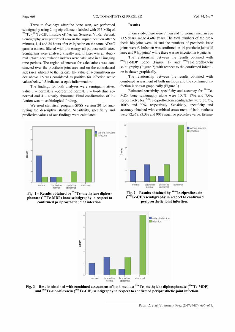
Page 668 VOJNOSANITETSKI PREGLED Vol. 74, No 7
Fig. 1 – Results obtained by 99mTc–methylene diphos-phonate (99mTc-MDP) bone scintigraphy in respect to
confirmed periprosthetic joint infection.
Fig. 2 – Results obtained by 99mTc-ciprofloxacin
(99mTc-CIP) scintigraphy in respect to confirmed periprosthetic joint infection.
Three to five days after the bone scan, we performed scintigraphy using 2 mg ciprofloxacin labeled with 555 MBq of 99mTc (99mTc-CIP, Institute of Nuclear Sciences Vinča, Serbia). Scintigraphy was performed also in the supine position after 5 minutes, 1, 4 and 24 hours after iv injection on the same ADAC gamma camera filtered with low energy all-purpose collimator. Scintigrams were analysed visually and, if there was an abnor-mal uptake, accumulation indexes were calculated in all imaging time periods. The region of interest for calculations was con-structed over the prosthetic joint area and on the contralateral side (area adjacent to the lesion). The value of accumulation in-dex above 1.5 was considered as positive for infection while values below 1.5 indicated aseptic inflammation.
The findings for both analyses were semiquantitative: value 1 – normal, 2 –borderline normal, 3 – borderline ab-normal and 4 – clearly abnormal. Final confirmation of in-fection was microbiological finding.
We used statistical program SPSS version 20 for ana-lyzing the descriptive statistic. Sensitivity, specificity and predictive values of our findings were calculated.
Results
In our study, there were 7 men and 13 women median age 73.5 years, range 43-82 years. The total numbers of the pros-thetic hip joint were 14 and the numbers of prosthetic knee joints were 6. Infection was confirmed in 14 prosthetic joints (5 knee and 9 hip joints) while there was no infection in 6 patients.
The relationship between the results obtained with 99mTc-MDP bone (Figure 1) and 99mTc-ciprofloxacin scintigraphy (Figure 2) with respect to the confirmed infecti-on is shown graphically.
The relationship between the results obtained with combined assessment of both methods and the confirmed in-fection is shown graphically (Figure 3).
Estimated sensitivity, specificity and accuracy for 99mTc-MDP bone scintigraphy alone were 100%, 17% and 75%, respectively; for 99mTc-ciprofloxacin scintigraphy were 85,7%, 100% and 90%, respectively. Sensitivity, specificity and accuracy obtained with combined assessment of both methods were 92,3%, 83,3% and 90% negative predictive value. Estima-
Fig. 3 – Results obtained with combined assessment of both metods: 99mTc–methylene diphosphonate (99mTc-MDP)
and 99mTc-ciprofloxacin (99mTc-CIP) scintigraphy in respect to confirmed periprosthetic joint infection.
Pucar D. et al, Vojnosanit Pregl 2017; 74(7): 666–671.
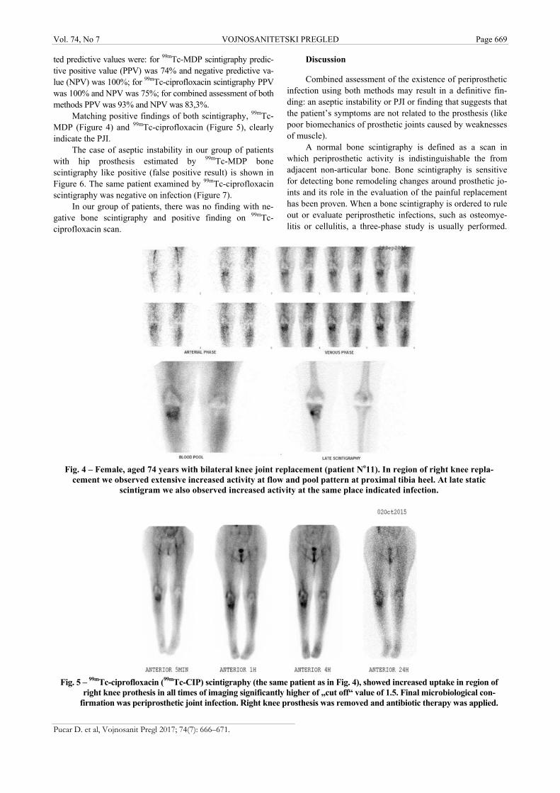
Vol. 74, No 7 VOJNOSANITETSKI PREGLED Page 669
ted predictive values were: for 99mTc-MDP scintigraphy predic-tive positive value (PPV) was 74% and negative predictive va-lue (NPV) was 100%; for 99mTc-ciprofloxacin scintigraphy PPV was 100% and NPV was 75%; for combined assessment of both methods PPV was 93% and NPV was 83,3%.
Matching positive findings of both scintigraphy, 99mTc-MDP (Figure 4) and 99mTc-ciprofloxacin (Figure 5), clearly indicate the PJI.
The case of aseptic instability in our group of patients with hip prosthesis estimated by 99mTc-MDP bone scintigraphy like positive (false positive result) is shown in Figure 6. The same patient examined by 99mTc-ciprofloxacin scintigraphy was negative on infection (Figure 7).
In our group of patients, there was no finding with ne-gative bone scintigraphy and positive finding on 99mTc-ciprofloxacin scan.
Discussion
Combined assessment of the existence of periprosthetic infection using both methods may result in a definitive fin-ding: an aseptic instability or PJI or finding that suggests that the patient’s symptoms are not related to the prosthesis (like poor biomechanics of prosthetic joints caused by weaknesses of muscle).
A normal bone scintigraphy is defined as a scan in which periprosthetic activity is indistinguishable the from adjacent non-articular bone. Bone scintigraphy is sensitive for detecting bone remodeling changes around prosthetic jo-ints and its role in the evaluation of the painful replacement has been proven. When a bone scintigraphy is ordered to rule out or evaluate periprosthetic infections, such as osteomye-litis or cellulitis, a three-phase study is usually performed.
Fig. 4 – Female, aged 74 years with bilateral knee joint replacement (patient No11). In region of right knee repla-
cement we observed extensive increased activity at flow and pool pattern at proximal tibia heel. At late static scintigram we also observed increased activity at the same place indicated infection.
Fig. 5 – 99mTc-ciprofloxacin (99mTc-CIP) scintigraphy (the same patient as in Fig. 4), showed increased uptake in region of
right knee prothesis in all times of imaging significantly higher of „cut off“ value of 1.5. Final microbiological con-firmation was periprosthetic joint infection. Right knee prosthesis was removed and antibiotic therapy was applied.
Pucar D. et al, Vojnosanit Pregl 2017; 74(7): 666–671.
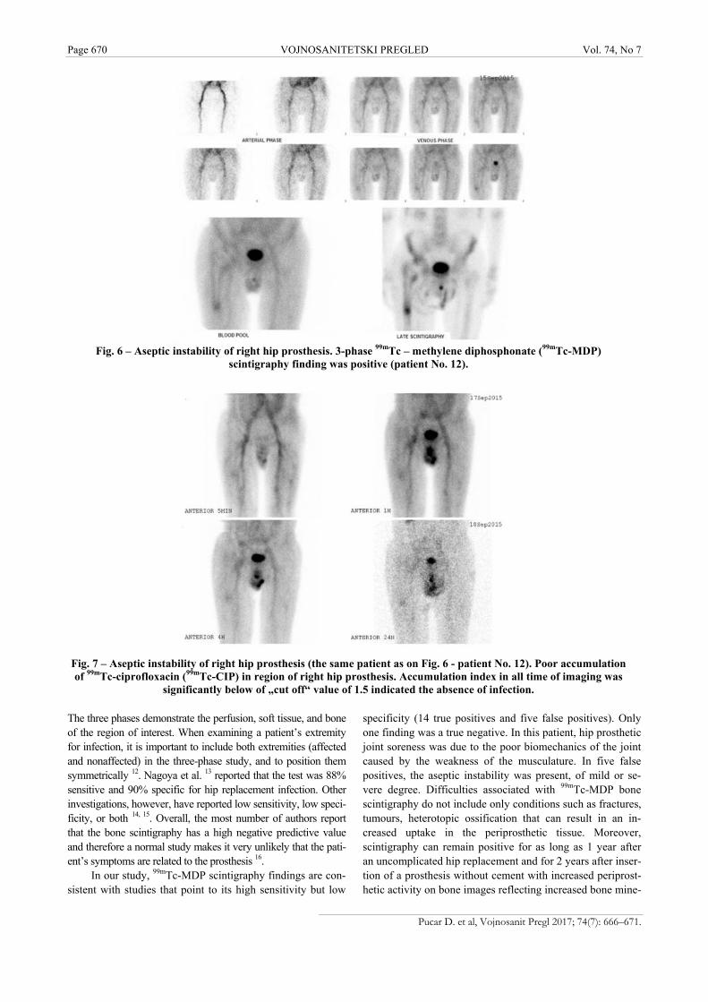
Page 670 VOJNOSANITETSKI PREGLED Vol. 74, No 7
Fig. 6 – Aseptic instability of right hip prosthesis. 3-phase 99mTc – methylene diphosphonate (99mTc-MDP)
scintigraphy finding was positive (patient No. 12).
Fig. 7 – Aseptic instability of right hip prosthesis (the same patient as on Fig. 6 - patient No. 12). Poor accumulation of 99mTc-ciprofloxacin (99mTc-CIP) in region of right hip prosthesis. Accumulation index in all time of imaging was
significantly below of „cut off“ value of 1.5 indicated the absence of infection.
The three phases demonstrate the perfusion, soft tissue, and bone of the region of interest. When examining a patient’s extremity for infection, it is important to include both extremities (affected and nonaffected) in the three-phase study, and to position them symmetrically 12. Nagoya et al. 13 reported that the test was 88% sensitive and 90% specific for hip replacement infection. Other investigations, however, have reported low sensitivity, low speci-ficity, or both 14, 15. Overall, the most number of authors report that the bone scintigraphy has a high negative predictive value and therefore a normal study makes it very unlikely that the pati-ent’s symptoms are related to the prosthesis 16.
In our study, 99mTc-MDP scintigraphy findings are con-sistent with studies that point to its high sensitivity but low
specificity (14 true positives and five false positives). Only one finding was a true negative. In this patient, hip prosthetic joint soreness was due to the poor biomechanics of the joint caused by the weakness of the musculature. In five false positives, the aseptic instability was present, of mild or se-vere degree. Difficulties associated with 99mTc-MDP bone scintigraphy do not include only conditions such as fractures, tumours, heterotopic ossification that can result in an in-creased uptake in the periprosthetic tissue. Moreover, scintigraphy can remain positive for as long as 1 year after an uncomplicated hip replacement and for 2 years after inser-tion of a prosthesis without cement with increased periprost-hetic activity on bone images reflecting increased bone mine-
Pucar D. et al, Vojnosanit Pregl 2017; 74(7): 666–671.
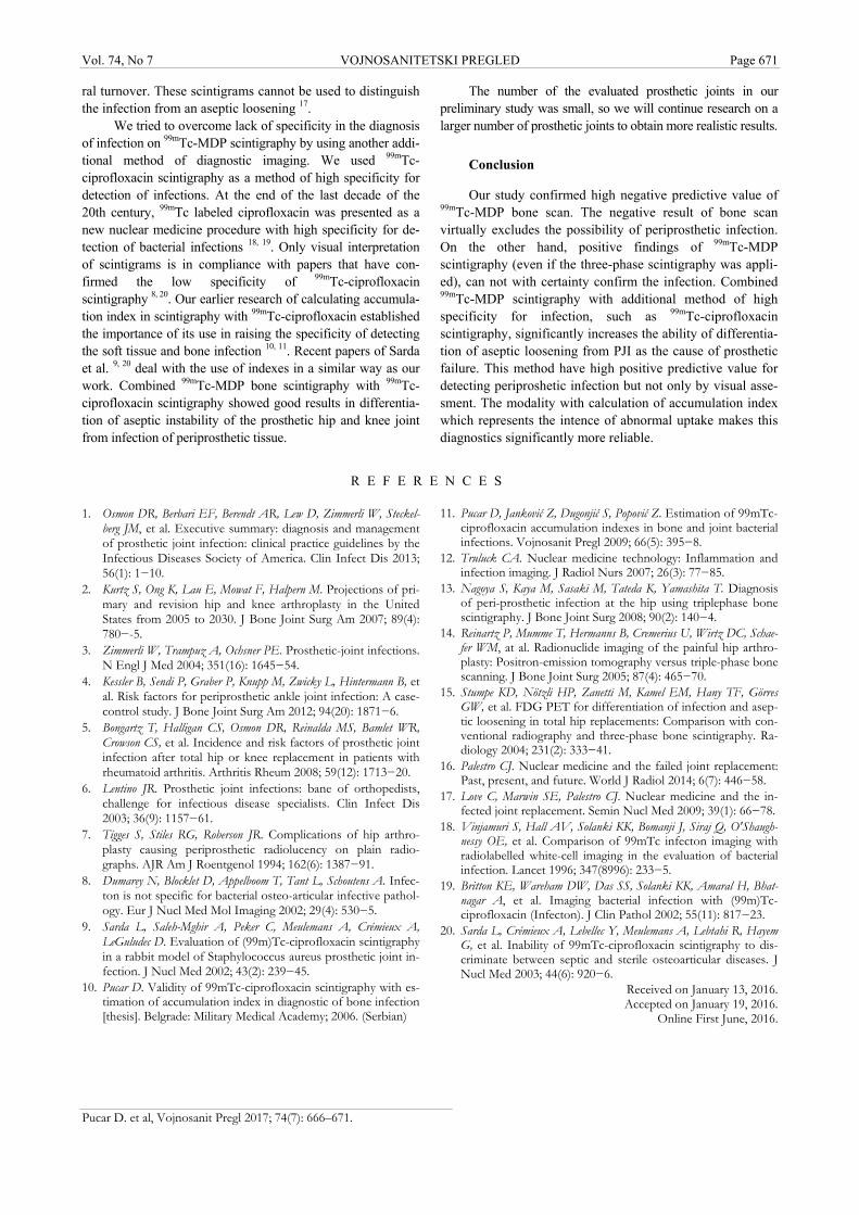
Vol. 74, No 7 VOJNOSANITETSKI PREGLED Page 671
Pucar D. et al, Vojnosanit Pregl 2017; 74(7): 666–671.
ral turnover. These scintigrams cannot be used to distinguish the infection from an aseptic loosening 17.
We tried to overcome lack of specificity in the diagnosis of infection on 99mTc-MDP scintigraphy by using another addi-tional method of diagnostic imaging. We used 99mTc-ciprofloxacin scintigraphy as a method of high specificity for detection of infections. At the end of the last decade of the 20th century, 99mTc labeled ciprofloxacin was presented as a new nuclear medicine procedure with high specificity for de-tection of bacterial infections 18, 19. Only visual interpretation of scintigrams is in compliance with papers that have con-firmed the low specificity of 99mTc-ciprofloxacin scintigraphy 8, 20. Our earlier research of calculating accumula-tion index in scintigraphy with 99mTc-ciprofloxacin established the importance of its use in raising the specificity of detecting the soft tissue and bone infection 10, 11. Recent papers of Sarda et al. 9, 20 deal with the use of indexes in a similar way as our work. Combined 99mTc-MDP bone scintigraphy with 99mTc-ciprofloxacin scintigraphy showed good results in differentia-tion of aseptic instability of the prosthetic hip and knee joint from infection of periprosthetic tissue.
The number of the evaluated prosthetic joints in our preliminary study was small, so we will continue research on a larger number of prosthetic joints to obtain more realistic results.
Conclusion
Our study confirmed high negative predictive value of 99mTc-MDP bone scan. The negative result of bone scan virtually excludes the possibility of periprosthetic infection. On the other hand, positive findings of 99mTc-MDP scintigraphy (even if the three-phase scintigraphy was appli-ed), can not with certainty confirm the infection. Combined 99mTc-MDP scintigraphy with additional method of high specificity for infection, such as 99mTc-ciprofloxacin scintigraphy, significantly increases the ability of differentia-tion of aseptic loosening from PJI as the cause of prosthetic failure. This method have high positive predictive value for detecting periproshetic infection but not only by visual asse-sment. The modality with calculation of accumulation index which represents the intence of abnormal uptake makes this diagnostics significantly more reliable.
R E F E R E N C E S
1. Osmon DR, Berbari EF, Berendt AR, Lew D, Zimmerli W, Steckel-berg JM, et al. Executive summary: diagnosis and management of prosthetic joint infection: clinical practice guidelines by the Infectious Diseases Society of America. Clin Infect Dis 2013; 56(1): 1−10.
2. Kurtz S, Ong K, Lau E, Mowat F, Halpern M. Projections of pri-mary and revision hip and knee arthroplasty in the United States from 2005 to 2030. J Bone Joint Surg Am 2007; 89(4): 780−-5.
3. Zimmerli W, Trampuz A, Ochsner PE. Prosthetic-joint infections. N Engl J Med 2004; 351(16): 1645−54.
4. Kessler B, Sendi P, Graber P, Knupp M, Zwicky L, Hintermann B, et al. Risk factors for periprosthetic ankle joint infection: A case-control study. J Bone Joint Surg Am 2012; 94(20): 1871−6.
5. Bongartz T, Halligan CS, Osmon DR, Reinalda MS, Bamlet WR, Crowson CS, et al. Incidence and risk factors of prosthetic joint infection after total hip or knee replacement in patients with rheumatoid arthritis. Arthritis Rheum 2008; 59(12): 1713−20.
6. Lentino JR. Prosthetic joint infections: bane of orthopedists, challenge for infectious disease specialists. Clin Infect Dis 2003; 36(9): 1157−61.
7. Tigges S, Stiles RG, Roberson JR. Complications of hip arthro-plasty causing periprosthetic radiolucency on plain radio-graphs. AJR Am J Roentgenol 1994; 162(6): 1387−91.
8. Dumarey N, Blocklet D, Appelboom T, Tant L, Schoutens A. Infec-ton is not specific for bacterial osteo-articular infective pathol-ogy. Eur J Nucl Med Mol Imaging 2002; 29(4): 530−5.
9. Sarda L, Saleh-Mghir A, Peker C, Meulemans A, Crémieux A, LeGuludec D. Evaluation of (99m)Tc-ciprofloxacin scintigraphy in a rabbit model of Staphylococcus aureus prosthetic joint in-fection. J Nucl Med 2002; 43(2): 239−45.
10. Pucar D. Validity of 99mTc-ciprofloxacin scintigraphy with es-timation of accumulation index in diagnostic of bone infection [thesis]. Belgrade: Military Medical Academy; 2006. (Serbian)
11. Pucar D, Janković Z, Dugonjić S, Popović Z. Estimation of 99mTc-ciprofloxacin accumulation indexes in bone and joint bacterial infections. Vojnosanit Pregl 2009; 66(5): 395−8.
12. Truluck CA. Nuclear medicine technology: Inflammation and infection imaging. J Radiol Nurs 2007; 26(3): 77−85.
13. Nagoya S, Kaya M, Sasaki M, Tateda K, Yamashita T. Diagnosis of peri-prosthetic infection at the hip using triplephase bone scintigraphy. J Bone Joint Surg 2008; 90(2): 140−4.
14. Reinartz P, Mumme T, Hermanns B, Cremerius U, Wirtz DC, Schae-fer WM, at al. Radionuclide imaging of the painful hip arthro-plasty: Positron-emission tomography versus triple-phase bone scanning. J Bone Joint Surg 2005; 87(4): 465−70.
15. Stumpe KD, Nötzli HP, Zanetti M, Kamel EM, Hany TF, Görres GW, et al. FDG PET for differentiation of infection and asep-tic loosening in total hip replacements: Comparison with con-ventional radiography and three-phase bone scintigraphy. Ra-diology 2004; 231(2): 333−41.
16. Palestro CJ. Nuclear medicine and the failed joint replacement: Past, present, and future. World J Radiol 2014; 6(7): 446−58.
17. Love C, Marwin SE, Palestro CJ. Nuclear medicine and the in-fected joint replacement. Semin Nucl Med 2009; 39(1): 66−78.
18. Vinjamuri S, Hall AV, Solanki KK, Bomanji J, Siraj Q, O'Shaugh-nessy OE, et al. Comparison of 99mTc infecton imaging with radiolabelled white-cell imaging in the evaluation of bacterial infection. Lancet 1996; 347(8996): 233−5.
19. Britton KE, Wareham DW, Das SS, Solanki KK, Amaral H, Bhat-nagar A, et al. Imaging bacterial infection with (99m)Tc-ciprofloxacin (Infecton). J Clin Pathol 2002; 55(11): 817−23.
20. Sarda L, Crémieux A, Lebellec Y, Meulemans A, Lebtahi R, Hayem G, et al. Inability of 99mTc-ciprofloxacin scintigraphy to dis-criminate between septic and sterile osteoarticular diseases. J Nucl Med 2003; 44(6): 920−6.
Received on January 13, 2016. Accepted on January 19, 2016.
Online First June, 2016.
Page 672 VOJNOSANITETSKI PREGLED Vojnosanit Pregl 2017; 74(7): 672–675.
Correspondence to: Marko Andjelković, Faculty of Dental Medicine, Department of Prosthodontics, Rankeova 4, 11 000 Belgrade, Serbia. E-mail: [email protected]
S H O R T C O M M U N I C A T I O N UDC: 579.61::616.31-002.4-08 DOI: 10.2298/VSP151014113L
Eradication of Helicobacter pylori in patients without gastric symptoms suffering from recurrent aphthous stomatitis: A pilot study
Eradikacija Helicobacter pylori kod bolesnika bez gastričkih simptoma koji imaju rekurentni aftozni stomatitis: pilot studija
Marina Latković*, Lazar Ranin†, Nevenka Teodorović*, Marko Andjelković‡
University of Belgrade, Faculty of Dental Medicine, *Department of Restorative Dentistry and Endodontics, ‡Department of Prosthodontics, Belgrade, Serbia;
University of Belgrade, Faculty of Medicine, †Institute of Microbiology and Immunology, Belgrade, Serbia
Abstract Background/Aim. Helicobacter (H.) pylori is a widespread bacterium and its involvement in pathogenesis of gastric diseases is well-known. However, H. pylori role in etiology of other histologically similar conditions, especially recurrent aphthous stomatitis (RAS) is still controversial. Research re-garding H. pylori and its association with RAS, as well as the treatment options were always conducted on patients with diagnosed gastric problems. The aim of this study was to determine whether H. pylori is present in the oral cavity of patients suffering from RAS but without any symptoms or medical history of gastric disease. Methods. A total of 15 patients with RAS participated in the study. None of the participants suffered from any gastrointestinal disorders. Two dental plaque samples from each participant were col-lected. The first was analyzed using rapid urease test and the second one was put in transport medium and sent for culti-vation. The sensitivity of H. pylori to antibiotics was estab-lished using disk diffusion method of sensitivity testing for every patient individually and adequate therapy was pre-scribed. Results. Before the treatment the mean annual re-currence rate of RAS was 8.1 ± 2.1, with the average num-ber of lesions being 3.9 ± 1.9. During the 12-month obser-vation period after the eradication therapy, none of the pa-tients reported recurrence of aphthous lesions. The treat-ment was successful in all cases. Conclusion. This study shows that RAS can be effectively treated by successful era-dication of oral H. pylori, and that RAS could be possibly considered as an early warning sign of potential gastric in-fection by H. pilory. Key words: stomatitis, aphthous; recurrence; helicobacter pylori; comorbidity; diagnosis; treatment outcome.
Apstrakt Uvod/Cilj. Helicobacter (H.) pylori je široko rasprostanjena bakterija i njen uticaj na nastanak gastričkih oboljenja vrlo dobro je dokumentovan. Međutim, uloga H. pylori u patogenezi histološki sličnih oboljenja, posebno rekurentnog aftoznog stomatitisa (RAS), nije dovoljno istražena. Dosadašnje studije, u kojima je ispitivana veza između H. pylori i RAS, kao i moguće terapijske opcije, bile su usmerene ka bolesnicima sa prethodno dijagnostikovanim gastričkim smetnjama. Cilj ovog istraživanja bio je da se utvrdi da li je H. pylori prisutan u usnoj duplji i kod bolesnika bez simptoma i istorije gastričkih oboljenja koji pate od RAS. Metode. U studiji je učestvovalo 15 bolesnika koji pate od RAS. Bolesnici nisu imali smetnje vezane za gornji deo digestivnog trakta. Po dva uzorka dentalnog plaka prikupljena su od svakog bolesnika. Jedan plak je ispitivan uz pomoć brzog ureaza testa, dok je drugi stavljen u transportni medijum i poslat na kultivaciju. Osetljivost H. pylori na antibiotike određivana je uz pomoć antibiograma za svakog bolesnika posebno i, u skladu sa rezultatima, prepisivana je odgovarajuća terapija. Rezultati. Pre lečenja prosečan broj epizoda RAS tokom godine iznosio je 8,1 ± 2,1, sa prosečno 3,9 ± 1,9 aftoznih lezija. Tokom 12-mesečnog perioda nakon eradikacione terapije, ni kod jednog bolesnika nije došlo do ponovne pojave afti. Terapija je bila uspešna kod svih bolesnika. Zaključak. Rezultati ovog istraživanja pokazuju da se RAS može uspešno lečiti eradikacijom H. pylori i da se sama pojava RAS može posmatrati kao rano upozorenje na moguću gastričku infekciju. Ključne reči: stomatitis, aftozni; recidiv; helicobacter pylori; komorbiditet; dijagnoza; lečenje, ishod.
Vol. 74, No 7 VOJNOSANITETSKI PREGLED Page 673
Latković M, et al. Vojnosanit Pregl 2017; 74(7): 672–675.
Introduction
Helicobacter (H.) pylori is a widespread microaerophi-lic, Gram-negative, spiral bacterium associated with gastro-intestinal disorders. Its involvement in pathogenesis of gas-tritis, gastric ulcers and malignancies is well-known 1, however, its role in etiology of other histologically similar conditions, especially recurrent aphthous stomatitis (RAS), is still controversial. Several studies tried to determine if H. pylori is present in aphthous lesions but the results were not very convincing 2–4. Despite that, other authors 5, 6 suggested that H. pylori could be one of the important causative factors in RAS pathogenesis. Another conflicting issue is whether H. pylori is a resident or transient member of oral microflora, and can oral cavity act as a reservoir of these bacteria 7, 8.
Eradication treatment (triple therapy) of H. pylori is the therapy of choice for patients suffering from gastroduodenal diseases. It consists of two antibiotics and proton pump inhi-bitor, and this therapy course is supported by a wide consen-sus 9, 10. Studies showed that eradication can also have positi-ve effects on patients suffering from RAS, regarding recur-rence rate, the number of lesions and severity of symptoms 5, 11. However, this treatment may fail mostly due to H. pylori resistance to one of the antibiotics used 12.
Research regarding H. pylori and its association with RAS, as well as the treatment options were always conducted on patients with diagnosed gastric problems. In this study we tried to determine whether H. pylori is present in the oral cavity of patients suffering from RAS but without any symptoms or medical history implying gastric disease and can eradication, based on antibiotic sensitivity testing results, eliminate or reduce RAS symptoms.
Methods
A total of 15 patients (7 men and 8 women, aged 30 to 50) with RAS participated in the study. None of the partici-pants suffered from any gastrointestinal disorders (dyspepsia, heartburn or peptic ulcer) and had not consumed any antibio-tics at least 1 month prior to the sample collecting. Samples were collected during the active phase of RAS; diagnosis was made clinically at the time of examination at the Depar-tment of Restorative Dentistry and Endodontics, Faculty of Dental Medicine, Belgrade, Serbia.
The patients were given detailed questionnaire regar-ding history of aphthae appearance, their number and locali-zation, as well as any pain and unpleasantness associated with the condition. Plaque index by Silness-Löe was measu-red for each patient.
Per two dental plaque subgingival samples from each participant were collected using a dental probe. The first was analyzed using rapid urease test (Bramio H. pylori test, The Institute for Immunology and Virology, Torlak, Belgrade, Serbia). The urease test was kept at 37°C and the result was read after 1, 2, and between 3 and 24 h; the values +++, ++, + were assigned, respectively, if the results were positive; the test was regarded positive if color had changed from yellow to red. The second sample was put into transport medium
and sent for cultivation (Institute of Microbiology and Immunology, Faculty of Medicine, University of Belgrade, Belgrade, Serbia). Columbia agar (bioMerieux, France) enri-ched with 7% horse blood (The Institute for Immunology and Virology, Torlak, Belgrade, Serbia), 5% yeast extracts an essential amino acids was used as culture medium for cul-tivating H. pylori. Cultivation was done in microaerophilic conditions (Generbag, bioMerieux, France) at 37°C. Identifi-cation of H. pylori was done by typical colony (grey, circular and translucent) and microscopic characteristics (Gram-negative curved, thin bacterium), and biochemical tests.
The sensitivity of H. pylori to antibiotics was establis-hed using the disk diffusion method for isolates from each patient individually, and adequate therapy was prescribed. Antibiotics taken into consideration were amoxicillin, doxycycline, erythromycin, ciprofloxacin, clarithromycin and metronidazole. Treatment consisted of two antibiotics that H. pylori was most sensitive on, and lasted for ten days.
One month after the eradication treatment patients were recalled and control samples were obtained. Procedures for identifying H. pylori were repeated. Plaque index by Silness-Löe was also measured.
A control period was 12 months. During this time pati-ents were monitored for potential recurrence of aphthous le-sions.
Data were analyzed using the statistical package (SPSS version 17.01, SPSS Inc, Chicago, IL, USA). The sample si-ze was established in 15 patients, to obtain the power higher than 80% (α = 0.05), on the basis of the results from the study of Whitley and Ball 13. Paired t-test was used to com-pare plaque index and χ2-test for cultivation and rapid urease test results before and after eradication treatment. A p-value of < 0.05 was used to assign statistical significance for all tests. All descriptive statistics are presented as mean ± sta-ndard deviation (SD).
Results
All of the 15 patients chosen for this study completed the protocol. The average age of patients was 38 ± 6 years. Aphthous lesions were located on lower lip (20.0%), buccal mucosa (53.3%) and hard palate (26.7%).
The average plaque index by Silness-Löe at the first examination was 1.67 ± 0.20. One month after the treatment, there was no statistical difference in plaque index values (1.64 ± 0.21).
Rapid urease test was positive for all the patients, and for 13 of them the color of the test changed during the first hour (8 of them in the first 20 min). Two samples were labe-led positive within the second hour. All of the colonies showed growth and were identified as H. pylori. After the treatment, rapid urease tests were negative for 14 patients and one test showed discrete color change after 24 h (p < 0.05), and there were no visible colonies in culture mediums (p < 0.05) (Table 1).
Before the treatment the mean annual recurrence rate was 8.1 ± 2.1, with the average number of lesions being 3.9 ± 1.9. During the 12-month follow-up after the eradication
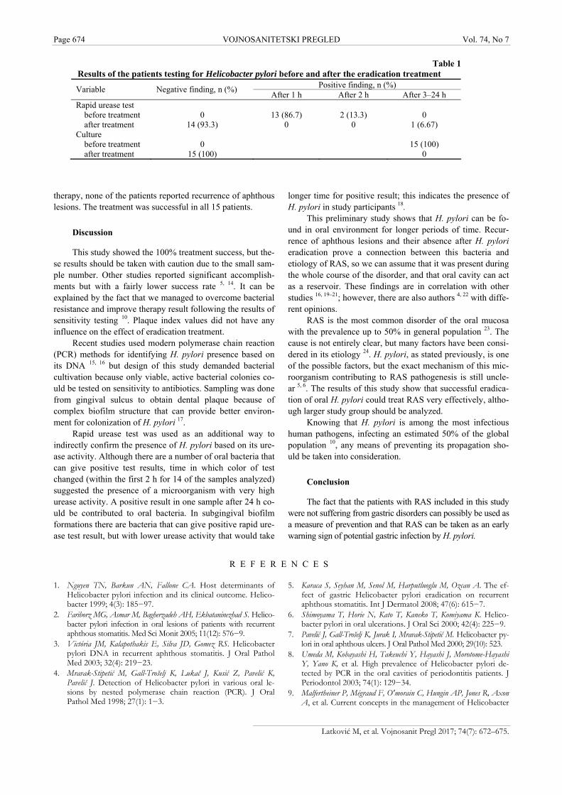
Page 674 VOJNOSANITETSKI PREGLED Vol. 74, No 7
Latković M, et al. Vojnosanit Pregl 2017; 74(7): 672–675.
Table 1 Results of the patients testing for Helicobacter pylori before and after the eradication treatment
Positive finding, n (%) Variable Negative finding, n (%) After 1 h After 2 h After 3–24 h
Rapid urease test before treatment 0 13 (86.7) 2 (13.3) 0 after treatment 14 (93.3) 0 0 1 (6.67)
Culture before treatment 0 15 (100) after treatment 15 (100) 0
therapy, none of the patients reported recurrence of aphthous lesions. The treatment was successful in all 15 patients.
Discussion
This study showed the 100% treatment success, but the-se results should be taken with caution due to the small sam-ple number. Other studies reported significant accomplish-ments but with a fairly lower success rate 5, 14. It can be explained by the fact that we managed to overcome bacterial resistance and improve therapy result following the results of sensitivity testing 10. Plaque index values did not have any influence on the effect of eradication treatment.
Recent studies used modern polymerase chain reaction (PCR) methods for identifying H. pylori presence based on its DNA 15, 16 but design of this study demanded bacterial cultivation because only viable, active bacterial colonies co-uld be tested on sensitivity to antibiotics. Sampling was done from gingival sulcus to obtain dental plaque because of complex biofilm structure that can provide better environ-ment for colonization of H. pylori 17.
Rapid urease test was used as an additional way to indirectly confirm the presence of H. pylori based on its ure-ase activity. Although there are a number of oral bacteria that can give positive test results, time in which color of test changed (within the first 2 h for 14 of the samples analyzed) suggested the presence of a microorganism with very high urease activity. A positive result in one sample after 24 h co-uld be contributed to oral bacteria. In subgingival biofilm formations there are bacteria that can give positive rapid ure-ase test result, but with lower urease activity that would take
longer time for positive result; this indicates the presence of H. pylori in study participants 18.
This preliminary study shows that H. pylori can be fo-und in oral environment for longer periods of time. Recur-rence of aphthous lesions and their absence after H. pylori eradication prove a connection between this bacteria and etiology of RAS, so we can assume that it was present during the whole course of the disorder, and that oral cavity can act as a reservoir. These findings are in correlation with other studies 16, 19–21; however, there are also authors 4, 22 with diffe-rent opinions.
RAS is the most common disorder of the oral mucosa with the prevalence up to 50% in general population 23. The cause is not entirely clear, but many factors have been consi-dered in its etiology 24. H. pylori, as stated previously, is one of the possible factors, but the exact mechanism of this mic-roorganism contributing to RAS pathogenesis is still uncle-ar 5, 6. The results of this study show that successful eradica-tion of oral H. pylori could treat RAS very effectively, altho-ugh larger study group should be analyzed.
Knowing that H. pylori is among the most infectious human pathogens, infecting an estimated 50% of the global population 10, any means of preventing its propagation sho-uld be taken into consideration.
Conclusion
The fact that the patients with RAS included in this study were not suffering from gastric disorders can possibly be used as a measure of prevention and that RAS can be taken as an early warning sign of potential gastric infection by H. pylori.
R E F E R E N C E S
1. Nguyen TN, Barkun AN, Fallone CA. Host determinants of Helicobacter pylori infection and its clinical outcome. Helico-bacter 1999; 4(3): 185−97.
2. Fariborz MG, Asmar M, Bagherzadeh AH, Ekbataninezhad S. Helico-bacter pylori infection in oral lesions of patients with recurrent aphthous stomatitis. Med Sci Monit 2005; 11(12): 576−9.
3. Victória JM, Kalapothakis E, Silva JD, Gomez RS. Helicobacter pylori DNA in recurrent aphthous stomatitis. J Oral Pathol Med 2003; 32(4): 219−23.
4. Mravak-Stipetić M, Gall-Trošelj K, Lukač J, Kusić Z, Pavelić K, Pavelić J. Detection of Helicobacter pylori in various oral le-sions by nested polymerase chain reaction (PCR). J Oral Pathol Med 1998; 27(1): 1−3.
5. Karaca S, Seyhan M, Senol M, Harputluoglu M, Ozcan A. The ef-fect of gastric Helicobacter pylori eradication on recurrent aphthous stomatitis. Int J Dermatol 2008; 47(6): 615−7.
6. Shimoyama T, Horie N, Kato T, Kaneko T, Komiyama K. Helico-bacter pylori in oral ulcerations. J Oral Sci 2000; 42(4): 225−9.
7. Pavelić J, Gall-Trošelj K, Jurak I, Mravak-Stipetić M. Helicobacter py-lori in oral aphthous ulcers. J Oral Pathol Med 2000; 29(10): 523.
8. Umeda M, Kobayashi H, Takeuchi Y, Hayashi J, Morotome-Hayashi Y, Yano K, et al. High prevalence of Helicobacter pylori de-tected by PCR in the oral cavities of periodontitis patients. J Periodontol 2003; 74(1): 129−34.
9. Malfertheiner P, Mégraud F, O'morain C, Hungin AP, Jones R, Axon A, et al. Current concepts in the management of Helicobacter
Vol. 74, No 7 VOJNOSANITETSKI PREGLED Page 675
Latković M, et al. Vojnosanit Pregl 2017; 74(7): 672–675.
pylori infection: The Maastricht 2-2000 Consensus Report. Aliment Pharmacol Ther 2002; 16(2): 167−80.
10. Malfertheiner P, Megraud F, O'morain C, Atherton J, Axon A, Baz-zoli F, et al. Management of Helicobacter pylori infection: The Maastricht IV/Florence consensus report. Gut 2012; 61(5): 646−64.
11. Taş AD, Yakar T, Sakalli H, Serin E. Impact of Helicobacter pylori on the clinical course of recurrent aphthous stomatitis. J Oral Pathol Med 2013; 42(1): 89−94.
12. Mégraud F. H pylori antibiotic resistance: Prevalence, impor-tance, and advances in testing. Gut 2004; 53(9): 1374−84.
13. Whitley E, Ball J. Statistics review 4: Sample size calculations. Crit Care 2002; 6(4): 335−41.
14. Albanidou-Farmaki E, Giannoulis L, Markopoulos A, Fotiades S, Aggouridaki X, Farmakis K, et al. Outcome following treatment for Helicobacter pylori in patients with recurrent aphthous stomatitis. Oral Dis 2005; 11(1): 22−6.
15. Al-Ahmad A, Kürschner A, Weckesser S, Wittmer A, Rauberger H, Jakob T, et al. Is Helicobacter pylori resident or transient in the human oral cavity. J Med Microbiol 2012; 61(Pt 8): 1146−52.
16. Gebara EC, Faria CM, Pannuti C, Chehter L, Mayer MP, Lima LA. Persistence of Helicobacter pylori in the oral cavity after systemic eradication therapy. J Clin Periodontol 2006; 33(5): 329−33.
17. Morales-Espinosa R, Fernandez-Presas A, Gonzalez-Valencia G, Flo-res-Hernandez S, Delgado-Sapien G, Mendez-Sanchez JL, et al. Helicobacter pylori in the oral cavity is associated with gas-troesophageal disease. Oral Microbiol Immunol 2009; 24(6): 464−8.
18. Burne RA, Chen YY. Bacterial ureases in infectious diseases. Microbes Infect 2000; 2(5): 533−42.
19. Liu Y, Yue H, Li A, Wang J, Jiang B, Zhang Y, et al. An epide-miologic study on the correlation between oral Helicobacter pylori and gastric H. pylori. Curr Microbiol 2009; 58(5): 449−53.
20. Gürbüz AK, Ozel MA, Yazgan Y, Celik M, Yildirim S. Oral colo-nization of Helicobacter pylori: Risk factors and response to eradication therapy. South Med J 2003; 96(3): 244−7.
21. Opavski N, Spuran M, Djukić S, Mijac V, Ranin L. Comparison of three diagnostic methods to confirm Helicobacter pylori in-fection. Srp Arh Celok Lek 2007; 135(1−2): 26−30. (Serbian)
22. Oshowo A, Tunio M, Gillam D, Botha AJ, Holton J, Boulos P, et al. Oral colonization is unlikely to play an important role in Heli-cobacter pylori infection. Br J Surg 1998; 85(6): 850−2.
23. Ship JA, Chavez EM, Doerr PA, Henson BS, Sarmadi M. Recur-rent aphthous stomatitis. Quintessence Int 2000; 31(2): 95−112.
24. Brocklehurst P, Tickle M, Glenny AM, Lewis MA, Pemberton MN, Taylor J, et al. Systemic interventions for recurrent aphthous stomatitis (mouth ulcers). Cochrane Database Syst Rev 2012; 9: CD005411.
Received on October 14, 2015. Revised on January 5, 2016.
Accepted on January 12, 2016. Online First May, 2016.
Page 676 VOJNOSANITETSKI PREGLED Vojnosanit Pregl 2017; 74(7): 676–680.
Correspondence to: Aleksandar Jevtić, Military Medical Academy, Clinic for Orthopedic Surgery and Traumatology, Crnotravska 17, 11040 Belgrade, Serbia, E-mail: [email protected]
C U R R E N T T O P I C UDC: 616.38::617-089 https://doi.org/10.2298/VSP151218255J
Autologous transfusions for elective surgery – from existing approaches to upcoming challenges
Autologne transfuzije za elektivnu hirurgiju – od postojećih pristupa do predstojećih izazova
Aleksandar Jevtić*, Milena Todorović†, Gordana Ostoji懧, Saša Vasilijić§║,
Mirjana Pavlović¶, Bela Balint‡§**
Military Medical Academy, *Clinic for Orthopedic Surgery, ‡Institute for Transfusiology and Hemobiology, ║Institute for Medical Research, Belgrade Serbia;
Clinical Center of Serbia, †Clinic for Hematology, Belgrade, Serbia; University of Defence, §Faculty of Medicine of the Military Medical Academy, Belgrade, Serbia;
Florida Atlantic University, ¶Department of Computer and Electrical Engineering and Computer Science, BocaRaton, Florida, United States of America; University of
Belgrade, **Institute for Medical Research, Belgrade, Serbia
Key words: blood transfusion, autologous; blood component transfusion; elective surgical procedures.
Ključne reči: autotransfuzija; transfuzija krvnih komponenata; hirurgija, elektivna, procedure.
Introduction
A well-balanced and rationalized decision making in regard to the implementation and/or accomplishment of haemotherapy or blood component therapy (BCT) is typically based on the individual consideration of responsible medical practitioner, her/his experience with the policy or guidelines of BCT, and the critical values of crucial clinical and laboratory parameters – first of all, the hemoglobin (Hb) concentration. However, when it comes to BCT, there are still sizeable differences in the strategies and approaches in practice. Namely, the same or similar pathological conditi-ons/disorders and specific surgical requirements/requests ha-ve not been yet standardized. In addition, recommendations and principles of BCT are not always respected and applied necessarily by all therapists 1–3. The final and correct decision regarding the realization of BCT, the type and quantity of required blood product should be in compliance with the ge-neral clinical condition, estimated and predicted blood (com-ponent) loss, as well as with the capacity of compensatory mechanisms of the patient 3–7. Typically, BCT for surgical procedures involves red blood cell (RBC) transfusions, wherein one should consider minimum hematocrit (Hct) and Hb values required to provide sufficient or high-quality oxygen transporting capacity (blood oxiform function).
Transfusion supported by platelets, plasma, cryoprecipitate, and other blood products is justified under conditions ac-companied just with isolated blood constituent deficiencies and following extensive blood loss, preceded by massive RBC transfusions 1, 8–10.
Considering the Hb levels, when assessing indications for RBC support ("transfusion trigger" or "threshold"), a re-vised recommendation aroused as a consensus at Conference of the National Institutes of Health of the USA that patients having Htc 0.21 and Hb 70.0 g/L should not be treated by RBCs 5–7. For an optimized decision of the BCT – especially for allogeneic RBC transfusions – the most impor-tant parameters are the ones that inform on blood oxiform function. Therefore, in order to determine an authentic and valid "transfusion trigger" for RBC usage it is thought that additional parameters, such as cardiac minute volume, oxygen blood saturation and its consumption in tissues, pati-ent's age etc, should be applied. It is clear that amongst pati-ents – potential candidates for surgical procedures – there are those requiring higher Hct/Hb levels. Namely, for a sizeable number of patients the most appropriate Htc would be ≥ 0.24 and Hb ≥ 80.0 g/L. Regarding elderly patients, those values should be even higher, Htc ≥ 0.30 and Hb ≥ 100.0 g/L 3, 7.
Regardless of the implementation of different effective preventive approaches and procedures [donor selection, blood
Vol. 74, No 7 VOJNOSANITETSKI PREGLED Page 677
Jevtić A, et al. Vojnosanit Pregl 2017; 74(7): 676–680.
screening procedures, white blood cell (WBC) and virus inac-tivation technology, the use of revised or reduced "transfusion thresholds"] – the use of allogeneic BCT in a sizeable number of patients is associated with a potential risk of post-transfusion morbidity, and sometimes even mortality – which justified the search for new solutions. That is why autologous transfusion (AT) of blood or blood products, as well as the ap-plication of other medical options – the use of blood substitu-tes, hematopoietic growth factors (HGF), antifibrinolytics and other pharmacological agents – are of particular significance as an alternative to allogeneic BCT 1, 11–13.
In a few words, AT consists of the collection of pati-ent's own blood and reinfusion of autologous blood or its components to the same patient during or immediately after the surgical procedure. The goal is to satisfy the patient's ne-ed for blood products, but without the use of allogeneic tran-sfusion. With the use of AT, the patient helps himself and contributes to acquiring higher safety of overall BCT. Namely, application of AT eliminates the risk of alloimmu-nization to RBC, WBC, platelet or plasma protein antigens, reduces of immunosuppression, and eradicates the risk of transmission of viruses or other infectious agents. Besides, certain AT procedures stimulate autologous donor's hema-topoietic system needed for endogenous erythrocytopoiesis and in doing so they support and justify the safety of this type of transfusion. Considering that, and unfavorable ef-fects of allogeneic transfusion, AT is the "safest manner" of BCT 1, 11–13.
Concerning AT, patient's blood can be collected using different strategies among which the most frequently applied are the following ones: 1) preoperative autologous collecti-on/donation (PAD); 2) acute normovolemic hemodilution (ANH); and 3) perioperative blood recovery/salvage (PBR). In addition, the scheme of PBR includes: a) intraoperative blood collection/salvage (IBC) from the surgical field, and b) postoperative blood collection/drainage (PBC) by aspirati-on 1, 14–22. Independently or combined, these AT-strategies proved to reduce the needs for allogeneic blood in elective surgery. Application of blood substitutes, hematopoietic growth factors and pharmacological agents (for the preventi-on/treatment of hemostasis disorders) can further reduce tho-se requirements 1–3.
Finally, the examination of potential transfusion side ef-fects and events should be the same or similar for autologous and allogeneic transfusions 5–7, 22–24. Namely, autologous transfusions have no risk of infectious (virus) and immune-mediated complications (reduced "biohazard"), but carry a similar risk of bacterial contamination following storage (PAD), circulation volume overload, and misadministration compared with normal allogeneic units. For these reasons, autologous blood should not be transfused without a clear indication for BCT 1, 5–7.
Preoperative blood collection/donation
Briefly, candidates for PAD are stable patients with adequate general condition planned for surgical procedures in which transfusion is expected. PAD is the most frequently
used AT technique intended for the elective surgery. It is simple to use, and applicable to the majority of patients un-dergoing the elective surgical procedure. If a patient is in go-od general condition – having Hb 110 g/L, Hct 0.34 and without inflammation symptoms and signs and if surgery can be performed within 2 to 5 weeks, it is recommended to ma-ke a PAD for AT blood. Patients who will probably not be needing blood transfusion support are not to be included in this category of AT program 5–7.
The use of PAD in elective surgery increased in the past few years. Considering that autologous donors are not voluntary blood donors, selection to incorporate patients into AT program is not based on the criteria for selection of nor-mal blood donors. According to the requirements, the patient can donate up to 450 mL of autologous blood every fourth day, although weekly donations are most common. Thus, PAD can be performed in 7 days intervals in adults (exfused blood volume = 10–12% of the total circulating volume) – stipulated that Hb content prior to each donation should not be below 4, 6. Duration of autologous donations in weekly in-tervals can last (according to the blood requirements) as long as the patient does not develop anemia symptoms and has Htc 0.30. It should be stated that the practice throughout the world is based on the "leapfrog" principle which allows obtaining five blood units within 29 days 1–3, 6.
It is recommended that the last autologous collection should take place at least 72 hours before the surgical proce-dure. Children older than 12 can also be included in the AT program, with the previously obtained parents' approval. Smaller quantities of blood are collected from children, in accordance with their body mass. Finally, persons 65 years are also candidates for PAD, in case they meet previously stated criteria referring to autologous blood donors 4–6. The quantity of collected autologous blood depends on the expected blood requirements, although it can be limited by the available time interval preceding the surgical intervention and the patient's initial Hb level and his/her ability to mainta-in adequate ( 110 g/L) Hb concentration. Autologous blood units can be processed into components if it is foreseen that those components will be used intraoperatively. However, considering protection of other patients' health, autologous blood might carry certain risks and therefore, it should not be ever used for allogeneic transfusion 1, 6.
Generally, for the majority of patients PAD is safe and it always will be if criteria required for autologous donors are respected. When performing PAD, precaution should be ta-ken regarding hypertension, hypotension, in elderly persons, in children and pregnant. Namely, pregnancy is not a contra-indication for AT in a case of planned caesarean section. In essence, just severe aortic stenosis and coronary diseases are medical conditions considered as contraindications for PAD. In those patients, additional circulating volume and Hb dec-rease could have unfavorable/unexpected consequences. However, there are data reporting safe PAD in patients with certain cardiac diseases, even in those prior to cardiopulmonary transplantation 3, 25–28.
In summary, PAD is a safe and helpful BCT manner that, commonly, prevents patients’ exposure to the applicati-
Page 678 VOJNOSANITETSKI PREGLED Vol. 74, No 7
Jevtić A, et al. Vojnosanit Pregl 2017; 74(7): 676–680.
on of allogeneic blood products, supported by an ever increa-sing number of reports. In order to achieve higher efficiency of the PAD program, additional research in the following areas are required: 1) determination of the optimal autologo-us blood quantity for specific surgical procedures (in order to reduce/eliminate the use of allogeneic blood) and to have minimum disposal of collected blood units; 2) investigation of the safety and potential risks of PAD (supported by the follow-up of a corresponding control groups); 3) evaluation the role of erythropoietin (EPO) and iron support to improve the PAD-effectiveness; and 4) improvement of the methods for autologous blood collection and storage.
Acute normovolemic hemodilution
Acute normovolemic hemodilution (ANH) is performed immediately before or after the introduction of anesthesia by exfusion of autologous blood, associated with the immediate and necessary replacement of circulating volume by the infu-sion of adequate fluid(s) – crystalloids, rarely colloids or both 1, 6. All vital functions, arterial blood pressure, Hb and Hct value in patients' blood must be maintained within carefully determined ranges. The total volume of infused re-placement fluid depends on its type and composition, stipula-ted that normovolemic dilution must be maintained during the whole surgical intervention 3–5. If blood replacement is obtained by crystalloids, the ratio between the volumes of exfused vs infused volume should be 1: 3 (because crystalloids rapidly leave from intra- to extra-vascular spa-ce). When colloids are used for replacement (most frequently synthetic solutions such us dextrin or gelatin), the withdrawn vs infused volume ratio is equal, that is 1: 1. The combinati-on of crystalloids and colloids can be also used to replace the exfused autologous blood 1.
The collected autologous blood (usually one to two units) is kept in a surgical room in the course of intervention at 20 2°C. Reinfusion of collected blood is performed once sur-gical (vascular) hemostasis is achieved, usually at the end of the surgical procedure. Blood can also be transfused during the surgery process, depending on the patient's condition 1–5.
The objective of ANH is to achieve temporary reducti-on of all of the patient's blood constituents (blood cells and plasma). Precisely, the moderate and monitored anemia as-sociated with normal intravenous volume, induced in that manner, shows the following favorable effects: 1) reduction of the blood viscosity and of the total peripheral resistance in patient's circulation; 2) increased cardiac output; 3) reduced formation of cell aggregates; and 4) reduced quantity of RBCs in the same volume of lost blood. Precisely, a patient having Htc = 0.45 and a loss of one liter of whole blood loses 450 mL of RBCs; however, if his Htc = 0.25, only 250 mL of RBCs are contained in lost whole blood. As a result – thanks to the hemodiluted reduction of Hct from 0.45 to 0.25 and – 200 mL of RBCs are preserved or "saved". However, the lowest and for patients the safest Hct value in AHN is yet to be exactly determined 1, 5–7.
ANH can be of particular significance in patients suita-ble for PAD, not having the time needed to collect required
autologous blood units. However, this AT-strategy should not be used in all patients. It is indicated in patients expected to have intraoperative blood loss of 1,000–2,000 mL, and with a value of Hb 120 g/L. Severe coronary disease, respiratory or renal insufficiency or coagulation factors deficiency are contraindications for performing ANH 4–6.
Perioperative blood recovery/salvage
Generally, the system of PBR implies a collection of patient's blood from the surgical field using cell-savers (followed by ex vivo blood processing) and postoperative as-piration of blood from drains (with its subsequent reinfusi-on). Consequently, that is an AT-strategy that implies salva-ge of autologous blood, that patient had lost during IBC or immediately upon PBC surgical procedure. The routine of PBR provides a surgical and reanimation team with conside-rable quantities of blood or RBCs needed for urgent BCT of patients. Previously used very seldom, almost exclusively in cardiothoracic surgery, PBR has nowadays become a sta-ndard procedure in other surgeries, as well. In addition, it is used for life saving of the wounded in war conflicts 1, 19–22. Finally, a number of practitioners believe that preoperative donation or intraoperative collection of plasma and/or plate-lets in cardiopulmonary bypass surgery may significantly improve hemostasis 6.
As mentioned, the name IBC or autologous blood recovery describes the technique of collecting and reinfusing blood lost by a patient during surgery. The "saved" blood is a source of RBCs having the adequate quality required for transfusion support that is for the performance of the surgical procedure. Namely, the oxygen-transport properties of reco-vered cells are equivalent to (or comparable with) stored al-logeneic RBCs. PBC denotes the recovery of blood from surgical drains followed by reinfusion, typically without pro-cessing. This blood-salvage procedure can be initiated immediately after the surgical intervention and it is usually performed within the first 4–12 hours following the surgery. It has been demonstrated that survival of perioperatively col-lected and reinfused RBCs is compatible with the survival of transfused allogeneic RBCs 1, 6.
However, the use of AT is not a "nonhazardous" BCT approach. Artificially induced anemia is the most common unfavorable effect of ANH, while bacterial con-tamination is a potential, although very rarely, the risk of PBR. Besides, PBR procedures can sometimes be associa-ted with air embolism and/or hemostasis malfunction. Namely, the salvaged blood has decreased coagulation factors, increased contents of the fibrinogen/fibrin degra-dation products and a considerable quantity of anticoagu-lants. These hemostatic defects are primarily manifested as bleeding moistening of the surgical field or bleeding along the surgical sutures due to the removal of platelets and plasma constituents during the ex vivo processing of "saved" whole blood. Seldom, due to platelet and WBC activation (in the course blood processing), transfusion-related acute lung injury (TRALI) can occur – termed also as the "syndrome of salvaged blood". Finally, infection
Vol. 74, No 7 VOJNOSANITETSKI PREGLED Page 679
Jevtić A, et al. Vojnosanit Pregl 2017; 74(7): 676–680.
and malignancy in the surgical field are contraindications for performing of the PBR manner 1, 3–6, 22–24.
Efficient IBC is enabled by the use of specific devices (cell-savers) for rapid processing of whole blood collected from the surgical field. Although there are reports on specific aspects of the clinical use of this type of AT-strategy, certain aspects require additional research including the following: 1) investigation of the risks of IBC when there is a probability that blood contains malignant cells or is contami-nated with bacteria; 2) conducting controlled prospective studies on benefits and risks of the use of autologous plasma and platelets (or platelet rich plasma) in cardiac surgery 6, 26.
Efficacy and safety of PBC in cardio-surgery has been demonstrated indisputably 7, 26. Opinions regarding efficiency of PBC in orthopedic surgery are not unanimous and the following should be taken into consideration: 1) prospect stu-dies to illustrate the benefits of transfusion of "saved" blood; b) make analyses of risks of transfusion of unprocessed blood, taking into special consideration possible contamination of blood with bacteria or chemical agents (e.g. metylmetakrylat) and 3) analysis of cost vs benefit aspects 6, 14–22.
In summary, not all AT-strategies are applicable or necessary for all patients. The choice should be reasonably complied with the expected BCT-needs, based on patient's general condition, a surgical procedure to be performed and available preoperative time interval. In order to make the right and correct use of AT, it should be considered not only by treating physicians, but also by patients, and it should be done before a decision is made regarding surgical procedure. Program of AT procedures shall be more functional if consi-dered on the integration of all strategies needed for its reali-zation and if continuous cooperation with transfusion and therapeutic establishments is ensured.
Cytokine/HGF stimulated erythrocytopoiesis
According to its chemical structure, erythropoietin (EPO) belongs to glycoproteins, a group of hematopoietic cytokines. It is the main regulator of erythrocytopoiesis – which practically expresses its stimulating effects starting from the population "oligopotent" stem cell stage all the way to the ma-ture cell formation. The largest portion of EPO is produced in kidneys and a smaller portion in the liver. Hypoxia being a stimulator and the increase of Hb blood level is acting as an inhibitor of EPO synthesis by "negative feedback". In a simpli-fied manner, EPO is initially bound to its receptor, followed by internalization of EPO complex and that receptor, then by sig-nal transduction from the membrane to the core and finally by the expression of the erythrocytopoiesis regulatory gene 3. Alt-hough RBC support for kidney patients (with endogenous EPO deficiency) is still in use, these transfusions should be completely replaced with the application of recombinant EPO (rHuEPO) 25. Clinical use of rHuEPO is indicated for some ot-her conditions associated anemia (with no deficiency of endo-genous EPO) – e.g. for pretreatment of patients in which PAD for elective surgery planned 12, 13, 29–32.
Within the PAD policy (according to the American As-sociation of Blood Banks – AABB standards), a collection of 450 mL of autologous blood is recommended on the third, and if it is necessary at the 10th and the 17th days before elective surgery. However, if the patient's Hb 110 g/L or if the blood requirements are elevated – a short term erythrocytopoiesis stimulation by high-dose rHuEPO pret-reatment (300–800 IJ/kg, three times weekly) along with the iron therapy are indicated (initiated two weeks proceeding the first blood collection) 6, 29–32.
It has been shown that in patients included in the PAD program with the use of rHuEPO, it is possible to achieve a significant reduction of the need for the perioperative alloge-neic transfusion. In one such comparative study 32 compari-son was made between autologous donors treated and untrea-ted with rHuEPO. Namely, subjects of the first (investigated) group were pretreated with rHuEPO (500 IU/kgbm applied twice weekly, during three weeks). Blood was collected after the first week of rHuEPO administration. Autologous blood was also collected from the subjects of the control group (they were not treated by rHuEPO). Iron therapy was admi-nistered to all patients, starting one week before the first blo-od collection. In the group of investigated subjects, Hb valu-es remained equal to the initial values (recorded before the first blood collection). Contrary to that, considerable decrea-se and slow Hb normalization were noted upon PAD in con-trol group. Likewise, only 10% of the investigated subjects (treated by rHuEPO) were transfused with allogeneic RBCs. In contrast, 40% of the control group subjects were treated with allogeneic transfusion. This study shows that the use of rHuEPO is especially beneficial in patients having a total circulating volume of about 4,000 mL, with the collection of around 2,000 mL of autologous blood 32.
Finally, even with the use of high doses of rHuEPO, side effects of this therapy are relatively rare 6, 12, 13. Commonly, ap-plication of rHuEPO can be associated with the occurrence of side effects – the most frequent ones being an increase of the blood pressure (despite the use of antihypertensive medication), muscle cramps, "flu-like syndrome", bleeding or thrombosis. In some patients thrombocytosis can develop after several weeks of the use of rHuEPO or biochemical changes may occur (e.g. inc-rease of potassium concentration). The rate of side effects rarely exceeds 5–l0%. Thus, patients usually tolerate well the rHuEPO pretreatment, used either intravenously or subcutaneously 6, 30.
Conclusion
Literature data describe general concepts and specific AT protocols; however, there are just seldom clinical inves-tigations related to the most effective AT-strategy for all pa-tients/situations. In future, relevant guidelines are needed with additional investigations in this area, including the following tasks: a) to define benefits and risks of AT-treatments and b) determination of criteria for selection of patients and optimal surveillance methods to perform the most satisfactory type of AT-strategy.
Page 680 VOJNOSANITETSKI PREGLED Vol. 74, No 7
Jevtić A, et al. Vojnosanit Pregl 2017; 74(7): 676–680.
R E F E R E N C E S
1. Radović M, Balint B. Intervention for reducing homologous transfusion use. Belgrade: Association ART; 1995. (Serbian)
2. Gligorović V, Balint B. Clinical transfusiology. Belgrade: The In-stitute for Textbooks Teaching Aids; 1998. (Serbian)
3. Balint B. Clinical transfusiology. Belgrade: The Institute for Textbooks Teaching Aids; 2004. (Serbian)
4. Balint B, Trkuljić M, Todorović M. Basic principles of haemother-apy. Belgrade: Čigoja; 2010. (Serbian)
5. Balint B, Vucetic D, Ostojic G, Ljubenov M. Basic of transfusiol-ogy with haemotiology. Belgrade: Faculty of Medicine of the Military Medical Academy – Media Center „Odbrana”; 2015. (Serbian)
6. Roback JD, Grossman BJ, Harris T, Hillyer CD. Technical man-ual. 17th ed. Bethesda, MD: AABB Press; 2011.
7. Klein HG, Anstee DJ. Mollison's Blood transfusion in clinical medicine. 12th ed. Oxford: Wiley-Blackwell; 2014.
8. Long M, Liu Z, Zhu J. Comparative analysis of autologous blood transfusion and allogeneic blood transfusion in surgical patients. Int J Clin Exp Med 2014; 7(9): 2889−94.
9. Rhee P, Inaba K, Pandit V, Khalil M, Siboni S, Vercruysse G, et al. Early autologous fresh whole blood transfusion leads to less allogeneic transfusions and is safe. J Trauma Acute Care Surg 2015; 78(4):729-34. PubMed PMID: 25807402
10. Catalano L, Campolongo A, Caponera M, Berzuini A, Bontadini A, Furlò G, et al. Indications and organisational methods for auto-logous blood transfusion procedures in Italy: Results of a na-tional survey. Blood Transfus 2014; 12(4): 97−508.
11. Guo J, Xu F, Jin X, Shen H, Liu Y, Zhang Y, et al. Impact of al-logenic and autologous transfusion on immune function in pa-tients with tumors. Asian Pac J Cancer Prev 2014; 15(1): 467−74.
12. Stanković B, Balint B, Taseski J, Trkuljić M, Jovanović Z, Kilibarda N. Perioperative use of rhEPO in female patient included in the program of autologous blood transfusion. Anest Reanim Transf 1997; 26: 53−6. (Serbian)
13. Stankovic B, Balint B, Taseski J, Trkuljic M, Jovanovic Z. Preopera-tive use of the ferrous sulfate and rhEPO in the autologous transfusion (AT) program. Vox Sang 1998; 74(Suppl 1): 1595, E.
14. Menendez ME, Ring D. Minorities are less likely to receive auto-logous blood transfusion for major elective orthopaedic sur-gery. Clin Orthop Relat Res 2014; 472(11): 3559−66.
15. Yoo M, Park H, Ryu J, Kim J. The efficacy and safety of autolo-gous transfusion in unilateral total knee arthroplasty. Knee Surg Relat Res 2015; 27(3): 168−72.
16. Schneider MM, Kendoff D, Oloughlin PF, Hessling C, Gehrke T, Ci-tak M. Effectiveness of autologous transfusion system in pri-mary total hip and knee arthroplasty. Technol Health Care 2014; 22(1): 123−8.
17. Hawi N, Kendoff DO, Hessling U, Haasper C, Gehrke T, Citak M. Effectiveness of an autologous transfusion system following cemented and non-cemented revisions of total hip arthro-plasty. Int Orthop 2014; 38(8): 1603−8.
18. Horstmann W, Kuipers B, Ohanis D, Slappendel R, Kollen B, Ver-heyen C. Autologous re-transfusion drain compared with no drain in total knee arthroplasty: A randomised controlled trial. Blood Transfus 2014;12(Suppl 1): s176−81.
19. Horstmann WG, Swierstra MJ, Ohanis D, Rolink R, Kollen BJ, Ver-heyen C. Favourable results of a new intraoperative and postop-erative filtered autologous blood re-transfusion system in total
hip arthroplasty: A randomised controlled trial. Int Orthop 2014; 38(1): 13−8.
20. Li N, Li P, Liu M, Wang D, Xia L. Comparison between auto-logous blood transfusion drainage and no drainage/closed-suction drainage in primary total hip arthroplasty: A meta-analysis. Arch Orthop Trauma Surg 2014; 134(11): 1623−31.
21. Tesic I, Sekulic J, Arbutinov V, Popov D, Velisavljev D. Autologous blood transfusion in patients undergoing hip replacement sur-gery. Med Pregl 2014; 67(3−4): 101−7.
22. Newman ET, Watters TS, Lewis JS, Jennings JM, Wellman SS, At-tarian DE, et al. Impact of perioperative allogeneic and autolo-gous blood transfusion on acute wound infection following to-tal knee and total hip arthroplasty. J Bone Joint Surg Am 2014; 96(4): 279−84.
23. Kumar S, Goyal K, Dubey S, Bindra A, Kedia S. Anaphylactic re-action after autologous blood transfusion: A case report and review of the literature. Asian J Neurosurg 2015; 10(2): 145−7.
24. Xing Y, Wang Y. Influence of autologous and homologous blood transfusion on interleukins and tumor necrosis factor-α in peri-operative patients with esophageal cancer. Asian Pac J Cancer Prev 2014; 15(18): 7831−4.
25. Suzuki Y, Okai K, Ohashi H, Aota S, Sakurai K, Terawaki H, et al. Autologous blood transfusion for hemodialysis patients: A case report and review of clinical reports and therapeutic fea-tures. Transf Apher Sci 2015; 52(2): 204−7.
26. Trapp C, Schiller W, Mellert F, Halbe M, Lorenzen H, Welz A, et al. Retrograde Autologous Priming as a Safe and Easy Method to Reduce Hemodilution and Transfusion Requirements dur-ing Cardiac Surgery. Thorac Cardiovasc Surg 2015; 63(7): 628−34.
27. Chalfin HJ, Frank SM, Feng Z, Trock BJ, Drake CG, Partin AW, et al. Allogeneic versus autologous blood transfusion and sur-vival after radical prostatectomy. Transfusion 2014; 54(9): 2168−74.
28. Shukla P, Dwivedi S, Bhargava M, Singh R, Singh S. Intraoperative autologous blood transfusion of peritoneal blood during lapa-rotomy for ectopic pregnancy: Prospective study. J Obstet Gynaecol India 2014; 64(5): 358−61.
29. Mercuriali F, Zanella A, Barosi G, Inghilleri G, Biffi E, Vinci A, et al. Use of erythropoietin to increase the volume of autologous blood donated by orthopedic patients. Transfusion 1993; 33(1): 55−60.
30. Mercuriali F, Inghilleri G, Biffi E, Colotti MT, Vinci A, Sinigaglia L, et al. Comparison between intravenous and subcutaneous re-combinant human erythropoietin (Epoetin alfa) administration in presurgical autologous blood donation in anemic rheuma-toid arthritis patients undergoing major orthopedic surgery. Vox Sang 1997; 72(2): 93−100.
31. Price TH, Goodnough LT, Vogler WR, Sacher RA, Hellman RM, Johnston MF, et al. The effect of recombinant human erythro-poietin on the efficacy of autologous blood donation in pa-tients with low hematocrits: A multicenter, randomized, dou-ble-blind, controlled trial. Transfusion 1996; 36(1): 29−36.
32. Biesma DH, Marx JJ, Kraaijenhagen RJ, Franke W, Messinger D, Van de Wiel A. Lower homologous blood requirement in auto-logous blood donors after treatment with recombinant human erythropoietin. Lancet 1994; 344(8919): 367−70.
Received on December 18, 2015. Accepted on December 29, 2015.
Online First September, 2016.
Vojnosanit Pregl 2017; 74(7): 681–686. VOJNOSANITETSKI PREGLED Page 681
Correspondence to: Sanja Djajić, University of Novi Sad, Faculty of Law, Department for International Law, Trg Dositeja Obradovića 1, 21 000 Novi Sad, Serbia. E-mail: [email protected]
I N F O C U S UDC: 61-051::355.357]:341 https://doi.org/10.2298/VSP170516072D
International legal protection of medical personnel in warfare and peace missions
Međunarodna pravna zaštita medicinskog osoblja u ratu i mirovnim misijama
Sanja Djajić
University of Novi Sad, Faculty of Law, Department for International Law
Introduction
According to the International Committee of the Red Cross (ICRC) “2,400 targeted attacks had been carried out in the last three years against patients and health-care workers, transport and centres in 11 countries” 1. According to the Physicians for Human Rights in Syria there were 400 attacks against medical facilities and 768 deaths of medical person-nel since March 2011 2. World Health Organisation (WHO) stated that as many as 645 medical personnel were killed du-ring the armed conflict in Syria until September 2015 3 and that around 60% of hospitals became dysfunctional during the same period. All these circumstances, including several heavy attacks on hospitals in Syria and Afghanistan in 2015 and 2016a 4 prompted the UN Security Council to reaffirm the international law protection of medical and humanitarian personnel by adopting Resolution 2286 (2016). Despite the rising number of casualties in armed conflicts worldwide, there is still relentless support for international peace missi-ons, which invariably involve medical personnel, by both United Nations (UN) and other international organisations. Against the background of recent events, UN Security Coun-cil (UNSC) Resolution and widespread practice of peace missions, this article will try to provide a brief overview of international legal framework relevant for medical and hu-manitarian personnel covering both the status of domestic medical personnel and that of international missions in warfare and for peace operations.
a Attack on the hospital of Doctors without Frontiers in Kunduz,
Afghanistan on 3 October 2015 killing 42 people including doctors,
nurses and patients. Reports show that only in 2015 and during the first
three months of 2016 there were attacks on medical personnel and facili-
ties in 19 countries in armed conflicts or political violence.
General legal framework – Geneva Conventions and international humanitarian law
International humanitarian law traces back to the mid of XIX century 5. Rules applicable today stem from the four 1949 Geneva Conventions (GC) 6, their Additional Protocols (AP) adopted in 1979 and 2005 7, and the so-called Hague rules codified almost a century ago. Regardless of different sources, there is a common understanding today that interna-tional humanitarian law is “one single complex system” 8.
The fundamental rule relevant for medical personnel at-tached to their armed forces guarantees a specially protected status which means that they must be respected and protected at all times and in all circumstances (GCI Art. 24, GCII Art. 36, GCIV Art. 20, API Art. 15, and APII Art. 9) 6, 7. This pro-tection covers immunity from the attacks and obligation to en-sure uninterrupted performance of providing medical help and health care even if they fall into the hands of the adverse party (GCI, Arts. 19-23) 6, including the right of medical personnel to be allowed to search for and to collect wounded and sick (GCI, Art. 15, APII, Art. 8) 6, 7. Medical personnel is entitled to wear a distinctive emblem of Red Cross (GCI Arts. 40, 41; GCII Art. 42; GCIV Art. 20; API Art. 18; APII Art. 12) 6, 7, to carry small arms and to provide medical assistance in all cir-cumstances (GCI, Art. 22) 6. Immunity includes the prohibition of requisition of medical equipment and supplies (GCI Arts. 33–35, GCIV Art. 57, and API Art. 14) 6, 7. Upon the outbreak and during the course of hostilities parties to the conflict may establish hospital zones and localities whereas protecting powers and ICRC are invited to facilitate the institution and recognition of these hospitals and localities (GCI, Art. 23, GCIV, Art. 14) 6, 7. This protection can be removed once the abuse of protected status is revealed (GCI, Art. 19, 24) 6. However, under no circumstances shall the medical aid be considered as an abuse of privileged status.
Page 682 VOJNOSANITETSKI PREGLED Vol. 74, No 7
Djajić S. Vojnosanit Pregl 2017; 74(7): 681–686.
The privileged status is further confirmed by the rule in Article 28 of the Geneva Convention I according to which medical personnel who fall into the hands of the adverse Party, may be retained only in so far as the state of health of priso-ners of war requires or otherwise be released. Pursuant to Arti-cle 30 of the GCI 6: “medical personnel whose retention is not indispensable by virtue of the provisions of Article 28 shall be returned to the Party to the conflict to whom they belong, as soon as a road is open for their return and military requirements permit. Pending their return, they shall not be deemed prisoners of war but they shall continue to fulfill their duties under the orders of the adverse Party and shall preferably be engaged in the care of the wounded and sick of the Party to the conflict to which they themselves belong. On their departure, they shall take with them the effects, personal belongings, valuables and instruments belonging to them.”
Therefore, medical personnel will not have the status of prisoners of war (POWs) but will still benefit from all the provisions of the GCIII (GCI, Art. 28, 29, GCII Art. 37, GCIII, Arts. 32 and 33) 6. This is why they have the status of retained personnel unlike other persons in captivity who are detained persons or prisoners. In these circumstances medi-cal personnel are entitled to certain special privileges such as the right to visit periodically prisoners of war in hospitals outside the detention camp, right to have the senior medical officer of the highest rank to monitor the professional activity of the retained medical personnel, the right to direct access to medical personnel under their supervision, right to be released of all other duties other than health services. However, medical personnel belonging to neutral powers or organisations entrusted with medical aid, which are not dee-med as a party to the conflict, may not even be retained (GCI, Art. 32) 6.
The 1949 Fourth Geneva Convention (GCIV)6 was the first international treaty to provide special protection to civili-an population thereby extending the application of internatio-nal humanitarian law to civilian medical personnel and hospi-tals. Under the GCIV medical personnel and hospitals are to be respected and protected from deliberate attacks (GCIV, Art. 18) 6 unless hospitals are used for military operations causing harm to the enemy but only after a due warning is given (GCIV, Art. 19) 6 medical personnel are to be given the safe passage for evacuation during the attack (GCIV, Art. 17) 6, while hospital staff, defined as “persons regularly and solely engaged in the operation and administration of civilian hospi-tals, including the personnel engaged in the search for, remo-val and transporting of and caring for wounded and sick civili-ans, the infirm and maternity cases” [GCIV, Art. 20(1)] 6 are protected and respected and will be recognizable by means of an identity card certifying their status, bearing the photograph of the holder and embossed with the stamp of the responsible authority, and also by means of a stamped, water-resistant ar-mlet which they shall wear on the left arm while carrying out their duties (GCIV, Art. 20) 6. This protection is in line with the duty of the parties to the conflict as well as of the occupying power to provide medical care and supplies to civi-lians (GCIV, Arts. 55-56) 6, so medical personnel are always allowed to carry out their duties (GCIV, Art. 56) 6. In addition,
internees are entitled to the attention of medical personnel of their own nationality (GCIV, Art. 91(3)) 6. Internees who are doctors, dentists or other medical personnel may be employed by the detaining power to provide medical aid to other interne-es (GCIV, Art. 95).
Additional Protocols to Geneva Convention (1977) 7 extend the protection to civilian medical personnel, medical supplies and units. The protection originally provided only to medical personnel attached to parties to the conflict is now extended for humanitarian purposes to practically all catego-ries of medical personnel: of a neutral or other State which is not a Party to that conflict; of a recognized and authorized aid society of such a State, and of an impartial international humanitarian organization [API, Art. 9(2)] 7, but according to Protocol I there need to be identity cards issued to both per-manent and temporary civilian medical personnel (API, Annex I (1993), Arts. 2, 3). All necessary help and assistance shall be provided to civilian medical personnel in areas where health services cease due to combats (API, Art. 15) 7. Pursuant to Article 16 of API any person able to perform medical activities for the benefit of the wounded should be able to do so without fear or any form of coercion. This me-ans that medical personnel are immune from punishments for providing medical aid or assistance provided that assistance is in line with medical ethics (GCI Art. 18, API Art. 16, 17 and APII Art. 10). According to the Commentary of the API “medical activity” is not limited to giving treatment: “He [doctor] may be called upon also to diagnose (which may re-veal that nothing is wrong), report as an expert consultant, give proof of death, or merely advice, and so forth” 9. There is also prohibition of coercing medical personnel to provide assistance that is contrary to medical ethics (API Art. 16, APII Art. 10) 7. Furthermore, according to Article 16(3) of the API medical personnel can withhold information on pati-ents “under his care, if such information would, in his opini-on, prove harmful to the patients concerned or to their fami-lies.” This seems to extend the standard rule of “medical confidentiality” as it implies that such patients are not to be denounced 9. Still, it is not entirely clear to whom this obli-gation is actually addressed: to those who can compel medi-
cal personnel or to medical personnel itself b. However, there
is an exception from this rule in relation to the compulsory
notification of communicable diseases [API, Art. 16(3)] 7. The minimum standard of protection of medical per-
sonnel that is applicable to national medical personnel of the parties to the conflict places health workers in a better positi-on than persons falling into the category of combatants. Such protection can cease only if these privileges are used to commit hostile acts outside of their humanitarian function, and only upon a due and timely warning 10. The same applies
b “[T]here is no obligation upon those exercising medical activities
to remain silent. They may denounce the presence of the wounded to the
authorities even when they know that this will be prejudicial to the
wounded person or his family, if such denunciation is in their view nec-
essary for saving lives. The prohibition is aimed at those who could
compel such denunciations.” – The 1987 Commentary of the API9.
Vol. 74, No 7 VOJNOSANITETSKI PREGLED Page 683
Djajić S. Vojnosanit Pregl 2017; 74(7): 681–686.
to members of the medical profession who take part in hosti-lities regardless of their medical background 11. The practical problem that may arise in a warfare is the re-assigning per-sonnel from medical to non-medical and vice versa as it significantly changes the status of combat immunity 12.
Breaches of the rules of protection of medical personnel in a warfare or during military occupation fall into the category of war crimes as prescribed by international crimi-nal law, more precisely by Article 8(2)(b)(xxiv) of the Statu-te of the International Criminal Court: “intentionally direc-ting attacks against…personnel using the distinctive em-blems of the Geneva Conventions in conformity with inter-national law” and civilian medical personnel that is to be re-spected at all times constitute a war crime in international armed conflicts. Since medical personnel are entitled to use these emblems it follows that they are protected by this cri-minal law provision. In addition, a number of national military manuals and national criminal legislation provide for criminal punishment in case of disrespect for medical personnel, units and hospitals. Also, denial of medical servi-ces equally amounts to a war crime 13. Conversely, war cri-mes committed by medical personnel have also been recog-nized by international law and are usually referred to as “medical crimes” 14.
Special legal regimes: Peace missions
There is an estimate that around 100,000 persons have been engaged in UN peace missions today 15, so the signifi-cance of their legal status from the international, humanitari-an and national law is self-evident. While the general legal framework provides for rules applicable during the armed conflicts and in occupied territories, contemporary employment of international forces quite often fall outside such context given that international forces organized under the auspices of the UN and other international organisations will usually find themselves outside the war zones 16 for the purposes of so-called peace-keeping, peace-making or peace-building missions c. On the other hand, the possibility for pe-ace corps to get involved in the armed conflict cannot be fully excluded. Therefore, the international legal regime for peace missions is a complex one that depends on the circum-stances in which they operate, i.e. whether they are involved in the armed conflict and what their role is therein, or whether international forces are operating in peace zones where there is still a risk of hostilities which may require the use of enforcement measures.
Although the idea of peace missions originated at the very beginning of the work of the United Nations, peace missions as such are not envisaged in the UN Charter. Originally the peace missions were organized under Chapter cThere are also more specialised peace operations such as UNMEER (United Nations Mission for Ebola Emergency Response - UN GA 69/1(2014) and UN SC Res. 2177(2014)) that was the first-ever UN emergency health mission entrusted with the task to scale up the re-sponse to this disease on the ground. After the mission’s mandate ex-pired on 31 July 2015 it was overtaken by several agencies including UN peace mission in Liberia and WHO. Such missions inevitably in-volve a considerable number of medical personnel.
VI of the UN Charter whereas today they have been mostly authorized by the UN Security Council acting under Chapter VII of the UN Charter. The difference lies in the authority of the decision adopted. The whole legal framework was to be-constructed in the years to come and significantly improved during the last 20 years. The assumption that UN peace corps were to be employed outside war zones significantly shaped the rules for peace missions. However, this assumption would turn out to be rebuttable so the rules applicable to pe-ace missions in armed conflicts were also reconsidered. The-refore, rules applicable for peace missions and medical per-sonnel which constitute their integral part shall be presented here along these lines.
UN peace missions consist of military and civilian per-sonnel of contributing states on the basis of agreement between these states and the UN. Its employment in foreign territories is further based on the decisions of UNSC and agreement between the UN and receiving State [Status of Forces Agreement (SOFA) or Status of Mission Agreement (SOMA)], as well as on the memorandums of understanding entered into between contributing states and the UN.17 Despi-te the fact that there is a model SOFA, each UN peace missi-on has a specific legal framework within which it operates.
On the other hand, there are some significant general rules applicable to all UN peace missions mostly in terms of immunities and privileges. The main privilege enjoyed by members of peace missions is immunity from the jurisdiction of a receiving state. Probably the most relevant international treaty for granting this privilege and other forms of protecti-on of personnel participating in UN peace missions would be the 1946 Convention on the Privileges and Immunities (CPI) of the United Nations 18 and the 1994 Convention on the Safety of United Nations and Associated Personnel (Safety Convention) 19 and its 2005 Optional Protocol. According to its Article 1(c) this Convention applies to all UN operations undertaken “for the purpose of maintaining or restoring in-ternational peace and security” or when the Security Council or the General Assembly “has declared, for the purposes of this Convention, that there exists an exceptional risk to the safety of the personnel participating in the operation” 19. Pur-suant to the Convention all parties have a duty to ensure the safety and security of UN and associated personnel and to take appropriate steps for their protection while on a mission. The Optional Protocol extends such protection to humanita-rian, political and development assistance. In its Article 4, the Safety Convention mandates the UN to conclude special agreements with host states that would include provisions on immunities of members of the peace mission. This is not without relevance given that a number of existing and pros-pective host states have not ratified the Safety Convention. In practice these agreements are known as, already mentioned Status-of-Force Agreements (SOFA) and Status-of-Mission Agreements (SOMA) which individually settle a number of outstanding issues including those of privileges and immuni-ties: “The conclusion of SOFAs or SOMAs is of practical va-lue for each mission. While the sovereign immunity of pea-cekeepers derives from customary law rather than SOFAs and SOMAs, the latter may have three important effects: to
Page 684 VOJNOSANITETSKI PREGLED Vol. 74, No 7
Djajić S. Vojnosanit Pregl 2017; 74(7): 681–686.
confirm the principle of immunity; to jointly agree on certain limitations to existing privileges where this may be appropri-ate, and to establish rules and procedures for cooperation be-tween the sending state and the host state” 16. These agree-ments also regulate which laws of the host state remain ap-plicable for members of peace missions as well as which laws would not apply (such as the rules on carrying arms, traffic rules, social security and salaries legislation, etc.).
For example, there were several peace-keeping missi-ons in Chad and Central African Republic (CAR) caused by the increase of refugees from Sudan and CAR d. The current peace-keeping missions are the United Nations Multidimen-sional Integrated Stabilization Mission in the Central African Republic (MINUSCA) and the European Union Training Mission in the CAR (EUTM-RCA). The MINUSCA Status of Forces Agreement was concluded on 2 September 2014 20. Given the grave situations in neighbouring countries and ci-vil war within CARe the issue remained constantly under the Security Council monitoring which resulted, inter alia, in UN sanctions against CAR involving arms embargo, travel ban and freezing of assets 21. According to the CAR SOFA and SC Resolutions, the mandate and powers of the MINU-SCA were widely tailored. For example, SC Res. 2301 (2016) adapted the mandate of the MINUSCA by allowing “proactive and robust posture without prejudice to the basic principles of peacekeeping“ 21 and “to actively seize, confis-cate and destroy, as appropriate, the weapons and ammuniti-ons of armed elements, including all militias and other non-state armed groups, who refuse or fail to lay down their arms“ 21 which implies the right to use force.
Civilian and military personnel of MINUSCA, its contrac-tors and national contingencies of participating States, as well as their property, assets and funds, thus enjoy judicial immunity from the CAR (paras. 4, 15, 26-34 CAR SOFA; II, V-VII CPI) 18, 20. However, the CAR SOFA permits the arrest of MINUSCA military personnel but only by MINUSCA military police in or-der to be transferred to their commander for further disciplinary measures. Military members of the military component of MI-NUSCA shall be subject to the exclusive jurisdiction of their res-pective participating State in respect of any criminal offence that may be committed by them in the Central African Republic. Immunity from civil jurisdiction depends on the prior authoriza-tion of the UN Special Representative depending on whether the case is related to official duties (CAR SOFA, Art. 53) 20. However, they are not relieved of responsibility that is to be as-sessed before their domestic courts whose jurisdiction remain in-tact by their international status. As to civil liability for damages
d The MINURCAT mission (established on the basis of SC Res.
1778 (2007) of 25 September 2007 followed by the SOMA between UN
and Central African Republic concluded on 20 November 2008. MIN-
URCAT completed its mission in 2010 due to the decision of Chad to
withdraw from the agreement) was transformed to a new mission, BI-
NUCA that was eventually, following the adoption of the SC Res. 2149
(2014) subsumed in the newly established peacekeeping operation –
MINUSCA. e Prosecutor of the International Criminal Court opened on 24
September 2014 investigation into alleged crimes committed since 2012.
that may occur in the course of the performance of their duties any claim for damages can be addressed only to the UN (CAR SOFA, Art. 56), which is the result of both the immunity of per-sonnel but also of the legal capacity of the UN f, 16. MINUSCA is entitled to premises free of charge, to fiscal privileges, tax-free imports and establishment of commissaries, full freedom of mo-vement without any restrictions or prior approvals, including the unrestricted entry into and departure from the CAR.
However, immunities are not the only privilege assig-ned to personnel 22 – it extends to the prohibition of arrest and hostage-taking (Safety Convention, Art. 8) g. They are equally protected from any form of attack that is punishable by laws of receiving states. The status of peace corps under the Safety Convention assumes that there is no armed con-flict that would replace the applicability of norms of peace to norms of war, i.e. the international humanitarian law. The problem is one on the ground: the level of hostilities which may involve members of peace operations does not have to rise to the level of armed conflict but would certainly involve some use of force. Such “robust peace operations” 16 are not rare, as can be seen from the authority vested to MINUSCA described above. The principal position is that such situati-ons are those of enforcement rather than hostilities so the in-ternational humanitarian law does not apply16 or even that the level of use of force by peace operations can be of a hig-her level than for other armed forces before the international humanitarian law applies 23. However, there are situations when the existence of the armed conflict would be recogni-zed by the peace mission. There are several possible regimes for the peace corps in the armed conflict: if international for-ces take part in hostilities and become a party to the armed conflict, and if they are engaged in the armed conflict but without being a party to it. In the first instance, members of the military component of the peace mission become comba-tants and they are thus fully under the international humani-tarian law regime – which moves medical personnel under the scope of Geneva Conventions both in terms of its rights and duties. In the second instance peace corps enjoy the pro- f While the rules envisaged in the CAR SOFA follow the general prac-tice in this respect, there is also emerging practice according to which certain exceptions seem to have been carved out before several national courts precisely because of the immunities of the UN as an international organization, and on the basis of command control over national contin-gencies. “In Mothers of Srebrenica et al. v. Netherlands and the United Nations, the Hague Court of Appeal ruled that it is impossible to bring the UN before a Dutch court due to the immunity from prosecution granted to the UN pursuant to international conventions, and it accepted that the Netherlands should share UN immunity in this respect. Later on, in Netherlands v. Hasan Nuhanovic´, the Supreme Court of the Nether-lands concluded that the Netherlands was responsible for the death of three Muslim men from Srebrenica and stated that the pertinent conduct of Dutchbat, as part of a UN peacekeeping force, could be attributed to the Netherlands because public international law allows the conduct to be attributed in this specific case to the sending state and not to the UN, insofar as the state had effective control over the disputed conduct. Im-munity was not invoked here, because the Court was deciding on the conduct of national military personnel. It rather concluded that the UN did not have (or no longer had) exclusive operational control over Dut-chbat, and that the state of the Netherlands was responsible for those ac-tions in terms of domestic tort law.”.
g During the operation of UN Peace Mission in Sierra Leone
(UNAMSIL) 500 peacekeepers were taken hostage in May 2000.
Vol. 74, No 7 VOJNOSANITETSKI PREGLED Page 685
Djajić S. Vojnosanit Pregl 2017; 74(7): 681–686.
tection as civilians and cannot be treated as a legitimate tar-get despite the warfare situation 24.
Structure and levels of medical support for UN peaceke-eping missions have been standardized and generally suppor-ted by the medical support unit of the Department for Peace Keeping Operations (DPKO) of the UN 25. According to the surveys of professional medical personnel of certain contribu-ting states, there seem to be some chronical and typical prob-lems arising out of complex multinational peace operations re-garding the status and functioning of medical support for pea-cekeeping missions 26. During the UNAMSIL it was discove-red that national contingencies were not able to provide satisfactory medical support. Problems that were identified were in relation to inadequate medical care for the civilian members of the mission either in terms of the lack of specia-lists (tropical diseases, gynaecologists) or lack of hygiene 27. The problems do not end with organizational or resource issu-es but may involve some ethical considerations especially re-garding the medical aid to the local population.28 The ethical dilemma lies in the conflict between the UN medical mandate limited to UN personnel, on one hand, and professional duties of medical workers, international humanitarian and human rights considerations toward local population, on the other. For example, in the 2014 Memorandum of Understanding between the UN and Republic of Serbia for Contributing Resources to the United Nations Interim Force in Lebanon (UNIFIL) 29 explicitly sets forth that medical support facilities can provide care to UN and other authorized personnel, either under self-sustainment or as a fee-for-service medical care (except for emergency care). However, this regime is not applicable to the local population: “Care provided to non-eligible personnel (e.g. local population) by a troop/police contributor is not re-imbursable by the United Nations.”
Responsibilities of contributing states do not cease with sending their personnel to act under the UN mandate. Even more, it seems that it is their “primary responsibility to ensu-re that units are properly equipped, trained and prepared for a peacekeeping mission” 27. The command structure may re-main with the contributing state together with the need to es-tablish a mechanism for potential responsibility claims and issues regarding its personnel.
National legal framework of Serbia for multinational operations
While the general legal framework is international, the-re are also national legal rules the scope of which is limited to national contingencies. These rules cannot overturn or outweigh international legal regime but they are of immedia-te relevance for national members of peace mission and to certain extent supplement international legal regime by pro-viding additional protection to national troops or by imple-menting rules on responsibility for acts of national contin-gencies. In case of Serbia, there is a special Law on Deployment of Serbian Army and Other Defence Forces in Multinational Operations outside the Territory of Republic of Serbia (2009). This Law allows that only those members of
armed forces of Serbia who had adequate training and were given certificate are eligible to join national contingency of peace corps (Art. 12), where they cannot stay longer than one year (Art. 22). Their engagement is contract-based (Art. 30) and is subject to disciplinary, criminal and civil liability rules set forth in both national legislation and international treaties (Art. 27), and most notably to international rules on the use of force and international humanitarian law. There are a number of social benefits (Arts. 25 and 34) as well as legal, diplomatic and material assistance (Art. 28) for Serbian military personnel engaged in multinational operations. While all members of peace missions represent UN or other international organization and while they enjoy immunity from the courts of the receiving state 29, the command struc-ture remains with the commander of national contingency unless the applicable international agreement provide for a different command structure (Art. 21) and Republic Serbia, as a contributing state, remains liable for civil damages in-curred on third parties during the mission (Art. 38).
Conclusion
International humanitarian law applicable during the armed conflict has equalized military and civilian medical personnel in terms of their protection. However, the rules on military medical personnel are more detailed and precise re-garding both their rights and responsibilities in a warfare gi-ven their special position. The protection extended to medi-cal personnel and hospitals is on the higher end of protection granted by the international humanitarian law the breach of which can be qualified as war crimes. The protection of me-dical personnel within peace missions is of equal value but of different source and character as it amounts to the protection available to diplomatic missions. While there exists differen-ce between military personnel as opposed to civilian compo-nent of peace missions, where the former enjoy absolute immunity from jurisdiction of the host state, the concept of protection of UN places also civilian part of the mission quite high given the condition of prior authorization of the Special Representative of the UN Secretary General for any enforcement measure to be undertaken against personnel be-fore the courts of the host state. The particular regime is, however, always to be assessed against the rules envisaged in SOFA or SOMA agreement concluded for each particular peace mission. The problem that peace missions are facing today is the one of characterization of the hostilities where they operate, i.e. which rules governing the use of force would be applicable. If hostilities are qualified as armed con-flict it is the international humanitarian law that applies, but if such hostilities do not reach the level of armed conflict measures undertaken would be treated as enforcement mea-sures outside international humanitarian law context. There remains the fact that today peace missions function in quite volatile environments with increasing risks for peace corps personnel which has been indirectly confirmed by UN Security Council resolutions authorizing peace missions to act in a more proactive and robust manner.
Page 686 VOJNOSANITETSKI PREGLED Vol. 74, No 7
Djajić S. Vojnosanit Pregl 2017; 74(7): 681–686.
R E F E R E N C E S
1. United Nations. Security Council Adopts Resolution 2286 (2016), Strongly Condemning Attacks against Medical Facili-ties, Personnel in Conflict Situations. Available from: https://www.un.org/press/en/2016/sc12347.doc.htm
2. Amnesty International. Annual Report: Syria 2016/2017. Avail-able from: https://www.amnesty.org/en/countries/middle-east-and-north-africa/syria/report-syria/.
3. WHO. Whole of Syria Health Sector/Cluster. Newsletter Vol-ume. 2015 September. Available at: http://www.who.int/hac/crises/syr/sitreps/syria_health_sector_cluster_news_september2015.pdf?ua=1.
4. Safeguarding Health in Conflict. No Protection, No Respect, Health Workers and Health Facilities under Attack, 2015 and early 2016. Available from: https://www.safeguardinghealth.org/sites/shcc/files/SHCC2016final.pdf
5. Shaw MN. International Law. 4th ed. Cambridge: Cambridge University Press; 1997.
6. Geneva Conventions. Convention (I) for the Amelioration of the Condition of the Wounded and Sick in Armed Forces in the Field. Geneva, 12 August 1949 (GCI); Convention (II) for the Amelioration of the Condition of Wounded, Sick and Shipwrecked Members of Armed Forces at Sea. Geneva, 12 August 1949 (GCII); Convention (III) relative to the Treat-ment of Prisoners of War. Geneva, 12 August 1949 (GCIII); Convention (IV) relative to the Protection of Civilian Persons in Time of War. Geneva, 12 August 1949. (GCIV). Geneva; International Committee of the Red Cross; 1949.
7. Geneva Conventions Protocols. Protocol Additional to the Geneva Conventions of 12 August 1949, and relating to the Protection of Victims of International Armed Conflicts (Pro-tocol I), 8 June 1977 (API); Protocol Additional to the Geneva Conventions of 12 August 1949, and relating to the Protection of Victims of Non-International Armed Conflicts (Protocol II), 8 June 1977 (APII); Protocol additional to the Geneva Conventions of 12 August 1949, and relating to the Adoption of an Additional Distinctive Emblem (Protocol III), 8 Decem-ber 2005 (APIII). Geneva; International Committee of the Red Cross; 2005.
8. International Court of Justice. Legality of the Threat or Use of Nu-clear Weapons. Advisory Opinion. ICJ Reports 1996, para. 75. Hague: International Court of Justice; 1996.
9. ICRC. Additional Protocol I to the Geneva Conventions: Commentary of 1987 – General Protection of Medical Duties. Available from: https://ihl-databases.icrc.org/ applic/ ihl/ ihl.nsf/ Comment.xsp? action= openDocument&documentId = C63B87837232A165C12563CD00430C81
10. Goniewicz M, Goniewicz K. Protection of medical personnel in armed conflicts-case study: Afghanistan. Eur J Trauma Emerg Surg 2013; 39(2): 107–12.
11. Stover E. Crimes of War. Medical Personnel. Available from: http://www.crimesofwar.org/a-z-guide/medical-personnel/
12. De Waard P, Tarrant J. Protection of Military Medical Personnel in Armed Conflicts. UWA Law Rev 2010; 35(1): 157‒ 84.
13. ICRC. Rule 156 – Definition of War Crimes. Available at: https://ihl-databases.icrc.org/customary-ihl/ eng/ docs/ v1_rul_rule 156
14. Mehring S. Medical War Crimes. In: Bogdandy A, Wolfrum R, editors. Max Planck Yearbook of United Nations Law. Leiden: Koninklijke Brill NV; 2011. 15: p. 229‒ 79.
15. United Nations. Peacekeeping. Available at: http://www.un.org/en/peacekeeping/issues/military/.
16. Fleck D. The legal status of personnel involved in United Na-tions peace operations. Int Rev Red Cross 2013; 95(891‒ 892): 613–36.
17. Oswald B, Durham H, Bates A. Documents on the Law of UN Peace Operations. Oxford: Oxford University Press; 2010.
18. United Nations. Convention on the Privileges and Immunities of the United Nations, 13 February 1946. New York: United Nations, Treaty Series; 1946; 1: 15.
19. United Nations. Convention on the Safety of United Nations and Associated Personnel, 9 December 1994, 2051 UNTS 363 (entered into force 15 January 1999) (hereinafter: Safety Con-vention); and the Optional Protocol thereto, 8 December 2005, UN Doc. A/RES/60/42, Annex, Registration No. A-35457 (entered into force 19 August 2010) New York: United Nations, Treaty Series; 1999; 2051: 363.
20. United Nations. United Nations and Central African Republic Status of Forces Agreement between the United Nations and the Government of the Central African Republic relating to the United Nations Multidimensional Integrated Stabilization Mission in the Central African Republic (MINUSCA). Bangui, 2 September 2014, entry into force 2 September 2014 by signa-ture, in accordance with paragraph 63. No. 52177. (CAR SOFA). Bangui: MINUSCA; 2014.
21. United Nations. UN SC Res. 2127(2013) and 2339(2017). Available from: https://www.un.org/press/en/2017/sc12697.doc.htm
22. Engdahl O. Protection of Personnel in Peace Operations. Lei-den-Boston: Martinus Nijhoff Publishers; 2007.
23. Greenwood C. International Humanitarian Law and United Na-tions Military Operations. Yearbook Int Humanit Law 1998; 1: 3‒ 34.
24. Ferraro T. The applicability and application of international humanitarian law to multinational forces. Int Rev Red Cross 2013; 95(891/892): 561‒ 612.
25. United Nations. Medical Support Manual for United Nations Peacekeeping Operations. 2nd ed. New York: UN; 1999.
26. Davis LM, Hosek SD, Tate MG, Perry M, Hepler G, Steinberg PS. Army Medical Support for Peace Operations and Humanitar-ian Assistance. Washington D.C.: Rand; 1996.
27. United Nations – Department of Peacekeeping Operations. Les-sons Learned from United Nations Peacekeeping Experiences in Sierra Leone, September 2003. New York: United Nations; 2003.
28. Tobin J. The challenges and ethical dilemmas of a military medical officer serving with a peacekeeping operation in re-gard to the medical care of the local population. J Med Ethics 2005; 31(10): 571–4.
29. United Nations. 2014 Memorandum of Understanding between the UN and Republic of Serbia for Contributing Resources to the United Nations Interim Force in Lebanon (UNIFIL. DFS/UNIFIL/SER/01. Official Journal of Republic of Serbia – International Agreements, No. 7/2014.
Received on May 16, 2017. Accepted on May 19, 2017.
Online First May, 2017.
Vojnosanit Pregl 2017; 74(7): 687–692. VOJNOSANITETSKI PREGLED Page 687
Correspondence to: Ksenija Bubnjević, University of Belgrade, Faculty of Sport and Physical Education, The Research Centre, Blagoja Parovića 156, 11 000, Belgrade, Serbia, Phone: +381 11 3 555 000. E-mail: [email protected]
C A S E R E P O R T S UDC: 796.012.412.5:[616.441::616.2/.4
https://doi.org/10.2298/VSP151013253B
Aerobic physical exercise in the third trimester in pregnant woman with Hashimoto's thyroiditis – A case report
Fizičko vežbanje aerobnog tipa tokom trećeg trimestra kod trudnice sa Hašimotovim sindromom
Ksenija Bubnjević*†, Dušan Ugarković*
University of Belgrade, *Faculty of Sport and Physical Education, Belgrade, Serbia; University of Defence, †Military Academy, Belgrade, Serbia
Abstract Introduction. Physical exercise and thyroid function affect the course and outcome of pregnancy. Pregnancy conversely has an effect on exercise and the secretion of thyroid hormones. It is recommended that pregnant women without medical or ob-stetric complications engage in physical exercise, as correct ex-ercise and suitable hormonal therapy reduce the risk of a nega-tive course and outcome of the pregnancy. Case report. A 33-year-old marathon runner with diagnosed Hashimoto’s thyroid-itis continued to run until she was ready to give birth. The re-corded parameters were body mass, and scope and intensity of running. In the third trimester, additional recorded parameters were blood pressure, pulse, blood glucose, prolactin, cortisol and thyroid hormones [thyroxine (T4 and thyroid-stimulating hormone (TSH)]. Foetus growth and development, as well as the status of the subject’s health, were monitored at regular en-docrinological and gynaecological examinations. There was an expected increase in body mass during pregnancy, which re-sulted in a reduction in the running distance and intensity. TSH, cortisol and prolactin blood levels were increased. Statis-tically, significant correlation has been obtained between TSH and the intensity of running (r = 0.864; p = 0.027). Using the method of cardiotocography (CTG), the average pulse rate in the lower reference range has been recorded (118 bpm). The delivery was induced at the scheduled date. There were no complications in the course and outcome of the pregnancy. Conclusion. Moderate to light aerobic physical exercise had no negative effect on the course and the outcome of the preg-nancy in the subject with Hashimoto’s thyroiditis. Key words: pregnancy; fetal development; hashimoto thyroiditis; exercise; delivery, obstetric.
Apstrakt Uvod. Fizičko vežbanje i rad štitaste žljezde utiču na tok i ishod trudnoće, kao što i trudnoća utiče na vežbanje i lu-čenje tiroidnih hormona. Trudnicama bez medicinskih ili akušerskih komplikacija preporučuje se fizičko vežbanje. Pravilnim vežbanjem i odgovarajućom hormonskom terapi-jom smanjuje se mogućnost negativnog toka i ishoda trud-noće. Prikaz bolesnika. Prikazana je 33-godišnja mara-tonka sa dijagnostikovanim Hašimoto tireoiditisom koja je nastavila sa trčanjem do porođaja. Od parametara praćeni su: telesna masa, obim i intenzitet trčanja, a u trećem tri-mestru: krvni pritisak, puls, nivoi glukoze, prolaktina, kor-tizola i tiroidnih hormona [tiroksina (T4) i tiroidnog stimu-lišućeg hormona (TSH)] u krvi. Na redovnim endokri-nološkim i ginekološkim pregledima praćen je rast i razvoj fetusa, kao i zdravstveno stanje trudnice. Tokom trudnoće došlo je do očekivanog povećanja telesne mase koji je uti-calo na smanjenje obima i intenziteta trčanja. Nivoi TSH, kortizola i prolaktina u krvi bili su povećani. Utvrđena je statistički značajna korelacija između TSH i intenziteta trčanja (r = 0,864; p = 0,027). Kardiotahografijom (KTG) zabeležena je srednja vrednost pulsa (118 udara u minuti) koja se kretala u granicama donje referentne vrednosti. U predviđenom terminu indukovan je porođaj. Tok i ishod trudnoće protekli su bez komplikacija. Zaključak. Fizičko vežbanje aerobnog tipa, umerenog do lakog intenziteta, nije imalo negativnog uticaja na tok i ishod trudnoće kod trud-nice sa Hašimoto tireoiditisom. Ključne reči: trudnoća; trudnoća, razvoj fetusa; tireoiditis, limfomatozni; vežbanje; porođaj.
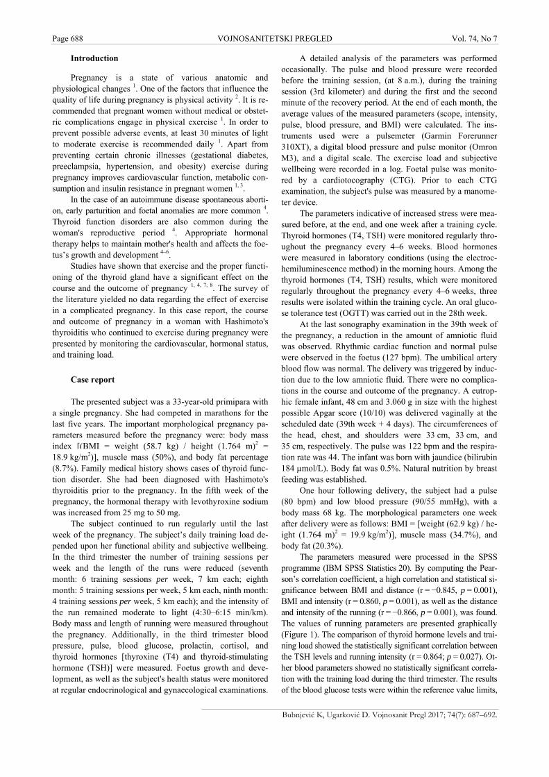
Page 688 VOJNOSANITETSKI PREGLED Vol. 74, No 7
Bubnjević K, Ugarković D. Vojnosanit Pregl 2017; 74(7): 687–692.
Introduction
Pregnancy is a state of various anatomic and physiological changes 1. One of the factors that influence the quality of life during pregnancy is physical activity 2. It is re-commended that pregnant women without medical or obstet-ric complications engage in physical exercise 1. In order to prevent possible adverse events, at least 30 minutes of light to moderate exercise is recommended daily 1. Apart from preventing certain chronic illnesses (gestational diabetes, preeclampsia, hypertension, and obesity) exercise during pregnancy improves cardiovascular function, metabolic con-sumption and insulin resistance in pregnant women 1, 3.
In the case of an autoimmune disease spontaneous aborti-on, early parturition and foetal anomalies are more common 4. Thyroid function disorders are also common during the woman's reproductive period 4. Appropriate hormonal therapy helps to maintain mother's health and affects the foe-tus’s growth and development 4–6.
Studies have shown that exercise and the proper functi-oning of the thyroid gland have a significant effect on the course and the outcome of pregnancy 1, 4, 7, 8. The survey of the literature yielded no data regarding the effect of exercise in a complicated pregnancy. In this case report, the course and outcome of pregnancy in a woman with Hashimoto's thyroiditis who continued to exercise during pregnancy were presented by monitoring the cardiovascular, hormonal status, and training load.
Case report
The presented subject was a 33-year-old primipara with a single pregnancy. She had competed in marathons for the last five years. The important morphological pregnancy pa-rameters measured before the pregnancy were: body mass index [(BMI = weight (58.7 kg) / height (1.764 m)2 = 18.9 kg/m2)], muscle mass (50%), and body fat percentage (8.7%). Family medical history shows cases of thyroid func-tion disorder. She had been diagnosed with Hashimoto's thyroiditis prior to the pregnancy. In the fifth week of the pregnancy, the hormonal therapy with levothyroxine sodium was increased from 25 mg to 50 mg.
The subject continued to run regularly until the last week of the pregnancy. The subject’s daily training load de-pended upon her functional ability and subjective wellbeing. In the third trimester the number of training sessions per week and the length of the runs were reduced (seventh month: 6 training sessions per week, 7 km each; eighth month: 5 training sessions per week, 5 km each, ninth month: 4 training sessions per week, 5 km each); and the intensity of the run remained moderate to light (4:30–6:15 min/km). Body mass and length of running were measured throughout the pregnancy. Additionally, in the third trimester blood pressure, pulse, blood glucose, prolactin, cortisol, and thyroid hormones [thyroxine (T4) and thyroid-stimulating hormone (TSH)] were measured. Foetus growth and deve-lopment, as well as the subject's health status were monitored at regular endocrinological and gynaecological examinations.
A detailed analysis of the parameters was performed occasionally. The pulse and blood pressure were recorded before the training session, (at 8 a.m.), during the training session (3rd kilometer) and during the first and the second minute of the recovery period. At the end of each month, the average values of the measured parameters (scope, intensity, pulse, blood pressure, and BMI) were calculated. The ins-truments used were a pulsemeter (Garmin Forerunner 310XT), a digital blood pressure and pulse monitor (Omron M3), and a digital scale. The exercise load and subjective wellbeing were recorded in a log. Foetal pulse was monito-red by a cardiotocography (CTG). Prior to each CTG examination, the subject's pulse was measured by a manome-ter device.
The parameters indicative of increased stress were mea-sured before, at the end, and one week after a training cycle. Thyroid hormones (T4, TSH) were monitored regularly thro-ughout the pregnancy every 4–6 weeks. Blood hormones were measured in laboratory conditions (using the electroc-hemiluminescence method) in the morning hours. Among the thyroid hormones (T4, TSH) results, which were monitored regularly throughout the pregnancy every 4–6 weeks, three results were isolated within the training cycle. An oral gluco-se tolerance test (OGTT) was carried out in the 28th week.
At the last sonography examination in the 39th week of the pregnancy, a reduction in the amount of amniotic fluid was observed. Rhythmic cardiac function and normal pulse were observed in the foetus (127 bpm). The umbilical artery blood flow was normal. The delivery was triggered by induc-tion due to the low amniotic fluid. There were no complica-tions in the course and outcome of the pregnancy. A eutrop-hic female infant, 48 cm and 3.060 g in size with the highest possible Apgar score (10/10) was delivered vaginally at the scheduled date (39th week + 4 days). The circumferences of the head, chest, and shoulders were 33 cm, 33 cm, and 35 cm, respectively. The pulse was 122 bpm and the respira-tion rate was 44. The infant was born with jaundice (bilirubin 184 µmol/L). Body fat was 0.5%. Natural nutrition by breast feeding was established.
One hour following delivery, the subject had a pulse (80 bpm) and low blood pressure (90/55 mmHg), with a body mass 68 kg. The morphological parameters one week after delivery were as follows: BMI = [weight (62.9 kg) / he-ight (1.764 m)2 = 19.9 kg/m2)], muscle mass (34.7%), and body fat (20.3%).
The parameters measured were processed in the SPSS programme (IBM SPSS Statistics 20). By computing the Pear-son’s correlation coefficient, a high correlation and statistical si-gnificance between BMI and distance (r = −0.845, p = 0.001), BMI and intensity (r = 0.860, p = 0.001), as well as the distance and intensity of the running (r = −0.866, p = 0.001), was found. The values of running parameters are presented graphically (Figure 1). The comparison of thyroid hormone levels and trai-ning load showed the statistically significant correlation between the TSH levels and running intensity (r = 0.864; p = 0.027). Ot-her blood parameters showed no statistically significant correla-tion with the training load during the third trimester. The results of the blood glucose tests were within the reference value limits,
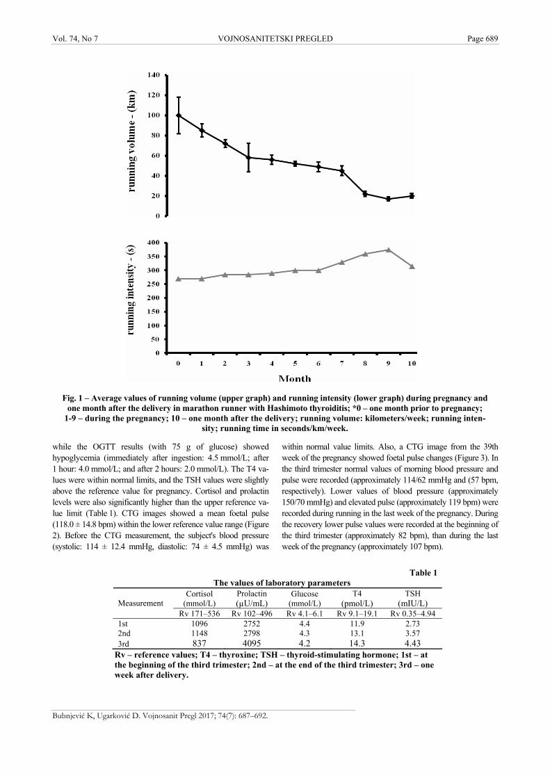
Vol. 74, No 7 VOJNOSANITETSKI PREGLED Page 689
Bubnjević K, Ugarković D. Vojnosanit Pregl 2017; 74(7): 687–692.
Fig. 1 – Average values of running volume (upper graph) and running intensity (lower graph) during pregnancy and
one month after the delivery in marathon runner with Hashimoto thyroiditis; *0 – one month prior to pregnancy; 1-9 – during the pregnancy; 10 – one month after the delivery; running volume: kilometers/week; running inten-
sity; running time in seconds/km/week.
Table 1
The values of laboratory parameters Cortisol
(mmol/L) Prolactin (µU/mL)
Glucose (mmol/L)
T4 (pmol/L)
TSH (mIU/L) Measurement
Rv 171–536 Rv 102–496 Rv 4.1–6.1 Rv 9.1–19.1 Rv 0.35–4.94 1st 1096 2752 4.4 11.9 2.73 2nd 1148 2798 4.3 13.1 3.57 3rd 837 4095 4.2 14.3 4.43
Rv – reference values; T4 – thyroxine; TSH – thyroid-stimulating hormone; 1st – at the beginning of the third trimester; 2nd – at the end of the third trimester; 3rd – one week after delivery.
while the OGTT results (with 75 g of glucose) showed hypoglycemia (immediately after ingestion: 4.5 mmol/L; after 1 hour: 4.0 mmol/L; and after 2 hours: 2.0 mmol/L). The T4 va-lues were within normal limits, and the TSH values were slightly above the reference value for pregnancy. Cortisol and prolactin levels were also significantly higher than the upper reference va-lue limit (Table 1). CTG images showed a mean foetal pulse (118.0 ± 14.8 bpm) within the lower reference value range (Figure 2). Before the CTG measurement, the subject's blood pressure (systolic: 114 ± 12.4 mmHg, diastolic: 74 ± 4.5 mmHg) was
within normal value limits. Also, a CTG image from the 39th week of the pregnancy showed foetal pulse changes (Figure 3). In the third trimester normal values of morning blood pressure and pulse were recorded (approximately 114/62 mmHg and (57 bpm, respectively). Lower values of blood pressure (approximately 150/70 mmHg) and elevated pulse (approximately 119 bpm) were recorded during running in the last week of the pregnancy. During the recovery lower pulse values were recorded at the beginning of the third trimester (approximately 82 bpm), than during the last week of the pregnancy (approximately 107 bpm).
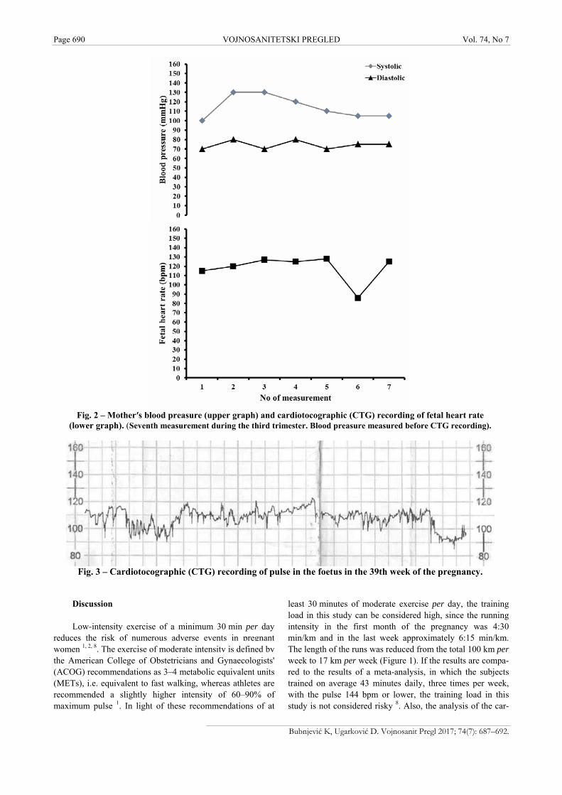
Page 690 VOJNOSANITETSKI PREGLED Vol. 74, No 7
Bubnjević K, Ugarković D. Vojnosanit Pregl 2017; 74(7): 687–692.
Fig. 2 – Mother′s blood preasure (upper graph) and cardiotocographic (CTG) recording of fetal heart rate (lower graph). (Seventh measurement during the third trimester. Blood preasure measured before CTG recording).
Fig. 3 – Cardiotocographic (CTG) recording of pulse in the foetus in the 39th week of the pregnancy.
Discussion
Low-intensity exercise of a minimum 30 min per day reduces the risk of numerous adverse events in pregnant women 1, 2, 8. The exercise of moderate intensity is defined by the American College of Obstetricians and Gynaecologists' (ACOG) recommendations as 3–4 metabolic equivalent units (METs), i.e. equivalent to fast walking, whereas athletes are recommended a slightly higher intensity of 60–90% of maximum pulse 1. In light of these recommendations of at
least 30 minutes of moderate exercise per day, the training load in this study can be considered high, since the running intensity in the first month of the pregnancy was 4:30 min/km and in the last week approximately 6:15 min/km. The length of the runs was reduced from the total 100 km per week to 17 km per week (Figure 1). If the results are compa-red to the results of a meta-analysis, in which the subjects trained on average 43 minutes daily, three times per week, with the pulse 144 bpm or lower, the training load in this study is not considered risky 8. Also, the analysis of the car-
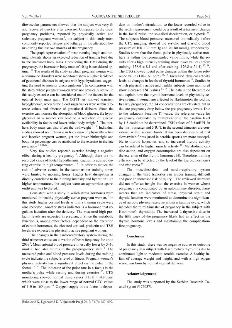
Vol. 74, No 7 VOJNOSANITETSKI PREGLED Page 691
Bubnjević K, Ugarković D. Vojnosanit Pregl 2017; 74(7): 687–692.
diovascular parameters showed that the subject was very fit and recovered quickly after exercise. Compared to the usual pregnancy problems, reported by physically active and sedentary pregnant women 9, the subject in this study most commonly reported fatigue and lethargy in the afternoon ho-urs during the last two months of the pregnancy.
The graph representation of mean running length and run-ning intensity shows an expected reduction of training load due to the increased body mass. Considering the BMI during the pregnancy, the increase in body mass of 10 kg is considered op-timal 10. The results of the study in which pregnant women with autoimmune disorders were monitored show a higher incidence of gestational diabetes in subjects with hypothyroidism, sugges-ting the need to monitor glucoregulation 7. In comparison with the study where pregnant women were not physically active, in this study exercise can be considered one of the factors for the optimal body mass gain. The OGTT test showed transient hypoglycemia, whereas the blood sugar values were within refe-rence values and absence of gestational diabetes. Since the exercise can increase the absorption of blood glucose, the hypo-glycemia in a mother can lead to a reduction of glucose availability in foetus and lower infant body weight 3. The mot-her's body mass can also affect the birthweight 8, 10. Individual studies showed no difference in body mass in physically active and inactive pregnant women, yet the lower birthweight and body fat percentage can be attributed to the exercise in the late pregnancy 1, 8, 11, 12.
Very few studies reported exercise having a negative effect during a healthy pregnancy 8. Although there are no recorded cases of foetal hyperthermia, caution is advised du-ring exercise in high temperatures 1, 8. In order to reduce the risk of adverse events, in the summertime training times were limited to morning hours. Higher heat dissipation is directly correlated to the running intensity and hydration 1. In higher temperatures, the subject wore an appropriate sports outfit and was hydrated.
Consistent with a study in which stress hormones were monitored in healthy physically active pregnant women, 3 in this study higher cortisol levels within a training cycle were also recorded. Another stress indicator is a hormone that re-gulates lactation after the delivery. The measured high pro-lactin levels are expected in pregnancy. Since the metabolic function is, among other factors, dependent on the excretion of certain hormones, the elevated cortisol, prolactin and TSH levels are expected in physically active pregnant women.
The changes in the cardiorespiratory system during the third trimester cause an elevation of heart frequency for up to 20% 1. Mean arterial blood pressure in usually lower by 5–10 mmHg, but later returns to the pre-pregnancy state 1. The measured pulse and blood pressure levels during the training cycle indicate the subject's level of fitness. Pregnant women's physical activity has a significant effect on the pulse in the foetus 13, 14. The indicator of the pulse rate in a foetus is the mother's pulse while resting and during exercise 15. CTG monitoring showed normal pulse values (118.0 ± 14.8 bpm) which were close to the lower range of normal CTG values of 110 to 160 bpm 16. Oxygen supply in the foetus is depen-
dent on mother's circulation, so the lower recorded value in the sixth measurement could be a result of a transient change in the foetal pulse, the so-called deceleration, or hypoxia 16. The subject's blood pressure, measured immediately before the CTG imaging, showed the systolic and diastolic blood pressure of 100–130 mmHg and 70–80 mmHg, respectively. Studies show that the foetal pulse in physically active mot-hers is within the recommended value limits, while the re-sults after a high intensity training show lower values (before training: 138.9 ± 8.1 and after training: 126.8 ± 34.4) 13, 14. The CTG showed foetal pulse changes within the lower refe-rence value (110–160 bpm) 13, 16. Increased physical activity leads to changes in levels of thyroid hormones 17. Studies in which physically active and healthy subjects were monitored show increased TSH values 17, 18. The data in the literature do not explain how the thyroid hormone levels in physically ac-tive pregnant women are affected by Hashimoto's thyroiditis. In early pregnancy, the T4 concentrations are elevated, but in the late pregnancy drop below the pre-pregnancy levels. Due to the unknown baseline T4 value, the reference value for pregnancy, calculated by multiplication of the baseline level by 1.5 could not be determined. TSH values of 2.5 mIU/L in the first trimester and 3 IU/L in the second trimester are con-sidered within normal limits. It has been demonstrated that slow-twitch fibres (used in aerobic sports) are more suscepti-ble to thyroid hormones, and so increased thyroid activity can be related to higher muscle activity 18. Metabolism, car-diac action, and oxygen consumption are also dependent on the excretion of the thyroid hormones (4). Therefore, training efficacy can be affected by the level of the thyroid hormones and vice versa 18.
The musculoskeletal and cardiorespiratory system changes in the third trimester can render training difficult and pose an increased risk of injury 1. The reviewed literature did not offer an insight into the exercise in women whose pregnancy is complicated by an autoimmune disorder. Para-meters that are indicators of stress, physical strain and thyroid function were monitored to determine the significan-ce of aerobic physical exercise within a training cycle, which included the third trimester of pregnancy in the subject with Hashimoto's thyroiditis. The increased L-thyroxine dose in the fifth week of the pregnancy likely had an effect on the thyroid hormone levels and maintaining the complication-free pregnancy.
Conclusion
In this study, there was no negative course or outcome of pregnancy in a subject with Hashimoto’s thyroiditis due to continuous light to moderate aerobic exercise. A healthy in-fant of average weight and height, and with a high Apgar score, was born by normal vaginal delivery.
Acknowledgement
The study was supported by the Serbian Research Co-uncil (grant #175037).
Page 692 VOJNOSANITETSKI PREGLED Vol. 74, No 7
Bubnjević K, Ugarković D. Vojnosanit Pregl 2017; 74(7): 687–692.
R E F E R E N C E S
1. Artal R, O'Toole M. Guidelines of the American College of Ob-stetricians and Gynecologists for exercise during pregnancy and the postpartum period. Br J Sports Med 2003; 37(1): 6−12.
2. Montoya Arizabaleta AV, Orozco Buitrago L, Aguilar de Plata AC, Mosquera Escudero M, Ramirez-Velez R. Aerobic exercise during pregnancy improves health-related quality of life: a randomised trial. J Physiother 2010; 56(4): 253−8.
3. Bessinger RC, McMurray RG, Hackney AC. Substrate utilization and hormonal responses to moderate intensity exercise during pregnancy and after delivery. Am J Obstet Gynecol 2002; 186(4): 757−64.
4. Bicanin M, Varjacic M. Thyroid gland and pregnancy. Med Čas 2011; 45(3): 32−7. (Serbian)
5. Negro R, Formoso G, Mangieri T, Pezzarossa A, Dazzi D, Hassan H. Levothyroxine treatment in euthyroid pregnant women with autoimmune thyroid disease: Effects on obstetrical com-plications. J Clin Endocrinol Metab 2006; 91(7): 2587−91.
6. Glinoer D. Management of hypo- and hyperthyroidism during pregnancy. Growth Horm IGF Res 2003; 13 Suppl A: S45−54.
7. Gudović A, Spremović-Radjenović S, Lazović G, Marinković J, Glisić A, Milićević S. Maternal autoimmune thyroid disease and preg-nancy complication. Vojnosanit Pregl 2010; 67(8): 617−21. (Serbian)
8. Sport Medicine Australia.SMA statement the benefits and risks of exercise during pregnancy. J Sci Med Sport 2002; 5(1): 11−9.
9. Horns PN, Ratcliffe LP, Leggett JC, Swanson MS. Pregnancy out-comes among active and sedentary primiparous women. J Ob-stet Gynecol Neonatal Nurs 1996; 25(1): 49−54.
10. American College of Obstetricians and Gynecologists. ACOG Com-mittee opinion no. 548: weight gain during pregnancy. Obstet Gynecol 2013; 121(1): 210−2.
11. Clapp JF 3rd, Kim H, Burciu B, Schmidt S, Petry K, Lopez B. Con-tinuing regular exercise during pregnancy: effect of exercise volume on fetoplacental growth. Am J Obstet Gynecol 2002; 186(1): 142−7.
12. Sokol RJ, Kazzi GM, Kalhan SC, Pillay SK. Identifying the preg-nancy at risk for intrauterine growth retardation: Possible use-fulness of the intravenous glucose tolerance test. Am J Obstet Gynecol 1982; 143(2): 220−3.
13. May LE, Suminski RR, Langaker MD, Yeh HW, Gustafson KM. Regular maternal exercise dose and fetal heart outcome. Med Sci Sports Exerc 2012; 44(7): 1252−8.
14. Szymanski LM, Satin AJ. Strenuous exercise during pregnancy: Is there a limit. Am J Obstet Gynecol 2012; 207(3): 179.e1–179.e6.
15. Rafla NM, Cook JR. The effect of maternal exercise on fetal heart rate. J Obstet Gynecol 1999; 19(4): 381−4.
16. Adler J, Brown K, Craig M, Price DJ, Wardlaw JM. Thyroid func-tion and physical activity a preliminary communication. Brit J Sports Med 1981; 15(4): 261−4.
17. Ciloglu F, Peker I, Pehlivan A, Karacabey K, Ilhan N, Saygin O, et al. Exercise intensity and its effects on thyroid hormones. Neuro Endocrinol Lett 2005; 26(6): 830−4.
18. Spremović Rađenović S. Specific characteristics of thyroid dys-function during pregnancy and postpartum period. Med Glas 2011; 15(37): 16−23. (Serbian)
Received on October 13, 2015.
Revised on December 28, 2015. Accepted on January 5, 2016.
Online First September, 2016.
Vojnosanit Pregl 2017; 74(7): 693–698. VOJNOSANITETSKI PREGLED Page 693
Correspondence to: Sveta Janković, Clinical Center Kragujevac, Pediatric Clinic, Zmaj Jovina 30, 34 000 Kragujevac, Serbia. Phone: +381 34 355 231. E-mail: [email protected]
C A S E R E P O R T UDC: 616-053.2::[616.831.9-002-06:616.61 https://doi.org/10.2298/VSP150819252V
Pneumococcal meningitis associated with glomerulonephritis: A case report
Pneumokokni meningitis udružen sa glomerulonefritisom
Biljana Vuletić*†, Andjelka Stojković*†, Zoran Igrutinović*†, Lidija Stanković*, Raša Medović*, Katerina Dajić†, Tanja Stojković*, Marijana Janković†,
Sveta Janković*, Ana Vujić*†
Clinical Center Kragujevac, *Pediatric Clinic, Kragujevac, Serbia; University of Kragujevac, †Faculty of Medical Sciences, Kragujevac, Serbia
Abstract Introduction. Streptococcus pneumoniae is the second most common cause of meningitis in children, producing more serious complications than other bacteria. Streptococcus pneu-moniae infections are a rare trigger of glomerulonephritis. We presented a case of glomerulonephritis developing concur-rently with meningitis in a young male child. Case report. Gross haematuria, significant proteinuria, hypertension and decreased level of C3 alongside the signs of central nervous system involvement occurred in a male patient of 5 years and 3 months of age. Spontaneous resolution of renal afflic-tion parameters followed the successful treatment of men-ingitis. The disease course was strongly suggestive of postin-fectious glomerulonephritis, although it manifested at the same time as meningitis. The absence of the latent period might point to the development of IgA nephropathy, but since the renal function was stable, without any abnormali-ties in urine tests documented during follow-up, our opin-ion is that this was rather the case of postinfectious neph-ropathy. Conclusion. The presented case is a unique clini-cal form of postinfectious glomerulonephritis. An accurate diagnosis of this entity should ensure the adequate treat-ment and follow-up of these patients. Keywords: pneumococcal infections; streptococcus pneumoniae; child; meningitis; glomerulonephritis; diagnosis.
Apstrakt Uvod. Streptococcus pneumoniae je drugi najčešći uzročnik meningitisa kod dece i izaziva značajno ozbiljnije komplikacije od drugih bakterija. Glomerulonefritis je retka posledica infekcije ovom bakterijom. Prikazali smo razvoj glomerulonefritisa istovremeno sa meningitisom kod dečaka. Prikaz bolesnika. Makrohematurija, značajna proteinurija, hipertenzija i snižen nivo C3 komponente javili su se uporedo sa znacima infekcije centralnog nervnog sistema kod dečaka u životnom dobu od 5 godina i 3 meseca. Spontana normalizacija parametara bubrežnog oštećenja usledila je nakon uspešnog izlečenja meningitisa. Tok bolesti je snažno ukazivao na razvoj postinfektivnog glomerulonefritisa, iako se manifestovao istovremeno sa meningitisom. Odsustvo latentnog perioda može sugerisati razvoj IgA nefropatije, ali s obzirom na stabilnu bubrežnu funkciju, bez ikakvih abnormalnosti u nalazu urina tokom perioda praćenja bolesnika, mišljenja smo da se u ovom slučaju radilo o postinfektivnom glomerulonefritisu. Zaključak. Predstavljeni slučaj pokazuje jedinstven klinički oblik postinfektivnog glomerulonefritisa. Tačna dijagnoza ovog entiteta trebalo bi da osigura adekvatno lečenje i praćenje obolelih. Ključne reči: infekcija, pneumococcus; streptococcus pneumoniae; deca; meningitis; glomerulonefritis; dijagnoza.
Introduction
Bacterial meningitis is a serious infection of the surface of the brain, affecting most commonly children and the elderly. Streptococcus pneumoniae (pneumococcus) is a commensal or-ganism of the human upper respiratory tract 1, and the second most common cause of meningitis in children 2, 3. The risk of complications of bacterial meningitis is higher for Streptococcus
pneumoniae than for other causative agents 4, 5. Glomerulonep-hritis is common in childhood. It represents an acute or chronic nonsuppurative inflammatory process in the glomeruli leading to impaired renal function. The pathogenesis is not fully under-stood, but it is suggested that glomerulonephritis is usually due to an immunologic response to a variety of etiologic agents. Streptococcus pneumoniae infections are a rare trigger of glo-merulonephritis, and their exact contribution is unknown.
Page 694 VOJNOSANITETSKI PREGLED Vol. 74, No 7
Vuletić B, et al. Vojnosanit Pregl 2017; 74(7): 693–698.
We presented a case of glomerulonephritis developing concurrently with meningitis in a young male child.
Case report
A 5-year and 3 months old male patient was admitted to our intensive care unit due to fever, somnolence and dark urine. His illness developed over the course of 3 days. His condition was initially manifested with a slight fever. On the evening of the next day, he developed a high fever, reaching 39.6°C which continued through the night. The following morning he vomited repeatedly and passed odd smelling and dark urine. Prior to hospitalization, he received an antipyretic for fever and diazepam for seizure prophylaxis. From his personal history, we learned that he had 3 episodes of febrile seizures during the previous year, and was suffering from un-recognized nocturnal enuresis. Also, skin prick test was posi-tive for house dust. He was fully vaccinated according to the Serbian vaccination calendar which did not include the anti-pneumococcal vaccine. Family history did not reveal any in-formation about renal diseases. On admission his body tem-perature was 39.4°C, he was vitally stable, conscious but somnolent, moderately dehydrated, a systolic murmur was noted, his blood pressure was increased and meningeal signs were negative. Blood tests had shown elevated count of white blood cells (WBC) 23.7 × 10⁹/L [normal range (NR) 4.8–10.8 109/L], polymorphonuclear cells (PMNs) 92.6% NR 40–74%), C-reactive protein (CRP) 243.7 mg/L (NR 0–5 mg/L) and procalcitonin (Pct) 9.210 ng/mL (NR 0.5–2.0 ng/mL), and increased erythrocyte sedimentation rate (ESR) 50 mm/h (NR 0–5 mm/h). Urine was dusky and morning urine sample test demonstrated pyuria (25–30 WBCs) [nor-mal value is less than 5 WBCs per high-powered field (HPF)], hematuria (40–50 red blood cells (RBC)/HPF, NR 3–5 RBC/HPF) and proteinuria (+++) – normal finding is negative, with hyaline and erythrocyte cylinders. Arterial blood gas analysis revealed a mild acidosis and hypokalemia. Upon admission, a lumbar puncture was performed and a cloudy cerebrospinal fluid (CSF) was attained, with a high count of WBC dominated by PMNs cells 3,925/mm3 (normal count < 5 WBCs/mm3). CSF biochemistry demonstrated ele-vated proteins [3.0 g/L (NR 0.15–0.3 g/L)] and decreased glucose level (CSF glucose ratio 0.37 mmol/L, normal ratio 0.6 mol/L). Based on these results he was treated under the diagnosis of bacterial meningitis with third generation cepha-losporins and vancomycin, dexamethasone and phenobarbi-tone. Further tests were ordered to evaluate the renal functi-on. The day after admission we received the result of the latex particle agglutination (LPA) test which indicated a pneumococcal infection that was later confirmed by CSF cul-ture detection of penicillin-resistant Streptococcus pneumo-niae. According to the pneumococcus susceptibility, antimic-robial treatment was continued with vancomycin and mero-penem for 14 days and additional 7 days with cefotaxime. Nasal and throat swab cultures were both positive exclusively with Staphylococcus aureus. Urine culture result demonstrated Klebsiella-Enterobacter, 5,000 colony forming units (CFU) per mL (CFU/mL). Since the first hospital day
his blood pressure levels were frequently above the 95th per-centile for age, gender and height. Because of significant proteinuria and hypertension, a diuretic and enalapril were added to his treatment. Our patient’s general condition im-proved, his neurological status was normal since the 8th day at the Clinic, but he continued with multiple daily spikes of fever for the total of 14 consecutive days. Gross haematuria also continued while the coagulation status was normal and there were no other signs of the haemorrhagic syndrome. Te-sting the relatives for haematuria was negative. The initial elevation of urea [10.2 mmol/L (NR 3–8 mmol/L)] and crea-tinine [94 µmol/L (NR 49–106 µmol/L)] in the serum were normalized after carefully balanced hydration. Antistreptolysin O titer (ASOT) was in NR (< 250 U/mL), C3 level was decreased [0.25 g/L) (NR 0.9–1.8 g/L)], while C4 was increased [0.51 g/L (NR 0.1–0.4 g/L)]. His 24-hour urine collection test had shown normal creatinine clearance ranging from 0.82 to 2.07 mL/s (NR 1.47–2.28 mL/s; Refe-rence values have not been established for the age < 18 years) but revealed significant proteinuria reaching 2.03 g/day (NR < 0.14 g/day) which continued until gross haema-turia subsided during the fourth week of illness. By that po-int, the levels of C3 and C4 were beginning to normalize along with blood pressure levels. Specific therapy for the re-nal disorder was not introduced. The findings of the control lumbar puncture, performed 7 days after the initial, were normal including the culture, with the exception of micros-copic examination that revealed 48 PMNs and 32 lymphocytes. The results of the brain magnetic resonance examination were normal. Electrocardiographic (ECG), car-diac and abdominal ultrasound examination findings were al-so normal. During the second week of illness the patient de-veloped anaemia, with normal peripheral blood smear fin-dings and normal reticulocyte count. After CRP level norma-lized, he was discharged with stable renal function after spending 26 days at our clinic. Two months after disease on-set C3 and C4 levels were normal, but microscopic haematu-ria and mild proteinuria could still be registered in the mor-ning urine samples. In the 24-hour urine collection test prote-inuria was still present but reduced. By that time enalapril was discontinued and his blood pressure was normal in all measurements. After four months of further follow-up prote-inuria and microscopic haematuria disappeared, blood pres-sure, complement levels and overall renal function remained normal, and neurological sequelae did not develop.
Discussion
A young boy was diagnosed with pneumococcal me-ningitis at our clinic during late September 2014. The diag-nosis was made quickly after admission based on clinical presentation and CSF findings. He was accordingly treated with antibiotics adhering to recommendations for bacterial meningitis. Initial antimicrobial therapy was the combination of ceftriaxone and vancomycin. Isolated Streptococcus pne-umoniae was penicillin resistant and sensitive to third gene-ration cephalosporins, meropenem, and vancomycin. The minimal inhibitory concentration (MIC) for cephalosporins
Vol. 74, No 7 VOJNOSANITETSKI PREGLED Page 695
Vuletić B, et al. Vojnosanit Pregl 2017; 74(7): 693–698.
was higher than for meropenem. Having in mind the patient's general condition, the treatment choice for pneumococcal meningitis was the combination of meropenem and vancomycin, since both of them were suggested to be effec-tive by the The National Institute for Health and Care Excellence (NICE) clinical guidelines 2. The rationale for this decision was that the pneumococcal infection was the main reason for the clinical presentation, including the renal disorder. Meropenem was chosen over cephalosporins due to better MIC. The dose of vancomycin was 15 mg/kg/6h, adju-sted for achieving the target trough level recommended for pneumococcal CNS infections 6 which is in fact around the demonstrated threshold for nephrotoxicity 7. However, in the setting of an acute renal failure, manifesting during a serious infection, we maintained the recommended dose as it is usually done in cases of acute renal impairment secondary to sepsis. Frequent 24-hour urine collection tests demonstrated normal creatinine clearance, and therefore no dosage correc-tion was needed.
Pneumococcal meningitis is one of the clinical manifes-tations of invasive pneumococcal disease (IPD). IPD is usually seen in the very young (under the age of 2 years) and the elderly (older than 50) patients, also in patients with chronic diseases such as chronic liver or kidney diseases, di-abetics, patients with primary and secondary immunological deficits, then in cases of meningeal membrane damage due to trauma, and in children with cochlear implants 8–13. The hu-man nasopharynx is the main reservoir for Streptococcus pneumoniae, where it commonly leads to only asymptomatic colonization. We found in available reports that carriage ra-tes of Streptococcus pneumoniae go as high as 53% among young children, especially those attending day care cen-tres 14. Clinical examination and visualisation studies in the presented case did not determine the entry point for Strepto-coccus pneumoniae in the upper respiratory tract nor elsewhere. Common colonizers of the upper respiratory tract, such as Haemophilus influenzae and Staphylococcus aureus antagonise Streptococcus pneumoniae colonization 15. De-monstrated overgrowth of Staphylococcus aureus in our pa-tients' respiratory mucosa might imply that there was some other point of entry for pneumococci, but since those two microorganisms share the same nishe and resources 16 it is possible that the same preexisting disorder created favourab-le conditions for Streptococcus pneumoniae invasion and Staphylococcus aureus overgrowth. Our patient had prior confirmation of house dust hypersensitivity although was not diagnosed for a particular allergic disease, but subclinical damage to the naso- and oropharyngeal mucosae by allergy might have led to pneumococcal adherence and migration through the respiratory epithelium and later development of invasive disease 17. Pneumococcus was not detected in the blood culture of our patient, but haematogenous disseminati-on could not be excluded since 75% of pneumococcal me-ningitis patients have a positive result 8, 18. It is important to note that Streptococcus pneumoniae is capable for an unmi-tigated invasion of endothelium and epithelium, via its surfa-ce antigens binding to receptors on host cells, which facilitate epithelial cell transcytosis 19–21. The development of IPD is
greatly dependent on host’s ability to defend itself, thus any state that impairs necessary defence mechanism increases the chance of serious illness caused by Streptococcus pneumoniae. There have been discovered rare forms of primary immunodeficiencies that predispose to pneumococcal infections 22–26. Investigation in a Swedish cohort of 40 patients with a homozygous C2 deficiency, revealed 23 (58%) cases with invasive, mainly pne-umococcal infections 27 implying that an inborn impairment of the complement system might predispose to IPD.
The incubation period for this type of infection can be as short as 1–3 day sand the onset of IPD is usually sudden as it was in the presented case. It is estimated that in the case of intact meningeal membranes a significant bacteriaemia during 12–24 hours is necessary before the breach of the blood-brain barrier.
During multiplication, pneumococci concurrently un-dergo autolysis 28, also the application of effective antibio-tics induces massive destruction of microorganisms and re-lease of bacterial products that are highly immunogenic and may lead to an increased inflammatory response in the host 29, 30, which was manifested in our patient with prolon-ged high fever and elevated levels of inflammatory mar-kers. The clinical course was complicated by haematuria, almost from the very beginning. Haematuria has long been accepted as a sign that should prompt the investigation of bacterial endocarditis 31 because of possible infarctions in the urinary tract that cause loin pain and haematuria. Bacte-rial endocarditis has so far been described as a complicati-on of pneumococcal infections 32. Other than haematuria and fever, our patient’s findings did not support the diagno-sis of bacterial endocarditis according to Duke criteria 33, since the blood culture was negative and there was no evi-dence for endocardial involvement. One of the possible complications of IPD is disseminated intravascular coagu-lation (DIC) 34. It is well known that the regulation of thrombin formation is disrupted during inflammation 35. Diagnosis of DIC in our patient was made unlikely by nor-mal coagulation status and fibrinogen level. Thus, in this setting, without any other bleeding site and normal coagu-lation screen, it would be reasonable to infer that a local damage in the urinary tract was the cause of haematuria, rather than a systemic impairment of the coagulation casca-de. Elevation of serum urea and creatinine associated with haematuria raised suspicion of hemolytic uremic syndrome (HUS). IPD may be complicated by HUS and pneumococ-cal-associated cases account for 14% of all HUS diagnosed in the United Kingdom 36. Although our patient had deve-loped anaemia by the second week of illness, normal levels of liver enzymes, the normal erythrocyte and thrombocyte count, along with normal peripheral blood smear findings and the presence of erythrocyte casts and later normally shaped erythrocytes in urinalysis, made the diagnosis of HUS unlikely, according to the Center for Disease Con-trol's definition of HUS 37 and the Canadian Paediatric Society’s Streptococcus pneumoniae associated haemolytic uremic syndrome case definitions 38.
Intensive immune response elicits immune complex de-position which may result in glomerulonephritis. Haematu-
Page 696 VOJNOSANITETSKI PREGLED Vol. 74, No 7
Vuletić B, et al. Vojnosanit Pregl 2017; 74(7): 693–698.
ria, proteinuria, and elevated blood pressure indicated the development of nephritic syndrome in our patient. Cases of adults and children developing a clinically apparent pneumo-coccal disease and subsequently acute glomerulonephritis, have been previously described 39–42. Streptococcus pneumo-niae is now a recognized cause of postinfectious glomerulo-nephritis (PIGN). Acute poststreptococcal glomerulonephri-tis is the classic example of PIGN. The pathogenesis remains unknown, and there is still no definitive insight into the natu-re of the main causative antigen43. Unfortunately, we were unable to test for serotype of the pneumococcus. So far pne-umococcus types 5, 6C, 7, 9, 14, 15 and 17F have been isola-ted from patients who developed a glomerulonephritis following a pneumococcal infection, and those serotypes ha-ve been suggested to be nephritogenic strains 40, 44–48. Altho-ugh, in one of these patients, a 4-year-old girl described by Hyman et al. 48, type 14 pneumococcal antigen was detected in the kidney 48 implying that the pneumococcal antigens may play a role in the local activation of the immune re-sponse, we could not find any evidence supporting signifi-cance of particular serotypes in the development of nephritis from the available literature. This was also the only case de-scribing the histology of a mesangial proliferative glomeru-lonephritis. The other available reported case in which a re-nal biopsy was performed describes a membranoproliferative glomerulonephritis after pneumococcal pneumonia 49. These findings correlate with acute PIGN histology, particularly in the early stages 43. Complete resolution of all parameters of renal function deterred us from performing a kidney biopsy in our patient.
The disease course of PIGN is usually mild with spon-taneous resolution of clinical parameters. The clinical course, resolution of haematuria, hypertension and transient decrease of C3 level in the presented case were in line with the typical presentation of PIGN with a significant exception of the time that passed from infection to glomerulonephritis onset. The similar disease course was described by other authors, de-
monstrating an acute glomerulonephritis following the pne-umococcal infection within 24–48 hours 41, 45, 47. Those cases also presented with a decrease of C3, but unlike in previous reports, the elevation of ASOT was not demonstrated in our patient. Renal function normalised in all, with various peri-ods needed, spanning from 5 days to 8 weeks. A usual latent period of 1–3 weeks is seen in PIGN, however, the nephritis of IgA nephropathy (IgAN) may occur either at the same ti-me or just 12–72 hours after precipitating event 50. It was demonstrated that antibody levels specific for various strep-tococci antigens, including those of the pneumococcus, are increased in patients with IgAN 51, 52 which suggests that pneumococcal antigens are pathogenic in this disease. IgAN commonly occurs in patients older than 15 years of age, the duration of gross haematuria is usually less than 3 days in IgAN, the degree of proteinuria is low, and the episodes of haematuria and proteinuria recur 50. Although IgAN can ma-nifest itself as nephrotic syndrome or as an acute nephritic syndrome, overall disease course of our patient was rather in favour of PIGN.
Conclusion
Pneumococcal meningitis is a rare cause of glomerulo-nephritis. It is important for clinicians to be aware of possib-le clinical presentations and the development of glomerulo-nephritis following a pneumococcal infection. Considering that only a few cases of PIGN caused by pneumococcal in-fections have been reported in children, and the early onset of glomerulonephritis, the presented case is a unique clinical form of PIGN. PIGN should be considered in any child who presents with an acute form of glomerulonephritis, regardless that non-typical infectious agents are detected. Most patients recover full renal function and are not biopsied, but an accu-rate clinical diagnosis of this entity is nonetheless possible and should ensure the adequate treatment and follow-up of such patients.
R E F E R E N C E S
1. Bogaert D, De Groot R, Hermans PW. Streptococcus pneumoniae colonisation: The key to pneumococcal disease. Lancet Infect Dis 2004; 4(3): 144−54.
2. National Collaborating Centre for Women's and Children's Health (UK). Bacterial meningitis and meningococcal septicaemia: Management of bacterial meningitis and meningococcal septi-caemia in children and young people younger than 16 years in primary and secondary care. NICE Clinical Guidelines, No. 102. London, UK: London: RCOG Press; 2010.
3. Tunkel AR, Hartman BJ, Kaplan SL, Kaufman BA, Roos KL, Scheld MW, et al. Practice guidelines for the management of bacterial meningitis. Clin Infect Dis 2004; 39(9): 1267−84.
4. Stockmann C, Ampofo K, Byington CL, Filloux F, Hersh AL, Blaschke AJ, et al. Pneumococcal meningitis in children: Epi-demiology, serotypes, and outcomes from 1997-2010 in Utah. Pediatrics 2013; 132(3): 421−8.
5. Kim KS. Acute bacterial meningitis in infants and children. Lancet Infect Dis 2010; 10(1) 32−42.
6. All Sites-Edmonton Zone. Vancomycin Monitoring and Dosing Guideline. [cited 2015 Oct 22]. Available from: http://www.albertahealthservices.ca/assets/Workflows/LabServices/wf-lab-vancomycin-monitoring-and-dosing-guideline.pdf
7. Pritchard L, Baker C, Leggett J, Sehdev P, Brown A, Bayley BK. In-creasing vancomycin serum trough concentrations and inci-dence of nephrotoxicity. Am J Med 2010; 123(12): 1143−9.
8. Arditi M, Mason EO, Bradley JS, Tan TQ, Barson WJ, Schutze GE, et al. Three-year multicenter surveillance of pneumococcal meningitis in children: Clinical characteristics, and outcome re-lated to penicillin susceptibility and dexamethasone use. Pedi-atrics 1998; 102(5): 1087−97.
9. Biernath KR, Reefhuis J, Whitney CG, Mann EA, Costa P, Eichwald J, et al. Bacterial meningitis among children with cochlear implants be-yond 24 months after implantation. Pediatrics 2006; 117(2): 284−9.
10. Miller ML, Gao G, Pestina T, Persons D, Tuomanen E. Hypersus-ceptibility to invasive pneumococcal infection in experimental sickle cell disease involves platelet-activating factor receptor. J Infect Dis 2007; 195(4): 581−4.
Vol. 74, No 7 VOJNOSANITETSKI PREGLED Page 697
Vuletić B, et al. Vojnosanit Pregl 2017; 74(7): 693–698.
11. Beek D, Gans J, Tunkel AR, Wijdicks EF. Community-acquired bacterial meningitis in adults. N Engl J Med 2006; 354(1): 44−53.
12. Weisfelt M, Gans J, Poll T, Beek D. Pneumococcal meningitis in adults: New approaches to management and prevention. Lan-cet Neurol 2006; 5(4): 332−42.
13. Brouwer MC, Tunkel AR, Beek D. Epidemiology, diagnosis, and antimicrobial treatment of acute bacterial meningitis. Clin Mi-crobiol Rev 2010; 23(3): 467−92.
14. Regev-Yochay G, Raz M, Dagan R, Porat N, Shainberg B, Pinco E, et al. Nasopharyngeal carriage of Streptococcus pneumoniae by adults and children in community and family settings. Clin Infect Dis 2004; 38(5): 632−9.
15. Margolis E, Yates A, Levin BR. The ecology of nasal coloniza-tion of Streptococcus pneumoniae, Haemophilus influenzae and Staphylococcus aureus: The role of competition and inter-actions with host's immune response. BMC Microbiology 2010; 10: 59.
16. Carratalà J, Mykietiuk A, Fernández-Sabé N, Suárez C, Dorca J, Verdaguer R, et al. Health care-associated pneumonia requiring hospital admission: Epidemiology, antibiotic therapy, and clin-ical outcomes. Arch Intern Med 2007; 167(13): 1393−9.
17. Juhn YJ, Kita H, Yawn BP, Boyce TG, Yoo KH, McGree ME, et al. Increased risk of serious pneumococcal disease in patients with asthma. J Allergy Clin Immunol 2008; 122(4): 719−23.
18. Chao Y, Chiu N, Huang F. Clinical features and prognostic fac-tors in childhood pneumococcal meningitis. J Microbiol Im-munol Infect 2008; 41(1): 48−53.
19. Okada N, Liszewski MK, Atkinson JP, Caparon M. Membrane cofactor protein (CD46) is a keratinocyte receptor for the M protein of the group A streptococcus. Proc Natl Acad Sci U.S.A. 1995; 92(7): 2489−93.
20. Elm C, Braathen R, Bergmann S, Frank R, Vaerman J, Kaetzel CS, et al. Ectodomains 3 and 4 of human polymeric Immu-noglobulin receptor (hpIgR) mediate invasion of Streptococ-cus pneumoniae into the epithelium. J Biol Chem 2004; 279(8): 6296−304.
21. Kaetzel CS. Polymeric Ig receptor: Defender of the fort or Tro-jan horse. Curr Biol 2001; 11(1): R35−8.
22. Ku C, Picard C, Erdös M, Jeurissen A, Bustamante J, Puel A, et al. IRAK4 and NEMO mutations in otherwise healthy children with recurrent invasive pneumococcal disease. J Med Genet 2007; 44(1): 16−23.
23. Ku CL, Yang K, Bustamante J, Puel A, von Bernuth H, Santos OF, et al. Inherited disorders of human Toll-like receptor signaling: Immunological implications. Immunol Rev 2005; 203: 10−20.
24. Currie AJ, Davidson DJ, Reid GSD, Bharya S, MacDonald KL, De-von RS, Speert DP. Primary immunodeficiency to pneumococ-cal infection due to a defect in Toll-like receptor signaling. J Pediatr 2004; 144(4): 512−8.
25. von Bernuth H, Picard C, Jin Z, Pankla R, Xiao H, Ku C, et al. Pyogenic bacterial infections in humans with MyD88 defi-ciency. Science 2008; 321(5889): 691−6.
26. Picard C, Casanova J, Puel A. Infectious diseases in patients with IRAK-4, MyD88, NEMO, or IκBα deficiency. Clin Microbiol Rev 2011; 24(3): 490−7.
27. Jonsson G, Truedsson L, Sturfelt G, Oxelius VA, Braconier JH, Sjöholm AG. Hereditary C2 deficiency in Sweden: Frequent oc-currence of invasive infection, atherosclerosis, and rheumatic disease. Medicine (Baltimore) 2005; 84(1): 23−34.
28. Tomasz A, Moreillon P, Pozzi G. Insertional inactivation of the major autolysin gene of Streptococcus pneumoniae. J Bacteriol 1988; 170(12): 5931−4.
29. Tuomanen E, Liu H, Hengstler B, Zak O, Tomasz A. The induc-tion of meningeal inflammation by components of the pneu-mococcal cell wall. J Infect. Dis 1985; 151(5): 859−68.
30. Nau R, Eiffert H. Minimizing the release of proinflammatory and toxic bacterial products within the host: A promising ap-proach to improve outcome in life-threatening infections. FEMS Immunol. Med Microbiol 2005; 44(1): 1−16.
31. Horstkotte D, Follath F, Gutschik E, Lengyel M, Oto A, Pavie A, et al. Guidelines on Prevention, Diagnosis and Treatment of In-fective Endocarditis Executive Summary The Task Force on Infective Endocarditis of the European Society of Cardiology. Eur Heart J 2004; 25(3): 267−76.
32. Gatzoulis MA, Winrow A, Kovar IZ. Acute endocarditis follow-ing pneumococcal meningitis. Eur J Pediatr 1993; 152(4): 323−4.
33. Ashley EA, Niebauer J. Cardiology Explained. London: Reme-dica; 2004.
34. Lee C, Chen M, Chiou Y, Shen C, Wu C, Chiou Y. Invasive pneumococcal pneumonia is the major cause of paediatric haemolytic-uraemic syndrome in Taiwan. Nephrology (Carl-ton) 2012; 17(1): 48−52.
35. Levi M, Poll T. Inflammation and coagulation. Crit Care Med 2010; 38(2): 26−34.
36. Waters AM, Kerecuk L, Luk D, Haq MR, Fitzpatrick MM, Gilbert RD, et al. Hemolytic uremic syndrome associated with invasive pneumococcal disease: The United kingdom experience. J Pe-diatr 2007; 151(2): 140−4.
37. Centers for Disease Control and Prevention. Case definitions for infectious conditions under public health surveillance. MMWR Recomm Rep 1997; 46(RR−10):1−55.
38. CHARGE A/S Canadian Paediatric Surveillance Program 2002 Results. Ottawa: Canadian Paediatric Society; 2003.
39. Corriere JN Jr, Lipshultz LI. Pneumococcal nephritis. Urology 1974; 3(5): 557−61.
40. Schachter J, Pomeranz A, Berger I, Wolach B. Acute glomeru-lonephritis secondary to lobar pneumonia. Int J Pediatr Nephrol 1987; 8(4): 211−4.
41. Phillips J, Palmer A, Baliga R. Glomerulonephritis associated with acute pneumococcal pneumonia: A case report. Pediatr Nephrol 2005; 20(10): 1494−5.
42. Ismail IH, Zainudin Z, Othman N. Pneumococcal glomeru-lonephritis in a healthy child: A case report and literature re-view. Singapore Med J 2014; 55(5): e69−72.
43. Tasic V. Postinfectious glomerulonephritis. In: Geary DF, Franz SF, editors. Comprehensive Pediatric Nephrology. 1th ed. Philadelphia: Mosby Elsevier; 2008. p. 309−17.
44. Kaehny WD, Ozawa T, Schwarz MI, Stanford RE, Kohler PF, McIn-tosh RM. Acute nephritis and pulmonary alveolitis following pneumococcal pneumonia. Arch Intern Med 1978; 138(5): 806−8.
45. Hibino S, Hoshino A, Fujii T, Abe Y, Watanabe S, Uemura O, et al. Post-streptococcal acute glomerulonephritis associated with pneumococcal infection. Pediatr Int 2013; 55(5): e136−8.
46. Wolach B, Uziel Y, Berger I, Pomeranz A. Cat-scratch bacillus and Streptococcus pneumoniae causing submandibular suppurative adenitis and acute glomerulonephritis. Child Nephrol Urol 1990; 10(3): 158−60.
Page 698 VOJNOSANITETSKI PREGLED Vol. 74, No 7
Vuletić B, et al. Vojnosanit Pregl 2017; 74(7): 693–698.
47. Carceller LF, Torre EM, Porto AR, Écija PJ. Acute glomeru-lonephritis associated with pneumonia: A review of three cases. Pediatr Nephrol 2010; 25(1): 161−4.
48. Hyman LR, Jenis EH, Hill GS, Zimmerman SW, Burkholder PM. Alternative C3 pathway activiation in pneumococcal glomeru-lonephritis. Am J Med 1975; 58(6): 810−4.
49. Morin D, Ichay I, Dumas ML, Valette H, Dumas R. Mem-branoproliferative glomerulonephritis in a 10-year-old boy with pneumococcal pneumonia. Pediatr Nephrol 1990; 13: C58.
50. Coppo R, Amore A. IgA nephropathy. In: Geary DF, Schaefer F, editors. Comprehensive Pediatric Nephrology. 1th ed. Phila-delphia: Mosby Elsevier; 2008. p. 291−9.
51. Drew PA, Nieuwhof WN, Clarkson AR, Woodroffe AJ. Increased concentration of serum IgA antibody to pneumococcal poly-saccharides in patients with IgA nephropathy. Clin Exp Im-munol 1987; 67(1): 124−9.
52. Kukuminato Y, Hamamoto M, Kataura A. Role of serum antibod-ies to streptococci in patients with IgA nephropathy. Acta Otolaryngol Suppl 1993; 508: 6−10.
Received on August 19, 2015. Revised on February 01, 2016.
Accepted on February 09, 2016. Online First September, 2016.
Vojnosanit Pregl 2017; 74(7): 699–702. VOJNOSANITETSKI PREGLED Page 699
Correspondence to: Silvija Stević-Carević, Military Medical Academy, Clinic for Rheumatology and Clinical Immunology, Crnotravska 17, 11 000, Belgrade, Serbia. E-mail: [email protected]
C A S E R E P O R T UDC: 616-002.52:616.438 https://doi.org/10.2298/VSP160317256S
Systemic lupus erythematosus and thymus persistens – A case report
Sistemski eritemski lupus i perzistentni timus
Silvija Stević-Carević*, Branislava Glišić*†, Jelena Dedović*
Military Medical Academy, *Clinic for Rheumatology and Clinical Immunology, Belgrade, Serbia; University of Defence, †Faculty of Medicine of the Military Medical
Academy, Belgrade, Serbia
Abstract Introduction. Thymus plays an important role in the maturation of T-lymphocytes and in the development of immune tolerance. Its involution comes after puberty. If thymic tissue remains preser-ved in an advanced age it is considered to be the thymus persistens. According to the available data, 5% of patients with a thymoma have some of the autoimmune disorders. Medical data on the sys-temic lupus erythematosus (SLE) association with the thymus persistens are scarce. Case report. A 29-year-old patient was diagnosed with SLE at the age of 12. She was treated with continuous doses of corticosteroids and an antimalarial drug (chloroquine). After ten years, the first, and then two more recurrences of the disease with the last recurrence in 2011 occurred. The performed laboratory analyses indicated the disease activity. The radiography of thorax showed a change on the right lung, with enlarged mediastinal shadow. Therefore the multislice computed tomography (MSCT) of thorax was made. The pathohistology findings confirmed that the change on the right lung was focus of chronic pneumonitis, while the change in mediastinum was thymus persistens. The thymectomy was performed. Due to pneumonitis, the treatment of SLE was continued with corticosteroids, antimalarial drug and pul-se doses of cyclophosphamide. The patient received six monthly and six quarterly pulsed doses of the drug. The remission of the di-sease maintained all the time. Conclusion. The disorder of thymic function should be considered as a possible cause in the develop-ment of SLE. Though the effect of thymectomy is difficult to as-sess, patients should be carefully monitored. Keywords: lupus erythematosus, systemic; thymus gland; comorbidity; diagnosis, differential; drug therapy.
Apstrakt Uvod. Timus ima važnu ulogu u sazrevanju T limfocita i razvo-ju imunološke tolerancije. Nakon puberteta dolazi do njegove involucije. Ukoliko tkivo timusa ostane očuvano i u odmaklom životnom dobu govorimo o perzistentnom timusu (thymus persi-stens). Prema dostupnoj literaturi 5% bolesnika sa timomom ima neki od autoimunskih poremećaja. Podaci o udruženosti si-stemskog eritemskog lupusa (SLE) i pezistentnog timusa su os-kudni. Prikaz bolesnika. Prikazali smo bolesnicu staru 29 go-dina kod koje je dijagnoza SLE postavljena u 12. godini života. Lečena je kontinuiranim dozama kortikosteroida i antimalari-kom hlorokinom. Nakon deset godina dolazi do prvog, a zatim još dva recidiva bolesti. Poslednji recidiv bio je 2011. godine. Učinjene laboratorijske analize ukazivale su na aktivnost bolesti. Tada je na radiografiji pluća uočena promena u desnom plućnom krilu i proširena senka medijastinuma, zbog čega je učinjen multislajsna kompjuterizovana tomografija (MSCT) grudnog koša. Biopsijom je utvrđeno da je promena u desnom plućnom krilu žarište pneumonitisa, a promena u medijastinu-mu perzistentni timus. Učinjena je timektomija. Lečenje SLE je zbog pneumonitisa nastavljeno kortikosteroidima, antimalari-kom i pulsnim dozama ciklofosfamida. Bolesnica je primila šest mesečnih i šest tromesečnih pulsnih doza leka. Sve vreme odr-žava se remisija bolesti. Zaključak. Poremećaj funkcije timusa treba razmatrati kao mogući uzrok nastanka SLE. Koliko time-ktomija utiče na tok autoimunske bolesti teško je proceniti, ali bolesnike svakako treba pažljivo pratiti. Ključne reči: lupus, eritematozni, sistematski; timus; komorbiditet; dijagnoza, diferencijalna; lečenje lekovima.
Introduction
Systemic lupus erythematosus (SLE) is a chronic inflammatory disease caused by unknown etiology. Its main characteristic is a tissue and cell damage, which is caused by immune complexes and pathogenic autoantibodies. A disor-der of immune response regulation with a development of
the phenomenon of autoimmunity plays an important role in the pathogenesis. The disease is characterized by the perma-nent creation of pathogenic autoantibodies to certain cellular components as well as by the formation of immune complexes. A hyperactivity of autoreactive B- and T- cells is expressed, which is caused by a series of disorders. A clini-cal profile of this multisystem disease is quite diverse. At the
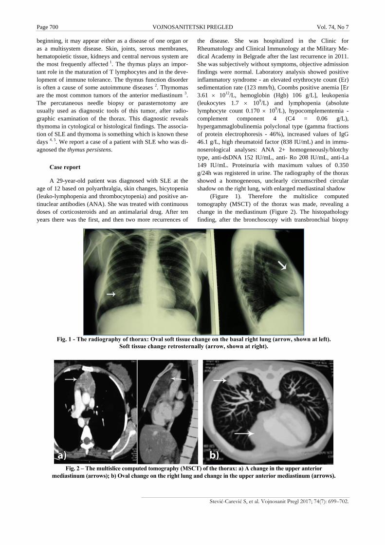
Page 700 VOJNOSANITETSKI PREGLED Vol. 74, No 7
Stević-Carević S, et al. Vojnosanit Pregl 2017; 74(7): 699–702.
Fig. 1 - The radiography of thorax: Oval soft tissue change on the basal right lung (arrow, shown at left).
Soft tissue change retrosternally (arrow, shown at right).
Fig. 2 – The multislice computed tomography (MSCT) of the thorax: a) A change in the upper anterior
mediastinum (arrows); b) Oval change on the right lung and change in the upper anterior mediastinum (arrows).
beginning, it may appear either as a disease of one organ or as a multisystem disease. Skin, joints, serous membranes, hematopoietic tissue, kidneys and central nervous system are the most frequently affected 1. The thymus plays an impor-tant role in the maturation of T lymphocytes and in the deve-lopment of immune tolerance. The thymus function disorder is often a cause of some autoimmune diseases 2. Thymomas are the most common tumors of the anterior mediastinum 3. The percutaneous needle biopsy or parasternotomy are usually used as diagnostic tools of this tumor, after radio-graphic examination of the thorax. This diagnostic reveals thymoma in cytological or histological findings. The associa-tion of SLE and thymoma is something which is known these days 4, 5. We report a case of a patient with SLE who was di-agnosed the thymus persistens.
Case report
A 29-year-old patient was diagnosed with SLE at the age of 12 based on polyarthralgia, skin changes, bicytopenia (leuko-lymphopenia and thrombocytopenia) and positive an-tinuclear antibodies (ANA). She was treated with continuous doses of corticosteroids and an antimalarial drug. After ten years there was the first, and then two more recurrences of
the disease. She was hospitalized in the Clinic for Rheumatology and Clinical Immunology at the Military Me-dical Academy in Belgrade after the last recurrence in 2011. She was subjectively without symptoms, objective admission findings were normal. Laboratory analysis showed positive inflammatory syndrome - an elevated erythrocyte count (Er) sedimentation rate (123 mm/h), Coombs positive anemia [Er 3.61 1012/L, hemoglobin (Hgb) 106 g/L], leukopenia (leukocytes 1.7 109/L) and lymphopenia (absolute lymphocyte count 0.170 109/L), hypocomplementemia - complement component 4 (C4 = 0.06 g/L), hypergammaglobulinemia polyclonal type (gamma fractions of protein electrophoresis - 46%), increased values of IgG 46.1 g/L, high rheumatoid factor (838 IU/mL) and in immu-noserological analyses: ANA 2+ homogeneously/blotchy type, anti-dsDNA 152 IU/mL, anti- Ro 208 IU/mL, anti-La 149 IU/mL. Proteinuria with maximum values of 0.350 g/24h was registered in urine. The radiography of the thorax showed a homogeneous, unclearly circumscribed circular shadow on the right lung, with enlarged mediastinal shadow
(Figure 1). Therefore the multislice computed tomography (MSCT) of the thorax was made, revealing a change in the mediastinum (Figure 2). The histopathology finding, after the bronchoscopy with transbronchial biopsy
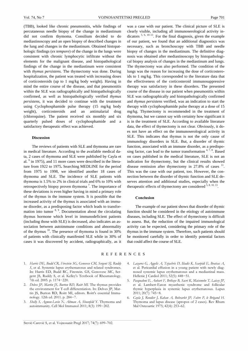
Vol. 74, No 7 VOJNOSANITETSKI PREGLED Page 701
Stević-Carević S, et al. Vojnosanit Pregl 2017; 74(7): 699–702.
(TBB), looked like chronic pneumonitis, while findings of percutaneous needle biopsy of the change in mediastinum did not confirm thymoma. Consilium decided to do mediastinoscopy and an open biopsy of described changes in the lung and changes in the mediastinum. Obtained histopat-hologic findings (ex tempore) of the change in the lungs were consistent with chronic lymphocytic infiltrate without the elements for the malignant disease, and histopathological findings of the change in the mediastinum were consistent with thymus persistens. The thymectomy was done. During hospitalization, the patient was treated with increasing doses of corticosteroids (up to 1 mg/kg body weight). Having in mind the entire course of the disease, and that pneumonitis within the SLE was radiographically and histopathologically confirmed, as well as histopathologically verified thymus persistens, it was decided to continue with the treatment using Cyclophosphamide pulse therapy (15 mg/kg body weight), corticosteroids and an antimalarial drug (chloroquine). The patient received six monthly and six quarterly pulsed doses of cyclophosphamide and a satisfactory therapeutic effect was achieved.
Discussion
The reviews of patients with SLE and thymoma are rare in medical literature. According to the available medical da-ta, 2 cases of thymoma and SLE were published by Cayla et al. 6 in 1975), and 11 more cases were described in the litera-ture from 1922 to 1975. Searching MEDLINE for the period from 1975 to 1998, we identified another 18 cases of thymoma and SLE. The incidence of SLE patients with thymoma is 1.5% to 2% in clinical trials and 6% to 10% with retrospectively biopsy proven thymoma 7. The importance of these deviations is even higher having in mind a primary role of the thymus in the immune system. It is possible that the increased activity of the thymus is associated with an immu-ne disorder, as a predisposing factor which leads to transfor-mation into tumor 8, 9. Documentation about the circulating thymus hormone which level in immunodeficient patients (including those with SLE) is decreased, also indicates an as-sociation between autoimmune conditions and abnormality of the thymus 10. The presence of thymoma is found in 30% of patients with clinically manifested SLE while in 30% of cases it was discovered by accident, radiographically, as it
was a case with our patient. The clinical picture of SLE is clearly visible, including all immunoserological activity in-dicators 5, 6, 10–13. For the final diagnosis, given the example of our patient, we found that an additional diagnostics was necessary, such as bronchoscopy with TBB and needle biopsy of changes in the mediastinum. The definitive diag-nosis was obtained after mediastinoscopy by histopathologi-cal biopsy analysis of changes in the mediastinum and lungs. The thymectomy was also performed. The condition of the lungs was the reason for increasing the dose of corticostero-ids to 1 mg/kg. This corresponded to the literature data that the effectiveness of the corticosteroid immunosuppressive therapy was satisfactory in these disorders. The presented course of the disease in our patient when pneumonitis within SLE was radiographically and histopathologically confirmed and thymus persistens verified, was an indication to start the therapy with cyclophosphamide pulse therapy at a dose of 15 mg/kg. Thymectomy is recommended in the treatment of thymoma, but we cannot say with certainty how significant it is in the treatment of SLE. According to available literature data, the effect of thymectomy is not clear. Obviously, it do-es not have an effect on the immunoserological activity in SLE. This indicates that thymus is not the only cause of immunology disorders in SLE. But, a disorder of thymic function, associated with an immune disorder, as a predispo-sing factor, can lead to the tumor transformation 4, 7, 8. Based on cases published in the medical literature, SLE is not an indication for thymectomy, but the clinical results showed disease remission after thymectomy in 27.8% of cases 11. This was the case with our patient, too. However, the con-nection between the disorder of thymic function and SLE de-serves attention and additional studies, especially when the therapeutic effects of thymectomy are considered 5, 8, 10, 13.
Conclusion
The example of our patient shows that disorder of thymic function should be considered in the etiology of autoimmune diseases, including SLE. The effect of thymectomy is difficult to assess. But, the reduction of the impaired immunological activity can be expected, considering the primary role of the thymus in the immune system. Therefore, such patients should be monitored carefully in order to identify potential factors that could affect the course of SLE.
R E F E R E N C E S
1. Harris DE, Budd CR, Firestein SG, Genovese CM, Sergent SJ, Ruddy S, et al. Systemic lupus erythematosus and related syndromes. In: Harris ED, Budd RC, Firestein. GS, Genovese MC, Ser-gent JS, Ruddy S, et al. Kelley's Textbook of Rheumatology. 7th ed. 2005. p. 1174−220.
2. Delves JP, Martin JS, Burton RD, Roitt MI. The thymus provides the environment for T-cell differentiation. In: Delves JP, Mar-tin JS, Burton RD, Roitt MI, editors. Roitt's essential Immu-nology. 12th ed. 2011. p. 284−7.
3. Shelly S, Agmon-Levin N, Altman A, Shoenfeld Y. Thymoma and autoimmunity. Cell Mol Immunol 2011; 8(3): 199−202.
4. Lazaros G, Aggelis A, Tziachris D, Iliadis K, Scarpidi E, Bratsas A, et al. Pericardial effusion in a young patient with newly diag-nosed systemic lupus erythematosus and a mediastinal mass. Hellenic J Cardiol 2011; 52(5): 448−51.
5. Pasqualoni E, Aubart F, Brihaye B, Sacré K, Maisonobe T, Laissy JP, et al. Lambert-Eaton myasthenic syndrome and follicular thymic hyperplasia in systemic lupus erythematosus. Lupus 2011; 20(7): 745−8.
6. Cayla J, Rondier J, Kahan A, Bohvarlet JP, Fabre P, le Brigand H. Thymoma and lupus disease (apropos of 2 cases). Rev Rhum Mal Osteoartic 1975; 42(4): 253–62.
Page 702 VOJNOSANITETSKI PREGLED Vol. 74, No 7
Stević-Carević S, et al. Vojnosanit Pregl 2017; 74(7): 699–702.
7. Boonen A, Rennenberg R, Linden S. Thymoma-associated sys-temic lupus erythematosus, exacerbating after thymectomy. A case report and review of the literature. Rheumatology (Ox-ford) 2000; 39(9): 1044−6.
8. Steven MM, Westedt ML, Eulderink F, Hazevoet HM, Dijkman JH, Cats A. Systemic lupus erythematosus and invasive thymoma: Report of two cases. Ann Rheum Dis 1984; 43(6): 825−8.
9. Menon S, Snaith ML, Isenberg DA. The association of malig-nancy with SLE: An analysis of 150 patients under long-term review. Lupus 1993; 2(3): 177−81.
10. Simeone FJ, Mccloud T, Putman EC, Marsh J. Thymoma and sys-temic lupus erythematosus. Thorax 1975; 30: 697−700.
11. Zhang L, Dong J, Leng XM, Zeng XF. A meta-analysis of thy-moma-associated systemic lupus erythematosus from 1975-
2008 world wide. Zhonghua Nei Ke Za Zhi 2009; 48(8): 643−6. (Chinese)
12. Mevorach D, Perrot S, Buchanan NM, Khamashta M, Laoussadi S, Hughes GR, et al. Appearance of systemic lupus erythematosus after thymectomy: Four case reports and review of the litera-ture. Lupus 1995; 4(1): 33−7.
13. Zandman-Goddard G, Lorber M, Shoenfeld Y. Systemic lupus ery-thematosus and thymoma -a double-edged sword. Int Arch Al-lergy Immunol 1995; 108(1): 99−102.
Received on March 17, 2016. Accepted on April 7, 2016.
Online First September, 2016.
Vojnosanit Pregl 2017; 74(7): 703–707. VOJNOSANITETSKI PREGLED Page 703
Correspondence to: Jovanović D. Mirko, Military Medical Academy, Urology Clinic, Crnotravska 17, 11 000 Belgrade, Serbia. E-mail: [email protected]
C A S E R E P O R T UDC: 616.6-08-06::616.617-007.271 https://doi.org/10.2298/VSP150728096J
Kidney auto-transplantation due to upper and middle ureter defect after ureteroscopy injury
Autotransplantacija bubrega zbog defekta gornjeg i srednjeg uretera nakon povrede pri ureteroskopiji
Mirko D Jovanović*, Predrag Marić*, Vladimir Bančević*†, Radovan Milošević*†,
Ivica Nikolić*, Dejan Simić*, Aleksandar Spasić*, Novak Milović*†
Military Medical Academy, *Urology Clinic, Belgrade, Serbia; Univeristy of Defence, †Faculty of Medicine of the Military Medical Academy, Belgrade, Serbia
Abstract Introduction. Surgery complications during and after uret-eroscopic procedures are rare, mostly temporary and require no special additional procedures. However, major complica-tions, nevertheless their small incidence (less than 1–2%), can be very serious and they include ureter perforation and avulsion. Case report. We represented a patient with com-plicated ureter iatrogenic stenosis developed after uret-eroscopic procedure of stone removal. After several hospi-talizations, septic condition with retroperitoneal abscess and an attempt of endoscopic and open surgical solution, the kidney auto-transplantation was successfully done. Conclu-sion. There are a small number of complications with uret-eroscopic procedures, but when ureter avulsion or perfora-tion occurs, and when the mission is to preserve pair or-gans, kidney auto-transplantation has the advantage over other methods of ureter reconstruction. Key words: ureteroscopy; iatrogenic disease; ureteral obstruction; kidney; transplantation, autologous; treatment outcome.
Apstrakt Uvod. Hirurške komplikacije tokom i nakon ureteroskopskih procedura su retke, uglavnom su prolazne i ne zahtevaju posebne dodatne procedure. Međutim, velike komplikacije i pored male incidence (manje od 1–2%), mogu biti veoma ozbiljne i tu spadaju perforacija i avulzija uretera. Prikaz slučaja. U radu je prikazan bolesnik sa komplikovanom jatrogenom stenozom uretera koja se razvila nakon ureteroskopske procedure razbijanja kamena. Nakon brojnih hospitalizacija, pojave septičnog stanja sa retroperitonealnim apscesom i pokušaja endoskopskog i otvorenog hirurškog rešavanja, uspešno je urađena autotransplantacija bubrega. Zaključak. Ureteroskopske procedure imaju mali broj komplikacija, ali u slučaju avulzije ili perforacije uretera, u cilju očuvanja parnih organa, autotransplantacija bubrega je u prednosti u odnosu na druge metode rekonstrukcije uretera. Ključne reči: ureteroskopija; jatrogena bolest; ureter, opstrukcija; bubreg; transplantacija, autologna; lečenje, ishod.
Introduction
Surgical complications during and after ureteroscopic procedures are rare, mostly of temporary character and do not require special additional procedures 1. Most of them be-long to a minor complications such as bleeding, mucosal la-ceration, and kidney colic, and all are successfully treated conservatively or endoscopically 2.
However, major complications, nevertheless their small incidence (less than 1–2%), can be very serious and cause si-gnificant morbidity. They include ureter perforation and avulsion 3. All these complications are early and arise in the course of the surgical procedure or immediately after it (in
the first 24 hours). The complications are dealt with endoscopy or open surgery 4, 5. Some of the most often late complications, that can arise from several weeks to several months after the primary treatment, are ureter stenoses 6, that occur in 0–0.2% cases 7, 8.
Ureteral stenoses lead to urin excretion obstruction and can be non-complicated or complicated by urinary infection, kidney insufficiency and kidney dysfunction. Complicated ureter stenoses require urinary tract decompression by pla-cing “double J” ureteral stent or percutaneous nephrostomy catheter. With some patients there is a necessity for ureteral “double J” stent constant replacement on a three months ba-sis. The consequence of these interventions is the stent bacte-
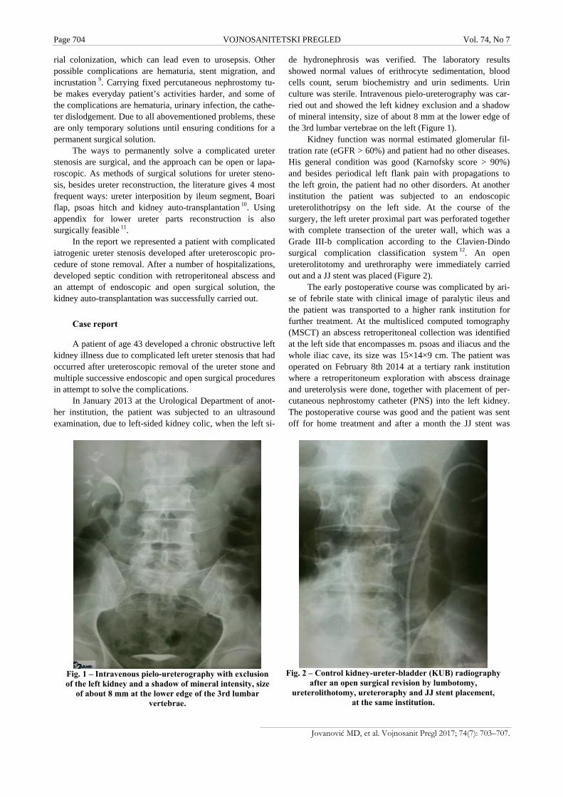
Page 704 VOJNOSANITETSKI PREGLED Vol. 74, No 7
Jovanović MD, et al. Vojnosanit Pregl 2017; 74(7): 703–707.
Fig. 1 – Intravenous pielo-ureterography with exclusion of the left kidney and a shadow of mineral intensity, size
of about 8 mm at the lower edge of the 3rd lumbar vertebrae.
Fig. 2 – Control kidney-ureter-bladder (KUB) radiography
after an open surgical revision by lumbotomy, ureterolithotomy, ureteroraphy and JJ stent placement,
at the same institution.
rial colonization, which can lead even to urosepsis. Other possible complications are hematuria, stent migration, and incrustation 9. Carrying fixed percutaneous nephrostomy tu-be makes everyday patient’s activities harder, and some of the complications are hematuria, urinary infection, the cathe-ter dislodgement. Due to all abovementioned problems, these are only temporary solutions until ensuring conditions for a permanent surgical solution.
The ways to permanently solve a complicated ureter stenosis are surgical, and the approach can be open or lapa-roscopic. As methods of surgical solutions for ureter steno-sis, besides ureter reconstruction, the literature gives 4 most frequent ways: ureter interposition by ileum segment, Boari flap, psoas hitch and kidney auto-transplantation 10. Using appendix for lower ureter parts reconstruction is also surgically feasible 11.
In the report we represented a patient with complicated iatrogenic ureter stenosis developed after ureteroscopic pro-cedure of stone removal. After a number of hospitalizations, developed septic condition with retroperitoneal abscess and an attempt of endoscopic and open surgical solution, the kidney auto-transplantation was successfully carried out.
Case report
A patient of age 43 developed a chronic obstructive left kidney illness due to complicated left ureter stenosis that had occurred after ureteroscopic removal of the ureter stone and multiple successive endoscopic and open surgical procedures in attempt to solve the complications.
In January 2013 at the Urological Department of anot-her institution, the patient was subjected to an ultrasound examination, due to left-sided kidney colic, when the left si-
de hydronephrosis was verified. The laboratory results showed normal values of erithrocyte sedimentation, blood cells count, serum biochemistry and urin sediments. Urin culture was sterile. Intravenous pielo-ureterography was car-ried out and showed the left kidney exclusion and a shadow of mineral intensity, size of about 8 mm at the lower edge of the 3rd lumbar vertebrae on the left (Figure 1).
Kidney function was normal estimated glomerular fil-tration rate (eGFR > 60%) and patient had no other diseases. His general condition was good (Karnofsky score > 90%) and besides periodical left flank pain with propagations to the left groin, the patient had no other disorders. At another institution the patient was subjected to an endoscopic ureterolithotripsy on the left side. At the course of the surgery, the left ureter proximal part was perforated together with complete transection of the ureter wall, which was a Grade III-b complication according to the Clavien-Dindo surgical complication classification system 12. An open ureterolitotomy and urethroraphy were immediately carried out and a JJ stent was placed (Figure 2).
The early postoperative course was complicated by ari-se of febrile state with clinical image of paralytic ileus and the patient was transported to a higher rank institution for further treatment. At the multisliced computed tomography (MSCT) an abscess retroperitoneal collection was identified at the left side that encompasses m. psoas and iliacus and the whole iliac cave, its size was 15×14×9 cm. The patient was operated on February 8th 2014 at a tertiary rank institution where a retroperitoneum exploration with abscess drainage and ureterolysis were done, together with placement of per-cutaneous nephrostomy catheter (PNS) into the left kidney. The postoperative course was good and the patient was sent off for home treatment and after a month the JJ stent was
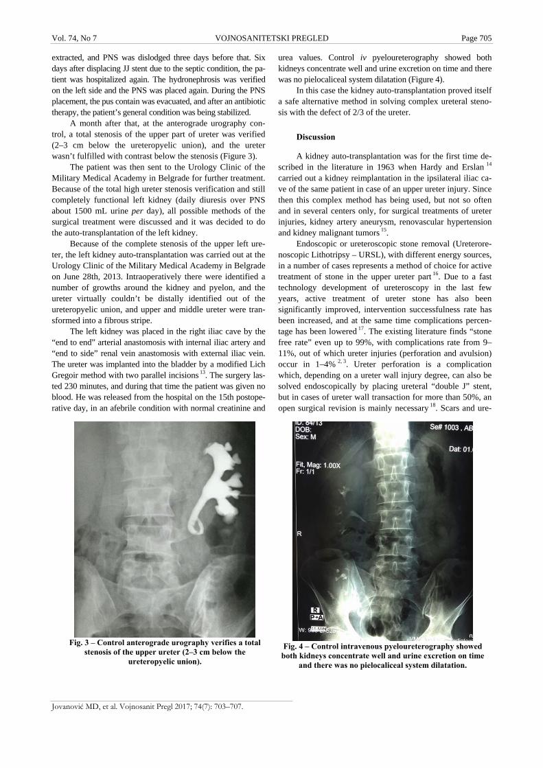
Vol. 74, No 7 VOJNOSANITETSKI PREGLED Page 705
Jovanović MD, et al. Vojnosanit Pregl 2017; 74(7): 703–707.
Fig. 3 – Control anterograde urography verifies a total
stenosis of the upper ureter (2–3 cm below the ureteropyelic union).
Fig. 4 – Control intravenous pyeloureterography showed
both kidneys concentrate well and urine excretion on time and there was no pielocaliceal system dilatation.
extracted, and PNS was dislodged three days before that. Six days after displacing JJ stent due to the septic condition, the pa-tient was hospitalized again. The hydronephrosis was verified on the left side and the PNS was placed again. During the PNS placement, the pus contain was evacuated, and after an antibiotic therapy, the patient’s general condition was being stabilized.
A month after that, at the anterograde urography con-trol, a total stenosis of the upper part of ureter was verified (2–3 cm below the ureteropyelic union), and the ureter wasn’t fulfilled with contrast below the stenosis (Figure 3).
The patient was then sent to the Urology Clinic of the Military Medical Academy in Belgrade for further treatment. Because of the total high ureter stenosis verification and still completely functional left kidney (daily diuresis over PNS about 1500 mL urine per day), all possible methods of the surgical treatment were discussed and it was decided to do the auto-transplantation of the left kidney.
Because of the complete stenosis of the upper left ure-ter, the left kidney auto-transplantation was carried out at the Urology Clinic of the Military Medical Academy in Belgrade on June 28th, 2013. Intraoperatively there were identified a number of growths around the kidney and pyelon, and the ureter virtually couldn’t be distally identified out of the ureteropyelic union, and upper and middle ureter were tran-sformed into a fibrous stripe.
The left kidney was placed in the right iliac cave by the “end to end” arterial anastomosis with internal iliac artery and “end to side” renal vein anastomosis with external iliac vein. The ureter was implanted into the bladder by a modified Lich Gregoir method with two parallel incisions 13. The surgery las-ted 230 minutes, and during that time the patient was given no blood. He was released from the hospital on the 15th postope-rative day, in an afebrile condition with normal creatinine and
urea values. Control iv pyeloureterography showed both kidneys concentrate well and urine excretion on time and there was no pielocaliceal system dilatation (Figure 4).
In this case the kidney auto-transplantation proved itself a safe alternative method in solving complex ureteral steno-sis with the defect of 2/3 of the ureter.
Discussion
A kidney auto-transplantation was for the first time de-scribed in the literature in 1963 when Hardy and Erslan 14 carried out a kidney reimplantation in the ipsilateral iliac ca-ve of the same patient in case of an upper ureter injury. Since then this complex method has being used, but not so often and in several centers only, for surgical treatments of ureter injuries, kidney artery aneurysm, renovascular hypertension and kidney malignant tumors 15.
Endoscopic or ureteroscopic stone removal (Ureterore-noscopic Lithotripsy – URSL), with different energy sources, in a number of cases represents a method of choice for active treatment of stone in the upper ureter part 16. Due to a fast technology development of ureteroscopy in the last few years, active treatment of ureter stone has also been significantly improved, intervention successfulness rate has been increased, and at the same time complications percen-tage has been lowered 17. The existing literature finds “stone free rate” even up to 99%, with complications rate from 9–11%, out of which ureter injuries (perforation and avulsion) occur in 1–4% 2, 3. Ureter perforation is a complication which, depending on a ureter wall injury degree, can also be solved endoscopically by placing ureteral “double J” stent, but in cases of ureter wall transaction for more than 50%, an open surgical revision is mainly necessary 18. Scars and ure-
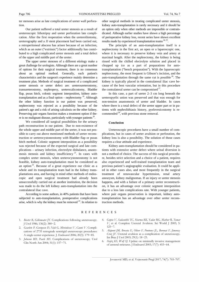
Page 706 VOJNOSANITETSKI PREGLED Vol. 74, No 7
Jovanović MD, et al. Vojnosanit Pregl 2017; 74(7): 703–707.
ter stenoses arise as late complications of ureter wall perfora-tion.
Our patient suffered a total ureter stenosis as a result of ureteroscopic lithotripsy and ureter perforation late compli-cation. After the first reoperation when the ureterolitotomy, ureterography and a JJ stent placement had been carried out, a retroperitoneal abscess has arisen because of an infection, which as an outer (“extrinsic”) factor additionally has contri-buted to a high complicated ureter stenosis and a total defect of the upper and middle part of the ureter.
The upper ureter stenoses of a different etiology make a great challenge for urologists. Although there are a great number of options for their surgical treatment, there is no consensus about an optimal method. Generally, each patient's characteristics and the surgeon's experience mainly determine a treatment plan. Methods of surgical treatment of a complicated ureter stenosis or ureter defect are: ureteroureterostomy, transureterostomy, nephropexy, ureterocalicostomy, Bladder flap, psoas hitch, colonic segment interposition, kidney auto-transplantation and as a final option – nephrectomy 19. Although the other kidney function in our patient was preserved, nephrectomy was rejected as a possibility because of the patient's age and a risk of arising calculosis on the other kidney. Preserving pair organs function makes a treatment option if the-re is no malignant disease, particularly with younger patients 20.
We considered all surgical possibilities for the urinary path reconstruction in our patient. Due to non-existence of the whole upper and middle part of the ureter, it was not pos-sible to carry out above mentioned methods of ureter recons-truction or ureterocystoneostomy with Bladder flap or psoas hitch method. Colonic segment interposition as a possibility was rejected because of the expected surgical and late com-plications – urinary infection, electrolyte disbalance, anasto-mosis stenosis and kidney insufficiency 21. In cases with complex ureter stenosis, when ureterocystoneostomy is not feasible, kidney auto-transplantation must be considered as an option 22. Because of a great experience our clinic as a whole and its transplantation team had in the kidney trans-plantations area, and having in mind other methods of endos-copic and open surgical treatment had already been unsuccessfully carried out at another institution, the decision was made to do the left kidney auto-transplantation into the contralateral iliac cave.
According to some authors, in 40% patients that have been subjected to auto-transplantation, postoperative complications arise, which is why the kidney must be removed 23. In relation to
other surgical methods in treating complicated ureter stenosis, kidney auto-transplantation is rarely necessary and it should be an option only when other methods are impossible or contrain-dicated. Although earlier studies have shown a high percentage of perioperative kidney loss, recent series have shown excellent results made by experienced transplantation teams 24, 25.
The principle of an auto-transplantation itself is a nephrectomy in the first act, an open or a laparoscopic one, where it is necessary to preserve kidney vein and artery at maximal length. After the nephrectomy, the kidney is being rinsed with the chilled electrolyte solution and placed in chopped up ice as a part of preparations for auto-transplantation (“bench preparation”). With the laparoscopic nephrectomy, the most frequent is Gibson’s incision, and the auto-transplantation through the same cut is possible 26. The kidney is typically placed in the contralateral iliac cave be-cause of the best vascular orientation, but in this procedure the contralateral ureter can be compromised 27.
In this case, a part of ureter 2–3 cm long beneath the ureteropyelic union was preserved and that was enough for non-tension anastomosis of ureter and bladder. In cases where there is a total defect of the ureter upper part or in pa-tients with nephrolithiasis history, pyelovesicostomy is re-commended 28, with previous stone removal.
Conclusion
Ureteroscopic procedures have a small number of com-plications, but in cases of ureter avulsion or perforation, the kidney loss is also a possibility. The solution of those cases requires a clear attitude and experience.
Kidney auto-transplantation should be considered in pa-tients with extensive ureter defect where urinal diversion is not a method of choice. The success of this surgical procedu-re, besides strict selection and a choice of a patient, requires also experienced and well-trained transplantation team and previous patient’s angiographic evaluation. It could be appli-ed in other cases also, and most frequently in the surgical treatment of renovascular hypertension, renal artery aneurysm, kidney malignomas. If an injury or ureter stenosis happens, and with a failure of a primary ureter reconstructi-on, it has an advantage over colonic segment interposition due to a less late complications rate. With younger patients, where pair organs preservation is important, kidney auto-transplantation has an advantage over other ureter recons-truction methods.
R E F E R E N C E S
1. Biester R, Gillenwater JY. Complications following ureteroscopy. J Urol 1986; 136(2): 380−2.
2. Geavlete P, Georgescu D, Niţă G, Mirciulescu V, Cauni V. Compli-cations of 2735 retrograde semirigid ureteroscopy procedures: A single-center experience. J. Endourol 2006; 20(3): 179−85.
3. Johnson BD, Pearle MS. Complications of ureteroscopy. Urol Clin North Am 2004; 31(1): 157−71.
4. Gupta V, Sadasukhi TC, Sharma KK, Yadav RG, Mathur R, Tomar V, et al. Complete Ureteral Avulsion. Sci World J 2005; 5: 125−7.
5. Alapont JM, Broseta E, Oliver F, Pontones JL, Boronat F, Jimenez-Cruz JF. Ureteral avulsion as a complication of ureteroscopy. Int Braz J Urol 2003; 29(1): 18−23.
6. Hafez KS, Wolf SJ. Update on minimally invasive management of ureteral strictures. J Endourol 2003; 17(7): 453−64.
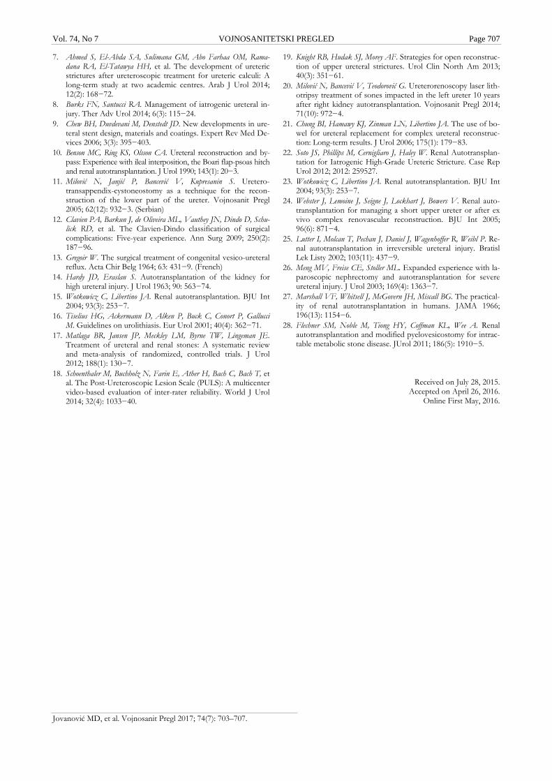
Vol. 74, No 7 VOJNOSANITETSKI PREGLED Page 707
Jovanović MD, et al. Vojnosanit Pregl 2017; 74(7): 703–707.
7. Ahmed S, El-Abda SA, Sulimana GM, Abo Farhaa OM, Rama-dana RA, El-Tatawya HH, et al. The development of ureteric strictures after ureteroscopic treatment for ureteric calculi: A long-term study at two academic centres. Arab J Urol 2014; 12(2): 168−72.
8. Burks FN, Santucci RA. Management of iatrogenic ureteral in-jury. Ther Adv Urol 2014; 6(3): 115−24.
9. Chew BH, Duvdevani M, Denstedt JD. New developments in ure-teral stent design, materials and coatings. Expert Rev Med De-vices 2006; 3(3): 395−403.
10. Benson MC, Ring KS, Olsson CA. Ureteral reconstruction and by-pass: Experience with ileal interposition, the Boari flap-psoas hitch and renal autotransplantation. J Urol 1990; 143(1): 20−3.
11. Milović N, Janjić P, Bancević V, Kupresanin S. Uretero-transappendix-cystoneostomy as a technique for the recon-struction of the lower part of the ureter. Vojnosanit Pregl 2005; 62(12): 932−3. (Serbian)
12. Clavien PA, Barkun J, de Oliveira ML, Vauthey JN, Dindo D, Schu-lick RD, et al. The Clavien-Dindo classification of surgical complications: Five-year experience. Ann Surg 2009; 250(2): 187−96.
13. Gregoir W. The surgical treatment of congenital vesico-ureteral reflux. Acta Chir Belg 1964; 63: 431−9. (French)
14. Hardy JD, Eraslan S. Autotransplantation of the kidney for high ureteral injury. J Urol 1963; 90: 563−74.
15. Wotkowicz C, Libertino JA. Renal autotransplantation. BJU Int 2004; 93(3): 253−7.
16. Tiselius HG, Ackermann D, Alken P, Buck C, Conort P, Gallucci M. Guidelines on urolithiasis. Eur Urol 2001; 40(4): 362−71.
17. Matlaga BR, Jansen JP, Meckley LM, Byrne TW, Lingeman JE. Treatment of ureteral and renal stones: A systematic review and meta-analysis of randomized, controlled trials. J Urol 2012; 188(1): 130−7.
18. Schoenthaler M, Buchholz N, Farin E, Ather H, Bach C, Bach T, et al. The Post-Ureteroscopic Lesion Scale (PULS): A multicenter video-based evaluation of inter-rater reliability. World J Urol 2014; 32(4): 1033−40.
19. Knight RB, Hudak SJ, Morey AF. Strategies for open reconstruc-tion of upper ureteral strictures. Urol Clin North Am 2013; 40(3): 351−61.
20. Milović N, Bancević V, Teodorović G. Ureterorenoscopy laser lith-otripsy treatment of sones impacted in the left ureter 10 years after right kidney autotransplantation. Vojnosanit Pregl 2014; 71(10): 972−4.
21. Chung BI, Hamawy KJ, Zinman LN, Libertino JA. The use of bo-wel for ureteral replacement for complex ureteral reconstruc-tion: Long-term results. J Urol 2006; 175(1): 179−83.
22. Soto JS, Phillips M, Cernigliaro J, Haley W. Renal Autotransplan-tation for Iatrogenic High-Grade Ureteric Stricture. Case Rep Urol 2012; 2012: 259527.
23. Wotkowicz C, Libertino JA. Renal autotransplantation. BJU Int 2004; 93(3): 253−7.
24. Webster J, Lemoine J, Seigne J, Lockhart J, Bowers V. Renal auto-transplantation for managing a short upper ureter or after ex vivo complex renovascular reconstruction. BJU Int 2005; 96(6): 871−4.
25. Lutter I, Molcan T, Pechan J, Daniel J, Wagenhoffer R, Weibl P. Re-nal autotransplantation in irreversible ureteral injury. Bratisl Lek Listy 2002; 103(11): 437−9.
26. Meng MV, Freise CE, Stoller ML. Expanded experience with la-paroscopic nephrectomy and autotransplantation for severe ureteral injury. J Urol 2003; 169(4): 1363−7.
27. Marshall VF, Whitsell J, McGovern JH, Miscall BG. The practical-ity of renal autotransplantation in humans. JAMA 1966; 196(13): 1154−6.
28. Flechner SM, Noble M, Tiong HY, Coffman KL, Wee A. Renal autotransplantation and modified pyelovesicostomy for intrac-table metabolic stone disease. JUrol 2011; 186(5): 1910−5.
Received on July 28, 2015. Accepted on April 26, 2016.
Online First May, 2016.
Page 708 VOJNOSANITETSKI PREGLED Vol. 74, No. 7
INSTRUCTIONS TO THE AUTHORS The Vojnosanitetski pregled (VSP) is an Open Access Journal.
All articles can be downloaded free from the web-site (http://www.vma.mod.gov.rs/sr/vojnosanitetski-pregled) with the use of li-cense: the Creative Commons — Attribution-ShareAlike (CC BY-SA) (http://creativecommons.org/licenses/by-as/4.0/).
The VSP publishes only papers not published before, nor submitted to any other journals, in the order determined by the Editorial Board. Any attempted plagiarism or self-plagiarism will be punished. When submitting a paper to the VSP electronic editing system (http://aseestant.ceon.rs/index.php), the following should be enclosed: a statement on meeting any technical require-ments, a statement signed by all the authors that the paper on the whole and/or partly has not been submitted nor accepted for publication elsewhere, a statement specifying the actual contribution of each author, no conflict of interest statement that make them responsible for meeting any requirements set. What follows subsequently is the acceptance of a paper for further editing procedure. The manuscripts submitted to the VSP pass in-house and external peer review. All authors pay “Article Processing Charge” for coverage all ed-iting and publishing expenses. Domestic authors pay 5,000 RSD, and those from aboard 150 euros. The editing and publishing fee is required for sub-stantive editing, facts and references validations, copy editing, and publishing online and in print by editorial staff of the Journal. No additional fees, other than stated above, are required even if an author who already paid the fee would have more articles accepted for publishing in the year when fee was paid. All authors who pay this fee may, if want, receive printed version of the Journal in year when fee is payed. Please note that the payment of this charge does not guarantee acceptance of the manuscript for publication and does not influence the outcome of the review procedure. The requirement about pay-ing “Article Processing Charge” does not apply to reviewers, members of the Editorial Board and the Publisher’s Council of the Journal, young researchers and students, as well as any of the subscribers of the Journal.
The VSP publishes: editorials, original articles, short communica-tions, reviews/meta-analyses, case reports, medical history (general or military), personal views, invited comments, letters to the editor, reports from scientific meetings, book reviews, and other. Original articles, short communications, meta-analyses and case reports are published with ab-stracts in both English and Serbian.
General review papers will be accepted by the Editorial Board only if the authors prove themselves as the experts in the fields they write on by citing not less than 5 self-citations.
Papers should be written on IBM-compatible PC, using 12 pt font, and double spacing, with at least 4 cm left margin. Bold and italic letters should be avoided as reserved for subtitles. Original articles, reviews, meta-analyses and articles from medical history should not exceed 16 pages; current topics 10; case reports 6; short communications 5; letters to the editor and com-ments 3, and reports on scientific meetings and book reviews 2.
All measurements should be reported in the metric system of the International System of Units (SI), and the standard internationally accepted terms (except for mmHg and °C).
MS Word for Windows (97, 2000, XP, 2003) is recommended for word processing; other programs are to be used only exceptionally. Il-lustrations should be made using standard Windows programs, Micro-soft Office (Excel, Word Graph). The use of colors and shading in graphs should be avoided.
Papers should be prepared in accordance with the Vancouver Convention.
Papers are reviewed anonymously by at least two editors and/or in-vited reviewers. Remarks and suggestions are sent to the author for final composition. Galley proofs are sent to the corresponding author for final agreement.
Preparation of manuscript
Parts of the manuscript are: Title page; Abstract with Key words; Text; Acknowledgements (to the authors’ desire), References, Enclosures.
1. Title page
a) The title should be concise but informative, while subheadings should be avoided;
b) Full names of the authors signed as follows: *, †, ‡, §, ||, ¶, **, ††, ... . c) Exact names and places of department(s) and institution(s) of af-
filiation where the studies were performed, city and the state for any au-thors, clearly marked by standard footnote signs;
d) Conclusion could be a separate chapter or the last paragraph of the discussion;
e) Data on the corresponding author.
2. Abstract and key words
The second page should carry a structured abstract (250-300 words for original articles and meta-analyses) with the title of the article. In short, clear sentences the authors should write the Background/Aim, major procedures – Methods (choice of subjects or laboratory animals; meth-ods for observation and analysis), the obtained findings – Results (con-crete data and their statistical significance), and the Conclusion. It should emphasize new and important aspects of the study or observa-tions. A structured abstract for case reports (up to 250 words) should contain subtitles Introduction, Case report, Conclusion). Below the
abstract Key words should provide 3–10 key words or short phrases that indicate the topic of the article.
3. Text
The text of the articles includes: Introduction, Methods, Results, and Discussion. Long articles may need subheadings within some sections to clarify their content.
Introduction. After the introductory notes, the aim of the article should be stated in brief (the reasons for the study or observation), only significant data from the literature, but not extensive, detailed consideratuion of the subject, nor data or conclusions from the work being reported.
Methods. The selection of study or experimental subjects (patients or experimental animals, including controls) should be clearly described. The methods, apparatus (manufacturer's name and address in parenthe-ses), and procedures should be identified in sufficient detail to allow other workers to reproduce the results. Also, give references to estab-lished methods, including statistical methods. Identify precisely all drugs and chemicals used, with generic name(s), dose(s), and route(s) of ad-ministration. State the approval of the Ethnics Committee for the tests in humans and animals.
Results should be presented in logical sequence in the text, tables and illustrations. Emphasize or summarize only important observations.
Discussion is to emphasize the new and significant aspects of the study and the conclusions that result from them. Relate the observations to other relevant studies. Link the conclusions with the goals of the study, but avoid unqualified statements and conclusions not completely supported by your data.
References
References should be superscripted and numerated consecutively in the order of their first mentioning within the text. All the authors should be listed, but if there are more than 6 authors, give the first 6 followed by et al. Do not use abstracts, secondary publications, oral communications, un-published papers, official and classified documents. References to papers accepted but not yet published should be cited as ”in press“. Information from manuscripts not yet accepted should be cited as ”unpublished data“. Data from the Internet are cited with the date of citation.
Examples of references: Jurhar-Pavlova M, Petlichkovski A, TrajkovD, Efinska-Mladenovska O, Arsov T, Strezova A, et al. Influence of the elevated ambient temperature on immunoglobulin G and immunoglobulin G subclasses in sera of Wis-tar rats. Vojnosanit Pregl 2003; 60(6): 657–612.
DiMaio VJ. Forensic Pathology. 2nd ed. Boca Raton: CRC Press; 2001. Blinder MA. Anemia and Transfusion Therapy. In: Ahya NS, Flood K, Paranjothi S, editors. The Washington Manual of Medical Therapeutics, 30th edition. Boston: Lippincot, Williams and Wilkins; 2001. p. 413-28. Christensen S, Oppacher F. An analysis of Koza's computational effort statistic for genetic programming. In: Foster JA, Lutton E, Miller J, Ryan C, Tettamanzi AG, editors. Genetic programming. EuroGP 2002: Proceedings of the 5th European Conference on Genetic Programming; 2002 Apr 3-5; Kinsdale, Ireland. Berlin: Springer; 2002. p. 182-91. Abood S. Quality improvement initiative in nursing homes: the ANA acts in an advisory role. Am J Nurs [serial on the Internet]. 2002 Jun [cited 2002 Aug 12]; 102(6): [about 3 p.]. Available from: http://www.nursingworld.org/AJN/2002/june/Wawatch.htm
Tables Each table should be typed double-spaced 1,5 on a separate sheet,
numbered in the order of their first citation in the text in the upper right corner and supplied with a brief title each. Explanatory notes are printed under a table. Each table should be mentioned in the text. If data from another source are used, acknowledge fully.
Illustrations Any forms of graphic enclosures are considered to bi figures and
should be submitted as additional databases in the System of Assistent. Letters, numbers, and symbols should be clear and uniform, of sufficient size that when reduced for publication, each item will still be legible. Each figure should have a label on its back indicating the number of the figure, author's name, and top of the figure (Figure 1, Figure 2 and so on). If a figure has been published, state the original source.
Legends for illustrations are typed on a separate page, with Arabic numbers corresponding to the illustrations. If used to identify parts of the illustrations, the symbols, arrows, numbers, or letters should be iden-tified and explained clearly in the legend. Explain the method of staining in photomicrographs.
Abbreviations and acronyms Authors are encouraged to use abbreviations and acronyms in the
manuscript in the following manner: abbreviations and acronyms must be defined the first time they are used in the text consistently throughout the whole manuscript, tables, and graphics; abbreviations should be used only for terms that appear more than three times in text; abbreviations should be sparingly used.
An alphabetical list of all abbreviations used in the paper, followed by their full definitions, should be provided on submission.
Detailed Instructions are available at the web site: www.vma.mod.gov.rs/vsp
Vol. 74, No. 7 VOJNOSANITETSKI PREGLED Page 709
UPUTSTVO AUTORIMA
Vojnosanitetski pregled (VSP) je dostupan u režimu otvorenog pris-tupa. Članci objavljeni u časopisu mogu se besplatno preuzeti sa sajta časopisa http://www.vma.mod.gov.rs/sr/ uz primenu licence Creative Commons Autorstvo-Deliti pod istim uslovima (CC BY-SA) (http://creativecommons.org/licenses/by-sa/4.0).
VSP objavljuje radove koji nisu ranije nigde objavljivani, niti preda-ti za objavljivanje redosledom koji određuje uređivački odbor. Svaki pokušaj plagijarizma ili autoplagijarizma kažnjava se. Prilikom prijave rada u sistem elektronskog uređivanja „Vojnosanitetskog pregle-da“(http://aseestant.ceon.rs/index.php) neophodno je priložiti izjavu da su ispunjeni svi postavljeni tehnički zahtevi uključujući i izjavu koju potpisuju svi autori da rad nije ranije ni u celini, niti delimično objavljen niti prihvaćen za štampanje u drugom časopisu. Izjavu o pojedinačnom doprinosu svakog od autora rada potpisanu od svih autora, treba skenirati i poslati uz rad kao dopunsku datoteku. Takođe, autori su obavezni da dostave i potpisanu izjavu o nepostojanju sukoba interesa čime postaju odgovorni za ispunjavanje svih postavljenih uslova. Ovome sledi odluka o prihvatanju za dalji uređivački po-stupak. Rukopisi pristigli u Redakciju časopisa podležu internoj i eksternoj recenziji. Svi autori dužni su da plate “Article Processing Charge” za pokriće troškova jezičke, stručne i tehničke obrade rukopisa, kao i njegovog objavlji-vanja. Domaći autori plaćaju iznos od 5 000 dinara, a inostrani 150 eura. Dodatna plaćanja nisu predviđena čak i u slučaju da autor koji je već pretho-dno platio traženi iznos, ima više prihvaćenih radova za objavljivanje u godi-ni u kojoj je izvršio uplatu. Svi autori koji su platili “Article Processing Charge” mogu, ukoliko žele, dobijati štampanu verziju časopisa tokom godi-ne u kojoj je izvršena uplata. Plaćanje ovog iznosa ne garantuje prihvatanje rukopisa za objavljivanje i ne utiče na ishod recenzije. Od obaveze plaćanja pokrića navedenih troškova oslobođeni su recenzenti, članovi Uređivačkog odbora i Izdavačkog saveta VSP, studenti i mladi istra-živači, kao i pretplatnici časopisa.
U VSP-u se objavljuju uvodnici, originalni članci, prethodna ili kratka saopštenja, revijski radovi tipa opšteg pregleda (uz uslov da autori navođenjem najmanje 5 autocitata potvrde da su eksperti u oblasti o kojoj pišu), aktuelne teme, metaanalize, kazuistika, seminar prak-tičnog lekara, članci iz istorije medicine, lični stavovi, naručeni ko-mentari, pisma uredništvu, izveštaji sa naučnih i stručnih skupova, pri-kazi knjiga i drugi prilozi. Radovi tipa originalnih članaka, prethodnih ili kratkih saopštenja, metaanalize i kazuistike objavljuju se uz apstrakte na srpskom i engleskom jeziku.
Rukopis se piše sa proredom 1,5 sa levom marginom od 4 cm. Koristi-ti font veličine 12, a načelno izbegavati upotrebu bold i italic slova, koja su rezervisana za podnaslove. Originalni članci, opšti pregledi i metaanali-ze i članci iz istorije medicine ne smeju prelaziti 16 stranica (bez priloga); aktuelne teme – deset, seminar praktičnog lekara – osam, kazuistika – šest, prethodna saopštenja – pet, a komentari i pisma uredniku – tri, izveštaji sa skupova i prikazi knjiga – dve stranice.
U celom radu obavezno je korišćenje međunarodnog sistema mera (SI) i standardnih međunarodno prihvaćenih termina (sem mm Hg i °C).
Za obradu teksta koristiti program Word for Windows verzije 97, 2000, XP ili 2003. Za izradu grafičkih priloga koristiti standardne gra-fičke programe za Windows, poželjno iz programskog paketa Micro-soft Office (Excel, Word Graph). Kod kompjuterske izrade grafika izbegavati upotrebu boja i senčenja pozadine.
Radovi se pripremaju u skladu sa Vankuverskim dogovorom.
Prispeli radovi kao anonimni podležu uređivačkoj obradi i recenziji najmanje dva urednika/recenzenta. Primedbe i sugestije uredni-ka/recenzenata dostavljaju se autoru radi konačnog oblikovanja. Pre ob-jave, rad se upućuje autoru određenom za korespodenciju na konačnu saglasnost.
Priprema rada
Delovi rada su: naslovna strana, apstrakt sa ključnim rečima, tekst rada, zahvalnost (po želji), literatura, prilozi.
1. Naslovna strana a) Poželjno je da naslov bude kratak, jasan i informativan i da odgovara
sadržaju, podnaslove izbegavati. b) Ispisuju se puna imena i prezimena autora sa oznakama redom: *, †,
‡, §, ||, ¶, **, ††, ... . c) Navode se puni nazivi ustanove i organizacijske jedinice u kojima je
rad obavljen mesta i države za svakog autora, koristeći standardne znake za fusnote.
d) Zaključak može da bude posebno poglavlje ili se iznosi u poslednjem pasusu diskusije.
e) Podaci o autoru za korespodenciju. 2. Apstrakt i ključne reči Na drugoj stranici nalazi se strukturisani apstrakt (250-300 reči za
originalne članke i meta-analize) sa naslovom rada. Kratkim rečeni-cama na srpskom i engleskom jeziku iznosi se Uvod/Cilj rada, osnov-ne procedure – Metode (izbor ispitanika ili laboratorijskih životinja; metode posmatranja i analize), glavni nalazi – Rezultati (konkretni podaci i njihova statistička značajnost) i glavni Zaključak. Naglasiti nove i značajne aspekte studije ili zapažanja. Strukturisani apstrakt za kazuistiku (do 250 reči), sadrži podnaslove Uvod, Prikaz bolesnika i
Zaključak). Ispod apstrakta, „Ključne reči“ sadrže 3–10 ključnih reči ili kratkih izraza koje ukazuju na sadržinu članka.
3. Tekst članka
Tekst sadrži sledeća poglavlja: uvod, metode, rezultate i diskusiju. Uvod. Posle uvodnih napomena, navesti cilj rada. Ukratko izneti razloge za studiju ili posmatranje. Navesti samo važne podatke iz literature a ne opširna razmatranja o predmetu rada, kao ni podatke ili zaključke iz rada o kome se izveštava.
Metode. Jasno opisati izbor metoda posmatranja ili eksperimentnih metoda (ispitanici ili eksperimentne životinje, uključujući kontrolne). Identifikovati metode, aparaturu (ime i adresa proizvođača u zagradi) i proceduru, dovoljno detaljno da se drugim autorima omogući reproduk-cija rezultata. Navesti podatke iz literature za uhodane metode, uključu-jući i statističke. Tačno identifikovati sve primenjene lekove i hemika-lije, uključujući generičko ime, doze i načine davanja. Za ispitivanja na ljudima i životinjama navesti saglasnost nadležnog etičkog komiteta.
Rezultate prikazati logičkim redosledom u tekstu, tabelama i ilustra-cijama. U tekstu naglasiti ili sumirati samo značajna zapažanja.
U diskusiji naglasiti nove i značajne aspekte studije i izvedene zak-ljučke. Posmatranja dovesti u vezu sa drugim relevantnim studijama, u načelu iz poslednje tri godine, a samo izuzetno i starijim. Povezati zak-ljučke sa ciljevima rada, ali izbegavati nesumnjive tvrdnje i one zaključ-ke koje podaci iz rada ne podržavaju u potpunosti.
Literatura
U radu literatura se citira kao superskript, a popisuje rednim broje-vima pod kojima se citat pojavljuje u tekstu. Navode se svi autori, ali ako broj prelazi šest, navodi se prvih šest i et al. Svi podaci o citiranoj literaturi moraju biti tačni. Literatura se u celini citira na engleskom jeziku, a iza naslova se navodi jezik članka u zagradi. Ne prihvata se citiranje apstrakata, sekundarnih publikacija, usmenih saopštenja, ne-objavljenih radova, službenih i poverljivih dokumenata. Radovi koji su prihvaćeni za štampu, ali još nisu objavljeni, navode se uz dodatak „u štampi“. Rukopisi koji su predati, ali još nisu prihvaćeni za štampu, u tekstu se citiraju kao „neobjavljeni podaci“ (u zagradi). Podaci sa Interneta citiraju se uz navođenje datuma pristupa tim podacima.
Primeri referenci:
Đurović BM. Endothelial trauma in the surgery of cataract. Vojnosanit Pregl 2004; 61(5): 491–7. (Serbian)
Balint B. From the haemotherapy to the haemomodulation. Beograd: Zavod za udžbenike i nastavna sredstva; 2001. (Serbian)
Mladenović T, Kandolf L, Mijušković ŽP. Lasers in dermatology. In: Karadaglić Đ, editor. Dermatology. Beograd: Vojnoizdavački zavod & Verzal Press; 2000. p. 1437–49. (Serbian)
Christensen S, Oppacher F. An analysis of Koza's computational ef-fort statistic for genetic programming. In: Foster JA, Lutton E, Miller J, Ryan C, Tettamanzi AG, editors. Genetic programming. EuroGP 2002: Proceedings of the 5th European Conference on Genetic Programming; 2002 Apr 3-5; Kinsdale, Ireland. Berlin: Springer; 2002. p. 182-91.
Abood S. Quality improvement initiative in nursing homes: the ANA acts in an advisory role. Am J Nurs [serial on the Internet]. 2002 Jun [cited 2002 Aug 12]; 102(6): [about 3 p.]. Available from: http://www.nursingworld.org/AJN/2002/june/Wawatch.htm
Tabele
Sve tabele pripremaju se sa proredom 1,5 na posebnom listu. Obeležavaju se arapskim brojevima, redosledom pojavljivanja, u desnom uglu (Tabela 1), a svakoj se daje kratak naslov. Objašnjenja se daju u fus-noti, ne u zaglavlju. Svaka tabela mora da se pomene u tekstu. Ako se koriste tuđi podaci, obave-zno ih navesti kao i svaki drugi podatak iz literature.
Ilustracije Slikama se zovu svi oblici grafičkih priloga i predaju se kao dopunske dato-
teke u sistemu aseestant. Slova, brojevi i simboli treba da su jasni i ujednačeni, a dovoljne veličine da prilikom umanjivanja budu čitljivi. Slike treba da budu jasne i obeležene brojevima, onim redom kojim se navode u tekstu (Sl. 1; Sl. 2 itd.). Ukoliko je slika već negde objavljena, obavezno citirati izvor.
Legende za ilustracije pisati na posebnom listu, koristeći arapske brojeve. Ukoliko se koriste simboli, strelice, brojevi ili slova za objašnjavanje pojedi-nog dela ilustracije, svaki pojedinačno treba objasniti u legendi. Za fotomi-krografije navesti metod bojenja i podatak o uvećanju.
Skraćenice i akronimi
Skraćenice i akronimi u rukopisu treba da budu korišćeni na sledeći način: definisati skraćenice i akronime pri njihovom prvom pojavljivanju u tekstu i koristiti ih konzistentno kroz čitav tekst, tabele i slike; koristiti ih samo za termine koji se pominju više od tri puta u tekstu; da bi se olakšalo čitaocu, skraćenice i aktinome treba štedljivo koristiti.
Abecedni popis svih skraćenica i akronima sa objašnjenjima treba dosta-viti pri predaji rukopisa.
Detaljno uputstvo može se dobiti u redakciji ili na sajtu: www.vma.mod.gov.rs/vsp