Yellow Laser Stimulation at GV2 Acupoint Mitigates Apoptosis,...
13
Research Article Yellow Laser Stimulation at GV2 Acupoint Mitigates Apoptosis, Oxidative Stress, Inflammation, and Motor Deficit in Spinal Cord Injury Rats Parichat On-ong-arj, 1 Jintanaporn Wattanathorn , 2,3 Supaporn Muchimapura , 2,3 and Wipawee Thukham-mee 2,3 Department of Physiology (Neuroscience Program), Faculty of Medicine, Khon Kaen University, Khon Kaen, , ailand Department of Physiology, Faculty of Medicine, Khon Kaen University, Khon Kaen, , ailand Integrative Complementary Alternative Medicine Research and Development Center, Khon Kaen University, , ailand Correspondence should be addressed to Jintanaporn Wattanathorn; [email protected] Received 7 June 2018; Revised 6 September 2018; Accepted 20 September 2018; Published 8 October 2018 Academic Editor: Sakthivel Muniyan Copyright © 2018 Parichat On-ong-arj et al. is is an open access article distributed under the Creative Commons Attribution License, which permits unrestricted use, distribution, and reproduction in any medium, provided the original work is properly cited. Currently, the suppression of oxidative stress and inflammation is considered as the treatment targets of spinal cord injury due to their roles on the hindrance of recovery process. Since laser acupuncture decreased oxidative stress and enhanced the survival of neurons from oxidative stress damage and GV2 stimulation was selected as one stimulated acupoint in order to enhance the recovery of spinal cord injury, we hypothesized that laser acupuncture at GV2 should enhance the recovery of spinal cord injury. To test this hypothesis, male Wistar rats were induced spinal cord injury at T10 level and they were exposed to a 10 minute-stimulation at GV2 by yellow laser. Laser acupuncture was performed at 0.25 and 1, 2, 6, and 12 hours aſter spinal cord injury. en, the stimulation was performed once daily for 7 days. Locomotor assessment was carried out on days 3 and 7 aſter injury. At the end of study period, the densities of polymorphonuclear of leukocyte, Bax, Caspase-3, Bcl-2, and BDNF positive stained cells in ventral horn of spinal cord were determined. Cyclooxygenase-2 (COX-2), interleukin-6 (IL-6), and oxidative stress status was also assessed. e results showed that laser acupuncture at GV2 increased BBB score, gross motor score, and densities of Bcl-2 and BDNF positive stained cells but decreased density with polymorphonuclear leukocyte, the densities of Bax and Caspase-3 positive stained cells, COX-2 level, and oxidative stress status in ventral horn of the lesion spinal cord. e reduction of serum COX-2 was also decreased. erefore, GV2 stimulation by yellow laser might enhance the recovery of spinal cord via the increase in BDNF and the decrease in inflammation, apoptosis, and oxidative stress status in the lesion spinal cord. 1. Introduction Traumatic spinal cord injury, one of the most devastat- ing neurological disorders, produces the profound negative impacts on a patient’s life and socioeconomic burdens. It has been reported that the annual Asian incidence is around 12.06 to 61.6 per million [1]. is rate is varied between developing and developed countries. However, it has been estimated that the global prevalence of spinal cord injury (SCI) each year is between 250 000 and 500 000 cases [2]. Despite the high impacts on socioeconomic burdens, no effective therapeutic strategy is available. SCI consists of 2 phases of injury including the primary and secondary injuries. Primary injury occurs as the result of the compression, contusion, stretching, or kinking of the spinal cord induced by mechanical insult [3]. Following this phase, secondary injury including inflammation, oxidative stress damage, and apoptosis occurs [4]. Currently, most of the therapeutic strategies target the secondary injury because this phase plays an important role on the hindrance of recovery process following SCI. Recent studies have demonstrated that the stimulation of the governor vessel acupoints such as GV1, GV2, and GV6 can promote the regeneration of nerve fiber at the injury site Hindawi Evidence-Based Complementary and Alternative Medicine Volume 2018, Article ID 5407052, 12 pages https://doi.org/10.1155/2018/5407052
Transcript of Yellow Laser Stimulation at GV2 Acupoint Mitigates Apoptosis,...

Research ArticleYellow Laser Stimulation at GV2 Acupoint MitigatesApoptosis Oxidative Stress Inflammation and Motor Deficit inSpinal Cord Injury Rats
Parichat On-ong-arj1 JintanapornWattanathorn 23
SupapornMuchimapura 23 andWipawee Thukham-mee23
1Department of Physiology (Neuroscience Program) Faculty of Medicine Khon Kaen University Khon Kaen 40002ailand2Department of Physiology Faculty of Medicine Khon Kaen University Khon Kaen 40002ailand3Integrative Complementary Alternative Medicine Research and Development Center Khon Kaen University 40002ailand
Correspondence should be addressed to JintanapornWattanathorn jintanapornwyahoocom
Received 7 June 2018 Revised 6 September 2018 Accepted 20 September 2018 Published 8 October 2018
Academic Editor Sakthivel Muniyan
Copyright copy 2018 Parichat On-ong-arj et al This is an open access article distributed under the Creative Commons AttributionLicense which permits unrestricted use distribution and reproduction in any medium provided the original work is properlycited
Currently the suppression of oxidative stress and inflammation is considered as the treatment targets of spinal cord injury due totheir roles on the hindrance of recovery process Since laser acupuncture decreased oxidative stress and enhanced the survival ofneurons fromoxidative stress damage andGV2 stimulationwas selected as one stimulated acupoint in order to enhance the recoveryof spinal cord injury we hypothesized that laser acupuncture at GV2 should enhance the recovery of spinal cord injury To test thishypothesis maleWistar rats were induced spinal cord injury at T10 level and they were exposed to a 10 minute-stimulation at GV2by yellow laser Laser acupuncturewas performed at 025 and 1 2 6 and 12 hours after spinal cord injuryThen the stimulation wasperformed once daily for 7 days Locomotor assessment was carried out on days 3 and 7 after injury At the end of study period thedensities of polymorphonuclear of leukocyte Bax Caspase-3 Bcl-2 and BDNF positive stained cells in ventral horn of spinal cordwere determined Cyclooxygenase-2 (COX-2) interleukin-6 (IL-6) and oxidative stress statuswas also assessedThe results showedthat laser acupuncture at GV2 increased BBB score gross motor score and densities of Bcl-2 and BDNF positive stained cells butdecreased density with polymorphonuclear leukocyte the densities of Bax and Caspase-3 positive stained cells COX-2 level andoxidative stress status in ventral horn of the lesion spinal cordThe reduction of serum COX-2 was also decreased Therefore GV2stimulation by yellow laser might enhance the recovery of spinal cord via the increase in BDNF and the decrease in inflammationapoptosis and oxidative stress status in the lesion spinal cord
1 Introduction
Traumatic spinal cord injury one of the most devastat-ing neurological disorders produces the profound negativeimpacts on a patientrsquos life and socioeconomic burdens It hasbeen reported that the annual Asian incidence is around 1206to 616 per million [1] This rate is varied between developingand developed countries However it has been estimated thatthe global prevalence of spinal cord injury (SCI) each yearis between 250 000 and 500 000 cases [2] Despite the highimpacts on socioeconomic burdens no effective therapeuticstrategy is available
SCI consists of 2 phases of injury including the primaryand secondary injuries Primary injury occurs as the resultof the compression contusion stretching or kinking of thespinal cord induced by mechanical insult [3] Following thisphase secondary injury including inflammation oxidativestress damage and apoptosis occurs [4] Currently most ofthe therapeutic strategies target the secondary injury becausethis phase plays an important role on the hindrance ofrecovery process following SCI
Recent studies have demonstrated that the stimulation ofthe governor vessel acupoints such as GV1 GV2 and GV6can promote the regeneration of nerve fiber at the injury site
HindawiEvidence-Based Complementary and Alternative MedicineVolume 2018 Article ID 5407052 12 pageshttpsdoiorg10115520185407052
2 Evidence-Based Complementary and Alternative Medicine
of spinal cord [5 6] by decreasing secondary damage via thesuppression of inflammation and the stimulation of nervegrowth factor release [6] In addition to the acupuncturelaser therapy is also reported to promote axonal regrowthafter spinal cord injury [7] and serves as the potential strategyfor neurorehabilitation for spinal cord injury [8] More-over accumulative lines of evidence have demonstrated thatlaser can stimulate acupoint and restore numerous neuronaldeficits such as brain damage and memory [9ndash12] Basedon the beneficial effects of laser and acupuncture togetherwith the capability to stimulate acupoint with laser theimprovement of functional recovery of spinal cord followingtraumatic injury induced by laser acupuncture has gainedmuch attention Several studies demonstrate that the stimu-lation of acupoint even at single acupoint can effectively pro-duce the desired effects [9ndash12] Therefore we hypothesizedthat the stimulation at GV2 acupoint which is commonlyused for treating weakness or atrophy of lower limbs byyellow laser might improve apoptosis and oxidative stress andneurological deficit in SCI rats To elucidate this issue thisstudy aimed to determine the effect of laser acupuncture atGV2 acupoint on locomotor activity oxidative stress andapoptosis in SCI rats
2 Materials and Methods
21 Animals and Experimental Protocol Adult male Wistarrats weighed 250-300 gramswere used in this experiment Allrats were purchased from National Laboratory Animal Cen-tre Mahidol University Salaya Thailand Rats were housedin a temperature-controlled room under a 12-hour lightdarkcycle and given access to food and water ad libitum Theanimals were acclimatized to the laboratory to the laboratorycondition for 1 week The protocols conducted in this studywere approved by the Institutional Animal Care and UseCommittee Khon Kaen University Khon Kaen Thailand(AEMDKKU 0022558)
All animals were randomly divided into 5 groups asfollows
Group I Naıve intact group all rats in this group receivedno treatment
Group II Sham operation+ sham laser acupuncturegroup all rats were subjected to sham operation surgery andreceived no treatment
Group III SCI + sham laser acupuncture rats in thisgroup were subjected to traumatic injury at T10 level
Group IV SCI+GV2 laser acupuncture the experimentalrats in this group were induced traumatic spinal cord injuryand received GV2 stimulation induced by yellow laser
After the induction of spinal cord injury induced bytraumatic injury rats in group III-group IV were subjectedto the 10-minute stimulation period by various interventionsas described earlier The stimulation was performed at 15minutes 6 12 and 24 hours after spinal cord injury (SCI)After the first day the 10-minute stimulation by variousinterventions were performed once daily for 7 days Theneurological deficit was assessed by using a battery test com-prising of Basso Beattie and Bresnahan (BBB) locomotorrating scale gross motor score These tests were performed
on days 3 and 7 after SCI At the end of study periodhistomorphology of spinal cord at the lesion level was alsoexplored by determining the densities of survival neuronpolymorphonuclear leukocytes Bax-positve (Bax+) caspase3-positive (caspase3+) and Bcl-2 positive (Bcl-2+) cells inventral horn by using histology and immunohistochem-istry techniques In addition the activity of cyclooxygenase-2 (COX-2) interleukin-6 (IL-6) and oxidative stress statusparameters including malondialdehyde (MDA) level and theactivities of superoxide dismutase (SOD) catalase (CAT) andglutathione peroxidase (GPx) were also investigated to probefor the possible underlying mechanism 24 hours after the firstday of intervention
22 Induction of Traumatic Spinal Cord Injury After theanesthetization with Pentobarbital Sodium (50mgkg BW)the paravertebral muscles of experimental animals wereexposed by a longitudinal incision at the midline of the backand exposed T9 to T11 vertebrae and spinal cord The crushinjury at T10 level was performed by exposing to a 15-second-extradural compression (50 g) Following this process mus-cles were closed in layers and the incision was closed by usingsilk sutures no4The surgical woundwas caredwith Betadineand rats were returned to their cages with the providedfood and water The rat bladders were manually voided threetimes a day until they were able to regain normal bladderfunction Tramadol (analgesic drug) at dose of 10kg BWand Tetracycline (antibiotics) at dose of 50mgkg BW wereadministered via subcutaneous route every 12 hours for 3 days[13]
23 GV2 Acupoint Stimulation The stimulation of GV2 anacupoint located on the posterior midline and in the hiatus ofthe sacrum in prone position was performed by yellow laserThe laser equipment used in this study was WeberneedleCompact (Lauenforde Germany) which can emit a wave-length of 589 nm and an output power of 50mW and a diam-eter of the laser beam was 500120583m At the end of 10 minute-intervention period the interventions were terminated Onthe first day of SCI the interventions were applied at 15minutes 6 12 and 24 hours after spinal cord injury (SCI)After the first day the 10 minute-stimulation were performedonce daily for 7 days
24 Neurological Deficit Assessments
241 Locomotor Activity Evaluation (Basso Beatie and Bres-nahan or BBB Score Test) The locomotion weight supportand coordination capacity following SCI of animals wereassessed by using the Basso Beattie and Bresnahan (BBB)scale [14] was evaluated According to this method a score of0-21was gradedThehighest score or 21 represented completemobility whereas 0 represented no spontaneous movement[15]
242 Gross Motor Score Evaluation In addition to BBB testthe locomotor activity was also determined by assessingmotor function through movement in hindlimb and weightbearing The locomotor impairments of rats were observed
Evidence-Based Complementary and Alternative Medicine 3
using openfield grading scores ranging from0 (nomovementin hindlimb) to 10 (normal walking) The locomotor testingwas carried out for 4 minutes [16]
25 Biochemical Assays
251 Tissue Preparation andProteinDetermination After thelast intervention the lesion spinal tissue was removed andhomogenized in 05mL of ice-cold Tris-HCl buffer (50mMpH 74) Following this process the homogenate was sub-jected to a 3000 rounds per minute (rpm)-centrifugationat 4∘C for 15 minutes The supernatant was harvested andstored at -80∘C until used The protein concentration in thehomogenate was determined by using a Thermo ScientificNanoDrop 2000c spectrophotometer (Thermo Fisher Scien-tific USA and the optical density at the wavelength of 280 nmwas determined [17]
252 Determination of Oxidative Stress Status Parameters Toassess the effect of GV2 stimulation on oxidative stress statusparameters the oxidative stress status parameters includingmalondialdehyde (MDA) and the activities of superoxidedismutase (SOD) catalase (CAT) and glutathione peroxidase(GPx) in the lesion spinal cord were determined
The level ofMDAwas determined by using thiobarbituricreaction [18] In brief an aliquot of sample tissue at thevolume of 100120583l was mixed with 100120583l of 81 sodiumdodecyl sulphate (SDS) (Sigma-Aldrich USA) 375120583l of 08of thiobarbituric acid (TBA) (Sigma-Aldrich USA) 375120583l of20 acetic acid (Sigma-Aldrich USA) and 150120583l of distilledwater (DW) Then the mixture was boiled in a water bathat 95∘C for 60 minutes After cooling at room temperature500120583l of water and 25ml of the mixture of n-butanol andpyridine at the ratio of 15 1 were added mixed togetherand centrifuged at 4000 rpm for 10minutesThe supernatantwas harvested and determined an absorbance of 532nm byspectrophotometer MDA level was expressed as nmolmgprotein
SOD activity was measured based on the ability of SODto inhibit the reduction of cytochrome c by competing for thesuperoxide radical In brief 20120583l of tissue sample was addedto the assay mixture containing 57mM phosphate buffersolution (KH2PO4) (Sigma-Aldrich USA) 01mM EDTA(Sigma-Aldrich USA) 10mM cytochrome C (Sigma-Al-drich USA) solution and 50120583Mof xanthine (Sigma-AldrichUSA) solution at the volume of 200 120583l Then 20 120583l of xan-thine oxidase (090mUml) (Sigma-Aldrich USA) solutionwas addedThe absorbancewasmeasured at 415 nmusingmi-croplate reader SODenzyme (Sigma-AldrichUSA) activitiesat the concentrations of 0-25 unitsml were used as standardand the results were expressed as unitsmg protein [19]
Catalase activity was measured based on the rate ofH2O2 disappearance Briefly 10120583l of spinal cord homogenatewas mixed with the assay mixture containing 50120583l of30mM hydrogen peroxide ((in 50mM phosphate buffer pH70) (BDH Chemicals Ltd UK) 25120583l of H2SO4 (Sigma-Aldrich USA) and 150 120583l of 5mM KMnO4 (Sigma-AldrichUSA) Absorbance at 490 nm was measured using a spec-trophotometer CAT enzyme (Sigma-Aldrich USA) at the
concentration range of 0-100unitsml was used as standardand the result was expressed as unitsmg protein [20]
GPx was measured according to the procedure previ-ously described [21] In brief 20 120583l of sample supernatantwas mixed with the reaction mixture consisting of 10120583lof 1mM dithiothreitol (DTT) (Sigma-Aldrich USA) in667mM potassium phosphate buffer (pH 7) 100120583l of 1mMsodium azide (Sigma-Aldrich USA) in 667mM potassiumphosphate buffer (pH 7) 10120583l of 50mM glutathione (Sigma-Aldrich USA) solution and 100 120583l of 30 hydrogen peroxide(BDH Chemicals Ltd UK) Then the mixture was shakenfor 5 minutes before adding 10120583l of DTNB (55-dithiobis-2-nitrobenzoic acid) (Sigma-Aldrich USA) The absorbanceat 412 nm was recorded using a spectrophotometer Thestandard calibration curve was prepared by using GPxenzyme (Sigma-Aldrich USA) at the concentration range of0-5 unitsml GPx activity was expressed as unitsmg proteinThen 10 120583l of 10mM DTNB (55-dithiobis-2-nitrobenzoicacid) (Sigma-Aldrich USA) was added and the optical den-sity at 412 nmwas recorded at 25∘Cover a period of 5minutesThe standard calibration curve was prepared by using GPxenzyme (Sigma-Aldrich USA) at the concentration range of0-5 unitsml GPx activity was expressed as unitsmg protein
253 Assessment of Cyclooxygenase-2 (COX-2) COX-2activity was measured by using a commercial COX activityassay kit (Cayman Chemical Ann Arbor Michigan USA)COX-2 is an enzyme that involve in inflammatory event byconverting arachidonic acid to prostaglandin an inflamma-tory mediator Briefly 10120583l of tissue sample or serum 20120583l of10120583M TMPD (NNN1015840N1015840-Tetramethyl-p-phenylenediaminedihydrochloride) and 20120583l of 100120583M arachidonic acid weretransferred into 96-well microliter plates Following a 30minute-incubation at room temperature the absorbance at590nm was recorded [22 23]
254 Assessment of Interleukin-6 (IL-6) The level of IL-6 was determined using ELISA kit (Sigma-Aldrich USA)The determination was performed according to the guidelineprotocol provided with the kit and data were expressed aspgmg protein In brief an aliquot of 100120583l of tissue sampleor serum was added to a 96 well-plate which coated withantibody against IL-6 and incubated at room temperature for150minutes At the end of incubation period washing processwas carried out Then the biotinylated antibody againstIL-6 antibody at 100120583l was added and incubated at roomtemperature for 60 minutes After washing and draining thesolution HRP-Streptavidin solution at 100 120583l was added andincubated for 45 minutes The washing process was carriedout following the incubation Then an ELISA ColorimetricTMB Reagent (Item H) at volume of 100120583l was added andincubated at room temperature for 30 minutes At the endof incubation period a stop solution (Item I) at 50120583l wasadded and the absorbance at 450 nm was measured Datawere presented as pgml
26 Histological Study Rats were sacrificed and subjected toa transcardial perfusion with 09 sterile saline for 5minutes
4 Evidence-Based Complementary and Alternative Medicine
Table 1 Effect of yellow laser acupuncture on the BBB score in rats induced spinal cord injury
Treatment groups Baseline Day 3 Day 7Naıve intact 2100plusmn0000 2100plusmn0000 2100plusmn0000Sham operation+ sham laser acupuncture 2100plusmn0000 2100plusmn0000 2100plusmn0000SCI + sham laser acupuncture 2100plusmn0000 000plusmn0000lowastlowastlowastaaa 000plusmn0000lowastlowastaaSCI+GV2 laser acupuncture 2100plusmn0000 067plusmn0333 917plusmn0167Data are expressed as meanplusmnSEM and lowastlowastlowastlowastlowastP-valuelt01 and 001 respectively compared with naıve intact aaaaaP-valuelt01 and 001 respectively comparedwith sham operation+ sham laser acupuncture P-valuelt 001 respectively compared with SCI + sham laser acupuncture
The spinal cordwas removed and placed in 4 paraformalde-hyde overnight Following this process the tissue was cry-oprotected by incubating in 30 sucrose for 72 hours at4∘C Serial cross sections of the lesion spinal cord werecut at 6 120583m thick by using a cryostat (Thermo ScientificHM 525 Cryostat) Then hematoxylin and eosin (HampE)stain was performed Briefly the sections were stained withhematoxylin (Sigma-Aldrich USA) 10 minutes and washedwith water 1minute (2 times)Then the slides were immersedin 50 and 70 ethanol for 3 minutes each and stained witheosin (Sigma-Aldrich USA) for 1 minute and dehydratedthrough 70 95 100 ethanol for 2 minutes each Thesections were cleared with xylene for 5 minutes (2 times) andmounted using DPX mountant (Merck Germany) [24] Thephotographs of sections were taken using amicroscope (AxioImagerA1 Carl Zeiss Oberkochen Germany) equipped witha digital camera system (Axio Cam MRc 5 Carl Zeiss) Thetotal number of polymorphonuclear leukocyte (neutrophil)per section was counted in three randomly selected fieldsfrom the lesion site per sample at 40x magnification [25]
To assess the surviving neurons density Nissl stain wasperformed The spinal cord tissue sections were immersedin 02 cresyl violet (Sigma-Aldrich USA) for 2 minutesrinsed with double distilled water dehydrated in ethanolsolutions with increasing concentrations (70 95 100 2x)Then the sections were mounted with DPX mountant andobserved under a light microscope Normal neurons werecharacterized by blue staining Nissl bodies [26] Counts wereperformed in three adjacent fields and expressed as the meannumber of Nissl positive cells255120583m2
The determination of densities of Bax-positive (Bax+)Caspase 3-positive (Caspase 3+) Bcl-2-positive (Bcl-2+)and brain derived nerve growth factor positive (BDNF+)cells were performed by using immunohistochemistry tech-nique In brief spinal cord was removed and placed in 4paraformaldehyde overnight Then the tissues were fixedin 30 sucrose as a cryoprotectant for 72 hour at 4∘CThe spinal cord tissues were then frozen and sectioned at20120583m thick by using cryostat The consecutive sections werecollected into six-well plates containing 01M phosphatebuffered saline (pH 74) and heated with microwave (800watt) for 10 minutes After heating the sections were cooledat room temperature Then the sections were washed in PBS3 times 5 minutes each Following this process the sectionswere incubated in 03 hydrogen peroxide for 20 minutesAfter the incubation the sectionswerewashed in PBS 3 times5 minutes each again Following this step the sections wereblocked nonspecific binding antibodies by incubating the
sections in a mixture containing 03 Triton X-100 (FlukaChemika Buchs Switzerland) 1 (wv) bovine serum album(BSA) and 10 normal goat serum for 20 minutes at roomtemperature After rinsing 3 times with PBS the sectionswere incubated with one of the following primary antibodiesincluding primary antibody against Bcl-2 (1 500 AbcamCambridgeMAUSA) Bax (1 500 AbcamCambridgeMAUSA) Caspase 3 (1 500 Cell Signal Technology DanversMA) and BDNF (1 500 Sigma Aldrich Saint Louis USA)at 4∘C overnight After washing in PBS primary antibod-ies were detected with the REAL EnVision DetectionSystem PeroxidaseDAB+ rabbitmouse (Dako GlostrupDenmark) by a 30 minute-incubation period for 30 minutesat room temperature The sections were rinsed with PBSand incubated for 5 minutes with 331015840-diaminobenzidinetetrahydrochloride (DAB) (Sigma-Aldrich USA) Positivestainingwas recognized as a brown colorThe control sectionswere stained with secondary antibody and no immunore-active neuron was detected The sections were mounted ongelatin-coated slides and counterstained with cresyl violetand dehydratedwith graded alcohols clearedwith xylene andmounted with DPX mountant and observed under a lightmicroscope Immunoreactive neurons were characterized bybrown granules in the cytoplasm Counts were performed inthree adjacent fields and expressed as the mean number ofpositive cells255 120583m2 [27 28]
27 Statistical Analysis Data were expressed as mean plusmn SEMThe significance of differences among the groupswas assessedby using one way analysis of variance (ANOVA) test followedby Tukey test The significant change was regarded when P-value lt 005
3 Results
31 Effect of GV2 Stimulation onNeurological Deficits Table 1showed the effect of GV2 stimulation on BBB score of theanimals on days 3 and 7 after the operation When comparedto naıve intact group sham operation group showed nosignificant BBB score throughout the study period SCI ratswith sham laser stimulation at GV2 acupoint significantlydecreased BBB score than sham operation group throughoutthe study period (P-valuelt01 all compared with shamoperation group) GV2 laser acupuncture could improve BBBscore at 7 days after SCI (P-valuelt01 compared with SCIrats+ sham laser) The effects of GV2 laser acupuncture ongross motor score in SCI rats also showed similar patternas BBB score as shown in Table 2 SCI rats with sham laser
Evidence-Based Complementary and Alternative Medicine 5
Table 2 Effect of yellow laser acupuncture on the gross motor score in rats induced spinal cord injury
Group Baseline Day 3 Day 7Naıve intact 1000plusmn0000 1000plusmn0000 1000plusmn0000Sham operation+ sham laser acupuncture 1000plusmn0000 1000plusmn0000 1000plusmn0000SCI + sham laser acupuncture 1000plusmn0000 000plusmn0000lowastlowastlowastaaa 000plusmn0000lowastlowastlowastaaa
SCI+GV2 laser acupuncture 1000plusmn0000 100plusmn0447 367plusmn061
Data are expressed as meanplusmnSEM lowastlowastlowastP-valuelt001 compared with naıve intact aaaP-valuelt001 compared with sham operation+ sham laser acupunctureand P-valuelt001 compared with SCI + sham laser acupuncture
Table 3 Effect of laser acupuncture at GV2 on the alterations of cyclooxygenase-2 (COX-2) and interleukin-6 (IL-6) levels in spinal cord andserum
Treatment Spinal cord SerumCOX-2 (ngdL) IL-6 (pgmL) COX-2 (ngdL) IL-6 (pgmL)
Naıve intact 1794plusmn054 041plusmn008 1724plusmn089 005plusmn031Sham operation +sham acupuncture 1875plusmn153 041plusmn011 1859plusmn163 005plusmn002SCI+sham acupuncture 2864plusmn150lowastlowastlowastaaa 160plusmn060lowastlowastaa 2740plusmn135lowastlowastlowastaaa 021plusmn001lowastlowastaa
SCI+laser acupuncture GV2 1996plusmn125 065plusmn016 1991plusmn120 011plusmn001Data are expressed as mean plusmn SEM lowastlowastlowastlowastlowastP-valuelt01 and 001 respectively compared with naıve intact aaaaaP-valuelt 01 and 001 respectively comparedwith sham operation+ sham laser acupuncture P-valuelt01 compared with SCI + sham laser acupuncture
stimulation also showed the decreased gross motor score (P-valuelt001 compared to sham operation) The stimulation atGV2 by laser significantly enhanced gross motor score (P-valuelt001 compared to SCI rats+ sham laser)
32 Effects ofGV2 Stimulation on Inflammation In this studyIL-6 and COX-2 activity were used as indicators reflectinginflammation The effects of GV2 stimulation on IL-6 andCOX-2 in the lesion spinal cord were assessed and datawere shown in Table 3 Sham operation failed to produce thesignificant change on both parameters justmentioned in bothspinal cord and serum SCI rats significantly increased bothparameters (P-valuelt01 and 001 respectively comparedwith sham operation group) in spinal cord and serumThe stimulation of GV2 acupoint by yellow laser failedto produce significant change of IL-6 in spinal cord andserum Interestingly the stimulation of GV2 by yellow lasersignificantly attenuated the elevation COX-2 in both spinalcord and serum of SCI rats (P-valuelt01 all compared withSCI rats+sham laser group)
33 Effects of GV2 Stimulation on Oxidative Stress StatusTable 4 showed the effect of GV2 stimulation on oxidativestress parameters It was found that sham operation producedno significant changes in MDA level and the activities ofSOD CAT and GPx in spinal cord SCI rats which receivedsham laser significantly increased MDA level but decreasedSOD and GPx activities (P-valuelt01 P-valuelt001 and P-valuelt001 respectively compared with sham operationgroup) Interestingly GV2 laser acupuncture could decreaseMDA level but increased SOD activity in the lesion spinalcord (P-valuelt01 and P-valuelt001 respectively comparedwith SCI rats+sham laser group)
34 Histological Study Figure 1 showed the neuron densityin ventral horn of spinal cord SCI rats which received sham
laser significantly decreased the density of survival neurons(P-valuelt001 compared to sham operation rats) The stim-ulation of GV2 by laser acupuncture significantly increasedneuron density in ventral horn (P-valuelt001 respectivelycompared with SCI rats+sham laser group) In additionthe data obtained from this study also showed that SCIrats which received sham operation significantly increasedpolymorphonuclear density in ventral horn of spinal cord (P-valuelt001 compared to sham operation rats) This elevationwas mitigated by the stimulation of GV2 acupoint by yellowlaser (P-valuelt001 compared with SCI rats+sham lasergroup) as shown in Figure 2
The effect of GV2 stimulation on apoptosis in ventralhornwas also explored and resultswere shown inFigures 3ndash6SCI rats which received sham laser acupuncture significantlyincreased Bax and Caspase 3 but decreased Bcl-2 and BDNFpositive cells densities in ventral horn (P-valueslt001 allcompared to sham operation) When compare to naıvecontrol no significant changes of all parameters just men-tioned were observed in ventral horn of sham operatedrats However the increase in Bax and Caspase 3 in ventralhorn of SCI rats were attenuated by GV2 laser acupuncture(P-valuelt001all compared with sham operation group) Inaddition the reduction in densities of Bcl-2 and BDNFpositive cells was alsomitigated byGV2 laser acupuncture (P-valuelt001 all compared with SCI rats+sham laser group)
4 Discussion
The current study has demonstrated that the stimulation ofGV2 acupoint by yellow laser can enhance the structurallesion and functional recovery in spinal cord followingtraumatic cord injury Locomotor activity of SCI rats whichreceived GV2 laser acupuncture was improved The decreasein COX-2 oxidative stress status polymorphonuclear den-sity and apoptosis but increase in BDNF-positive cell density
6 Evidence-Based Complementary and Alternative Medicine
Table4Th
eeffectof
yello
wlasera
cupu
nctureon
oxidatives
tressparametersinspinalcord
Group
MDAlevel(nm
olm
gprotein)
SODactiv
ity(U
mgProtein)
CAT
activ
ity(U
mgprotein)
GPx
activ
ity(U
mgprotein)
Naıve
intact
007plusmn000
6
2713plusmn14
34
4937plusmn5691
514plusmn0753
Sham
operation+
sham
lasera
cupu
ncture
007plusmn000
9
2537plusmn17
97
4616plusmn10049
491plusmn0658
SCI+
sham
lasera
cupu
ncture
013plusmn0014lowastlowastaa
1090plusmn
1039lowastlowastlowastaa
a1347plusmn
0992lowastlowastlowast
154plusmn
0114lowastlowastlowastaa
a
SCI+GV2lasera
cupu
ncture
008plusmn0003
1970plusmn
1062lowastlowast
3201plusmn10
47lowastlowast
339plusmn0091
Dataareexpressedas
meanplusmnSE
MlowastlowastlowastlowastlowastP-valuelt
01a
nd001
respectiv
elycomparedwith
naıveintacta
aaaaP-valuelt
01and
001
respectiv
elycomparedwith
sham
operation+
sham
lasera
cupu
ncture
P
-valuelt
01and
001respectivelycomparedwith
SCI+
sham
lasera
cupu
ncture
Evidence-Based Complementary and Alternative Medicine 7
Naiumlve intact Sham operation+ sham laser acupuncture
SCI + sham laseracupuncture
SCI+GV2 laser acupuncture
50 m 50 m 50 m 50 m
(a)
000200400600800
1000120014001600
Den
sity
of su
rviv
al n
euro
ns
Cresyl violet
Naiumlve intact Sham operation+sham laser acupunctureSCI + sham laser acupuncture SCI+GV2 laser acupuncture
aaa
(Neu
rons
255G
2)
lowastlowastlowast
50m
(b)
Figure 1 Effect of yellow laser acupuncture on survival neurons (ventral horn) in the spinal cord Data are expressed as mean plusmn SEM lowastlowastlowastP-valuelt 001 comparedwith naıve intact aaaP-valuelt001 comparedwith shamoperation+ sham laser acupuncture P-valuelt 001 comparedwith SCI + sham laser acupuncture
were also observed in SCI rats which received GV2 laseracupuncture
It has been reported that IL-6 is the principal proin-flammatory cytokine in SCI [29] This cytokine plays rolesin regulating various steps in inflammatory reactions suchas the activation and infiltration of neutrophils monocytesmacrophages and lymphocytes [30] In addition amongvarious types of leukocytes mentioned earlier neutrophil isregarded as the most potent triggers of post-traumatic spinalcord damage Based on the crucial roles of both IL-6 and poly-morphonuclear leukocyte (neutrophil) density both parame-ters were explored in this study Interestingly the stimulationof GV2 acupoint by yellow laser decreased polymorphonu-clear leukocyte density Therefore the increased survivalneurons in ventral horn of spinal cord and the improved loco-motor activity of SCI rats which received laser stimulation atGV2 might occur partly via the decreased inflammation andvia the reduction of polymorphonuclear leukocyte densityIt was found that COX-2 activity was also upregulated inSCI rats and this change was mitigated by yellow laseracupuncture at GV2 acupoint Based on this information
we did suggest that the decrease in inflammation might alsooccur partly via the reduction of COX-2
The data obtained from this study demonstrated thatthe SCI rats showed the reduction of the main scavengerenzymes including SOD and GPx leading to the excessof superoxide anion and hydrogen peroxide and the attackof polyunsaturated fatty acid (PUFA) in the membranes ofvarious organelles resulting in the elevation of malondialde-hyde (MDA) level These results were in agreement manythe previous studies [31 32] GV2 stimulation by yellow lasercould attenuate the reduction of SOD and the elevation ofMDA level of SCI rats In addition it has been reportedthat oxidative stress can induce inflammatory responsewhichin turn leads to apoptosis and neurological deficit [33]Therefore the decreased inflammation in SCI rats mightoccur not only via the reduction of polymorphonuclearleukocyte density but also via the reduction of oxidativestress However the reduction of inflammation can alsodecrease oxidative stress [34] Therefore the reduction ofoxidative stress induced by the stimulation induced by thestimulation of GV2 also occurs partly due to the reduction of
8 Evidence-Based Complementary and Alternative Medicine
Naiumlve intact Sham operation+ sham laser acupuncture
SCI + sham laseracupuncture
SCI+GV2 laser acupuncture
50m 50m 50m 50m
(a)
0
5
10
15
20
25
30
35
Poly
mor
phon
ucle
ar le
ukoc
yte aaa
Naiumlve intact Sham operation+ sham laser acupunctureSCI + sham laser acupuncture SCI+GV2 laser acupuncture
(255G
2)
lowastlowastlowast
(b)Figure 2 Effect of yellow laser acupuncture on polymorphonuclear leukocyte (ventral horn) in the spinal cord Data are expressed as meanplusmn SEM lowastlowastlowastP-valuelt001 compared with naıve intact aaaP-valuelt001 compared with sham operation+ sham laser acupuncture and P-valuelt001 compared with SCI + sham laser acupuncture
Naiumlve intact Sham operation+ sham laser acupuncture
SCI + sham laseracupuncture
SCI+GV2 laser acupuncture
(a)
000
100
200
300
400
500
600
700
Den
sity
of B
ax p
ositi
ve n
euro
ns
Bax
aaa
Naiumlve intact Sham operation+sham laser acupunctureSCI + sham laser acupuncture SCI+GV2 laser acupuncture
(Neu
rons
255G
2)
lowastlowastlowast
(b)Figure 3 Effect of yellow laser acupuncture on Bax positive stained neurons density in ventral horn of spinal cord injury rats Dataare expressed as mean plusmn SEM lowastlowastlowastP-valuelt001 compared with Naıve intact aaaP-valuelt001 compared with sham operation+ sham laseracupuncture P-valuelt001 compared with SCI + sham laser acupuncture
Evidence-Based Complementary and Alternative Medicine 9
Naiumlve intact Sham operation+ sham laser acupuncture
SCI + sham laseracupuncture
SCI+GV2 laser acupuncture
(a)
000
200
400
600
800
1000
1200
1400
Den
sity
of ca
spas
e-3
posit
ive n
euro
ns
Caspase3
aaa
Naiumlve intact Sham operation+sham laser acupunctureSCI + sham laser acupuncture SCI+GV2 laser acupuncture
(Neu
rons
255G
2)
lowastlowastlowast
(b)Figure 4 Effect of yellow laser acupuncture on Caspase 3 positive stained neurons density in ventral horn of spinal cord injury rats Dataare expressed as mean plusmn SEM lowastlowastlowastP-valuelt001 compared with naıve intact aaaP-valuelt0001 compared with sham operation+ sham laseracupuncture andP-valuelt001 compared with SCI + sham laser acupuncture
Naiumlve intact Sham operation+ sham laser acupuncture
SCI + sham laseracupuncture
SCI+GV2 laser acupuncture
(a)
000
200
400
600
800
1000
1200
1400
Den
sity
of B
cl-2
posit
ive n
euro
ns
Bcl-2
aaa
Naiumlve intact Sham operation+sham laser acupunctureSCI + sham laser acupuncture SCI+GV2 laser acupuncture
(Neu
rons
255G
2)
lowastlowastlowast
(b)Figure 5 Effect of yellow laser acupuncture on Bcl-2 positive stained neurons density in ventral horn of spinal cord injury rats Dataare expressed as meanplusmnSEM lowastlowastlowastP-valuelt001 compared with naıve intact aaaP-valuelt001 compared with sham operation+ sham laseracupuncture and P-valuelt 001 respectively compared with SCI + sham laser acupuncture
10 Evidence-Based Complementary and Alternative Medicine
Naiumlve intact Sham operation+ sham laser acupuncture
SCI + sham laseracupuncture
SCI+GV2 laser acupuncture
(a)
000
200
400
600
800
1000
1200
1400
Den
sity
of B
DN
F po
sitiv
e neu
rons
(Neu
rons
255
m
2))
BDNF
Naive intact Sham operation+sham laserSCI+sham laser acupuncture GV2 SCI+laser acupuncture GV2
aaa
lowastlowastlowast
(b)Figure 6 Effect of yellow laser acupuncture on BDNF-positive stained neurons density in ventral horn of spinal cord injury rats Dataare expressed as meanplusmnSEM lowastlowastlowastP-valuelt001 compared with naıve intact aaaP-valuelt001 compared with sham operation+ sham laseracupuncture and P-valuelt 001 respectively compared with SCI + sham laser acupuncture
inflammation Interestingly the stimulation of GV2 by yellowlaser could enhance SOD activity It has been reported thatSOD an important antioxidant enzyme catalyzes the dismu-tation of superoxide anion (O2
minus) to H2O2 [35]Therefore thereduction MDA observed in SCI rats which received GV2stimulation by yellow lasermight occur partly via the increasein SOD which in turn decreased H2O2 and the attack offree radicals to lipid component of membrane leading to thereduction of neurodegeneration
Following SCI apoptosis also occurs and plays a role onthe functional disability [36 37] This process is regulated byBcl-2 Caspase 3 and Bax [38 39] Our data revealed that fol-lowing SCI Bax and Caspase 3 positive cells were upregulatedwhereas Bcl-2 positive cell was downregulated These datawere in agreement with the previous study [39] Interestinglyour data demonstrated that the stimulation at GV2 acupointsignificantly increased the density of Bcl-2 positive cell butdecreased the densities of Bax and Caspase-3 positive cells inventral horn of the lesion spinal cord
The current data showed that the density of survivalneurons in ventral horn of spinal cord increased The under-lying mechanism is most likely to be associated with thedecreased apoptosis (density of Bax-positive cell decreasedbut density of Bcl-2-positive cell increased) Therefore thegrowth factormay contribute a role on this process Howeverthe growth factor which contributes essential role on therecovery of spinal cord following injury and has gained
much attention as therapeutic strategy against spinal cordinjury is brain derived growth factor (BDNF) [40] It canrescue neurons from degenerative atrophy and apoptoticcell death [41 42] The current results showed that laseracupuncture at GV2 acupoint significantly enhanced BDNFpositive cell in SCI rats Therefore the increased survivalneuron density together with the decreased apoptosis mightbe associated with the increase in BDNF which in turnenhanced the survival of neuron and suppressed apoptosis
Taking all data together our data suggested that SCI ratswhich received GV2 stimulation by yellow laser significantlyenhanced BDNF producing cells which in turn increased thesurvival neuron and suppressed apoptosis of neurons in ven-tral horn of spinal cord In addition GV2 laser acupuncturealsomitigated oxidative stress status and inflammationTheseprocesses also play the roles on the increase neuron densityin ventral horn which in turn improved locomotor activityof SCI rats The elevation of glial cell derived growth factor(GDNF) which plays an important role on the promotionof axonal regeneration and myelination may also involve theimprovement of functional outcome of spinal cord followingtraumatic injury However this required further studies
5 Conclusion
In conclusion the stimulation at GV2 acupoint by yellowlaser is the potential novel intervention to improve both
Evidence-Based Complementary and Alternative Medicine 11
motor deficit and neurodegeneration after traumatic injury inventral horn of spinal cord It should provide health benefitfor many disability patients and decrease annual healthcarebudget related to the management of disability of traumaticspinal cord injury However the clinical trial study is stillessential before moving forward for further application
Data Availability
The data used to support the findings of this study areavailable from the corresponding author upon request
Conflicts of Interest
The authors declare that they have no conflicts of interest
Authorsrsquo Contributions
Parichat On-ong-arj collected data and prepared figureand table Jintanaporn Wattanathorn designed experimentanalyzed data and prepared whole manuscripts SupapornMuchimapura handled training surgery and cord injuryinduction Wipawee Thukham-mee prepared data and datapresentation
Acknowledgments
This study was supported by National Research Coun-cil of Thailand Integrative Complementary AlternativeResearch and Development Center and Invitation Researchof Research Division Faculty of Medicine Khon KaenUniversity Khon Kaen Thailand Since the work submittedhere is some part of PhD thesis of Parichat On-ong-arjthe authors also would like to thank Graduate School KhonKaen University Khon Kaen Thailand for providing theopportunity to publish this work
References
[1] G-Z Ning Q Wu Y-L Li and S-Q Feng ldquoEpidemiology oftraumatic spinal cord injury in Asia a systematic reviewrdquo eJournal of Spinal CordMedicine vol 35 no 4 pp 229ndash239 2012
[2] World Health Organization ldquoInternational Perspectives onSpinal Cord Injuryrdquo 2013 httpwwwwhointdisabilitiespoliciesspinal cord injuryen Accessed on 1 February 2018
[3] P F Stahel T Vanderheiden and M A Finn ldquoManagementstrategies for acute spinal cord injury Current options andfuture perspectivesrdquo Current Opinion in Critical Care vol 18no 6 pp 651ndash660 2012
[4] C A Oyinbo ldquoSecondary injury mechanisms in traumaticspinal cord injury a nugget of this multiply cascaderdquo ActaNeurobiologiae Experimentalis vol 71 no 2 pp 281ndash299 2011
[5] Q Yan J-W Ruan Y Ding W-J Li Y Li and Y-S ZengldquoElectro-acupuncture promotes differentiation ofmesenchymalstem cells regeneration of nerve fibers and partial functionalrecovery after spinal cord injuryrdquo Experimental and ToxicologicPathology vol 63 no 1-2 pp 151ndash156 2011
[6] Y-T Zhang H Jin J-H Wang et al ldquoTail nerve electricalstimulation and electro-acupuncture can protect spinal motor
neurons and alleviate muscle atrophy after spinal cord transec-tion in ratsrdquo Neural Plasticity vol 2017 Article ID 7351238 11pages 2017
[7] XWu A E Dmitriev M J Cardoso et al ldquo810 nmwavelengthlight An effective therapy for transected or contused rat spinalcordrdquo Lasers in Surgery and Medicine vol 41 no 1 pp 36ndash412009
[8] J T Hashmi Y Huang B Z Osmani S K Sharma M ANaeser and M R Hamblin ldquoRole of low-level laser therapy inneurorehabilitationrdquo PMampR e Journal of Injury Functionand Rehabilitation vol 2 no 12 pp S292ndashS305 2010
[9] J Khongrum and J Wattanathorn ldquoLaser acupuncture at HT7improves the cerebellar disorders in valproic acid-rat model ofautismrdquo JAMS Journal of Acupuncture andMeridian Studies vol10 no 4 pp 231ndash239 2017
[10] J Khongrum and J Wattanathorn ldquoLaser acupunctureimproves behavioral disorders and brain oxidative stress statusin the valproic acid ratmodel of autismrdquo Journal of Acupunctureand Meridian Studies vol 8 no 4 pp 183ndash191 2015
[11] N Phunchago J Wattanathorn K Chaisiwamongkol SMuchimapura and W Thukham-mee ldquoAcupuncture reducesmemory impairment and oxidative stress and enhances cholin-ergic function in an animal model of alcoholismrdquo JAMS Journalof Acupuncture and Meridian Studies vol 8 no 1 pp 23ndash292015
[12] C Sutalangka JWattanathorn S MuchimapuraWThukham-Mee P Wannanon and T Tong-un ldquoLaser acupunctureimprovesmemory impairment in an animal model of Alzheim-errsquos diseaserdquo Journal of Acupuncture and Meridian Studies vol6 no 5 pp 247ndash251 2013
[13] J C BruceM A Oatway and L CWeaver ldquoChronic pain afterclip-compression injury of the rat spinal cordrdquo ExperimentalNeurology vol 178 no 1 pp 33ndash48 2002
[14] D M Basso M S Beattie and J C Bresnahan ldquoA sensitiveand reliable locomotor rating scale for open field testing in ratsrdquoJournal of Neurotrauma vol 12 no 1 pp 1ndash21 1995
[15] T E P De Barros Filho and A E I S Molina ldquoAnalysis of thesensitivity and reproducibility of the Basso Beattie Bresnahan(BBB) scale in wistar ratsrdquo Clinics vol 63 no 1 pp 103ndash1082008
[16] P Anand D C Mathangi M Jeraud A Namasivayam and RS Babu ldquoBehavioral analysis after sciatic nerve compression inalbino ratsrdquo Annals of Neurosciences vol 18 no 2 pp 37ndash432011
[17] Y H O Yang Z Wang J Zheng and R Wang ldquoProtectiveeffects of gallic acid against spinal cord injury-induced oxidativestressrdquoMolecularMedicine Reports vol 12 no 2 pp 3017ndash30242015
[18] H Ohkawa N Ohishi and K Yagi ldquoAssay for lipid peroxidesin animal tissues by thiobarbituric acid reactionrdquo AnalyticalBiochemistry vol 95 no 2 pp 351ndash358 1979
[19] Y Sun L W Oberley and Y Li ldquoA simple method for clinicalassay of superoxide dismutaserdquo Clinical Chemistry vol 34 no3 pp 497ndash500 1988
[20] L Goth ldquoA simple method for determination of serum catalaseactivity and revision of reference rangerdquo Clinica Chimica Actavol 196 no 2-3 pp 143ndash151 1991
[21] J T Rotruck A L Pope H E Ganther A B Swanson D GHafeman and W G Hoekstra ldquoSelenium biochemical role asa component of glatathione peroxidaserdquo Science vol 179 no4073 pp 588ndash590 1973
12 Evidence-Based Complementary and Alternative Medicine
[22] H Cho C-W YunW-K Park et al ldquoModulation of the activityof pro-inflammatory enzymes COX-2 and iNOS by chrysinderivativesrdquo Pharmacological Research vol 49 no 1 pp 37ndash432004
[23] A McDonough A Monterrubio J Ariza and V Martınez-Cerdeno ldquoCalibrated forceps model of spinal cord compressioninjuryrdquo Journal of Visualized Experiments vol 2015 no 98 2015
[24] K-L Zhou D-H Chen H-M Jin et al ldquoEffects of calcitriol onexperimental spinal cord injury in ratsrdquo Spinal Cord vol 54 no7 pp 510ndash516 2016
[25] L Fan KWang Z Shi J Die C Wang and X Dang ldquoTetram-ethylpyrazine protects spinal cord and reduces inflammation ina rat model of spinal cord ischemia-reperfusion injuryrdquo Journalof Vascular Surgery vol 54 no 1 pp 192ndash200 2011
[26] M H Chen Q X Ren W F Yang et al ldquoInfluences of HIF-la on BaxBcl-2 and VEGF expressions in rats with spinalcord injuryrdquo International Journal of Clinical and ExperimentalPathology vol 6 no 11 pp 2312ndash2322 2013
[27] J Sun C Xie W Liu et al ldquoThe effects of simvastatin onhippocampal caspase-3 and Bcl-2 expression following kainate-induced seizures in ratsrdquo International Journal of MolecularMedicine vol 30 no 4 pp 739ndash746 2012
[28] S OkadaMNakamura YMikami et al ldquoBlockadeof Interleu-kin-6 Receptor Suppresses Reactive Astrogliosis and Amelio-rates Functional Recovery in Experimental Spinal Cord InjuryrdquoJournal of Neuroscience Research vol 76 no 2 pp 265ndash2762004
[29] S Lacroix L Chang S Rose-John and M H TuszynskildquoDelivery of hyper-interleukin-6 to the injured spinal cordincreases neutrophil and macrophage infiltration and inhibitsaxonal growthrdquo Journal of Comparative Neurology vol 454 no3 pp 213ndash228 2002
[30] M Y Kaynar M Hanci C Kuday A Belce K Gumustas andE Kokoglu ldquoChanges in the activity of antioxidant enzymes(SOD GPX CAT) after experimental spinal cord injuryrdquo eTokushima journal of experimental medicine vol 41 no 3-4 pp133ndash136 1994
[31] C-Y Wang J-K Chen Y-T Wu et al ldquoReduction in antiox-idant enzyme expression and sustained inflammation enhancetissue damage in the subacute phase of spinal cord contusiveinjuryrdquo Journal of Biomedical Science vol 18 no 13 pp 1ndash162011
[32] T Genovese E Esposito E Mazzon et al ldquoAbsence of endoge-nous interleukin-10 enhances secondary inflammatory processafter spinal cord compression injury in micerdquo Journal of Neuro-chemistry vol 108 no 6 pp 1360ndash1372 2009
[33] O H Bedreag A F Rogobete M Sarandan et al ldquoOxidativestress and antioxidant therapy in traumatic spinal cord injuriesrdquoRomanian Journal of Anaesthesia and Intensive Care vol 21 no2 pp 123ndash129 2014
[34] O Ighodaro and O Akinloye ldquoFirst line defence antioxidants-superoxide dismutase (SOD) catalase (CAT) and glutathioneperoxidase (GPX) Their fundamental role in the entire antiox-idant defence gridrdquo Alexandria Journal of Medicine 2017
[35] L Zhang P Tang H Hou et al ldquoAutophagy reduces neuronaldamage and promotes locomotor recovery via inhibition ofapoptosis after spinal cord injury in ratsrdquo Molecular Neurobi-ology vol 49 no 1 pp 276ndash287 2014
[36] X Chen X Chen X Huang et al ldquoSoluble epoxide hydrolaseinhibition provides multi-target therapeutic effects in rats afterspinal cord injuryrdquo Molecular Neurobiology vol 53 no 3 pp1565ndash1578 2016
[37] G M Cohen ldquoCaspases The executioners of apoptosisrdquo Bio-chemical Journal vol 326 part 1 pp 1ndash16 1997
[38] DW Choi ldquoNeuronal and glial apoptosis after traumatic spinalcord injuryrdquoe Journal ofNeuroscience vol 17 no 14 pp 5395ndash5406 1997
[39] J Yuan and B A Yankner ldquoApoptosis in the nervous systemrdquoNature vol 407 no 6805 pp 802ndash809 2000
[40] A H Nagahara and M H Tuszynski ldquoPotential therapeuticuses of BDNF in neurological and psychiatric disordersrdquoNatureReviews Drug Discovery vol 10 no 3 pp 209ndash219 2011
[41] N R Kobayashi D-P Fan K M Giehl A M Bedard S JWiegand and W Tetzlaff ldquoBDNF and NT-45 prevent atrophyof rat rubrospinal neurons after cervical axotomy stimulateGAP-43 and T1205721-tubulin mRNA expression and promoteaxonal regenerationrdquoe Journal of Neuroscience vol 17 no 24pp 9583ndash9595 1997
[42] P Lu A Blesch andM H Tuszynski ldquoNeurotrophism withoutneurotropism BDNF promotes survival but not growth oflesioned corticospinal neuronsrdquo Journal of Comparative Neurol-ogy vol 436 no 4 pp 456ndash470 2001
Stem Cells International
Hindawiwwwhindawicom Volume 2018
Hindawiwwwhindawicom Volume 2018
MEDIATORSINFLAMMATION
of
EndocrinologyInternational Journal of
Hindawiwwwhindawicom Volume 2018
Hindawiwwwhindawicom Volume 2018
Disease Markers
Hindawiwwwhindawicom Volume 2018
BioMed Research International
OncologyJournal of
Hindawiwwwhindawicom Volume 2013
Hindawiwwwhindawicom Volume 2018
Oxidative Medicine and Cellular Longevity
Hindawiwwwhindawicom Volume 2018
PPAR Research
Hindawi Publishing Corporation httpwwwhindawicom Volume 2013Hindawiwwwhindawicom
The Scientific World Journal
Volume 2018
Immunology ResearchHindawiwwwhindawicom Volume 2018
Journal of
ObesityJournal of
Hindawiwwwhindawicom Volume 2018
Hindawiwwwhindawicom Volume 2018
Computational and Mathematical Methods in Medicine
Hindawiwwwhindawicom Volume 2018
Behavioural Neurology
OphthalmologyJournal of
Hindawiwwwhindawicom Volume 2018
Diabetes ResearchJournal of
Hindawiwwwhindawicom Volume 2018
Hindawiwwwhindawicom Volume 2018
Research and TreatmentAIDS
Hindawiwwwhindawicom Volume 2018
Gastroenterology Research and Practice
Hindawiwwwhindawicom Volume 2018
Parkinsonrsquos Disease
Evidence-Based Complementary andAlternative Medicine
Volume 2018Hindawiwwwhindawicom
Submit your manuscripts atwwwhindawicom
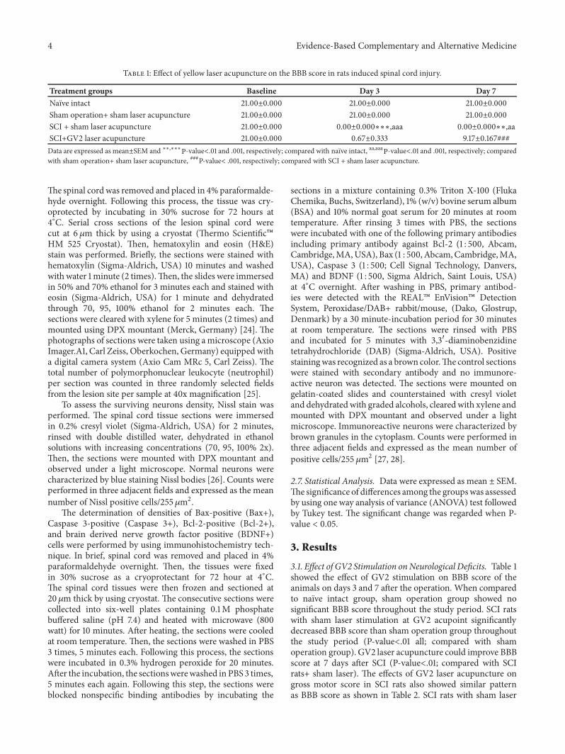