Yanfei Hu To cite this version - Accueil - TEL
210
HAL Id: tel-01203760 https://tel.archives-ouvertes.fr/tel-01203760 Submitted on 23 Sep 2015 HAL is a multi-disciplinary open access archive for the deposit and dissemination of sci- entific research documents, whether they are pub- lished or not. The documents may come from teaching and research institutions in France or abroad, or from public or private research centers. L’archive ouverte pluridisciplinaire HAL, est destinée au dépôt et à la diffusion de documents scientifiques de niveau recherche, publiés ou non, émanant des établissements d’enseignement et de recherche français ou étrangers, des laboratoires publics ou privés. Synthesis, self-assembly and controlled drug delivery of novel thermo-responsive and amphiphilic block copolymers based on polylactide, polyacrylamide and poly(oligo(ethylene glycol) methacrylate) Yanfei Hu To cite this version: Yanfei Hu. Synthesis, self-assembly and controlled drug delivery of novel thermo-responsive and amphiphilic block copolymers based on polylactide, polyacrylamide and poly(oligo(ethylene gly- col) methacrylate). Human health and pathology. Université Montpellier, 2015. English. NNT : 2015MONT3502. tel-01203760
Transcript of Yanfei Hu To cite this version - Accueil - TEL

HAL Id: tel-01203760https://tel.archives-ouvertes.fr/tel-01203760
Submitted on 23 Sep 2015
HAL is a multi-disciplinary open accessarchive for the deposit and dissemination of sci-entific research documents, whether they are pub-lished or not. The documents may come fromteaching and research institutions in France orabroad, or from public or private research centers.
L’archive ouverte pluridisciplinaire HAL, estdestinée au dépôt et à la diffusion de documentsscientifiques de niveau recherche, publiés ou non,émanant des établissements d’enseignement et derecherche français ou étrangers, des laboratoirespublics ou privés.
Synthesis, self-assembly and controlled drug delivery ofnovel thermo-responsive and amphiphilic block
copolymers based on polylactide, polyacrylamide andpoly(oligo(ethylene glycol) methacrylate)
Yanfei Hu
To cite this version:Yanfei Hu. Synthesis, self-assembly and controlled drug delivery of novel thermo-responsive andamphiphilic block copolymers based on polylactide, polyacrylamide and poly(oligo(ethylene gly-col) methacrylate). Human health and pathology. Université Montpellier, 2015. English. NNT :2015MONT3502. tel-01203760

Délivré par Université de Montpellier
Préparée au sein de l’école doctorale Sciences Chimiques et
Biologiques pour la Santé
Et de l’unité de recherche UMR CNRS 5247
Spécialité : Chimie et Physico-chimie des Polymères
Présentée par
Yanfei Hu
Soutenue le 8 avril 2015 devant le jury composé de
Mr Didier GIGMES, Directeur de recherche, Aix-Marseille
Université
Rapporteur
Mr Christophe MINGOTAUD, Directeur de recherche,
Université Paul Sabatier
Rapporteur
Mme Catherine LADAVIERE, Chargé de recherche,
Université de Lyon 1
Examinateur
Mr André DERATANI, Directeur de recherche,
Université de Montpellier
Examinateur
Mr Suming LI, Directeur de recherche, Université de
Montpellier
Directeur de thèse
Mr Vincent DARCOS, Ingénieur d’étude, Université de
Montpellier
Co-encadrant
Synthèse, auto-assemblage et libération contrôlée de principes
actifs de nouveaux copolymères à blocs
thermo-sensibleset amphiphiles à base de polylactide, de
polyacrylamide et de poly(oligo(éthylène glycol) méthacrylate)

- 1 -
Table of contents
Résumé .......................................................................................................................... I
Abstract .......................................................................................................................IX
List of Tables ........................................................................................................ .XVII
List of Schemes .............................................................................................. ……..XIX
List of Figures ......................................................................................................... .XXI
Abbreviation List .................................................................................................. XXV
General Introduction ........................................................................................... XXXI
Chapter 1 Bibiography ................................................................................................ 1
1.1 “Magic bullet” for cancer therapy ............................................................................ 1
1.1.1 Passive and active targeting in cancer therapy.................................................. 1
1.1.2 Polymeric delivery systems .............................................................................. 3
1.1.3 Thermo-responsive targeted drug delivery ....................................................... 5
1.2 Nanocarrier .............................................................................................................. 6
1.2.1 Liposome........................................................................................................... 7
1.2.2 Polymersome..................................................................................................... 9
1.2.3 Nanoparticle .................................................................................................... 12
1.2.4 Polymeric Micelle ........................................................................................... 13
1.3 Amphiphilic copolymers for uses as nanocarrier ................................................... 15
1.3.1 PLA/PEG block copolymers ........................................................................... 15
1.3.1.1 Polylactide................................................................................................ 15
1.3.1.2 Poly(ethylene glycol) ............................................................................. 16
1.3.1.3 PLA/PEG copolymers .............................................................................. 18
1.3.2 Thermo-responsive PLA/PNIPAAm copolymers ........................................... 19
1.3.2.1 Poly(N-isopropylacrylamide) ………………………………………….. 19
1.3.2.2 PLA/PNIPAAm copolymers .................................................................... 22
1.3.3 Thermo-responsive copolymers from PLA/PEG analogues ........................... 26

- 2 -
1.3.3.1 PEG analogues ......................................................................................... 26
1.3.3.2 PLA/PEG analogues block copolymers ................................................... 31
1.4 Summary and work plan ........................................................................................ 33
1.5 Reference ............................................................................................................... 34
Chapter 2 Synthesis and self-assembling of PNIPAAm-b-PLLA-b-PNIPAAm
thermo-responsive triblock copolymers prepared by combination of ROP and
ATRP ........................................................................................................................... 47
2.1 Introduction ............................................................................................................ 48
2.2 Experimental section .............................................................................................. 50
2.2.1 Materials ......................................................................................................... 50
2.2.2 Characterization .............................................................................................. 50
2.2.3 Synthesis of HO-PLLA-OH ............................................................................ 52
2.2.4 Synthesis of Br-PLLA-Br ............................................................................... 53
2.2.5 Synthesis of PNIPAAm-b-PLLA-b-PNIPAAm .............................................. 53
2.3 Results and discussion ........................................................................................... 54
2.3.1 Synthesis of PNIPAAm-b-PLLA-b-PNIPAAm .............................................. 54
2.3.2 Self-assembly of copolymers .......................................................................... 61
2.3.3 Morphology and size distribution of micelles ................................................ 64
2.3.4 Thermo-responsive behavior of micelles ........................................................ 67
2.4 Conclusion ............................................................................................................. 70
2.5 Reference ............................................................................................................... 71
Chapter 3 Tunable thermo-responsive
P(NIPAAm-co-DMAAm)-b-PLLA-b-P(NIPAAm-co-DMAAm) triblock
copolymer micelles as drug carrier .......................................................................... 75
3.1 Introduction ............................................................................................................ 76
3.2 Experimental section .............................................................................................. 78
3.2.1 Materials ......................................................................................................... 78
3.2.2 Typical synthesis of
P(NIPAAm-co-DMAAm)-b-PLLA-b-P(NIPAAm-co-DMAAm) ........................... 79

- 3 -
3.2.3 Drug release studies ........................................................................................ 79
3.2.4 Cytotoxicity..................................................................................................... 80
3.2.5 Characterization .............................................................................................. 81
3.3 Results and discussion ........................................................................................... 84
3.3.1 Synthesis of P(NIPAAm-co-DMAAm)-b-PLLA-b-P(NIPAAm-co-DMAAm)
triblock copolymer ................................................................................................. 84
3.3.2 Self-assembly of triblock copolymers micelles in aqueous medium .............. 89
3.3.3 Morphology and size distribution of micelles ................................................ 91
3.3.4 Thermo-responsive behavior of micelles ........................................................ 92
3.3.5 In vitro drug release ........................................................................................ 96
3.3.6 Cytocompatibility ........................................................................................... 98
3.4 Conclusion ........................................................................................................... 101
3.5 Reference ............................................................................................................. 102
Chapter 4 Thermo-responsive release of curcumin from
P(NIPAAm-co-DMAAm)-b-PLLA-b-P(NIPAAm-co-DMAAm) triblock
copolymers micelles ................................................................................................. 107
4.1 Introduction .......................................................................................................... 109
4.2 Materials and methods ......................................................................................... 111
4.2.1 Materials ....................................................................................................... 111
4.2.2 Synthesis of P(NIPAAm-co-DMAAm)-b-PLLA-b-P(NIPAAm-co-DMAAm)
triblock copolymer .. .............................................................................................. 111
4.2.3 Preparation of curcumin loaded polymeric micelles .................................... 112
4.2.4 In vitro drug release ...................................................................................... 112
4.2.5 In vitro cytotoxicity assay ............................................................................. 113
4.2.5 Characterization ............................................................................................ 114
4.3 Results and discussion ......................................................................................... 116
4.3.1 Synthesis of triblock copolymers .................................................................. 116
4.3.2 Characterization of polymer micelles ........................................................... 117
4.3.3 Preparation and characterization of drug loaded micelles ............................ 119

- 4 -
4.3.4 Phase transition of curcumin loaded polymeric micelles ............................. 123
4.3.5 In vitro drug release ...................................................................................... 124
4.3.6 In vitro cytocompatibility .............................................................................. 128
4.4 Conclusion ........................................................................................................... 131
4.5 Reference ............................................................................................................. 131
Chapter 5 Thermo-responsive micelles from comb-like block copolymers of
polylactide and poly(ethylene glycol) analogues: synthesis, micellization and in
vitro drug release ..................................................................................................... 135
5.1 Introduction .......................................................................................................... 136
5.2 Experimental section ............................................................................................ 138
5.2.1 Materials ....................................................................................................... 138
5.2.2 Synthesis of P(MEO2MA-co-OEGMA)-b-PLLA-b- P(MEO2MA-co-OEGMA)
copolymer .............................................................................................................. 138
5.2.3 Preparation of curcumin loaded micelles ...................................................... 139
5.2.4 In vitro drug release .................................................................................... 139
5.2.5 Characterization ............................................................................................ 140
5.3 Results and discussion ......................................................................................... 142
5.3.1 Synthesis of copolymers by ATRP ................................................................ 142
5.3.2 Self-assembly polymer micelles in aqueous medium ................................... 145
5.3.3 Phase transition of polymeric micelles ......................................................... 147
5.3.4 Size distribution of polymeric micelles ........................................................ 148
5.3.5 Preparation and characterization of drug loaded micelles ............................ 149
5.3.6 Phase transition of curcumin loaded micelles ............................................... 154
5.3.7 In vitro drug release ...................................................................................... 155
5.4 Conclusion ........................................................................................................... 158
5.5 Reference ............................................................................................................. 158
Conclusion and perspectives ................................................................................... 163
Publications .............................................................................................................. 167
Acknowledgements .................................................................................................. 168
I
Résumé
Dans les dernières décennies, les polymères sensibles aux stimulants ont attiré une
grande attention en raison de leur potentiel pour la libération ciblée de principes actifs,
en particulier pour la libération ciblée d’anti-tumoraux. Le support idéal de principes
actifs devrait être thermo-sensible au tissu tumoral où la température est d’environ
42 °C, c’est-à-dire bien supérieure à la température corporelle. Parmi les divers
polymères thermo-sensibles, le poly(N-isopropylacrylamide) (PNIPAAm) est
considéré comme le "standard d’or" parce qu’il est biocompatible et présente une
température critique inférieure de solution (LCST) autour de 32 °C qui est
relativement insensible aux changements environnementaux. Variation du pH, de la
concentration ou de l'environnement chimique n’influence que légèrement la LCST
du PNIPAAm. De plus, l'introduction d'un comonomère hydrophile de type
méthacrylamide, comme le N,N-diméthylacrylamide (DMAAm), permet d'augmenter
et d'ajuster précisément la LCST des copolymères obtenus. Néanmoins, ces
copolymères thermo-sensibles ne peuvent pas s’auto-assembler en nano-agrégats tels
que des micelles capables d'encapsuler des principes actifs.
Poly(L-lactide) (PLLA) est un polyester biodégradable et biocompatible largement
utilisé pour des applications biomédicales et pharmaceutiques telles que les implants
chirurgicaux, les supports en ingénierie tissulaire et des supports de principes actifs.
Les copolymères à blocs de PNIPAAm et de PLLA combinent la thermo-sensibilité et
l'hydrophilie du PNIPAAm avec la dégradabilité et l’hydrophobie du PLLA. En outre,
des micelles "intelligentes" peuvent être obtenues par auto-assemblage des
copolymères amphiphiles PLLA/PNIPAAm ou PLLA/P(NIPAAm-co-DMAAm) en
milieu aqueux. Cependant, les copolymères préparés en utilisant des méthodes de
synthèse traditionnelles présentent généralement des structures macromoléculaires
mal définies conduisant à une large transition de phase autour de la LCST, ce qui
n’est pas bénéfique pour les applications pharmaceutiques, notamment comme
II
support pour la libération ciblée des anti-tumoraux.
D'autre part, les polyméthacrylates thermo-sensibles contenant de courtes chaînes
pendantes d’oligo(éthylène glycol) ont été récemment proposés comme une
alternative au PNIPAAm. Ces analogues de PEG présentent des propriétés
remarquables comme l’hydrophilie, la biocompatibilité et la thermo-sensibilité, et
sont considérées comme une nouvelle génération de biomatériaux "intelligents". La
valeur de LCST peut être contrôlée dans une gamme de température allant de
l’ambiante à 90 °C en utilisant des comonomères oligo(éthylène glycol) méthacrylate
(OEGMA) de longueurs de chaîne différentes entre 2 et 9. Les copolymères thermo-
sensibles et amphiphiles dérivés des blocs P(OEGMA) et PLLA sont capables de
s’auto-assembler dans un milieu aqueux pour donner des micelles "intelligentes"
comme dans le cas des copolymères PLLA/PNIPAAm ou PLLA/P(NIPAAm-co-
DMAAm). Néanmoins, au mieux de notre connaissance, les propriétés
d'encapsulation et de libération de principes actifs des copolymères de PLLA et
d’analogues de PEG n’ont pas été étudiées en détail jusqu’à présent.
Dans ce travail de thèse, une étude détaillée est réalisée sur la synthèse, la
caractérisation, l'auto-assemblage et la libération de principes actifs des copolymères
à base de PLLA et P(NIPAAm-co-DMAAm) et à base de PLLA et d’analogues de
PEG. Les copolymères triblocs possèdent le même bloc central PLLA et deux blocs
hydrophiles latéraux. La composition des blocs hydrophiles est modifiée en utilisant
différents rapports de NIPAAm/DMAAm ou de comonomères OEGMA de façon à
ajuster la LCST des copolymères. Les copolymères obtenus présentent une structure
de chaînes bien définie et une faible dispersité des masses molaires. Les micelles
obtenues par auto-assemblage présentent une très faible CMC, une taille en dessous
de 100 nm et une libération de principes actifs thermo-sensible, ce qui est très
prometteur pour la libération ciblée des anti-tumoraux. Le contenu principal de cette
thèse est présenté ci-dessous:
III
1) Une série de copolymères triblocs thermo-sensibles PNIPAAm-b-PLLA-b-
PNIPAAm est synthétisée par la polymérisation radicalaire par transfert d’atomes
(ATRP) du NIPAAm en présence d'un macro-amorceur de PLLA possédant une degré
de polymérisation en unité acide lactique de 40. La polymérisation est réalisée en
utilisant un complexe CuCl/tris(2-diméthylaminoéthyl) amine (Me6TREN) comme
catalyseur à 25 °C dans un mélange DMF/eau (50/50 v/v) pendant 30 minutes. La
masse molaire des copolymères obtenus varie entre 18000 à 38000 g.mol-1
, et la
dispersité entre 1,10 et 1,28. Les suivies cinétiques de ln([M]0/[M]) vs. t2/3
présentent
une relation linéaire pendant les 10 premières minutes pour un rapport
NIPAAm/macro-amorceur de 100, et pendant les 30 premières minutes pour un
rapport NIPAAm/macro-amorceur de 200 et 300, ce qui indique que la concentration
d'espèces actives reste constante durant la période indiquée.
Les micelles sont formées par auto-assemblage de copolymères dans un milieu
aqueux à température ambiante. La structure cœur-couronne des micelles est
confirmée par la spectroscopie RMN 1H dans deux solvants différents (CDCl3 et D2O).
La concentration micellaire critique (CMC) déterminée par la spectroscopie de
fluorescence se trouve dans la gamme de 0,0077 à 0,016 mg mL-1
. Les copolymères
présentent une LCST entre 32,1 et 32,8 °C obtenue par des mesures de transmittance
UV-Vis. Les micelles sont sous forme sphériques avec un diamètre moyen compris
entre 31,4 et 83,3 nm déterminé par la TEM et la DLS. La taille des micelles
augmente avec la longueur des blocs hydrophiles de PNIPAAm parce que ceux-ci
sont étendus dans l'eau en dessous de la LCST. Les micelles présentent différents
comportements thermo-sensibles en fonction de la concentration. A forte
concentration (3,0 mg ml-1
), l'agrégation des micelles se produit lorsque la
température est élevée au-dessus de la LCST, conduisant à l'augmentation de la taille
de micelles. En revanche, à faible concentration (0,2 mg ml-1
), une diminution de la
taille de micelles est observée à cause de l'effondrement de blocs de PNIPAAm au-
dessus de la LCST.

IV
2) Dans cette seconde partie, des copolymères triblocs thermo-sensibles P(NIPAAm-
co-DMAAm)-b-PLLA-b-P(NIPAAm-co-DMAAm) sont synthétisés par l’ATRP du
NIPAAm et du DMAAm en utilisant le même macro-amorceur PLLA que
précédemment. La polymérisation est réalisée dans les mêmes conditions que dans le
cas des copolymères PNIPAAm-b-PLLA-b-PNIPAAm. Le DMAAm est incorporé
dans les chaînes de copolymère en tant que comonomère plus hydrophile afin de
réguler et d’augmenter la LCST. Le rapport NIPAAm/DMAAm varie de 100/0 à
66/24. Les copolymères obtenus présentent une structure de chaînes bien définie avec
des masses molaires Mn allant de 23600 à 27000 g mol-1
et de faibles dispersités (Đ =
1.10 à 1.18).
Des micelles de taille entre 37 et 54 nm avec une distribution étroite sont obtenues par
auto-assemblage de copolymères en milieu aqueux. Des valeurs très faibles de CMC
sont obtenues, allant de 0,010 à 0,015 mg ml-1
. La LCST augmente linéairement de
32,2 à 39,1 °C avec l'augmentation de la proportion en monomère DMAAm de 0 à
24 % dans la partie hydrophile. La présence de motifs DMAAm dans les copolymères
conduit à une transition de phase extrêmement étroite (∆T < 0,5 °C) par rapport aux
copolymères PNIPAAm-b-PLLA-b-PNIPAAm (∆T = 2,8 °C). Des changements
réversibles de taille des micelles sont observés avec une variation de température à
travers la LCST. En fait, lorsque les motifs DMAAm sont introduits, les chaînes de
P(NIPAAm-co-DMAAm) deviennent plus hydrophiles par rapport à celles de
PNIPAAm. Ainsi, la LCST augmente avec la transition de phase plus étroite.
L'amphotéricine B (AmpB), un principe actif faiblement hydrosoluble pour le
traitement de mycose systémique, est utilisée comme modèle afin d’évaluer le
potentiel de copolymères pour la libération de principes actifs. L’AmpB est
encapsulée dans les micelles en utilisant la méthode de dialyse avec une efficacité
d’encapsulation au-dessus de 59 %. Les tests de libération in vitro sont effectués à 37
ou 38 ° C dans l'eau en utilisant un copolymère ayant une LCST de 37,8 °C. Une
libération initiale brutale est observée dans tous les cas. La vitesse de libération à

V
38 °C est beaucoup plus rapide que celle à 37 °C, ce qui suggère que les micelles de
copolymères triblocs P(NIPAAm-co-DMAAm)-b-PLLA-b-P(NIPAAm-co-DMAAm)
pourraient permettre une libération thermo-sensible de principes actifs après
administration in vivo.
La cytotoxicité des copolymères est évaluée par le test MTT après incubation avec les
myocytes cardiaques de souris (MCM) et les fibroblastes embryonnaires de souris
(MEF). Les valeurs de densité optique (OD) sur le substrat polymère sont légèrement
supérieures à celles de la solution de culture comme témoin négatif, et bien
supérieures à celles de solution de phénol comme témoin positif. Les valeurs de taux
de croissance relative (RGR) des copolymères sont bien au-dessus de 100 % pendant
4 jours d'incubation avec les cellules MCF et MEF, correspondant à un niveau de
cytotoxicité de 0. En outre, l'adhésion et la prolifération de cellules sont observées sur
le substrat polymère. Les cellules présentent des formes broche, polygone ou ovale, et
le pseudopode de cellules s’étend vers l’extérieur. Ainsi, le test MTT et l'observation
de la morphologie des cellules indiquent que les copolymères P(NIPAAm-co-
DMAAm)-b-PLLA-b-P(NIPAAm-co-DMAAm) présentent une très bonne
cytocompatibilité.
3) La troisième partie de thèse porte sur une étude approfondie sur les propriétés
d'encapsulation et de libération de principes actifs des copolymères triblocs thermo-
sensibles P(NIPAAm-co-DMAAm)-b-PLLA-b-(NIPAAm-co-DMAAm). Quatre
copolymères sont d'abord synthétisés par l’ATRP comme décrit précédemment. Le
rapport NIPAAm/DMAAm varie de 68,2/31,8 à 60,6/39,4, et la masse molaire de
18000 à 26000 g mol-1
avec une faible dispersité (Đ = 1,1). Les copolymères
présentent une très faible CMC qui augmente légèrement de 0,0113 à 0,0144 mg mL-1
avec l’augmentation de la teneur en DMAAm de 31,8 à 39,4 mol% dans les blocs
hydrophiles P(NIPAAm-co-DMAAm). Parallèlement, la LCST des copolymères
augmente de 44,7 °C à 49,4 °C dans de l'eau et diminue d’environ 3.5 °C dans le
tampon phosphate salin (PBS). D'autre part, la taille des micelles augmente
VI
légèrement de 36,7 à 44,1 nm avec une teneur croissante en DMAAm. Le potentiel
zêta de micelles varie dans la gamme de -12,4 à -18,7 mV, en accord avec la structure
de micelles ayant les blocs PLLA dans le cœur et P(NIPAAm-co-DMAAm) dans la
couronne. Un copolymère avec une LCST de 42,1 °C dans le PBS est sélectionné
pour une étude approfondie de libération de principes actifs.
Un principe actif anticancéreux, la curcumine, est encapsulé dans les micelles de
copolymère à l'aide de la méthode dite "évaporation de solvant/hydratation de
membrane". Des charges en curcumine jusqu'à 20,4% sont obtenues avec une
excellente efficacité d’encapsulation (> 94%). La LCST diminue de 42,1 °C pour les
micelles seules dans le PBS à 38,0, 37,8 et 37,5°C avec une charge en curcumine de
6,0, 12,1 et 20,4%, respectivement. La transition de phase reste très étroite (<0,5 °C)
dans tous les cas. D'autre part, la taille augmente de 38,6 nm pour les micelles seules à
47,5, 53,7 et 88,2 nm pour les micelles avec une charge en curcumine de 6,0, 12,1 et
20,4 %, respectivement, ce qui indique que l'incorporation de curcumine conduit à de
plus grandes tailles de micelles. En outre, le potentiel zêta dans l'eau augmente de -
12,4 mV pour micelles seules jusqu'à -18,1 mV pour 20,4% de charge. Des valeurs de
potentiel zêta inférieures sont obtenues dans le PBS en raison de l'effet d’écran
ionique et agrégation de micelles. Toutes les solutions micellaires restent homogènes
et transparentes sur plus d’un mois, ceci étant dû à la charge négative en surface de
smicelles.
Des études de libération de principes actifs sont effectuées dans des conditions in vitro
à 37 et 40 °C, c’est-à-dire en dessous ou au-dessus de la LCST des micelles chargées.
Une libération initiale brutale est observée dans tous les cas, suivie d'une libération
plus lente. La vitesse de libération à 40 °C est plus rapide que celle à 37 °C en raison
de la libération thermo-sensible au-dessus de la LCST. D'autre part, les micelles
faiblement chargées présentent une vitesse de libération plus élevée que celles à plus
forte charge. Ce résultat pourrait être attribué à l'effet de la solubilité. La théorie de
Peppas est appliquée pour décrire les comportements de libération. Les résultats

VII
indiquent un mécanisme de libération contrôlée par une combinaison de diffusion et
de dégradation. Par ailleurs, les tests de cytotoxicité in vitro utilisant les cellules
fibrosarcome murin (L929) et lungcarcinoma humaine (A549) confirment une très
bonne cytocompatibilité des copolymères.
4) Dans la dernière partie de thèse, des copolymères triblocs en peigne P(MEO2MA-
co-OEGMA)-b-PLLA-b-P(MEO2MA-co-OEGMA) sont synthétisés par ATRP des
comonomères MEO2MA et OEGMA (Mn = 475) en utilisant le même macro-
amorceur Br-PLLA-Br dans des conditions similaires. Les copolymères obtenus
présentent une structure de chaînes bien définie avec des masses molaires Mn variant
de 27100 à 9800 g mol-1
et une dispersité d’environ 1,40. Le rapport
MEO2MA/OEGMA dans les blocs hydrophiles varie de 79/21 à 42/58.
Des micelles thermo-sensibles sont obtenues par auto-assemblage des copolymères
dans un milieu aqueux. La CMC des copolymères augmente légèrement de 0,0113 à
0,0130 mg ml-1
avec l'augmentation de la teneur en monomère OEGMA de 21 à 58
mol% grâce à son plus grande hydrophilie par rapport à MEO2MA. La LCST des
micelles dans l'eau augmente de 36,4 à 56,7 °C avec la teneur en monomère OEGMA
passant de 21% à 43%. Les données ne sont pas disponibles pour les copolymères
avec des teneurs en OEGMA plus élevées, ceci étant dû à l'évaporation rapide de l'eau
au-dessus de 60 °C. La présence de sel diminue de manière significative la LCST des
copolymères. Une diminution de près de 17 °C est observée lorsque l'on compare les
valeurs de LCST dans le PBS et dans l'eau, ce qui suggère un effet de déshydratation
très fort des ions phosphate. La taille des micelles augmente de 20,7 à 102,5 nm avec
la teneur en OEGMA passant de 21% à 58%. D'autre part, le potentiel zêta de micelles
est compris entre -0,77 et -1,99 mV, ce qui implique une charge de surface presque
neutre.
La curcumine est encapsulée dans les micelles en utilisant la méthode "évaporation de
solvant/hydratation de membrane" avec une efficacité d’encapsulation de plus de 90%.

VIII
La taille des micelles diminue de 102,2 nm pour micelles seules à 37,6 nm avec
10,8% de charge en curcumine, et la LCST diminue de 45,1 °C à 38,3 °C.
Parallèlement, le potentiel zêta augmente de -1,99 jusqu'à -44,9 mV. Ces résultats
indiquent que la charge en curcumine influence fortement la procédure d'auto-
assemblage. Les copolymères triblocs sont supposés s’auto-assembler en micelles
avec un cœur PLA, une mésophase PMA et une couronne hydrophile PEG.
L’encapsulation de la curcumine dans le cœur et dans la mésophase est susceptible de
conduire à une diminution de la taille et de la LCST des micelles, et une augmentation
du potentiel zêta.
La libération in vitro de curcumine est réalisée dans le PBS (pH = 7,4) à 37 °C et
41 °C, c’est-à-dire en dessous ou au-dessus de la LCST, respectivement. Une
libération initiale brutale est observée dans tous les cas, suivie d’une libération plus
lente. 81,8 et 92,6% de libération sont observés pour les micelles avec 5,9% de charge
en curcumine après 14 jours à 37 et 41 °C, respectivement. Une libération plus faible
est obtenue pour les micelles avec 10,8% de charge. La libération totale atteint 59,5 et
75,9 % à 37 et 41 °C dans la même période, respectivement. Par conséquent, la
libération de curcumine n’est pas seulement dépendante de la transition de phase à la
LCST, mais aussi de la charge en curcumine car la solubilité est un facteur limitant.
En résumé, deux séries de copolymères triblocs thermo-sensibles, à savoir
PLA/P(NIPAAm-co-DMAAm) et PLA/P(MEO2MA-co-OEGMA) sont synthétisées
avec succès par combinaison de la ROP et l’ATRP. Les copolymères amphiphiles
obtenus présentent des propriétés intéressantes d'auto-assemblage, une LCST
ajustable autour de la température du corps et une excellente cytocompatiblity. La
forte teneur en principe actif, l’excellente efficacité d'encapsulation, et la libération
thermo-sensible indiquent que ces copolymères sont prometteurs pour l'administration
ciblée de principes actifs anticancéreux.

IX
Abstract
In the past decades, stimuli-responsive polymers have drawn great attention due to
their potential for targeted drug delivery, especially for targeted delivery of anti-tumor
drugs. The ideal drug carrier should be thermo-responsive to the tumor tissue where
the temperature is ~42 oC, i.e. higher than the body temperature. Among the various
thermo-responsive polymers, poly(N-isopropyl acrylamide) (PNIPAAm) is considered
as the “gold standard” as it is biocompatible and exhibits a lower critical solution
temperature (LCST) around 32 oC which is relatively insensitive to environmental
changes. Variation of pH, concentration or chemical environment only slightly affects
the LCST of PNIPAAm. In contrast, introduction of an acrylamide comonomer, N, N-
dimethyl acrylamide (DMAAm) allows to increase and precisely adjust the LCST of
the resulting copolymers. Nevertheless, these thermo-responsive copolymers cannot
self-assemble into nanocarriers such as micelles capable of encapsulating drugs.
Poly(L-lactide) (PLLA) is a biodegradable and biocompatible polyester widely used
in biomedical and pharmaceutical applications such as surgical implants, tissue
engineering scaffolds and drug carriers. Block copolymers based on PNIPAAm and
PLLA combine the thermo-sensitivity and hydrophilicity of PNIPAAm and the
degradability and hydrophobicity of PLLA. Moreover, “smart” micelles can be
obtained by self-assembly of amphiphilic PLLA/PNIPAAm or PLLA/P(NIPAAm-co-
DMAAm) copolymers in aqueous medium. However, copolymers prepared using
traditional synthetic methods generally exhibit poorly defined macromolecular
structures with broad phase transition around the LCST, which is not beneficial for
pharmaceutical applications as carrier for targeted drug delivery.
On the other hand, thermo-responsive polymethacrylates containing short
oligo(ethylene glycol) side chains have been recently proposed as an alternative to
PNIPAAm or P(NIPAAm-co-DMAAm). These PEG analogues exhibit outstanding
properties such as hydrophilicity, biocompatibility and thermo-sensitivity, and are

X
regarded as a new generation of “smart” biomaterials. The LCST can be adjusted
from the room temperature up to 90 °C by using oligo(ethylene glycol) methacrylate
(OEGMA) comonomers of different chain lengths ranging from 2 to 9. Thermo-
responsive and amphiphilic copolymers derived from OEGMA and PLLA are able to
self-assemble in aqueous medium to yield “smart” micelles as in the case of
PLLA/PNIPAAm or PLLA/P(NIPAAm-co-DMAAm) copolymers. Nevertheless, to
the best of our knowledge, the drug encapsulation and drug release properties of
PLLA/PEG analogues in physiological conditions have not been investigated in detail,
so far.
In this work, a detailed investigation is performed on the synthesis, characterization,
self-assembly and drug release behavior of PLLA/P(NIPAAm-co-DMAAm) and
PLLA/PEG analogues. The triblock copolymers possess the same PLLA central block
and two lateral hydrophilic blocks. The composition of hydrophilic blocks is varied
using different NIPAAm/DMAAm ratios or OEGMA comonomers so as to adjust the
LCST of the copolymers. The obtained copolymers exhibit well-defined structure and
narrow molecular weight dispersity. Self-assembled micelles present very low CMC,
size below 100 nm and thermo-responsive drug release behavior, which is most
promising for targeted delivery of antitumor drugs. The main contents of this thesis
are shown in the following:
1) A series of thermo-responsive PNIPAAm-b-PLLA-b-PNIPAAm triblock
copolymers were successfully prepared by atom transfer radical polymerization
(ATRP) of NIPAAm in the presence of a α, ω-Br-PLLA-Br macroinitiator with a
block length of 40. The reaction was realized using a CuCl/tris(2-dimethylaminoethyl)
amine (Me6TREN) complex as catalyst at 25 oC in a DMF/water (50/50 v/v) mixture
for 30 min. The molecular weight of the resulting copolymers ranges from 18000 to
38000 g mol-1
, and the dispersity from 1.10 to 1.28. Kinetic plots of ln([M]0/[M])
against t2/3
exhibit a linear relationship during the first 10 min for
NIPAAm/macroinitiator ratio of 100, and during the first 30 min for

XI
NIPAAm/macroinitiator ratios of 200 and 300, indicating that the concentration of
active species remain constant at the initial stage.
Micelles are formed by self-assembly of copolymers in aqueous medium at room
temperature. Core-shell structure micellization of the copolymers was confirmed by
1H NMR spectroscopy in two different solvents (CDCl3 and D2O). The critical
micelle concentration (CMC) determined by fluorescence spectroscopy is in the range
of 0.0077-0.016 mg mL-1
. The copolymer micelles exhibit a lower critical temperature
(LCST) between 32.1 and 32.8 o
C obtained from UV-Vis transmittance measurements.
The micelles are spherical in shape with a mean diameter between 31.4 and 83.3 nm,
as determined by TEM and DLS. The size of micelles increases with increasing the
length of hydrophilic PNIPAAm blocks since they are extended in water below the
LCST. The micelles exhibit different thermo-responsive behaviors as a function of the
concentration. At high concentration (3.0 mg mL-1
), aggregation of micelles occurs
when the temperature is raised above the LCST, leading to micelle size increase. In
contrast, at low concentration (0.2 mg mL-1
), a decrease of micelle size is detected
because of the collapse of PNIPAAm blocks above the LCST.
2) In this second part, thermo-responsive P(NIPAAm-co-DMAAm)-b-PLLA-b-
P(NIPAAm-co-DMAAm) triblock copolymers were synthesized by ATRP of
NIPAAm and DMAAm using the same α, ω-Br-PLLA-Br macroinitiator. The
polymerization was realized under the same conditions as in the case of PNIPAAm-b-
PLLA-b-PNIPAAm copolymers. DMAAm was incorporated in copolymer chains as a
more hydrophilic comonomer in order to tune the LCST. The NIPAAm/DMAAm
ratio ranges from 100/0 to 66/24. The resulting copolymers present well defined chain
structures with Mn ranging from 23600 to 27000 g mol-1
and low dispersity (Đ=1.10-
1.18).
Nano-size micelles (37 to 54 nm) with narrow distribution were obtained by self-
assembly of copolymers in aqueous medium. Very low CMC values were obtained,
XII
ranging from 0.010 to 0.015 mg mL-1
. The LCST linearly increases from 32.2 to 39.1
oC with increasing the DMAAm content from 0 to 24 % in the hydrophilic part. The
presence of DMAAm units in the copolymers leads to extremely narrow phase
transition (∆T<0.5 oC), as compared to PNIPAAm-b-PLLA-b-PNIPAAm copolymers
(∆T=2.8 oC). Reversible micelle size changes are observed with temperature variation
across the LCST. In fact, when randomly distributed DMAAm is introduced, the
P(NIPAAm-co-DMAAm) chains become more hydrophilic as compared to PNIPAAm.
Thus the LCST phase transition shifts to higher temperature and becomes sharper.
Amphotericin B (AmpB), a poorly water-soluble drug for the treatment of systemic
mycosis, was used as a model drug to evaluate the potential of copolymers for drug
delivery. AmpB was encapsulated in micelles using dialysis method with drug loading
efficiency above 59 %. In vitro drug release was performed at 37 or 38 °C in water
using a copolymer with a LCST of 37.8 °C. An initial burst release is observed in all
cases. The release rate at 38 °C is much faster than that at 37 °C, suggesting that
P(NIPAAm-co-DMAAm)-b-PLLA-b-P(NIPAAm-co-DMAAm) triblock copolymer
micelles could achieve thermo-responsive release after in vivo administration.
The cytotoxicity of copolymers was evaluated by MTT assay after incubation with
mouse cardiac myocytes (MCM) and mouse embryonic fibroblasts (MEF). The values
of optical density (OD) on polymer substrate are slightly higher than those in the
culture solution as the negative control, and much higher than those in phenol as the
positive control. The relative growth rate (RGR) values of the copolymer are well
above 100 % during 4 days incubation with MCF and MEF cells, corresponding to a
cytotoxicity level of 0. Moreover, cell adhesion and proliferation are observed on the
copolymer substrate. Cells exhibit spindle, polygon or oval shapes, and the
pseudopodium of cells stretches out. Thus, MTT assay and cell morphology
observation indicate that P(NIPAAm-co-DMAAm)-b-PLLA-b-P(NIPAAm-co-
DMAAm) copolymers presents outstanding cytocompatibility.

XIII
3) The third part deals with a detailed investigation of the drug encapsulation and drug
release properties of thermo-responsive P(NIPAAm-co-DMAAm)-b-PLLA-b-
P(NIPAAm-co-DMAAm) triblock copolymers. Four copolymers were first
synthesized by ATRP as described previously. The NIPAAm/DMAAm ratio varies
from 68.2/31.8 to 60.6/39.4, and the molecular weight from 18000 to 26000 g mol-1
with narrow dispersity (Đ = 1.1). The copolymers exhibit very low CMC which
slightly increases from 0.0113 to 0.0144 mg mL-1
while the DMAAm content
increases from 31.8 to 39.4 mol% in the hydrophilic P(NIPAAm-co-DMAAm) blocks.
Meanwhile, the LCST of copolymers increases from 44.7 °C to 49.4 °C in water and
decreases by ca.3.5 °C in phosphate buffered saline (PBS). On the other hand, the
micelle size slightly increases from 36.7 to 44.1 nm with increasing DMAAm content.
The zeta-potential of micelles varies in the -12.4 to -18.7 mV range, in agreement
with the structure of micelles having PLLA block in the core and P(NIPAAm-co-
DMAAm) blocks at the corona. A copolymer with a LCST of 42.1 °C in PBS was
selected for detailed drug release studies.
An anticancer drug, curcumin, was encapsulated in the core of micelles using solvent
evaporation/membrane hydration method. High drug loading up to 20.4 % is achieved
with high loading efficiency (>94 %). The LCST decreases from 42.1 °C for blank
micelles in PBS to 38.0, 37.8 and 37.5 oC with drug loading of 6.0, 12.1 and 20.4 %,
respectively. The phase transition intervals remain very sharp (<0.5 oC) in all cases.
On the other hand, the micelle size increases from 38.6 nm for blank micelles to 47.5,
53.7 and 88.2 nm with drug loading of 6.0, 12.1 and 20.4 %, respectively, indicating
that curcumin incorporation leads to larger micelles. Moreover, the zeta potential in
water decreases from -12.4 mV for blank micelles up to -18.1 mV for 20.4 % drug
loading. Higher zeta potential values are obtained in PBS due to the ionic screening
effect and aggregation of micelles. All micelles remain homogeneously transparent
over 1 month due to the negative surface charge.

XIV
Drug release studies were performed under in vitro conditions at 37 and 40 °C, i.e.
below or above the LCST of drug loaded micelles. Initial burst release is observed in
all cases, followed by a slower release. The release rate is higher at 40 °C than that at
37 °C due to thermo-responsive release above the LCST. On the other hand, micelles
with lower drug loading exhibit higher release rate than those with higher drug
loading. This finding could be assigned to the solubility effect. Peppas’ theory is
applied to describe the release behaviors. The results indicate a combination of
diffusion and degradation controlled release. Moreover, the in vitro cytotoxicity tests
using murine fibrosarcoma (L929) and human lungcarcinoma (A549) cells confirm
the good cytocompatibility of the copolymers.
4) In the last part of the work, thermo-responsive comb-like P(MEO2MA-co-
OEGMA)-b-PLLA-b-P(MEO2MA-co-OEGMA) triblock copolymers were
synthesized by ATRP of MEO2MA and OEGMA (Mn = 300 g mol-1
) comonomers
using the same α, ω-Br-PLLA-Br macroinitiator under similar conditions. The
resulting copolymers present well defined chain structures with Mn varying from
27100 to 9800 g mol-1
and dispersity of c.a 1.40. The MEO2MA/OEGMA ratio in the
hydrophilic blocks varies from 79/21 to 42/58.
Thermo-responsive micelles are obtained by self-assembly of copolymers in aqueous
medium. The CMC of copolymers slightly increases from 0.0113 to 0.0130 mg mL-1
with increasing the OEGMA content from 21 to 58 % due to its higher hydrophilicity.
The LCST of micelles in water increases from 36.4 to 56.7 oC with the content of
OEGMA increasing from 21 % to 43 %. Data are not available for copolymers with
higher OEGMA contents due to fast evaporation of water above 60 oC. The presence
of salt significantly decreases the LCST of copolymers. A decrease of nearly 17 oC is
observed when comparing the LCST values in PBS and in water, suggesting the
strong dehydration effect of phosphate ions. The micelle size increases from 20.7 to
102.5 nm with the content of OEGMA increasing from 21 % to 58 %. On the other

XV
hand, the zeta potential of micelles ranges from -0.77 to -1.99 mV, which implies a
nearly neutral surface charge.
Curcumin is encapsulated in micelles by using solvent evaporation/membrane
hydration method with loading efficiency above 90 %. The micelle size decreases
from 102.2 nm for blank micelles to 37.6 nm with 10.8 % drug loading, and the LCST
decreases from 45.1 °C to 38.3 oC. Interestingly, the zeta potential decreases from -
1.99 up to -44.9 mV. These findings indicate that drug loading strongly affects the
self-assembly procedure. Triblock copolymers are assumed to self-assemble in
micelles with a PLA core, a PMA mesophase and a hydrophilic corona. Curcumin
encapsulation in the core and in the mesophase could lead to decrease of micelle size
and LCST and increase of zeta potential.
In vitro drug release is performed in pH 7.4 PBS at 37 °C and 41 oC, i.e. below or
above the LCST, respectively. An initial fast release from micelles is observed in all
cases, followed by slower release. 81.8 % and 92.6 % release is observed for micelles
with 5.9 % drug loading after 14 days at 37 oC and 41
oC, respectively. Slower release
is obtained for micelles with 10.8 % drug loading. The total release reaches 59.5 %
and 75.9 % at 37 oC and 41
oC in the same period, respectively. Therefore, drug
release is not only dependent on the phase transition across the LCST, but also on the
drug loading because the solubility of curcumin is a limiting factor.
Therefore, two series of thermo-responsive triblock copolymers, i.e.
PLA/P(NIPAAm-co-DMAAm) and PLA/P(MEO2MA-co-OEGMA) are successfully
synthesized by combination of ROP and ATRP. The resulting amphiphilic copolymers
present interesting self-assembling properties, variable LCST, and outstanding
cytocompatiblity. The high drug content and encapsulation efficiency, and thermo-
responsive drug release behavior indicate that these copolymers are promising
candidate for targeted delivery of anticancer drugs.

XVI

XVII
List of Tables
Table 2.1 Molecular characteristics of PNIPAAm-b-PLLA-b-PNIPAAm triblock
copolymers ................................................................................................................... 56
Table 2.2 Characterization of PNIPAAm-b-PLLA-b-PNIPAAm Triblock Copolymer
Micelles ........................................................................................................................ 65
Table 3.1 Relationship between the RGR value and cytotoxicity level ...................... 81
Table 3.2 Characteristics of P(NIPAAm-co-DMAAm)-b-PLLA-b-P(NIPAAm-co-
DMAAm) triblock copolymers .................................................................................... 86
Table 3.3 Properties of P(NIPAAm-co-DMAAm)-b-PLLA-b-P(NIPAAm-co-
DMAAm) micelles....................................................................................................... 90
Table 3.4 Drug loading content and loading efficiency of ampotericin B in polymeric
micelles ........................................................................................................................ 96
Table 3.5 RGR values of copolymers T4 with MCM and MEF cells during 4 days
incubation ................................................................................................................... 100
Table 4.1 Relationship between the RGR value and cytotoxicity level .................... 114
Table 4.2 Characteristics of P(NIPAAm-co-DMAAm)-b-PLA-b-P(NIPAAm-co-
DMAAm) triblock copolymers .................................................................................. 116
Table 4.3 Characterization of triblock copolymer micelles ...................................... 118
Table 4.4 Properties of curcumin-loaded micelles prepared from copolymer T2 .... 119
Table 4.5 Release exponent (n), rate constant (k) and correlation coefficient (R2) for
drug release from cur-polymer micelles .................................................................... 128
Table 4.6 RGR values of copolymers T2 with A549 and L929 cells during 4 days
incubation ................................................................................................................... 130
Table 5.1 Molecular characteristics of P(MEO2MA-co-OEGMA)-b-PLLA-b-
P(MEO2MA-co-OEGMA) triblock copolymers ........................................................ 144

XVIII
Table 5.2 Properties of P(MEO2MA-co-OEGMA)-b-PLLA-P(MEO2MA-co-OEGMA)
triblock copolymer micelles ....................................................................................... 147
Table 5.3 Properties of curcumin-loaded polymer (T5) micelles ............................. 150
XIX
List of Schemes
Scheme 1.1 Drug release from thermo-responsive polymeric micelles ........................ 5
Scheme 1.2 Self-assembly micelles from amphiphilic block copolyme ..................... 13
Scheme 1.3 Chemical structures of lactic acid enantiomers, and lactide
diastereoisomers ........................................................................................................... 15
Scheme 1.4 Synthesis of PEG/PLA block copolymers by ring opening
polymerization………………………………………………………………………..18
Scheme 1.5 Phase transition of PNIPAAm chains in response to temperature
changes……………………………………………………………………………….20
Scheme 1.6 Synthesis of PLA-b-PNIPAAm diblock copolymers by free radical
polymerization and ring-opening polymerization ........................................................ 22
Scheme 1.7 Synthesis of PLA-b-PNIPAAm-b-PLA triblock copolymers by
combination of ROP and RAFT ................................................................................... 23
Scheme 1.8 Phase transition mechanism of copolymer from PEG analogues in
aqueous medium .......................................................................................................... 27
Scheme 1.9 Synthesis of PEG analogues by ATRP of OEGMA and MEO2MA ......... 30
Scheme 1.10 Strategy for the click chemistry functionalization of well-defined
POEGMA ..................................................................................................................... 31
Scheme 2.1 Synthesis of PNIPAAm-b-PLLA-b-PNIPAAm triblock copolymers via
combination of ring-opening polymerization and atom transfer radical
polymerization………………………………………………………………………..56
Scheme 2.2 Illustration of the temperature responsive behavior of micelles with
varying temperatures across the LCST at low or high concentrations ........................ 69
Scheme 3.1 Synthesis of P(NIPAAm-co-DMAAm)-b-PLLA-b-P(NIPAAm-co-
DMAAm) triblock copolymers via combination of ROP and ATRP........................... 85
Scheme 5.1 Synthesis of P(MEO2MA-co-OEGMA)-b-PLLA-b-P(MEO2MA-co-
OEGMA) triblock copolymers .................................................................................. 142
Scheme 5.2 Illustration of micellar structures during drug loading .......................... 153

XX

XXI
List of Figures
Figure 1.1 Passive targeting (A) and active targeting (B) of nanocariers ..................... 2
Figure 1.2 Drug concentrations at site of therapeutic action after delivery as a
conventional injection (black) and as a temporal controlled release system (red) ........ 4
Figure 1.3 Different families of nanocarrier for drug delivery ..................................... 7
Figure 1.4 lower critical solution temperatures (LCST) as a function of the content of
OEGMA units per chain .............................................................................................. 26
Figure 1.5 Plots of transmittance as a function of temperature of (A) P(MEO2MA-co-
OEGMA), and (B) PNIPAAm in water at 3 mg mL-1
(solid line: heating, dashed line:
cooling) ....................................................................................................................... 28
Figure 1.6 Metabolic cell viability measured for human hepatocellular carcinoma
(HepG2) cell lines incubated at 37.8 oC in the presence of PEG, P(MEO2MA-co-
OEGMA) containing 10 mol % of OEGMA and 30 mol % of OEGMA .................... 29
Figure 1.7 Intensity size distribution (A) and volume size distribution (B) of
P(MEO2MA-co-OEGMA) measured by DLS at room temperature at 3 mg mL-1
in
aqueous solution........................................................................................................... 32
Figure 2.1 1H NMR spectra of HO-PLLA-OH (A), Br-PLLA-Br (B) and PNIPAAm-
b-PLA-b-PNIPAAm (C) in CDCl3 ............................................................................... 55
Figure 2.2 SEC chromatograms in DMF of Br-PLLA-Br and copolymers T1, T2, T3
and T4 .......................................................................................................................... 58
Figure 2.3 Kinetic plots for the polymerization of N-isopropylacrylamide in a
DMF/water mixture at 25 °C using a PLLA macroinitiator ........................................ 58
Figure 2.4 Mn vs. conversion plots of the polymerization of N-isopropylacrylamide in
DMF/water mixture solution at 25 °C using Br-PLLA-Br macroinitiator .................. 59
Figure 2.5 SEC traces for the polymerization of N-isopropylacrylamide in a
DMF/water mixture at 25 °C using a PLLA macroinitiator ........................................ 60
Figure 2.6 The influence of temperature on the kinetics: () 25 oC and () 0
oC ...... 61

XXII
Figure 2.7 1H NMR spectra of PNIPAAm-b-PLLA-b-PNIPAAm triblock copolymer
T1 in (A) CDCl3 and (B) D2O...................................................................................... 62
Figure 2.8 Fluorescence excitation spectra (λ=394 nm) of pyrene (6×10⁻⁷
M) in
aqueous solutions of PNIPAAm-b-PLLA-b-PNIPAAm triblock copolymer (T1) (A);
Plots of the intensity ratios (I337/I333 from pyrene excitation spectra) versus the
concentration of PNIPAAm-b-PLLA-b-PNIPAAm copolymer (T1) in water (B) ..... 63
Figure 2.9 TEM (A) and DLS (B) results of self-assembling micelles of copolymer
T4 at 1.0 mg mL-1
........................................................................................................ 66
Figure 2.10 Plot of transmittance changes as a function of temperature for aqueous
solution (3.0 mg mL-1
) of triblock copolymer T2 ........................................................ 67
Figure 2.11 Hydrodynamic diameter distributions obtained for micelles of copolymer
T2: (A) at a concentration of 3 mg mL-1
, and (B) at a concentration of 0.2 mg mL-1
.
() T= 20 °C, () T= 40 °C, () T= 20 °C ............................................................... 68
Figure 3.1 1H NMR spectrum of P(NIPAAm-co-DMAAm)-b-PLLA-b-(PNIPAAm-
co-DMAAm) triblock copolymer in CDCl3 ................................................................ 86
Figure 3.2 DOSY NMR spectrum of P(NIPAAm-co-DMAAm)-b-PLLA-P(NIPAAm-
co-DMAAm) triblock copolymer in DMSO-d6 ........................................................... 88
Figure 3.3 SEC chromatograms of Br-PLLA-Br macroinitiator and triblock
copolymers (T1 and T4) .............................................................................................. 88
Figure 3.4 Plots of the I337/I333 ratio changes from pyrene excitation spectra versus the
concentration of P(NIPAAm-co-DMAAm)-b-PLLA-b-P(NIPAAm-co-DMAAm) .... 90
Figure 3.5 TEM (A) and DLS (B) results of the self-assembling micelles of
copolymerT4 in aqueous medium. ............................................................................... 92
Figure 3.6 Plots of transmittance as a function of temperature (A) and (B) plot of
LCST values as a function of DMAAm content for copolymer solutions at 3.0 mg
mL-1
. ............................................................................................................................ .93
Figure 3.7 Reversible changes of the hydrodynamic diameter for T4 micelles at 3 mg
mL-1
when the temperature is raised from 25 () to 45 oC () (A), and cooled down
to 25 oC () (B) ......................................................................................................... 95

XXIII
Figure 3.8 Drug release profiles of AmpB-loaded polymeric micelles (Samples 1 and
2) at 37 and 38 oC in water........................................................................................... 97
Figure 3.9 Optical density values of MCM (A) and MEF (B) solutions after 2, 3 and 4
days culture in DMEM with copolymer substrate (T4), and controls (DMEM and 5 %
phenol). ........................................................................................................................ 99
Figure 3.10 Microscopic images of cells stained by NR after 3 days culture in
different media: (A) Copolymer T4 substrate without cell; (B) MCM cells on polymer
substrate in DMEM; (C) MEF cells on polymer substrate in DMEM; (D) MEF cells in
5 % phenol medium ................................................................................................... 100
Figure 4.1 1H NMR spectra of triblock copolymer T2in (A) CDCl3, and (B) D2O ..116
Figure 4.2 Image of (A) 6.0 % curcumin in H2O; (B) 6.0 % curcumin-loaded micelles
in PBS; (C) 12.1 % curcumin-loaded micelles in PBS; (D) 20.4 % curcumin-loaded
micelles in PBS .......................................................................................................... 120
Figure 4.3 DLS spectra and TEM micrographs of curcumin loaded micelles. (A) and
(B): Sample 1 (DL=6.0 %); (C) and (D): Sample 2 (DL=12.1 %); (E) and (F): Sample
3 (DL=20.4 %) ........................................................................................................... 121
Figure 4.4 Illustration of drug release from thermo-responsive polymeric micelles
controlled by phase transition .................................................................................... 123
Figure 4.5 Phase transition of curcumin-loaded micelles at 1 mg mL-1
determined by
UV-Vis spectroscopy. ()DL=0 in water; () DL=0 in PBS; () DL=6.0 % in PBS;
() DL=12.1 % in PBS; () DL=20.4 % in PBS ..................................................... 123
Figure 4.6 Calibration curve (A) and drug release profiles (B) of curcumin-loaded
micelles at 37 oC (red) and 40
oC (black): a and b -sample 1 (DL=6.0 %); c and d -
sample 2 (DL=12.1 %); e and f - sample 3 (DL=20.4 %) ......................................... 125
Figure 4.7 Plots of theoretical fitting to Peppas model for curcumin release from
curcumin-loaded micelles (the symbols are the same as in Figure 4.6) ................... 127
Figure 4.8 Optical density values of L929 (A)and A549 (B) solutions after 2, 3 and 4
days culture in DMEM with copolymer substrate (T2), and controls (DMEM and 5%
phenol) ...................................................................................................................... 129
XXIV
Figure 4.9 Microscopic images of cells stained by NR after 3 days culture in different
media: (A) Copolymer T2 substrate without cells; (B) L929 cells on polymer substrate
in DMEM; (C) A549 cells on polymer substrate in DMEM; (D) A549 cells in 5 %
phenol medium........................................................................................................... 130
Figure 5.1 1H NMR of P(MEO2MA-co-OEGMA)-b-PLLA-b-P(MEO2MA-co-
OEGMA) triblock copolymers .................................................................................. 143
Figure 5.2 SEC chromatograms of triblock copolymers ........................................... 144
Figure 5.3 Plots of the I337/I333 ratio changes from pyrene excitation spectra vs. the
concentration of copolymer T5 .................................................................................. 146
Figure 5.4 Intensity size distribution (A) and number size distribution (B) of triblock
copolymer T1 measured by DLS at room temperature .............................................. 148
Figure 5.5 Images of curcuminin solution: (A) 6.0 % curcumin in PBS; (B) sample 2
(DL=5.9 %); (C) sample 3 (DL=10.8 %) ................................................................... 150
Figure 5.6 DLS spectra and TEM micrographs of curcumin loaded micelles. (A) and
(B): sample 1 (DL=0); (C) and (D): sample 2 (DL=5.9 %); (E) and (F): sample 3
(DL=10.8 %) .............................................................................................................. 151
Figure 5.7 Zeta potential of curcumin-loaded micelles: () sample 1 (DL=0); ()
sample 2 (DL=5.9 %); () sample 3 (DL=10.8 %) .................................................. 152
Figure 5.8 Phase transition of blank and curcumin-loaded micelles determined by
UV-Vis spectroscopy at 1mg mL-1
in PBS. () blank micelles; () DL=5.9 %; ()
DL=10.8 % ................................................................................................................. 154
Figure 5.9 Calibration curve (A) and drug release profiles (B) of curcumin-loaded
micelles: a) sample 2 (DL=5.9 %) at 41 oC; b) sample 2 (DL=5.9 %) at 37
oC; c)
sample 3 (DL=10.8 %) at 41 oC; d) sample 3 (DL=10.8 %) at 37
oC ....................... 156

XXV
Abbreviation List
A
A549: Lungcarcinoma cell
AIBN: Azobisisobutyronitrile
AmpB: Amphotericin B
ATRP: Atom transfer radical polymerization
B
BPO: Benzoyl peroxide
Bpy: Bipyridine
Br-PCL-Br: α, ω-Bromopropionyl poly(ε-caprolactone)
Br-PLLA-Br: α, ω-Bromopropionyl poly(L-lactide)
C
CaH2: Calcium hydride
CLSM: Confocal laser scanning microscopy
CMC: Critical micellar concentration
CMT: Critical micelle temperature
CuCl: Copper (I) chloride
D
DDS: Drug delivery systems
DTBz: Dithiobenzoate
DL: Drug loading
D-LA: D-lactide
DLS: Dynamic laser scattering
DMAAm: N, N-dimethylacrylamide
DMEM: Dulbecco’s Modified Eagle’s Meduim

XXVI
DMF: N, N-dimethyformamide
DMSO: Dimethyl sulfoxide
DOSY NMR: Diffusion-ordered NMR
E
EDTA:
EE: Encapsulation efficiency
EPR: Enhanced permeability and retention
F
FDA: Food and Drug Administration
H
1H NMR: Proton Nuclear Magnetic Resonance
HepG2: Human hepatocellular carcinoma cell line
HMTETA: Hexamethyltriethylenetetramine
HO-PLLA-OH: α,ω-Hydroxy Poly(L-lactide)
I
ICP-MS: Inductively coupled plasma-mass spectrometey
L
L929: Murine fibrosarcoma
L-LA: L-lactide
LCST: Lower critical solution temperature
M
Mal: Maleimide
MCM: Mouse cardiac myocytes
MEF: Mouse embryonic fibroblasts

XXVII
MEO2MA: 2-(2-Methoxyethoxy) ethyl methacrylate
Me6TREN: Tris(2-dimethylaminoethyl) amine
mPEG-b-PCL: Poly(ethylene glycol)-b-poly(ε-caprolactone)
MTT: 3-(4, 5-Dimethylthiazol-2-yl)-2,5-diphenyltetrazolium bromide
MWCO: Molecular weight cut off
N
NMP: Nitroxide mediated polymerization
O
OD: Optical density
OEGMA: Oligo(ethylene glycol) methacrylate
P
PDLA: Poly(D-lactide)
PEG (or PEO): Poly(ethylene glycol)
PLA: Polylactide
PLGA-b-PEG-b-PLGA :
Poly(D,L-lactide-co-glycolide)-b-poly(ethyleneglycol)-b-poly(D,L-lactide-co-glycolide)
PLGA-b-POEGMA:
Poly(lactide-co-glycolide)-b-poly(oligo(ethyleneglycol) methacrylate)
PLLA: Poly(L-lactide)
PMA: Polymethacrylate
PMEOMA: Poly(2-methoxy ethyl) methacrylate
PMDETA: Pentamethyldiethylenetriamine
PNIPAAm: Poly(N-isopropylacrylamide)
P(NIPAAm-co-DMAAm)-b-PLGA:
Poly(N-isopropylacrylamide-co-N,N-dimethylacrylamide)-b-poly(D,L-lactide-co-
glycolide)
PNIPAAm-b-PLLA-b-PNIPAAm:

XXVIII
Poly(N-isopropylacrylamide)-b-poly(L-lactide)-b-poly(N-isopropylacylamide)
P(NIPAAm-co-DMAAm)-b-PLLA-b-P(NIPAAm-co-DMAAm):
Poly(N-isopropylacrymine-co-N,N-dimethylacrylamine)-b-poly(L-lactide)-b-poly(N-
isopropylacrymine-co-N,N-dimethylacrylamine)
P(NIPAAm-co-DMAAm)-b-PCL:
Poly(N-isopropylacrylamide-co-N,N-dimethylacrylamide-b-ε-caprolactone)
PNIPAAm-b-PS:
Poly(N-isopropylacrylamide)-b-polystylene
PNIPAAm-b-PMMA:
Poly(N-isopropylacrylamide)-b-poly(methyl methylacrylate)
PNIPAAm-b-PtBA: Poly(N-isopropylacrylamide)-b-poly(tert-butyl acrylate)
PSMA: Prostate specific membrane antigen
PTMC-b-PGA: Poly(trimethylene carbonate)-b-poly(L-glutamic acid)
R
RAFT: Reversible addition-fragmentation chain transfer
RES: Reticuloendothelial system
RGR: Relative growth ratio
ROP: ring-opening polymerization
S
SEC: Size exclusion chromatography
SG1: N-tert-butyl-N-(1-diethylphosphono-2, 2-dimethylpropyl) nitroxide
Sn(Oct)2: Tin(II)2-ethylhexanoate
T
TEM: Transmission electron microscopy
THF: Tetrahydrofuran
WHO: World Health Organization

XXIX
Z
ZP: Zeta potential
Symbol List
Ð: Molecular weight dispersity
D: Diffusion coefficients of the solutes
D0 : The translational diffusion coefficient at finite dilution
G : Relaxation frequency
h : The viscosity of the medium
kB: Boltzmann constant
Mn: Molecular weight
q: Wave vector
RH: Apparent equivalent hydrodynamic radius
T: Temperature
t: Time
∆T: Phase transition interval

XXX

XXXI
General introduction
Vectors prepared from amphiphilic block copolymers have been widely investigated
for pharmaceutical applications such as gene or cancer therapy. Among them,
nanocarrier has drawn great attention due to the passive enhanced permeability and
retention effect (EPR). Polymer micelles with small size (<200 nm) and low critical
micelle concentration (CMC) exhibit outstanding stability, high drug loading and
loading efficiency, and targeted drug delivery by EPR, which makes them promising
candidate as nanocarrier of anticancer drugs. However, most of the reported micelles
are not stimuli-responsive.
The aim of this work is to develop novel triblock copolymer micelles as nanocarrier
which combines both the passive targeting by EPR and active targeting by thermo-
responsiveness.
The first chapter of the thesis is a bibliography which provides an overview on the
state of the art in this domain. Nanocarriers including liposomes, polymersomes,
nanoparticles and polymeric micelles are introduced. Then, amphiphilic
polylactide/poly(ethylene glycol) (PLA/PEG) block copolymers are discussed as the
most studied bioresorbable and biocompatible micelles for controlled drug delivery.
The principle of thermo-responsive polymeric micelles was presented. Two different
families of thermo-responsive (co)polymers, polyacrylamide and poly(oligo(ethylene
glycol) methacrylate) or PEG analogues, as well as their amphiphilic block
copolymers with PLA, are then introduced, including the synthesis, self-assembly and
drug release properties.
The second chapter focuses on the synthesis, characterization and self-assembly of
poly(N-isopropylacrylamide)-b-poly(L-lactide)-b-poly(N-isopropylacrylamide)
(PNIPAAm-b-PLLA-b-PNIPAAm) triblock copolymers. Herein, for the first time, we

XXXII
report the synthesis of triblock copolymers by by ATRP of NIPAAm using a Br-
PLLA-Br macroinitiator. Kinetic studies are realized to illustrate the relationship
between the molecular weight and NIPAAm/macroinitiator ratio. Self-assembly
properties of copolymers and phase transition across the lower critical solution
temperature (LCST) are determined by CMC, DLS, TEM and UV transmittance
measurements.
In the third chapter, an acrylamide comonomer, N, N-dimethylacrylamide (DMAAm)
is introduced to increase and precisely adjust the LCST of the resulting P(NIPAAm-
co-DMAAm)-b-PLLA-b-P(NIPAAm-co-DMAAm) triblock copolymers. The
copolymers are successfully synthesized under the same conditions and fully
characterized. Self-assembly and phase transition of copolymers are studied.
Preliminary drug release is realized in water using Amphotericin B as a model. The
cytotoxicity of copolymers is evaluated by MTT assay after incubation with mouse
cardiac myocytes (MCM) and mouse embryonic fibroblasts (MEF).
The fourth chapter deals with the properties and in vitro drug release behavior of drug
loaded micelles. Curcumin, an anticancer drug, is loaded in P(NIPAAm-co-
DMAAm)-b-PLLA-b-P(NIPAAm-co-DMAAm) triblock copolymer micelles by
solvent evaporation/membrane rehydration method. The properties such as drug
loading, loading efficiency and the effect of drug loading on the LCST, size and
morphology are investigated. In vitro drug release is performed at temperatures below
or above LCST. Peppas’ theory is applied to describe the drug release behavior. The
cytotoxicity is further evaluated using murine fibrosarcoma (L929) and human lung
carcinoma cell (A549) cells to confirm the good cytocompatibility of the copolymers.
The fifth chapter describes the synthesis, micellization and in vitro drug release from
P(MEO2MA-co-OEGMA)-b-PLLA-b-P(MEO2MA-co-OEGMA) triblock copolymers.
The copolymers are synthesized by ATRP using the same macroinitiator and catalyst
system as in the case of PLA/PNIPAm copolymers. Micellization is performed in

XXXIII
aqueous medium and confirmed by CMC, DLS and TEM measurements. In vitro drug
release is performed in pH 7.4 PBS at 37 °C (below the LCST) and 41 oC (above the
LCST). The effects of temperature and drug loading on the drug release are discussed.

XXXIV

1
Chapter 1 Bibliography
1.1 “Magic bullet” for cancer therapy
Cancer is nowadays a leading cause of death in the word. The World Health
Organization (WHO) reported the approximately 14 million new cases and 8.2 million
cancer related deaths in 2012 [1]. The number of new cases is expected to rise by
about 70 % over the next two decades. For efficient cancer therapy, it is of key
importance to improve our knowledge of cancer physiopathology, to discover new
anti-cancer drugs and to develop novel biomedical technologies. Currently, the cancer
therapy has become a multidisciplinary challenge requiring close collaboration among
clinicians, biological and materials scientists, and biomedical engineers.
The concept of “magic bullet” that hits its target without damaging healthy issues
proposed by the Nobel laureate Paul Ehrlich in 1906 turned to reality for cancer
treatment and serious infectious diseases [2]. The challenge of the targeting is triple: (i)
to find the drug that effectively treats this disease, (ii) to find how to carry the drug,
and (iii) to find the proper target for a particular disease.
In the past decade, “magic bullet” from nanotechnology has been considered as one of
the most challenging innovations in pharmacology, and is now widely used in
prevention, diagnosis and therapy of cancer [3-6]. Nanocarriers have been exploited
to protect the drug, to optimize its targeting, to limit its accumulation in healthy
organs, to reduce its potential toxicity and to control its release [7].
1.1.1 Passive and active targeting in cancer therapy
The specific tumor targeting requires better profiles of pharmacokinetics and
pharmacodynamics, controlled and sustained release of drugs, improved specificity,
increased internalization and intracellular delivery and, more importantly, lower

2
systemic toxicity [8]. As shown in Figure 1.1, one can distinguish two kinds of tumor
targeting, i.e. passive targeting and active targeting. Drug carriers with size ranging
from 10 to 200 nm can reach tumor passively through the leaky vasculature
surrounding the tumors by the enhanced permeability and retention (EPR) effect. In
contrast, ligands grafted at the surface of drug carriers allow active targeting by
binding to the receptors over expressed by cancer cells or angiogenic endothelial cells,
followed by drug release in tumor [9]. However, the active targeting process cannot
be separated from the passive one because it occurs only after passive accumulation in
tumors [10].
Figure 1.1 Passive targeting (A) and active targeting (B) of nanocarriers. From ref.
[9].

3
Indeed, all nanocarriers use the “passive targeting” EPR effect as a guiding principle
[11]. As shown in Figure 1.1A, nanocarriers reach tumors selectively through the
leaky vasculature surrounding the tumors. Small drug molecules (unencapsulated by a
polymeric matrix) diffuse freely in and out the tumor blood vessels because of their
small size. Thus their effective concentration in the tumor decreases rapidly. In
contrast, drug-loaded nanocarriers cannot diffuse back into the blood stream because
of their large size (10-200 nm), thus resulting in progressive accumulation by the EPR
effect, which has become the “gold standard” in cancer-targeting drug designing.
The EPR effect is applicable for almost all rapidly growing solid tumors [12]. Indeed,
it can be observed in almost all human cancers with the exception of hypovascular
tumors such as prostate cancer or pancreatic cancer [13]. The EPR effect will be
optimal if nanocarriers can circulate for a long period of time. Very high local
concentration of drug-loaded nanocarriers can be achieved at the tumor site, for
instance 10–50 times higher than in normal tissue within 1–2 days [14].
1.1.2 Polymeric delivery system
Because of the specific characteristics of the tumor microenvironment and tumor
angiogenesis, it is possible to design drug delivery systems that specifically deliver
anti-cancer drugs to tumors. However, most of the conventional therapeutic agents
have poor pharmacokinetic profiles and are distributed nonspecifically in the body
leading to systemic toxicity associated with serious side effects.
In the past decades, polymeric delivery system has been widely exploited for various
biomedical and pharmaceutical applications [15]. Early in 1970s, Yolles et al.
proposed the concept of polymeric delivery system which is expected to deliver the
right amount of active agent at the right time to the right site [16, 17]. From then on,
polymeric delivery system has attracted more and more attention [18, 19]. Controlled
release systems are expected to deliver the drug into the systemic circulation at a

4
predetermined rate, while targeted delivery systems should release medications at or
near the targeted site. The advantage of targeted delivery is that high local
concentration of drug can be achieved. The drug is predominantly delivered to the
tumor rather than distributed throughout the whole body.
Figure 1.2 Drug concentrations at site of therapeutic action after delivery as a
conventional injection (black) and as a temporal controlled release system (red). From
ref. [20] with modification.
Indeed, drugs usually have an optimum concentration range within which maximum
benefit is achieved. Concentrations above or below this range are ether toxic or
exhibit no therapeutic effect [20]. Controlled release over an extended duration is
highly beneficial for drugs that are rapidly metabolized and eliminated from the body
after administration. As shown in Figure 1.2, the concentration of drug at the site of
activity after release from conventional administration (4 injections administered at 6
hourly intervals) is compared with that after release from a controlled release system.
In the case of conventional drug release, the drug concentration strongly fluctuates
during the 24 h period, and only a portion of the treatment period is in the therapeutic
window. With the controlled release system, in contrast, the drug concentration is
within the therapeutic window for almost 24 h. Controlled drug release could provide

5
a significant improvement in clinic therapy. A temporally controlled release system
would ensure that the maximum possible benefit is derived from the drug.
Consequently, polymeric delivery systems could allow to deliver the drugs in vivo
with the following benefits: 1) maintenance of optimum therapeutic drug
concentration in the blood with minimum fluctuation; 2) predictable and reproducible
release rate for extended duration; 3) prolongation of activity period for short half-life
drugs; 4) elimination or decrease of side effects, frequent dosing and wastage of drug;
and 5) optimized therapy and improved patient compliance.
1.1.3 Thermo-responsive targeted drug delivery
Scheme 1.1 Drug release from thermo-responsive polymeric micelles.
A new targeting strategy consists in developing “activable” or “activated”
nanocarriers due to the fast cancer cell metabolism. It is well known that different
from normal tissues, tumor has higher temperature (~42 oC) and lower pH (~5.3) due
to fast cell metabolism [21]. Recently, “smart” polymers which are responsive to
stimuli such as pH, acid and heat have also drawn great attention as drug delivery

6
systems [4, 22-29]. Thus, active target-specific delivery of drugs from these systems
to various sites in the body can be achieved to improve the therapeutic therapy,
meanwhile minimizing undesired side-effects.
Temperature is the most widely used stimulus in environmentally responsive
polymeric drug delivery systems. The change of temperature is not only relatively
easy to control, but also easily applicable both in vitro and in vivo. Therefore,
temperature-responsive nanocarriers have been largely developed [30, 31]. These
polymers generally exhibit a lower critical solution temperature (LCST) above which
phase separation occurs. As shown in Scheme 1.1, micelles can be obtained by
self-assembly of amphiphilic and thermo-responsive block copolymers below the
LCST, encapsulating a hydrophobic drug in the core of micelles. With increasing the
temperature above the LCST, the hydrophilic shell composed of thermo-responsive
blocks becomes hydrophobic and collapsed, and the micelles shrink or aggregate,
leading to faster drug release.
1.2 Nanocarrier
Various nanocarriers have been investigated for controlled or targeted drug delivery.
Figure 1.3 shows the various families of nanocarrier from 1 nm to 1 µm, including
branched polymers, micelles, liposomes, polymersomes, nanoparticles and micelles.
The size limit strongly depends on the target. Nanocarriers below 400 nm are more
suitable for accumulation in most tumors depending on cut-off size of vascular pores.
The lower limit is determined by the sieving coefficients for the glomerular capillary
wall in the kidney to avoid rapid renal filtration (that clears particles below 10 nm
such as branched polymers). Among the various systems, nanocarriers with sizes
between 10 and 200 nm are most promising candidate for targeted drug delivery
because they can avoid being cleared by the reticuloendothelial system (RES) or
mechanically filtered through the spleen and liver, and are small enough to efficiently

7
accumulate in tumors by the EPR effect.
Figure 1.3 Different families of nanocarrier for drug delivery. From ref. [7] with
modification.
1.2.1 Liposome
The first liposome was reported by Gregoriadis et al. in 1974 [32]. Liposomes are
defined as spherical lipid vesicles composed of amphiphilic phospholipids with a
bilayer membrane structure, and constitute an excellent tool for the understanding of
biological events such as cell adhesion, signal transduction or endocytosis. The closed
bilayers are formed by protecting the hydrophobic groups from the aqueous
environment which is in contact with the hydrophilic head group [3]. The polar
character of the liposomal core enables polar drug molecules to be encapsulated,
while lipophilic molecules are solubilized within the bilayer according to their affinity
towards phospholipids. Channel proteins can be incorporated without loss of activity
within the hydrophobic domain of vesicle bilayers, acting as a size-selective filter
only allowing passive diffusion of small solutes such as ions, nutrients and antibiotics.
Drugs that are encapsulated in a nanocage functionalized with channel proteins are

8
effectively protected from premature degradation by proteolytic enzymes. Drug
molecules are able to diffuse through the channel, driven by the concentration
difference between the interior and the exterior of the nanocage. Depending upon the
size and number of bilayers, liposomes can be divided in three categories:
multilamellar vesicles, large unilamellar vesicles, and small unilamellar vesicles. On
the other hand, liposomes can also be classified in five types in terms of composition
and mechanism of intracellular delivery, i.e. conventional liposomes, pH-sensitive
liposomes, cationic liposomes, immune-liposomes, and long-circulating liposomes
[33].
Liposomes can be prepared by using film casting and hydration method. Materials are
first dissolved in a solvent to yield a homogenous solution, and then a thin film is
obtained by using a rotary evaporator. Subsequently, the film is hydrated by addition
of water. Water permeation in the thin film driven by hydration forces allows to form
bilayers, and finally yields vesicles upon separation from the recipient’s surface.
Liposomes have been investigated for the delivery of vaccine, toxoids, gene,
anticancer, and anti-HIV drugs [33-37]. However, although liposome technology was
discovered over 40 years ago, liposome-based drug formulations have rarely entered
the stage of clinical applications. The major drawback of liposomes is their low
mechanical stability and high leakiness, mainly due to their small membrane thickness
(3–5 nm). Other factors such as limited versatility of chemical structures, poor
batch-to-batch reproducibility, difficulties in sterilization, large size (up to 400 nm)
and low drug loading also contribute to restrain the development of liposomes as drug
carrier [7, 38].
Recently, modification of liposome by PEG has been successfully developed [36].
Doxil® (Sequus Pharmaceuticals, Menlo Park, California), the first FDA-approved
nano-drug (1995), is a long-acting PEGylated liposomal formulation of doxorubicin.

9
It alters dramatically the pharmacokinetic properties of doxorubicin with prolonged
drug circulation time, escaping from the RES, high loading of doxorubicin and
targeted release at the tumor due to the EPR effect [39, 40].
1.2.2 Polymersome
In the past decade, polymersomes (also referred to as polymeric vesicles) have
attracted growing interest based on their intriguing aggregation phenomena, cell and
virus-mimicking dimensions and functions, as well as tremendous potential
applications in medicine, pharmacy, and biotechnology [41, 42]. Unlike liposomes
self-assembled from low molecular weight lipids, polymersomes are in general
prepared from macromolecular amphiphiles of various architectures including
amphiphilic diblock, triblock, graft as well as dendritic copolymers [43-45].
The preparation methods of polymersomes are generally classified into two groups:
solvent-switching and film hydration. Solvent-switching techniques have been widely
used in the preparation of polymeric vesicles (and also for other morphologies) as
most amphiphilic block copolymers are not directly soluble in water. Initially the
amphiphilic copolymer is dissolved in a common organic solvent for both blocks and
then water is added gradually to the copolymer solution. The hydrophobic blocks tend
to associate together in the polar environment to form a bilayer, whereas the
hydrophilic blocks are solvated to form the corona which colloidally stabilizes the
vesicle. Film hydration technique is the same as that used for liposomes preparation
mentioned above, involving film casting and hydration to induce self-assembly and
polymersome formation.
Polymersomes and liposomes present similar architectures. Both are nano- to
micrometer-sized capsules with a bilayered membrane enclosing an aqueous
compartment. They are able to encapsulate hydrophilic molecules within the aqueous
core and also hydrophobic molecules within the membrane. Moreover, because

10
polymersomes are composed of block copolymers instead of small molecules, they
present lower permeability and higher mechanical stability, and their architecture is
more tunable as compared to liposomes [7].
Polymersome is one of the most interesting drug delivery systems because of multi
drug loading capacity, membrane robustness and stealth properties [46, 47]. Some
authors have studied polymersomes for targeted drug delivery [48]. Two anticancer
drugs, paclitaxel and doxorubicin have been encapsulated in polymersomes for cancer
therapy as reported by Ahmed et al. [49]. The drugs are loaded into polymersomes by
a pH-gradient method established for liposomes. A single systemic injection of the
dual drug combination shows a higher maximum tolerated dose than the free drug
cocktail and shrinks tumors more effectively and more sustainably than free drug:
50 % smaller tumors are observed at 5 days with polymersomes. Drug accumulation
within tumors was also observed, showing the potential of multi-drug delivery by
polymersomes for clinical applications.
However, the major drawback of polymersome delivery systems is the long
encapsulation procedure (up to weeks). Recently, Lecommandoux et al. proposed a
convenient and reproducible nanoprecipitation method to obtain stable and
pH-inducible poly(trimethylene carbonate)-b-poly(L-glutamic acid) (PTMC-b-PGA)
polymersomes loaded with a remarkably high content of doxorubicin (47 %, w/w %)
at pH 10.5 [42, 50]. Doxorubicin and polymer are dissolved in dimethyl sulfoxide
(DMSO). Then an aqueous phase is quickly added to the organic phase under stirring.
Excess drug and DMSO are removed by dialysis against a buffer solution. After
neutralization, an aqueous dispersion of drug-loaded vesicles is obtained which
remains stable for a prolonged period of time (at least 6 months) without vesicle
disruption or drug precipitation.
Furthermore, to prevent premature drug leakage, Lecommandoux et al. reported the
11
preparation of polymeric vesosomes, that is, polymersomes in polymersomes [51].
Vesosomes present controlled permeability by encapsulating nano-size polymersomes
inside larger polymersomes. A double-membrane diffusion barrier effect was
demonstrated since the observed in vitro release rate of doxorubicin from vesosomes
was twice slower than that from polymersomes.
In principle, drug release from polymersomes is governed by the diffusion of drug
through the membrane. According to the diffusion-interface theory, the driving force
is the concentration gradient of drug between the polymersome and the surrounding
medium [52]. When drug molecules diffuse from the core of polymersomes to the
surrounding medium, the release rate is a function of the square root of time. The size
distribution of polymersomes also influences the overall release rate [53]. Based on
theoretical approaches, polymersomes suitable for the delivery of specific drugs can
be designed and the release kinetics predicted. Nevertheless, in many cases, the
control for drug release cannot be adjusted to the desired level because the properties
of polymersomes cannot be varied to a large extent due to lack of versatility for the
composition of block copolymers [54].
Recently, to achieve targeted drug delivery, significant efforts have been devoted to
develop “smart” polymersomes which are responsive to pH, heat, light as well as
magnetic field [28, 55-66]. Stimuli-responsive polymersomes have emerged as novel
programmable delivery systems allowing to readily modulate the release of
encapsulated contents by the stimulus. Stimuli-responsive release could significantly
enhance the therapeutic efficacy and minimize the side effects. It is also feasible to
form and disassemble polymersomes in water simply by applying an appropriate
stimulus. Lecommandoux et al. reported biologically active polymersomes from
polypeptide block copolymers prepared by “click chemistry” [67-69]. Hybrid
polymer/lipid vesicles were also developed when the content of lipids was below 20
mol %, in which lipids are randomly distributed in the polymer-rich membrane [70,

12
71].
1.2.3 Nanoparticle
Nanoparticles include both nanospheres and nanocapsules in solid state, either
amorphous or crystalline. They are able to absorb or encapsulate a drug, thus
protecting it against chemical and enzymatic degradation [18]. Nanoparticles as drug
carrier can be obtained from both biodegradable polymers and non-biodegradable
ones [3, 72, 73]. Recently, biodegradable polymeric nanoparticles have attracted great
attention in view of their applications in controlled and targeted delivery of drugs [11,
74-76].
Polymeric nanoparticles can be prepared from emulsion or precipitation method.
Formation of hollow structures, named nanocapsules, can also be achieved. Their size
is generally between 10 and 1000 nm and strongly depends on the preparation method.
The solid nature of nanoparticles confers great stability and controlled drug release
rate. Nanoparticles with size ranging from 10 to 200 nm can preferentially concentrate
in tumor by virtue of the EPR effect as mentioned above. In addition, multiple drug
loadings can be achieved within nanoparticles [7].
Moreover, surface PEGylated nanoparticles also have been developed to provide
excellent pharmacokinetic control, and are suitable for the entrapment and delivery of
a wide range of therapeutic agents [77-79]. PEGylated nanoparticles allow to reduce
the immunogenicity, and to limit the phagocytosis of nanoparticles by the RES, thus
resulting in increased blood concentration of drug in organs such as the brain,
intestines, and kidneys.
Nanoparticles are matrix systems in which the drug is physically and uniformly
dispersed. Drug release kinetics can be adjusted by careful selection of polymers or
polymer blends. Thus, drug release from nanoparticles generally occurs either by

13
diffusion or by matrix degradation or both.
1.2.4 Polymeric micelle
In the past decades, self-assembly of amphiphilic block copolymers in aqueous
solutions into various micellar structures has been extensively investigated. Bader et
al. first proposed micelles as potential drug carrier in 1984 [80]. Since then, micelles
attracted more and more interest for controlled release of drugs due to their numerous
advantages including reduced side effects of drugs, targeted drug delivery, and
prolonged blood circulation time [81-86]. Drug encapsulation can be simply realized
by physical mixing, which is attractive as compared to chemical conjugate [85, 87].
Scheme 1.2 Self-assembly micelles from amphiphilic block copolymer.
In an aqueous medium, polymeric micelles generally present a core-corona structure
driven by hydrophobic interactions above the critical micellar concentration (CMC) as
shown in Scheme 1.2 [88]. The hydrophobic inner core allows encapsulation of
hydrophobic drugs by physical entrapment, whereas the hydrophilic shell acts as a
stabilizing/interacting corona [89]. Micelles are thus used to encapsulate drugs with
poor water solubility [90, 91]. Compared to polymersome systems, micelles have
advantages such as simple preparation and efficient drug loading.
Polymeric micelles can be prepared by using different methods: direct dissolution,
dialysis, nanoprecipitation (solvent-switching), and solution casting/membrane
hydration. Direct dissolution involves simply dissolving a water soluble amphiphilic

14
copolymer in an aqueous medium. Dialysis consists in dialyzing a copolymer solution
against water. Nanoprecipitation and membrane hydration methods are the same as
those used for the preparation of polymersomes mentioned above. Yang et al.
compared micelles prepared by direct dissolution and dialysis methods [92, 93].
Similar drug loading and encapsulated efficiency were obtained for both methods.
Micelles by direct dissolution method present major advantages such as easy
formulation and absence of toxic organic solvents, which shows great potential as
carrier of hydrophobic drugs.
Polymeric micelles generally have small sizes (i.e. <200 nm) with narrow distribution,
which allows them to escape from non-specific uptake by the RES cells and to
achieve long circulation in the blood stream [81, 87, 94]. Drug molecules can be
incorporated by means of chemical, physical or electrostatic interactions, depending
on their physicochemical properties. Amphiphilic block copolymers are attractive for
drug delivery applications since their chemical composition, molecular weight and
block lengths can be easily changed, which allows control of the size and morphology
of micelles. Polymeric micelles can target tumor sites by passive as well as active
targeting mechanisms. In fact, the inherent properties of micelles including size in the
nano range, stability in plasma, long circulation in vivo, and the pathological
characteristics of tumor allow them to target the tumor site by the EPR effect [88,
95-101].
Recently, growing interest has been paid to amphiphilic block copolymer micelles as
novel carrier for drug targeting [84, 91, 102-104]. The distribution of drug loaded
polymeric micelles in the body is mainly determined by their size and surface
properties, and less affected by the properties of drug embedded in the inner core of
micelles. In this regard, the size and surface properties of polymeric micelles have
crucial importance in achieving controllable drug delivery with efficacy [84, 105].
Prolonged in vivo circulation time and adequate retention of drug within the carrier

15
are prerequisites to successful drug targeting [106]. Thermo-responsive and pH
polymeric micelles have also been developed to achieve targeted drug release at the
tumor site [29, 103, 107, 108].
1.3 Amphiphilic copolymers for uses as nanocarrier
1.3.1 PLA/PEG block copolymers
1.3.1.1 Polylactide
From a theoretical point of view, micelles prepared from biodegradable polymers
would be more beneficial in applications since no accumulation of polymers occurs
after drug release. As a biodegradable polymer, polylactide (PLA) has been
extensively investigated as controlled drug release systems for biomedical and
pharmaceutical applications [72, 109, 110]. PLA presents outstanding
biocompatibility and variable biodegradability. The final degradation product, lactic
acid, is a metabolite and can be easily eliminated from the human body via the Kreb’s
cycle.
Scheme 1.3 Chemical structures of lactic acid enantiomers, and lactide
diastereoisomers.

16
It should be noted that lactic acid is chiral molecule due to the presence of an
asymmetric carbon atom, and thus exists in the form of L-lactic and D-lactic acid
enantiomers. Similarly, the cyclic diester of lactic acid, lactide, exists in the form of
three kinds of diastereoisomers, i.e. L-lactide, D-lactide and meso-lactide. In the case
of LA-containing polymer chains, chirality of LA units provides a worthwide means
to adjust the physical-chemical, biological and mechanical characteristics of polymers.
The structure of various enantiomers and diastereoisomers of lactic acid and lactide is
shown in Scheme 1.3.
Consequently, PLA exists in the form of poly(L-lactide) (PLLA), poly(D-lactide)
(PDLA), and various stereocopolymers. In 1987, Ikada et al. first reported that
physical interaction between PLLA and PDLA, namely stereocomplexation, strongly
affects the crystallization behavior, thermal resistance, mechanical properties as well
as the hydrolytic resistance of PLA based materials [111]. These improvements are
due to the stronger interaction between L-LA and D-LA sequences. Novel
stereocomplexation-induced micelles and hydrogels have been reported in the case of
PLA/PEG block copolymers [86, 93, 112-114].
1.3.1.2 Poly(ethylene glycol)
Poly(ethylene glycol) (PEG), also as known as poly(ethylene oxide) (PEO) depending
on the synthetic conditions, is a linear polyether terminated with hydroxyl end groups.
PEG has been approved by the US Food and Drug Administration (FDA), and is
probably the most widely applied synthetic polymer in the domains of biotechnology,
pharmacology, and medicine [15, 100]. PEG is a cheap, neutral, water soluble and
biocompatible polymer with good hydrolytic resistance. PEG generally has narrow
molecular weight distributions which are readily accessible by anionic polymerization
of ethylene oxide. The water solubility of PEG results from the strong tendency to
form hydrogen bonds with water via -O- groups. The hydrophilic character with
surface-wetting properties is especially important for the prevention of protein

17
adsorption and the blood compatibility in biomedical applications.
“Stealth” effect is a key property of PEG, which allows avoiding a fast recognition by
the immune system followed by rapid clearance from the body. Chemical attachment
of PEG to the drug of choice is a process called PEGylation [110]. The concept of
PEGylation was first introduced by Davis et al. in 1970s [77]. Unexpected finding by
PEGylation was observed, including improved pharmacokinetic and
pharmacodynamic properties of polypeptide drugs due to increased water solubility,
reduced renal clearance and toxicity. PEGylation reduces kidney clearance simply by
making the molecules larger as the kidneys filter substances according to their size.
Studies of PEG in solution revealed that ethylene oxide subunit is tightly associated
with two or three water molecules. This binding of water molecules makes PEGylated
compounds function as though they are five to ten times larger compared to a soluble
protein of similar molecular weight, as confirmed by size exclusion chromatography
and gel electrophoresis [78, 115]. When attached to hydrophobic drugs or carriers,
PEG provides them with good physical and thermal stability, preventing or reducing
aggregation of drugs in storage and in vivo as a result of the steric hindrance.
Therefore, PEG with associated water molecules behaves like a shield to protect the
attached drug from enzymatic degradation, rapid renal clearance and interactions with
cell surface proteins, thereby limiting adverse immunological effects.
PEGylated drugs are also more stable over a range of pH and temperature changes as
compared to their unPEGylated counterparts. Consequently, PEGylation confers to
drugs a number of clinical benefits, such as sustained blood levels that enhance the
drug efficacy, fewer adverse reactions, longer shelf life and improved patient
convenience [116]. The ability of PEGylation to influence the pharmacokinetic
properties of drugs and drug carriers is widely utilized in pharmaceutics [77, 78, 117].
In fact, all polymer based stealth drug delivery systems that have been brought to the
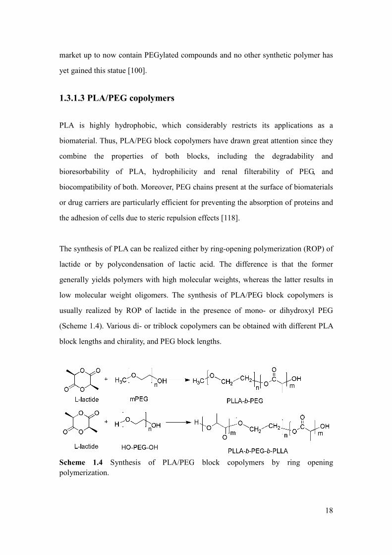
18
market up to now contain PEGylated compounds and no other synthetic polymer has
yet gained this statue [100].
1.3.1.3 PLA/PEG copolymers
PLA is highly hydrophobic, which considerably restricts its applications as a
biomaterial. Thus, PLA/PEG block copolymers have drawn great attention since they
combine the properties of both blocks, including the degradability and
bioresorbability of PLA, hydrophilicity and renal filterability of PEG, and
biocompatibility of both. Moreover, PEG chains present at the surface of biomaterials
or drug carriers are particularly efficient for preventing the absorption of proteins and
the adhesion of cells due to steric repulsion effects [118].
The synthesis of PLA can be realized either by ring-opening polymerization (ROP) of
lactide or by polycondensation of lactic acid. The difference is that the former
generally yields polymers with high molecular weights, whereas the latter results in
low molecular weight oligomers. The synthesis of PLA/PEG block copolymers is
usually realized by ROP of lactide in the presence of mono- or dihydroxyl PEG
(Scheme 1.4). Various di- or triblock copolymers can be obtained with different PLA
block lengths and chirality, and PEG block lengths.
Scheme 1.4 Synthesis of PLA/PEG block copolymers by ring opening
polymerization.

19
In the past decades, PLA/PEG copolymer micelles have been largely investigated as
drug carrier [44, 119-121]. Anti-tumor drugs such as paclitaxel, beta-lapachone and
doxorubicin have been loaded in PLA/PEG micelles [110, 122-124]. Currently, the
most advanced micellar formulations encapsulating hydrophobic drugs such as
doxorubicin and paclitaxel have already entered the market in India (Nanoxel®
) and
South Korea (Genexol-PM®
) [104, 125]. Most recently, the first “active” docetaxel
encapsulated PLA/PEG micelles (BIND-014®
) with tumor target prostate specific
membrane antigen (PSMA) has been approved in USA for the treatment of solid
tumors and metastatic cancers [126]. The micelles present major advantages such as
decreased side effects, better pharmacological profiles, and improved drug tolerance
over the current clinical formulations.
Nevertheless, PEG/PLA micelles are not responsive to environment changes. In fact,
many bioactive agents used in pharmacotherapy exhibit side effects that may affects
the drug efficacy. For instance, compounds used for cancer therapy can kill not only
cancer cells, but also normal cells in the body, which results in undesired side effects
[87]. The EPR effect is considered as passive targeting method. Drug targeting could
be further enhanced by introducing a polymeric matrix which is responsive to
variation of temperature [127, 128].
1.3.2 Thermo-responsive PLA/PNIPAAm copolymers
1.3.2.1 Poly(N-isopropylacrylamide)
Poly(N-isopropylacrylamide) (PNIPAAm) is the most studied thermo-responsive
polymer with a LCST at 32 oC where it exhibits a sharp phase transition from
hydrophilic coils to hydrophobic globules [129]. Hydrogen bonding is formed
between amide groups and water molecules in PNIPAAm solutions under the LCST.
In contrast, the alkyl backbone and isopropyl groups present a competitive
hydrophobic effect [129]. The phase transition, as shown in Scheme 1.5, is attributed

20
to the entropic gain since water molecules associated to the amide groups are released
to the aqueous phase when the temperature increases above the LCST [130, 131].
Scheme 1.5 Phase transition of PNIPAAm chains in response to temperature changes.
From Ref. [131] with modification.
As suggested by Cho et al., the phase transition of PNIPAAm across the LCST
involves at least three phenomena: (i) rearrangement of bound water around either
hydrophobic isopropyl or hydrophilic amide groups of PNIPAAm, (ii) intermolecular
and intramolecular hydrogen bonding between the amide groups, and (iii) association
of hydrophobic groups in PNIPAAm mainchains [132]. When increasing the
temperature above the LCST, polymer-polymer interactions are thermo-dynamically
favored as compared to polymer-water interactions. Water molecules around the
hydrophilic groups also are released to the medium. The intermolecular and
intramolecular interactions between PNIPAAm chains include not only hydrophobic
association of the hydrophobic groups, but also hydrogen bonding between the amide
groups. PNIPAAm chains thus become dehydrated globules in the form of a
suspension.
In Ono’s work, the number of water molecules per NIPAAm unit in homogeneous
solution was determined to be 11 exactly below the LCST, employing the
high-frequency dielectric relaxation technique [132]. Thus, PNIPAAm is regarded as a
“golden standard” thermo-responsive polymer due to the fact that its LCST is
relatively insensitive to physical and chemical environment [27]. Slight variations of

21
pH, concentration or chemical environment only affect the LCST of PNIPAAm by a
few degrees [129].
PNIPAAm homopolymer was initially synthesized by free radical polymerization. It
can be functionalized by reaction with chain transfer agents having functional acid,
hydroxyl or amino groups [129]. Introduction of functional end groups could allow to
improve the properties of polymers including thermo-sensitivity, pH sensitivity or
fine tuning of the LCST. However, the molecular weight and dispersity of polymers
were not controlled.
The atom transfer radical polymerization (ATRP) of NIPAAm was first reported by
Masci et al. [133] using CuCl/tris(2-dimethylaminoethyl) amine (Me6TREN) as a
catalytic system. The polymerization was carried out in a DMF/water (50:50 v/v)
solvent mixture at 20 oC. Kinetic studies showed that the reaction was of first order up
to 92 % conversion at 35 minutes. Molecular weights up to 22000 and low dispersities
(Đ =1.19) were obtained. In addition, block copolymer of dimethylacrylamide
(DMAAm) and NIPAAm was obtained by successive ATRP. Later, Ye et al. reported
well controlled ATRP of NIPAAm using the same catalyst system in various aqueous
solvent mixtures at room temperature and at 0 oC [134].
Generally, the ATRP in aqueous medium is fast and hardly controllable, which leads
to polymers of relatively low molecular weights with broad molecular weight
distributions. The lack of control could be attributed to the occurrence of side
reactions. In particular, hydrolysis of the deactivator leads to faster polymerization
and eventually loss of control. However, the ATRP of NIPAAm can be well
controlled due to the high stability of the catalyst complex CuCl/Me6TREN in water
at low temperature (<50 oC).
Further modification of PNIPAAm by combination of controlled radical
polymerization and click chemistry has also been investigated. Ooi et al. and Boere et

22
al. reported hydrogels with peptide based on PNIPAAm by combination of reversible
addition-fragmentation chain transfer (RAFT) polymerization and click chemistry
[135, 136]. De et al. reported block copolymers of NIPAAm and DMAAm by RAFT
polymerization [137]. A biological ligand, folic acid, was then clicked on the
copolymer chain ends. Pan et al. synthesized miktoarm star-shaped PNIPAAm-DNA
by click chemistry [138]. The modification of PNIPAAm based polymers with target
ligand and DNA could provide active targeting properties for anticancer applications.
1.3.2.2 PLA/PNIPAAm copolymers
PNIPAAm based polymers could be used for controlled delivery of anticancer drugs
or DNA [139-146]. However, these polymers are hydrophilic and non degradable,
which limits their potential applications as drug carrier. Block copolymers based on
PNIPAAm and PLA combine both the thermo-sensitivity and hydrophilicity of
PNIPAAm and the degradability and hydrophobicity of PLA [147-149]. Increasing
the temperature above the LCST could result in collapse of micelles and burst-like
release of encapsulated drug. Thus, targeted drug delivery could be achieved by using
thermo-sensitive and partially degradable micelles prepared from PLA/PNIPAAm
copolymers.
Scheme 1.6 Synthesis of PLA-b-PNIPAAm diblock copolymers by free radical
polymerization and ring-opening polymerization.

23
In the past decades, most PLA/PNIPAAm block copolymers were prepared by
combination of free radical polymerization and ring-opening polymerization (Scheme
1.6) [146-150]. First, hydroxyl-terminated PNIPAAm-OH was obtained by free
radical polymerization of NIPAAm initiated by benzoyl peroxide (BPO), using
2-hydroxyethanethiol as chain transfer agent. Then, PNIPAAm-OH is used to initiate
lactide polymerization using tin(II)2-ethylhexanoate (Sn(Oct)2) as catalyst. However,
the obtained copolymers have poorly controlled molecular weight with large
dispersity (Ð>2), which leads to a broad phase transition at the LCST. Moreover, only
PLA-b-PNIPAAm diblock copolymers could be obtained by this method.
Scheme 1.7 Synthesis of PLA-b-PNIPAAm-b-PLA triblock copolymers by
combination of ROP and RAFT polymerization. From Ref. [198].
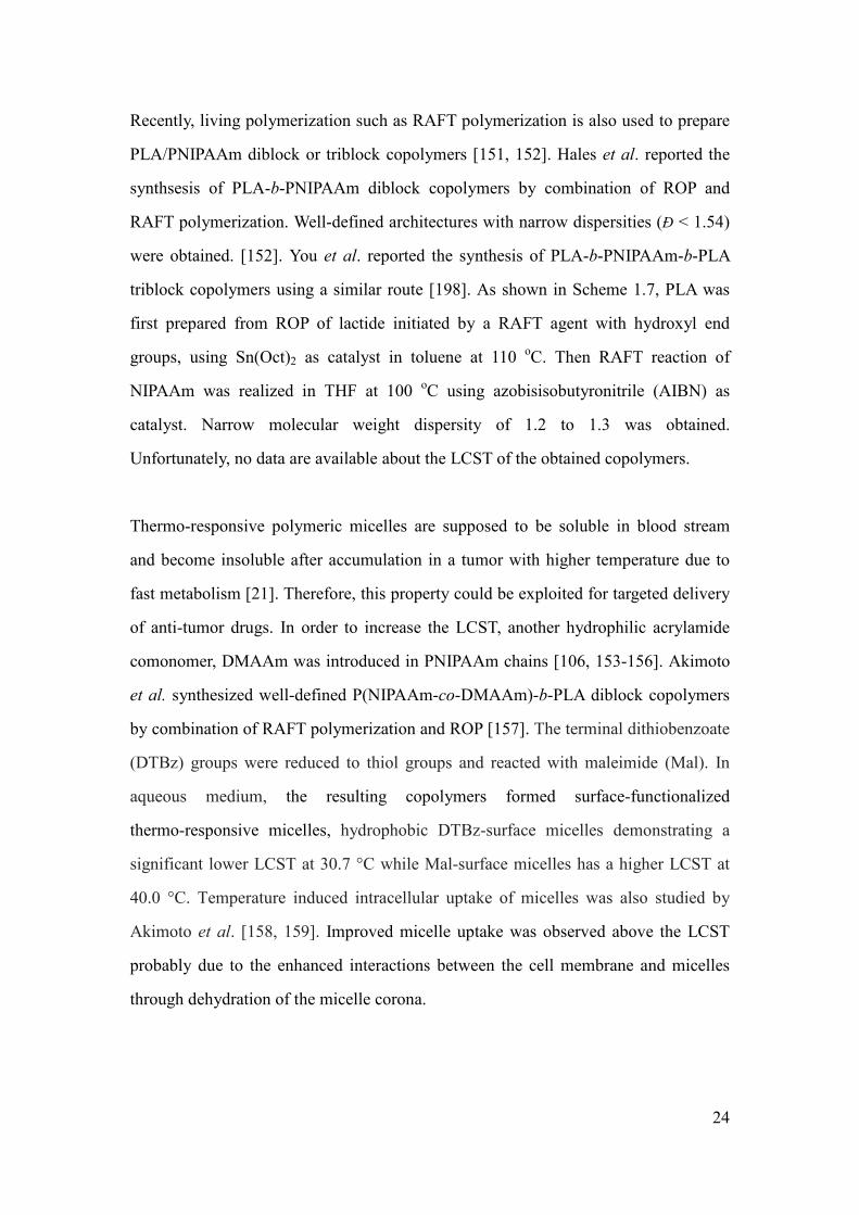
24
Recently, living polymerization such as RAFT polymerization is also used to prepare
PLA/PNIPAAm diblock or triblock copolymers [151, 152]. Hales et al. reported the
synthsesis of PLA-b-PNIPAAm diblock copolymers by combination of ROP and
RAFT polymerization. Well-defined architectures with narrow dispersities (Đ < 1.54)
were obtained. [152]. You et al. reported the synthesis of PLA-b-PNIPAAm-b-PLA
triblock copolymers using a similar route [198]. As shown in Scheme 1.7, PLA was
first prepared from ROP of lactide initiated by a RAFT agent with hydroxyl end
groups, using Sn(Oct)2 as catalyst in toluene at 110 oC. Then RAFT reaction of
NIPAAm was realized in THF at 100 oC using azobisisobutyronitrile (AIBN) as
catalyst. Narrow molecular weight dispersity of 1.2 to 1.3 was obtained.
Unfortunately, no data are available about the LCST of the obtained copolymers.
Thermo-responsive polymeric micelles are supposed to be soluble in blood stream
and become insoluble after accumulation in a tumor with higher temperature due to
fast metabolism [21]. Therefore, this property could be exploited for targeted delivery
of anti-tumor drugs. In order to increase the LCST, another hydrophilic acrylamide
comonomer, DMAAm was introduced in PNIPAAm chains [106, 153-156]. Akimoto
et al. synthesized well-defined P(NIPAAm-co-DMAAm)-b-PLA diblock copolymers
by combination of RAFT polymerization and ROP [157]. The terminal dithiobenzoate
(DTBz) groups were reduced to thiol groups and reacted with maleimide (Mal). In
aqueous medium, the resulting copolymers formed surface-functionalized
thermo-responsive micelles, hydrophobic DTBz-surface micelles demonstrating a
significant lower LCST at 30.7 °C while Mal-surface micelles has a higher LCST at
40.0 °C. Temperature induced intracellular uptake of micelles was also studied by
Akimoto et al. [158, 159]. Improved micelle uptake was observed above the LCST
probably due to the enhanced interactions between the cell membrane and micelles
through dehydration of the micelle corona.

25
Thermo-responsive release of doxorubicin from P(NIPAAm-co-DMAAm)-b-PLA has
been investigated by Kohori et al. [160]. No cytotoxicity of micelles was observed at
37 or 42.5 °C. Enhanced interactions between micelles and cells above the LCST
seem to increase the incorporation rate of drug loaded micelles into cells. Liu et al.
reported in vitro release of doxorubicin from
poly(N-isopropylacrylamide-co-N,N-dimethylacrylamide)-b-poly(D,L-lactide
-co-glycolide) [P(NIPAAm-co-DMAAm)-b-PLGA] micelles. Confocal laser scanning
microscopy revealed that free drug molecules enter cell nuclei very fast, and that drug
loaded micelles accumulate mostly in cytoplasm after endocytosis. At a temperature
above the LCST, more drug molecules release from the micelles and enter the nuclei
as compared to the case below the LCST. Drug loaded micelles exhibit greater
cytotoxicity at a temperature above the LCST due to the collapse of micelles. Later,
Li et al. reported similar maleimide end-functional P(NIPAAm-co-DMAAm)-b-PLA
and poly(N-isopropylacrylamide-co-N,N-dimethylacrylamide-b-ε-caprolactone)
(P(NIPAAm-co-DMAAm)-b-PCL) copolymers with LCST of 39 and 40.5 oC,
respectively [108]. Doxorubicin was also used as model to investigate the drug release
behavior. Comparing with the normal physiological conditions (pH=7.4, 37 oC), drug
release and hydrophobic core degradation were obviously enhanced under simulated
tumor tissue conditions (pH=5.3, T=40 oC). Both flow cytometry and fluorescent
microscopy showed that doxorubicin released from micelles is about 4 times higher
than that by the commercial formulation Taxotere®
. In vitro cytotoxicity assay against
N-87 cancer cell and confocal laser scanning microscopy (CLSM) also confirmed
enhanced drug efficiency. However, the drug loading is quite low (<5 %). Besides,
whether the drug release is controlled by thermo-responsive phase transition or
degradation is uncertain. In fact, PLA shows faster degradation at pH=5.3 and 40 °C
than at pH=7.4 and 37 °C.

26
1.3.3 Thermo-responsive copolymers from PLA/PEG
analogues
1.3.3.1 PEG analogues
Recently, thermo-responsive polymers containing short oligo(ethylene glycol) side
chains of different chain lengths were proposed as an attractive alternative to
PNIPAAm [161-167]. These PEG analogues exhibit not only the properties such as
hydrophilicity and biocompatibility, but also thermo-sensitivity. They are regarded as
a new generation of “smart” biocompatible materials. Zhou et al. reported microgels
based on oligo(ethylene glycol) methacrylate (OEGMA) for drug delivery
applications. The microgels are nontoxic and released drug molecules remain active to
kill cancer cells [168].
Figure 1.4 Lower critical solution temperature (LCST) of PEG analogues as a
function of the content of OEGMA units per chain. From ref. [170].
PEG macromonomers, i.e. molecules composed of a polymerizable moiety connected
to a short OEG chain were firstly reported by Neugebauer in 1980s [169]. Novel
macromolecular architectures with PEG side chains were obtained. The significant
advantage of PEG macromomomers is the possibility to access high molecular weight
PEG based copolymers using mild conditions. Random copolymers of

27
2-(2-methoxyethoxy) ethyl methacrylate (MEO2MA) and OEGMA (Mn=475) exhibit
a LCST value between 26 and 90 oC, which could be precisely adjusted by varying
the comonomer composition (Figure 1.4) [170]. For example, LCST values of either
32.8 oC (comparable to the standard LCST of PNIPAAm), 37.8
oC (body temperature)
or 39–40.8 oC (fever temperatures) were obtained in pure water for copolymers
possessing in average 5, 8, or 10% of OEGMA units, respectively.
The solubility of PEG analogues is dependent on the number of repeat units of PEG
side chains. These non linear PEG analogues could be insoluble in water, or soluble
up to 100 oC. Poly(2-methoxy ethyl) methacrylate (PMEOMA) with only 1 EO unit is
not soluble in water at room temperature. PEG analogues with side chain length
between 2 and 10 exhibit a LCST in aqueous solution. As the length increases,
polymers derived from MEO2MA, MEO3MA and OEGMA (5 to 9 units) showed a
LCST at 26, 52 and 60-90 oC, respectively [161, 171].
Scheme 1.8 Phase transition mechanism of PEG analogues in aqueous medium. From
ref. [172].
In fact, similar to PNIPAAm, the phase transition is also related to the balance
between hydrophilic and hydrophobic moieties in the macromolecular structures. As
shown in Scheme 1.8, hydrogen bonding between the ether oxygen of PEG and
surrounding water molecules is the driving force for the solubilization of PEG
analogues at room temperature [172-174]. This solubilizing effect is counterbalanced
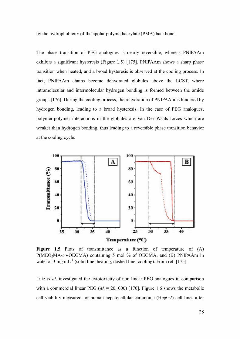
28
by the hydrophobicity of the apolar polymethacrylate (PMA) backbone.
The phase transition of PEG analogues is nearly reversible, whereas PNIPAAm
exhibits a significant hysteresis (Figure 1.5) [175]. PNIPAAm shows a sharp phase
transition when heated, and a broad hysteresis is observed at the cooling process. In
fact, PNIPAAm chains become dehydrated globules above the LCST, where
intramolecular and intermolecular hydrogen bonding is formed between the amide
groups [176]. During the cooling process, the rehydration of PNIPAAm is hindered by
hydrogen bonding, leading to a broad hysteresis. In the case of PEG analogues,
polymer-polymer interactions in the globules are Van Der Waals forces which are
weaker than hydrogen bonding, thus leading to a reversible phase transition behavior
at the cooling cycle.
Figure 1.5 Plots of transmittance as a function of temperature of (A)
P(MEO2MA-co-OEGMA) containing 5 mol % of OEGMA, and (B) PNIPAAm in
water at 3 mg mL-1
(solid line: heating, dashed line: cooling). From ref. [175].
Lutz et al. investigated the cytotoxicity of non linear PEG analogues in comparison
with a commercial linear PEG (Mn = 20, 000) [170]. Figure 1.6 shows the metabolic
cell viability measured for human hepatocellular carcinoma (HepG2) cell lines after
29
incubation at 37.8 oC in the presence of PEG and P(MEO2MA-co-OEGMA)
containing 10, 30 and 100 mol % of OEGMA. In all cases, in vitro cell assay
demonstrates excellent cell viability, thus showing that PEG analogues are apparently
not cytotoxic as linear PEG.
Figure 1.6 Metabolic cell viability measured for human hepatocellular carcinoma
(HepG2) cell lines incubated at 37.8 oC in the presence of PEG and
P(MEO2MA-co-OEGMA)of various compositions. From ref. [170].
Free radical polymerization is the most versatile method for the synthesis of non-liner
PEG analogues [169]. The resulting polymers could be promising for biomedical
applications as they are principally composed of biocompatible OEG segments.
However, the polymers prepared from radical polymerization are characterized by
high dispersity with poorly controlled molecular weight and architecture.
Han et al. reported homopolymers and copolymers of MEO2MA and MEO3MA
synthesized by living anionic polymerization [161]. The obtained polymers have
narrow molecular weight distribution (Đ<1.1). The LCST tends to decrease with
increasing the Mn of polymers. In addition, polymers with higher molecular weights
exhibit sharper phase transition at the LCST, suggesting that longer polymer chains
easily forms the intermolecular and intramolecular aggregation at lower temperature,
which results in faster phase separation.
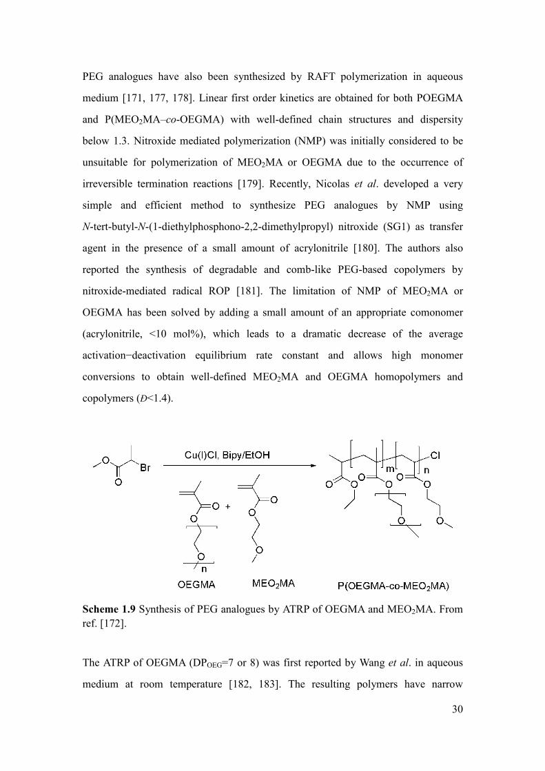
30
PEG analogues have also been synthesized by RAFT polymerization in aqueous
medium [171, 177, 178]. Linear first order kinetics are obtained for both POEGMA
and P(MEO2MA–co-OEGMA) with well-defined chain structures and dispersity
below 1.3. Nitroxide mediated polymerization (NMP) was initially considered to be
unsuitable for polymerization of MEO2MA or OEGMA due to the occurrence of
irreversible termination reactions [179]. Recently, Nicolas et al. developed a very
simple and efficient method to synthesize PEG analogues by NMP using
N-tert-butyl-N-(1-diethylphosphono-2,2-dimethylpropyl) nitroxide (SG1) as transfer
agent in the presence of a small amount of acrylonitrile [180]. The authors also
reported the synthesis of degradable and comb-like PEG-based copolymers by
nitroxide-mediated radical ROP [181]. The limitation of NMP of MEO2MA or
OEGMA has been solved by adding a small amount of an appropriate comonomer
(acrylonitrile, <10 mol%), which leads to a dramatic decrease of the average
activation−deactivation equilibrium rate constant and allows high monomer
conversions to obtain well-defined MEO2MA and OEGMA homopolymers and
copolymers (Đ<1.4).
Scheme 1.9 Synthesis of PEG analogues by ATRP of OEGMA and MEO2MA. From
ref. [172].
The ATRP of OEGMA (DPOEG=7 or 8) was first reported by Wang et al. in aqueous
medium at room temperature [182, 183]. The resulting polymers have narrow

31
molecular weight distribution. Matyjaszewski et al. investigated the successive ATRP
of MEO2MA and MEO3MA in toluene or anisole solution [184, 185]. Recently, Lutz
et al. obtained well-defined thermo-responsive P(MEO2MA-co-OEGMA) copolymers
by ATRP in pure ethanol at 60 oC (Scheme 1.9) [170, 186, 187].
PEG analogues could be further modified by “click chemistry” (Scheme 1.10) [188,
189]. Well-defined POEGMA synthesized by ATRP could be post-functionalized via
copper catalyzed 1, 3-dipolar cycloaddition. In fact, the bromine chain-ends of
POEGMA can be transformed into azide groups and subsequently reacts with alkynes.
For example, click chemistry can be used for the reaction of POEGMA with
functional biopolymers such as oligopeptides [190]. In addition, target molecules such
as folic acid could also be clicked on POEGMA using this method [191, 192].
Scheme 1.10 Strategy for the functionalization of well-defined POEGMA using click
chemistry. From ref. [193].
1.3.3.2 PLA/PEG analogues block copolymer
Although the hydrophilic/hydrophobic balance exists in the case of PEG analogues,
whether the polymers can induce aggregation such as micelles in water still remains
controversial. Ito et al. reported that PEG analogues generally adopt a compact coil
conformation in water. Fluorimetry studies suggest micelle formation of PEG
analogues in water due to weak hydrophobic interactions [193-195].
In the case of P(OEGMA-co-MEO2MA) copolymers, the bimodal Dynamic laser

32
scattering (DLS) intensity distribution indicated coexistence of free polymer coils of
3.7 nm and larger aggregates of 150 nm (Figure 1.7). However, the latter are indeed a
negligible minority, i.e. less than 0.01 % of the overall volume distribution (Figure
1.7B). These findings confirm that the conformation of P(OEGMA-co-MEO2MA) is
predominantly random coil in water. Hence, hydrophobic blocks should be introduced
in PEG analogue chains so as to obtain amphiphilic copolymers for applications in the
field of drug delivery.
Figure 1.7 Intensity size distribution (A) and volume size distribution (B) of
P(MEO2MA-co-OEGMA) determined by DLS at room temperature at 3 mg mL-1
in
aqueous solution. From ref. [172].
Saeed prepared PLGA-b-OEGMA copolymers by combining ROP and RAFT
polymerization [196]. The resulting copolymers self-assembled to form micelles able
to encapsulate carboxyfluorescein and fluorescein isothiocyanate as model of
hydrophobic drugs. Selected copolymer micelles were incubated with 3T3 fibroblasts
as a model cell line, and were rapidly taken up as indicated by fluorescence
microscopy assays. Later on, Luzon et al. obtained POEGMA-b-PCL-b-POEGMA
triblock copolymers by ATRP, using α,ω-bromopropionyl Br-PCL-Br as
macroinitiator [197]. The CMC and critical micelle temperature (CMT) were

33
determined. Self-assembly is temperature and concentration dependent. Decrease of
the CMT is observed as the concentration increases. The same synthetic route was
adopted by Bakkour et al. for the synthesis of POEGMA-b-PLLA-b-POEGMA
triblock copolymers [198]. The self-assembly and drug encapsulation potential of the
copolymers were reported. Most recently, Fenyves et al. synthesized amphiphilic
bottlebrush block copolymers containing PLA and PEG side chains using a
grafting-from method, and studied their self-assembly in aqueous medium [199].
Spherical, cylindrical micelles as well as bilayer structures were observed. However,
the copolymers shown above exhibit very high LCST (>80 °C) and no drug release
was reported.
1.4 Summary and work plan
PLA/PEG amphiphilic copolymer micelles have been widely studied for biomedical
applications due to the passive targeted drug delivery by the EPR effect. However,
these micelles present no active stimuli response to environmental changes. The ideal
intelligent drug carrier should be stimuli responsive to the tumor so as to improve the
drug efficacy. It is well known that fast metabolism of tumor cells after mutation
makes the tumor issue very different from normal tissues with higher temperature
(~42 oC), lower pH (pH~5.3) and anomalistic vessel (intercellular gap junction=
300-700 nm) [15, 200]. Therefore, novel thermo-responsive micelles based on
PNIPAAm or PEG analogues have drawn much attention for active targeted delivery
of anti-tumor drugs. However, only PLA-b-PNIPAAm diblock copolymers were
obtained from combination of ROP and radical polymerization, and few
PLA-b-PNIPAAm-b-PLA triblock copolymers were prepared from combination of
ROP and RAFT polymerization. Moreover, the LCST of these copolymers could not
be precisely controlled around the body temperature, which limits the potential
applications in targeted drug delivery.

34
Herein, in this work, PNIPAAm-b-PLLA-b-PNIPAAm triblock copolymers are first
prepared by combination of ROP and ATRP, using a Br-PLLA-Br macroinitiator.
Then, a comonomer, DMAAm, is introduced to copolymer chains in order to increase
and precisely adjust the LCST of the resulting
P(NIPAAm-co-DMAAm)-b-PLLA-b-P(NIPAAm-co-DMAAm) copolymers. The
physico-chemical properties of the various copolymers and self-assembly behavior
are fully characterized by NMR, SEC, UV, DLS and TEM, etc. An anticancer drug,
curcumin, is used as model drug to monitor the drug encapsulation and release
properties of micelles under physiological conditions.
In the case of thermo-responsive PLA/PEG analogues reported in literature, the LCST
is much higher than the body temperature. Moreover, to the best of our knowledge, no
drug release data are available for these micelles. In this contribution,
P(OEGMA-co-MEO2MA)-b-PLLA-b-P(OEGMA-co-MEO2MA) triblock copolymers
are prepared by combination of ROP and ATRP, using the same Br-PLLA-Br
macroinitiator. The self-assembly and drug release properties are also investigated.
Thus, this work should allow to evaluate the potential of two different
thermo-responsive systems for targeted delivery of anti-tumor drugs.
1.5 References
[1] B. W. Stewart, C. P. Wild, World Cancer Report 2014 IARC Nonserial Publication
(2014).
[2] P. Ehrlich, The collected papers of paul Ehrlich. pergamon, London (1960).
[3] S. K. Sahoo, V. Labhasetwar, Nanotech approaches to drug delivery and imaging. Drug
Discov. Today 8 (2003) 1112-1120.
[4] P. Couvreur, C. Vauthier, Nanotechnology: Intelligent design to treat complex disease.
Pharm. Res. 23 (2006) 1417-1450.
[5] D. Peer, J. M. Karp, S. Hong, O. C. FaroKhzad, R. Margalit, R. Langer, Nanocarriers as
an emerging platform for cancer therapy. Nat. Nanotechnol. 2 (2007) 751-760.
[6] M. Sun, X. Su, B. Y. Ding, X. L. He, X. J. Liu, A. H. Yu, H. X. Lou, G. X. Zhai,
Advances in nanotechnology-based delivery systems for curcumin. Nanomedicine 7 (2012)
1085-1100.
[7] M. Beija, R. Salvayre, N. Lauth-de Viguerie, J. D. Marty, Colloidal systems for drug
delivery: from design to therapy. Trends Biotechnol. 30 (2012) 485-496.

35
[8] N. Bertrand, J. Wu, X. Y. Xu, N. Kamaly, O. C. Farokhzad, Cancer nanotechnology: The
impact of passive and active targeting in the era of modern cancer biology. Adv. Drug Deliv.
Rev. 66 (2014) 2-25.
[9] F. Danhier, O. Feron, V. Prat, To exploit the tumor microenvironment: Passive and active
tumor targeting of nanocarriers for anti-cancer drug delivery. J. Controlled Release 148 (2010)
135-146.
[10] H. Hillaireau, P. Couvreur, Nanocarriers' entry into the cell: relevance to drug delivery.
Cell. Mol. Life Sci. 66(17) (2009) 2873-2896.
[11] B. Haley, E. Frenkel, Nanoparticles for drug delivery in cancer treatment. Urol.
Oncol.-Semin. Orig. Investig. 26(1) (2008) 57-64.
[12] H. Maeda, G. Y. Bharate, J. Daruwalla, Polymeric drugs for efficient tumor-targeted drug
delivery based on EPR-effect. Eur. J. Pharm. Biopharm. 71 (2009) 409-419.
[13] H. Maeda, T. Sawa, T. Konno, Mechanism of tumor-targeted delivery of macromolecular
drugs, including the EPR effect in solid tumor and clinical overview of the prototype
polymeric drug SMANCS. J. Controlled Release 74 (2001) 47-61.
[14] A. K. Iyer, G. Khaled, J. Fang, H. Maeda, Exploiting the enhanced permeability and
retention effect for tumor targeting. Drug Disco. Today 11 (2006) 812-818.
[15] R. Duncan, The dawning era of polymer therapeutics. Nat Rev Drug Discov. 2 (2003)
347-360.
[16] J. H. Woodland, S. Yolles, Long-acting delivery systems For Narcotic Antagonists.1. J.
Med. Chem. 16(8) (1973) 897-901.
[17] S. Yolles, T. D. Leafe, J.H. Woodland, F. J. Meyer, Long-acting delivery systems For
Narcotic Antagonists 2. Release rates of naltrexone from poly(lactic acid) Composites. J.
Pharm. Sci. 64 (1975) 348-349.
[18] T. M. Allen, P. R. Cullis, Drug delivery systems: Entering the mainstream. Science 303
(2004) 1818-1822.
[19] R. Langer, Drug delivery and targeting. Nature 392 (1998) 5-10.
[20] K. E. Uhrich, S. M. Cannizzaro, R. S. Langer, K. M. Shakesheff, Polymeric Systems for
Controlled Drug Release. Chem. Rev.99 (1999) 3181-3198.
[21] A. Chilkoti, M. R. Dreher, D. E. Meyer, D. Raucher, Targeted drug delivery by thermally
responsive polymers. Adv. Drug Deliv. Rev. 54 (2002) 613-630.
[22] A. K. Bajpai, S. K. Shukla, S. Bhanu, S. Kankane, Responsive polymers in controlled
drug delivery. Prog. Polym. Sci. 33 (2008) 1088-1118.
[23] S. Ganta, H. Devalapally, A. Shahiwala, M. Amiji, A review of stimuli-responsive
nanocarriers for drug and gene delivery. J. Control. Release 126 (2008) 187-204.
[24] V. P. Torchilin, Multifunctional nanocarriers. Adv. Drug Deliv. Rev. 58 (2006)
1532-1555.
[25] D. Schmaljohann, Thermo- and pH-responsive polymers in drug delivery. Adv. Drug
Deliv. Rev. 58 (2006) 1655-1670.
[26] C. D. H. Alarcon, S. Pennadam, C. Alexander, Stimuli responsive polymers for
biomedical applications. Chem. Soc. Rev. 34 (2005) 276-285.
[27] E. S. Gil, S. M. Hudson, Stimuli-reponsive polymers and their bioconjugates. Prog.
Polym. Sci. 29((2004) 1173-1222.
[28] F. H. Meng, Z. Y. Zhong, J. Feijen, Stimuli-Responsive Polymersomes for Programmed

36
Drug Delivery. Biomacromolecules 10 (2009) 197-209.
[29] N. Rapoport, Physical stimuli-responsive polymeric micelles for anti-cancer drug
delivery. Prog. Polym. Sci. 32 (2007) 962-990.
[30] D. Roy, W. L. A. Brooks, B. S. Sumerlin, New directions in thermoresponsive polymers.
Chem. Soc. Rev. 42 (2013) 7214-7243.
[31] I. S. Kim, Y. I. Jeong, C. S. Cho, S. H. Kim, Thermo-responsive self-assembled
polymeric micelles for drug delivery in vitro. Inter.l J. Pharm. 205 (2000) 165-172.
[32] G, Gregoria, E. J. Wills, C. P. Swain, A. S. Tavill, Drug carrier potential of liposomes in
cancer chemotherap. Lancet 1 (1974) 1313-1316.
[33] W. E. Bawarski, E. Chidlowsky, D. J. Bharali, S. A. Mousa, Emerging
nanopharmaceuticals. Nanomed. Nanotechnol. Biol. Med. 4 (2008) 273-282.
[34] A. Bochot, E. Fattal, Liposomes for intravitreal drug delivery: A state of the art. J.
Controlled Release 161 (2012) 628-634.
[35] D. J. Bharali, S. A. Mousa, Emerging nanomedicines for early cancer detection and
improved treatment: Current perspective and future promise. Pharmacol. Ther. 128(2) (2010)
324-335.
[36] T. Lammers, F. Kiessling, W. E. Hennink, G. Storm, Nanotheranostics and Image-Guided
Drug Delivery: Current Concepts and Future Directions. Molecular Pharmaceutics 7(6) (2010)
1899-1912.
[37] R. B. Campbell, D. Fukumura, E. B. Brown, L. M. Mazzola, Y. Izumi, R. K. Jain, V. P.
Torchilin, L. L. Munn, Cationic charge determines the distribution of liposomes between the
vascular and extravascular compartments of tumors. Cancer Res. 62 (2002) 6831-6836.
[38] B. Mishra, B. B. Patel, S. Tiwari, Colloidal nanocarriers: a review on formulation
technology, types and applications toward targeted drug delivery. Nanomedicine:
Nanotechnology, Biology and Medicine 6 (2009) 9-24.
[39] S. Unezaki, K. Maruyama, O. Ishida, A. Suginaka, J. Hosoda, M. Iwatsuru, Enhanced
tumor targeting and improved antitumor activity of doxorubicin by long-circulating liposomes
containing amphipathic poly(ethylene glycol). Inter. J. Pharm. 126 (1995) 41-48.
[40] T. Daemen, J. Regts, M. Meesters, M. T. TenKate, I. BakkerWoudenberg, G. L.
Scherphof, Toxicity of doxorubicin entrapped within long-circulating liposomes. J. Controlled
Release 44 (1997) 1-9.
[41] C. J. F. Rijcken, O. Soga, W. E. Hennink, C. F. van Nostrum, Triggered destabilisation of
polymeric micelles and vesicles by changing polymers polarity: An attractive tool for drug
delivery. J. Controlled Release 120 (2007) 131-148.
[42] K. K. Upadhyay, A. N. Bhatt, A. K. Mishra, B. S. Dwarakanath, S. Jain, C. Schatz, J. F.
Le Meins, A. Farooque, G. Chandraiah, A. K. Jain, A. Misra, S. Lecommandoux, The
intracellular drug delivery and anti tumor activity of doxorubicin loaded poly(gamma-benzyl
L-glutamate)-b-hyaluronan polymersomes. Biomaterials 31 (2010) 2882-2892.
[43] D. E. Discher, A. Eisenberg, Polymer vesicles. Science 297 (2002) 967-973.
[44] F. Ahmed, D. E. Discher, Self-porating polymersomes of PEG-PLA and PEG-PCL:
hydrolysis-triggered controlled release vesicles. J. Controlled Release 96 (2004) 37-53.
[45] K. Kita-Tokarczyk, J. Grumelard, T. Haefele, W. Meier, Block copolymer vesicles using
concepts from polymer chemistry to mimic biomembranes. Polymer 46 (2005) 3540-3563.
[46] M. Massignani, H. Lomas, G. Battaglia, Modern Techniques for Nano- and
37
Microreactors/-Reactions, Springer-Verlag Berlin, Berlin,Vol. 229, (2010) pp. 115-154.
[47] R. P. Brinkhuis, F. Rutjes, J. C. M. van Hest, Polymeric vesicles in biomedical
applications. Polym. Chem. 2 (2011) 1449-1462.
[48] S. L. Li, B. Byrne, J. Welsh, A. F. Palmer, Self-assembled
poly(butadiene)-b-poly(ethylene oxide) polymersomes as paclitaxel carriers. Biotechnol. Prog.
23 (2007) 278-285.
[49] F. Ahmed, R. I. Pakunlu, A. Brannan, F. Bates, T. Minko, D. E. Discher, Biodegradable
polymersomes loaded with both paclitaxel and doxorubicin permeate and shrink tumors,
inducing apoptosis in proportion to accumulated drug. J. Controlled Release 116 (2006)
150-158.
[50] C. Sanson, C. Schatz, J. F. Le Meins, A. Soum, J. Thevenot, E. Garanger, S.
Lecommandoux, A simple method to achieve high doxorubicin loading in biodegradable
polymersomes. J. Controlled Release 147 (2010) 428-435.
[51] M. Marguet, L. Edembe, S. Lecommandoux, Polymersomes in Polymersomes: Multiple
Loading and Permeability Control. Angew. Chem.-Int. Edit. 51(5) (2012) 1173-1176.
[52] D. M. Saylor, C. S. Kim, D. V. Patwardhan, J. A. Warren, Diffuse-interface theory for
structure formation and release behavior in controlled drug release systems. Acta
Biomaterialia 3 (2007) 851-864.
[53] J. S. Lee, J. Feijen, Polymersomes for drug delivery: Design, formation and
characterization. J. Controlled Release 161 (2012) 473-483.
[54] A. Mecke, C. Dittrich, W. Meier, Biomimetic membranes designed from amphiphilic
block copolymers. Soft Matter 2 (2006) 751-759.
[55] W. Chen, F. H. Meng, R. Cheng, Z. Y. Zhong, pH-Sensitive degradable polymersomes
for triggered release of anticancer drugs: A comparative study with micelles. J. Controlled
Release 142 (2010) 40-46.
[56] Y. F. Du, W. Chen, M. Zheng, F. H. Meng, Z.Y. Zhong, pH-sensitive degradable
chimaeric polymersomes for the intracellular release of doxorubicin hydrochloride.
Biomaterials 33 (2012) 7291-7299.
[57] S. B. Lecommandoux, O. Sandre, F. Checot, J. Rodriguez-Hernandez, R. Perzynski,
Magnetic nanocomposite micelles and vesicles. Adv. Mater. 17 (2005) 712-+.
[58] X. Tong, G. Wang, A. Soldera, Y. Zhao, How can azobenzene block copolymer vesicles
be dissociated and reformed by light? J. Phys. Chem. B 109 (2005) 20281-20287.
[59] S. H. Qin, Y. Geng, D. E. Discher, S. Yang, Temperature-controlled assembly and release
from polymer vesicles of poly(ethylene oxide)-block-poly(N-isopropylacrylamide). Adv.
Mater. 18 (2006) 2905-2909.
[60] O. Onaca, R. Enea, D. W. Hughes, W. Meier, Stimuli-Responsive Polymersomes as
Nanocarriers for Drug and Gene Delivery. Macromol. Biosci. 9 (2009) 129-139.
[61] W. Agut, A. Brulet, C. Schatz, D. Taton, S. Lecommandoux, pH and Temperature
Responsive Polymeric Micelles and Polymersomes by Self-Assembly of
Poly[2-(dimethylamino)ethyl methacrylate]-b-Poly(glutamic acid) Double Hydrophilic Block
Copolymers. Langmuir 26 (2010) 10546-10554.
[62] H. De Oliveira, J. Thevenot, S. Lecommandoux, Smart polymersomes for therapy and
diagnosis: fast progress toward multifunctional biomimetic nanomedicines. Wiley Interdiscip.
Rev.-Nanomed. Nanobiotechnol. 4 (2012) 525-546.
38
[63] H. Oliveira, E. Perez-Andres, J. Thevenot, O. Sandre, E. Berra, S. Lecommandoux,
Magnetic field triggered drug release from polymersomes for cancer therapeutics. J.
Controlled Release 169 (2013) 165-170.
[64] L. Pourtau, H. Oliveira, J. Thevenot, Y. L. Wan, A. R. Brisson, O. Sandre, S. Miraux, E.
Thiaudiere, S. Lecommandoux, Antibody-Functionalized Magnetic Polymersomes: In vivo
Targeting and Imaging of Bone Metastases using High Resolution MRI. Adv. Healthc. Mater.
2 (2013) 1420-1424.
[65] C. Sanson, O. Diou, J. Thevenot, E. Ibarboure, A. Soum, A. Brulet, S. Miraux, E.
Thiaudiere, S. Tan, A. Brisson, V. Dupuis, O. Sandre, S. Lecommandoux, Doxorubicin
Loaded Magnetic Polymersomes: Theranostic Nanocarriers for MR Imaging and
Magneto-Chemotherapy. ACS Nano 5 (2011) 1122-1140.
[66] J. Thevenot, H. de Oliveira, O. Sandre, L. Pourtau, E. Andres, S. Miraux, E. Thiaudiere,
E. Berra, S. Lecommandoux, Multifunctional polymersomes for cancer theranostics. J.
Controlled Release 172 (2013) E44-E45.
[67] J. Huang, C. Bonduelle, J. Thevenot, S. Lecommandoux, A. Heise, Biologically Active
Polymersomes from Amphiphilic Glycopeptides. J. Am. Chem. Soc. 134 (2012) 119-122.
[68] E. Garanger, S. Lecommandoux, Towards Bioactive Nanovehicles Based on Protein
Polymers. Angew. Chem.-Int. Edit. 51 (2012) 3060-3062.
[69] M. Marguet, O. Sandre, S. Lecommandoux, Polymersomes in "Gelly" Polymersomes:
Toward Structural Cell Mimicry. Langmuir 28 (2012) 2035-2043.
[70] M. Chemin, P. M. Brun, S. Lecommandoux, O. Sandre, J. F. Le Meins, Hybrid
polymer/lipid vesicles: fine control of the lipid and polymer distribution in the binary
membrane. Soft Matter 8 (2012) 2867-2874.
[71] J. F. Le Meins, C. Schatz, S. Lecommandoux, O. Sandre, Hybrid polymer/lipid vesicles:
state of the art and future perspectives. Mater. Today 16 (2014) 397-402.
[72] A. Kumari, S. K. Yadav, S. C. Yadav, Biodegradable polymeric nanoparticles based drug
delivery systems. Colloid Surf. B-Biointerfaces 75 (2010) 1-18.
[73] P. Anand, H.B. Nair, B. Sung, A.B. Kunnumakkara, V.R. Yadav, R.R. Tekmal, B.B.
Aggarwal, Design of curcumin-loaded PLGA nanoparticles formulation with enhanced
cellular uptake, and increased bioactivity in vitro and superior bioavailability in vivo.
Biochem. Pharma. 79 (2009) 330-338.
[74] P. Couvreur, Nanoparticles in drug delivery: Past, present and future. Adv. Drug Deliv.
Rev. 65 (2012) 21-23.
[75] S. Parveen, R. Misra, S.K. Sahoo, Nanoparticles: a boon to drug delivery, therapeutics,
diagnostics and imaging. Nanomed.: Nanotech., Biology and Medicine 8 (2011) 147-166.
[76] H. D. Tang, C. J. Murphy, B. Zhang, Y. Q. Shen, M. H. Sui, E. A. Van Kirk, X. W. Feng,
W. J. Murdoch, Amphiphilic curcumin conjugate-forming nanoparticles as anticancer prodrug
and drug carriers: in vitro and in vivo effects. Nanomedicine 5 (2010) 855-865.
[77] J. M. Harris, R. B. Chess, Effect of pegylation on pharmaceuticals. Nat. Rev. Drug
Discov. 2 (2003) 214-221.
[78] G. Molineux, Pegylation: engineering improved pharmaceuticals for enhanced therapy.
Cancer Treat. Rev. 28 (2002) 13-16.
[79] A. H. Faraji, P. Wipf, Nanoparticles in cellular drug delivery. Bioorg. Med. Chem. 17
(2009) 2950-2962.
39
[80] H. Bader, H. Ringsdorf, B. Schmidt, Watersoluble polymer medicine. Angew. Makromol.
Chem. 123 (1984) 457-485.
[81] G. S. Kwon, K. Kataoka, Block-copolymer micelles as long-circulating drug vesicles.
Adv. Drug Deliv. Rev. 16 (1995) 295-309.
[82] M. C. Jones, J. C. Leroux, Polymeric micelles - a new generation of colloidal drug
carriers. Eur. J. Pharm. Biopharm. 48 (1999) 101-111.
[83] K. Letchford, H. Burt, A review of the formation and classification of amphiphilic block
copolymer nanoparticulate structures: micelles, nanospheres, nanocapsules and polymersomes.
Eur. J. Pharm. Biopharm. 65 (2007) 259-269.
[84] K. Kataoka, A. Harada, Y. Nagasaki, Block copolymer micelles for drug delivery: design,
characterization and biological significance. Adv. Drug Deliv. Rev. 47 (2001) 113-131.
[85] M. L. Adams, A. Lavasanifar, G. S. Kwon, Amphiphilic block copolymers for drug
delivery. Journal of Pharmaceutical Sciences 92 (2003) 1343-1355.
[86] B. Jeong, Y. H. Bae, D. S. Lee, S. W. Kim, Biodegradable block copolymers as injectable
drug-delivery systems. Nature 388 (1997) 860-862.
[87] A. Rosler, G. W. M. Vandermeulen, H. A. Klok, Advanced drug delivery devices via
self-assembly of amphiphilic block copolymers. Adv. Drug Deliv. Rev. 53 (2001) 95-108.
[88] G. Gaucher, M. H. Dufresne, V. P. Sant, N. Kang, D. Maysinger, J. C. Leroux, Block
copolymer micelles: preparation, characterization and application in drug delivery. J.
Controlled Release 109 (2005) 169-188.
[89] C. O. Rangel-Yagui, A. Pessoa, L. C. Tavares, Micellar solubilization of drugs. J. Pharm.
Pharm. Sci. 8 (2005) 147-163.
[90] Y. Tian, S. R. Mao, Amphiphilic polymeric micelles as the nanocarrier for peroral
delivery of poorly soluble anticancer drugs. Expert Opin. Drug Deliv. 9 (2012) 687-700.
[91] Y. Yu, X. L. Zhang, L. Y. Qiu, The anti-tumor efficacy of curcumin when delivered by
size/charge-changing multistage polymeric micelles based on amphiphilic poly(beta-amino
ester) derivates. Biomaterials 35 (2014) 3467-3479.
[92] L. Yang, X. H. Wu, F. Liu, Y. R. Duan, S. M. Li, Novel Biodegradable
Polylactide/poly(ethylene glycol) Micelles Prepared by Direct Dissolution Method for
Controlled Delivery of Anticancer Drugs. Pharm. Res. 26 (2009) 2332-2342.
[93] L. Yang, X. Qi, P. Liu, A. El Ghzaoui, S. M. Li, Aggregation behavior of self-assembling
polylactide/poly(ethylene glycol) micelles for sustained drug delivery. Inter. J. Pharm. 394
(2010) 43-49.
[94] U. Kedar, P. Phutane, S. Shidhaye, V. Kadam, Advances in polymeric micelles for drug
delivery and tumor targeting. Nanomed.: Nanotech., Biology and Medicine 6 (2010) 714-729.
[95] Y. Yan, G. K. Such, A. P. R. Johnston, J. P. Best, F. Caruso, Engineering Particles for
Therapeutic Delivery: Prospects and Challenges. ACS Nano 6 (2012) 3663-3669.
[96] C. Oerlemans, W. Bult, M. Bos, G. Storm, J. F. Nijsen, W. Hennink, Polymeric Micelles
in Anticancer Therapy: Targeting, Imaging and Triggered Release. Pharm. Res. 27 (2010)
2569-2589.
[97] I. Hevus, A. Modgil, J. Daniels, A. Kohut, C. Sun, S. Stafslien, A. Voronov, Invertible
Micellar Polymer Assemblies for Delivery of Poorly Water-Soluble Drugs.
Biomacromolecules 13 (2012) 2537-2545.
[98] K. Letchford, R. Liggins, H. Burt, Pharmaceutics, preformulation and drug delivery.

40
Journal of Pharmaceutical Sciences 97 (2008) 1179-1190.
[99] H. Maeda, J. Wu, T. Sawa, Y. Matsumura, K. Hori, Tumor vascular permeability and the
EPR effect in macromolecular therapeutics: a review. J. Controlled Release 65 (2000)
271-284.
[100] K. Knop, R. Hoogenboom, D. Fischer, U. S. Schubert, Poly(ethylene glycol) in Drug
Delivery: Pros and Cons as Well as Potential Alternatives. Angew. Chem.-Int. Edit. 49 (2010)
6288-6308.
[101] L. Liu, L. Sun, Q. Wu, W. Guo, L. Li, Y. Chen, Y. Li, C. Gong, Z. Qian, Y. Wei,
Curcumin loaded polymeric micelles inhibit breast tumor growth and spontaneous pulmonary
metastasis. Inter. J. of Pharm. 443 (2013) 175-182.
[102] R. L. Yang, S. A. Zhang, D. L. Kong, X. L. Gao, Y. J. Zhao, Z. Wang, Biodegradable
Polymer-Curcumin Conjugate Micelles Enhance the Loading and Delivery of Low-Potency
Curcumin. Pharm. Res. 29 (2012) 3512-3525.
[103]V. A. Sethuraman, M. C. Lee, Y. H. Bae, A biodegradable pH-sensitive micelle system
for targeting acidic solid tumors. Pharm. Res. 25 (2008) 657-666.
[104] C. Deng, Y. Jiang, R. Cheng, F. Meng, Z. Zhong, Biodegradable polymeric micelles for
targeted and controlled anticancer drug delivery: Promises, progress and prospects. Nano
Today 7 (2012) 467-480.
[105] J. Nicolas, S. Mura, D. Brambilla, N. Mackiewicz, P. Couvreur, Design,
functionalization strategies and biomedical applications of targeted
biodegradable/biocompatible polymer-based nanocarriers for drug delivery. Chem. Soc. Rev.
42 (2013) 1147-1235.
[106] S. Q. Liu, N. Wiradharma, S. J. Gao, Y. W. Tong, Y. Y. Yang, Bio-functional micelles
self-assembled from a folate-conjugated block copolymer for targeted intracellular delivery of
anticancer drugs. Biomaterials 28 (2007) 1423-1433.
[107] J. Z. Du, R. K. O'Reilly, Advances and challenges in smart and functional polymer
vesicles. Soft Matter 5 (2009) 3544-3561.
[108] W. Li, J. F. Li, J. Gao, B. H. Li, Y. Xia, Y. C. Meng, Y. S. Yu, H. W. Chen, J. X. Dai, H.
Wang, Y. J. Guo, The fine-tuning of thermosensitive and degradable polymer micelles for
enhancing intracellular uptake and drug release in tumors. Biomaterials 32 (2011) 3832-3844.
[109] A. C. Albertsson, I. Varma, Degradable Aliphatic Polyesters, Vol. 157, Springer Berlin
Heidelberg, 2002, pp. 1-40.
[110] G. S. He, L. L. Ma, J. Pan, S. Venkatraman, ABA and BAB type triblock copolymers of
PEG and PLA: A comparative study of drug release properties and"stealth particle
characteristics. Inter. J. Pharm.334 (2007) 48-55.
[111] Y. Ikada, K. Jamshidi, H. Tsuji, S. H. Hyon, Stereocomplex formation between
enantiomeric poly(lactides). Macromolecules 20 (1987) 904-906.
[112] S. M. Li, M. Vert, Synthesis, characterization, and stereocomplex-induced gelation of
block copolymers prepared by ring-opening polymerization of L(D)-lactide in the presence of
poly(ethylene glycol). Macromolecules 36 (2003) 8008-8014.
[113] Y. Zhang, X. H. Wu, Y. R Han, F. Mo, Y. Duan, S. M Li, Novel thymopentin release
systems prepared from bioresorbable PLA-PEG-PLA hydrogels. Inter. J. Pharm. 386 (2010)
15-22.
[114] L. Yang, A. El Ghzaoui, S. M. Li, In vitro degradation behavior of

41
poly(lactide)-poly(ethylene glycol) block copolymer micelles in aqueous solution. Inter. J.
Pharm. 400 (2010) 96-103.
[115] A. Kozlowski, J. M. Harris, Improvements in protein PEGylation: pegylated interferons
for treatment of hepatitis C. J. Controlled Release 72 (2001) 217-224.
[116] A. Kozlowski, S. A. Charles, J. M. Harris, Development of pegylated interferons for the
treatment of chronic hepatitis C. Biodrugs 15 (2001) 419-429.
[117] R. Gref, M. Luck, P. Quellec, M. Marchand, E. Dellacherie, S. Harnisch, T. Blunk, R. H.
Muller, 'Stealth' corona-core nanoparticles surface modified by polyethylene glycol (PEG):
influences of the corona (PEG chain length and surface density) and of the core composition
on phagocytic uptake and plasma protein adsorption. Colloid Surf. B-Biointerfaces 18 (2000)
301-313.
[118] S. I. Jeon, J. H. Lee, J. D. Andrade, P. G. Degennes, Protein surface interactions in the
presence of polyethylene oxide 1. Simplified theory. J. Colloid Interface Sci. 142 (1991)
149-158.
[119] T. Riley, C. R. Heald, S. Stolnik, M. C. Garnett, L. Illum, S. S. Davis, S. M. King, R. K.
Heenan, S. C. Purkiss, R. J. Barlow, P. R. Gellert, C. Washington, Core-shell structure of
PLA-PEG nanoparticles used for drug delivery. Langmuir 19 (2003) 8428-8435.
[120] T. Govender, T. Riley, T. Ehtezazi, M. C. Garnett, S. Stolnik, L. Illum, S. S. Davis,
Defining the drug incorporation properties of PLA-PEG nanoparticles. Inter. J. Pharm. 199
(2000) 95-110.
[121] T. Riley, S. Stolnik, C. R. Heald, C. D. Xiong, M. C. Garnett, L. Illum, S. S. Davis, S. C.
Purkiss, R. J. Barlow, P. R. Gellert, Physicochemical evaluation of nanoparticles assembled
from poly(lactic acid)-poly(ethylene glycol) (PLA-PEG) block copolymers as drug delivery
vehicles. Langmuir 17 (2001) 3168-3174.
[122] S. S. Venkatraman, P. Jie, F. Min, B. Y. C. Freddy, G. Leong-Huat, Micelle-like
nanoparticles of PLA-PEG-PLA triblock copolymer as chemotherapeutic carrier. Inter. J.
Pharm. 298 (2005) 219-232.
[123] R. T. Liggins, H. M. Burt, Polyether-polyester diblock copolymers for the preparation
of paclitaxel loaded polymeric micelle formulations. Adv. Drug Deliv. Rev. 54 (2002)
191-202.
[124] E. Blanco, E. A. Bey, Y. Dong, B. D. Weinberg, D. M. Sutton, D. A. Boothman, J. M.
Gao, beta-Lapachone-containing PEG-PLA polymer micelles as novel nanotherapeutics
against NQO1-overexpres sing tumor cells. J. Controlled Release 122 (2007) 365-374.
[125] S. C. Kim, D. W. Kim, Y. H. Shim, J. S. Bang, H. S. Oh, S. W. Kim, M. H. Seo, In vivo
evaluation of polymeric micellar paclitaxel formulation: toxicity and efficacy. J. Controlled
Release 72 (2001) 191-202.
[126] J. Hrkach, D. Von Hoff, M. M. Ali, E. Andrianova, J. Auer, T. Campbell, D. De Witt, M.
Figa, M. Figueiredo, A. Horhota, S. Low, K. McDonnell, E. Peeke, B. Retnarajan, A. Sabnis,
E. Schnipper, J. J. Song, Y. H. Song, J. Summa, D. Tompsett, G. Troiano, T. Van Geen Hoven,
J. Wright, P. LoRusso, P. W. Kantoff, N. H. Bander, C. Sweeney, O. C. Farokhzad, R. Langer,
S. Zale, Preclinical Development and Clinical Translation of a PSMA-Targeted Docetaxel
Nanoparticle with a Differentiated Pharmacological Profile. Sci. Transl. Med. 4 (2012)
128-139.
[127] S. Cammas, K. Suzuki, C. Sone, Y. Sakurai, K. Kataoka, T. Okano, Thermo-responsive

42
polymer nanoparticles with a core-shell micelle structure as site-specific drug carriers.
J..Controlled Release 48 (1997) 157-164.
[128] J. Chung, M. Yokoyama, T. Aoyagi, Y. Sakurai, T. Okano, Effect of molecular
architecture of hydrophobically modified poly(N-isopropylacrylamide) on the formation of
thermoresponsive core-shell micellar drug carriers. J. Controlled Release 53 (1998) 119-130.
[129] G. Schild, Poly(N-isopropylacrylamide)-experiment, theory and application. Prog.
Polym. Sci. (1992) 163-249.
[130] S. Fujishige, K. Kubota, I. Ando, Phase-Transition of Aqueous-Solutions of
Poly(N-Isopropylacrylamide) and Poly(N-isopropylmethacrylamide). J. Phys. Chem. 93 (1989)
3311-3313.
[131] Y. Okada, F. Tanaka, Cooperative Hydration, Chain Collapse, and Flat LCST Behavior
in Aqueous Poly(N-isopropylacrylamide) Solutions. Macromolecules 38 (2005) 4465-4471.
[132] Y. Ono, T. Shikata, Hydration and Dynamic Behavior of Poly(N-isopropylacrylamide)s
in Aqueous Solution:A Sharp Phase Transition at the Lower Critical Solution Temperature. J.
Am. Chem. Soc. 128 (2006) 10030-10031.
[133] G. Masci, L. Giacomelli, V. Crescenzi, Atom transfer radical polymerization of
N-isopropylacrylamide. Macromol. Rapid Commun. 25 (2004) 559-564.
[134] J. D. Ye, R. Narain, Water-Assisted Atom Transfer Radical Polymerization of
N-isopropylacrylamide: Nature of Solvent and Temperature. J. Phys. Chem. B 113 (2009)
676-681.
[135] K. W. M. Boere, B. G. Soliman, D. T. S. Rijkers, W. E. Hennink, T. Vermonden,
Thermoresponsive Injectable Hydrogels Cross-Linked by Native Chemical Ligation.
Macromolecules 47 (2014) 2430-2438.
[136] H. W. Ooi, K. S. Jack, H. Peng, A. K. Whittaker, "Click" PNIPAAm hydrogels - a
comprehensive study of structure and properties. Polym. Chemi. 4 (2013) 4788-4800.
[137] P. De, S. R. Gondi, B. S. Sumerlin, Folate-conjugated thermoresponsive block
copolymers: Highly efficient conjugation and solution self-assembly. Biomacromolecules 9
(2008) 1064-1070.
[138] P. J. Pan, M. Fujita, W. Y. Ooi, K. Sudesh, T. Takarada, A. Goto, M. Maeda,
Thermoresponsive Micellization and Micellar Stability of
Poly(N-isopropylacrylamide)-b-DNA Diblock and Miktoarm Star Polymers. Langmuir 28
(2012) 14347-14356.
[139] W. Y. Ooi, M. Fujita, P. J. Pan, H. Y. Tang, K. Sudesh, K. Ito, N. Kanayama, T. Takarada,
M. Maeda, Structural characterization of nanoparticles from thermoresponsive
poly(N-isopropylacrylamide)-DNA conjugate. J. Colloid Interface Sci. 374 (2012) 315-320.
[140] A. Wang, C. Tao, Y. Cui, L. Duan, Y. Yang, J. Li, Assembly of environmental sensitive
microcapsules of PNIPAAm and alginate acid and their application in drug release. J. Colloid
Interface Sci. 332 (2009) 271-279.
[141] Y. Liu, X. Cao, M. Luo, Z. Le, W. Xu, Self-assembled micellar nanoparticles of a novel
star copolymer for thermo and pH dual-responsive drug release. J. Colloid Interface Sci. 329
(2009) 244-252.
[142] C. H. Hu, X. Z. Zhang, L. Zhang, X. D. Xu, R. X. Zhuo, Temperature- and pH-sensitive
hydrogels to immobilize heparin-modified PEI/DNA complexes for sustained gene delivery. J.
Mater. Chem. 19 (2009) 8982-8989.
43
[143] P. van Rijn, H. Park, K. Ozlem Nazli, N.C. Mougin, A. Boker, Self-Assembly Process
of Soft Ferritin-PNIPAAm Conjugate Bionanoparticles at Polar-Apolar Interfaces. Langmuir
29 (2013) 276-284.
[144] N. M. Matsumoto, P. Prabhakaran, L. H. Rome, H. D. Maynard, Smart Vaults:
Thermally-Responsive Protein Nanocapsules. ACS Nano 7 (2013) 867-874.
[145] H. Wei, S. X. Cheng, X. Z. Zhang, R.X. Zhuo, Thermo-sensitive polymeric micelles
based on poly(N-isopropylacrylamide) as drug carriers. Prog. Polym. Sci. 34 (2009) 893-910.
[146] H. Wei, X. Z. Zhang, C. Cheng, S. X. Cheng, R.X. Zhuo, Self-assembled,
thermosensitive micelles of a star block copolymer based on PMMA and PNIPAAm for
controlled drug delivery. Biomaterials 28 (2007) 99-107.
[147] K. Fukashi, S. Kiyotaka, A. Takao, Y. Masayuki, S. Yasuhisa, O. Teruo, Preparation and
characterization of thermally responsive block copolymer micelles comprising
poly(N-isopropylacrylamide-b-DLlactide). J.Controlled Release 55 (1998) 87-98.
[148] J. Qin, Y. S. Jo, J. E. Ihm, D. K. Kim, M. Muhammed, Thermosensitive Nanospheres
with a Gold Layer Revealed as Low-Cytotoxic Drug Vehicles. Langmuir 21 (2005)
9346-9351.
[149] M. K. Eri Ayano, Tsutomu Ishihara, Hideko Kanazawa, Teruo Okano, Poly
(N-isopropylacrylamide)-PLA and PLA blend nanoparticles for temperature-controllable drug
release and intracellular uptake. Colloids Surf. B: Biointerfaces 99 (2012) 67-73.
[150] S. Q. Liu, Y. Y. Yang, X. M. Liu, Y. W. Tong, Preparation and Characterization of
Temperature-Sensitive Poly(N-isopropylacrylamide)-b-poly(d,l-lactide) Microspheres for
Protein Delivery. Biomacromolecules 4 (2003) 1784-1793.
[151] Y. You, C. Hong, W. Wang, W. Lu, C. Pan, Preparation and Characterization of
Thermally Responsive and Biodegradable Block Copolymer Comprised of PNIPAAM and
PLA by Combination of ROP and RAFT Methods. Macromolecules 37 (2004) 9761-9767.
[152] M. Hales, C. Barner-Kowollik, T. P. Davis, M. H. Stenzel, Shell-Cross-Linked Vesicles
Synthesized from Block Copolymers of Poly(D,L-lactide) and Poly(N-isopropylacrylamide)
as Thermoresponsive Nanocontainers. Langmuir (20) (2004) 10809-10817.
[153] F. Kohori, K. Sakai, T. Aoyagi, M. Yokoyama, M. Yamato, Y. Sakurai, T. Okano,
Control of adriamycin cytotoxic activity using thermally responsive polymeric micelles
composed of poly(N-isopropylacrylamide-co-N,N-dimethylacrylamide)-b-poly(d,l-lactide).
Colloids and Surfaces B: Biointerfaces 16 (1999) 195-205.
[154] S. Q. Liu, Y. W. Tong, Y. Y. Yang, Incorporation and in vitro release of doxorubicin in
thermally sensitive micelles made from
poly(N-isopropylacrylamide-co-N,N-dimethylacrylamide)-b-poly(d,l-lactide-co-glycolide)
with varying compositions. Biomaterials 26 (2005) 5064-5074.
[155] S. Q. Liu, Y. Y. Yang, X. M. Liu, Y. W. Tong, Preparation and Characterization of
Temperature-Sensitive Poly(N-isopropylacrylamide)-b-poly(d,l-lactide) Microspheres for
Protein Delivery. Biomacromolecules 4 (2003) 1784-1793.
[156] S. Q. Liu, Y. W. Tong, Y. Y. Yang, Thermally sensitive micelles self-assembled from
poly(N-isopropylacrylamide-co-N,N-dimethylacrylamide)-b-poly(D,L-lactide-co-glycolide)
for controlled delivers of paclitaxel. Mol. Biosyst. 1 (2005) 158-165.
[157] J. Akimoto, M. Nakayama, K. Sakai, T. Okano, Molecular Design of Outermost Surface
Functionalized Thermoresponsive Polymeric Micelles with Biodegradable Cores. J. Polym.

44
Sci. Part a-Polym. Chemi. 46 (2008) 7127-7137.
[158] J. Akimoto, M. Nakayama, K. Sakai, T. Okano, Thermally Controlled Intracellular
Uptake System of Polymeric Micelles Possessing Poly(N-isopropylacrylamide)-Based Outer
Coronas. Mole. Pharm. 7 (2010) 926-935.
[159] J. Akimoto, M. Nakayama, K. Sakai, T. Okano, Temperature-Induced Intracellular
Uptake of Thermoresponsive Polymeric Micelles. Biomacromolecules 10 (2009) 1331-1336.
[160] F. Kohori, K. Sakai, T. Aoyagi, M. Yokoyama, M. Yamato, Y. Sakurai, T. Okano,
Control of adriamycin cytotoxic activity using thermally responsive polymeric micelles
composed of poly(N-isopropylacrylamide-co-N,N-dimethylacrylamide)-b-poly(d,l-lactide).
Colloids and Surfaces B: Biointerfaces 16 (1999) 195-205.
[161] S. Han, M. Hagiwara, T. Ishizone, Synthesis of Thermally Sensitive Water-Soluble
Polymethacrylates by Living Anionic Polymerizations of Oligo(ethylene glycol) Methyl Ether
Methacrylates. Macromolecules 36 (2003) 8312-8319.
[162] H. Kitano, T. Hirabayashi, M. Gemmei-Ide, M. Kyogoku, Effect of macrocycles on the
temperature-responsiveness of poly[(methoxy diethylene glycol methacrylate)-graft-PEG].
Macromol. Chem. Phys. 205 (2004) 1651-1659.
[163] M. M. Ali, H. D. H. Stover, Well-defined amphiphilic thermosensitive copolymers
based on poly(ethylene glycol monomethacrylate) and methyl methacrylate prepared by atom
transfer radical polymerization. Macromolecules 37 (2004) 5219-5227.
[164] J. A. Jones, N. Novo, K. Flagler, C.D. Pagnucco, S. Carew, C. Cheong, X. Z. Kong, N.
A. D. Burke, H. D. H. Stover, Thermoresponsive copolymers of methacrylic acid and
poly(ethylene glycol) methyl ether methacrylate. J. Polym. Sci. Pol. Chem. 43 (2005)
6095-6104.
[165] S. Sugihara, S. Kanaoka, S. Aoshima, Double thermosensitive diblock copolymers of
vinyl ethers with pendant oxyethylene groups: Unique physical gelation. Macromolecules 38
(2005) 1919-1927.
[166] D. Zhang, C. Macias, C. Ortiz, Synthesis and solubility of (mono-) end-functionalized
poly(2-hydroxyethyl methacrylate-g-ethylene glycol) graft copolymers with varying
macromolecular architectures. Macromolecules 38 (2005) 2530-2534.
[167] B. Zhao, D. J. Li, F. J. Hua, D. R. Green, Synthesis of thermosensitive water-soluble
polystyrenics with pendant methoxyoligo(ethylene glycol) groups by nitroxide-mediated
radical polymerization. Macromolecules 38 (2005) 9509-9517.
[168] T. Zhou, W. Wu, S. Zhou, Engineering oligo(ethylene glycol)-based thermosensitive
microgels for drug delivery applications. Polymer 51 (2010) 3926-3933.
[169] D. Neugebauer, Graft copolymers with poly(ethylene oxide) segments. Polym. Int. 56
(2007) 1469-1498.
[170] J. F. Lutz, Polymerization of oligo(ethylene glycol) (meth)acrylates: Toward new
generations of smart biocompatible materials. J. Polym. Sci. Part A: Polym. Chemi. 46 (2008)
3459-3470.
[171] M. Mertoglu, S. Garnier, A. Laschewsky, K. Skrabania, J. Storsberg, Stimuli responsive
amphiphilic block copolymers for aqueous media synthesised via reversible addition
fragmentation chain transfer polymerisation (RAFT). Polymer 46 (2005) 7726-7740.
[172] J. F. Lutz, K. Weichenhan, O. Akdemir, A. Hoth, About the Phase Transitions in
Aqueous Solutions of Thermoresponsive Copolymers and Hydrogels Based on

45
2-(2-methoxyethoxy)ethyl Methacrylate and Oligo(ethylene glycol) Methacrylate.
Macromolecules 40 (2007) 2503-2508.
[173] R. Begum, H. Matsuura, Conformational properties of short poly(oxyethylene) chains
in water studied by IR spectroscopy. J. Chem. Soc.-Faraday Trans. 93 (1997) 3839-3848.
[174] K. Tasaki, Poly(oxyethylene)-water interactions: A molecular dynamics study. J. Am.
Chem. Soc. 118 (1996) 8459-8469.
[175] J. F. Lutz, O. Akdemir, A. Hoth, Point by Point Comparison of Two Thermosensitive
Polymers Exhibiting a Similar LCST: Is the Age of Poly(NIPAM) Over? J. Am. Chem. Soc.
128 (2006) 13046-13047.
[176] X. H. Wang, X. P. Qiu, C. Wu, Comparison of the coil-to-globule and the
globule-to-coil transitions of a single poly(N-isopropylacrylamide) homopolymer chain in
water. Macromolecules 31 (1998) 2972-2976.
[177] S. Garnier, A. Laschewsky, Synthesis of new amphiphilic diblock copolymers and their
self-assembly in aqueous solution. Macromolecules 38 (2005) 7580-7592.
[178] C. Pietsch, M. W. M. Fijten, H. M. L. Lambermont-Thijs, R. Hoogenboom, U. S.
Schubert, Unexpected reactivity for the RAFT copolymerization of oligo(ethylene glycol)
methacrylates. J. Polym. Sci. Part A: Polym. Chemi. 47 (2009) 2811-2820.
[179] G. S. Ananchenko, H. Fischer, Decomposition of model alkoxyamines in simple and
polymerizing systems. I. 2, 2, 6, 6-tetramethylpiperidinyl-N-oxyl-based compounds. J. Polym.
Sci. Pol. Chem. 39 (2001) 3604-3621.
[180] M. Chenal, S. Mura, C. Marchal, D. Gigmes, B. Charleux, E. Fattal, P. Couvreur, J.
Nicolas, Facile Synthesis of Innocuous Comb-Shaped Polymethacrylates with PEG Side
Chains by Nitroxide-Mediated Radical Polymerization in Hydroalcoholic Solutions.
Macromolecules 43 (2010) 9291-9303.
[181] V. Delplace, A. Tardy, S. Harrisson, S. Mura, D. Gigmes, Y. Guillaneuf, J. Nicolas,
Degradable and Comb-Like PEG-Based Copolymers by Nitroxide-Mediated Radical
Ring-Opening Polymerization. Biomacromolecules 14 (2013) 3769-3779.
[182] X. S. Wang, S. F. Lascelles, R. A. Jackson, S. P. Armes, Facile synthesis of well-defined
water-soluble polymers via atom transfer radical polymerization in aqueous media at ambient
temperature. Chem. Commun. (18) (1999) 1817-1818.
[183] X. S. Wang, S. P. Armes, Facile atom transfer radical polymerization of
methoxy-capped oligo(ethylene glycol) methacrylate in aqueous media at ambient
temperature. Macromolecules 33 (2000) 6640-6647.
[184] S. I. Yamamoto, J. Pietrasik, K. Matyjaszewski, The effect of structure on the
thermoresponsive nature of well-defined poly(oligo(ethylene oxide) methacrylates)
synthesized by ATRP. J. Polym. Sci. Part A: Polym. Chemi. 46 (2008) 194-202.
[185] S. Yamamoto, J. Pietrasik, K. Matyjaszewski, ATRP synthesis of thermally responsive
molecular brushes from oligo(ethylene oxide) methacrylates. Macromolecules 40 (2007)
9348-9353.
[186] J. F. Lutz, A. Hoth, Preparation of Ideal PEG Analogues with a Tunable
Thermosensitivity by Controlled Radical Copolymerization of 2-(2-Methoxyethoxy)ethyl
Methacrylate and Oligo(ethylene glycol) Methacrylate. Macromolecules 39 (2006) 893-896.
[187] J. F. Lutz, J. Andrieu, S. uzgun, C. Rudolph, S. Agarwal, Biocompatible,
Thermoresponsive, and Biodegradable:Simple Preparation of "All-in-One" Biorelevant

46
Polymers. Macromolecules 40 (2007) 8540-8543.
[188] J. F. Lutz, H. G. Borner, K. Weichenhan, Combining ATRP and "click" chemistry: a
promising platform toward functional biocompatible polymers and polymer bioconjugates.
Macromolecules 39 (2006) 6376-6383.
[189] J. F. Lutz, H.G. Borner, K. Weichenhan, Combining atom transfer radical
polymerization and click chemistry: A versatile method for the preparation of end-functional
polymers. Macromole. Rapid Commun. 26 (2005) 514-518.
[190] Y. Cheng, C. He, C. Xiao, J. Ding, X. Zhuang, X. Chen, Versatile synthesis of
temperature-sensitive polypeptides by click grafting of oligo(ethylene glycol). Polym. Chemi.
2 (2011) 2627-2634.
[191] J. F. Lutz, H.G. Borner, Modern trends in polymer bioconjugates design. Prog. Polym.
Sci. 33 (2008) 1-39.
[192] S. J. T. Rezaei, M. R. Nabid, H. Niknejad, A. A. Entezami, Folate-decorated
thermoresponsive micelles based on star-shaped amphiphilic block copolymers for efficient
intracellular release of anticancer drugs. Inter. J. Pharm. 437 (2012) 70-79.
[193] K. Ito, K. Tanaka, H. Tanaka, G. Imai, S. Kawaguchi, S. Itsuno, Poly(ethylene oxide)
macromonomers. 7. Micellar polymerization in water. Macromolecules 24 (1991) 2348-2354.
[194] K. Ito, Y. Tomi, S. Kawaguchi, Poly(ethylene oxide) macromonomers. 10.
Characterization and solution properties of the regular comb polymers with polystyrene main
chains and poly(ethylene oxide) side chains. Macromolecules 25 (1992) 1534-1538.
[195] S. Kawaguchi, A. Yekta, J. Duhamel, M.A. Winnik, K. Ito, Fluorescence Probe Study of
Micelle Formation of Poly(ethylene oxide) Macromonomers in Water. The Journal of
Physical Chemistry 98 (1994) 7891-7898.
[196] A.O. Saeed, S. Dey, S.M. Howdle, K.J. Thurecht, C. Alexander, One-pot controlled
synthesis of biodegradable and biocompatible co-polymer micelles. J. Mater. Chem. 19 (2009)
4529-4535.
[197] M. Luzón, T. Corrales, F. Catalina, V. San Miguel, C. Ballesteros, C. Peinado,
Hierarchically organized micellization of thermoresponsive rod-coil copolymers based on
poly[oligo(ethylene glycol) methacrylate] and poly(ε-caprolactone). J. Polym. Sci. Part A:
Polym. Chemi. 48 (2010) 4909-4921.
[198] Y. Bakkour, V. Darcos, F. Coumes, S. M Li, J. Coudane, Brush-like amphiphilic
copolymers based on polylactide and poly(ethylene glycol): Synthesis, self-assembly and
evaluation as drug carrier. Polymer 54 (2013) 1746-1754.
[199] R. Fenyves, M. Schmutz, I. J. Horner, F.V. Bright, J. Rzayev, Aqueous Self-Assembly
of Giant Bottlebrush Block Copolymer Surfactants as Shape-Tunable Building Blocks. J. Am.
Chem. Soc. 136 7762-7770.
[200] T. Yahara, T. Koga, S. Yoshida, S. Nakagawa, H. Deguchi, K. Shirouzu, Relationship
Between Microvessel Density and Thermographic Hot Areas in Breast Cancer. Surgery Today
33 (2003) 243-248.

47
Chapter 2 Synthesis and self-assembling of
PNIPAAm-b-PLLA-b-PNIPAAm thermo-responsive
triblock copolymers prepared by combination of ROP and
ATRP
Abstract: In this work, novel thermo-responsive
poly(N-isopropylacrylamide)-block-poly(L-lactide)-block-poly(N-isopropylacylamide)
(PNIPAAm-b-PLLA-b-PNIPAAm) triblock copolymers were successfully prepared
by ATRP of NIPAAm with Br-PLLA-Br macroinitiator, using a
CuCl/tris(2-dimethylaminoethyl) amine (Me6TREN) complex as catalyst at 25 oC in a
DMF/water mixture for 30 mins. The molecular weight of the copolymers ranges
from 18, 000 to 38, 000 g mol-1
, and the dispersity from 1.10 to 1.28. Micelles are
formed by self-assembly of copolymers in aqueous medium at room temperature, as
evidenced by 1H NMR, DLS and TEM. Core-shell structure micellization of the
triblock copolymers was confirmed by 1H NMR spectra in two selective solvent
environments (CDCl3 and D2O). The critical micelle concentration determined by
fluorescence spectroscopy is in the range of 0.0077-0.016 mg mL-1
. The copolymer
micelles exhibit a LCST between 32.1 and 32.8 o
C. The micelles are spherical in
shape with a mean diameter between 31 and 83 nm, as determined by TEM and DLS.
When the temperature is raised above the LCST, micelle size increases at high
copolymer concentrations due to aggregation. In contrast, at low copolymer
concentrations, decrease of micelle size is observed due to collapse of PNIPAAm
chains.
Keywords: poly(N-isopropylacrylamide); poly(L-lactide); ring-opening
polymerization; atom transfer radical polymerization; self-assembly; micelle;
thermo-responsive.

48
2.1 Introduction
In the past decade, self-assembling micelles prepared from amphiphilic block
copolymers have been widely investigated for their potential applications in the
pharmaceutical field as drug carrier [1-4]. These polymeric micelles present a
core-shell structure: an inner core able to encapsulate hydrophobic drugs with
improved solubility, and a hydrophilic shell that stabilizes the micelles in aqueous
solution and inhibits aggregation. Furthermore, they possess a nano-scale size range
(i.e. <200 nm) with a narrow distribution, and can achieve higher accumulation at the
target site through an enhanced permeability and retention effect (EPR effect). Other
advantages of micelles include reduced side effects of anticancer drugs, targeted drug
delivery, and prolonged blood circulation time, which make them promising as carrier
of hydrophobic drugs.
Among the various micellar systems, “smart” micelles derived from
stimuli-responsive polymers have drawn great attention in the past years [5, 6].
Thermo-responsive polymers, in particular poly(N-isopropylacrylamide) (PNIPAAm),
are probably the most promising stimuli-responsive polymers. PNIPAAm shows a
lower critical solution temperature (LCST) of about 32 oC, which is relatively
insensitive to slight variation of environmental conditions [7]. Below the LCST,
thermo-sensitive polymers exhibit a random coil structure whereas above the LCST, a
collapsed globular structure is obtained. With the development of living
polymerization during the last decade, novel thermo-sensitive amphiphilic block
copolymer micelles involving PNIPAAm, such as
poly(N-isopropylacrylamide)-b-polystylene (PNIPAAm-b-PS),
poly(N-isopropylacrylamide)-b-poly(methyl methylacrylate) (PNIPAAm-b-PMMA)
and poly(N-isopropylacrylamide)-b-poly(tert-butyl acrylate) PNIPAAm-b-PtBA, have
been prepared [8-11]. Nevertheless, these polymers are nondegradable, which limits
their potential applications as drug carrier.

49
In this context, the use of polylactide (PLA) appears very interesting for biomedical
and pharmaceutical applications such as controlled drug release systems, medical
implants and scaffolds in tissue engineering [12-15]. Copolymerization of NIPAAm
with lactide allows combining the thermo-sensitivity of PNIPAAm and the
degradability of PLA [16-19]. To date, few examples of block copolymers based on
PNIPAAm and PLA described in the literature are obtained by ring-opening
polymerization (ROP) of lactide initiated by hydroxyl-terminated PNIPAAm
precursor [16-20]. The precursor is first obtained by free radical polymerization
initiated by benzoyl peroxide (BPO), using 2-hydroxyethanethiol as chain transfer
agent. The ROP of lactide is then carried out with tin (II) 2-ethylhexanoate (Sn(Oct)2)
as catalyst. The resulting copolymers exhibit poorly controlled molecular weights and
large dispersity (Đ=Mw/Mn) [16-20]. Besides, only PNIPAAm-b-PLA diblock
copolymers were obtained using this approach.
To improve the control, living/controlled polymerization, especially reversible
addition-fragmentation chain transfer (RAFT), was also used to synthesize
PNIPAAm/PLA diblock or triblock as such technique allows to obtain copolymers
with well controlled molecular weight and narrow dispersity [21, 22]. Hales et al.
synthesized PNIPAAm-b-PLA diblock copolymers by combination of RAFT and
ROP [23]. Shell cross-linked vesicles with sizes above 230nm were prepared from
the copolymers. You et al. reported the synthesis of a PLA-b-PNIPAAm-b-PLA
triblock copolymer via the same route [24]. However, only the LCST of the
copolymer was determined. Most recently, Akimoto et al. reported
P(NIPAAm-co-DMAAm)-b-PLA diblock micelles prepared by combination of
RAFT and ROP [25-27]. The thermo-responsive micelles showed time-dependent
intracellular uptake above LCST without exhibiting cytotoxicity.
In this contribution, we report for the first time, to the best of our knowledge, the
synthesis of self-assembly of novel PNIPAAm-b-PLLA-b-PNIPAAm ABA triblock

50
copolymers prepared by combination of ROP and ATRP.
PNIPAAm-b-PLLA-b-PNIPAAm was prepared by a controlled ATRP process using a
poly(L-lactide) as macroinitiator, allowing an excellent control over the molecular
weight under mild condition reactions. The kinetics of the polymerization and the
self-assembly behavior of the resulting copolymers were investigated.
2.2 Experimental Section
2.2.1 Materials
L-lactide was purchased from PuracBiochem (Goerinchem, The Netherlands).
N-isopropylacrylamide (NIPAAm), stannous 2-ethylhexanoate (Sn(Oct)2,
1,4-benzenedimethanol, 2-bromopropionyl bromide, triethylamine,
tris(2-dimethylaminoethyl)amine (Me6TREN), copper (I) chloride (CuCl),
N,N-dimethy formamide (DMF), diethyl ether and methanol were obtained from
Sigma-Aldrich (St-Quentin Fallavier, France), and were used without further
purification. Dichloromethane and toluene were supplied by Sigma-Aldrich, and were
dried over calcium hydride (CaH2) for 24 hours at room temperature and distilled
under reduced pressure. Ultrapure water with a conductivity of 18 MΩ was produced
using a Millpore Mill-Q water system.
2.2.2 Characterization
Proton Nuclear Magnetic Resonance (1H NMR)
1H NMR spectra were recorded on
a Bruker spectrometer (AMX300) at 300 MHz Chemical shifts were referenced to the
peak of residual non-deuterated solvents.
Size Exclusion Chromatography Size exclusion chromatography (SEC) was
performed on a Varian 390-LC equipped with a refractive index detector and two
ResiPore columns (300×7.5 mm). Measurements were performed at 60 °C at a flow

51
rate of 1 mL min-1,The eluent was DMF (0.1 wt% LiBr). Calibration was realized
with PMMA standards.
Fluorescence Spectroscopy The CMC of the copolymers was determined by
fluorescence spectroscopy using pyrene as a hydrophobic fluorescent probe.
Measurements were carried outon an RF 5302 Shimadzu spectrofluorometer (Japan)
equipped with a Xenon light source (UXL-150S, Ushio, Japan). Briefly, an aliquot of
pyrene solution (6×10⁻⁶
M in acetone, 1 mL) was added to different vials, and the
solvent was evaporated. Then, 10 mL of aqueous solutions of different concentrations
were added to the vials. The final concentration of pyrene in each vial was 6×10⁻⁷
M.
After equilibrating at room temperature overnight, the fluorescence excitation spectra
of the solutions were recorded from 300 to 360 nm at an emission wavelength of 394
nm. The emission and excitation slit widths were 3 nm and 5 nm, respectively. The
excitation fluorescence values I337 and I333, respectively at 337 and 333 nm, were used
for subsequent calculations.
Cloud Point The thermo-sensitivity of the copolymers was estimated from
transmittance changes with temperature. The measurements were carried out on
copolymer solutions at 3.0 mg mL-1
at a wavelength of 500 nm, using a Perkin Elmer
Lambda 35 UV-Visible spectrometer equipped with a Peltier temperature programmer
PTP-1+1. The temperature ramp was 0.1 °C min-1
.
Dynamic Light Scattering Dynamic light scattering (DLS) was performed with a
Malvern Instrument Nano-ZS equipped with a He-Ne laser (λ = 632.8 nm). Polymer
solutions at 1.0 mg mL-1
were filtered through a 0.45 µm PTFE microfilter before
measurements. The correlation function was analyzed via the general purpose method
(NNLS) to obtain the distribution of diffusion coefficients (D) of the solutes. The
apparent equivalent hydrodynamic radius (RH) was determined from the cumulant
method using the Stokes-Einstein equation:
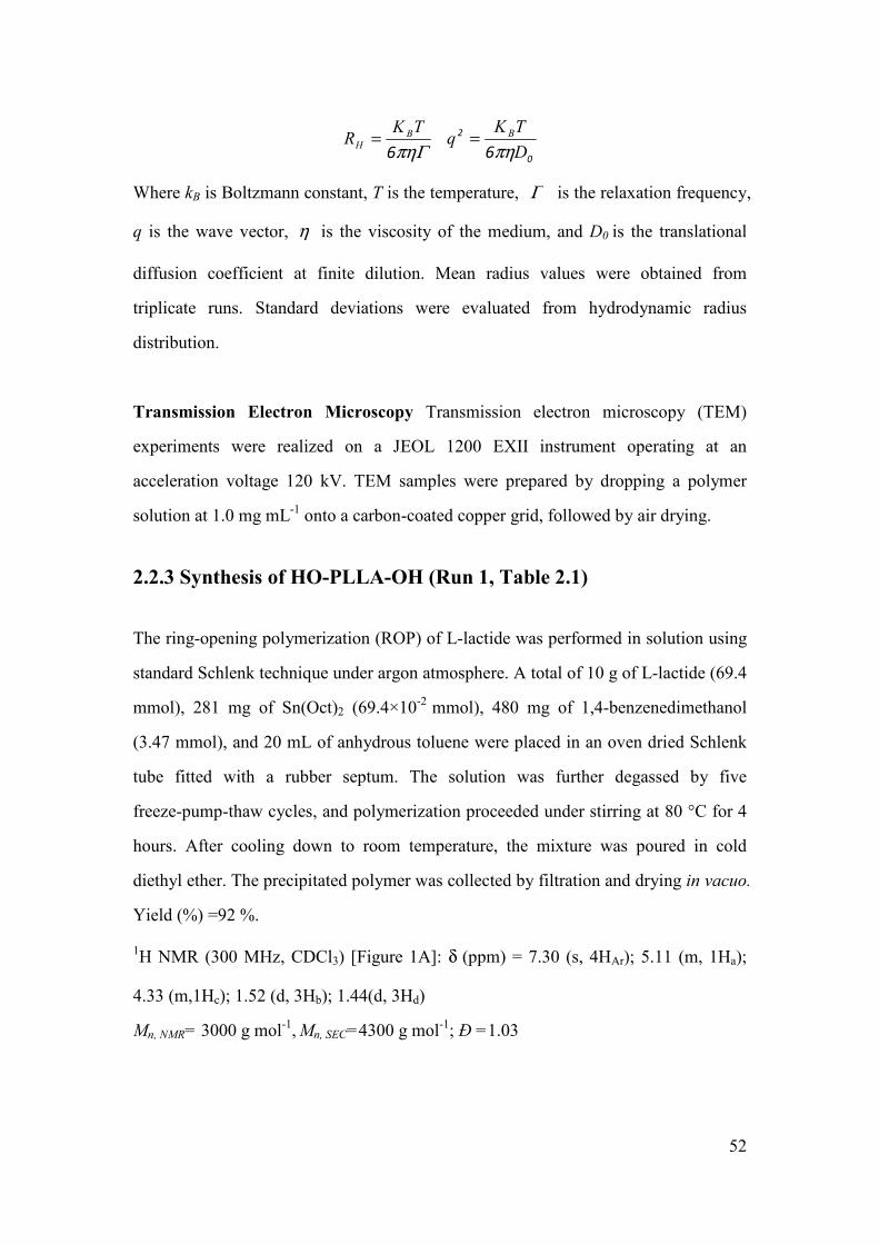
52
Γπη6
TKR B
H = 0
2
6 D
TKq B
πη=
Where kB is Boltzmann constant, T is the temperature, Γ is the relaxation frequency,
q is the wave vector, η is the viscosity of the medium, and D0 is the translational
diffusion coefficient at finite dilution. Mean radius values were obtained from
triplicate runs. Standard deviations were evaluated from hydrodynamic radius
distribution.
Transmission Electron Microscopy Transmission electron microscopy (TEM)
experiments were realized on a JEOL 1200 EXII instrument operating at an
acceleration voltage 120 kV. TEM samples were prepared by dropping a polymer
solution at 1.0 mg mL-1
onto a carbon-coated copper grid, followed by air drying.
2.2.3 Synthesis of HO-PLLA-OH (Run 1, Table 2.1)
The ring-opening polymerization (ROP) of L-lactide was performed in solution using
standard Schlenk technique under argon atmosphere. A total of 10 g of L-lactide (69.4
mmol), 281 mg of Sn(Oct)2 (69.4×10⁻²
mmol), 480 mg of 1,4-benzenedimethanol
(3.47 mmol), and 20 mL of anhydrous toluene were placed in an oven dried Schlenk
tube fitted with a rubber septum. The solution was further degassed by five
freeze-pump-thaw cycles, and polymerization proceeded under stirring at 80 °C for 4
hours. After cooling down to room temperature, the mixture was poured in cold
diethyl ether. The precipitated polymer was collected by filtration and drying in vacuo.
Yield (%) =92 %.
1H NMR (300 MHz, CDCl3) [Figure 1A]: δ (ppm) = 7.30 (s, 4HAr); 5.11 (m, 1Ha);
4.33 (m,1Hc); 1.52 (d, 3Hb); 1.44(d, 3Hd)
Mn, NMR= 3000 g mol-1
, Mn, SEC=4300 g mol
-1; Đ =1.03

53
2.2.4 Synthesis of Br-PLLA-Br
In a dried round bottom flask equipped with a drying tube filled with CaCl2, 2 g
(0.667 mmol) of HO-PLLA-OH was dissolved in 20 mL of dried dichloromethane
with stirring (Run 2, Table 2.1). Then, 0.298 mL (2 mmol) of triethylamine and 0.209
mL (2 mmol) of 2-bromopropionylbromide were added dropwise to the solution in an
ice bath. The reaction mixture was then stirred for 72 hours at room temperature. The
precipitated by-product (i.e. Et3N·HBr) was removed by filtration. The filtrate was
washed using 5 % sodium bicarbonate (3 × 30 mL) and then water (3 ×30 mL). The
organic phase was dried over anhydrous MgSO4 and filtered. The filtrate was poured
in cold methanol. The precipitated polymer was collected by filtration and drying in
vacuo.
1H NMR (300 MHz, CDCl3) [Figure 1B] δ (ppm) = 7.29 (S, 4HAr), 5.11 (m, 1Ha),
4.33 (m, 1Hc+1He), 1.8 (d, 3Hf), 1.52 (d, 3Hb).
Mn, NMR = 3300 g mol-1
, Mn, SEC = 4700 g mol-1
, Đ=1.04
2.2.5 Synthesis of PNIPAAm-b-PLLA-b-PNIPAAm
Triblock copolymers were prepared using a standard Schlenk technique (Run T1,
Table 2.1). Typically, 100 mg (0.0304 mmol) of Br-PLA-Br, 344 mg (3.04 mmol) of
NIPAAm and 6.0 mg (0.0608 mmol) of CuCl were dissoloved in 1.5 mL DMF, then
0.3 mL of H2O were introduced into a Schlenk tube. After five freeze-pump-thaw
cycles, 16.3 µL (0.0608 mmol) of Me6TREN was added under argon atmosphere. The
reaction was then allowed to proceed under stirring at 25 oC for 30 minutes. During
the reaction, samples were taken from the solution at preset time intervals for NMR
and SEC analyses. After quenching with liquid N2, the viscous solution was
precipitated by addition of diethylether. The crude product was dissolved in 10 mL
CH2Cl2. The catalyst was removed by passing the dilute solution through an
aluminum oxide column. The resulting colorless solution was then concentrated,

54
followed by precipitation in cold diethylether. The final product was dried under
vacuum at room temperature for 24 h.
1H NMR (300 MHz, CDCl3) δ (ppm) = 7.29(S, 4HAr), 5.11 (m, 1Ha), 3.94 (m,1He),
2.07 (t, 1Hd), 1.76 (m, 2Hc), 1.52 (d, 3Hb), 1.09 (d, 6Hf).
Mn, NMR= 14000 g mol-1
, Mn, SEC = 18000 g mol-1
, Đ= 1.08
2.3 Results and discussion
2.3.1 Synthesis of PNIPAAm-b-PLLA-b-PNIPAAm
Amphiphilic and thermo-responsive triblock copolymers, namely
PNIPAAm-b-PLLA-b-PNIPAAm, were prepared in three steps according to the
following reaction sequence: (i) synthesis of α, ω-telechelic HO-PLLA-OH by ROP
of L-lactide, (ii) modification of HO-PLLA-OH using 2-bromopropionyl bromide to
obtain di-2-bromopropionyl-terminated Br-PLLA-Br macroinitiator, and (iii) ATRP
of NIPAAm from the Br-PLLA-Br macroinitiator yielding
PNIPAAm-b-PLLA-b-PNIPAAm triblock copolymers.
The synthesis of Br-PLLA-Br macroinitiator was performed according to a procedure
previously reported [28]. In the first step, PLLA bearing two hydroxyl end groups
(HO-PLLA-OH) was obtained by ROP of L-lactide using Sn(Oct)2 as catalyst and
1,4-dimethanol benzene as initiator. The molecular weight determined by 1H NMR
(Mn,NMR= 3000 g mol-1
) (Figure 2.1A) is close to the targeted value (2900 g mol-1
),
while the Mn determined by SEC (Mn,SEC= 4400 g mol-1
) with PS standards is higher.
The polymer exhibits a very low dispersity (Đ= 1.03), in agreement with a good
control of the reaction.

55
Figure 2.1 1H NMR spectra of HO-PLLA-OH (A), Br-PLLA-Br (B) and
PNIPAAm-b-PLA-b-PNIPAAm (C) in CDCl3.
In the second step, HO-PLLA-OH reacted with 2-bromopropionyl bromide in the
presence of triethylamine, yielding Br-PLLA-Br as ATRP macroinitiator (Scheme
2.1). The attachment of the bromopropionyl group is confirmed by 1H NMR (Figure
2.1B). A doublet appears at 1.8 ppm corresponding to the methyl groups (f) adjacent
to the bromine. The transformation of hydroxyl endgroups into corresponding
2-bromopropionyl moiety is almost quantitative (98 %), as determined by comparing
the peak area of methyl (f) protons of 2-bromopropionyl at 1.8 ppm with that of the
aromatic protons at 7.29 ppm. Br-PLLA-Br shows a slight increase of molecular
weight (Mn, NMR= 3300 g mol-1
, Mn, SEC =4700 g mol-1
) as determined by SEC and 1H
NMR analyses, indicating that no chain cleavage occurred during the esterification
reaction. The dispersity of Br-PLLA-Br remains unchanged (Đ = 1.04) as compared
to HO-PLLA-OH.

56
Scheme 2.1 Synthesis of PNIPAAm-b-PLLA-b-PNIPAAm triblock copolymers via
combination of ring-opening polymerization and atom transfer radical polymerization
Table 2.1 Molecular characteristics of PNIPAAm-b-PLLA-b-PNIPAAm triblock
copolymers
Run [NIPAAm]0/
[PLLA]0
Time
(min)
Conversiona
%
[NIPAAm]/
[LA]a
Mn,NMRa
(g mol-1
)
Mn,SECb
(g mol-1
)
Ðb
HO-PLLA-OH - - 92 - 3000 4400 1.03
Br-PLLA-Br - - 98 - 3300 4700 1.04
T1a 100 30 71 2.4 14000 18000 1.08
T2a 200 30 58 4.2 22100 26700 1.10
T3a 300 30 53 5.4 27500 31500 1.13
T4a 300 120 65 6.9 34400 37500 1.28
a determined by
1H NMR,
b determined by SEC in DMF using PMMA standards for calibration
cATRP of NIPAAm. Conditions of polymerization: [NIPAAm]0=1.69 M, T=25 °C,
[initiator]/[CuCl]/[Me6TREN]=1/2/2, solvent mixture DMF/water=5/1.
Finally, the ATRP of NIPAAm initiated by Br-PLLA-Br macroinitiator was
successfully performed at 25 °C in a DMF/water mixture using CuCl/Me6TREN
complex as catalyst system. A series of amphiphilic thermo-responsive
PNIPAAm-b-PLLA-b-PNIPAAm triblock copolymers were obtained by varying the

57
ratio of NIPAAm to PLLA from 100 to 300 equivalents and the reaction time from 30
to 120 min (Run T1-T4, Table 2.1). The molecular weight ranges between 14000 and
35000 g mol-1
with narrow dispersity (Ð= 1.08-1.28), indicating good control of the
reaction. The molecular weights determined by 1H NMR well agree with those
obtained by SEC in DMF with PMMA standards.
Loh et al. reported the synthesis of
poly(N-isopropylacrylamide)-b-poly(ε-caprolactone)-b-poly(N-isopropylacrylamide)
(PNIPAAm-b-PCL-b-PNIPAAm) in dioxane using CuBr/HMTETA as catalyst
system [29]. No kinetic study was performed, and the copolymer exhibited large
dispersity ranging from 1.4 to 1.7. In the case of our
PNIPAAm-b-PLLA-b-PNIPAAm, these conditions led to a poor control of the ATRP
polymerization with low kinetics. In this context, ATRP was performed using
experimental conditions similar to those described by Masci et al., who first reported
controlled polymerization of N-isopropylacrylamide using ATRP in DMF/water (1:1)
with CuCl/Me6TREN as catalyst system [30]. More recently, the effect of both solvent
and temperature on the ATRP of NIPAAm was investigated by Ye et al. [31]. Water
was proved to facilitate the reaction in organic-aqueous solvent systems. In this work,
a DMF/water mixture with volume ratio of 5/1 was used as reaction solvent. The
CuCl/Me6TREN catalyst system was selected due to its high stability at low
temperature which allows achieving a good control on the polymerization [30, 32].
Figure 2.1C shows a typical NMR spectrum of PNIPAAm-b-PLLA-b-PNIPAAm
obtained for run T2 (Table 2.1). The signals at 3.94 and 1.09 ppm are assigned to the
protons of PNIPAAm, while those at 5.11 and 1.52 ppm are characteristic of protons
from PLLA. The composition and the molecular weight of copolymers were
calculated from the integrations of the methine groups of PNIPAAm and PLLA at
3.94 and 5.11 ppm, respectively. Figure 2.2 shows the SEC traces of different triblock
copolymers in comparison with Br-PLLA-Br. A shift to higher molecular weight is

58
detected for the copolymers, indicating successful initiation of the ATRP of NIPAAm
by the macroinitiator.
Figure 2.2 SEC chromatograms in DMF of Br-PLLA-Br and copolymers T1, T2, T3
and T4.
Figure 2.3 Kinetic plots for the polymerization of N-isopropylacrylamide in a
DMF/water mixture at 25 °C using a PLLA macroinitiator: ()
[NIPAAm]/[Br-PLLA-Br]/[CuCl]/[Me6TREN]= 100/1/2/2; ()
[NIPAAm]/[Br-PLLA-Br]/[CuCl]/[Me6TREN]= 200/1/2/2; ()
[NIPAAm]/[Br-PLLA-Br]/[CuCl]/ [Me6TREN]= 300/1/2/2. [NIPAAm]0 = 1.69 M.
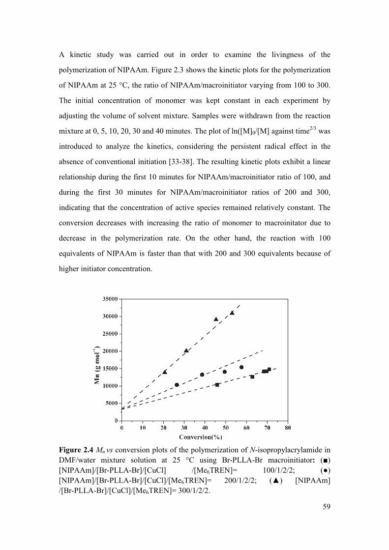
59
A kinetic study was carried out in order to examine the livingness of the
polymerization of NIPAAm. Figure 2.3 shows the kinetic plots for the polymerization
of NIPAAm at 25 °C, the ratio of NIPAAm/macroinitiator varying from 100 to 300.
The initial concentration of monomer was kept constant in each experiment by
adjusting the volume of solvent mixture. Samples were withdrawn from the reaction
mixture at 0, 5, 10, 20, 30 and 40 minutes. The plot of ln([M]0/[M] against time2/3
was
introduced to analyze the kinetics, considering the persistent radical effect in the
absence of conventional initiation [33-38]. The resulting kinetic plots exhibit a linear
relationship during the first 10 minutes for NIPAAm/macroinitiator ratio of 100, and
during the first 30 minutes for NIPAAm/macroinitiator ratios of 200 and 300,
indicating that the concentration of active species remained relatively constant. The
conversion decreases with increasing the ratio of monomer to macroinitator due to
decrease in the polymerization rate. On the other hand, the reaction with 100
equivalents of NIPAAm is faster than that with 200 and 300 equivalents because of
higher initiator concentration.
Figure 2.4 Mn vs conversion plots of the polymerization of N-isopropylacrylamide in
DMF/water mixture solution at 25 °C using Br-PLLA-Br macroinitiator: ()
[NIPAAm]/[Br-PLLA-Br]/[CuCl] /[Me6TREN]= 100/1/2/2; ()
[NIPAAm]/[Br-PLLA-Br]/[CuCl]/[Me6TREN]= 200/1/2/2; () [NIPAAm]
/[Br-PLLA-Br]/[CuCl]/[Me6TREN]= 300/1/2/2.

60
The evolution of Mn vs. conversion is shown in Figure 2.4. The molecular weight
linearly increases with conversion, which is indicative of the living character of the
reaction. Evolution of SEC traces for the ATRP of NIPAAM
(NIPAAm/macroinitiator ratio=100) was followed at different reaction time periods
(Figure 2.5). Shift to shorter elution time or higher Mn is noticed in all cases.
Therefore, all data suggest a well-controlled ATRP of NIPAAm using a hydrophobic
Br-PLLA-Br as macroinitiator to prepare triblock copolymers based on PLA and
PNIPAAm.
Figure 2.5 SEC traces for the polymerization of N-isopropylacrylamide in a
DMF/water mixture at 25 °C using a PLLA macroinitiator.
[NIPAAm]/[Br-PLLA-Br]/[CuCl]/[Me6TREN]= 100/1/2/2.
As shown in Figure 2.6, the influence of temperature on the kinetics was also
investigated. The ln([M]0/[M] vs. time2/3
plots shows a linear increase at 25 oC. On the
contrary, the kinetic plot shows much slower reaction rate at 0 oC because of the
lower reactivity of the catalyst system.

61
Figure 2.6 The influence of temperature on the kinetics: () 25 oC and () 0
oC.
[NIPAAm]/[Br-PLLA-Br]/[CuCl]/[Me6TREN]= 200/1/2/2.
2.3.2 Self-assembling of copolymers
Amphiphilic block copolymers are susceptible to self-assemble into various
aggregates in aqueous medium, including spherical micelles, anisotropic micelles,
filomicelles, nanotubes and so forth. Spherical micelles are the most commonly
observed aggregates because of minimum free energy. The hydrophobic parts
segregate in the core, while the hydrophilic blocks constitute the shell which interacts
with the external environment. The aggregation of core-forming blocks can result in
the disappearance of NMR signals [20, 23, 39]. Thus, 1H NMR analysis in selective
solvents provides an insight into the self-assembly process of
PNIPAAm-b-PLA-b-PNIPAAm [23].
Figure 2.7 shows the 1H NMR spectra of triblock copolymer T1 in CDCl3 and in
deuterated water (D2O). CDCl3 is a good solvent for PNIPAAm and PLLA, and
signals of both blocks are observed. The signals at 3.94 and 1.09 ppm are assigned to
the protons of PNIPAAm, while those at 5.11 and 1.52 ppm are characteristic of the

62
protons from PLLA. On the other hand, D2O only solubilizes hydrophilic PNIPAAm
segments of the copolymer. As a consequence, only signals corresponding to
PNIPAAm are detected (Figure 2.7B). Signals belonging to PLLA block disappear
due to the limited molecular motion of the moiety in aqueous medium. This finding
confirmed the formation of micelles composed of a hydrophobic PLLA core and a
hydrophilic PNIPAAm shell.
Figure 2.7 1H NMR spectra of PNIPAAm-b-PLLA-b-PNIPAAm triblock copolymer
T1 in (A) CDCl3 and (B) D2O.
The critical micellar concentration (CMC) of PNIPAAm-b-PLLA-b-PNIPAAm
copolymers was determined by fluorescence spectroscopy using pyrene as the
fluorescent probe [40]. All measurements were performed at room temperature to
ensure the solubilization in water of the PNIPAAm moieties. Figure 2.8A shows an
overlay of excitation spectra of pyrene (6×10⁻⁷
M) at different copolymer
concentrations. When the copolymer concentration of pyrene containing solutions

63
increases above the CMC, significant changes are observed on the excitation spectra
due to the transfer of free pyrene molecules into the hydrophobic micellar core.
Figure 2.8 Fluorescence excitation spectra (λem=394 nm) of pyrene (6×10⁻⁷
M) in
aqueous solutions of PNIPAAm-b-PLLA-b-PNIPAAm triblock copolymer (T1) (A);
Plots of the intensity ratios (I337/I333 from pyrene excitation spectra) vs. the
concentration of PNIPAAm-b-PLLA-b-PNIPAAm copolymer (T1) in water (B).

64
Figure 2.8B shows the plots of the I337/I333 intensity ratio against the copolymer
concentration. The intensity ratio exhibits a substantial increase at a particular
concentration, indicating the incorporation of pyrene into micelles. The CMC of the
different triblock copolymers was obtained from the intersection of the two linear
regression lines of the plot. The values range from 0.0077 to 0.016 mg mL-1
(Table
2.2), which are much lower than those of PNIPAAm-b-PLA diblock copolymers
described by Fukashi et al. [16]. Copolymers with longer PNIPAAm block exhibits
higher CMC due to higher hydrophilicity. In all cases, the very low CMC indicates
the strong tendency of triblock copolymers to self-assemble into highly stable
micelles in aqueous medium.
2.3.3 Morphology and size distribution of micelles
The size distribution and the morphology of micelles were investigated by DLS and
TEM, respectively. Figure 2.9A shows the TEM image of the copolymer T4. Most
micelles are spherical in shape with small size (78~120 nm). Few aggregated particles
are also detected. A typical DLS graph of T4 micelles at 1 mg mL-1
is shown in Figure
2.9B. The average diameter is 83 nm with a dispersity index of 0.167. The observed
monomodal size distribution indicates the formation of micelles with relatively
similar sizes. Figure 2.9B also shows the plot of the correlation coefficient versus the
decay time. The observed curve confirms a monomodal size distribution of micelles
[10].
The difference between TEM and DLS data could be attributed to the experimental
conditions. In fact, DLS determines the hydrodynamic diameter of micelles in
aqueous solutions, whereas TEM shows the dehydrated solid state of micelles. Table
2.2 shows the DLS data of all copolymer micelles in water at 25 oC. The average
diameter ranges from 31 to 83 nm with narrow size distribution (Đ = 0.167-0.220),
which are smaller than that of PNIPAAm-PCL-PNIPAAm triblock polymer micelles
(92-125 nm) reported by Loh et al. [29]. The size of micelles increases with
65
increasing ratio of NIPAAm to LA units. Copolymers with longer PNIPAAm blocks
tend to form larger micelle size because hydrophilic PNIPAAm blocks are extended
in water below LCST.
Table 2.2 Characterization of PNIPAAm-b-PLLA-b-PNIPAAm triblock copolymer
micelles.
Run [NIPAAm]/[LA] LCSTa
(°C)
CMCb
(mg mL-1
)
DHc
(nm)
PDIc
T1 2.4 32.1 0.0077 31 0.210
T2 4.2 32.3 0.0088 64 0.210
T3 5.4 32.4 0.0098 71 0.118
T4 6.9 32.8 0.0160 83 0.167
a) determined from turbidimetry measurements at 3 mg mL-1
.
b) determined at 25 °C by fluorescence using pyrene as fluorescent probe.
c) determined at 25 °C by dynamic light scattering at 1 mg mL-1

66
Figure 2.9 TEM (A) and DLS (B) results of self-assembling micelles of copolymer
T4 at 1.0 mg mL-1
.

67
2.3.4 Thermo-responsive behavior of micelles
PNIPAAm exhibits a LCST of 32 °C which is relatively insensitive to variation of the
environmental conditions. Thermo-responsive polymers become insoluble when the
temperature increases above the LCST due to coil to globule transition. This phase
transition can be evidenced by light transmission measurements of a micelle solution
as a function of temperature.
Figure 2.10 Plot of transmittance changes as a function of temperature for aqueous
solution (3.0 mg mL-1
) of triblock copolymer T2.
Figure 2.10 shows a typical phase transition of copolymer T2 at 3.0 mg mL-1
. A sharp
light transmittance decrease from 100 to 0 % is observed at 32.3 oC, which indicates
phase transition from a transparent solution to a precipitated suspension. Varying the
[NIPAAm]/[LLA] ratio from 2.4 to 6.9, the LCST only slightly increases from 32.1 to
32.8 oC (Table 2.2). Copolymer T1 exhibits the lowest LCST (32.1
oC) because of the
highest hydrophobic PLLA content. In contrast, copolymer T4 with the highest
PNIPAAm content shows the highest LCST at 32.8 oC. However, the LCST variation
is very limited although higher hydrophobic PLLA content favors copolymer
precipitation. Actually, the length of PNIPAAm blocks has no substantial effect on

68
the LCST since the hydrogen-bonding between PNIPAAm chains and neighboring
water molecules leads to flat LCST behavior with little block length dependence
[40-42]. Similar findings were previously reported in the literature [24].
Figure 2.11 Hydrodynamic diameter distributions obtained for micelles of copolymer
T2: (A) at a concentration of 3 mg mL-1
, and (B) at a concentration of 0.2 mg mL-1
.
() T= 20 °C, () T= 40 °C, () T= 20 °C

69
The thermo-responsive behavior of micelles was illustrated by DLS measurements
(Figure 2.11). In the case of high copolymer concentration (3.0 mg mL-1
), the size of
the micelles increases from 100 nm to 654 nm when the temperature is raised from 20
to 40 oC (Figure 2.11A). On the contrary, in the case of low concentrations (0.2 mg
mL-1
), the micelles exhibit a totally different thermo-responsive behavior. The
diameter of micelles decreases from 82 nm at 20 oC to 63 nm at 40
oC (Figure 2.11B).
In both cases, the initial micelle sizes are recovered after cooling the solution down to
room temperature.
Scheme 2.2 Illustration of the temperature responsive behavior of micelles with
varying temperatures across the LCST at low or high concentrations
These findings could be explained as follows (Scheme 2.2).
PNIPAAm-b-PLLA-b-PNIPAAm copolymers form micelles in water at 25 oC with a
hydrophobic PLA core and a hydrophilic PNIPAAm corona. As the temperature
increases, PNIPAAm chains become hydrophobic and eventually collapse. Micelles
aggregate to form larger particles with increased hydrophobicity of PNIPAAm [6, 7].
On the other hand, at low concentrations, PNIPAAm chains at the outer shell collapse

70
above the LCST, leading to decrease of micelle size. Micelles are well separated from
each other and can hardly aggregate.
Triblock copolymer micelles composed of degradable PLLA core and
thermo-responsive PNIPAAm corona exhibit nano-scale sizes and very low CMC
values. Once loaded with a hydrophobic drug, the micelles could be easily
administered by intravenous injection. With degradation of PLA, the drug would be
gradually released and the micelles would dissociate into PNIPAAm chains. It is
noteworthy that PNIPAAm with molecular weight below 50000 g mol-1
exhibits no
toxicity during long-term circulation and can be excreted by glomerular filtration [16,
43, 44]. Therefore, amphiphilic PNIPAAm-b-PLLA-b-PNIPAAm triblock
copolymers appear very promising as carrier of hydrophobic drugs.
2.4 Conclusion
In this work, a series of novel well-defined amphiphilic
PNIPAAm-b-PLLA-b-PNIPAAm triblock copolymers were successfully synthesized
by combining ROP and ATRP under mild conditions. The resulting copolymers
present well controlled molecular weight and narrow dispersity, and are susceptible to
self-assemble into spherical micelles in aqueous medium. The CMC of the
copolymers ranges from 0.0077 to 0.016 mg mL-1
. The very low CMC values
demonstrate the strong tendency of copolymers toward formation of micelles. The
LCST of the copolymer is in the range of 32.1-32.8 oC. Both the CMC and LCST
increase with increasing PNIPAAm block length. The size of micelles obtained from
DLS ranges from 31 to 83 nm with narrow distributions. The micelles exhibit
different thermo-responsive behaviors at high or low concentrations. At high
concentrations (3.0 mg mL-1
), aggregation of micelles occurs when the temperature is
raised above the LCST, leading to micelle size increase. In contrast, at low
concentrations (0.2 mg mL-1
), a decrease of micellesize is detected because of the
collapse of PNIPAAm blocks above the LCST.

71
Therefore, PNIPAAm-b-PLLA-b-PNIPAAm triblock copolymer micelles appear very
promising for application as drug carrier. Future work is underway to prepare
PNIPAAm/PLLA-based triblock copolymers with LSCT above the physiological
temperature (37 oC) by addition of a third comonomer, using similar ROP and ATRP
methods.
2.5 References
[1] E. L. Thomas, The ABCs of Self-Assembly. Science 286 (1999) 1307.
[2] Y. Yan, G. K. Such, A. P. R. Johnston, J. P. Best, F. Caruso, Engineering Particles for
Therapeutic Delivery: Prospects and Challenges. ACS Nano 6 (2012) 3663-3669.
[3] C. Oerlemans, W. Bult, M. Bos, G. Storm, J.F. Nijsen, W. Hennink, Polymeric Micelles in
Anticancer Therapy: Targeting, Imaging and Triggered Release. Pharm. Res. 27 (2010)
2569-2589.
[4] A. L. Monica, L. Adams, G. S. Kwon, Amphiphilic Block Copolymers for Drug Delivery. J.
Pharm. Sci. 92 (2003) 1343-1355.
[5] A. K. Bajpai, S. K. Shukla, S. Bhanu, S. Kankane, Responsive polymers in controlled drug
delivery. Prog. Polym. Sci. 33. (2008) 1088-1118.
[6] B. Guillerm, V. Darcos, V. Lapinte, S. Monge, J. Coudane, J. J. Robin, Synthesis and
evaluation of triazole-linked poly(ε-caprolactone)-graft-poly(2-methyl-2-oxazoline) copolymers
as potential drug carriers. Chem.Commun. 48 (2012) 2879-2881.
[7] H. senff, W. Richtering., Influence of cross-link density on rheological properties of
temperature-sensitive microgel suspensions. Collioid polym sci 278 (2000) 830-840.
[8] H. Wei, S. X. Cheng, X. Z. Zhang, R. X. Zhuo, Thermo-sensitive polymeric micelles based
on poly(N-isopropylacrylamide) as drug carriers. Prog. Polym. Sci. 34 (2009) 893-910.
[9] S. Q. Liu, N. Wiradharma, S. J. Gao, Y. W. Tong, Y. Y. Yang, Bio-functional micelles
self-assembled from a folate-conjugated block copolymer for targeted intracellular delivery of
anticancer drugs. Biomaterials 28 (2007) 1423-1433.
[10] B. Y. Du, A. X. Mei, Y. Yang, Q. F. Zhang, Q. Wang, J. T. Xu, Z. Q. Fan, Synthesis and
micelle behavior of (PNIPAm-PtBA-PNIPAm)m amphiphilic multiblock copolymer. Polymer 51
(2010) 3497-3502.
[11] T. H. Qu, A. R. Wang, J. F. Yuan, J. H. Shi, Q. Y. Gao, Preparation and characterization of
thermo-responsive amphiphilic triblock copolymer and its self-assembled micelle for controlled
drug release. Colloids Surf. B: Biointerfaces 72 (2009) 94-100.
[12] S. M. Li, I. Rashkov, J. L. Espartero, N. Manolova, M. Vert, Synthesis, characterization, and
hydrolytic degradation of PLA/PEO/PLA triblock copolymers with long poly(L-lactic acid) blocks.
Macromolecules 29 (1996) 57-62.
[13] A. C. Albertsson, I. Varma, Degradable Aliphatic Polyesters, Vol. 157, Springer Berlin
Heidelberg, 2002, pp. 1-40.

72
[14] G. S. He, L. L. Ma, J. Pan, S. Venkatraman, ABA and BAB type triblock copolymers of PEG
and PLA: A comparative study of drug release properties and "stealth" particle characteristics. Int.
J. Pharm. 334 (2007) 48-55.
[15] L. Yang, A. El Ghzaoui, S. M. Li, In vitro degradation behavior of
poly(lactide)-poly(ethylene glycol) block copolymer micelles in aqueous solution. Int. J. Pharm.
400 (2010) 96-103.
[16] K. Fukashi, S. Kiyotaka, A. Takao, Y. Masayuki, S. Yasuhisa, O. Teruo, Preparation and
characterization of thermally responsive block copolymer micelles comprising
poly(N-isopropylacrylamide-b-DLlactide). J. Controlled Release 55 (1998) 87-98.
[17] S. Q. Liu, Y. Y. Yang, X. M. Liu, Y. W. Tong, Preparation and Characterization of
Temperature-Sensitive Poly(N-isopropylacrylamide)-b-poly(d,l-lactide) Microspheres for Protein
Delivery. Biomacromolecules 4 (2003) 1784-1793.
[18] J. Qin, Y. S. Jo, J. E. Ihm, D. K. Kim, M. Muhammed, Thermosensitive Nanospheres with a
Gold Layer Revealed as Low-Cytotoxic Drug Vehicles. Langmuir 21 (2005) 9346-9351.
[19] E. Ayano, M. Karaki, T. Ishihara, H. Kanazawa, T. Okano,
Poly(N-isopropylacrylamide)-PLA and PLA blend nanoparticles for temperature-controllable drug
release and intracellular uptake. Colloids Surf. B: Biointerfaces 99 (2012) 67-73.
[20] H. Wei, X. Z. Zhang, C. Cheng, S. X. Cheng, R. X. Zhuo, Self-assembled, thermosensitive
micelles of a star block copolymer based on PMMA and PNIPAAm for controlled drug delivery.
Biomaterials 28 (2007) 99-107.
[21] K. Matyjaszewski, J. H. Xia, Atom Transfer Radical Polymerization. Chem. Rev. 101 (2001)
2921-2990.
[22] T. E. Patten, J. H. Xia, T. Abernathy, K. Matyjaszewski, Polymers with very low
polydispersities from atom transfer radical polymerization. Science 272 (1996) 866-868.
[23] M. Hales, C. Barner-Kowollik, T. P. Davis, M. H. Stenzel, Shell-Cross-Linked Vesicles
Synthesized from Block Copolymers of Poly(D,L-lactide) and Poly(N-isopropylacrylamide) as
Thermoresponsive Nanocontainers. Langmuir 20 (2004) 10809-10817.
[24] Y. Z. You, C. Y. Hong, W. P. Wang, W. Q. Lu, C. Y. Pan, Preparation and Characterization
of Thermally Responsive and Biodegradable Block Copolymer Comprised of PNIPAAM and
PLA by Combination of ROP and RAFT Methods. Macromolecules 37 (2004) 9761-9767.
[25] J. Akimoto, M. Nakayama, K. Sakai, T. Okano, Molecular Design of Outermost Surface
Functionalized Thermoresponsive Polymeric Micelles with Biodegradable Cores. J. Polym. Sci.
Polym. Chem. 46 (2008) 7127-7137.
[26] J. Akimoto, M. Nakayama, K. Sakai, T. Okano, Temperature-Induced Intracellular Uptake of
Thermoresponsive Polymeric Micelles. Biomacromolecules 10 (2009) 1331-1336.
[27] J. Akimoto, M. Nakayama, K. Sakai, T. Okano, Thermally Controlled Intracellular Uptake
System of Polymeric Micelles Possessing Poly(N-isopropylacrylamide)-Based Outer Coronas.
Mol. Pharm. 7 (2010) 926-935.
[28] Y. Bakkour, V. Darcos, F. Coumes, S. Li, J. Coudane, Brush-like amphiphilic copolymers
based on polylactide and poly(ethylene glycol): Synthesis, self-assembly and evaluation as drug
carrier. Polymer 54 (2013) 1746-1754.
[29] X. J. Loh, Y. L. Wu, W. T. J. Seow, M. N. I. Norimzan, Z. X. Zhang, F. Xu, E. T. Kang, K.
G. Neoh, J. Li, Micellization and phase transition behavior of thermosensitive

73
poly(N-isopropylacrylamide)-poly(ε-caprolactone)-poly (N-isopropylacrylamide) triblock
copolymers. Polymer 49 (2008) 5084-5094.
[30] G. Masci, L. Giacomelli, V. Crescenzi, Atom transfer radical polymerization of
N-isopropylacrylamide. Macromol. Rapid Commun. 25 (2004) 559-564.
[31] J. D. Ye, R. Narain, Water-Assisted Atom Transfer Radical Polymerization of
N-Isopropylacrylamide: Nature of Solvent and Temperature. J. Phys. Chem. B 113 (2009)
676-681.
[32] E. J. Lobb, I. Ma, N. C. Billingham, S. P. Armes, A. L. Lewis, Facile Synthesis of
Well-Defined, Biocompatible Phosphorylcholine-Based Methacrylate Copolymers via Atom
Transfer Radical Polymerization at 20 OC. J. Am. Chem. Soc. 123 (2001) 7913-7914.
[33] X. Lu, L. Zhang, L. Meng, Y. Liu, Synthesis of poly(N-isopropylacrylamide) by ATRP using
a fluorescein-based initiator. Polym. Bull. 59 (2007) 195-206.
[34] H. Fischer, The Persistent Radical Effect In living Radical Polymerization. Macromolecules
30 (1997) 5666-5672.
[35] H. Fischer, The persistent radical effect in controlled radical polymerizations. J. Polym. Sci.
Part A: Polym. Chem. 37 (1999) 1885-1901.
[36] K. Ohno, Y. Tsujii, T. Miyamoto, T. Fukuda, M. Goto, K. Kobayashi, T. Akaike, Synthesis
of a Well-Defined Glycopolymer by Nitroxide-Controlled Free Radical Polymerization.
Macromolecules 31 (1998) 1064-1069.
[37] T. Fukuda, A. Goto, K. Ohno, Mechanisms and kinetics of living radical polymerizations.
Macromol. Rapid Commun. 21 (2000) 151-165.
[38] E. Berndt, M. Ulbricht, Synthesis of block copolymers for surface functionalization with
stimuli-responsive macromolecules. Polymer 50 (2009) 5181-5191.
[39] H. Walderhaug, O. Soderman, NMR studies of block copolymer micelles. Curr. Opin.
Colloid Interface Sci. 14 (2009) 171-177.
[40] X. J. Loh, Z. X. Zhang, Y. L. Wu, T. S. Lee, J. Li, Synthesis of Novel Biodegradable
Thermoresponsive Triblock Copolymers Based on Poly[(R)-3-hydroxybutyrate] and
Poly(N-isopropylacrylamide) and Their Formation of Thermoresponsive Micelles.
Macromolecules 42 (2009) 194-202.
[41] S. Fujishige, K. Kubota, I. Ando, Phase-Transition of Aqueous-Solutions of
Poly(N-isopropylacrylamide) and Poly(N-isopropylmethacrylamide). J. Phys. Chem. 93 (1989)
3311-3313.
[42] Y. Okada, F. Tanaka, Cooperative Hydration, Chain Collapse, and Flat LCST Behavior in
Aqueous Poly(N-isopropylacrylamide) Solutions. Macromolecules 38 (2005) 4465-4471.
[43] C. Chang, H. Wei, C.Y. Quan, Y.Y. Li, J. Liu, Z.C. Wang, S.X. Cheng, X.Z. Zhang, R.X.
Zhuo, Fabrication of thermosensitive PCL-PNIPAAm-PCL triblock copolymeric micelles for drug
delivery. J. Polym. Sci. Pol. Chem. 46 (2008) 3048-3057.
[44] S. Cammas, K. Suzuki, C. Sone, Y. Sakurai, K. Kataoka, T. Okano, Thermo-responsive
polymer nanoparticles with a core-shell micelle structure as site-specific drug carriers. J.
Controlled Release 48 (1997) 157-164.

74

75
Chapter 3 Tunable thermo-responsive
P(NIPAAm-co-DMAAm)-b-PLLA-b-P(NIPAAm-co-DMAA
m) triblock copolymer micelles as drug carrier
Abstract: Thermo-responsive triblock copolymers were synthesized by atom
transfer radical polymerization of N-isopropylacrylamide (NIPAAm) and
N,N-dimethylacrylamide (DMAAm) using α,ω-bromopropionyl poly(L-lactide) as
macroinitiator and a CuCl/tris(2-dimethylaminoethyl) amine (Me6TREN) complex as
catalyst. The polymerization was realized at 25 oC in a DMF/water mixture. DMAAm
was incorporated in the copolymer as a hydrophilic comonomer in order to tune the
lower critical solution temperature (LCST). The LCST increases linearly from 32.2 oC
to 39.1 oC with increasing the DMAAm content from 0 to 24 %. The phase transition
of polymeric micelles at the LCST occurs in a narrow temperature interval below 0.5
oC. Reversible size changes are observed when the temperature increases from 25
oC
to 45 oC and then decreases down to 25
oC. Nano-size micelles (37 to 54 nm) with
narrow distribution were obtained by self-assembly of amphiphilic copolymers in
aqueous medium. The critical micelle concentration (CMC) ranges from 0.010 to
0.015 mg mL-1
. In vitro drug release studies show a much faster release at
temperatures above the LCST. MTT assay was conducted to evaluate the cytotoxicity
of copolymers. The nano-scale size, low CMC, rapid phase transition, LCST slightly
above the body temperature and thermo-responsive drug release indicate that these
copolymers could be potential candidates for applications in targeted delivery of
drugs.
Keywords: thermo-responsive copolymer; poly(N-isopropylacrylamide);
poly(L-lactide)

76
3.1 Introduction
Recently, “smart micelles” derived from stimuli-responsive polymers have drawn
great attention as drug delivery systems (DDS) [1-3]. Among them, thermo-sensitive
copolymers based on poly(N-isopropylacrylamide) (PNIPAAm) have been widely
investigated, notably to build polymeric micelles for controlled delivery of anticancer
drugs or DNA [4-10]. PNIPAAm could be considered as the “gold standard”
thermo-responsive polymer with a lower critical solution temperature (LCST) around
32 oC, which is induced by a reversible hydration-dehydration transition [11, 12].
Variation of pH, concentration, chemical structure or biological environment only
slightly affects the LCST. However, these copolymers are not degradable, which
limits their potential applications as drug carrier.
As a biodegradable polymer, polylactide (PLA) has been extensively investigated for
biomedical and pharmaceutical applications such as controlled drug release systems,
medical implants and scaffolds in tissue engineering [13-16]. Block copolymers based
on PNIPAAm and PLA combine both the thermo-sensitivity of PNIPAAm and the
degradability of PLA [17-22]. Increasing the temperature above the LCST results in
collapse of micelles and burst-like release of encapsulated drug [21]. Thus temporal
drug delivery could be achieved by using thermo-sensitive and degradable polymeric
micelles based on PLA and PNIPAAm.
The PNIPAAm moiety is supposed to be soluble in the blood stream, and becomes
insoluble after accumulation in a locally heated tumor tissue. This property could be
exploited for targeted delivery of drugs [23-26]. Therefore, polymeric micelles should
be designed so as to exhibit a LCST slightly above the body temperature. Introduction
of hydrophilic acrylamide comonomers such as dimethyl acrylamide (DMAAm) in
PNIPAAm chains has been used to increase the LCST of the resulting copolymers.
Most of the P(NIPAAm-co-DMAAm)-b-PLA diblock copolymers described in the
literature were synthesized by combination of free radical polymerization and

77
ring-opening polymerization (ROP) [27-33]. The resulting copolymers exhibit poor
compositions and large molecular weight distributions (dispersity Ð>2), which led to
a broad phase transition at the LCST. Later on, Akimoto et al. synthesized
well-defined P(NIPAAm-co-DMAAm)-b-PLA diblock copolymers by combination of
reversible addition-fragmentation chain transfer polymerization (RAFT) and ROP
[34-37]. The terminal dithiobenzoate (DTBz) groups were reduced to thiolgroups and
reacted with maleimide (Mal). In aqueous media, the resulting copolymers formed
surface-functionalized thermo-responsive micelles, hydrophobic DTBz-surface
micelles demonstrating a significant lower LCST (30.7 °C) than Mal-surface micelles
(40.0 °C). The LCST of micelle mixtures can be varied by changing the ratio of
DTBz/Mal end-functional diblock copolymers [34]. The authors also studied
thermally controlled intracellular uptake of copolymer micelle [35, 36]. Li et al.
reported similar maleimide end-functional P(NIPAAm-co-DMAAm)-b-PLA and
P(NIPAAm-co-DMAAm)-b-PCL copolymers with LCST of 39 °C and 40.5 °C,
respectively [37]. Both the adriamycin release and the intracellular uptake were
enhanced at 40 ° C as compared to 37 °C. However, precise tuning of the LCST could
not be achieved by modification of end groups or mixing two copolymers with
different end groups.
In the previous chapter, we reported the synthesis of thermo-sensitive
PNIPAAm-b-PLA-b-PNIPAAm triblock copolymers by atom transfer radical
polymerization (ATRP) using α, ω-bromopropionyl poly(L-lactide) (PLLA) as
macroinitiator [38]. Excellent control over the molecular weights was achieved under
mild conditions. The kinetics of polymerization and the self-assembly behavior of the
resulting copolymers were investigated. However, the LCST of the copolymers ranges
from 32.1 to 32.8 °C, i.e. much lower than the body temperature.
In this chapter, thermo-responsive and biodegradable
P(NIPAAm-co-DMAAm)-b-PLLA-b-P(NIPAAm-co-DMAAm) triblock copolymers

78
were prepared via combination of ROP and ATRP. A DMAAm comonomer was
introduced in order to tune the LCST slightly above body temperature for targeted
drug delivery. The resulting copolymers were fully characterized by 1H NMR, SEC
and DOSY NMR. The physico-chemical properties of copolymer micelles were
investigated in aqueous media. MTT assay was carried out to evaluate the cytotoxity
of copolymers. The drug release behavior of micelles containing an antimicrobial
agent, Amphotericin B, was investigated below and above the LCST.
3.2 Experimental Methods
3.2.1 Materials
L-lactide was purchased from Purac Biochem (Goerinchem, The Netherlands).
Stannous 2-ethylhexanoate (Sn(Oct)2), 1,4-benzene dimethanol, 2-bromopropionyl
bromide, tris(2-dimethyl aminoethyl) amine (Me6TREN), copper (I) chloride (CuCl),
N, N-dimethyl formamide (DMF) and Amphotericin B were obtained from
Sigma-Aldrich (St-Quentin Fallavier, France), and were used without further
purification. Dichloromethane and toluene from Sigma-Aldrich were dried over
calcium hydride for 24 h at room temperature and distilled under reduced pressure.
NIPAAm and DMAAm were obtained from Sigma-Aldrich and purified through a
basic aluminum oxide column. Triethylamine (Sigma-Aldrich) was dried over
potassium hydroxyde for 24 h at room temperature and distilled. Ultrapure water with
a conductivity of 18 MΩ was produced using a Millipore Milli-Q water system.
α,ω-bromopropionyl PLLA (Br-PLLA-Br) was prepared as previously reported [38].
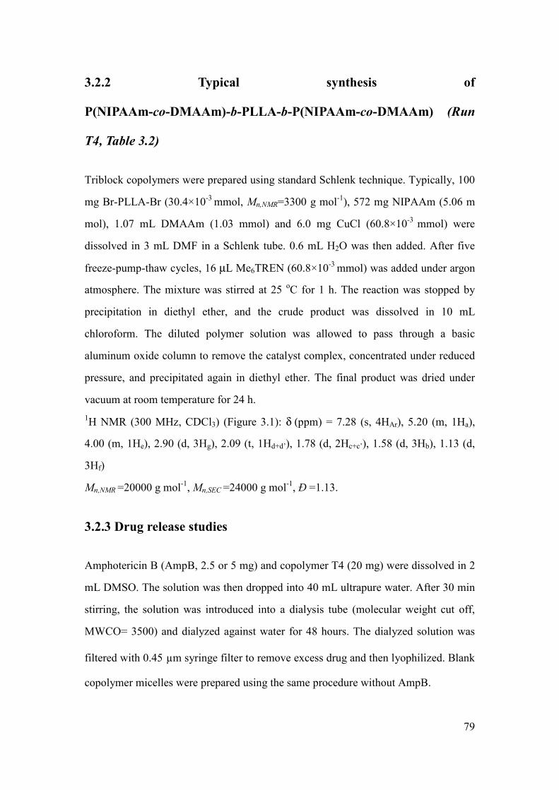
79
3.2.2 Typical synthesis of
P(NIPAAm-co-DMAAm)-b-PLLA-b-P(NIPAAm-co-DMAAm) (Run
T4, Table 3.2)
Triblock copolymers were prepared using standard Schlenk technique. Typically, 100
mg Br-PLLA-Br (30.4×10⁻³
mmol, Mn,NMR=3300 g mol-1
), 572 mg NIPAAm (5.06 m
mol), 1.07 mL DMAAm (1.03 mmol) and 6.0 mg CuCl (60.8×10⁻³
mmol) were
dissolved in 3 mL DMF in a Schlenk tube. 0.6 mL H2O was then added. After five
freeze-pump-thaw cycles, 16 µL Me6TREN (60.8×10⁻³
mmol) was added under argon
atmosphere. The mixture was stirred at 25 oC for 1 h. The reaction was stopped by
precipitation in diethyl ether, and the crude product was dissolved in 10 mL
chloroform. The diluted polymer solution was allowed to pass through a basic
aluminum oxide column to remove the catalyst complex, concentrated under reduced
pressure, and precipitated again in diethyl ether. The final product was dried under
vacuum at room temperature for 24 h.
1H NMR (300 MHz, CDCl3) (Figure 3.1): δ (ppm) = 7.28 (s, 4HAr), 5.20 (m, 1Ha),
4.00 (m, 1He), 2.90 (d, 3Hg), 2.09 (t, 1Hd+d’), 1.78 (d, 2Hc+c’), 1.58 (d, 3Hb), 1.13 (d,
3Hf)
Mn,NMR =20000 g mol-1
, Mn,SEC =24000 g mol-1
, Ð =1.13.
3.2.3 Drug release studies
Amphotericin B (AmpB, 2.5 or 5 mg) and copolymer T4 (20 mg) were dissolved in 2
mL DMSO. The solution was then dropped into 40 mL ultrapure water. After 30 min
stirring, the solution was introduced into a dialysis tube (molecular weight cut off,
MWCO= 3500) and dialyzed against water for 48 hours. The dialyzed solution was
filtered with 0.45 µm syringe filter to remove excess drug and then lyophilized. Blank
copolymer micelles were prepared using the same procedure without AmpB.

80
The drug content and loading efficiency were determined by UV spectroscopy. 5 mg
AmpB encapsulated polymeric micelles were dissolved in DMSO. UV measurements
were realized at 388 nm. The concentration of AmpB was determined using a
calibration curve obtained with a series of standard AmpB solutions in DMSO. The
drug loading content and loading efficiency were calculated using the following
equations:
100×=micelles loaded drug of weight
micelles in drug of weight%) wt( loading Drug (1)
100×=drug feeding of weight
micelles in drug of weight%) (wt efficiency Loading (2)
Drug release studies were carried out at two different temperatures. Briefly, 5 mg
lyophilized polymeric micelle was reconstituted in 5 mL ultrapure water and then
introduced into a dialysis tube (MWCO: 3500). The dialysis tube was placed in 20
mL ultrapure water in oven at 37 or 38 oC. At preset time interval, the release medium
was recovered for analysis and renewed with fresh ultrapure water. The drug
concentration was determined by UV spectroscopy at 388 nm.
3.2.4 Cytotoxicity
The cytotoxicity of polymers was evaluated by MTT assay. After sterilization by UV
for 5 h, the copolymer T4 was dissolved in dulbecco’s modified eagle’s meduim
(DMEM, Hyclone products) at a concentration of 6 mg mL-1
. Then the solution was
transferred to 96-well plates (Corning costar, USA), 100 µL per well. The wells were
placed in an incubator (NU-4850, NuAire, USA) at 37 oC under humidified
atmosphere containing 5 % CO2 for 24 h. After incubation, the copolymer in the form
of a hydrogel was used as cell culture substrate.
Mouse cardiac myocytes (MCM) and mouse embryonic fibroblasts (MEF) in
logarithmic growth phase were harvested and diluted with DMEM medium (10 % calf
serum, 100 µg mL-1
Penicillin, 100 µg mL-1
streptomycin) to a concentration of 1×10⁵

81
cells mL-1
. 100 µL cellular solution was added in hydrogel containing wells which
were then placed in the same incubator at 37 oC under humidified atmosphere
containing 5 % CO2. 100 µL fresh medium was used as the negative control, and 100
µL solution of phenol in water as the positive control. After 2, 3 and 4 days culture, 20
µL 3-(4,5-dimethylthiazol-2-yl)-2,5-diphenyltetrazolium bromide (MTT) at 5 mg
mL-1
were added. The medium was removed after 6 h incubation, and 150 µL
dimethylsulfoxide (DMSO) was added. After 15 min shaking, the optical density (OD)
was measured by using a microplate reader (Elx800, BioTek, USA) at 490 nm.
The relative growth ratio (RGR) was calculated by using the following equation:
RGR (%) = (ODtest sample/ODnegative Control)×100 (3)
The cytotoxicity is generally noted in 0-5 levels according to the RGR value as shown
in Table 3.1.
Table 3.1 Relationship between the RGR value and cytotoxicity level
RGR (%) ≥100 75-99 50-74 25-49 1-24 0
Level 0 1 2 3 4 5
The cellular morphology was observed using an Olympus CKX41 inverted
microscope. After 3 days seeding, the test medium was replaced with 150 µL neutral
red staining solution (NR) for 10 min at room temperature. The plates were rinsed 3
times with warm phosphate-buffered saline (PBS), and the morphology of cells
adhered to the hydrogel surface was immediately observed under microscope.
3.2.5 Characterization
Nuclear magnetic resonance (1H NMR)
1H NMR spectra were recorded on a Bruker
spectrometer (AMX300) operating at 300 MHz. Chemical shift was referenced to the
peak of residual non-deuterated solvents.

82
Diffusion-ordered NMR spectroscopy (DOSY NMR) DOSY 2D NMR
measurements were performed at 300 K on a BrukerAvance AQS600 NMR
spectrometer operating at 600 MHz and equipped with a Bruker multinuclear
z-gradient inverse probe head capable of producing gradients in the z direction with
strength of 55 G cm-1
. The DOSY spectra were acquired with the ledbpgp2s pulse
program from Bruker topspin software. All spectra were recorded with 32 K time
domain data points in t2 dimension and 32 t1 increments. The gradient strength was
logarithmically incremented in 32 steps from 2 % up to 95 % of the maximum
gradient strength. All measurements were performed with a compromise diffusion
delay ∆ of 200 ms in order to keep the relaxation contribution to the signal attenuation
constant for all samples. The gradient pulse length δ was 5 ms in order to ensure full
signal attenuation. The diffusion dimension of the 2D DOSY spectra was processed
by means of the Bruker topspin software (version 2.1).
Size exclusion chromatography (SEC) SEC measurements were performed on a
Varian 390-LC equipped with a refractive index detector and two ResiPore columns
(300×7.5 mm) at 60 °C at a flow rate of 1 mL min-1
. The eluent was DMF containing
0.1 wt % LiBr. Calibration was established with PMMA standards.
Fluorescence Spectroscopy The CMC of the copolymers was determined by
fluorescence spectroscopy using pyrene as a hydrophobic fluorescent probe.
Measurements were carried outon an RF 5302 Shimadzu spectrofluorometer (Japan)
equipped with a Xenon light source (UXL-150S, Ushio, Japan). Briefly, an aliquot of
pyrene solution (6×10⁻⁶
M in acetone, 1 mL) was added to different vials, and the
solvent was evaporated. Then, 10 mL aqueous solutions at different concentrations
were added to the vials. The final concentration of pyrene in each vial was 6×10⁻⁷
M.
After equilibrating at room temperature overnight, the fluorescence excitation spectra
of the solutions were recorded from 300 to 360 nm at an emission wavelength of 394
nm. The emission and excitation slit widths were 3 and 5 nm, respectively. The

83
excitation fluorescence values I337 and I333, respectively at 337 and 333 nm, were used
for subsequent calculations. The CMC was determined from the intersection of linear
regression lines on the I337/I333 ratio versus polymer concentration plots.
Cloud point The lower critical solution temperature (LCST) was estimated from the
changes in the transmittance through copolymer solutions at a concentration of 3.0 mg
mL-1
as a function of temperature. The measurements were carried out at a wavelength
of 500 nm with a Perkin Elmer Lambda 35 UV-Visible spectrometer equipped with a
Peltier temperature programmer PTP-1+1. The temperature ramp was 0.1 °C min-1
.
Dynamic light scattering (DLS) DLS measurements were carried out with a Malvern
Instrument Nano-ZS equipped with a He-Ne laser (λ = 632.8 nm). Polymer solutions
at a concentration of 1.0 mg mL-1
were filtered through a 0.45 µm PTFE microfilter
before measurements. The correlation function was analyzed via the general purpose
method (NNLS) to obtain the distribution of diffusion coefficients (D) of the solutes.
The apparent equivalent hydrodynamic radius ( HR ) was determined from the
cumulant method using the Stokes-Einstein equation:
Γπη6
TKR B
H = 0
2
6 D
TKq B
πη= (4)
Where kB is Boltzmann constant, T is the temperature, Γ is the relaxation frequency,
q is the wave vector, η is the viscosity of the medium, and D0 is the translational
diffusion coefficient at finite dilution. Mean radius values were obtained from
triplicate runs. Standard deviations were evaluated from hydrodynamic radius
distribution.
Transmission electron microscopy (TEM) TEM experiments were carried out on a
JEOL 1200 EXII instrument operating at an acceleration voltage 120 kV. The samples
were prepared by dropping a polymer solution at a concentration of 1.0 mg mL-1
onto
a carbon coated copper grid, followed by air drying.

84
Inductively Coupled plasma-mass spectrometry (ICP-MS) The residual copper
content in the copolymers was quantified using ThemoFinnigan Element XR sector
field ICP-MS previously calibrated using copper solutions in water. Typically, ICPMS
samples were prepared by dissolution of the copolymers in nitric acid. The solution
was then heated to fully decompose the polymer until no precipitate was visible. After
that, the samples were dissolved in 10 mL deionized water before analysis to
determine the copper concentration. Each sample was analyzed four times.
3.3 Results and discussion
3.3.1 Synthesis of
P(NIPAAm-co-DMAAm)-b-PLLA-b-P(NIPAAm-co-DMAAm)
triblock copolymer
Amphiphilic thermo-sensitive triblock copolymers, namely
P(NIPAAm-co-DMAAm)-b-PLLA-b-P(NIPAAm-co-DMAAm), were synthesized by
ATRP of NIPAAm and DMAAm using Br-PLLA-Br as macroinitiator (Scheme 3.1).
In the first step, ring opening polymerization of L-lactide was carried out using
1,4-benzyl methanol as initiator to yield α,ω-hydroxy PLLA (HO-PLLA-OH). The
targeted degree of polymerization (DP) of PLLA was 40 which correspond to a
theoretical Mn of 2900 g mol-1
. The Mn of HO-PLLA-OH determined by 1H NMR was
3000 g mol-1
, i.e. very close to the targeted value. Moreover, the polymer exhibited a
very low dispersity (Đ=1.03), in agreement with a good control of the reaction. In a
second step, HO-PLLA-OH reacted with 2-bromopropionyl bromide in the presence
of triethylamine, yielding α,ω-bromopropionyl PLLA (Br-PLLA-Br). The
incorporation of bromopropionyl moiety was evidenced by 1H NMR, and the
anchoring proved to be quantitative. 1H NMR and SEC analyses of Br-PLLA-Br
(Mn,NMR= 3300 g mol-1
, Ð= 1.04) showed a slight increase of molecular weight and

85
unchanged dispersity compared to the starting HO-PLLA-OH, indicating that no
chain cleavage occurred during the esterification reaction.
Scheme 3.1 Synthesis of
P(NIPAAm-co-DMAAm)-b-PLLA-b-P(NIPAAm-co-DMAAm) triblock copolymers
via combination of ROP and ATRP
Then, α, ω-bromopropionyl PLLA was used as macroinitiator for ATRP
copolymerization of NIPAAm and DMAAm at 25 °C in a DMF/water mixture using
CuCl/Me6TREN complex as catalytic system (Scheme 3.1). A series of amphiphilic
triblock copolymerswere prepared by varying the NIPAAm to DMAAm ratio, but
using a constant ratio of monomers to PLLA (200 equivalents) to yield copolymers
with similar molecular weights (Table 3.2). This should allow to elucidate the
influence of NIPAAm to DMAAm ratio on the LCST since the effect of hydrophobic
to hydrophiphilic ratio is discarded. The conversion of NIPAAm was found to be
around 70 %, while the conversion of DMAAm is almost complete. This finding
indicates that DMAAm exhibits higher reactivity than NIPAAm [39, 40].
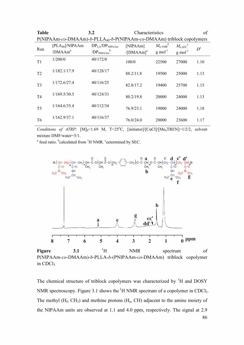
86
Table 3.2 Characteristics of
P(NIPAAm-co-DMAAm)-b-PLLA40-b-P(NIPAAm-co-DMAAm) triblock copolymers
Run [PLA40]/NIPAAm
/DMAAma
DPLA/DPNIPAAm
/DPDMAAmb
[NIPAAm]
/[DMAAm]a
Mn,NMRb
g mol-1
Mn,SECc
g mol-1
Ð
c
T1 1/200/0 40/172/0
100/0 22500 27000 1.10
T2 1/182.1/17.9 40/128/17
88.2/11.8 19500 25000 1.13
T3 1/172.6/27.4 40/116/25
82.8/17.2 19400 25700 1.15
T4 1/169.5/30.5 40/124/31
80.2/19.8 20000 24000 1.13
T5 1/164.6/35.4 40/112/34
76.9/23.1 19000 24000 1.18
T6 1/162.9/37.1 40/116/37
76.0/24.0 20000 23600 1.17
Conditions of ATRP: [M]0=1.69 M, T=25oC, [initiator]/[CuCl]/[Me6TREN]=1/2/2, solvent
mixture DMF/water=5/1. a feed ratio.
bcalculated from
1H NMR.
ceetermined by SEC.
Figure 3.1 1H NMR spectrum of
P(NIPAAm-co-DMAAm)-b-PLLA-b-(PNIPAAm-co-DMAAm) triblock copolymer
in CDCl3
The chemical structure of triblock copolymers was characterized by 1H and DOSY
NMR spectroscopy. Figure 3.1 shows the 1
H NMR spectrum of a copolymer in CDCl3.
The methyl (Hf, CH3) and methine protons (He, CH) adjacent to the amine moiety of
the NIPAAm units are observed at 1.1 and 4.0 ppm, respectively. The signal at 2.9

87
ppm is assigned to the methyl proton (Hg, CH3) of DMAAm. The signals at 1.5 and
5.1 ppm are characteristic of the methyl (Hb) and methine protons (Ha, CH) of PLLA.
The [LA]/[NIPAAm]/[DMAAm] ratio was calculated from the integrations of the
methine protons (-CH(CH3)-) of lactyl units at 5.1 ppm, methine protons
(-NHCH(CH3)2-) of NIPAAm units at 4.0 ppm and methyl protons (-(CH3)2) of
DMAAm at 2.9 ppm (equation 5). The DP of NIPAAm and DMAAm units, and the
Mn of copolymers were then obtained using following equations:
[LA]/[NIPAAm]/[DMAAm] = Ia / Ie / (Ig/6) (5)
DPNIPAAm = DPLA / ([LA]/[NIPAAm]) (6)
DPDMAAm = DPLA / ([LA]/[DMAAm]) (7)
MnNMR = 72 x DPLA + 113 x DPNIPAAm+ 98 x DPDMAAm (8)
Where 72, 113 and 98 are the molecular weight of LA, NIPAAm and DMAAm units,
respectively.
The efficiency of polymerization was also evaluated by DOSY NMR, one of the
powerful tools to characterize block copolymers [41]. DOSY NMR is a two
dimensional NMR technique in which the signal decays exponentially due to the
self-diffusion behavior of individual molecules. This leads to two dimensions: the first
dimension (F2) accounts for the conventional chemical shift and the second one (F1)
for self-diffusion coefficients (D). Thus, each component in a mixture can be virtually
separated, based on its own diffusion coefficient on the diffusion dimension. Figure
3.2 shows the DOSY pattern of copolymer T4 in dilute DMSO-d6. The 1H NMR
spectrum exhibits signals corresponding to PLLA (δ=5.1 and 1.5 ppm), NIPAAm
(δ=3.9 ppm) and DMAAm (δ=2.7, 2.8, 2.9 and 3.0 ppm). As shown on the DOSY
pattern, the 1H NMR signals of PLA and P(NIPAAm-co-DMAAm) present the same
diffusion coefficient D=2.291 × 10⁻¹¹
m2s
2, in agreement with efficient
copolymerization of NIPAAm and DMAAm on the Br-PLLA-Br macroinitiator. No
free PNIPAAm was observed since the diffusion coefficient of PNIPAAm in dilute
DMSO-d6 was found to be D=5.248×10⁻¹¹
m2s
2.

88
Figure 3.2 DOSY NMR spectrum of
P(NIPAAm-co-DMAAm)-b-PLLA-P(NIPAAm-co-DMAAm) triblock copolymer in
DMSO-d6.
Figure 3.3 SEC chromatograms of Br-PLLA-Br macroinitiator and triblock
copolymers (T1 and T4).
Figure 3.3 shows the SEC traces of the copolymers T1 and T4 in comparison with the
Br-PLLA-Br macroinitiator. All the polymers exhibit narrow and unimodal molecular
weight distributions. Furthermore, a shift of the copolymer traces towards higher

89
molecular weights is observed as compared to Br-PLLA-Br, indicating successful
synthesis of block copolymers.
Table 3.2 summarizes the characteristics of the various copolymers. The
[NIPAAm]/[DMAAm] molar ratio varies from 100/0 to 76/24 in the copolymers.
DPNIPAAm varies from 172 to 112, and DPDMAAm varies from 0 to 37. The Mn obtained
from SEC ranges from 23600 to 27000 g mol-1
with low dispersity (Đ=1.10-1.18). A
good agreement was observed between the Mn,NMR and Mn,SEC values although the
former is slightly lower than the latter.
After polymerization, the catalyst was removed by filtration through a basic alumina
column. The copolymers recovered by precipitation in diethyl ether appeared almost
colorless. ICP-MS measurements showed that the copper content in the copolymers
was between 1 and 3 ppm.
3.3.2 Self-assembling of triblock copolymer micelles in aqueous
medium
The physico-chemical properties of the amphiphilic copolymers in aqueous solution
were determined in order to evaluate their potential as drug carrier. The different
copolymers are water soluble and are able to form micellar aggregates by
self-assembly in water using the direct dissolution method. The critical micelle
concentration (CMC) was determined by fluorescence spectroscopy using pyrene as
hydrophobic probe. Figure 3.4 shows the I337/I333 ratio changes as a function of
polymer concentration. The intensity ratio exhibits a substantial increase at a
particular concentration, indicating the incorporation of pyrene into micelles. The
CMC was obtained from the intersection of the two linear regression lines of the plots.

90
Figure 3.4 Plots of the I337/I333 ratio changes from pyrene excitation spectra versus the
concentration of P(NIPAAm-co-DMAAm)-b-PLLA-b-P(NIPAAm-co-DMAAm)
(T4)
Table 3.3 Properties of
P(NIPAAm-co-DMAAm)-b-PLLA-b-P(NIPAAm-co-DMAAm) micelles
Run DPLA/DPNIPAAm
/DPDMAAma
[NIPAAm]
/[DMAAm]a
LCSTb
oC
CMCc
mg mL-1
DHd
nm
PDId
T1 40/172/0 100/0 32.3 0.010 40 0.12
T2 40/128/17 88.2/11.8 35.6 - 42 0.17
T3 40/116/25 82.8/17.2 37.1 0.013 42 0.21
T4 40/124/31 80.2/19.8 37.8 0.014 43 0.15
T5 40/112/34 76.9/23.1 38.8 - 55 0.22
T6 40/116/37 76.0:24.0 39.1 0.015 53 0.21
a) Calculated by NMR; b) Determined by UV spectroscopy; c) Determined by fluorescence
spectroscopy; d) Determined by DLS.
As shown in Table 3.3, the CMC value of PNIPAAm-b-PLLA-b-PNIPAAm (T1) is
0.010 mg mL-1
, while those of
P(NIPAAm-co-DMAAm)-b-PLLA-b-P(NIPAAm-co-DMAAm) are slightly higher.
The CMC is dependent on different factors such as the composition, the hydrophilic
to hydrophobic ratio, etc. The higher CMC value of
P(NIPAAm-co-DMAAm)-b-PLLA-b-P(NIPAAm-co-DMAAm) as compared to
PNIPAAm-b-PLLA-b-PNIPAAm is attributed to the presence of more hydrophilic

91
DMAAm units. It is also noted that the CMC values of the copolymers are lower that
of the diblock copolymer with NIPAAm/DMAAm/LA ratio of 54/29/14 (0.022 mg
mL-1
) reported by Akimoto et al., but higher than those of the copolymers
P(NIPAAm/DMAAm)118-PLA59 and P(NIPAAm/DMAAm)118-PCL60 reported by Li
et al. (0.00184 and 0.00398 mg mL-1
, respectively) [34, 37]. These findings can be
assigned to the fact that higher hydrophobic content leads to lower CMC. The very
low CMC values demonstrate the strong tendency of copolymers toward formation of
micelles, which is of major importance for the long circulation of micelles in the
blood stream after injection induced dilution [42, 43].
3.3.3 Morphology and size distribution of micelles
TEM experiments were performed to examine the morphology of the self-assembling
aggregates. As shown in Figure 3.5A, the micelles of copolymer T4 are spherical in
shape with an average diameter of 32 nm. The size distribution of the copolymer
micelles was determined by DLS (Figure 3.5B). The micelles exhibit a unimodal and
narrow size distribution with an average hydrodynamic diameter of 43 nm and a
dispersity of 0.15.
As frequently reported in the literature, the size obtained from DLS is larger than that
from TEM. This difference could be attributed to the experimental conditions. In fact,
DLS determines the hydrodynamic diameter of micelles in aqueous solution, whereas
TEM shows the dehydrated solid state of micelles. Table 3.3 shows the DLS data of
all copolymer micelles in water at 25 oC. The average diameter varies from 40 to 55
nm with narrow size distribution (Ð = 0.15 to 0.22). The size of copolymer micelles
increases with increasing the content of hydrophilic DMAAm units. The nano-size of
micelles should allow them to escape from the reticuloendothelial system (RES) and
preferentially accumulate in tumor tissues through the enhanced permeability and
retention (EPR) effect [44, 45].

92
Figure 3.5 TEM (A) and DLS (B) results of the self-assembling micelles of
copolymerT4 in aqueous medium.
It is noteworthy that the size of copolymer micelles is larger than that of the diblock
copolymer (23 nm) reported by Akimoto et al., but smaller than those of the
copolymers P(NIPAAm/DMAAm)118-PLA59 and P(NIPAAm/DMAAm)118-PCL60
(170 and 87 nm, respectively) reported by Li et al. [34].
3.3.4 Thermo-responsive behavior of micelles
Our strategy for drug release is to use thermo-sensitive copolymers to target tumor
tissue under hyperthermia. Therefore, the LCST should be slightly higher than the
body temperature. Thermo-responsive polymers become insoluble when the
temperature increases above the LCST due to coil-to-globule transition. This phase

93
transition can be evidenced by light transmission measurements of micellar solution
as a function of temperature.
Figure 3.6 Plots of transmittance as a function of temperature (A) and plot of LCST
values as a function (B) of DMAAm content for copolymer solutions at 3.0 mg mL-1
.
Figure 3.6 shows the transmission changes of different polymeric micelle solutions at
3.0 mg mL-1
with increasing temperature. A sharp transmittance decrease is detected
around the LCST. The resulting LCST values are listed in Table 3.3. As expected, the
LCST increases from 32.3 to 39.1 oC with the increase of DMAAm content in the

94
copolymers (Table 3.3). Moreover, a linear relationship is obtained between the LCST
and the molar fraction of DMAAm up to 24 %, as shown in Figure 3.6B.
Therefore, the LCST can be tuned to temperatures inside tumor tissue by simply
increasing the content of DMAAm units in the copolymers. This finding is consistent
with literature data. In fact, Shen et al. reported that the LCST linearly increases with
increasing the content of DMAAm in P(NIPAAm-co-DMAAm) copolymers [46]. The
LCST of P(NIPAAm-co-DMAAm)-b-PLLA diblock copolymers with maleimide
endgroup reported by Akimoto et al. and Li et al. is about 40 oC despite the different
P(NIPAAm-co-DMAAm) to PLA ratios [34, 37]. In fact, diblock copolymers with
maleimide endgroup lead to surface-functional thermo-responsive micelles, and the
LCST of micelles can be tuned by mixing hydrophobic DTBz-surface micelles and
Mal-surface ones [34]. In our case, the LCST of
P(NIPAAm-co-DMAAm)-b-PLLA-b-P(NIPAAm-co-DMAAm) triblock copolymers
can be precisely adjusted to above the body temperature by varying the NIPAAm to
DMAAm ratio, which presents great interest for targeted delivery of anticancer drugs.
Moreover, introduction of DMAAm not only leads to LCST increase of copolymers,
but also leads to an extremely narrow phase transition interval within 0.5 oC. As
shown in Figure 3.6A, the optical transmittance of PNIPAAm-b-PLLA-b-PNIPAAm
(T1) decreases from 100 % to 0 with increasing temperature between 30.0 to 32.8 oC
(∆T=2.8 oC). Similar behavior was reported in literature. Indeed, for
PNIPAAm-b-PLLA diblock copolymer, a broad phase transition interval of ∆T=5 oC
was observed [17, 18, 22]. In the case of DMAAm containing copolymer T4, the
transmittance decrease from 100 % to 0 occurs in the temperature range of 37.6 to
38.0 oC, with a phase transition interval ∆T=0.4
oC. Narrow phase transition intervals
below 0.5 oC were also observed for other DMAAm containing copolymers as shown
in Figure 3.6A. Nakayama et al. reported phase transition intervals above 3 oC for
P(NIPAAm-co-DMAAm)-b-PDLLA copolymers obtained by combination of radical

95
polymerization and ROP [32, 33]. On the contrary, Akimoto et al. reported narrow
phase transition intervals within 0.5 oC of such copolymers by combination of RAFT
and ROP, which is consistent with our work [34].
The phase transition of copolymers is illustrated by DLS. Figure 3.7 shows the
reversible changes of the hydrodynamic diameter distribution depending on
temperature. The size of the micelles increases from 35 to 640 nm when the
temperature is raised from 25 to 45 oC. After quickly cooling down to 25
oC, the
micelle size decreases to 38 nm. In fact, P(NIPAAm-co-DMAAm) chains are
extended and coil-like in water under the LCST. When the temperature approaches the
LCST, dehydration of P(NIPAAm-co-DMAAm) occurs, leading to precipitation of
polymers and aggregation.
Figure 3.7 Reversible changes of the hydrodynamic diameter for T4 micelles at 3 mg
mL-1
when the temperature is raised from 25 () to 45 oC (), and cooled down to 25
oC ().
The observed phase transition is due to a coil-to-globule transition governed by
cooperative dehydration of hydrophilic chains. In the case of PNIPAAm, the
hydrogen-bonding site (amide group) is blocked by a bulky hydrophobic group
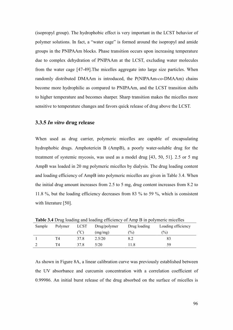
96
(isopropyl group). The hydrophobic effect is very important in the LCST behavior of
polymer solutions. In fact, a “water cage” is formed around the isopropyl and amide
groups in the PNIPAAm blocks. Phase transition occurs upon increasing temperature
due to complex dehydration of PNIPAAm at the LCST, excluding water molecules
from the water cage [47-49].The micelles aggregate into large size particles. When
randomly distributed DMAAm is introduced, the P(NIPAAm-co-DMAAm) chains
become more hydrophilic as compared to PNIPAAm, and the LCST transition shifts
to higher temperature and becomes sharper. Sharp transition makes the micelles more
sensitive to temperature changes and favors quick release of drug above the LCST.
3.3.5 In vitro drug release
When used as drug carrier, polymeric micelles are capable of encapsulating
hydrophobic drugs. Amphotericin B (AmpB), a poorly water-soluble drug for the
treatment of systemic mycosis, was used as a model drug [43, 50, 51]. 2.5 or 5 mg
AmpB was loaded in 20 mg polymeric micelles by dialysis. The drug loading content
and loading efficiency of AmpB into polymeric micelles are given in Table 3.4. When
the initial drug amount increases from 2.5 to 5 mg, drug content increases from 8.2 to
11.8 %, but the loading efficiency decreases from 83 % to 59 %, which is consistent
with literature [50].
Table 3.4 Drug loading and loading efficiency of Amp B in polymeric micelles
Sample Polymer LCST
(oC)
Drug/polymer
(mg/mg)
Drug loading
(%)
Loading efficiency
(%)
1 T4 37.8 2.5/20 8.2 83
2 T4 37.8 5/20 11.8 59
As shown in Figure 8A, a linear calibration curve was previously established between
the UV absorbance and curcumin concentration with a correlation coefficient of
0.99986. An initial burst release of the drug absorbed on the surface of micelles is

97
observed in all cases. Sample 2 with higher drug content exhibits slower drug release,
which could be attributed to the poor solubility of AmpB (Figure 3.8B).
Figure 3.8 Drug release profiles of AmpB loaded polymeric micelles (Samples 1 and
2) at 37 and 38 oC in water.
On the other hand, the in vitro drug release from polymer micelles presents a
thermo-sensitive behavior. At temperature below the LCST (37 °C), the release of
AmpB is relatively slow. After 3 days, nearly 50 % and 32 % of drug are released for

98
sample 1 and sample 2, respectively. In contrast, at 38 °C which is above the LCST,
the release rate is much faster with 90 and 75 % of released drug in both samples.
These preliminary results suggest that drug loaded micelles could be injected to a
human body and achieve quick drug release at the tumor site where the temperature is
higher than the LCST. Thus, the drug release behavior indicates that these copolymers
could be potential candidates for applications in targeted delivery of drugs.
Li et al. comparatively studied the release of adriamycin from maleimide
end-functional P(NIPAAm-co-DMAAm)-b-PLA and
P(NIPAAm-co-DMAAm)-b-PCL copolymer micelles under physiological conditions
(37 °C and pH = 7.3) and in simulated tumor environment (40 °C and pH = 5.3) [37].
The authors observed that only half of entrapped adriamycin was released out within
one week under physiological conditions, while about 90 % adriamycin was released
within 10 h in simulated tumor environment. Nevertheless, faster degradation of
micelles at 40 °C and pH = 5.3 could have contributed to the faster drug release
together with the LCST effect. Nakayama et al. studied the release of doxorubicin
from P(NIPAAm-co-DMAAm)-b-PCL and P(NIPAAm-co-DMAAm)-b-P(LA-co-CL)
copolymer micelles below and above the LCST [32]. No difference was detected in
the case of P(NIPAAm-co-DMAAm)-b-PCL at 42.5 °C and 37 °C. In contrast, the
release of doxorubicin is much faster at 41°C than that at 35 °C in the case of
P(NIPAAm-co-DMAAm)-b-P(LA-co-CL). Again, degradation of micelles plays a key
role in drug release because P(LA-co-CL) degrades much faster than PCL [52]. In our
case, the faster release of AmpB can be assigned to the destabilization of micelles
above the LCST since little difference of degradation could be expected at 38 °C and
37 °C.
3.3.6 Cytocompatibility
In vitro cytotoxicity is generally evaluated by MTT assay for the screening of
biomaterials [53]. MTT assay is based on the reaction between MTT and

99
mitochondrial succinate dehydrogenases in living cells to form a purple formazan
which is soluble in DMSO but insoluble in water. The OD value of formazan-DMSO
solution is considered to be proportional to the number of living cells.
Figure 3.9 Optical density values of MCM (A) and MEF(B) solutions after 2, 3 and 4
days culture in DMEM with copolymer substrate (T4), and controls (DMEM and 5 %
phenol
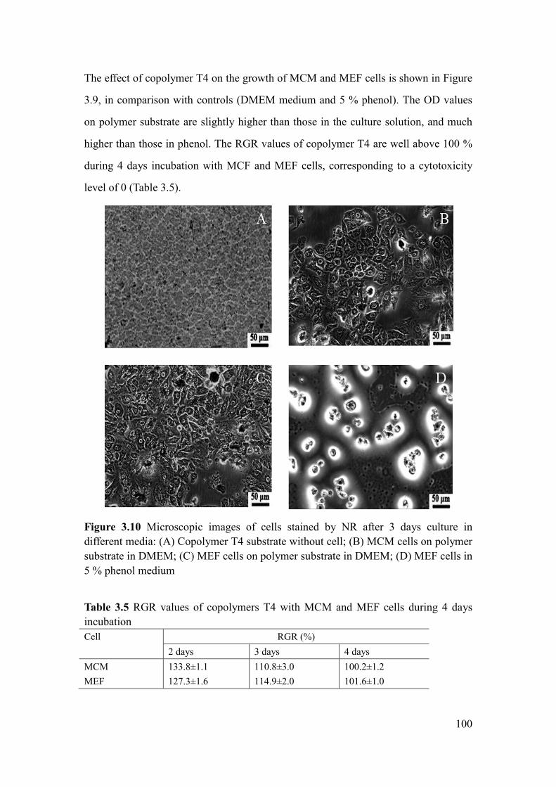
100
The effect of copolymer T4 on the growth of MCM and MEF cells is shown in Figure
3.9, in comparison with controls (DMEM medium and 5 % phenol). The OD values
on polymer substrate are slightly higher than those in the culture solution, and much
higher than those in phenol. The RGR values of copolymer T4 are well above 100 %
during 4 days incubation with MCF and MEF cells, corresponding to a cytotoxicity
level of 0 (Table 3.5).
Figure 3.10 Microscopic images of cells stained by NR after 3 days culture in
different media: (A) Copolymer T4 substrate without cell; (B) MCM cells on polymer
substrate in DMEM; (C) MEF cells on polymer substrate in DMEM; (D) MEF cells in
5 % phenol medium
Table 3.5 RGR values of copolymers T4 with MCM and MEF cells during 4 days
incubation
Cell RGR (%)
2 days 3 days 4 days
MCM 133.8±1.1 110.8±3.0 100.2±1.2
MEF 127.3±1.6 114.9±2.0 101.6±1.0
101
Figure 3.10 shows the morphology of cells after 3 days culture. Large number of cells
died in the toxic phenol medium (Figure 3.10D). On the contrary, cell adhesion and
proliferation are observed on the copolymer substrate (Figure 3.10B and Figure
3.10C). Cells exhibit spindle, polygon or oval shapes, and the pseudopodium of cells
stretches out. Therefore, MTT assay and cell morphology observation indicate that
P(NIPAAm-co-DMAAm)-b-PLLA-b-P(NIPAAm-co-DMAAm) copolymers presents
outstanding cytocompatibility and could be used for biomedical applications.
3.4 Conclusion
P(NIPAAm-co-DMAAm)-b-PLLA-b-P(NIPAAm-co-DMAAm) triblock copolymers
were prepared by ATRP using Br-PLLA-Br as macroinitiator. The resulting
copolymers present well defined molecular weights and narrow dispersities. The
LCST of the copolymers linearly increases from 32.3 to 39.1 °C with increasing the
DMAAm content. Self-assembling micelles were prepared by dissolving the
copolymers in aqueous medium. The copolymers have very low CMC (10 to 15 µg
mL-1
) and nano-size (40 to 55 nm). The presence of DMAAm units in the copolymers
leads to an extremely sharp phase transition, as compared to
PNIPAAm-b-PLLA-b-PNIPAAm copolymers. Moreover, reversible size changes
were observed when the micelle solutions was heated from 25 to 45 oC and then
cooled down to 25 oC. Much faster drug release is detected at temperatures above the
LCST in in vitro studies. The MTT assay and cell morphology observation confirm
the good biocompatibility of these thermo-sensitive copolymers.
Therefore, the small size and low CMC, the LCST slightly above the body
temperature, the rapid phase transition at LCST, the thermo-responsive drug release
behavior above LCST, and the good cytocompatilibity indicate that
P(NIPAAm-co-DMAAm)-b-PLLA-b-P(NIPAAm-co-DMAAm) copolymers are
promising candidates for applications in targeted delivery of anticancer drugs.

102
In this chapter, the preliminary study of drug release has been performed in water.
However, the properties of micelles in physiological conditions (pH=7.4, PBS) should
be investigated to determine the effects of salt and drug loading on the LCST, the size
and morphology of micelles. In the next chapter, an anticancer drug, curcumin, has
been used as drug model for a systemic study on the drug release properties of
P(NIPAAm-co-DMAAm)-b-PLLA-b-P(NIPAAm-co-DMAAm) copolymers.
3.5 References
[1] A. K. Bajpai, S. K. Shukla, S. Bhanu, S. Kankane, Responsive polymers in controlled drug
delivery. Prog. Polym. Sci. 33 (2008) 1088-1118.
[2] A. Kumar, A. Srivastava, I. Y. Galaev, B. Mattiasson, Smart polymers: Physical forms and
bioengineering applications. Prog. Polym. Sci. 32 (2007) 1205-1237.
[3] B. Guillerm, V. Darcos, V. Lapinte, S. Monge, J. Coudane, J. J. Robin, Synthesis and
evaluation of triazole-linked poly(ε-caprolactone)-graft-poly(2-methyl-2-oxazoline) copolymers
as potential drug carriers. Chemical Communications 48 (2012) 2879-2881.
[4] W. Y. Ooi, M. Fujita, P. J. Pan, H. Y. Tang, K. Sudesh, K. Ito, N. Kanayama, T. Takarada, M.
Maeda, Structural characterization of nanoparticles from thermoresponsive
poly(N-isopropylacrylamide)-DNA conjugate. J. Colloid Interface Sci. 374 (2012) 315-320.
[5] A. Wang, C. Tao, Y. Cui, L. Duan, Y. Yang, J. Li, Assembly of environmental sensitive
microcapsules of PNIPAAm and alginate acid and their application in drug release. J. Colloid
Interface Sci. 332 (2009) 271-279.
[6] C. H. Hu, X. Z. Zhang, L. Zhang, X. D. Xu, R. X. Zhuo, Temperature- and pH-sensitive
hydrogels to immobilize heparin-modified PEI/DNA complexes for sustained gene delivery. J.
Mater. Chem. 19 (2009) 8982-8989.
[7] Y. Liu, X. Cao, M. Luo, Z. Le, W. Xu, Self-assembled micellar nanoparticles of a novel star
copolymer for thermo and pH dual-responsive drug release. J. Colloid Interface Sci. 329 (2009)
244-252.
[8] P. VanRijn, H. Park, K. O. Nazli, N. C. Mougin, A. Boker, Self-Assembly Process of Soft
Ferritin-PNIPAAm Conjugate Bionanoparticles at Polar-Apolar Interfaces. Langmuir 29 (2013)
276-284.
[9] N. M. Matsumoto, P. Prabhakaran, L. H. Rome, H. D. Maynard, Smart Vaults:
Thermally-Responsive Protein Nanocapsules. ACS Nano 7 (2013) 867-874.
[10] H. Wei, S. X. Cheng, X. Z. Zhang, R. X. Zhuo, Thermo-sensitive polymeric micelles based
on poly(N-isopropylacrylamide) as drug carriers. Prog. Polym. Sci. 34 (2009) 893-910.
[11] H. G. Schild, Poly (N-isopropylacrymide)-experiment, theory and application. Prog. Polym.
Sci. 17 (1992) 163-249.
[12] W. R. H senff, Influence of cross-link density on rheological properties of
temperature-sensitive microgel suspensions. collioid polym sci 278 (2000) 830-840.
[13] A. C. Albertsson, I. Varma, Degradable Aliphatic Polyesters, Vol. 157, Springer Berlin

103
Heidelberg, 2002, pp. 1-40.
[14] G. S. He, L. L. Ma, J. Pan, S. Venkatraman, ABA and BAB type triblock copolymers of PEG
and PLA: A comparative study of drug release properties and"stealth" particle characteristics. Int.
J. Pharm. 334 (2007) 48-55.
[15] L. Yang, A. E. Ghzaoui, S. M. Li, In vitro degradation behavior of
poly(lactide)-poly(ethylene glycol) block copolymer micelles in aqueous solution. Int. J. Pharm.
400 (2010) 96-103.
[16] I. Barwal, A. Sood, M. Sharma, B. Singh, S. C. Yadav, Development of stevioside
Pluronic-F-68 copolymer based PLA-nanoparticles as an antidiabetic nanomedicine. Colloid Surf.
B-Biointerfaces 101 (2007) 510-516.
[17] K. Fukashi, S. Kiyotaka, A. Takao, Y. Masayuki, S. Yasuhisa, O. Teruo, Preparation and
characterization of thermally responsive block copolymer micelles comprising
poly(N-isopropylacrylamide-b-DLlactide). J. Controlled Release 55 (1998) 87-98.
[18] J. Qin, Y. S. Jo, J. E. Ihm, D. K. Kim, M. Muhammed, Thermosensitive Nanospheres with a
Gold Layer Revealed as Low-Cytotoxic Drug Vehicles. Langmuir 21 (2005) 9346-9351.
[19] E. Ayano, M. Karaki, T. Ishihara, H. Kanazawa, T. Okano, Poly
(N-isopropylacrylamide)-PLA and PLA blend nanoparticles for temperature-controllable drug
release and intracellular uptake. Colloids Surf. B: Biointerfaces 99 (2012) 67-73.
[20] Y. Z. You, C. Y. Hong, W. P. Wang, W. Q. Lu, C. Y. Pan, Preparation and Characterization of
Thermally Responsive and Biodegradable Block Copolymer Comprised of PNIPAAM and PLA
by Combination of ROP and RAFT Methods. Macromolecules 37 (2004) 9761-9767.
[21] M. Hales, C. Barner-Kowollik, T. P. Davis, M. H. Stenzel, Shell-Cross-Linked Vesicles
Synthesized from Block Copolymers of Poly(D,L-lactide) and Poly(N-isopropylacrylamide) as
Thermoresponsive Nanocontainers. Langmuir 20 (2004) 10809-10817.
[22] H. Wei, X. Z. Zhang, C. Cheng, S. X. Cheng, R. X. Zhuo, Self-assembled, thermosensitive
micelles of a star block copolymer based on PMMA and PNIPAAm for controlled drug delivery.
Biomaterials 28 (2007) 99-107.
[23] S. J. T. Rezaei, M. R. Nabid, H. Niknejad, A. A. Entezami, Folate-decorated
thermoresponsive micelles based on star-shaped amphiphilic block copolymers for efficient
intracellular release of anticancer drugs. Int. J. Pharm. 437 (2012) 70-79.
[24] S. J. T. Rezaei, M. R. Nabid, H. Niknejad, A. A. Entezami, Multifunctional and
thermoresponsive unimolecular micelles for tumor-targeted delivery and site-specifically release
of anticancer drugs. Polymer 53 (2012) 3485-3497.
[25] F. N. Chearuil, O. I. Corrigan, Thermosensitivity and release from poly
N-isopropylacrylamide-polylactide copolymers. Int. J. Pharm. 366 (2009) 21-30.
[26] J. Zhang, R. D. K. Misra, Magnetic drug-targeting carrier encapsulated with thermosensitive
smart polymer: Core-shell nanoparticle carrier and drug release response. Acta Biomater. 3 (2007)
838-850.
[27] F. Kohori, K. Sakai, T. Aoyagi, M. Yokoyama, M. Yamato, Y. Sakurai, T. Okano, Control of
adriamycin cytotoxic activity using thermally responsive polymeric micelles composed of
poly(N-isopropylacrylamide-co-N,N-dimethylacrylamide)-b-poly(d,l-lactide). Colloids and
Surfaces B: Biointerfaces 16 (1999) 195-205.
[28] S. Q. Liu, Y. Y. Yang, X. M. Liu, Y. W. Tong, Preparation and Characterization of
Temperature-Sensitive Poly(N-isopropylacrylamide)-b-poly(d,l-lactide) Microspheres for Protein
104
Delivery. Biomacromolecules 4 (2003) 1784-1793.
[29] S. Q. Liu, Y. W. Tong, Y. Y. Yang, Incorporation and in vitro release of doxorubicin in
thermally sensitive micelles made from
poly(N-isopropylacrylamide-co-N,N-dimethylacrylamide)-b-poly(d,l-lactide-co-glycolide) with
varying compositions. Biomaterials 26 (2005) 5064-5074.
[30] S. Q. Liu, Y. W. Tong, Y. Y. Yang, Thermally sensitive micelles self-assembled from
poly(N-isopropylacrylamide-co-N,N-dimethylacrylamide)-b-poly(D,L-lactide -co-glycolide) for
controlled delivers of paclitaxel. Mol. Biosyst. 1 (2005) 158-165.
[31] S. Q. Liu, N. Wiradharma, S. J. Gao, Y. W. Tong, Y. Y. Yang, Bio-functional micelles
self-assembled from a folate-conjugated block copolymer for targeted intracellular delivery of
anticancer drugs. Biomaterials 28 (2007) 1423-1433.
[32] M. Nakayama, T. Okano, T. Miyazaki, F. Kohori, K. Sakai, M. Yokoyama, Molecular design
of biodegradable polymeric micelles for temperature-responsive drug release. J. Controlled
Release 115 (2006) 46-56.
[33] M. Nakayama, J. E. Chung, T. Miyazaki, M. Yokoyama, K. Sakai, T. Okano, Thermal
modulation of intracellular drug distribution using thermoresponsive polymeric micelles. React.
Funct. Polym. 67 (2007) 1398-1407.
[34] J. Akimoto, M. Nakayama, K. Sakai, T. Okano, Molecular Design of Outermost Surface
Functionalized Thermoresponsive Polymeric Micelles with Biodegradable Cores. J. Polym. Sci.
Part A: Polym. Chem. 46 (2008) 7127-7137.
[35] J. Akimoto, M. Nakayama, K. Sakai, T. Okano, Temperature-Induced Intracellular Uptake of
Thermoresponsive Polymeric Micelles. Biomacromolecules 10 (2009) 1331-1336.
[36] J. Akimoto, M. Nakayama, K. Sakai, T. Okano, Thermally Controlled Intracellular Uptake
System of Polymeric Micelles Possessing Poly(N-isopropylacrylamide)-Based Outer Coronas.
Mol. Pharm. 7 (2010) 926-935.
[37] W. Li, J. F. Li, J. Gao, B. H. Li, Y. Xia, Y. C. Meng, Y. S. Yu, H. W. Chen, J. X. Dai, H. Wang,
Y. J. Guo, The fine-tuning of thermosensitive and degradable polymer micelles for enhancing
intracellular uptake and drug release in tumors. Biomaterials 32 (2011) 3832-3844.
[38] Y. F Hu, V. Darcos, S. Monge, S. M. Li, Synthesis and self-assembling of
poly(N-isopropylacrylamide-block-poly(L-lactide)-block-poly(N-isopropylacrylamide) triblock
copolymers prepared by combination of ring-opening polymerization and atom transfer radical
polymerization. J. Polym. Sci. Part A: Polym. Chem. 51 (2013) 3274-3283..
[39] G. Masci, L. Giacomelli, V. Crescenzi, Atom transfer radical polymerization of
N-isopropylacrylamide. Macromol. Rapid Commun. 25 (2004) 559-564.
[40] K. Bauri, S. Roy, S. Arora, R. Dey, A. Goswami, G. Madras, P. De, Thermal degradation
kinetics of thermo-responsive poly(N-isopropylacrylamide-co-N,N-dimethylacrylamide)
copolymers prepared via RAFT polymerization. J.Therm. Anal. Calorim. 111 (2013) 753-761.
[41] Y. Bakkour, V. Darcos, S. M. Li, J. Coudane, Diffusion ordered spectroscopy (DOSY) as a
powerful tool for amphiphilic block copolymer characterization and for critical micelle
concentration (CMC) determination. Polym. Chem. 3 (2012) 2006-2010.
[42] M. Yokoyama, T. Okano, Y. Sakurai, H. Ekimoto, C. Shibazaki, K. Kataoka, Toxicity and
Antitumor Activity against Solid Tumors of Micelle-forming Polymeric Anticancer Drug and Its
Extremely Long Circulation in Blood. Cancer Res.51 (1991) 3229-3236.
[43] M. L. Adams, A. Lavasanifar, G. S. Kwon, Amphiphilic Block Copolymers for Drug Delivery.

105
J. Pharm. Sci. 92 (2003) 1343-1355.
[44] Y. Yan, G. K. Such, A. P. R. Johnston, J. P. Best, F. Caruso, Engineering Particles for
Therapeutic Delivery: Prospects and Challenges. ACS Nano 6 (2012) 3663-3669.
[45] C. Oerlemans, W. Bult, M. Bos, G. Storm, J. F. Nijsen, W. Hennink, Polymeric Micelles in
Anticancer Therapy: Targeting, Imaging and Triggered Release. Pharm. Res. 27 (2010)
2569-2589.
[46] Z. Shen, K. Terao, Y. Maki, T. Dobashi, G. Ma, T. Yamamoto, Synthesis and phase behavior
of aqueous poly(N-isopropylacrylamide-co-acrylamide),
poly(N-isopropylacrylamide-co-N,N-dimethylacrylamide) and
poly(N-isopropylacrylamide-co-2-hydroxyethyl methacrylate). Colloid Polym. Sci. 284 (2006)
1001-1007.
[47] E. C. Cho, J. Lee, K. Cho, Role of Bound Water and Hydrophobic Interaction in Phase
Transition of Poly(N-isopropylacrylamide) Aqueous Solution. Macromolecules 36 (2003)
9929-9934.
[48] Y. Okada, F. Tanaka, Cooperative Hydration, Chain Collapse, and Flat LCST Behavior in
Aqueous Poly(N-isopropylacrylamide) Solutions. Macromolecules 38 (2005) 4465-4471.
[49] Y. Ono, T. Shikata, Hydration and Dynamic Behavior of Poly(N-isopropylacrylamide)s in
Aqueous Solution:A Sharp Phase Transition at the Lower Critical Solution Temperature. J. Am.
Chem. Soc. 128 (2006) 10030-10031.
[50] K. C. Choi, J. Y. Bang, P. I. Kim, C. Kim, C. E. Song, Amphotericin B-incorporated
polymeric micelles composed of poly(d,l-lactide-co-glycolide)/dextran graft copolymer. Int. J.
Pharm. 355 (2008) 224-230.
[51] T. A. Diezi, J. K. Takemoto, N. M. Davies, G. S. Kwon, Pharmacokinetics and nephrotoxicity
of amphotericin B-incorporated poly(ethylene glycol)-block-poly(N-hexyl stearate l-aspartamide)
micelles. J. Pharm. Sci. 100 (2011) 2064-2070.
[52] S. M. Li, J. L. Espartero, P. Foch, M. Vert, Structural characterization and hydrolytic
degradation of a Zn metal initiated copolymer of L-lactide and epsilon-caprolactone. J. Biomater.
Sci. Polym. Ed. 8 (1996) 165-187.
[53] Y. L. Luo, C. H. Zhang, F. Xu, Y. S. Chen, Novel THTPBA/PEG-derived highly branched
polyurethane scaffolds with improved mechanical property and biocompatibility. Polym. Adv.
Technol. 23 (2012) 551-557.

106

107
Chapter 4 Thermo-responsive release of curcumin from
P(NIPAAm-co-DMAAm)-b-PLLA-b-P(NIPAAm-co-DMAA
m) triblock copolymers micelles
Abstract Thermo-responsive micelles are prepared by self-assembly of amphiphilic
P(NIPAAm-co-DMAAm)-b-PLLA-b-P(NIPAAm-co-DMAAm) triblock copolymer
using solvent evaporation/film hydration method. The resulting micelles exhibit very
low critical micelle concentration (CMC) which slightly increases from 0.0113 to
0.0144 mg mL-1
while the DMAAm content increases from 31.8 to 39.4 % in the
hydrophilic P(NIPAAm-co-DMAAm) blocks. The lower critical solution temperatures
(LCST) of copolymers varies from 44.7 °C to 49.4 °C in water as determined by UV
spectroscopy, and decreases by ca. 3.5 °C in phosphate buffered saline (PBS).
Curcumin was encapsulated in the core of micelles. High drug loading up to 20 % is
obtained with high loading efficiency (>94 %). The LCST of drug loaded micelles
ranges from 37.5 to 38.0 oC with drug loading increasing from 6.0 to 20 %. The
micelles with diameters ranging from 48 to 88 nm remain stable over one month due
to the negative surface charge as determined by zeta potential (-12.4 to -18.7 mV).
Drug release studies were performed under in vitro conditions at 37 °C and 40 °C, i.e.
below and above the LCST, respectively. Initial burst release is observed in all cases,
followed by a slower release. The release rate is higher at 40 °C than that at 37 °C due
to thermo-responsive release across the LCST. Micelles with lower drug loading
exhibit higher release rate than those with higher drug loading, which is assigned to
the solubility effect. Peppas’ theory was applied to describe the release behaviors.
Moreover, the in vitro cytotoxicity of copolymers was evaluated using MTT assay.
The results show that the copolymers present good cytocompatibility.

108
Keywords: thermo-responsive; micelle; self-assembly; block copolymer;
poly(L-lactide); poly(N-isopropylacrylamide); curcumin

109
4.1 Introduction
Self-assembled micelles from amphiphilic block copolymers have been widely
investigated as nanocarrier for controlled release of drugs [1-4]. Nano-size polymeric
micelles exhibit outstanding properties such as reduced side effects, improved
therapeutic efficacy, selective targeting, stable storage, and prolonged blood
circulation time. Furthermore, polymeric micelles can achieve higher accumulation at
the target site through the enhanced permeability and retention (EPR) effect [5-7].
Curcumin, a natural polyphenol derived from curcuma longa, exhibits a wide
spectrum of pharmacological activities including anti-oxidant, anti-inflammatory,
anti-microbial, anti-amyloid and anti-tumor properties due to its diverse range of
molecular targets [8]. In the past decades, it was regarded as a prominent drug for
treating inflammation, cystic fibrosis, Alzheimer’s and malarial diseases and cancer
[9-12]. However, the therapeutic efficacy of curcumin is limited by its extremely low
solubility (11 ng mL-1
) in water [13]. Moreover, rapid metabolism and elimination
lead to poor bioactivity of curcumin [14].
Recently, polymer micelles have been used to encapsulate curcumin so as to enhance
the solubility, cellular uptake and anti-tumor bioactivity [15-19]. Micelles prepared
from poly(ethylene glycol)-b-poly(ε-caprolactone) (mPEG-b-PCL) diblock
copolymers have been largely studied for controlled delivery of curcumin. However,
mPEG-b-PCL micelles with drug loading of 9.9 % were unstable as aggregates were
observed in 72 hours [15]. Song et al. reported the preparation, pharmacokinetics and
distribution of curcumin loaded micelles using
poly(D,L-lactide-co-glycolide)-b-poly(ethylene glycol)-b-poly(D,L-lactide-co
-glycolide) (PLGA-b-PEG-b-PLGA) triblock copolymers [19]. The prolongation of
half-life, enhanced residence time and decreased total clearance indicate that the
micelles could prolong the acting time of curcumin in vivo. Nevertheless, both the
drug loading (6.4 %) and encapsulation efficiency (70 %) need to be improved.
110
Polymer-conjugate micelles were prepared to enhance the curcumin loading, but only
7.5 % cumulative drug release were obtained after 60 hours as drug release is
controlled by degradation of micelles [20].
Among the polymeric micelles described in literature, none is sensitive to
environmental changes. It is well known that different from normal tissues, tumor has
a particularly high temperature (~42 oC) [21]. Therefore, thermo-sensitive polymeric
micelles based on poly(N-isopropylacrylamide) (PNIPAAm) with a lower critical
solution temperature (LCST) at 32 oC attracted much attention for targeted
stimuli-responsive drug release [22-26]. The hydrophilic PNIPAAm outer shell
becomes hydrophobic by increasing the temperature above the LCST. The selective
accumulation of micelles at a specific site could be enhanced as a result of enhanced
micelle adsorption to cells by hydrophobic interactions between the polymeric
micelles. Therefore, thermo-responsive micelles could deliver the drugs via a
stimuli-responsive targeting process at tumor sites. In addition, nano-scaled
thermo-responsive polymeric micelles could combine the passive targeting by EPR
effect and active targeting by stimuli response, thus leading to accelerated release of
the encapsulated drug at desired sites.
To the best of our knowledge, no thermo-responsive curcumin encapsulated
polymeric micelles have been reported, so far. In this work, thermo-responsive
micelles were prepared from copolymers composed of a poly(L-lactide) (PLLA)
central block and two poly(N-isopropylacrylamide-co-N,N-dimethylacrylamide)
(P(NIPAAm-co-DMAAm)) lateral blocks, using solvent evaporation/film hydration
method. Curcumin was loaded in the core of micelles during the fabrication procedure.
Drug release studies were realized under in vitro conditions in a pH=7.4 phosphate
buffer saline at 37 and 40 oC. The cytotoxicity of micelles was evaluated using MTT
assay. The results are reported herein in comparison with literature data.

111
4.2 Materials and methods
4.2.1 Materials
α, ω-Bromopropionyl PLLA (Br-PLLA-Br) with degree of polymerization (DP) of 40
was prepared as previously reported [27]. Tris(2-dimethyl aminoethyl) amine
(Me6TREN), copper (I) chloride (CuCl), N-isopropylacrylamide (NIPAAm), N,
N-dimethyl formamide (DMF), chloroform, dicholoromethane, diethyl ether, basic
aluminum oxide, Tween 80, ethylenediaminetetraacetic acid tetrasodiumsalt hydrate
(EDTA-Na4·H2O) and curcumin were obtained from Sigma-Aldrich and used without
further purification. DMAAm was obtained from Sigma-Aldrich and purified through
a basic aluminum oxide column. Ultrapure water with a conductivity of 18 MΩ was
produced using a Millipore Milli-Q water system.
4.2.2 Synthesis of
P(NIPAAm-co-DMAAm)-b-PLA-b-P(NIPAAm-co-DMAAm) triblock
copolymer (T2, Table 2)
Triblock copolymers were prepared by atom transfer radical polymerization (ATRP)
at 25 oC using a DMF/water mixture solvent as reported previously [28]. Typically,
100 mg Br-PLLA-Br (30.4×10⁻³
mmol, Mn,NMR= 3300 g mol-1
), 511.4 mg NIPAAm
(4.52 mmol), 159.4 mg DMAAm (1.61 mmol) and 6.0 mg CuCl (60.8×10⁻³
mmol)
were dissolved in 3 mL DMF in a Schlenk tube. 0.5 mL H2O was then added. After
five freeze-pump-thaw cycles, 16µL Me6TREN (60.8×10⁻³
mmol) was added under
argon atmosphere. The mixture was stirred at 25 oC for 1 h. The reaction was stopped
by precipitation in diethyl ether. The crude product was dissolved in 10 mL
chloroform and passed through a basic aluminum oxide column for 3 times to remove
the catalyst complex. After concentration under reduced pressure, the solution was
precipitated again in diethyl ether. The product was dissolved in DMF and then
112
dialyzed for 3 days in a dialysis bag (MWCO: 3500) to remove the monomer and
trace copper against Mill-Q water adding ethylenediaminetetraacetic acid tetrasodium
salt hydrate. The product was then collected by lyophilization.
4.2.3 Preparation of curcumin-loaded polymeric micelles
Typically, 2.51mg curcumin and 17.53 mg copolymer T2 (Sample 2, Table 4.3) were
dissolved in 20 mL acetone. The solvent was evaporated in rotary evaporator at room
temperature to yield a membrane on the wall of the flask. After vacuum drying for 24
hours, 20 mL Milli-Q water was added to the flask. Drug loaded micelles were
obtained under stirring at room temperature. The micelle solution was then
centrifuged at 5000 rpm for 10 min to remove unload curcumin, and the supernatant
was lyophilized and stored at 4 oC before use.
4.2.4 In vitro drug release
The drug loading (DL) and encapsulation efficiency (EE) were determined as follows.
5 mg lyophilized micelles were dissolved in 5 mL acetonitrile/ammonium acetate (10
mM) mixture solution (pH= 4.0). Then the solution was diluted 100 times. The
concentration of curcumin was determined by UV-Vis spectroscopy at 424 nm, using
a previously established standard calibration curve. The drug loading and
encapsulation efficiency were calculated according to the following equations:
100×=micelles polymeric of weight
drug loaded of weight(%) loading Drug (1)
100×=loading drug ltheoretica
micelles in drug of weight(%) efficiency ionEncapsulat (2)
Drug loaded micelles (2 mg) were dispersed in 2 mL PBS solution (pH=7.4), and then
introduced into a dialysis bag (MWCO: 3500). The bag was placed in 10 mL PBS
containing 0.5 wt% Tween80 in an incubator (Heidolph 1000) with gentle shaking
(150 rpm) at 37 or 40 oC. At specific time intervals, the release medium was removed

113
and replaced by pre-warmed (37 oC) fresh medium. The withdrawn solution was then
diluted 10 times with pH=4.0 acetonitrile/ammonium acetate (10 mM) mixture
solution for UV measurement.
4.2.5 In vitro cytotoxicity
The cytotoxicity of copolymers was evaluated by MTT assay. After UV sterilization
for 5 hours, the copolymer T2 was dissolved in dulbecco’s modified eagle’s meduim
(DMEM, Hyclone products) at a concentration of 6 mg mL-1
. Then the solution was
transferred to 96-well plates (Corning costar, USA), 100 µL per well. The wells were
placed in an incubator (NU-4850, NuAire, USA) at 37 oC under humidified
atmosphere containing 5 % CO2 for 24 hours. After incubation, the copolymer in the
form of a hydrogel was used as cell culture substrate.
Murine fibrosarcoma (L929) and human lungcarcinoma cell (A549) in logarithmic
growth phase were harvested and diluted with DMEM medium (10 % calf serum, 100
µg mL-1
Penicillin, 100 µg mL-1
streptomycin) to a concentration of 1×10⁵
cells mL-1
.
100 µL cellular solution was added in each hydrogel containing well which was then
placed in the same incubator under humidified atmosphere containing 5 % CO2 at 37
oC. 100 µL fresh medium was used as the negative control, and 100 µL of 5 % phenol
solution as the positive control. After 2, 3 and 4 days, 20 µL
3-(4,5-dimethylthiazol-2-yl)-2,5-diphenyltetrazolium bromide (MTT) at 5 mg mL-1
were added. The medium was removed after 6 hours incubation, and 150 µL
dimethylsulfoxide (DMSO) was added. After 15 min shaking, the optical density (OD)
was measured by using a microplate reader (Elx800, BioTek, USA) at 490 nm. The
relative growth ratio (RGR) was calculated by using the following equation:
100)/OD(OD RGR control negative sampletest ×=(%) (3)
The cytotoxicity is generally noted in 0-5 levels according to the RGR value as shown
in Table 4.1.
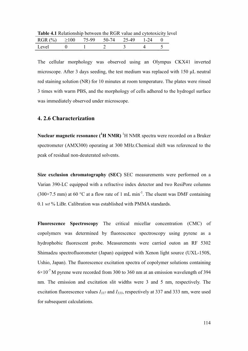
114
Table 4.1 Relationship between the RGR value and cytotoxicity level
RGR (%) ≥100 75-99 50-74 25-49 1-24 0
Level 0 1 2 3 4 5
The cellular morphology was observed using an Olympus CKX41 inverted
microscope. After 3 days seeding, the test medium was replaced with 150 µL neutral
red staining solution (NR) for 10 minutes at room temperature. The plates were rinsed
3 times with warm PBS, and the morphology of cells adhered to the hydrogel surface
was immediately observed under microscope.
4. 2.6 Characterization
Nuclear magnetic resonance (1H NMR)
1H NMR spectra were recorded on a Bruker
spectrometer (AMX300) operating at 300 MHz.Chemical shift was referenced to the
peak of residual non-deuterated solvents.
Size exclusion chromatography (SEC) SEC measurements were performed on a
Varian 390-LC equipped with a refractive index detector and two ResiPore columns
(300×7.5 mm) at 60 °C at a flow rate of 1 mL min-1
. The eluent was DMF containing
0.1 wt % LiBr. Calibration was established with PMMA standards.
Fluorescence Spectroscopy The critical micellar concentration (CMC) of
copolymers was determined by fluorescence spectroscopy using pyrene as a
hydrophobic fluorescent probe. Measurements were carried outon an RF 5302
Shimadzu spectrofluorometer (Japan) equipped with Xenon light source (UXL-150S,
Ushio, Japan). The fluorescence excitation spectra of copolymer solutions containing
6×10⁻⁷
M pyrene were recorded from 300 to 360 nm at an emission wavelength of 394
nm. The emission and excitation slit widths were 3 and 5 nm, respectively. The
excitation fluorescence values I337 and I333, respectively at 337 and 333 nm, were used
for subsequent calculations.

115
Cloud point The LCST was estimated from the changes in the transmittance through
copolymer solutions at 1.0 mg mL-1
as a function of temperature. The measurements
were carried out at a wavelength of 500 nm with a Perkin ElmerLambda 35
UV-Visible spectrometer equipped with a Peltier temperature programmer PTP-1+1.
The temperature ramp was 0.1 °C min-1
.
Dynamic light scattering (DLS) and Zeta-Potential (ZP) The size and
zeta-potential of micelles were determined by using Zetasizer Nano-ZS (Malvern
Instrument Ltd. UK) equipped with a He-Ne laser (λ = 632.8 nm). Polymer solutions
at 1.0 mg mL-1
were filtered through a 0.45 µm PTFE microfilter before
measurements.
Transmission electron microscopy (TEM) TEM experiments were carried out on a
JEOL 1200 EXII instrument operating at an acceleration voltage of 120 kV. The
samples were prepared by dropping a polymer solution at 1.0 mg mL-1
onto a carbon
coated copper grid, followed by air drying.
Inductively Coupled plasma-mass spectrometry (ICP-MS) The residual copper
content in the copolymers was quantified using ThemoFinnigan Element XR sector
field ICP-MS previously calibrated using copper solutions in water. Typically,
samples were prepared by dissolution of the copolymers in nitric acid. The solution
was then heated to fully decompose the polymer until no precipitate was visible. After
that, the samples were dissolved in 10 mL deionized water before analysis to
determine the copper concentration. Each sample was analyzed four times.

116
4.3 Results and discussion
4.3.1 Synthesis of triblock copolymers
A series of P(NIPAAm-co-DMAAm)-b-PLA-b-P(NIPAAm-co-DMAAm) triblock
copolymers were successfully synthesized by ATRP as described in a previous work
[28]. The DP of PLLA macroinitiator is 40, while that of NIPAAm and DMAAm
ranges from 63 to 74, and from 32 to 41, respectively (Table 4.2).
Table 4.2 Characteristics of
P(NIPAAm-co-DMAAm)-b-PLA-b-P(NIPAAm-co-DMAAm) triblock copolymers
Run PLA/NIPAAm
/DMAAm a
[LA]/[NIPAAm]
/[DMAAm]b
NIPAAm/
DMAAmb
Mn, NMRb
g mol-1
Mn, SECc
gmol-1
Đc
T1 1/148.8/51.2 40/74/34 68.2/31.8 18300 26000 1.1
T2 1/147.5/52.5 40/65/32 65.8/34.2 19700 25000 1.1
T3 1/142.6/57.4 40/66/36 64.8/35.2 14300 21000 1.1
T4 1/136.2/63.8 40/63/41 61.6/39.4 14500 18000 1.1
a) feed ratio (in equivalents); b) calculated by NMR; c) determined by SEC
Figure 4.1 1H NMR spectra of triblock copolymer T2 in (A) CDCl3, and (B) D2O.

117
Various NIPAAm/DMAAm ratios were used to tailor the properties of the resulting
copolymers. The molecular weights determined by SEC (Mn,SEC) range from 18,000 to
26,000 g mol-1
with narrow dispersity (Đ= 1.1), whereas the molecular weights
determined by 1H NMR (Mn,NMR) are lower. Mn,SEC values are obtained with respect to
poly(methyl methacrylate) standards, and are generally higher than absolute
molecular weights.
The chemical structure of triblock copolymers was characterized by 1H NMR
spectroscopy. As shown in Figure 4.1A, the peaks at 1.1 and 4.0 ppm were attributed
respectively to the methyl (Hf, CH3) and the methine protons (He, CH) adjacent to the
amine moiety of the NIPAAm units. The signal at 2.9 ppm is assigned to the methyl
proton (Hg, CH3) of the DMAAm units. The signals at 1.6 and 5.2 ppm are
characteristic of the methyl (Hb) and methine protons (Ha, CH) of PLLA. The
composition of copolymers, the DP of NIPAAm and DMAAm in the hydrophilic
block, and the molecular weight (Mn) of copolymers were calculated from the
integrations o f the methine protons of lactyl units at 5.1 ppm, the methine protons of
NIPAAm units at 4.0 ppm, and the methyl protons of DMAAm at 2.9 ppm.
After polymerization, the catalyst was removed by filtration through a basic alumina
column. The copolymers recovered by precipitation in diethyl ether appeared almost
colorless. ICP-MS measurements showed that the copper content in the copolymers
was between 1 and 3 ppm.
4.3.2 Characterization of polymer micelles
It is well-known that amphiphilic copolymers can form polymeric micelles by
self-assembly in aqueous medium. Figure 4.1B shows the 1H NMR spectrum of
copolymer T2 obtained in D2O. Compared with the 1H NMR spectrum in CDCl3
(Figure 4.2A), the peaks belonging to PLLA moieties at 5.2 and 1.6 ppm totally

118
disappear in D2O. This finding evidences the aggregation of copolymers into micelles
with the PLLA block in the core and P(NIPAAm-co-DMAAm) blocks at the corona.
Table 4.3 Characterization of triblock copolymer micelles
Copolymer CMCa
g mL-1
Db
(nm)
PDIb LCST
c
(oC)
LCSTd
(oC)
ZPe
(mV)
T1 11.3 37 0.11 44.7 41.4 -18.7
T2 13.1 39 0.11 45.6 42.1 -12.4
T3 13.7 41 0.14 45.9 42.5 -15.3
T4 14.4 44 0.17 49.4 46.2 -16.4
a) by fluorescence spectroscopy; b) by DLS; c) by UV spectroscopy in deionzed water; d) by UV
spectroscopy in PBS; e) by Zetasizer Nano-ZS
The CMC is an important parameter which reflects the stability of micelles upon
dilution. The lower the CMC, the higher the stability of micelle. In this work, the
CMC was determined by fluorescence spectroscopy using pyrene as the probe. As
shown in Table 4.3, the CMC of copolymers slightly increases from 0.0113 to 0.0144
mg mL-1
while the content of DMAAm increases from 31.8 % for T1 to 39.4 % for T4
in the hydrophilic part. In fact, the CMC is dependent on different factors such as the
composition, the hydrophilic to hydrophobic ratio, the molecular weight, etc. The
DMAAm units are more hydrophilic than NIPAAm ones. Thus, higher DMAAm
contents lead to higher CMC. The very low CMC values indicate the strong tendency
of copolymers toward formation of micelles, which is of major importance for the
stability of micelles in the bloodstream after injection induced dilution [29].
The LCST of copolymers was determined by light transmission measurements using
UV spectroscopy. As shown in Table 4.3, the LCST value in distilled water increases
from 44.7 to 49.4 °C with increasing DMAAm content in the hydrophilic blocks.
LCST measurements were also performed in a pH 7.4 phosphate buffered saline (PBS)
to mimic the in vivo conditions. A decrease of ca. 3.5 degrees in LCST values is
observed in PBS as compared to those in distilled water.

119
The size and size distribution of copolymer micelles were determined by dynamic
light scattering (DLS). The average diameter varies from 37 to 44 nm, and the
dispersity (PDI) from 0.11 to 0.17 with increasing DMAAm content in the
hydrophilic blocks. The zeta-potential of copolymer micelles varies in the -12.4 to
-18.7 mV range. The zeta potential value decreases with the increase of hydrophilic
content. This finding well agrees with the structure of micelles having PLLA block in
the core and P(NIPAAm-co-DMAAm) blocks at the corona.
4.3.3 Preparation and characterization of drug loaded micelles
Curcumin is a natural anticancer drug, but its application is limited due to poor
solubility. Copolymer T2 with a LCST of 42.1 °C in PBS was selected for drug
release studies. Different ratios of curcumin and copolymer T2 were used to prepare
drug loaded micelles by solvent evaporation/membrane rehydration method (Table
4.4). After lyophilization, the micelles were dispersed in PBS solution for drug release
studies.
Table 4.4 Properties of curcumin-loaded micelles prepared from copolymer T2
Sample Drug/Polymer
(mg/mg)
DL
(%)
EE
(%)
LCSTa
(oC)
Db
(nm)
PDIb Zeta-potential (mV)
In water In PBS
1 1.2/18.9 6.0 97.2 38.0 48 0.19 -13.9 -7.51
2 2.5/17.5 12.1 96.6 37.8 54 0.21 -15.5 -7.68
3 4.5/16.3 20.4 94.1 37.5 88 0.25 -18.1 -2.39
a) by UV in PBS; b) by DLS in water
The drug loading (DL) and encapsulation efficiency (EE) of micelles were obtained
by light transmission measurements using UV spectroscopy. When the initial drug
amount increases from 1.2 to 4.50 mg with approximately the same amount of
polymer, drug loading increases from 6.0 to 20.4 %. The loading efficiency is above
94 % in all cases.
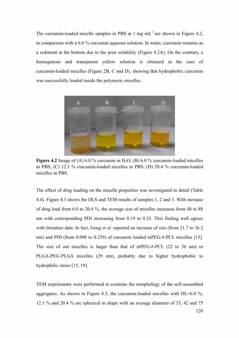
120
The curcumin-loaded micelle samples in PBS at 1 mg mL-1
are shown in Figure 4.2,
in comparison with a 6.0 % curcumin aqueous solution. In water, curcumin remains as
a sediment at the bottom due to the poor solubility (Figure 4.2A). On the contrary, a
homogenous and transparent yellow solution is obtained in the case of
curcumin-loaded micelles (Figure 2B, C and D), showing that hydrophobic curcumin
was successfully loaded inside the polymeric micelles.
Figure 4.2 Image of (A) 6.0 % curcumin in H2O; (B) 6.0 % curcumin-loaded micelles
in PBS; (C) 12.1 % curcumin-loaded micelles in PBS; (D) 20.4 % curcumin-loaded
micelles in PBS.
The effect of drug loading on the micelle properties was investigated in detail (Table
4.4). Figure 4.3 shows the DLS and TEM results of samples 1, 2 and 3. With increase
of drug load from 6.0 to 20.4 %, the average size of micelles increases from 48 to 88
nm with corresponding PDI increasing from 0.19 to 0.25. This finding well agrees
with literature data. In fact, Gong et al. reported an increase of size (from 21.7 to 36.2
nm) and PDI (from 0.098 to 0.259) of curcumin loaded mPEG-b-PCL micelles [15].
The size of our micelles is larger than that of mPEG-b-PCL (22 to 36 nm) or
PLGA-PEG-PLGA micelles (29 nm), probably due to higher hydrophobic to
hydrophilic ratios [15, 19].
TEM experiments were performed to examine the morphology of the self-assembled
aggregates. As shown in Figure 4.3, the curcumin-loaded micelles with DL=6.0 %,
12.1 % and 20.4 % are spherical in shape with an average diameter of 33, 42 and 75

121
nm, respectively. The size obtained from DLS is larger than that from TEM due to
different experimental conditions. In fact, DLS allows determining the hydrodynamic
diameter of micelles in aqueous solution, whereas TEM shows the dehydrated solid
state of micelles.
Figure 4.3 DLS spectra and TEM micrographs of curcumin loaded micelles. (A) and
(B): sample 1 (DL=6.0 %); (C) and (D): sample 2 (DL=12.1 %); (E) and (F): sample
3 (DL=20.4 %).

122
The zeta potential of curcumin-loaded micelles in water ranges from -13.9 mV for
Sample 1 to -18.1 mV for Sample 3, indicating good micellar stability. Compared to
blank polymer micelles, curcumin-loaded micelles present higher charge due to larger
surface area, resulting from the increase of micelle size. The ZP of sample 1, sample 2
and sample 3 decreases to -7.51, -7.68 and -2.39 mV in PBS, respectively. This
significant decrease can be attributed to the ionic screening effect and clustered
aggregate [23]. All the samples remained homogeneously transparent over 1 month. It
is known that more pronounced zeta potential absolute values tend to stabilize
particles in suspension. The electrostatic repulsion between micelles with the same
electrical charge prevents aggregation of micelles [30, 31]. Thus, the negatively
charged surface of micelles could help to stabilize the micelles via both the steric and
electrostatic mechanisms. It is of interest to note that mPEG-PCL micelles with 9.9 %
curcumin loading remained stable for 72 h [15]. On the other hand, Liu et al. reported
stable mPEG-PCL micelles with small size (28.2 nm) with low zeta potential (0.41
mV) [17]. The stability was attributed to the stereospecific blockade of micelles
although the authors did not mention the duration of micelle stability.
Both the size and zeta potential are important physicochemical properties of micelles
because they determine the physical stability as well as the biopharmaceutical
properties. The nano-size (48-88 nm) of curcumin-loaded micelles allows them to
escape from the reticuloendothelial system (RES) and preferentially accumulate in
tumor tissues through the EPR effect [32-35]. The charge of nanomaterials is also an
important factor to affect the biocompatibility [35]. Generally, positive charges are
considered as harmful to the plasma exposure of nanomaterials since they could
induce the formation of primary platelet clots [36-38]. On the contrary, negative
charges of micelles are beneficial to negatively charged human blood as they could
enhance the binding of micelles to proteins, reduce undesirable clearance by the
reticuloendothelial system such as liver, improve the blood compatibility, and thus
deliver the anti-cancer drugs more efficiently to the tumor sites [37, 39].

123
4.3.4 Phase transition of curcumin loaded polymeric micelles
It is well known that the drug release of thermo-responsive polymeric micelles is
controlled by the phase transition (Figure 4.4). Generally, the
P(NIPAAm-co-DMAAm) block is hydrophilic and the solution is transparent below
the LCST. When the temperature increases above the LCST, dehydration of
P(NIPAAm-co-DMAAm) occurs due to coil-to-globule transition, leading to
precipitation of polymers and the release of drug from polymer matrix.
Figure 4.4 Illustration of drug release from thermo-responsive polymeric micelles
controlled by phase transition.
Figure 4.5 Phase transition of curcumin-loaded micelles at 1 mg mL-1
determined by
UV-Vis spectroscopy. () DL=0 in water; () DL=0 in PBS; () DL=6.0 % in PBS;
() DL=12.1 % in PBS; () DL=20.4 % in PBS.

124
Figure 4.5 shows the phase transition of various curcumin-loaded micelles in water or
in PBS. The initially transparent solution of curcumin-loaded micelles becomes turbid
when the temperature increases above the LCST. In the meantime, the transmittance
of the solution rapidly decreases from 100 % to 0.
The presence of salt greatly affects the behavior of thermo-responsive polymers. As
shown in Figure 4.5, the LCST of blank micelles decreases from 45.6 oC in water to
42.1 oC in PBS due to the high ionic strength in PBS and screening effect from
counter-ions. The presence of phosphate leads to partial dehydration of the
macromolecules and consequently to a decrease of the LCST. The influence of drug
loading on the LCST was also considered in PBS. The LCST of curcumin-loaded
micelles with 6.0 % drug load is 38.0 oC in PBS, which is lower by 4.1
oC than that of
drug free micelles (Figure 4.5, Table 3). This finding is assigned to the enhanced core
hydrophobicity due to encapsulated drug. Higher drug load leads to lower LCST, but
the decrease is very limited. In fact, the LCST only slightly decreases from 38.0 to
37.5 oC when the drug load increases from 6.0 % to 20.4 %. In addition, the phase
transition intervals are very sharp (<0.5 oC) in all cases, as reported in our previous
work [28]. The fast phase transition favors rapid response of drug release when the
micelles were delivered to the tumor site.
4.3.5 In vitro drug release
Drug release was performed under physiological conditions (PBS, pH=7.4) at 37 °C
(below the LCST) and 40 oC (above the LCST), respectively. The amount of curcumin
was determined from the UV-visible absorbance at 424 nm. A linear calibration curve
was previously established between the UV absorbance and curcumin concentration
with a correlation coefficient of 0.99991 (Figure 4.6A).
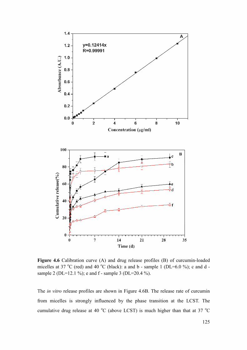
125
Figure 4.6 Calibration curve (A) and drug release profiles (B) of curcumin-loaded
micelles at 37 oC (red) and 40
oC (black): a and b - sample 1 (DL=6.0 %); c and d -
sample 2 (DL=12.1 %); e and f - sample 3 (DL=20.4 %).
The in vitro release profiles are shown in Figure 4.6B. The release rate of curcumin
from micelles is strongly influenced by the phase transition at the LCST. The
cumulative drug release at 40 oC (above LCST) is much higher than that at 37
oC

126
(below LCST). In all cases, burst-like release is observed. The cumulative release for
samples 1, 2 and 3 after 1 hour at 40 °C is 23.6 %, 14.7 % and 9.0 %, respectively,
while it is 16.3 %, 10.4 % and 4.1 % at 37 °C. The burst-like release at 37 oC can be
attributed to the release of drug located at the interface between the core and corona
of micelles. The larger burst-like release at 40 oC is attributed not only to the fast
release because of phase transition above the LCST, but also to the release of drug
located at the interface.
Sample 1 with 6.0 % drug loading shows the fastest drug release. After the first 3 days,
89.3 % and 74.9 % of the loaded curcumin are released at 40 °C and 37 °C,
respectively. Beyond, the release rate slows down. The cumulative release of
curcumin reaches 84 % at 37 °C after 30 days and 92.3 % at 40 oC after 10 days. The
release rate is slower for sample 2 with initial drug loading of 12.1 %. The cumulative
release is 85.2 % and 48.9 % after 14 days, and 91.1 % and 53.6 % after 30 days at 40
oC and 37
oC, respectively. Sample 3 with initial drug loading of 20.4 % shows the
slowest drug release. After 30 days, curcumin release reaches 59.8 % and 35.7 % at
40 °C and 37 °C, respectively. Therefore, drug release is not only dependent on the
phase transition across the LCST, but also on the drug loading. In fact, the solubility
of curcumin in the release medium is a limiting factor. Samples with higher drug
loading exhibits lower release rate because of the solubility effect. It should also be
noted that the LCST of cur-micelles is dynamically changing with the release of
curcumin. Theoretically, the LCST will increase up to 42.1 oC when the drug release
reaches 100 % for all the samples as shown in Figure 4.5.
Peppas et al. proposed the following semi-empirical equation to describe the drug
release mechanism from a polymeric matrix [40-42]:
0
ntMkt
M= (4)
0log( ) log logtM M n t k= + (5)

127
Where Mt and M0 are the cumulative amount of released drug at time t and the drug
loading, respectively, k is a constant incorporating structural and geometric
characteristic of the device and n is the release exponent indicating the drug release
mechanism. For spherical particles, the value of n is equal to 0.43 for Fickian
diffusion, and equal to 0.85 for swelling-controlled mechanism. In contrast, n<0.43
means combination of diffusion and erosion controlled release, and 0.43<n<0.85 is
assigned to anomalous transport mechanism.
Figure 4.7 Plots of theoretical fitting to Peppas model for curcumin release from
curcumin-loaded micelles (the symbols are the same as in Figure 4.6).
Figure 4.7 presents the theoretical fitting plots. The various release curves are divided
in two stages: from 0 to 7 hours and from 1 to 30 days. The fitting parameters n, k and
the correlation coefficient R2
are summarized in Table 5. For the first stage, the n
values at 40 oC are in the range of 0.43 to 0.85, suggesting that the release of
curcumin involves anomalous transport probably due to phase transition across the
LCST. The n values of sample 1 and 2 at 37 oC are also in the range of 0.43 to 0.85,
which could be assigned to the initial burst release. The n value of sample 3 at 37 oC
is 0.349, indicating diffusion and degradation controlled release according to Peppas’

128
theory. It might also be attributed to the highest drug loading and compact micelle
structure. For the second stage, the n values in all cases are well below 0.43,
suggesting a combination of diffusion and degradation controlled release. For both
stages, the k values at 40 oC are higher than those at 37
oC, which confirms a faster
drug release above the LCST due to collapse of micelles.
Table 4.5 Release exponent (n), rate constant (k) and correlation coefficient (R2) for
drug release from drug loaded polymer micelles.
Sample Time interval 40oC 37
oC
n k R2 n k R
2
Sample 1 0-7h 0.6238 1.6667 0.997 0.5969 1.1033 0.992
1-30d 0.0843 0.7780 0.943 0.0552 0.6834 0.974
Sample 2 0-7h 0.4430 0.6002 0.995 0.5563 0.5904 0.997
1-30d 0.22693 0.4421 0.994 0.17396 0.2990 0.991
Sample 3 0-7h 0.5444 0.5376 0.975 0.3490 0.2187 0.974
1-30d 0.1638 0.3448 0.994 0.2576 0.1477 0.972
Therefore, the release of curcumin from thermo-responsive curcumin-loaded micelles
is strongly affected by the phase transition across the LCST. Such release would favor
drug accumulation at a specific tumor site where the temperature is above the LCST.
Thus the micelles could combine the passive targeting by EPR effect due to the small
size ranging from 47.5 to 88.2 nm, and the active targeting by thermo-responsive
phase transition.
4.3.6 In vitro cytocompatibility
Curcumin is a natural polyphenolic compound with low intrinsic toxicity. The in vitro
cytotoxicity of copolymers is evaluated by MTT assay, a widely used method for the
screening of biomaterials. MTT assay is based on the reaction between MTT and
mitochondrial succinate dehydrogenases in living cells to form a purple formazan
which is soluble in DMSO but insoluble in water. The OD value of formazan-DMSO
solution is considered to be proportional to the number of living cells.

129
Figure 4.8 Optical density values of L929 (A) and A549 (B) solutions after 2, 3 and 4
days culture in DMEM with copolymer substrate (T2), and controls (DMEM and 5 %
phenol).
The effects of copolymer T2 on the proliferation of L929 and A549 cells are shown in
Figure 4.8. In comparison with controls (DMEM medium and 5 % phenol), the OD
values on polymer substrate are close to those in the culture medium, and much
higher than those in phenol. The RGR values (Table 4.6) of copolymer T2 are well

130
above 92.6 % during 4 days incubation with L929 and A549 cells, corresponding to a
cytotoxicity level of 0 and 1 as defined in Table 1, thus indicating that the copolymers
present no cytotoxicity.
Table 4.6 RGR values of copolymers T2 with A549 and L929 cells during 4 days
incubation.
Cell type RGR (%)
2 days 3 days 4 days
L929 118.9 102.0 118.8
A549 124.9 115.2 92.6
Figure 4.9 Microscopic images of cells stained by NR after 3 days culture in different
media: (A) Copolymer T2 substrate without cells; (B) L929 cells on polymer substrate
in DMEM; (C) A549 cells on polymer substrate in DMEM; (D) A549 cells in 5 %
phenol medium.
Figure 4.9 shows the morphology of cells after 3 days culture. Large number of cells
died in the toxic phenol medium (Figure 4.9). On the contrary, cell adhesion and
proliferation are observed on the copolymer substrate (Figure 4.9B and Figure 4.9C).
Cells exhibit spindle, polygon or oval shapes, and the pseudopodium of cells stretches

131
out. Therefore, MTT assay and cell morphology results indicate that
P(NIPAAm-co-DMAAm)-b-PLLA-b-P(NIPAAm-co-DMAAm) copolymers present
outstanding cytocompatibility and could be used for biomedical applications.
4.4 Conclusions
In this work, thermo-responsive micelles are prepared by self-assembly of
P(NIPAAm-co-DMAAm)-b-PLLA-b-P(NIPAAm-co-DMAAm) triblock copolymers
as drug carrier, where curcumin was used as a model drug. Compared to
mPEG-b-PCL or PLGA-b-PEG-b-PLGA micelles, the thermo-responsive micelles
described in this manuscript present several advantages. First, lower CMC values
ranging from 0.0113 to 0.0144 mg mL-1
are obtained, which should enhance the
stability of micelles during long circulation in the bloodstream after injection induced
dilution. Second, high curcumin loading up to 20.4 % was achieved. Meanwhile, the
micelles remain stable over 1 month due to the negative surface charge as shown by
zeta potential measurements. Last, the micelles with sizes ranging from 47.5 to 88.2
nm could combine the passive targeting by EPR effect and thermo-responsive targeted
release. Such responsive behavior will favor the release at a specific tumor site (~42
oC). Therefore, all these properties show that the thermo-responsive micelles
described in this work are promising as a functional drug carrier.
Recently, due to the possible concerns on the toxicity of N-isopropylacrylamide
monomer, Lutz et al. reported new thermo-responsive and biocompatible copolymer
from ATRP of two PEG Analogues: 2-(2-methoxyethoxy) ethyl methacrylate
(MEO2MA) and oligo(ethylene glycol) methacrylate (OEGMA) [43-47]. Thus, in
order to make a comparison, following work will focus on the synthesis and
self-assembly of PLA/PEG analogues as drug carrier.

132
4.5 Reference
[1] L. Yang, A. El Ghzaoui, S. M. Li, In vitro degradation behavior of
poly(lactide)-poly(ethylene glycol) block copolymer micelles in aqueous solution. Int. J. Pharm.
400 (2010) 96-103.
[2] L. Yang, X. Qi, P. Liu, A. El Ghzaoui, S. M. Li, Aggregation behavior of self-assembling
polylactide/poly(ethylene glycol) micelles for sustained drug delivery. Int. J. Pharm.394 (2010)
43-49.
[3] H. Z. Zhao, L. Y. L. Yung, Selectivity of folate conjugated polymer micelles against different
tumor cells. Inter. J. Pharm. 349 (2008) 256-268.
[4] M. Watanabe, K. Kawano, M. Yokoyama, P. Opanasopit, T. Okano, Y. Maitani, Preparation of
camptothecin-loaded polymeric micelles and evaluation of their incorporation and circulation
stability. Inte. J. Pharm. 308 (2006) 183-189.
[5] G. Gaucher, M. H. Dufresne, V. P. Sant, N. Kang, D. Maysinger, J. C. Leroux, Block
copolymer micelles: preparation, characterization and application in drug delivery. J. Control.
Release 109 (2005) 169-188.
[6] K. Kataoka, A. Harada, Y. Nagasaki, Block copolymer micelles for drug delivery: design,
characterization and biological significance. Adv. Drug Deliv. Rev. 47 (2001) 113-131.
[7] V. P. Torchilin, Structure and design of polymeric surfactant-based drug delivery systems. J.
Control. Release 73 (2001) 137-172.
[8] R. K. Maheshwari, A. K. Singh, J. Gaddipati, R. C. Srimal, Multiple biological activities of
curcumin: A short review. Life Sci. 78 (2006) 2081-2087.
[9] B. B. Aggarwal, A. Kumar, A. C. Bharti, Anticancer potential of curcumin: Preclinical and
clinical studies. Anticancer Res. 23 (2003) 363-398.
[10] D. K. Agrawal, P. K. Mishra, Curcumin and its analogues: Potential anticancer agents. Med.
Res. Rev. 30 (2009) 818-860.
[11] P. Anand, C. Sundaram, S. Jhurani, A .B. Kunnumakkara, B. B. Aggarwal, Curcumin and
cancer: An "old-age" disease with an "age-old" solution. Cancer Lett. 267 (2008) 133-164.
[12] A. B. Kunnumakkara, P. Anand, B. B. Aggarwal, Curcumin inhibits proliferation, invasion,
angiogenesis and metastasis of different cancers through interaction with multiple cell signaling
proteins. Cancer Lett. 269 (2008) 199-225.
[13] H. H. Tonnesen, Solubility, chemical and photochemical stability of curcumin in surfactant
solutions - Studies of curcumin and curcuminolds, XXVIII. Pharmazie 57 (2002) 820-824.
[14] P. Anand, A. B. Kunnumakkara, R. A. Newman, B. B. Aggarwal, Bioavailability of curcumin:
Problems and promises. Mol. Pharm. 4 (2007) 807-818.
[15] C. Gong, S. Deng, Q. Wu, M. Xiang, X. Wei, L. Li, X. Gao, B. Wang, L. Sun, Y. Chen, Y. Li,
L. Liu, Z. Qian, Y. Wei, Improving antiangiogenesis and anti-tumor activity of curcumin by
biodegradable polymeric micelles. Biomaterials 34 (2013) 1413-1432.
[16] M. L. Gou, K. Men, H. S. Shi, M. L. Xiang, J. A. Zhang, J. Song, J. L. Long, Y. Wan, F. Luo,
X. Zhao, Z. Y. Qian, Curcumin-loaded biodegradable polymeric micelles for colon cancer therapy
in vitro and in vivo. Nanoscale 3 (2011) 1558-1567.
[17] L. Liu, L. Sun, Q. Wu, W. Guo, L. Li, Y. Chen, Y. Li, C. Gong, Z. Qian, Y. Wei, Curcumin

133
loaded polymeric micelles inhibit breast tumor growth and spontaneous pulmonary metastasis.
Inter. J. Pharm. 443 (2013) 175-182.
[18] L. Song, Y. Shen, J. Hou, L. Lei, S. Guo, C. Qian, Polymeric micelles for parenteral delivery
of curcumin: Preparation, characterization and in vitro evaluation. Colloids Surf. A: Physicochem.
Eng. Aspects 390 (2011) 25-32.
[19] Z. M. Song, R. L. Feng, M. Sun, C. Y. Guo, Y. Gao, L. B. Li, G.. X. Zhai, Curcumin-loaded
PLGA-PEG-PLGA triblock copolymeric micelles: Preparation, pharmacokinetics and distribution
in vivo. J. Colloid Inter. Sci. 354 (2011) 116-123.
[20] R. L. Yang, S. A. Zhang, D. L. Kong, X. L. Gao, Y. J. Zhao, Z. Wang, Biodegradable
Polymer-Curcumin Conjugate Micelles Enhance the Loading and Delivery of Low-Potency
Curcumin. Pharm. Res. 29 (2012) 3512-3525.
[21] A. Chilkoti, M. R. Dreher, D. E. Meyer, D. Raucher, Targeted drug delivery by thermally
responsive polymers. Adv. Drug Deliv. Rev. 54 (2002) 613-630.
[22] H. Wei, S. X. Cheng, X. Z. Zhang, R. X. Zhuo, Thermo-sensitive polymeric micelles based
on poly(N-isopropylacrylamide) as drug carriers. Prog. Polym. Sci. 34 (2009) 893-910.
[23] F. Xu, S. Z. Zheng, Y. L. Luo, Thermosensitive t-PLA-b-PNIPAAm tri-armed star block
copolymer nanoscale micelles for camptothecin drug release. J. Polym. Sci. Part A: Polym. Chem.
51 (2013) 4429-4439.
[24] C. Chang, H. Wei, D. Q. Wu, B. Yang, N. Chen, S. X. Cheng, X. Z. Zhang, R. X. Zhuo,
Thermo-responsive shell cross-linked PMMA-b-P(NIPAAm-co-NAS) micelles for drug delivery.
Inter. J. Pharm. 420 (2011) 333-340.
[25] I. S. Kim, Y. I. Jeong, C. S. Cho, S. H. Kim, Thermo-responsive self-assembled polymeric
micelles for drug delivery in vitro. Int. J. Pharm. 205 (2000) 165-172.
[26] S. J. T. Rezaei, M. R. Nabid, H. Niknejad, A. A. Entezami, Folate-decorated
thermoresponsive micelles based on star-shaped amphiphilic block copolymers for efficient
intracellular release of anticancer drugs. Inter. J. Pharm. 437 (2012) 70-79.
[27] Y. F Hu, V. Darcos, S. Monge, S. M. Li, Synthesis and self-assembling of
poly(N-isopropylacrylamide-block-poly(L-lactide)-block-poly(N-isopropylacrylamide) triblock
copolymers prepared by combination of ring-opening polymerization and atom transfer radical
polymerization. J. Polym. Sci. Part A: Polym. Chem. 51 (2013) 3274-3283.
[28] Y. F Hu, V. Darcos, S. Monge, S. M Li, Y. Zhou, F. Su, Tunable thermo-responsive
P(NIPAAm-co-DMAAm)-b-PLLA-b-P(NIPAAm-co-DMAAm) triblock copolymer micelles as
drug carriers. J. Mater. Chem. B 2 (2014) 2738-2748.
[29] M. Yokoyama, T. Okano, Y. Sakurai, H. Ekimoto, C. Shibazaki, K. Kataoka, Toxicity and
Antitumor Activity against Solid Tumors of Micelle-forming Polymeric Anticancer Drug and Its
Extremely Long Circulation in Blood. Cancer Res. 51 (1991) 3229-3236.
[30] S. S. Feng, G. F. Huang, Effects of emulsifiers on the controlled release of paclitaxel (Taxol
(R)) from nanospheres of biodegradable polymers. J. Control. Release 71 (2001) 53-69.
[31] H. Ding, F. Wu, Y. Huang, Z.R. Zhang, Y. Nie, Synthesis and characterization of
temperature-responsive copolymer of PELGA modified poly(N-isopropylacrylamide). Polymer 47
(2006) 1575-1583.
[32] Y. Yan, G. K. Such, A. P. R. Johnston, J. P. Best, F. Caruso, Engineering Particles for
Therapeutic Delivery: Prospects and Challenges. ACS Nano 6 (2012) 3663-3669.
[33] C. Oerlemans, W. Bult, M. Bos, G. Storm, J. F. Nijsen, W. Hennink, Polymeric Micelles in
134
Anticancer Therapy: Targeting, Imaging and Triggered Release. Pharm. Res. 27 (2010)
2569-2589.
[34] H. Maeda, J. Wu, T. Sawa, Y. Matsumura, K. Hori, Tumor vascular permeability and the EPR
effect in macromolecular therapeutics: a review. J. Control. Release 65 (2000) 271-284.
[35] N. Bertrand, J. Wu, X. Y. Xu, N. Kamaly, O. C. Farokhzad, Cancer nanotechnology: The
impact of passive and active targeting in the era of modern cancer biology. Adv. Drug Deliv. Rev.
66 (2014) 2-25.
[36] Y. Yamamoto, Y. Nagasaki, Y. Kato, Y. Sugiyama, K. Kataoka, Long-circulating
poly(ethylene glycol)-b-poly(D,L-lactide) block copolymer micelles with modulated surface
charge. J. Control. Release 77 (2001) 27-38.
[37] C. B. He, Y. P. Hu, L. C. Yin, C. Tang, C. H. Yin, Effects of particle size and surface charge
on cellular uptake and biodistribution of polymeric nanoparticles. Biomaterials 31 (2010)
3657-3666.
[38] R. B. Campbell, D. Fukumura, E. B. Brown, L. M. Mazzola, Y. Izumi, R. K. Jain, V. P.
Torchilin, L. L. Munn, Cationic charge determines the distribution of liposomes between the
vascular and extravascular compartments of tumors. Cancer Res. 62 (2002) 6831-6836.
[39] K. Xiao, Y. P. Li, J. T. Luo, J. S. Lee, W. W. Xiao, A. M. Gonik, R. G. Agarwal, K. S. Lam,
The effect of surface charge on in vivo biodistribution of PEG-oligocholic acid based micellar
nanoparticles. Biomaterials 32 (2011) 3435-3446.
[40] P. L. Ritger, N. A. Peppas, A simple equation for description of solute release I. Fickian and
non-fickian release from non-swellable devices in the form of slabs, spheres, cylinders or discs. J.
Control. Release 5 (1987) 23-36.
[41] P. L. Ritger, N. A. Peppas, A simple equation for description of solute release II. Fickian and
anomalous release from swellable devices. J. Control. Release 5 (1987) 37-42.
[42] J. Siepmann, N. A. Peppas, Modeling of drug release from delivery systems based on
hydroxypropyl methylcellulose (HPMC). Adv. Drug Deliv. Rev. 64 (2012) 163-174.
[43] J. F. Lutz, A. Hoth, Preparation of Ideal PEG Analogues with a Tunable Thermosensitivity by
Controlled Radical Copolymerization of 2-(2-Methoxyethoxy)ethyl Methacrylate and
Oligo(ethylene glycol) Methacrylate. Macromolecules 39 (2006) 893-896.
[44] J. F. Lutz, K. Weichenhan, O. Akdemir, A. Hoth, About the Phase Transitions in Aqueous
Solutions of Thermoresponsive Copolymers and Hydrogels Based on 2-(2-methoxyethoxy)ethyl
Methacrylate and Oligo(ethylene glycol) Methacrylate. Macromolecules 40 (2007) 2503-2508.
[45] J. F. Lutz, J. Andrieu, S. uzgun, C. Rudolph, S. Agarwal, Biocompatible, Thermoresponsive,
and Biodegradable: Simple Preparation of "All-in-One" Biorelevant Polymers. Macromolecules
40 (2007) 8540-8543.
[46] J. F. Lutz, Polymerization of oligo(ethylene glycol) (meth)acrylates: Toward new generations
of smart biocompatible materials. J. Polym. Sci. Part A: Polym. Chem. 46 (2008) 3459-3470.
[47] J. F. Lutz, Thermo-Switchable Materials Prepared Using the OEGMA-Platform. Adv. Mater.
23 (2011) 2237-2243.

135
Chapter 5 Thermo-responsive micelles from comb-like block
copolymers of polylactide and poly(ethylene glycol)
analogues: Synthesis, Micellization and In vitro drug release
Abstract: Thermo-responsive comb-like
P(MEO2MA-co-OEGMA)-b-PLLA-b-P(MEO2MA-co-OEGMA) triblock copolymers
were synthesized by atom transfer radical polymerization of MEO2MA and OEGMA
co-monomers using a Br-PLLA-Br macroinitiator. The resulting copolymers with
MEO2MA/OEGMA molar ratio ranging from 79/21 to 42/58 were characterized by
1H NMR and SEC. Thermo-responsive micelles were obtained by self-assembly of
copolymers in aqueous medium. The properties of micelles were determined by CMC,
DLS, TEM and LCST measurements. The micelles are spherical in shape with sizes
varying from 21 to 103 nm. A hydrophobic anticancer drug, curcumin, was
encapsulated in micelles by using membrane hydration method. The micelles size
decreases from 103 to 38 nm with 10.8 % drug loading, suggesting that drug loading
strongly affects the self-assembly procedure. The LCST decreases from 45.1 °C for
blank micelles to 40.6 and 38.3 oC with 5.9 % and 10.8 % drug loading. In vitro drug
release was performed in pH=7.4 PBS at different temperatures. Data show that the
release rate significantly increased above the LCST. The burst-like release was
depressed by the enhanced interaction between drug and comb-like copolymer chains.
Keywords: Polylactide; PEG analogues; Comb-like copolymer; Micellization;
Curcumin; Thermo-responsive; Drug release

136
5.1 Introduction
Cancer is nowadays a leading cause of death in the world. The World Health
Organization reported the approximately 14 million new cases and 8.2 million cancer
related deaths in 2012. For cancer therapy, the ideal drug delivery system should be
stimuli responsive to the local environment of tumor [1-3]. In fact, the tumor issue is
very different from normal tissues with higher temperature (~42 oC), lower pH (<5.3)
and anomalous vessel (intercellular gap junction of 300-700 nm) [4-6]. Therefore,
thermo-responsive polymeric micelles have been increasingly investigated for
targeted delivery of anticancer drugs [7-11]. So far, poly(N-isopropylacylamide)
(PNIPAAm), which displays a lower critical solution temperature (LCST) at 32 oC, is
the most studied thermo-responsive polymer in biomedical applications [5, 12-15].
Slight variation of pH, concentration, or chemical environment only slightly affects
the LCST of PNIPAAm, which is considered as the “Golden standard” of
thermo-responsive polymers. Nevertheless, the presence of residual
N-isopropylacrylamide monomer in PNIPAAm based polymers has raised concerns
on the toxicity.
Recently, Lutz et al. reported novel thermo-responsive copolymers of
2-(2-methoxyethoxy) ethyl methacrylate (MEO2MA) and oligo(ethylene glycol)
methacrylate (OEGMA), and hydrogels of both monomers using ethylene glycol
dimethacrylate as cross-linker by atom transfer radical polymerization (ATRP)
[16-23]. These PEG analogues exhibit a LCST between 26 and 90 oC which can be
precisely adjusted by varying the co-monomers ratio. They are thus attractive for
targeted drug delivery due to combination of thermo-responsive properties and good
biocompatibility as linear PEG [21].
However, PEG analogues generally adopt a compact coil conformation in aqueous
medium [20]. Introduction of hydrophobic block is thus required in order to obtain
thermo-responsive polymeric micelles as potential carrier of targeted drug delivery.

137
Saeed et al. synthesized poly(lactide-co-glycolide)-b-poly(oligo(ethyleneglycol)
methacrylate) (PLGA-b-POEGMA) diblock copolymers by combining ring opening
polymerization (ROP) and reversible addition fragmentation chain transfer (RAFT)
polymerization, and studied the self-assembly, drug release and cytotoxicity of the
copolymer micelles [24]. Later on, Luzon et al. obtained
POEGMA-b-PCL-b-POEGMA triblock copolymers by ATRP, using
α,ω-bromopropionyl poly(ε-caprolactone) (Br-PCL-Br) as macroinitiator [25]. The
critical micelle concentration (CMC) and critical micelle temperature (CMT) were
determined. The same synthetic route was adopted by Bakkour et al. for the synthesis
of POEGMA-b-PLLA-b-POEGMA triblock copolymers [26]. The self-assembly and
drug encapsulation potential of the copolymers were reported. Most recently, Fenyves
et al. synthesized amphiphilic bottlecomb block copolymers containing PLA and PEG
side chains using a grafting-from method, and studied their self-assembly in aqueous
medium. Spherical, cylindrical micelles as well as bilayer structures were observed
[27]. However, the LCST of these copolymers was in the range of 75 to 85 °C, i.e.
much higher than the body temperature.
In our previous work, well defined thermo-responsive
poly(N-isopropylacrymine-co-N,N-dimethylacrylamine)-b-poly(L-lactide)-b-poly(N-i
sopropylacrymine-co-N,N-dimethylacrylamine) P(NIPAAm-co-DMAAm)-b-PLLA-b
-P(NIPAAm-co-DMAAm) micelles has been investigated as drug carrier [28, 29].
The cytotoxicity evaluated by MTT assay showed that unreacted NIPAAm was
eliminated by dialysis. In vitro drug release studies present a much faster release at
temperatures above the LCST than below, suggesting that the copolymer micelles
could be promising for targeted delivery of antitumor drugs.
In this work, for the first time, we report a series of amphiphilic comb-like triblock
polymers composed of a biodegradable PLLA central block and two lateral
thermo-responsive P(MEO2MA-co-OEGMA) blocks prepared by combination of

138
ROP and ATRP. The self-assembly and thermo-responsive properties of the
copolymers were determined by CMC, DLS, TEM and LCST measurements. The
LCST was adjusted precisely by varying MEO2MA/OEGMA ratio.
Thermo-responsive drug release from copolymer micelles was investigated below or
above the LCST, and compared with that from
P(NIPAAm-co-DMAAm)-b-PLLA-b-P(NIPAAm-co-DMAAm) micelles.
5.2 Experimental Section
5.2.1 Materials
L-lactide was purchased from PuracBiochem (Goerinchem, The Netherlands).
Stannous 2-ethylhexanoate (Sn(Oct)2), 1,4-benzene dimethanol, 2-bromopropionyl
bromide, tris(2-dimethyl aminoethyl) amine (Me6TREN), copper (I) chloride (CuCl),
N,N-dimethyl formamide (DMF) and curcumin were obtained from Sigma-Aldrich
(St-Quentin Fallavier, France), and were used without further purification.
Dichloromethane and toluene from Sigma-Aldrich were dried over calcium hydride
for 24 h at room temperature and distilled under reduced pressure. MEO2MA and
OEGMA (Mn=300 g mol-1
) were obtained from Sigma-Aldrich and purified through a
basic aluminum oxide column. Triethylamine (Sigma-Aldrich) was dried over
potassium hydroxide for 24 h at room temperature and distilled. Ultrapure water with
a conductivity of 18 MΩ was produced using a Millipore Milli-Q water system.
α,ω-Bromopropionyl PLLA (Br-PLLA-Br) with degree of polymerization (DP) of 40
was prepared as previously reported [30].
5.2.2 Synthesis of P(MEO2MA-co-OEGMA)-b-PLLA-b-
P(MEO2MA-co-OEGMA) copolymer (copolymer T5, Table 5.1)
Triblock copolymers were prepared using standard Schlenk technique. Typically, 100
mg Br-PLLA-Br (30.4×10⁻³
mmol, Mn,NMR= 3300 g mol-1
), 461 mg MEO2MA ( 2.451

139
mmol), 1.1 g OEGMA (3.676 mmol) and 6 mg CuCl (60.6×10⁻³
mmol) were
dissolved in 2 mL DMF. After five freeze-pump-thaw cycles, 16 µL Me6TREN
(60.8×10⁻³
mmol) was added under argon atmosphere. The mixture was stirred at 80
oC for 6 h. The reaction was stopped by exposition to air for 2 h at room temperature.
A dialysis was then carried out for 3 days in a dialysis tube (MWCO: 3500 D) to
remove residual monomers and copper against Mill-Q water containing
ethylenediaminetetraacetic acid tetrasodium salt hydrate. The product was finally
collected by lyophilization.
1H NMR (300 MHz, CDCL3) (Figure 5.1) δ (ppm)=7.26 (s, 4HAr); 5.14 (m, 1Ha);
4.08 (t, 2Hc); 3.63+3.55(t, 2Hd); 3.37 (s, 3He); 1.56 (d, 3Hb)
5.2.3 Preparation of curcumin loaded micelles
The micelles were prepared by solvent evaporation/membrane hydration method.
Typically, 1.2 mg curcumin and 18.8 mg copolymer T5 (Sample 1, Table 5.2) were
dissolved in 10 mL acetone, and then the solvent was evaporated in rotary evaporator
at room temperature to yield a membrane at the wall of the round flask. After vacuum
drying for 24 h, 20 mL Mill-Q water was added to the flask, yielding self-assembled
micelles at room temperature. The resulting micellar solution was then centrifuged at
5000 rpm for 10 min to remove unloaded curcumin. The supernatant was lyophilized,
and dispersed in deionized water or PBS to yield a drug loaded micellar solution at a
concentration of 1 mg mL-1
for physiochemical characterization and drug release
studies.
5.2.4 In vitro drug release
The drug loading (DL) and encapsulation efficiency (EE) of micelles were determined
as follows. 0.1 mL drug loaded micellar solution (1 mg mL-1
) was dissolved in 2.4 mL
60/40 acetonitrile/ammonium acetate (pH=4.0, 10 mM) mixture solution. The
concentration of curcumin was determined by UV-Vis spectroscopy at λ=424 nm,

140
using a calibration curve previously established from a series of standard curcumin
solutions with concentrations from 0.008 to 10 µg mL-1
. The DL and EE were
calculated from following formulae:
100×=micelles polymeric of weight
drug loaded of weight(%) loading Drug (1)
100×=loading drug ltheoretica
micelles in drug of weight(%) efficiency ionEncapsulat (2)
3 mL drug loaded micelles in PBS (pH= 7.4, 1 mg mL-1
) were introduced into a
dialysis tube (MWCO: 3500 D). The tube was incubated in 10 mL PBS containing
0.5 % Tween80 in an incubator (Heidolph 1000) with gentle shaking (150 rpm) at 37
or 41 oC. At specific time intervals, the release medium was totally removed and
replaced by pre-warmed fresh PBS. The solution was then diluted 5-10 times with
60/40 acetonitrile/ammonium acetate (pH=4.0, 10 mM) mixture, and the amount of
released curcumin was determined from UV measurements using a calibration curve.
5.2.5 Characterization
Nuclear magnetic resonance (NMR) 1H NMR spectra were recorded on a Bruker
spectrometer (AMX300) operating at 300 MHz. Chemical shift was referenced to the
peak of residual non-deuterated solvents.
Size exclusion chromatography (SEC) SEC measurements were performed on a
Varian 390-LC equipped with a refractive index detector and two ResiPore columns
(300×7.5 mm) at 60 °C at a flow rate of 1 mLmin-1
. The eluent was DMF containing
0.1 wt % LiBr. Calibration was established with PMMA standards.
Fluorescence spectroscopy The CMC of the copolymers was determined by
fluorescence spectroscopy using pyrene as a hydrophobic fluorescent probe.
Measurements were carried out on an RF 5302 Shimadzu spectrofluorometer (Japan)
equipped with a Xenon light source (UXL-150S, Ushio, Japan). Briefly, 1 mL pyrene

141
solution (6×10⁻⁶
M) in acetone was added to different vials, and the solvent was
evaporated. Then, 10 mL aqueous solutions of different copolymer concentrations
were added to the vials. After equilibrating at room temperature overnight, the
fluorescence excitation spectra of the solutions were recorded from 300 to 360 nm at
an emission wavelength of 394 nm. The emission and excitation slit widths were 3
and 5 nm, respectively. The excitation fluorescence values I337 and I333, respectively at
337 and 333 nm, were used for subsequent calculations. The CMC was determined
from the intersection of linear regression lines on the I337/I333 ratio versus polymer
concentration plots.
Phase transition The LCST of copolymers was estimated from the transmittance
changes of 1.0 mg mL-1
solutions as a function of temperature. The measurements
were carried out at a wavelength of 500 nm with a Perkin Elmer Lambda 35
UV-Visible spectrometer equipped with a Peltier temperature programmer PTP-1+1.
The temperature ramp was 0.1 °C min-1
.
Dynamic light scattering (DLS) The size and zeta-potential of micelles in aqueous
medium were measured by Zetasizer Nano-ZS (Malvern Instrument Ltd. UK)
equipped with a He-Ne laser (λ= 632.8 nm). The correlation function of DLS was
analyzed via the general purpose method (NNLS) to obtain the distribution of
diffusion coefficients (D) of the solutes. The apparent equivalent hydrodynamic radius
(RH) was determined from the cumulant method using the Stokes-Einstein equation.
Mean radius values were obtained from triplicate runs. Standard deviations were
evaluated from hydrodynamic radius distribution.
Transmission electron microscopy (TEM) TEM experiments were carried out on a
JEOL 1200 EXII instrument operating at an acceleration voltage 120 kV. The samples
were prepared by dropping a polymer solution at 1.0 mg mL-1
onto a carbon coated
copper grid, followed by air drying.

142
Inductively coupled plasma-mass spectrometry (ICP-MS) The residual copper
content in the copolymers was quantified using ThemoFinnigan Element XR sector
field ICP-MS previously calibrated using copper solutions in water. Typically,
ICP-MS samples were prepared by dissolving copolymers in nitric acid. The solution
was then heated to fully decompose the polymers. After that, the samples were
dissolved in 10 mL deionized water before analysis to determine the copper
concentration. Each sample was analyzed four times.
5.3 Results and discussion
5.3.1 Synthesis of copolymers by ATRP
P(MEO2MA-co-OEGMA)-b-PLLA-b-P(MEO2MA-co-OEGMA) triblock copolymers
were successfully synthesized by ATRP of MEO2MA and OEGMA, using a
α,ω-telechelic Br-PLLA-Br as macroinitiator (Scheme 5.1). The reaction was
performed in DMF at 80 oC for 6 h using a CuCl/Me6TREN catalytic system. The
conversion of monomers is ca. 58 % as determined by 1HNMR.
Scheme 5.1 Synthesis of
P(MEO2MA-co-OEGMA)-b-PLLA-b-P(MEO2MA-co-OEGMA) triblock copolymers
Figure 5.1 shows a typical 1H NMR spectrum of triblock copolymers. The signals at
5.2 and 1.6 ppm are characteristic of methine (Ha, CH) and the methyl (Hb) protons of
PLLA, respectively. The signal at 4.0 ppm is assigned to the methylene protons (Hc)
adjacent to the ester bond of both MEO2MA and OEGMA. Other methylene protons
(Hd) of MEO2MA and OEGMA are observed at 3.6 ppm. The signal at 3.4 ppm is

143
assigned to the methyl end group of MEO2MA and OEGMA. The DP of MEO2MA
and OEGMA and the Mn, NMR of copolymers were calculated from the integrations of
the methine protons of LA units at 5.2 ppm, methylene protons adjacent to ester bond
at 4.0 ppm and other methylene protons at 3.6 ppm of both MEO2MA and OEGMA
moieties according to the following equations:
x/(y+z) = Ia/(Ic/2)
(2y+2z)/(6y+18z) = Ic/Id
Mn,NMR = 72x + 188y + 300z
Where x, y and z are the DP of PLLA, MEO2MA and OEGMA, and 72, 188 and 300
are the molecular weight of LA, MEO2MA and OEGMA repeat units, respectively.
Figure 5.1 1H NMR of
P(MEO2MA-co-OEGMA)-b-PLLA-b-P(MEO2MA-co-OEGMA) triblock
copolymers.
Table 5.1 shows the molecular characteristics of
P(MEO2MA-co-OEGMA)-b-PLLA-b-P(MEO2MA-co-OEGMA) triblock polymers
with MEO2MA to OEGMA molar ratios from 79/21 to 42/58. The composition

144
determined by 1H NMR is close to the feed ratio, which indicated the successful
polymerization of both monomers initiated by Br-PLA-Br macroinitiator. The DP of
PLLA is 40, and that of OEGMA and MEO2MA moieties varies from 22 to 66, and
from 85 to 48, respectively.
Table 5.1 Molecular characteristics of
P(MEO2MA-co-OEGMA)-b-PLLA-b-P(MEO2MA-co-OEGMA) triblock
copolymers.
Copolymer a)
[MEO2MA]0/
[OEGMA]0
[MEO2MA]/
[OEGMA] b)
x/y/z b)
Mn b)
Mn c)
Đ c)
T1 80/20 79/21 40/85/22 25900 25700 1.42
T2 70/30 68/32 40/86/33 28900 27100 1.37
T3 60/40 57/43 40/67/49 30600 18000 1.39
T4 50/50 49/51 40/65/53 31400 11700 1.37
T5 40/60 42/58 40/48/66 32100 9800 1.38
a) ATRP conditions: [PLLA]/[monomers]/[CuCl]/[Me6TREN]=1/200/2/2, [M]=1.74 M, T= 80 oC, Reaction time=6 h,
b) Calculated from NMR
c) Obtained from SEC.
Figure 5.2 SEC chromatograms of triblock copolymers.

145
The molecular weight of copolymers obtained from 1H NMR varies from 25, 900 to
32, 100 with increasing OEGMA content since OEGMA has higher Mn than
MEO2MA. Figure 5.2 presents the SEC curves of the copolymers. A monomodal
molecular weight distribution is observed in all cases with dispersity (Đ) of about 1.40.
The Mn obtained by SEC varies from 27, 100 to 9, 800 g mol-1
. A difference is
observed between the Mn values obtained by NMR and by SEC, in particular for
copolymers T3, T4 and T5 with high OEGMA contents.
The fact that the Mn obtained by SEC was lower than that determined by NMR could
be illustrated by the unusual behavior of POEGMA macromolecular combes in
solution [31]. Similar findings have been reported by Lutz et al. [19]. In our work, the
copolymers have the same central PLLA block (DP=40) and various terminal PEG
analogues. As the DP of POEGMA increases, the main chain becomes longer than the
side chain and the PEG analogues exhibit a cylindrical comb structure [25].
Experimental molecular weights measured for triblock copolymers are much lower
than those calculated from NMR, indicating that such copolymers most likely adopt a
random coil conformation in DMF solution. Comb-like polymers have different
relationship of retention volume vs. molecular weight compared to the PMMA
standards in SEC calibration [31, 32]. Moreover, anomalous elution was also
demonstrated for other comb-like polymers [33].
ICP-MS measurements showed that the copper content in the copolymers is between
1 and 3 ppm, which is acceptable for biomedical or pharmaceutical applications.
5.3.2 Self-assembly polymer micelles in aqueous medium
The amphiphilic copolymers are water soluble and are able to form micellar
aggregates by self-assembly in water using the direct dissolution method. The critical
micellar concentration (CMC) is an important parameter to evaluate the stability of
micelles. Figure 5.3 shows the I337/I333 ratio changes as a function of copolymer T5

146
concentration. The intensity ratio exhibits a substantial increase at a particular
concentration, indicating the incorporation of pyrene inside micelles. The CMC was
obtained from the intersection of the two linear regression lines of the plots.
Figure 5.3 Plots of the I337/I333 ratio changes from pyrene excitation spectra versus the
concentration of copolymer T5.
As summarized in Table 5.2, the CMC value of triblock copolymers increases from
0.0113 to 0.0130 mg mL-1
as the content of OEGMA increasing from 21 to 58 % due
to the higher hydrophilicity of OEGMA units [26]. The CMC is 10 times lower than
that of POEGMA-PLLA-POEGMA copolymers (0.08-0.12 mg mL-1
) reported by
Bakkour et al. and 2 times lower than that of POEGMA-PCL-POEGMA micelles
(0.0254-0.0372 mg mL-1
) reported by Mario et al. [25, 26]. In this work, the lower
CMC of copolymers could be assigned to the presence of MEO2MA moieties which
are less hydrophilic than OEGMA ones. The very low CMC demonstrates the strong
tendency of copolymers toward formation of micelles, which is of major importance
for the stability of micelles in long circulation in the bloodstream after injection
induced dilution [34].

147
5.3.3 Phase transition of polymeric micelles
The LCST of micelles in water increases from 36.4 to 56.7 oC with the content of
OEGMA increasing from 21 % to 43 %. Data are not available for copolymers with
higher OEGMA contents due to fast evaporation of water above 60 oC. The LCST of
micelles in PBS increases from 24.8 to 45.1 oC as the content of OEGMA increases
from 32 % to 58 %. The salt has significant influence on the thermal behavior of
copolymers. A decrease of nearly 17 oC was observed when comparing the LCST
values in PBS and in water in the case of copolymers T2 and T3. Interestingly, Lutz et
al. reported that the LCST of P(MEO2MA-co-OEGMA) in NaCl solution was 3 oC
lower than that in deionized water at a concentration of 3 mg mL-1
[16]. However, the
LCST of copolymers based on PEG analogues in PBS has not been reported, so far.
The difference can be assigned to the fact that phosphate has stronger dehydration
effect than NaCl.
Table 5.2 Properties of
P(MEO2MA-co-OEGMA)-b-PLLA-b-P(MEO2MA-co-OEGMA) triblock copolymer
micelles
Polymer [MEO2MA]/
[OEGMA] a)
CMC b)
(mg mL-1
)
DH c)
(nm)
PDI c)
LCST d)
(oC)
LCST e)
(oC)
ZP f)
T1 79/21 0.0113 21 0.384 36.4 - -0.77
T2 68/32 0.0120 39 0.274 41.0 24.8 -1.06
T3 57/43 0.0123 69 0.261 56.7 39.8 -1.22
T4 49/51 0.0125 90 0.259 - 42.2 -1.58
T5 42/58 0.0130 103 0.392 - 45.1 -1.99
a) determined by 1H NMR; b) determined by fluorometry; c) by DLS in water; d) by UV in
water; e) by UV in PBS; f) by DLS in water

148
5.3.4 Size distribution of polymeric micelles
Figure 5.4 Intensity size distribution (A) and number size distribution (B) of triblock
copolymer T1 measured by DLS at room temperature.

149
DLS was used to determine the size distribution of polymeric micelles. Figure 5.4
shows the typical size distributions of copolymer T1 at 1 mg mL-1
. Two populations
of micelles are observed on intensity size distribution: one population with average
size of 21 nm and another one around 197 nm (Figure 5.4A). Nevertheless, the latter
is indeed a negligible minority, as evidenced by the number size distribution shown in
Figure 5.4B. The larger aggregates represent less than 0.2 % of the overall number
distribution. Similar findings have been reported in literature in the case of
P(MEO2MA-co-OEGMA) copolymers [20]. Hence, the copolymers adopt mostly
micellar conformation with sizes of c.a 20 nm at room temperature.
Indeed, all the size distributions of micelles observed by DLS exhibit bimodal
distribution with intensity distribution and mostly unimodal distribution with number
distribution. The average diameter of micelles increases from 21 to 103 nm with the
content of OEGMA increasing from 21 % to 58 %, which is consistent with the CMC
results (Table 5.2).
Zeta potential denotes the charge difference between the layer of ions adsorbed onto
the micelle surface and the bulk of the dispersing medium. The ZP value of
copolymer micelles ranges from -0.77 to -1.99 mV, which implies a nearly neutral
surface charge.
5.3.5 Preparation and characterization of drug loaded micelles
Curcumin is a natural anticancer drug, but its application is limited by the poor water
solubility (11 ng mL-1
). Nanotechnology was used to encapsulate curcumin into the
hydrophobic core of polymeric micelles [29, 35-38]. In the present work, curcumin
loaded micelles were prepared with theoretical drug loading of 6.0 and 12.0 % using
solvent evaporation/membrane hydration method, in comparison with blank micelles.

150
The drug loading and encapsulation efficiency of micelles were obtained by light
transmission measurements using UV spectroscopy. As shown in Table 5.3, when the
initial drug amount increases from 1.2 to 2.4 mg, drug content increases from 5.9 % to
10.8 %. The encapsulation efficiency was above 90.2 % in both cases, which is higher
than the value of 70 % obtained for linear poly(lactide-co-glycolide)-b-poly(ethylene
glycol)-b-poly (lactide-co-glycolide) (PLGA-PEG-PLGA) reported by Song et al.
[38]. The improved loading efficiency may be attributed to the enhanced
drug-polymer interaction between curcumin with both PLA segments and
polymethacrylate (PMA) backbone [39].
Table 5.3 Properties of curcumin-loaded polymer (T5) micelles
Sample Drug/polymer a)
(mg/mg)
DLb)
(wt %)
E b)
(wt %)
LCST c)
(oC)
DHd)
(nm)
PDId)
ZPe)
1 0/20 - - 45.1 103 0.392 -1.99
2 1.2/18.8 5.9 98.3 40.6 57 0.364 -29.8
3 2.4/17.6 10.8 90.2 38.3 38 0.295 -44.9
a) in feed; b) determined by UV absorbance; c) by UV transmittance in PBS; d) by DLS; e) by
DLS in water
Figure 5.5 Images of curcuminin solution: (A) 6.0 % curcumin in PBS; (B) Sample 2
(DL=5.9 %); (C) Sample 3 (DL=10.8 %).

151
The images of curcumin-loaded micelles in PBS at 1 mg mL-1
are shown in Figure 5.5.
In water, curcumin remains as a sediment at the bottom due to the poor solubility
(Figure 5.5A). On the contrary, a homogenous and transparent yellow solution is
obtained in the case of curcumin-loaded micelles (Figure 5.5B and 5.5C), showing
that hydrophobic curcumin was successfully loaded inside polymeric micelles.
Figure 5.6 DLS spectra and TEM micrographs of curcumin loaded micelles. (A) and
(B): Sample 1 (DL=0); (C) and (D): Sample 2 (DL=5.9 %); (E) and (F): Sample 3
(DL=10.8 %).
The size and the morphology of micelles were obtained by DLS and TEM
measurements. The average size of micelles loaded with 5.9 and 10.8 % curcumin
was 57 and 38 nm (Figure 5.6 and Table 5.3), respectively, which is much smaller
than the size of blank micelles (103 nm). TEM shows that the micelles are spherical in
shape with average size apparently smaller than that obtained by DLS. In fact, the
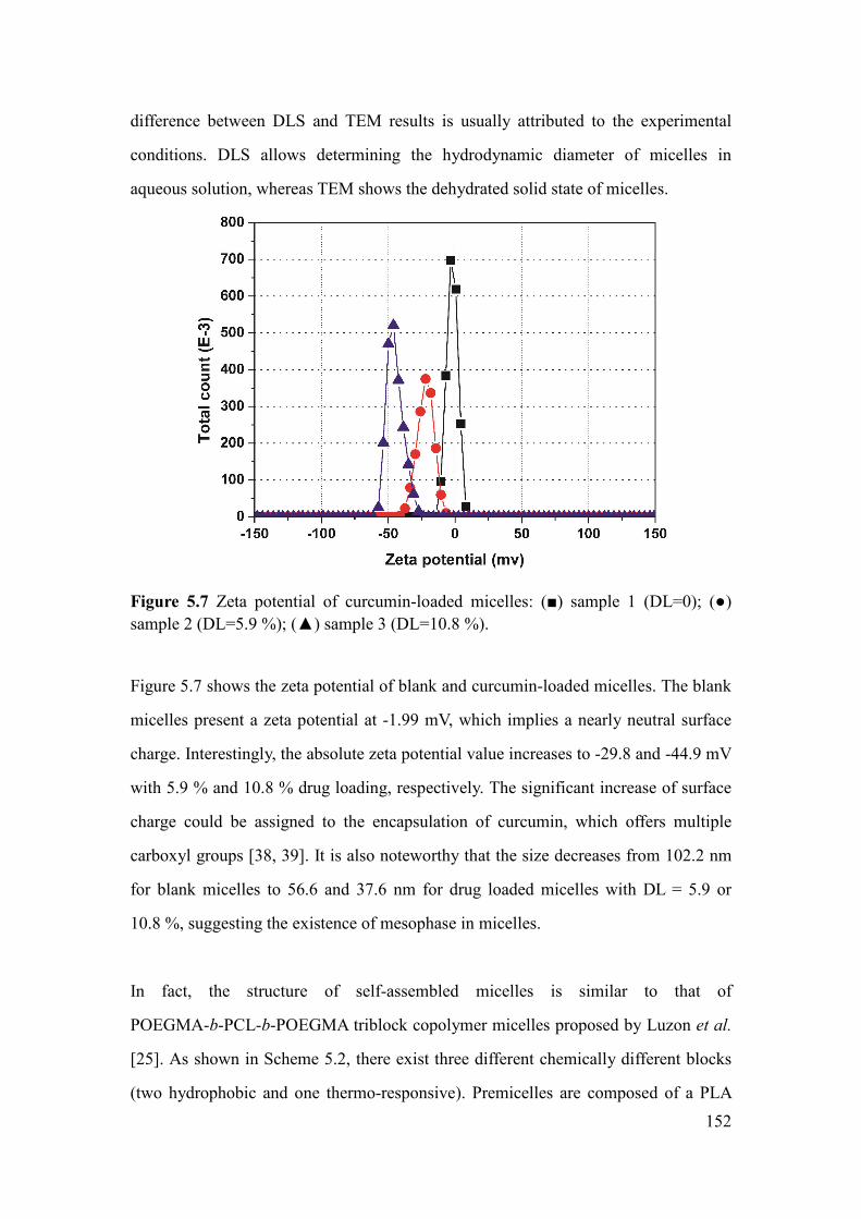
152
difference between DLS and TEM results is usually attributed to the experimental
conditions. DLS allows determining the hydrodynamic diameter of micelles in
aqueous solution, whereas TEM shows the dehydrated solid state of micelles.
Figure 5.7 Zeta potential of curcumin-loaded micelles: () sample 1 (DL=0); ()
sample 2 (DL=5.9 %); () sample 3 (DL=10.8 %).
Figure 5.7 shows the zeta potential of blank and curcumin-loaded micelles. The blank
micelles present a zeta potential at -1.99 mV, which implies a nearly neutral surface
charge. Interestingly, the absolute zeta potential value increases to -29.8 and -44.9 mV
with 5.9 % and 10.8 % drug loading, respectively. The significant increase of surface
charge could be assigned to the encapsulation of curcumin, which offers multiple
carboxyl groups [38, 39]. It is also noteworthy that the size decreases from 102.2 nm
for blank micelles to 56.6 and 37.6 nm for drug loaded micelles with DL = 5.9 or
10.8 %, suggesting the existence of mesophase in micelles.
In fact, the structure of self-assembled micelles is similar to that of
POEGMA-b-PCL-b-POEGMA triblock copolymer micelles proposed by Luzon et al.
[25]. As shown in Scheme 5.2, there exist three different chemically different blocks
(two hydrophobic and one thermo-responsive). Premicelles are composed of a PLA

153
core, a PMA shell and an oligoPEG corona. PMA blocks exhibit a relatively strong
affinity to water due to surface active ester groups. As the concentration increases
above the CMC, copolymer chains aggregate to form spherical micelles, which can be
regarded as soft matter dispersions with ordered inner structure stabilized by
hydrophilic corona. It could be assumed that triblock copolymers achieve steric
stabilization by having PLA blocks absorbed at interface of PMA, whereas
hydrophilic comb blocks extend into the external aqueous phase. Consequently,
copolymer micelles have an internal mesophase.
Scheme 5.2 Illustration of micellar structures during drug loading
In the present work, curcumin loaded micelles were prepared by solvent
evaporation/membrane rehydration method. Thus, curcumin could play an important
role in the micellization procedure. In fact, curcumin was encapsulated in the core and
in the mesophase, leading to decrease of micelle size and increase of zeta potential.
Therefore, curcumin loaded micelles exhibit more compacted core structure than
blank micelles. Similarly findings have been reported by Zhang et al. in the case of
amphotericin B loaded micelles prepared from comb-like block copolymers [42].
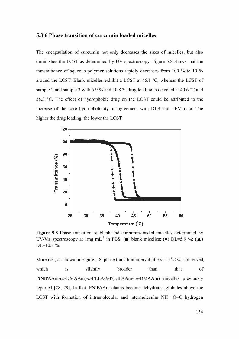
154
5.3.6 Phase transition of curcumin loaded micelles
The encapsulation of curcumin not only decreases the sizes of micelles, but also
diminishes the LCST as determined by UV spectroscopy. Figure 5.8 shows that the
transmittance of aqueous polymer solutions rapidly decreases from 100 % to 10 %
around the LCST. Blank micelles exhibit a LCST at 45.1 oC, whereas the LCST of
sample 2 and sample 3 with 5.9 % and 10.8 % drug loading is detected at 40.6 oC and
38.3 °C. The effect of hydrophobic drug on the LCST could be attributed to the
increase of the core hydrophobicity, in agreement with DLS and TEM data. The
higher the drug loading, the lower the LCST.
Figure 5.8 Phase transition of blank and curcumin-loaded micelles determined by
UV-Vis spectroscopy at 1mg mL-1
in PBS. () blank micelles; () DL=5.9 %; ()
DL=10.8 %.
Moreover, as shown in Figure 5.8, phase transition interval of c.a 1.5 oC was observed,
which is slightly broader than that of
P(NIPAAm-co-DMAAm)-b-PLLA-b-P(NIPAAm-co-DMAAm) micelles previously
reported [28, 29]. In fact, PNIPAAm chains become dehydrated globules above the
LCST with formation of intramolecular and intermolecular NH…O=C hydrogen

155
bonding. However, in the case of PEG analogues, the polymer-polymer interactions in
the globules are Van Der Waals forces which are weaker than H-bonding, thus leading
to a broader phase transition [16, 20].
5.3.7 In vitro drug release
Drug release was performed under physiological conditions (PBS, pH=7.4) at 37 °C
(below the LCST) and 41 oC (above the LCST), respectively. The amount of curcumin
was determined from UV-visible absorbance at 424 nm. As shown in Figure 9A, a
linear calibration curve was previously established between the UV absorbance and
curcumin concentration with a correlation coefficient of 0.99991.
The drug release profiles are shown in Fig. 9B. An initial fast release from micelles
was observed in all cases, followed by slower release. 7.7 % and 6.2 % of curcumin
were released from sample 2 and sample 3 at 1 h at 41°C, in contrast to 5.9 % and
3.5 % of curcumin released at 37 oC. Release of curcumin from sample 2 and sample
3 reached 80.3 % and 58.2 % after 4 day at 41 °C, in contrast to 66.1 and 45.5 % at
37 °C. Beyond, the release from sample 2 at 41 oC reached 89.5 % and 92.6 % at 7
and 14 days. For sample 3, 74.2 % and 75.9 % release were detected after 10 and 14
days at 41 °C. On the other hand, sample 2 and sample 3 showed a cumulative release
of 81.8 % and 59.5 % after 14 days at 37 oC, respectively.

156
Figure 5.9 Calibration curve (A) and drug release profiles (B) of curcumin-loaded
micelles: a) sample 2 (DL=5.9 %) at 41 oC; b) sample 2 (DL=5.9 %) at 37
oC; c)
sample 3 (DL=10.8 %) at 41 oC; d) sample 3 (DL=10.8 %) at 37
oC.

157
Therefore, drug release is not only dependent on the phase transition across the LCST,
but also on the drug loading. In fact, the solubility of curcumin in the release medium
is a limiting factor. Samples with higher drug loading exhibit lower release rate
because of the solubility effect. Besides, the micelles of sample 3 with higher drug
loading have a more compacted core as compared to sample 2 due to stronger
interactions between drug and hydrophobic blocks. It should also be noted that the
LCST of drug loaded micelles is dynamically changing with the release of curcumin.
Theoretically, the LCST should increase up to 45.1 oC when drug release reaches
100 % for all the samples.
It is of interest to compare the drug release behaviors from
P(NIPAAm-co-DMAAm)-b-PLLA-b-P(NIPAAm-co-DMAAm) and PLA/PEG
analogues micelles [29, 30]. Faster drug release is observed above the LCST than
below the LCST in both cases. However, the drug release rate from the former is
faster than that from the latter. In fact,
P(NIPAAm-co-DMAAm)-b-PLLA-b-P(NIPAAm-co-DMAAm) micelles exhibit
initially a larger burst-like release and then a slower release. In contrast, PLA/PEG
analogues micelles present smaller burst-like release, and then drug release gradually
increases. The depressed burst release and slower drug release may be related to the
enhanced interaction between the drug and hydrophobic blocks. In fact, as shown in
Scheme 2, both the inner PLA segment and intramolecular PMA blocks may
contribute to the enhanced interaction [39].
Therefore, curcumin loaded micelles with thermo-responsive
P(OEGMA-co-MEO2MA) corona, PMA interface and PLLA core exhibit low CMC
and nano size. The micelles could be easily administrated by intravenous injection.
The thermo-responsive drug release behavior shows that these polymeric micelles are
very promising as carrier of hydrophobic drugs.

158
5.4 Conclusion
Thermo-responsive comb-like
P(MEO2MA-co-OEGMA)-b-PLLA-b-P(MEO2MA-co-OEGMA) triblock copolymers
were synthesized by ATRP using a Br-PLLA-Br macroinitiator, and characterized by
1H NMR and SEC. The physico-chemical properties of copolymers are dependent on
the composition, namely the ratio of MEO2MA/OEGMA ranging from 79/21 to 42/58
(mol/mol). The LCST increases from 24.8 oC to 45.1
oC in PBS with increasing the
content of OEGMA. Micellization of copolymers was investigated by CMC, TEM
and DLS measurements. The CMC of copolymers increases from 0.0109 to 0.0137
mg mL-1
, and the size of micelles increases from 20.7 to 102.5 nm with increasing the
content of OEGMA. A hydrophobic anticancer drug, curcumin, was successfully
encapsulated in micelles. The drug loading was 5.9 % and 10.8 % with loading
efficiency higher than 90 %. The LCST of drug loaded micelles was 40.6 oC and 38.3
oC, respectively, which is lower than blank micelles by 4.5 and 6.8 °C. TEM and DLS
results show a strong decrease of micellar size, suggesting that the hydrophobic drug
plays a major role in the self-assembly procedure of drug loaded micelles. In vitro
drug release exhibits thermo-responsive dependence across the LCST. Micelles with
low drug loading show fast release than micelles with high drug loading. The higher
drug loading and depressed burst-like release could be assigned to the enhanced
interaction between drug and comb-like copolymers with two hydrophobic blocks, i.e.
PLLA and PMA. Therefore, the nanosize, low CMC and thermo-responsive drug
release indicate that P(MEO2MA-co-OEGMA)-b-PLLA-b-P(MEO2MA-co-OEGMA)
triblock copolymers are promising candidate for targeted delivery of anti-tumor drugs.
5.5 Reference
1. R. Savic, L. B. Luo, A. Eisenberg and D. Maysinger, Micellar nanocontainers distribute to
defined cytoplasmic organelles. Science 300 (2003) 615-618.
2. J. A. Hubbell, Enhancing drug function. Science 300 (2003) 595-596.
3. N. Bertrand, J. Wu, X. Y. Xu, N. Kamaly and O. C. Farokhzad, Cancer nanotechnology: The

159
impact of passive and active targeting in the era of modern cancer biology. Adv. Drug Deliv. Rev.
66 (2014) 2-25.
4. T. Yahara, T. Koga, S. Yoshida, S. Nakagawa, H. Deguchi, K. Shirouzu, Relationship Between
Microvessel Density and Thermographic Hot Areas in Breast Cancer. Surg. Today 33 (2003)
243-248.
5. A. Chilkoti, M. R. Dreher, D. E. Meyer, D. Raucher, Targeted drug delivery by thermally
responsive polymers. Adv. Drug Deliv. Rev. 54 (2002) 613-630.
6. F. Danhier, O. Feron, V. Prat, To exploit the tumor microenvironment: Passive and active tumor
targeting of nanocarriers for anti-cancer drug delivery. J. Controlled Release 148 (2010) 135-146.
7. S. Hocine and M. H. Li, Thermoresponsive self-assembled polymer colloids in water. Soft
Matter 9 (2013) 5839-5861.
8. J. Du and R. K. O’Reilly, Advances and challenges in smart and functional polymer vesicles.
Soft Matter 5 (2009) 3544-3561.
9. A. K. Bajpai, S. K. Shukla, S. Bhanu and S. Kankane, Responsive polymers in controlled drug
delivery. Prog. Polym. Sci. 33 (2008) 1088-1118.
10. S. Ganta, H. Devalapally, A. Shahiwala and M. Amiji, A review of stimuli-responsive
nanocarriers for drug and gene delivery. J. Controlled Release 126 (2008) 187-204.
11. D. Schmaljohann, Thermo- and pH-responsive polymers in drug delivery. Adv. Drug Deliv.
Rev. 58 (2006) 1655-1670.
12. H. Ding, F. Wu, Y. Huang, Z. R. Zhang, Y. Nie, Synthesis and characterization of
temperature-responsive copolymer of PELGA modified poly(N-isopropylacrylamide). Polymer 47
(2006) 1575-1583.
13. H. Wei, S. X. Cheng, X. Z. Zhang, R. X. Zhuo, Thermo-sensitive polymeric micelles based on
poly(N-isopropylacrylamide) as drug carriers. Prog. Polym. Sci.34 (2009) 893-910.
14. W. Li, J. F. Li, J. Gao, B. H. Li, Y. Xia, Y. C. Meng, Y. S. Yu, H. W. Chen, J. X. Dai, H.
Wang, Y. J. Guo, The fine-tuning of thermosensitive and degradable polymer micelles for
enhancing intracellular uptake and drug release in tumors. Biomaterials 32 (2011) 3832-3844.
15. F. Xu, S. Z. Zheng, Y. L. Luo, Thermosensitive t-PLA-b-PNIPAAm tri-armed star block
copolymer nanoscale micelles for camptothecin drug release. J. Polym. Sci. Part A: Polym. Chem.
51 (2013) 4429-4439.
16. J. F. Lutz, O. Akdemir, A. Hoth, Point by Point Comparison of Two Thermosensitive
Polymers Exhibiting a Similar LCST: Is the Age of Poly(NIPAM) Over? J. Am. Chem. Soc. 128
(2006) 13046-13047.
17. J. F. Lutz, J. Andrieu, S. uzgun, C. Rudolph, S. Agarwal, Biocompatible, Thermoresponsive,
and Biodegradable:Simple Preparation of "All-in-One" Biorelevant Polymers. Macromolecules 40
(2007) 8540-8543.
18. J. F. Lutz, H. G. Borner, Modern trends in polymer bioconjugates design. Prog. Polym.Sci. 33
(2008) 1-39.
19. J. F. Lutz, A. Hoth, Preparation of Ideal PEG Analogues with a Tunable Thermosensitivity by
Controlled Radical Copolymerization of 2-(2-Methoxyethoxy) ethyl Methacrylate and
Oligo(ethylene glycol) Methacrylate. Macromolecules 39 (2006) 893-896.
20. J. F. Lutz, K. Weichenhan, O. Akdemir, A. Hoth, About the Phase Transitions in Aqueous
Solutions of Thermoresponsive Copolymers and Hydrogels Based on 2-(2-methoxyethoxy) ethyl
Methacrylate and Oligo(ethylene glycol) Methacrylate. Macromolecules 40 (2007) 2503-2508.

160
21. J. F. Lutz, Polymerization of oligo(ethylene glycol) (meth)acrylates: Toward new generations
of smart biocompatible materials. J. Polym. Sci. Part A: Polym. Chem. 46 (2008) 3459-3470.
22. N. Fechler, N. Badi, K. Schade, S. Pfeifer, J. F. Lutz, Thermogelation of PEG-Based
Macromolecules of Controlled Architecture. Macromolecules 42 (2008) 33-36.
23. J. F. Lutz, Thermo-Switchable Materials Prepared Using the OEGMA-Platform. Adv. Mater.
23 (2011) 2237-2243.
24. A. O. Saeed, S. Dey, S. M. Howdle, K. J. Thurecht and C. Alexander, One-pot controlled
synthesis of biodegradable and biocompatible co-polymer micelles. J. Mater. Chem. 19 (2009)
4529-4535.
25. M. Luzón, T. Corrales, F. Catalina, V. San Miguel, C. Ballesteros and C. Peinado,
Hierarchically organized micellization of thermoresponsive rod-coil copolymers based on
poly[oligo(ethylene glycol) methacrylate] and poly(ε-caprolactone). J. Polym. Sci. Part A: Polym.
Chem. 48 (2010) 4909-4921.
26. Y. Bakkour, V. Darcos, F. Coumes, S. M. Li and J. Coudane, Comb-like amphiphilic
copolymers based on polylactide and poly(ethylene glycol): Synthesis, self-assembly and
evaluation as drug carrier. Polymer 54 (2013) 1746-1754.
27. R. Fenyves, M. Schmutz, I. J. Horner, F. V. Bright and J. Rzayev, Aqueous Self-Assembly of
Giant Bottlecomb Block Copolymer Surfactants as Shape-Tunable Building Blocks. J. Am. Chem.
Soc. 136 (2014) 7762-7770.
28. Y. F. Hu, V. Darcos, S. Monge, S. M. Li, Y. Zhou, F. Su, Tunable thermo-responsive
P(NIPAAm-co-DMAAm)-b-PLLA-b-P(NIPAAm-co-DMAAm) triblock copolymer micelles as
drug carriers. J. Mater. Chem. B 2 (2014) 2738-2748.
29. Y. F. Hu, V. Darcos, S. Monge, S. M. Li, Y. Zhou, F. Su, Thermo-responsive release of
curcumin from micelles prepared by self-assembly of amphiphilic
P(NIPAAm-co-DMAAm)-b-PLLA-b-P (NIPAAm-co-DMAAm) triblock copolymers. Inter. J.
Pharm. 476 (2014) 31-40.
30. Y. F. Hu, V. Darcos, S. Monge and S. M. Li, Synthesis and self-assembling of
poly(N-isopropylacrylamide-block-poly(L-lactide)-block-poly(N-isopropylacrylamide) triblock
copolymers prepared by combination of ring-opening polymerization and atom transfer radical
polymerization. J. Polym. Sci. Part A: Polym. Chem. 51 (2013) 3274-3283.
31. M. Zhang and A. H. E. Müller, Cylindrical polymer combes. J. Polym. Sci. Part A: Polym.
Chem. 43 (2005) 3461-3481.
32. L. Gu, Z. Shen, S. Zhang, G. Lu, X. Zhang and X. Huang, Novel Amphiphilic Centipede-Like
Copolymer Bearing Polyacrylate Backbone and Poly(ethylene glycol) and Polystyrene Side
Chains. Macromolecules 40 (2007) 4486-4493
33. M. Gerle, K. Fischer, S. Roos, A. H. E. Muller, M. Schmidt, S. S. Sheiko, S. Prokhorova and
M. Muller, Main Chain Conformation and Anomalous Elution Behavior of Cylindrical Combes As
Revealed by GPC/MALLS, Light Scattering, and SFM. Macromolecules 32 (1999) 2629-2637.
34. M. Nakayama, J. E. Chung, T. Miyazaki, M. Yokoyama, K. Sakai and T. Okano, Thermal
modulation of intracellular drug distribution using thermoresponsive polymeric micelles. React.
Funct. Polym. 67 (2007) 1398-1407.
35. M. L. Gou, K. Men, H. S. Shi, M. L. Xiang, J. A. Zhang, J. Song, J. L. Long, Y. Wan, F. Luo,
X. Zhao and Z. Y. Qian, Curcumin-loaded biodegradable polymeric micelles for colon cancer
therapy in vitro and in vivo. Nanoscale 3 (2011) 1558-1567.

161
36. C. Mohanty, S. Acharya, A. K. Mohanty, F. Dilnawaz, S. K. Sahoo, Curcumin-encapsulated
MePEG/PCL diblock copolymeric micelles: a novel controlled delivery vehicle for cancer therapy.
Nanomed. 5 (2010) 433-449.
37. O. Naksuriya, S. Okonogi, R. M. Schiffelers, W. E. Hennink, Curcumin nanoformulations: A
review of pharmaceutical properties and preclinical studies and clinical data related to cancer
treatment. Biomaterials 35 (2014) 3365-3383.
38. Z. M. Song, R. L. Feng, M. Sun, C. Y. Guo, Y. Gao, L. B. Li, G. X. Zhai, Curcumin-loaded
PLGA-PEG-PLGA triblock copolymeric micelles: Preparation, pharmacokinetics and distribution
in vivo. J.Colloid Interface Sci.354 (2011) 116-123.
39. J. Z. Du, L. Y. Tang, W. J. Song, Y. Shi and J. Wang, Evaluation of Polymeric Micelles from
Comb Polymer with Poly(epsilon-caprolactone)-b-Poly(ethylene glycol) Side Chains as Drug
Carrier. Biomacromolecules 10 (2009) 2169-2174.
40. R. L. Yang, S. A. Zhang, D. L. Kong, X. L. Gao, Y. J. Zhao, Z. Wang, Biodegradable
Polymer-Curcumin Conjugate Micelles Enhance the Loading and Delivery of Low-Potency
Curcumin. Pharm. Res. 29 (2012) 3512-3525.
41. H. D. Tang, C. J. Murphy, B. Zhang, Y. Q. Shen, M. H. Sui, E. A. Van Kirk, X. W. Feng, W. J.
Murdoch, Amphiphilic curcumin conjugate-forming nanoparticles as anticancer prodrug and drug
carriers: in vitro and in vivo effects. Nanomed. 5 (2015) 855-865.
42. X. W. Zhang, X. Q. Zhu, F. Y. Ke, L. Ye, E. Q. Chen, A. Y. Zhang, Z. G. Feng, Preparation and
self-assembly of amphiphilic triblock copolymers with polyrotaxane as a middle block and their
application as carrier for the controlled release of Amphotericin B. Polymer 50 (2009) 4343-4351.

162
163
Conclusions and perspectives
In this thesis, two families of amphiphilic and thermo-responsive block copolymers
based on poly(L-lactide) (PLLA) and polyacrylamide or poly(oligo(ethylene glycol)
methacrylate) are successfully synthesized by ATRP using the same Br-PLLA-Br
(DP=40) macroinitiator. The self-assembly, phase transition, drug release and
cytotoxicity of the various copolymers are studied to evaluate their potential as
nanocarrier for thermo targeted delivery of antitumor drugs. The following
conclusions are drawn from this work:
1) PNIPAAm-b-PLLA-b-PNIPAAm triblock copolymers are obtained by ATRP of
NIPAAm using a α, ω-Br-PLLA-Br macroinitiator. The latter is prepared by ring
opening polymerization of L-lactide followed by reaction with 2-bromopropionyl
bromide. The ATRP of NIPAAm is performed in a DMF/water (5/1) mixture using
CuCl/Me6TREN catalyst system at room temperature, yielding copolymers with well
controlled molecular weights and narrow dispersities.
PNIPAAm-b-PLLA-b-PNIPAAm copolymers with NIPAAm/LA ratios from 2.4 to
6.9 are susceptible to self-assemble in aqueous medium to yields spherical micelles of
sizes from 31 to 83 nm. The CMC of copolymers is in the range of 0.0077-0.016 mg
mL-1
and the LCST in the range of 32.1-32.8 oC. The micelles exhibit different
thermo-responsive behaviors at high or low concentrations. At 3.0 mg mL-1
,
aggregation of micelles occurs when the temperature is raised above the LCST,
leading to micelle size increase. In contrast, at 0.2 mg mL-1
, a decrease of micelle size
is detected because of the collapse of PNIPAAm blocks above the LCST. The original
micelle size is recovered when the temperature is decreased below the LCST in both
cases, indicating that the system is thermo-reversible.
2) P(NIPAAm-co-DMAAm)-b-PLLA-b-P(NIPAAm-co-DMAAm) triblock
copolymers can be obtained under the same conditions. With the ratio of NIPAAm to

164
DMAAm ranging from 100/0 to 76/24 in the hydrophilic blocks, the LCST of
copolymers linearly increases the LCST to the range of 32.3 to 39.1. Moreover,
introduction of DMAAm leads to very narrow phase transition across LCST within
0.5 oC, in contrast to a phase transition interval of 2.8 °C for
PNIPAAm-b-PLLA-b-PNIPAAm. Increasing the DMAAm content also leads to larger
micelle size from 40 to 55 nm, and higher CMC from 0.010 to 0.015 mg mL-1
due to
the more hydrophilic nature of DMAAm as compared to NIPAAm. Copolymer
micelles are able to encapsulate hydrophobic drugs by using dialysis method with
loading efficiency above 59 %. In vitro drug release performed at 37 or 38 °C in water
exhibits an initial burst release. The release rate at 38 °C is much faster than that at
37 °C, suggesting that these copolymer micelles could achieve thermo-responsive
release after in vivo administration. Moreover, MTT assay and cell morphology
observation indicate that the copolymers present outstanding cytocompatibility.
3) The drug release properties of
P(NIPAAm-co-DMAAm)-b-PLLA-b-P(NIPAAm-co-DMAAm) are further studied
with higher DMAAm content from 31.8 to 39.4 % in the hydrophilic
P(NIPAAm-co-DMAAm) blocks. The LCST of copolymers varies from 44.7 °C to
49.4 °C in water and decreases by ca. 3.5 °C in PBS. Increase of DMAAm content
also leads to slight CMC increase from 0.0113 to 0.0144 mg mL-1
, and slight micelle
size increase from 37 to 44 nm. The zeta-potential of micelles varies in the -12.4 to
-18.7 mV range, in agreement with the structure of micelles having PLLA block in the
core and P(NIPAAm-co-DMAAm) blocks at the corona. A hydrophobic anticancer
drug, curcumin, is encapsulated by using film hydration method with drug loading
increases from 6.0 to 20.4 % and loading efficiency above 94 %. The LCST of blank
micelles decreases from 45.6 oC in water to 42.1
oC in PBS. When with 6.0 % drug,
the LCST further decreases from 42.1 to 38.0 oC in PBS. Higher drug loading leads to
lower LCST, but the decrease is very limited as the LCST only lightly decreases to
37.5 oC with 20.4 % drug loading. On the other hand, the micelle size increases from

165
39 nm for blank micelles to 88 nm with drug loading of 20.4 %, indicating that
curcumin incorporation leads to larger micelles. All micelles remain homogeneously
transparent over 1 month due to the negative surface charge.
In vitro drug release presents an initial burst release in all cases, followed by a slower
release. Thermo-responsive release is observed as the release rate is higher at 40 °C
than that at 37 °C. Application of Peppas’ theory indicates a combination of diffusion
and degradation controlled release.
4) Triblock comb-like copolymers of PLA/PEG analogues with MEO2MA/OEGMA
molar ratio ranging from 79/21 to 42/58 are synthesized by ATRP in DMF at 80 oC
using the same α, ω-Br-PLLA-Br macroinitiator and CuCl/Me6TREN catalyst system.
The LCST in PBS increases from 24.8 to 45.1 oC with the increase of OEGMA
content from 32 % to 58 %. Spherical micelles with size from 20.7 to 102.5 nm are
obtained by self-assembly of copolymers in aqueous medium with a CMC from
0.0109 to 0.0137 mg mL-1
. Curcumin is encapsulated by using film hydration method
with drug loading of 5.9 % and 10.8 % and loading efficiency higher than 90 %.The
LCST of drug loaded micelles was 40.6 oC and 38.3
oC, which is lower than blank
micelles by 4.5 and 6.8 °C, respectively. The size of curcumin loaded micelles
decreases from 103 to 38 nm while the zeta potential decreases from -1.99 mV to
-44.9 mV, which indicates that the hydrophobic drug plays an important role in the
self-assembly procedure of drug loaded micelles.
In vitro drug release exhibits thermo-responsive dependence across the LCST.
Micelles with low drug loading show fast release than micelles with high drug loading.
The higher drug loading and depressed burst-like release could be assigned to the
enhanced interaction between drug and comb-like copolymers with two hydrophobic
blocks, i.e. PLLA and PMA.

166
Therefore, an innovative work has been accomplished on thermo-responsive micelles
as nanocarrier for anticancer applications. The copolymers obtained by ATRP exhibit
well-defined chain structures. Self-assembled copolymer micelles exhibit nano-size,
very low CMC and thermo-responsive drug release behavior, which present great
potential in antitumor applications. Further investigations can be envisaged in future:
1) Synthesis and self-assembly of PLA-PNIPAAm (and PLA-PEG analogues) diblock
copolymers; 2) Preparation of other aggregate architectures including polymersomes,
worm-like micelles, nanotubes as well as hydrogels; 3) Synthesis of block copolymers
with PLLA or PDLA macroinitiator so as to obtain stereocomplex micelles; 4)
synthesis of thermo-responsive copolymers with targeting molecules by click
chemistry.

167
Publications
1 Y. F. Hu, V. Darcos, S. Monge, S. M. Li, Thermoresponse micelles from
comb-like copolymers of polylactide and PEG Analogues: Synthesis,
Micellization and In vitro drug release, Internal Journal of Pharmaceutics,
submitted.
2 Y. F. Hu, V. Darcos, S. Monge, S. M. Li, Y. Zhou, F. Su, Thermo-responsive
release of curcumin from micelles prepared by self-assembly of amphiphilic
P(NIPAAm-co-DMAAm)-b-PLA-b-P(NIPAAm-co-DMAAm) triblock
copolymers, Internal Journal of Pharmaceutics, 476 (2014) 31-40
3 Y. F. Hu, V. Darcos, S. Monge, S. M. Li, Y. Zhou, F. Su, Tunable
thermo-responsive
P(NIPAAm-co-DMAAm)-b-PLLA-b-P(NIPAAm-co-DMAAm) triblock
copolymer micelles as drug carriers. Journal of Materials Chemistry B, 2 (2014)
2738-2748.
4 Y. F. Hu, V. Darcos, S. Monge, S. M. Li, Synthesis and self-assembling of
poly(N-isopropylacrylamide-block-poly(L-lactide)-block-poly(N-isopropylacryla
mide) triblock copolymers prepared by combination of ring-opening
polymerization and atom transfer radical polymerization. Journal of Polymer
Science part A: Polymer Chemistry, 51 (2013) 3274-3283.

168
Acknowlegdements
After 3 and half years PH.D study, finally I could have my defense and have a
delighted time. I am a shy person and not so good at express myself. Herein, I write
this final part with cheerful and complicated mood. I have taken an interesting
experience in Montpellier. I was lost and stand in the cold wind in Montpellier due to
the delay of plane and train at 21th
, November 2011. I asked a passenger with my poor
French to call my supervisor Suming Li to pick me. Then I found Montpellier is really
nice city with nice people.
First, I would like to express my appreciation to my supervisor, Suming Li. We have
met and knew with each other for 10 years. I still remember the first time we met at
Fudan University, China when I was 22 years old and a fresh noob mater student.
Suming not only advised my thesis work but also took care of my daily life. I have
nice experience under his advising and I learn a lot of stuff from him, including how
to start a scientific project, write papers as well as interact with people.
I also want to thank Dr Vincent, my co-supervisor for his excellent direction on my
polymer synthesis. The valuable suggestion from him improved the thesis and my
profession skills, although we have different opinions at some times. I am want to
thank Dr sophie Monge for her support of my test. Perhaps I did most of the test in
her lab. She is a really nice person to teach me how to use the machine and book the
time for me. I am also grateful to Dr Jean Coudane for his full support as the director
of the laboratory and Dr Boustta Mahfoud for his favor as my seat adjacent to endure
my messy desk. I should send many thanks to Xiaohan Wu. He helped me everything
in my early life in France. Many thanks to the couple of Adrien Leroy and Fanny
Coumes for their help familiar with lab and solve the problems I asked with stunning
patience. Many thanks present to Marc-Alexandre with whom I really enjoy the talk.
Many thanks also should present to Dominique Arama who always say hello to me in

169
Chinese. Appreciation also should be presented to Benjamin Nottelet, Sylvie Hunger,
Xavier Dumont and Cedric paniagua for their support, to Frank Godiard for TEM
measurements, to all the members in our laboratory for providing a enjoyable work
environment.
A part of this thesis work was carried out in the College of Chemical Engineering,
Qingdao University of Science and Technology, China. Thanks Yang Zhou and Feng
Su for their excellent work.
At last, I wish to express my sincere gratitude to my wife. She accompanies me for 3
years in France. It is not easy for her especially she is always homesick and takes care
of my baby daughter alone for most of time. Finally, I finished my job and could have
a nice time with my family.
When I recall the past years, there are a lots of emotion, freshness, curiousness,
understanding, cheerfulness for my work, moved tears for the birth of my daughter. It
is interesting life in France as a foreigner for these 3 years. It will affect me for the
whole life. It is time to say goodbye to friends and people in the lab. I will miss you
all.

RESUME
Deux séries de copolymères tribloc thermo-sensibles et amphiphiles, à savoir
poly(L-lactide)/poly(N-isopropylacrylamide-co-N,N-diméthylacrylamide) et
poly(L-lactide)/poly(2-(2-méthoxyéthoxy) éthyl méthacrylate-co-oligo(éthylène glycol)
méthacrylate) ont été synthétisées par polymérisation radicalaire par transfert d’atomes
en utilisant le Br-PLLA-Br comme macroamorceur dans des conditions douces. Les
copolymères obtenus présentent une structure de chaînes bien définie avec une
dispersité étroite, et sont capable de s’auto-assembler dans un milieu aqueux pour
donner des micelles sphériques de taille en dessous de 100 nm et de faible concentration
micellaire critique (<0.016 mg mL-1
). La température critique inférieure de solution peut
être ajustée avec précision en faisant varier le rapport NIPAAm/DMAAm ou
MEO2MA/OEGMA. Un principe actif hydrophobe, curcumine, a été choisi comme
modèle pour déterminer les propriétés de libération des micelles à différentes
températures. Une libération thermo-sensible de curcumine a été observée, indiquant
que ces copolymères sont prometteurs pour la libération ciblée de principes actifs
anti-tumoraux.
Mots-clés: thermo-sensible, polymérisation radicalaire par transfert d’atomes,
poly(L-lactide), poly(N-isopropylacrylamide), oligo(éthyl glycol) méthacrylate,
auto-assemblage, libération ciblée de principes actifs.
Synthesis, self-assembly and controlled drug delivery of novel thermo-responsive
and amphiphilic block copolymers based on polylactide, polyacrylamide and
Poly[oligo(ethylene glycol) methacrylate]
ABSTRACT
Two series of thermo-responsive and amphiphilic triblock copolymers, i.e.
poly(L-lactide)/poly(N-isopropylacrylamide-co-N,N-dimethylacrylamide) and
poly(L-lactide)/poly(2-(2-methoxyethoxy) ethyl methacrylate-co-oligo(ethylene glycol)
methacrylate) were synthesized by atom transfer radical polymerization using
Br-PLLA-Br as macroinitiator under mild conditions. The obtained copolymers present
well defined chain structures with narrow dispersity, and are able to self-assemble in
aqueous medium yielding spherical micelles with size below 100 nm and low critical
micellization concentration (<0.016 mg mL-1
). The lower critical solution temperature is
precisely adjusted by changing the NIPAAm/DMAAm or MEO2MA/OEGMA ratio. A
hydrophobic drug, curcumin, is taken as a model to evaluate the drug release properties
of micelles at different temperatures. Thermo-responsive drug release behavior is
observed, indicating that these copolymers are promising candidate for targeted delivery
of anticancer drugs.
Keywords: thermo-responsive, atom transfer radical polymerization, poly(L-lactide),
poly(N-isopropylacrylamide), oligo(ethylene glycol) methacrylate, self-assembly,
targeted drug delivery.
Intitulé et addresse de l’unité ou laboratoire
Equipe Biopolymères Artificiels, Institut des Biomolécules Max Mousseron, UMR
CNRS 5247, Faculté de Pharmacie -Université de Montpellier- 15 avenue Charles
Flahaut, 34093 Montpellier, France