XXVI SICOT Triennial World Congresslhcnews.sicot.org/resources/File/Newsletter/NL136.pdf ·...
12
September 2013 – No. 136 x Editorial: 34 th SICOT Orthopaedic World Congress by Ashok Johari 2 x Fellowship News: Report of the “SICOT meets SICOT” Fellowship at CHOP, United States 3 x Training Around the World: Orthopaedic Training in Turkey 4 x Articles by SICOT Members: The Nigerian Orthopaedic Association meeting in Ife-Ife, Nigeria 5 x Expert Corner: How I do a Primary Total Hip Arthroplasty (THA) 6 x History of Orthopaedics: A very brief history of orthopaedic surgery in the United Kingdom 8 x Worldwide News: The value of the three-point index in predicting redisplacement of diaphyseal fractures of the forearm in children 10 x Congress News: XXVI SICOT Triennial World Congress combined with the 46 th SBOT Annual Meeting 12 XXVI SICOT Triennial World Congress combined with the 46 th SBOT Annual Meeting Rio de Janeiro, Brazil – 20-22 November 2014
Transcript of XXVI SICOT Triennial World Congresslhcnews.sicot.org/resources/File/Newsletter/NL136.pdf ·...

September 2013 – No. 136
Editorial: 34th SICOT Orthopaedic World Congress by Ashok Johari 2 Fellowship News: Report of the “SICOT meets SICOT” Fellowship at CHOP, United States 3 Training Around the World: Orthopaedic Training in Turkey 4 Articles by SICOT Members: The Nigerian Orthopaedic Association meeting in Ife-Ife, Nigeria 5
Expert Corner: How I do a Primary Total Hip Arthroplasty (THA) 6
History of Orthopaedics: A very brief history of orthopaedic surgery in the United Kingdom 8 Worldwide News: The value of the three-point index in predicting redisplacement of diaphyseal fractures of the
forearm in children 10 Congress News: XXVI SICOT Triennial World Congress combined with the 46th SBOT Annual Meeting 12
XXVI SICOT Triennial World Congresscombined with the 46th SBOT Annual Meeting
Rio de Janeiro, Brazil – 20-22 November 2014

Editorial
34th SICOT Orthopaedic World Congress Hyderabad, India – 17-19 October 2013
An event not to be missed – “Must go, must participate”
Ashok Johari Congress President – Mumbai, India
2
What does a surgeon desire from a congress? Great academics, great faculty, cutting-edge technology, camaraderie, collaborations, relaxing social events and fantastic tourism. The 34th SICOT Orthopaedic World Congress (Hyderabad OWC 2013) has it all aplenty and hence is an event not to be missed. Experts from different subspecialties in Orthopaedics from all over the globe will converge to Hyderabad between 16 and 19 October. Symposia, instructional courses, debates and case discussions will cover various frontier areas of Orthopaedics and Traumatology. An intense learning process has been set up covering 10 halls, 4 plenary lectures, 47 symposia and instructional courses, 46 free paper sessions, one best paper session, 11 focused oral communication sessions and a myriad of electronic posters. Hyderabad OWC 2013 has the highest number of abstract submissions ever, breaking the earlier record set by the Dubai OWC 2012. 2,047 abstracts have been selected for presentation in various forms. Undoubtedly, this will provide a very wide coverage of Orthopaedics. While frontier areas in Trauma, Spine, Hip, Knee, Ankle and Foot, Upper Extremity, Arthroscopy and Sports Medicine, Paediatrics, Oncology, Basic Research and Education are being covered, topics relevant to the developing world are being highlighted through various symposia. Academic organisations such as the American Academy of Orthopaedic Surgeons (AAOS), Asia Pacific Orthopaedic Association (APOA), Association for Rational Treatment of Fractures (ARTOF), Association for the Study and Application of the Method of Ilizarov (ASAMI), European Bone & Joint Infection Society (EBJIS), International Federation of Paediatric Orthopaedic Societies (IFPOS), Société Internationale de Recherche en Orthopédie et Traumatologie (SIROT), Société Française de Chirurgie Orthopédique et Traumatologique (SOFCOT), and World Orthopaedic Concern (WOC) will add lustre to the proceedings. Lunch symposia will provide a glance of the industry viewpoint further exemplified in the huge trade exhibition. The Educational Day on 16 October focuses on
Trauma and boasts a coverage of more than 40 areas by an eminent international faculty. Besides all this, invisible seepage and exchange of knowledge will take place through individual interaction during the various breaks and social events. Numerous social events are planned. A spectacular inauguration will have a touch of Indian culture and arts. The welcome reception and the Indian theme dinner will provide much needed relaxation after intense academics. A cricket match in the spirit of the Indies and a charity run are all added in for fun and social causes. Nations of the APOA and South Asian Association for Regional Cooperation (SAARC) member countries are ‘Friendship Nations’ and I am sure the OWC will witness a lot of delegates from these countries. India is a great tourist destination and Hyderabad has been ranked as one of the ten best cities in the world. Its airport is the world's best ranking airport in the 5 to 15 million capacity and connects with many international destinations. A treasure of sightseeing and shopping awaits those so inclined. From Hyderabad one can travel north or south, east or west, for seeing incredible India. I welcome each of you personally and promise you a great Congress. We will deem it an honour if you came. I am sure that the Hyderabad OWC 2013 will fulfil the cherished mission of SICOT which is to advance Orthopaedics, promote international education and research and camaraderie between orthopaedic colleagues from different parts of the world.
Read more about the Congress at: www.sicot.org/?id_page=657
Fellowship News
Report of the “SICOT meets SICOT” Fellowship at CHOP, United States
Islam Abouyoussef SICOT Associate Member – Cairo, Egypt
3
I had the honour of being selected by SICOT for the “SICOT meets SICOT” (SmS) Fellowship Programme at the Children's Hospital of Philadelphia (CHOP) during May 2013. Philadelphia is a pleasant city located in the eastern region of the United States. CHOP is the main paediatric hospital of the University of Pennsylvania. In 2012, CHOP was selected the best children's hospital in the United States. The division of Orthopaedic Surgery in CHOP is always busy in terms of providing quality patient care, training of medical staff and doing research work. My experience in CHOP was really great, starting from the hospital set-up to the detailed surgical techniques. The team of the Orthopaedic Division is wonderful. They provide integrated care to their patients.
With Prof John Dormans
Prof John P. Dormans, Chief of the Orthopaedic Surgery Division, is a great mentor and leader. My experience with him in the fields of limb salvage surgeries and spine surgeries was awe-inspiring. Since his clinic always has a high flow of patients, I had the chance to see some rare cases with him. I was also able to learn from him how to approach patients effectively in the clinic, how to teach them about their diseases and how to manage time.
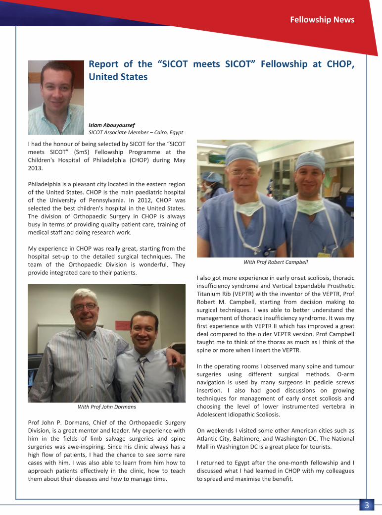
With Prof Robert Campbell
I also got more experience in early onset scoliosis, thoracic insufficiency syndrome and Vertical Expandable Prosthetic Titanium Rib (VEPTR) with the inventor of the VEPTR, Prof Robert M. Campbell, starting from decision making to surgical techniques. I was able to better understand the management of thoracic insufficiency syndrome. It was my first experience with VEPTR II which has improved a great deal compared to the older VEPTR version. Prof Campbell taught me to think of the thorax as much as I think of the spine or more when I insert the VEPTR. In the operating rooms I observed many spine and tumour surgeries using different surgical methods. O-arm navigation is used by many surgeons in pedicle screws insertion. I also had good discussions on growing techniques for management of early onset scoliosis and choosing the level of lower instrumented vertebra in Adolescent Idiopathic Scoliosis. On weekends I visited some other American cities such as Atlantic City, Baltimore, and Washington DC. The National Mall in Washington DC is a great place for tourists. I returned to Egypt after the one-month fellowship and I discussed what I had learned in CHOP with my colleagues to spread and maximise the benefit.

Training Around the World
Orthopaedic Training in Turkey
Ömer Cengiz Istanbul, Turkey
4
Undergraduate medical training in Turkey is five years in duration with an additional one year of internship. After completing six years of undergraduate study, a medical student is entitled to become a medical doctor. However, this does not mean that you can get your medical diploma right away. You are required to fulfil a civil service obligation ranging between 300 and 600 days that varies between regions. However, there is an exemption through sitting the medical specialty examination. By passing the examination before completing the civil service, you are then exempted from the compulsory service. The examination consists of multiple choice questions (MCQ). In order to enter any residency programme, one has to pass and get enough points which is really challenging. After that, your five-year orthopaedic residency programme begins. Orthopaedic training is done either in medical schools of universities or in the “Educational and Research Hospitals” under the Ministry of Health. Although the workload does not vary much, the universities are more theoretical and research based. On the other hand, more practical trainings are being done in the “Educational and Research Hospitals”, contrary to its name. The first year of training is the hardest. The working hours and on call schedule is organised by the clinical director or head of the department, usually up to 130 hours per week is required during your first 12 months. Due to this, residents tend to drop out from the programme during their first year of training. The standard of training in orthopaedics and traumatology in Turkey is monitored by the Turkish Orthopaedics and Traumatology Association (TOTBID). All residents are given an assistant report card provided by TOTBID. Residents are required to rotate in other specialties apart from orthopaedic surgery, such as general surgery, plastic and reconstructive surgery, cardiovascular surgery, physical therapy and rehabilitation, anaesthesia, and emergency medicine. Attendances at meetings and courses are also recorded in the report cards. At the end of each year, the report cards are collected, evaluated and graded by the clinical director.
In May of each year, an annual assessment is scheduled by TOTBID, which is compulsory for all orthopaedic residents to attend. The results are then sent to the heads of the departments for evaluation of any shortcomings in training where extra clinical seminars are held if necessary. Orthopaedic training in Turkey is a five-year training process. As a resident you will undergo intensive training in trauma during your first year. I think surgeons studying for a postgraduate orthopaedic degree in Turkey are lucky in this regard because, although this is sad, Turkey has a very high rate of road traffic accidents which provide a high number of trauma cases. In the second year, you need to gain more knowledge of orthopaedic surgery and at the same time improve your surgical skills. Participation in courses, conferences, seminars and workshops is recommended. Through five years of residency, residents will gain enough experience, skills, and knowledge to be able to perform the surgery by themselves in their last year of study. However, every resident is required to complete a master thesis in order to complete their training. The content of the thesis can either be a clinical, biomechanical or experimental study. On top of that, they also need to pass a specialty examination with two parts consisting of a practical and oral examination by five examiners where at least three of them are professors of orthopaedics and trauma surgery. Finally, at the end of five years of hard training, you will receive your diploma of specialisation, but only after completing a compulsory service with an average duration of 450 days. So, only those who really set their heart on this path are able to do it, as this journey requires a lot of effort and patience...

Articles by SICOT Members
The Nigerian Orthopaedic Association meeting in Ife-Ife, Nigeria
Hatem Said SICOT Editorial Secretary – Assiut, Egypt
5
I was pleased to receive an invitation to lecture at the Nigerian Orthopaedic Association (NOA) meeting in Ile-Ife in November 2012. The trip to Nigeria started several weeks before with the immunisation injections for meningitis, cholera and yellow fever. The yellow fever vaccine must be taken ten days before you go, in addition to the weekly tablet for malarial prophylaxis which starts two weeks before and continues four weeks after.
After a six-hour direct flight from Cairo to Lagos, I was picked up by my friend and ex-Assiut fellow Dr Orimolade. We took a bus to Ile-Ife, which is about 240 km away, but the trip took around four hours. Besides the bumps and hurdles that forces a driving pattern reminding you of the Mario driving game, there were accidents on the road side frequently to remind you of the excitement that you might face... We arrived at night time to the campus of Obafemi Awolowo University. Their definition of a campus is different from what we are used to, in which this is a small town, with large distances in between colleges and other facilities, where you may drive five to ten minutes to reach another site. It’s a huge place! One interesting thing was the thousands of bats flying back to their trees after sunset. We were hosted in the VIP University accommodation with two other international guests from India, Dr Umesh Nagare and Dr Rajesh Parasnis. The generators supplied back-up power for the constantly interrupted power supply, which seems to be the norm. The following two days included pre-congress activities such as workshops and instructional courses. On the first day, Dr Nagare and myself covered the Arthroplasty side, while Dr Parasnis lectured about Spine. Dr Nagare gave three to four talks about TKR, while I presented an instructional course on HTO, which included a hands-on
workshop with locked plates. The following day, Dr Nagare gave interesting talks about THR, while I presented a course on Basic Knee Arthroscopy, which ended with a hands-on workshop. We were impressed by the eagerness and passion of the participants to learn, despite the power shortages which turned the glass conference halls into a steamy sauna. It was refreshing to eat their cold delicious fruits on returning to the accommodation. We had a welcome reception on the Wednesday evening, where I was pleased to meet Dr Wahab Yinusa and all my Assiut fellows and friends. In total, 17 Nigerian fellows have passed through the Assiut/SICOT fellowship programme since 2002. The meeting attendance was moderate in which I expect around 200 participants were present from the 450 national orthopaedic surgeons. However, it is an annual social and political event for the NOA members. On Thursday morning, Dr Parasnis gave a talk about Spine Tumours, while Dr Nagare presented on the THR posterior approach. My talk was about TKR in severe varus deformities. At the opening ceremony, after a traditional musical dance, I was asked to deliver a talk about SICOT and its activities, in which I highlighted the contributions of SICOT to the educational process in Africa and the world, and the recent approval of the Education Centre in Lagos. So, after presenting ten talks and two workshops in 2.5 days, we then left for Lagos on another exciting road trip. On Friday I was escorted by Dr Mustafa Alimi to visit the National Orthopaedic Hospital in Lagos, one of the oldest specialised orthopaedic hospitals in the region. With 250 beds, 15 consultants, and around 50 trainees, the hospital receives around 8,000 patients per year, and performs around 1,000 operations per year. I had a chance to view the location of the new Education Centre with the preparations for the official opening in the near future. I would like to thank Dr Orimolade, the organising committee, the NOA, and all my friends who hosted me during this wonderful trip.

Expert Corner
How I do a Primary Total Hip Arthroplasty (THA)
Jacques Caton SICOT Publications & Communications Committee Chairman and SICOT Hip Arthroplasty Subspecialty Committee Chairman – Lyon, France
6
Introduction Today, Total Hip Arthroplasty (THA) is one of the most dramatically successful procedures performed in the field of medicine. The number of THAs performed in France in 2010 was estimated to be 147,513. The first modern, commonly accepted technique for THA was developed by Sir John Charnley in 1962 [5], 50 years ago, and has achieved very good long-term results. On that basis, since 1979, for my patients with osteoarthritis (OA), osteonecrosis (ON), or femoral neck fracture, I use a Charnley type THA with a 22.2 mm metallic or ceramic head and, in the last 33 years, I have performed this operation on more than 5,000 patients. The Operation THA is performed using a “no-touch technique”, after careful preoperative planning, and involves the application of templates to restore offset, determine the anatomic centre of the hip and ensure equal leg length. The patient is placed in strict lateral decubitus position. Surgery is conducted under general anaesthesia, closely following a checklist, and with two assistants. After skin preparation and draping, I perform a minimally-invasive posterolateral approach with an incision between 10 to 15 cm, with conservation of the piriformis tendon and quadratus femoris, as well as longitudinal section of the capsule, which is thus preserved. I remove the head after posterior dislocation of the hip and then, to avoid bleeding of the femur, I prepare first the acetabulum which is exposed with a MacKee retractor. I subsequently remove any osteophytes and the subchondral bone with an osteotome, then perform reaming using a reamer the same size as the size of the cup (size for size).
I preserve always the medial wall of the acetabulum and, if there are some cysts, they are debrided of soft tissue and graft by cancellous bone of the head. The cup is implanted at an abduction angle of 45° and an anteversion angle between 15° and 20°. I always use a metal/polyethylene torque with, according to the patient's age:
a cementless press-fit cup with HA coating and two screws before 60 years of age (Figure 1A);
a cemented Charnley polyethylene cup after 60 years of age (Figure 1B);
and a QUATTRO® cementless dual mobility cup (groupe Lépine – Genay, France), always with HA coating, for patients over 70 years of age, as well as in the case of patients with fractures of the femoral neck, neurological diseases or cognitive impairment (Figure 2).
Then I prepare the femur for implantation. I always use a STALLION® Charnley-type cemented stem (groupe Lépine – Genay, France) according to a third-generation cementing technique involving a resorbable plug with events AIR PLUG® (groupe Lépine – Genay, France) in order to avoid thrombo and fat embolisms. The antibiotics impregnated cement is inserted slowly by gradually applying pressure with a special syringe, the same for the stem. After verifying that the prosthesis shows proper stability, I suture the capsule and reattach the external rotator muscles. I conclude the operation by suturing the fascia lata totally closing the wound, with the addition of a drain that is left in place for two days. I administer Non-Steroidal Anti-Inflammatory Drugs (NSAIDs) for a period of eight days to prevent heterotopic ossifications. The patient is allowed full weight bearing 24 hours after the operation and full recovery requires eight days.

7
Results Long-term results are very good with very few complications: less than 0.5% infection and less than 0.5% of early dislocation. At 10 years' follow-up, there is only 1% of revision for dislocation with QUATTRO® cementless dual mobility cup [6,7,8]. We have observed a 5% risk of serious medical complications called “medical risk”. Over 25 years, survival after Charnley's total hip arthroplasty is very good in our series [3,4], with a survival curve of 85% as reported by DJ Berry et al [1] or JJ Callaghan et al [2]. After more than 40 years of experience, in surviving patients, there is wear of the cup as reported by M Wroblewski et al [9]. We can classify wear ranges in three categories of patients:
One-third show an excessive wear pattern of more than 0.1 mm/year;
One-third show a “normal” and regular wear pattern of 0.1 mm/year;
One-third show no wear at all. Conclusion Finally, there are many factors that can influence the longevity of the polyethylene and we hope that with the new polyethylenes (highly cross-linked or vitamin E diffused UHMPWPE) the long-term results will be better in the future. References The list of references can be found on the SICOT website: www.sicot.org/?id_page=704
Figure 1A: X-ray of Charnley STALLION® with HA cementless press-fit cup (left) Figure 1B: X-ray of Charnley STALLION® with cemented cup (right)
Figure 2: X-ray of HA cementless dual mobility cup
.

History of Orthopaedics
A very brief history of orthopaedic surgery in the United Kingdom
Alistair Ross Bath, United Kingdom
8
It is impossible, in a short article, to acknowledge the contribution of the United Kingdom to world orthopaedics. All one can do is to focus attention on some of the major advances and recognise the surgeons, and others, who made them.
Sir Robert Jones
One starts with Hugh Owen Thomas (1834-1891), long considered the father of orthopaedic surgery in Britain. The son of a bone setter in Liverpool, he trained as a doctor in Edinburgh and at University College Hospital, London before returning to Liverpool where he treated fractures and tuberculosis, in particular. He is best remembered for the Thomas splint, notably used by his nephew, Sir Robert Jones (1857-1933), in the First World War to reduce the mortality of femoral fractures, and for Thomas’s test for fixed flexion of the hip. Jones himself studied with his uncle before eventually becoming Surgeon Superintendent of the Manchester Ship Canal. He established the first accident service in the world to treat the injured workers, experience which stood him in good stead when he organised the system of military hospitals and became Inspector of Military Orthopaedics (1916). By this time, he and Alfred Tubby had also organised a group of surgeons into the British Orthopaedic Society (1894) and subsequently, in 1918, he was one of a small group of
surgeons who founded the British Orthopaedic Association. The Shropshire Orthopaedic Hospital, in Oswestry, was renamed the Robert Jones and Agnes Hunt Orthopaedic Hospital in 1933.
Sir Reginald Watson-Jones
One of Robert Jones’ protégés was Reginald (later Sir Reginald) Watson-Jones (1902-1972). After graduating from the University of Liverpool, he trained in London then returned to Liverpool as the honorary assistant surgeon in charge of the orthopaedic department and fracture clinic at the Royal Infirmary. Later, he too was appointed to the staff of the Shropshire Orthopaedic Hospital. He instituted an instructional course on fractures at the Liverpool Royal Infirmary which became so popular that he was encouraged to write the first edition of his “Fractures and Joint Injuries”. This was so successful that it ran to 15 editions under his authorship before being taken over, after his death, by JN (Ginger) Wilson under whose editorship it ran for a further seven editions. Watson-Jones adopted some of the European practices of internal fixation of the long bones but always believed these to be an “internal suture” which he supplemented externally with plaster of Paris. He never wholeheartedly adopted the principles of the Swiss school. He was appointed civilian consultant in orthopaedic surgery to the Royal Air
9
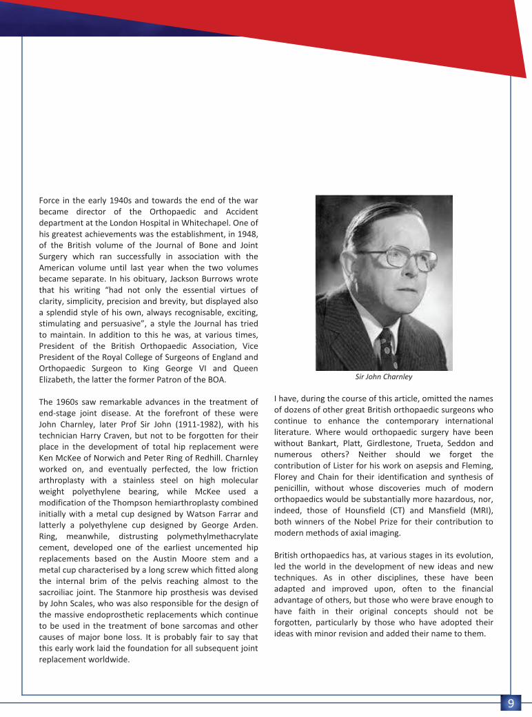
Force in the early 1940s and towards the end of the war became director of the Orthopaedic and Accident department at the London Hospital in Whitechapel. One of his greatest achievements was the establishment, in 1948, of the British volume of the Journal of Bone and Joint Surgery which ran successfully in association with the American volume until last year when the two volumes became separate. In his obituary, Jackson Burrows wrote that his writing “had not only the essential virtues of clarity, simplicity, precision and brevity, but displayed also a splendid style of his own, always recognisable, exciting, stimulating and persuasive”, a style the Journal has tried to maintain. In addition to this he was, at various times, President of the British Orthopaedic Association, Vice President of the Royal College of Surgeons of England and Orthopaedic Surgeon to King George VI and Queen Elizabeth, the latter the former Patron of the BOA. The 1960s saw remarkable advances in the treatment of end-stage joint disease. At the forefront of these were John Charnley, later Prof Sir John (1911-1982), with his technician Harry Craven, but not to be forgotten for their place in the development of total hip replacement were Ken McKee of Norwich and Peter Ring of Redhill. Charnley worked on, and eventually perfected, the low friction arthroplasty with a stainless steel on high molecular weight polyethylene bearing, while McKee used a modification of the Thompson hemiarthroplasty combined initially with a metal cup designed by Watson Farrar and latterly a polyethylene cup designed by George Arden. Ring, meanwhile, distrusting polymethylmethacrylate cement, developed one of the earliest uncemented hip replacements based on the Austin Moore stem and a metal cup characterised by a long screw which fitted along the internal brim of the pelvis reaching almost to the sacroiliac joint. The Stanmore hip prosthesis was devised by John Scales, who was also responsible for the design of the massive endoprosthetic replacements which continue to be used in the treatment of bone sarcomas and other causes of major bone loss. It is probably fair to say that this early work laid the foundation for all subsequent joint replacement worldwide.
Sir John Charnley
I have, during the course of this article, omitted the names of dozens of other great British orthopaedic surgeons who continue to enhance the contemporary international literature. Where would orthopaedic surgery have been without Bankart, Platt, Girdlestone, Trueta, Seddon and numerous others? Neither should we forget the contribution of Lister for his work on asepsis and Fleming, Florey and Chain for their identification and synthesis of penicillin, without whose discoveries much of modern orthopaedics would be substantially more hazardous, nor, indeed, those of Hounsfield (CT) and Mansfield (MRI), both winners of the Nobel Prize for their contribution to modern methods of axial imaging. British orthopaedics has, at various stages in its evolution, led the world in the development of new ideas and new techniques. As in other disciplines, these have been adapted and improved upon, often to the financial advantage of others, but those who were brave enough to have faith in their original concepts should not be forgotten, particularly by those who have adopted their ideas with minor revision and added their name to them.

Worldwide News
The value of the three-point index in predicting redisplacement of diaphyseal fractures of the forearm in children S. İltar, K.B. Alemdaroğlu, F. Say, N.H. Aydoğan Ankara Training and Research Hospital, Ulucanlar, Ankara, Turkey Samsun Training and Research Hospital, İlkadım, Samsun, Turkey Bone Joint J 2013;95-B:563–7.
Comment by Shalin Maheshwari
10
Abstract Introduction: Redisplacement is the most common complication of immobilisation in a cast for the treatment of diaphyseal fractures of the forearm in children. We have previously shown that the three-point index (TPI) can accurately predict redisplacement of fractures of the distal radius. In this prospective study we applied this index to assessment of diaphyseal fractures of the forearm in children and compared it with other cast-related indices that might predict redisplacement. Methods: A total of 76 children were included. Their ages, initial displacement, quality of reduction, site and level of the fractures and quality of the casting according to the TPI, Canterbury index and padding index were analysed. Logistic regression analysis was used to investigate risk factors for redisplacement. Results: A total of 18 fractures (24%) redisplaced in the cast. A TPI value of > 0.8 was the only significant risk factor for redisplacement (odds ratio 238.5 (95% confidence interval 7.063 to 8054.86); p < 0.001). Summary: The TPI was far superior to other radiological indices, with a sensitivity of 84% and a specificity of 97% in successfully predicting redisplacement. We recommend it for routine use in the management of these fractures in children. Research Analysis Primary research question: To test the hypothesis that the (Three-Point Index) TPI can be successfully modified to assess the reduction of forearm fractures in children and its success rate in predicting redisplacement can approach that in the distal radius. Methodology: Prospective case series. Study population: 84 consecutive children aged < 15 years with fractures of the forearm, 76 were considered eligible
for conservative treatment and were included. Exclusion criteria included an unsatisfactory reduction (four patients), open fractures (three) and floating elbow (one). None of the fractures included in the study had an associated neurovascular injury. There were no bilateral fractures. Outcome measurement: Radiographic measurements were made and acceptable initial reduction angulation was < 25° in children aged < four years, < 15° in those between four and nine years and < 10° in those aged ≥ ten years, considering the remodelling potential due to years of growth remaining. All radiological measurements were made on anteroposterior and lateral radiographs by three authors (SI, KBA, FS) using Image J 1.42q (National Institutes of Health, Bethesda, Maryland) with 0.5° or 0.1 mm accuracy. At weekly follow-up visits, redisplacement was defined as further angulation of > 10° in any direction.
Statistical analysis: Chi-squared test was used to compare the dichotomous variables between redisplaced and non-redisplaced fractures. The Mann-Whitney U test was used to evaluate the significance of the difference of the means between the independent groups. Multivariate logistic regression analysis was performed to determine the effect of possible risk factors on redisplacement when they were present together. Thus, on the basis of univariate analysis, any variable significantly related with fall occurrence and those with a result of p < 0.25 were drawn into the analysis. 15 SPSS v11.5 for Windows (SPSS Inc., Chicago, Illinois) was used for statistical analysis, and p-values < 0.05 were considered significant. Results: In 71 fractures (93%) the TPI predicted correctly whether there would be redisplacement, with a sensitivity of 84%, specificity of 97%, positive predictive value of 89% and negative predictive value of 95%. Comment Most paediatric diaphyseal fractures of the forearm are treated by manipulative reduction. The factors leading to

11
redisplacement can be related to the fracture or the surgeon. Unlike fractures of the distal radius, radiological indices have not aroused interest in diaphyseal fractures of the forearm, aside from one study by Bhatia et al (2006) where a retrospective/prospective study was conducted, in which the review of case records and radiographs of forearm and distal radius/ulna fractures were included. In this study, distal radius fractures were included for the first time and even though all indices were higher in the group that showed displacement, there was no subgroup analysis, which means that the forearm fractures were not differentiated from the distal radius fractures, thus acting as a possible confounder. The Padding index is the proportion of the dorsal gap (measured between the cast and the skin) to the largest interosseous distance between the radius and the ulna. The Cast index is the ratio of inner cast diameter at the fracture site on lateral and anteroposterior view. It proposes that the sectional geometry of the cast should be elliptical rather than circular in the distal forearm. Although later attempts were made to adapt the cast index to forearm fractures, the sectional geometry of the rest of the forearm is not as elliptical as at the wrist, and hence this index is not suitable for predicting redisplacement throughout the forearm. The Canterbury index combines the padding index and the cast index. Three-point index differs from the other indices because it not only takes into account the gaps at the fracture site, it also uses the gaps proximal and distal to the fracture sites, which are important points to maintain reduction against common displacement forces. In previous studies of paediatric distal radius fractures where these indices were used: Alemdaroğlu KB et al (2008) have shown that the limitation of these indices is that they are based on measurements of the gap at the fracture site alone and ignore proximal and distal cast fixation. Marcheix et al (2011) found poor moulding of the cast, as measured by the TPI, to be the only important risk factor for redisplacement and again found the TPI to reflect the quality of application of the cast and to be an
excellent predictor of redisplacement. Hang et al (2011) concluded that a high TPI was the most significant risk factor for redisplacement, and Devalia et al (2011) reported that the TPI and the quality of reduction were the most significant indicators of redisplacement, recommending its use when judging the technique of moulding the cast. This is the first report on the use of a modified TPI for the assessment of reduction of diaphyseal fractures of the forearm. The authors have compared all the above-mentioned indices with TPI and have found TPI to have higher sensitivity, specificity, positive predictive value and negative predictive value with acceptable interobserver reliability and intra-observer reliability. The authors were however not involved in the cast application but only in the assessment of the fractures on follow-up. They found a greater tendency for redisplacement of fractures in children aged under seven years which was attributed to difficulty in maintaining the reduction when casting a small forearm. Paediatric forearm fractures is a very complex subject to be explained by a single factor. In my experience, complete initial displacement of the fracture, anatomical reduction and obliquity of the fracture line seem to be the important risk factors for redisplacement. I feel that the casting indices should not be interpreted as a separate issue but in conjunction with fracture characteristics and patients factors. The three-point index has proven to be the superior of all these indices in paediatric distal radius fractures and now also seems promising in paediatric forearm fractures by this study. Overall, it is a good study and I would like to implement TPI assessment in paediatric forearm fractures in my routine practice. However, these results should be collaborated by further larger, multicentric, RCT’s to recommend it strongly as a protocol in radiological assessment of these forearm fractures under treatment and serve as a prognostic value for redisplacement.

Congress News
Editorial Secretary: Hatem Said Editorial Production: Linda Ridefjord Editorial Board: Ahmed Abdel Azeem, Syah Bahari, Kamal Bali, Bassel El-Osta, Anthony Hall, Shalin Maheshwari,
Maximilian Rudert
Rue Washington 40-b.9, 1050 Brussels, Belgium Tel.: +32 2 648 68 23 | Fax: +32 2 649 86 01 E-mail: [email protected] | Website: www.sicot.org
XXVI SICOT Triennial World Congress
combined with the 46th SBOT Annual Meeting Rio de Janeiro, Brazil – 20-22 November 2014
Abstract Submission and Congress Registration open on 1 November 2013!
More information will be available on the SICOT website soon!