XSS26 - Ophthalmology
27
SYMPTOM SUMMARIES OPHTHALMOLOGY DARWIN’S NOTEBOOK
Transcript of XSS26 - Ophthalmology

SYMPTOM SUMMARIES
OPHTHALMOLOGY
DARWIN’S NOTEBOOK

TABLE OF CONTENTS
RED EYE 1
TRAUMA 7
GRADUAL VISUAL LOSS 11
SUDDEN VISUAL LOSS 14
DIPLOPIA 18
EYELID PROBLEMS / BULGING EYE 22
OPHTHALMOLOGY – RED EYE
DARWIN’S NOTEBOOK 1
Introduction • Red eye is among the commonest ophthalmic presentations ® it is usually accompanied by some
degree of pain / discomfort, ranging from the mild (e.g. blepharitis, conjunctivitis) to the severe (e.g. scleritis, iritis, primary angle closure)
• The red, painful eye usually suggests a disorder in the anterior segment ® conjunctiva, sclera /
episclera, cornea, iris & lens
• A white, painful eye is more unusual and usually suggests pathology at the back of the eye (e.g.
posterior scleritis, optic / retrobulbar neuritis) ® however, in these conditions other symptoms will
predominate, particularly visual loss
o NB: Orbital pathology may also present with a painless white eye ® although symptoms of
proptosis, diplopia or visual loss are likely to predominate
• The commonest cause of a red, painless eye is spontaneous subconjunctival haemorrhage ® in this
condition, blood is seen overlying the sclera under the conjunctiva:
o It looks alarming, but is of no visual consequence
o Usually no cause is identified ® although it may occur in patients coughing, sneezing or
following any other Valsalva manoeuvre, or in patients taking aspiring or warfarin
o Usually one can reassure the patient ® if the haemorrhages are recurrent, one can consider
checking the patient’s BP or clotting
o NB: Always enquire about a history of trauma in cases of bilateral subconjunctival
haemorrhages ® base of skull fractures may present this way, and the posterior limit of the
haemorrhage may not be visible
• It is important to be able to distinguish between mild, self-limiting causes of red eye (e.g. conjunctivitis) and sight-threatening causes (e.g. primary angle closure, iritis, scleritis, microbial keratitis)
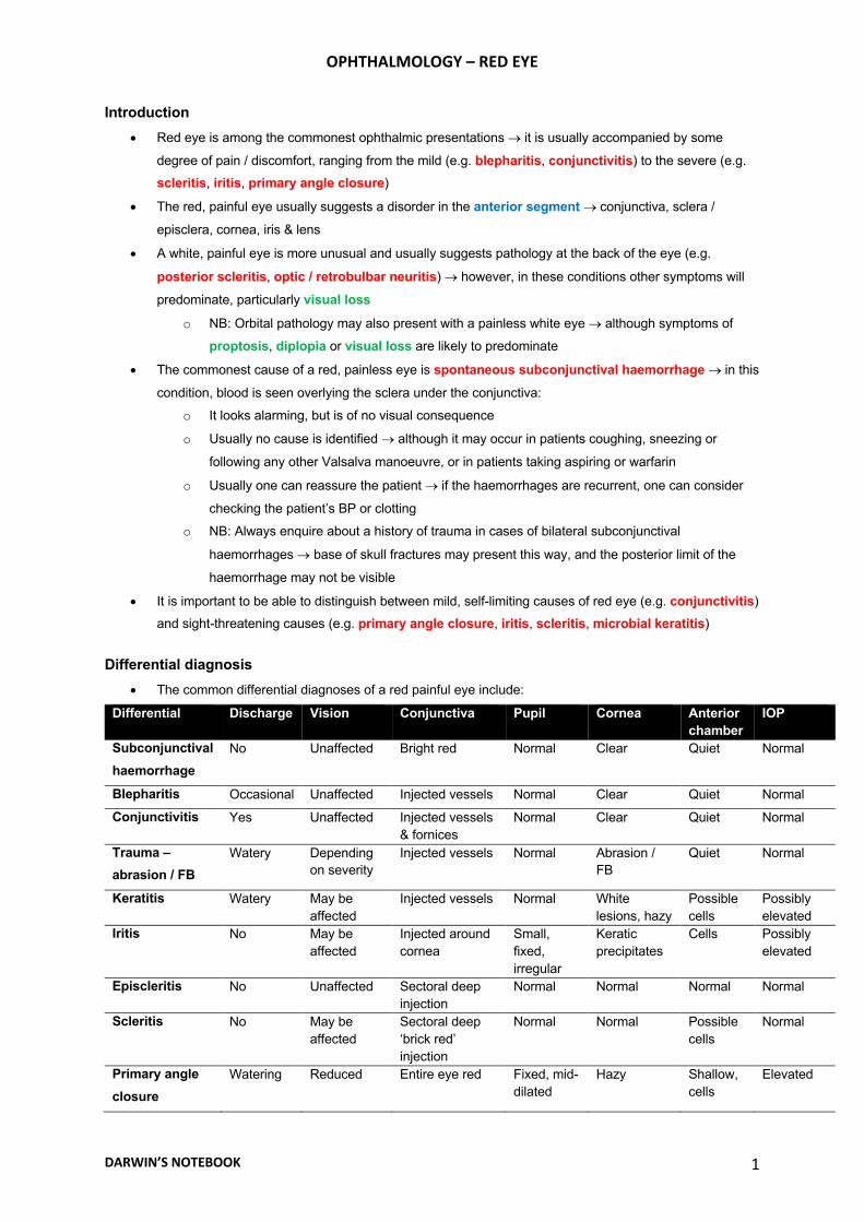
Differential diagnosis • The common differential diagnoses of a red painful eye include:
Differential Discharge Vision Conjunctiva Pupil Cornea Anterior chamber
IOP
Subconjunctival haemorrhage
No Unaffected Bright red Normal Clear Quiet Normal
Blepharitis Occasional Unaffected Injected vessels Normal Clear Quiet Normal
Conjunctivitis Yes Unaffected Injected vessels & fornices
Normal Clear Quiet Normal
Trauma – abrasion / FB
Watery Depending on severity
Injected vessels Normal Abrasion / FB
Quiet Normal
Keratitis Watery May be affected
Injected vessels Normal White lesions, hazy
Possible cells
Possibly elevated
Iritis No May be affected
Injected around cornea
Small, fixed, irregular
Keratic precipitates
Cells Possibly elevated
Episcleritis No Unaffected Sectoral deep injection
Normal Normal Normal Normal
Scleritis No May be affected
Sectoral deep ‘brick red’ injection
Normal Normal Possible cells
Normal
Primary angle closure
Watering Reduced Entire eye red Fixed, mid-dilated
Hazy Shallow, cells
Elevated

OPHTHALMOLOGY – RED EYE
DARWIN’S NOTEBOOK 2
• The key symptoms to ask about to help differentiate the causes of red eye are: o Pain
o Watery or sticky discharge
o Photophobia o Visual loss or reduced vision
History DESCRIPTION OF PAIN
• Grittiness & irritation ® suggestive of conjunctivitis or blepharitis
• Itching ® characteristic of allergic conjunctivitis
• Sharp pain (if experienced after trauma) ® suggestive of corneal foreign body or corneal abrasion (eye watering is usually an accompanying symptom)
• Photophobia ® usually a feature of anterior uveitis or iritis
• Dull, boring ache ® suggests anterior scleritis ® episcleritis can look similar, but the pain is usually
milder, and does not keep them up at night
DURATION OF SYMPTOMS • Patients who have experienced trauma will be symptomatic immediately
• Viral & bacterial conjunctivitis may have a period of latency ® whereby the eye may be mildly
irritated for up to 10 days before the redness & discharge develop
• A red, discharging eye over a period of months is unlikely to be bacterial or viral conjunctivitis (as these
resolve within 21 days) ® in these circumstances, one should consider chlamydia conjunctivitis (where there is a thick and rope-like mucopurulent discharge)
o Floppy eyelid syndrome may also present in a similar fashion ® such patients have very
easily everted upper lids, and have the habitus of patients with obstructive sleep apnoea o In the younger age group, another cause of chronic infective conjunctivitis is molluscum
contagiosum ® the lesions may be apparent on inspection of the lids
ASSOCIATED SYMPTOMS • Watering:
o Mild to moderate watering may be seen in blepharitis or conjunctivitis o Excessive lacrimation may be seen in corneal abrasions, corneal foreign bodies, subtarsal
foreign body, and trichiasis
• Discharge: o Clear discharge with stickiness of the lids in the morning is common in blepharitis
o Discharge may be indistinguishable between viral and bacterial conjunctivitis ® although in
bacterial it may be green / yellow, compared to yellow / white in viral o The discharge in chlamydia conjunctivitis is thick and accumulates in rope-like strands
o Gonococcal conjunctivitis in babies is excessive, even explosive ® it is known as
ophthalmia neonatorum
PHOTOPHOBIA • Photophobia is characteristic of anterior uveitis or iritis
• However, it may also be a symptom of any condition which causes ciliary muscle spasm ® including
microbial keratitis, herpetic keratitis, corneal abrasion, or recurrent erosion syndrome

OPHTHALMOLOGY – RED EYE
DARWIN’S NOTEBOOK 3
VISUAL LOSS • Conditions affected the external lids, the conjunctiva, and the episclera do not usually affect vision ®
except where the patient is viewing the world through a film of pus or tears
• Blurred vision in the context of a red, painful eye will usually suggest either corneal involvement (e.g. microbial keratitis, herpetic keratitis, corneal oedema in primary angle closure, large corneal abrasion) or a significant inflammatory component (e.g. scleritis or iritis)
Past medical history • A number of underlying medical conditions may be associated with red, painful eyes, including:
Ophthalmic condition Underlying conditions Scleritis Rheumatoid arthritis, lupus
Iritis Ankylosing spondylitis, sarcoidosis, JIA, IBD, AIDS / HIV-related opportunistic infections, toxoplasmosis, etc.
Dry eyes Sjögren’s syndrome
Thyroid eye disease Grave’s disease
• A recent history of upper respiratory tract viral illness (cough / sore throat) might be associated with the onset of viral conjunctivitis
Past ophthalmic history • Recent surgery ® acute iritis and endophthalmitis may occur following cataract surgery
o Endophthalmitis generally occurs within 10 days of surgery, whereas iritis may occur weeks
afterwards
• Patients with recurrent erosion syndrome give a history of traumatic corneal abrasion in the past,
which got better ® the patient now wakes up with a feeling of the lids sticking together on the side of the
previous abrasion, and then they open the eyes there is a feeling of ‘ripping’ or ‘tearing’, and the symptoms of the original abrasion return
• Anterior uveitis / iritis and herpetic keratitis are recurrent conditions, and the patients may volunteer their diagnosis from the outset
• Refractive error ® primary angle closure is more likely to occur in patients with short, hypermetropic
eyes with coexistent cataract
• Contact lenses predispose to microbial causes of keratitis, particularly in soft lenses ® ask about
whether the patient sleeps in contact lenses, or swims / showers in them (the latter may point to a diagnosis of acanthamoeba)
Examination • A systematic examination of the anterior segment structures should easily identify the cause of the red,
painful eye
EXTERNAL LIDS & ADNEXAE • Before examining a patient’s lids, look at their face:
o Do they have features of acne rosacea?
o Is there a vesicular rash over one side of the forehead? ® herpes zoster ophthalmicus
o Do they have features suggestive of thyroid eye disease (such as proptosis)?

OPHTHALMOLOGY – RED EYE
DARWIN’S NOTEBOOK 4
o Inflammation, redness & swelling of the lids indicate cellulitis (either orbital or pre-septal) ®
as it is the more severe condition, one must exclude features of orbital cellulitis (proptosis,
double vision, loss of colour vision, reduced vision, swollen optic disc)
o If you suspect conjunctivitis, palpate the pre-auricular lymph nodes ® tender pre-auricular lymphadenopathy is a feature of viral conjunctivitis
o Look for signs of herpetic lesions ® cold sores or the vesicular lesions of herpes zoster
o Examine the lid margin for features of blepharitis (crusting of the lashes, telangiectatic vessels at the lid margin, irregular lid margin, inflamed meibomian glands) ® also look for
other potentially infective lesions (e.g. mollusca)
INTERNAL LIDS • Always evert the lids ® this way you will be able to identify and remove subtarsal foreign bodies
• Inspection of the inner aspect of the lids is a reliable way of diagnosing conjunctivitis ® look for follicles
& papillae:
o Follicles are small and resemble grains of rice, with a surrounding vascular bed ® they are
characteristic of viral and chlamydial conjunctivitis
o Papillae are larger polygonal ‘bumps’ with a central vascular core ® they are non-specific and
may be seen in bacterial conjunctivitis, allergic conjunctivitis, foreign bodies (including chronic contact lens wear), and in floppy eyelid syndrome
CONJUNCTIVA • If the conjunctiva is inflamed, injected with follicles ± tender pre-auricular lymphadenopathy ® viral
conjunctivitis is likely o Subconjunctival haemorrhages & chemosis (‘waterlogged conjunctiva’) may also occur in
this condition
• If the conjunctiva is inflamed, injected with subtarsal & bulbar papillae, and there is excessive purulent
discharge ® then bacterial conjunctivitis is likely
• Acute chemosis & papillae are a feature of allergic / atopic conjunctivitis ® as well as chemical conjunctivitis, which may be seen following a chemical injury
SCLERA / EPISCLERA • The redness seen in scleritis can be distinguished from episcleritis in a number of ways:
o In scleritis, the redness is sectoral but takes on a brick-like, violet colour with the distention of
scleral & episcleral vessels, which may appear tortuous ® the patient may also report a severe
aching pain when the lid is pressed directly over the area of inflammation
o The injection in episcleritis is also sectoral, but much milder, with a less striking reddish hue, and a single drop of 2.5% phenylephrine will allow the episcleral vessels to constrict, making
the eye appear whiter ® this DOES NOT happen in scleritis, which may be a useful feature in
distinguishing between the two
o An inflammatory nodule may be present in both episcleritis and scleritis
CORNEA • The instillation of topical fluorescein and illumination with cobalt blue light is useful in detecting
corneal epithelial pathology, including:
o Corneal abrasions

OPHTHALMOLOGY – RED EYE
DARWIN’S NOTEBOOK 5
o Dendritic ulcers ® ulcers with tree like branches, characteristic of herpetic keratitis
o Dry eye ® characterised by punctate (spotty) fluorescein staining
• The appearance of whitish / cloudy lesions, called infiltrates, within the anterior corneal stroma is a feature of keratitis:
o Where corneal infiltrates with overlying epithelial fluorescein staining occur in the context of
contact lens wear ® microbial keratitis is the most likely diagnosis
o Corneal infiltrates occurring near to the limbus, in patients with lid-margin disease, in particular
if associated with rosacea ® are likely to be a sign of marginal keratitis
o Multiple discoid subepithelial infiltrates without fluorescein staining may occur as a sequela of
adenoviral conjunctivitis ® the patient may re-present complaining of reduced vision
secondary to these ‘adeno-spots’, which clear eventually without treatment, although topical steroids may speed the process
• Corneal oedema is usually apparent as a hazing and thickening of the cornea ® it may be present in a
localised form in any form of keratitis, and a more diffuse generalised form may occur in acute elevations of intraocular pressure (e.g. primary angle closure)
• Keratic precipitates are small spots or discs of inflammatory material adherent to the endothelium (the
innermost aspect of the cornea) ® they are usually seen in anterior uveitis
ANTERIOR CHAMBER • Full examination of the anterior chamber (the fluid space between the cornea & iris) is difficult without
the use of a slit lamp ® but there are some features which may be apparent using a direct ophthalmoscope:
o Hypopyon (a white fluid level within the eye) ® caused by a settling of inflammatory cells
within the anterior chamber:
§ In the context of contact lens wear and corneal infiltrate ® this is pathognomonic of
an infective keratitis
§ If seen within a few days of cataract surgery ® endophthalmitis should be
suspected
§ Hypopyon may also be a feature of severe anterior uveitis ® as may occur in
Behçet’s disease
o Hyphaema (a red fluid level in the eye) is caused by the settling of red blood cells in the
anterior chamber ® patients will invariably report trauma or surgery, and it is important to
check the intraocular pressures of such patients
o Anterior chamber shallowing / flattening is a feature of primary angle closure ® it can be
difficult to appreciate without the use of an angled slit beam
• Other features detectable using a slit lamp are:
o Cells & flares (circulating inflammatory cells & protein exudate) ® these are two characteristic
features of anterior uveitis, and can only be seen with a slit lamp
IRIS & PUPIL • Primary angle closure may present with a number of iris / pupil signs, including:
o Fixed, mid-dilated pupils o Anterior iris bowing (iris bombé)
o Peripheral iridocorneal touch, as well as shallowing / narrowing of the anterior chamber

OPHTHALMOLOGY – RED EYE
DARWIN’S NOTEBOOK 6
o The cornea may be hazy secondary to oedema, and the intraocular pressure is elevated
o Cataract may also be a predisposing factor
• Iritis may be associated with:
o Posterior synechiae ® these are inflammatory adhesions between the pupillary margin of the
iris and the anterior lens capsule, resulting in an irregularly shaped pupil
o Iris nodules ® especially in sarcoid-related anterior uveitis
• Rubeosis occurs when neovascularisation occurs in the iris and iridocorneal angle ® it is a feature of
ischaemic retinopathies, such as proliferative diabetic retinopathy, central retinal vein occlusion,
and certain forms of uveitis ® it may cause rubeotic glaucoma, whereby the eye becomes painful and
red with reduced vision and raised intraocular pressure
INTRAOCULAR PRESSURE • This may be elevated in a number of conditions, but is seldom in itself a cause of pain ® except when
extremely high
• Causes include: o Primary angle closure o Rubeosis
o Some forms of anterior uveitis ® known as ‘hypertensive uveitis’
§ Another rarer form, Posner-Schlossman syndrome, is associated with high IOP,
very mild anterior reaction, and one or two keratic precipitates § Hypertensive uveitis is a common presenting feature of toxoplasmosis-associated
uveitis ® so always remember to look in the fundus for evidence of active
toxoplasma retinitis
§ Herpes zoster uveitis is also frequently associated with raised pressure
Investigation • In ophthalmology, most clinical diagnoses can be made of the basis of history and examination
• However, there are a few important investigations to be aware of when a patient presents with a red
eye:
o Conjunctival swabs ® if an infective conjunctivitis is suspected
o Corneal scrapings ® taken if a contact lens-related ulcer / infiltrate is seen
• Systemic investigations are rarely performed in first presentations of scleritis and iritis ® investigations
are performed if the presentation is atypical or if recurrent:
o Iritis screen: § Chest X-ray ® tuberculosis, sarcoidosis
§ Serum ACE ® sarcoidosis
§ VDRL ® syphilis
§ HLA B27 ® ankylosing spondylitis
o Scleritis screen: § cANCA ® Wegener’s
• CT scan is useful in the diagnosis of orbital conditions
• B-scan ultrasonography may be required if a view of the posterior segment is not possible ® e.g.
because of hyphaema

OPHTHALMOLOGY – TRAUMA
DARWIN’S NOTEBOOK 7
Differential diagnosis • The eye’s response to trauma will vary according to the mechanism of injury:
Mechanism Site Traumatic consequence Blunt trauma Ocular Corneal abrasion, corneal foreign body, hyphaema, traumatic iritis, traumatic mydriasis,
iridodialysis, angle recession, cataract, lens subluxation / dislocation, commotio retinae, retinal tear / detachment, vitreous haemorrhage, suprachoroidal haemorrhage, globe rupture, traumatic optic neuropathy
Adnexal Orbital blow-out fracture, lid ecchymosis, retrobulbar haemorrhage, traumatic optic neuropathy
Penetrating trauma
Ocular Corneal laceration, conjunctival laceration, scleral rupture, retained intraocular foreign body
Adnexal Lid laceration
Burn (chemical / thermal)
Ocular Conjunctival ischaemia, symblepharon, corneal limbal stem cell failure, corneal opacity
Adnexal Skin burns
• By definition, penetrating trauma is more likely to result in laceration of the ocular tissues
• Blunt trauma, if mild, may only result in superficial tissue damage & bruising ® however, where blunt
trauma is more forceful, the forced may be transmitted through the globe, perhaps resulting in bruising
of the retina (commotio retinae) or globe rupture ® where the force of blunt trauma is transmitted to
the orbit, fracture may occur
• The eye may also be subject to injuries from chemical and thermal agents, which present different challenges from mechanical trauma
• Visual loss following trauma may result from:
o Corneal scarring ® from corneal laceration or opacity following chemical injury
o Optic nerve compression ® from retrobulbar haemorrhage or traumatic neuropathy
o Severely disordered globe anatomoy
o Retinal detachment / choroidal haemorrhage
History EXCLUDE SIGNIFICANT NON-OPHTHALMIC TRAUMA
• It is essential that potentially life-threatening injuries should be addressed before referral to ophthalmology
• Upon presentation, most severe traumatic injuries are identified and dealt with ® however, there is a
risk in more trivial injuries that only the ophthalmic problem is identified:
o Loss of consciousness or variable level of consciousness may indicate neurological trauma ®
likewise, other features of raised intracranial pressure or neurological symptoms should cause
concern
o Take into consideration the size and dimensions of any instrument responsible for penetrating
injury around the globe ® if an intracranial penetrating trauma is missed, the patient is at risk
of death from intracerebral haemorrhage, encephalitis or meningitis
o As routine, any patient with history of penetrating trauma should have their tetanus status
assessed
NATURE OF INJURY • For foreign body trauma, identify:

OPHTHALMOLOGY – TRAUMA
DARWIN’S NOTEBOOK 8
o Type of object ® whether metal or vegetative matter, as the latter may cause risk of fungal
infection ® in chemical injury, it is important to know whether acid or alkali was involved, as
alkali burns may cause more serious injury
o Velocity, trajectory, distance ® metal FBs ‘blown’ into the eye will usually just land on the
corneal surface, but objects at higher velocity and lower distance are more likely to penetrate
the globe
o Risk of retention ® the likelihood of a foreign body lodging in the globe or adnexal tissue will
vary according to velocity and distance, and try to establish whether part of a penetrating object (particularly wooden) may have broken off at the time of injury
• For blunt trauma, consider:
o Size of object ® objects smaller than the orbital rim will transmit forces directly to the globe,
and objects larger than the orbital rim will transmit forces to the lids and bony orbit ® so called
‘blow-out’ fractures occur because the weakest walls of the orbit (floor and medial) are most
prone to fracture when the orbital pressure is raised during direct trauma
PRESENTING SYMPTOMS • Red eye ® conjunctival haemorrhage, corneal abrasion, corneal foreign body, penetrating trauma
• Painful eye ® hyphaema, traumatic iritis, corneal abrasion, corneal foreign body, penetrating trauma
• Blurred vision ® hyphaema, cataract, vitreous haemorrhage, commotio retinae, retinal detachment,
traumatic optic neuropathy
• Flashing lights & floaters ® commotio retinae, vitreous haemorrhage, retinal tear / detachment
• Double vision ® orbital blow-out fracture
• Pain on eye movement ® orbital blow-out fracture
o NB: If you suspect a blow-out, always ask about numbness of teeth ® molars may become
numb if the fracture disturbs the infraorbital nerve
Examination • A systematic examination of the ocular system, starting from the lids and working backwards through all
the structures of the eye, should identify all potential sequelae of trauma
VISUAL ACUITY • It is absolutely essential that visual acuity is recorded at first presentation
• Some gauge of what the vision was like in the eye prior to injury should be established
EXTERNAL EYE • Lids:
o Ecchymosis / bruising o Lid lacerations
o Remember to evert the lids ® the inside of the lid and tarsal plate may split following eye lid
‘thumbing’
• Orbit:
o Proptosis ® suggests the presence of blood in the orbit

OPHTHALMOLOGY – TRAUMA
DARWIN’S NOTEBOOK 9
§ NB: An acute retrobulbar haemorrhage, where there is rapidly progressing
proptosis, diffuse subconjunctival haemorrhage, and reducing vision, is a sight-
threatening emergency ® urgent treatment by lateral cantholysis is required
o Enophthalmos ® this is a feature of a blow-out fracture with entrapment, in which part of the
orbital contents becomes caught within the fracture
o Ocular motility ® restriction in up or down gaze is a feature of a blow-out fracture with
entrapment ® 4th nerve palsies may occur as they are particularly susceptible to injury, and 6th
nerve palsies may be a sign of raised ICP
o Palpation ® check trigeminal sensation, and look for signs of bony ridges or crepitus (features
of orbital fracture)
ANTERIOR SEGMENT • Conjunctiva / sclera:
o Subconjunctival haemorrhage ® this is a common sequela of blunt trauma to the globe, but
beware of bilateral subconjunctival haemorrhage without visible posterior extents, as this may be due to a skull base fracture
o Scleral rupture / laceration ® beware of any brown or black material on the sclera
associated with haemorrhage, as this may represent choroidal tissue herniating through
disrupted sclera
• Cornea:
o Corneal lacerations may be visible by direct inspection ® the anterior chamber will be shallow
or flat if there is an aqueous leak
o Corneal abrasions are best seen with fluorescein & cobalt blue light ® vertical corneal
scratches may be secondary to a subtarsal foreign body
o Corneal foreign body may be visible by direct inspection if large enough ® otherwise, slit lamp examination will be required
o Following chemical injury, there may be corneal epithelial disturbance ® a particularly
suspicious feature is if there is staining or ischaemia (i.e. absent conjunctival vessels) at the
limbus
• Anterior chamber:
o Shallow ® if a laceration has caused an aqueous leak
o Deep ® if blunt trauma has caused iridocorneal angle recession (long-term risk of developing
glaucoma), or if the lens is avulsed or dislocates o Hyphaema
o Hypopyon ® would usually suggest an infection, and would be a late feature of untreated
trauma (e.g. corneal foreign body)
o Iritis ® cells may be present in the anterior chamber following direct blunt trauma to the globe
(traumatic iritis)
• Iris:
o Is there iris tissue outside the globe? ® a feature of globe rupture or corneal laceration
o Transillumination defects ® a penetrating foreign body may cause a small hole in the iris,
with the red reflex visible through the defect
o The iris sphincter muscle may rupture following blunt trauma ® resulting in traumatic mydriasis

OPHTHALMOLOGY – TRAUMA
DARWIN’S NOTEBOOK 10
• Relative afferent pupillary defect (RAPD): o This is a poor prognostic sign for visual outcome
o It is absolutely essential that the presence or absence of RAPD is assessed at first
presentation o An RAPD following trauma usually indicates a degree of optic nerve compromise:
§ Traumatic optic neuropathy
§ Optic nerve compression ® e.g. by retrobulbar haemorrhage or a piece of bone
fragment impinging on the nerve o Other causes include:
§ Maculae off retinal detachment
§ Massive choroidal haemorrhage
• Lens:
o Cataract may develop following trauma ® a posterior subcapsular cataract may develop some
time after trauma, or a focal cataract may develop where a foreign body has passed through
the lens
o The lens is held in place by zonular ligaments, which may give out if the force applied is
sufficiently high ® the lens may stay in place but wobble when the eye move (phacodynesis),
or subluxate (move up or down within the posterior chamber), or fully luxate (dislocate into
the vitreous or be expelled from the eye)
POSTERIOR SEGMENT • Vitreous:
o Vitreous haemorrhage should be suspected if there is no red reflex, or if there is no view of the fundus with an ophthalmoscope
• Retina:
o Commotio retinae is seen as pale, oedematous patches of retina ® the retina may also thin
out in areas of commotio, and the patient is subsequently at risk of retinal detachment
o Retinal detachment ® this frequently occurs in globe ruptures extending posteriorly
o Suprachoroidal / choroidal haemorrhage ® this is seen as an immobile black shadow on
fundoscopy, and if large, may have a disastrous prognosis in terms of vision
Investigation • X-rays ® facial and orbital views may be useful in detecting orbital fractures, but orbital CT is the fold
standard
• It may be possible to detect some radio-opaque (e.g. glass, metal) intraocular foreign bodies using X-
ray ® but if a metal foreign body is suspected, DO NOT use MRI
• In chemical injuries, check the pH of the affected eye using pH paper

OPHTHALMOLOGY – GRADUAL VISUAL LOSS
DARWIN’S NOTEBOOK 11
Differential diagnosis • Patients with gradual deterioration of vision often have a refractive error, and as such will usually
improve with glasses
• The vision in two of the more common presentations in older age, cataract and dry macular degeneration, will also improve, up to a level, with refraction
• Often, presentation to hospital eye services are made when no improvement ca be made on refraction
• Common causes of gradual visual loss include: o Refractive error o Cataract o Posterior capsule opacification ® post-cataract surgery
o Age-related macular degeneration
o Primary open angle glaucoma ® although presentation with visual loss is unusual
• Rarer causes of gradual visual loss include:
o Extremes of refractive error ® keratoconus myopic degeneration
o Inherited eye disease
o Toxic / drug-related
History DURATION OF SYMPTOMS
• It can be difficult to get an accurate history of gradual vision loss in terms of timescale ® many patients
only become aware of the problem when it is pointed out by an optometrist
• Detecting gradual monocular visual changes can be particularly difficult, as the other eye will compensate
ASSOCIATED SYMPTOMS • Common associated symptoms of gradual visual loss include
Symptom Pathology Glare Cataract
Floaters Vitritis, retinal detachment
Night blindness (nyctalopia) Inherited eye disease (particularly retinitis pigmentosa)
Peripheral visual loss (‘bumping into things’) Late stage glaucoma, inherited eye disease (e.g. RP)
Positive central (dark) scotoma Age-related macular degeneration (AMD)
Visual distortion (metamorphopsia) Wet AMD
• ‘Negative’ scotomata occur most commonly following optic nerve disease ® the patient is not aware
of their central field loss until they try to read
• By contrast, patients are aware of ‘positive’ scotomata ® patients with macular degeneration are
constantly aware of their central field defect
• Visual distortion (metamorphopsia), increase in image size (macropsia), and decrease in image size
(micropsia) are reflections of changes in the relative positions of photoreceptors ® they are a feature
of macular disease

OPHTHALMOLOGY – GRADUAL VISUAL LOSS
DARWIN’S NOTEBOOK 12
FAMILY HISTORY • Family history is important as it may point to an inherited condition ® such as retinitis pigmentosa or
Stargardt’s disease
DRUG HISTORY • Some chronic medications may cause poor visions ® such as vigabatrin, tamoxifen, and chloroquine
• Also, alcohol and tobacco intake may be useful in identifying patients at risk of B12 & folate deficiency
Examination VISUAL ACUITY
• An accurate assessment of visual acuity is essential ® if possible with the patient wearing their current
spectacles
• Re-checking visual acuity using a pinhole is essential ® an improvement in vision usually points to a
condition that may be improved with up-to-date glasses (e.g. cataract or refractive error), and visual
acuity remains reduced in retinal lesions, particularly macular
COLOUR VISION • Usually assessed using Ishihara colour plates
• Loss of colour vision is an early sign of optic neuritis / neuropathy
VISUAL FIELD • May be examined by confrontation, or more formally by static automated perimetry
• Central scotoma is a feature of macular disease
• An enlarged blind spot is a feature of chronic optic nerve swelling ® e.g. benign intracranial
hypertension
• An Amsler grid is useful for examining the central field in macular disease
PUPILS • A relative afferent pupillary defect is a feature of optic nerve compromise, as well as a chronic total
retinal detachment
RED REFLEX • Opacities within the red reflex may be observed in cataract • Hazy visual media may be seen in cataract, posterior capsular opacification, and corneal scarring
or opacity
FUNDOSCOPY • Optic nerve:
o Optic disc pallor suggests chronic optic neuropathy o Optic disc cupping is a characteristic feature of glaucomatous optic neuropathy
• Macula: o Drusen (deep yellow flecks) at the macula are a characteristic feature of macular
degeneration
o As dry AMD progresses, dark pigmentary changes, as well as atrophy, may be observed at the macula

OPHTHALMOLOGY – GRADUAL VISUAL LOSS
DARWIN’S NOTEBOOK 13
• Retinal periphery & vasculature:
o Peripheral patchy ‘bone spicule’ pigmentation is a feature of retinitis pigmentosa ® the
retinal vessels may also be attenuated, and the optic disc may display ‘waxy pallor’
Investigation • These are often not necessary, as the diagnosis should normally be apparent from history &
examination:
o B-scan ultrasound ® may be necessary of fundal examination is precluded by a dense
cataract
o Blood tests ® in particular B12 & folate, to rule out nutritional optic neuropathy
o Electrodiagnostic testing (visual evoked potential, electro-oculogram, electro-retinogram)
has two important roles in gradual visual loss: § Can identify the location (i.e. rod, cone, macular, optic nerve) of the visual dysfunction
§ Can be used to identify non-organic (i.e. functional) visual loss
• NB: Always consider imaging the brain ± the orbits by CT / MRI in any cases of unexplained non-functional visual loss

OPHTHALMOLOGY – SUDDEN VISUAL LOSS
DARWIN’S NOTEBOOK 14
Differential diagnosis • The causes of a sudden loss in vision may be classified as either painful or painless
• Common causes of painful sudden vision loss include:
Anatomical location Examples Anterior segment Iritis, scleritis, keratitis, primary angle closure
Optic nerve Optic neuritis Arteritic ischaemic optic neuropathy (giant cell arteritis)
Intracranial Migraine Benign intracranial hypertension
• Common causes of painless sudden vision loss include:
Anatomical location Examples Vitreous Vitritis, vitreous haemorrhage
Retinal – vascular Branch retinal vein occlusion, central retinal vein occlusion Branch retinal artery occlusion, central retinal artery occlusion
Retinal – macula Wet AMD Diabetic maculopathy
Retinal Retinal detachment, retinitis, retinochoroiditis
Optic nerve Non-arteritic ischaemic optic neuropathy Compressive optic neuropathy
Intracerebral Stroke
PATIENT DEMOGRAPHICS • Age:
o Some conditions are unlikely under the age of 60 ® particularly ischaemic optic neuropathy
o Retinovascular occlusive conditions are also relatively unusual in a younger age group ® if
encountered, one should suspect an underlying thrombophilia
• Sex: o Benign intracranial hypertension (BIH) is a condition that almost exclusively affects young
(usually overweight) females
o Demyelinating optic neuritis is also far more common in young females
• Race: o Always consider the possibility of sickle cell retinopathy in patients of Afro-Caribbean
descent ® this may present with a vitreous haemorrhage
o Diabetic patients from the Indian subcontinent are more prone to severe diabetic retinopathy o Some forms of uveitis are more common in certain groups, such as Behçet’s in Turks /
Greeks, toxoplasmosis in South Americans, and sarcoidosis in Africans
History DURATION OF SYMPTOMS
• Immediate loss of vision ® suggests a vascular event (i.e. venous / arterial occlusion)
• Progressive loss of field over hours / days ® suggests retinal detachment
• Progressive dimming of vision ® suggests optic nerve pathology
• Transient visual loss lasting seconds / hours ® e.g. migraine, giant cell arteritis, BIH

OPHTHALMOLOGY – SUDDEN VISUAL LOSS
DARWIN’S NOTEBOOK 15
ASSOCIATED SYMPTOMS • Painful eye ® iritis, scleritis, keratitis, and primary angle closure
o NB: All may also have photophobia
• Headache: o Headache on walking may be a feature of BIH (as is buzzing in the ear)
o The headache in giant cell arteritis is characteristically centred on the patient’s temple ®
there may be associated pain in the jaw on chewing (jaw claudication)
o Headache preceded by scintillating lights or fortification spectra is a feature of migraine
• Photopsia (flashing lights) ® this may precede a retinal detachment or retinal tear, and are
commonly bilateral in migraine
• Floaters ® a sudden increase in floaters may precede retinal detachment, or may be a sign of
vitreous haemorrhage or vitritis
• Loss of colour vision ® a characteristic feature of demyelinating optic neuritis
• Metamorphopsia ® wet AMD
PAST OPHTHALMIC HISTORY • Certain ophthalmic conditions predispose to particular causes of sudden vision loss:
o Myopia ® the risk of retinal detachment increases with high myopia
o Hypermetropia ® these patients are more prone to primary angle closure (particularly in
combination with dense cataract), and are more prone to ischaemic optic neuropathy
(especially if the patient is hypertensive)
o Ocular hypertension / glaucoma ® predisposes to central retinal vein occlusion
PAST MEDICAL HISTORY • Hypertension ® high blood pressure predisposes patients to venous & arterial occlusions, as well
as non-arteritic ischaemic optic neuropathy and stroke
• Diabetes ® the commonest presentation of sudden visual loss in a diabetic is vitreous haemorrhage in
proliferative diabetic retinopathy, although vein occlusions may also present in this way
• Thyroid / Grave’s disease ® visual loss in thyroid eye disease may be caused by three mechanisms:
o Corneal scarring
o Raised intraocular pressure
o Compressive optic neuropathy ® this one presents acutely, and thus colour vision and visual
field must be checked
Examination • Visual acuity
• Colour vision ® reduced in optic nerve pathologies
• Visual fields:
o Homonymous field defects ® are a feature of stroke
o Altitudinal defects ® are a feature of non-arteritic ischaemic optic neuropathy
o Central scotoma ® optic neuritis, macular degeneration
o Peripheral field loss ® retinal detachment
o Enlarged blind spots ® benign intracranial hypertension
OPHTHALMOLOGY – SUDDEN VISUAL LOSS
DARWIN’S NOTEBOOK 16
• Pupils:
o An RAPD is a feature of optic nerve pathology ® it may also be a feature of retinal detachment
o A fixed mid-dilated pupil is a feature of primary angle closure ® whereas a small irregular pupil is a feature of anterior uveitis / iritis
• Anterior segment examination ® helpful in identifying iritis, scleritis, and primary angle closure
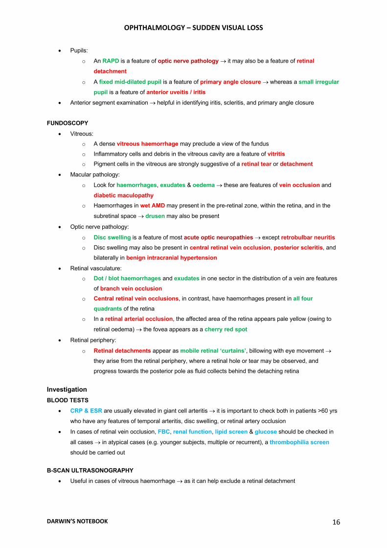
FUNDOSCOPY • Vitreous:
o A dense vitreous haemorrhage may preclude a view of the fundus
o Inflammatory cells and debris in the vitreous cavity are a feature of vitritis o Pigment cells in the vitreous are strongly suggestive of a retinal tear or detachment
• Macular pathology:
o Look for haemorrhages, exudates & oedema ® these are features of vein occlusion and
diabetic maculopathy
o Haemorrhages in wet AMD may present in the pre-retinal zone, within the retina, and in the
subretinal space ® drusen may also be present
• Optic nerve pathology:
o Disc swelling is a feature of most acute optic neuropathies ® except retrobulbar neuritis
o Disc swelling may also be present in central retinal vein occlusion, posterior scleritis, and
bilaterally in benign intracranial hypertension
• Retinal vasculature: o Dot / blot haemorrhages and exudates in one sector in the distribution of a vein are features
of branch vein occlusion o Central retinal vein occlusions, in contrast, have haemorrhages present in all four
quadrants of the retina
o In a retinal arterial occlusion, the affected area of the retina appears pale yellow (owing to
retinal oedema) ® the fovea appears as a cherry red spot
• Retinal periphery:
o Retinal detachments appear as mobile retinal ‘curtains’, billowing with eye movement ®
they arise from the retinal periphery, where a retinal hole or tear may be observed, and
progress towards the posterior pole as fluid collects behind the detaching retina
Investigation BLOOD TESTS
• CRP & ESR are usually elevated in giant cell arteritis ® it is important to check both in patients >60 yrs
who have any features of temporal arteritis, disc swelling, or retinal artery occlusion
• In cases of retinal vein occlusion, FBC, renal function, lipid screen & glucose should be checked in
all cases ® in atypical cases (e.g. younger subjects, multiple or recurrent), a thrombophilia screen
should be carried out
B-SCAN ULTRASONOGRAPHY • Useful in cases of vitreous haemorrhage ® as it can help exclude a retinal detachment

OPHTHALMOLOGY – SUDDEN VISUAL LOSS
DARWIN’S NOTEBOOK 17
FLUORESCEIN ANGIOGRAPHY • This is a particularly useful photographic test which enables the function of the retinal vasculature to be
subjectively assessed
• It is possible to identify areas of vascular leakage and vascular non-perfusion ® it is therefore
particularly useful in the diagnosis & management of exudative vascular conditions (e.g. AMD, diabetic retinopathy, vein occlusions, inflammatory vasculopathies) and ischaemic retinopathies (diabetes,
artery occlusions)
IMAGING • CT scan of the orbits is useful for excluding any compressive orbital or ‘intraconal’ causes of reduced
vision
• MRI of the brain is required to confirm demyelination as a cause of optic neuritis
• An MRI of the brain and orbits should be considered in any cases of unexplained reduced vision or field
loss
ELECTRODIAGNOSTIC TESTING • In patients with unexplained visual loss, electrodiagnostics may be required to identify a non-organic
cause

OPHTHALMOLOGY – DIPLOPIA
DARWIN’S NOTEBOOK 18
Introduction • Diplopia can be divided into two categories:
o Monocular causes o Binocular causes
Monocular diplopia DIFFERENTIAL DIAGNOSIS
• Monocular diplopia is by far the less common presentation of double vision
• It is usually caused by some disruption to the ocular media of the affected eye ® including:
o Corneal opacity / scarring / oedema
o Iris defects ® e.g. large iridectomy, post-cataract surgery iris trauma
o Subluxated natural lens o Decentred artificial intraocular lens
o Uncorrected astigmatism
HISTORY • In order to establish from the history whether or not the diplopia is monocular or binocular ® ask the
patient whether the double vision is still present when the unaffected eye is covered
• Establish the refractive status of the patient ® when did they last see an optician?
• Have they had any ophthalmic surgery or laser treatment? ® was cataract surgery complicated, making
the intraocular lens more likely to decentre?
• Has there been any direct trauma to the eye which may have caused the natural lens to become unstable in position or to decentre an intraocular lens?
• Does the patient have an underlying condition which predisposes them to lens subluxation? ® e.g.
Marfan’s or homocysteinuria
EXAMINATION • Visual acuity
• Refraction ® establish whether is patient is astigmatic
• Pinhole ® the use of a pinhole in front of the affected eye should abolish most causes of acquired
monocular diplopia
• Cover test & ocular motility ® it is important to perform this to document clearly that the patient does
not have binocular diplopia or a disorder of ocular motility
• Slit lamp examination ® this is essential for seeing whether there is a corneal cause of monocular
diplopia, and dilated examination will enable the position of the natural or intraocular lens to be
assessed
INVESTIGATION • Most causes of monocular diplopia will be identified through systematic examination
• However, monocular diplopia may sometimes be of a sensory and not an optical origin ® such cases
may be secondary to brain trauma or stroke, and do not improve with a pinhole ® CT / MRI may be
necessary to confirm the diagnosis

OPHTHALMOLOGY – DIPLOPIA
DARWIN’S NOTEBOOK 19
Binocular diplopia • Binocular diplopia is caused by a disturbance of the ocular motility system ® the two eyes either do
not move together in synchrony, or the point of regard of each eye is too far apart for them to be fused into a single image
• There is some important terminology regarding strabismus (squints):
o Convergent squint (esotropia) ® eyes deviate inwards
o Divergent squint (exotropia) ® eyes deviate outwards
o Hypertropia / hypotropia ® affected eye deviates upwards / downwards respectively
o The term ‘-tropia’ refers to a manifest squint ® this means that during a cover test, the
uncovered eye will be seen to take up fixation, moving away from the deviation
o The term ‘-phoria’ refers to a latent squint ® these will not be seen during a cover test in the
fellow eye, but rather in the cover / uncover or alternate cover tests o Manifest squints may be concomitant (where the deviation is fixed regardless of direction of
gaze), or incomitant (where the deviation varies according to the direction of gaze
• It is important to remember that a lot of patients will have a strabismus but will not necessarily have
diplopia ® in this circumstance:
o One of the images may be suppressed ® this is usually a feature of childhood concomitant
strabismus
o The patient may have a latent squint ® they may squint when tired, but they are able to fuse
the images as single
o The deviation has arisen in a non-fixating blind eye
DIFFERENTIAL DIAGNOSIS • The differential diagnosis of binocular diplopia includes:
Type of diplopia Cause Horizontal Decompensated pre-existing exo- / esophoria
VI nerve palsy Internuclear ophthalmoplegia Mechanical restriction ® e.g. medial orbital wall fracture
Vertical Decompensated pre-existing hypo- / hyperphoria IV nerve palsy III nerve palsy
Variable Thyroid eye disease Myasthenia gravis Orbital myositis, orbital cellulitis, orbital apex lesions
HISTORY
• Nature of the double vision: o Is it purely horizontal? ® if worse in the distance, this suggests VI nerve palsy
o If there is both a vertical and torsional component ® this suggests either IV nerve palsy or
combined III & IV nerve palsy
o Double vision changing during the course of the day (either direction or severity) ® suggests
myasthenia gravis
o Double vision changing over the course of weeks / months ® may suggest thyroid eye disease or myasthenia (or both)

OPHTHALMOLOGY – DIPLOPIA
DARWIN’S NOTEBOOK 20
• Headache: o This is vital in terms of a pupil-involved III nerve palsy ® as it is a strong indicator of a
compressive lesions (particularly posterior communicating artery aneurysm), which are a
neurosurgical emergency o Headache may also be a feature of a diabetic microvascular III nerve palsy
o Temporal arteritis may present with VI nerve palsy
• History of trauma: o Blunt trauma to the orbit may cause an orbital fracture
o Head injury (e.g. road traffic accident) may cause IV nerve palsy ® often bilateral
• Previous ophthalmic history: o Establish if there is a history of childhood squint, or of patching (amblyopia treatment)
o Myopic patients have a tendency to exophoria, and hypermetropic patients for esophoria
• Previous medical history: o By far the most common cause of an acquired III, IV or VI nerve palsy in patients >60 yrs is
microvascular ® therefore enquire whether the patient is diabetic or hypertensive
o The patient may also volunteer that they have Grave’s disease, MS, or myasthenia
EXAMINATION • Inspection:
o Observe the patient for a head posture ® these are adopted to compensate for a squint:
§ A face turn may be seen in a VI nerve palsy § A head tilt may be seen in a unilateral IV nerve palsy
§ Chin down tilt is seen in bilateral IV nerve palsy
o Does the patient have a ptosis?:
§ If variable or bilateral ® this may suggest myasthenia
§ If the eye under the ptosis is down and out ® this indicates III nerve palsy
o Does the patient have herpes zoster ophthalmicus? ® VI nerve palsy may be a presenting
feature
o Observe the patient’s orbits & adnexal tissues: § Are there features of thyroid eye disease? (e.g. proptosis, lid retraction, periorbital
oedema)
§ Are the lids red & inflamed? ® suggestive of orbital cellulitis or even cavernous
sinus thrombosis o A pen torch may be useful to see the deviation of the corneal reflexes to determine the
direction of the squint
• Cover test: o Performed at 1/3 metre and 6 metres
o Cover one eye, and observe movement of the fellow eye ® look for tropia
o Repeat for the other eye
o Look for movement of the covered eye when the cover is removed ® to see if it takes up
fixation
o In the alternating cover test, the cover is moved rapidly between the two eyes to see if there is any latent movement

OPHTHALMOLOGY – DIPLOPIA
DARWIN’S NOTEBOOK 21
• Ocular motility: o Versions ® i.e. both eyes moving together, are tested in the six positions of gaze
o Ductions ® i.e. with one eye covered, to determine if an eye is truly restricted in gaze (e.g.
due to mechanical restriction)
o Saccades ® a limitation in adduction in one eye and abducting nystagmus in the fellow eye ®
particularly useful for eliciting an internuclear ophthalmoplegia
• Cranial nerve examination & neurological examination: o It is important to identify a coexistent cranial nerve pathology ® suggesting a brainstem or
orbital apex pathology, or even temporal arteritis
o Neurological examination is useful to identify any features suggestive of stroke or space-occupying lesion
• Dilated fundoscopy: o It is essential to check whether the discs are swollen ® to help rule out raised ICP
INVESTIGATION • Bloods:
o ESR / CRP ® in patients over 60 yrs to rule out temporal arteritis
o Anti-acetylcholine receptor antibodies ® to establish a diagnosis of myasthenia gravis
o TFTs & thyroid antibodies ® to diagnose thyroid eye disease
o Blood glucose & lipids ® when a microvascular cause is suspected
• Imaging:
o CT of the orbits ® useful in diagnosing thyroid eye disease, orbital fractures, orbital myositis, and orbital cellulitis (if an abscess is suspected)
o B-scan ultrasound ® may be useful in diagnosing orbital myositis
o MRI of the brain ® useful in diagnosing MS in young patients with internuclear ophthalmoplegia (in patients >50 yrs, brainstem stroke / vascular event is far more common)
o MRA of the brain ® useful for the investigation of a suspected compressive III nerve lesion
§ NB: In general, presumed microvascular palsy does not require imaging
• Orthoptic assessment: o Orthoptists are specialists in the diagnosis & management of disorders of ocular motility
o In general, all patients with double vision should be assessed by an orthoptist
o In particular, the Hess chart is useful for diagnosing incomitant squint, and for monitoring progression or improvement over time

OPHTHALMOLOGY – EYELID PROBLEMS / BULGING EYE
DARWIN’S NOTEBOOK 22
Eyelid malposition • The commonest eyelid malpositions are:
o Ptosis ® droopy upper lid
o Entropion ® turned in lid margin
o Ectropion ® turned out lid margin
PTOSIS • The differential diagnosis of ptosis includes:
Cause Features Congenital ptosis Present since birth, if untreated affected eye may be amblyopic,
frontalis overaction (raise eyebrow to lift lid), low skin crease, reduced levator function
Aponeurotic dehiscence Usually older patients, also contact lens wearers High skin crease
III nerve palsy Full ptosis, affected eye deviated down & out ± miosis
Horner’s syndrome Partial ptosis, miosis, hemifacial anhidrosis If congenital, heterochromia may be present
Myasthenia Ptosis increases with fatigue ® variable or bilateral Ptosis improves with application of ice pack to closed lids
Pseudo-ptosis Lid retraction in fellow eye (i.e. thyroid eye disease) Empty socket syndrome in patients with prosthetic eye
• History:
o Headache ® important for III nerve palsy
o Diplopia ® III nerve palsy, myasthenia
o Fatiguability ® myasthenia
o History of contact lens use ® aponeurotic dehiscence
• Examination: o Look for anisocoria (Horner’s & III nerve palsy) and heterochromia (congenital Horner’s)
o Look for squint, prosthetic eye, or features of thyroid eye disease
• Ptosis assessment: o Using a ruler, you can measure the palpebral aperture (normally 10–12 mm), the height of the
skin crease (normally 4–5 mm), and levator function (normally >12 mm; fix the brow in place
and measure in down gaze and up gaze to counteract action of frontalis)
o Look to see if there is fatiguability by measuring the palpebral aperture before & after 1 minute of up gaze
• Ocular motility: o Check ocular motility to confirm III neve palsy
o Motility will also be disordered in myasthenia
• Investigations:
o MRI should be performed if an intracranial cause is suspected o ACh receptor antibodies are useful to confirm myasthenia

OPHTHALMOLOGY – EYELID PROBLEMS / BULGING EYE
DARWIN’S NOTEBOOK 23
ECTROPTION & ENTROPION • Both of these conditions are extremely common among the elderly ® where loss of tissue elasticity
results in the lid either slipping over the tarsal plate, or slipping downwards & withdrawing the lid margin away from the globe
• Lid margin malposition may also be a consequence of trauma, lid scarring (cicatricial), inflammatory conditions or tumours (e.g. basal cell carcinoma)
• Patients may present with epiphora ± red eye
Eyelid lumps & bumps • Common eyelid bumps are:
o Chalazion ® or meibomian cyst
o Hordeolum (stye)
o Inclusion cyst o Sebaceous cyst o Papilloma
• One must always be aware of potentially malignant transformations ® the commonest are:
o BCC o SCC o Melanoma o Rarer lesions include:
§ Meibomian gland carcinoma § Merkel cell carcinoma
• These should always be suspected in elderly patients with recurrent or atypical lumps
• Most lumps & bumps can be excised as a minor operative procedure ® this is usually done on cosmetic
grounds at the patient’s request
• If there is any suspicion of malignancy ® the excised material should be sent for histology
• In older patients presenting with a suspicious lid lesion ® a biopsy should be performed first, to ensure
that a complete excision can be planned
Eyelid swelling • There are many causes of a swollen eyelid, including:
o Allergy
o Inflamed cyst ® e.g. chalazion
o Conjunctivitis o Bruising / black eye
• The most important condition to be aware of is orbital cellulitis: o Red, inflamed lids may be pre-septal only (i.e. involving the most anterior lid tissues)
o It is important to recognise features suggestive of post-septal involvement, as this is sight-
threatening and potentially life-threatening (if the sepsis enters the meningeal cavity)
ORBITAL CELLULITIS • History:
o The episode may be preceded by trauma such as a bite
o The condition is rapidly progressive
o Children may be profoundly unwell

OPHTHALMOLOGY – EYELID PROBLEMS / BULGING EYE
DARWIN’S NOTEBOOK 24
o Ask about double vision or reduced vision
• Examination: o Crucial signs of orbital cellulitis that should not be missed are:
§ Proptosis
§ Reduced ocular motility ® in some cases the globe may be ‘frozen’
§ Reduced colour vision
§ Reduced vision
§ Swollen optic nerve o Any children with pre-septal cellulitis should be observed for development of these signs
• Investigation:
o Orbital cellulitis is an emergency and requires IV antibiotics as soon as it is suspected ® it is
therefore likely that treatment will commence before investigations can be performed
o CT of the orbits is useful for identifying whether there is a periosteal or sinus abscess requiring drainage
o Blood cultures should also be performed if the patient is febrile
Proptosis • By far the commonest cause of proptosis, whether bilateral or unilateral, is thyroid eye disease
• Other causes include: o Orbital myositis
o Orbital cellulitis
o Orbital apex tumours ® e.g. lymphoma
o Orbital inflammatory disorders ® e.g. Wegener’s, sarcoidosis
o Vascular anomalies ® e.g. orbital varices, carotico-cavernous sinus fistula
• The eye may also look proptosed if it is very big ® as in high myopia
THYROID EYE DISEASE • Features include:
o Periorbital oedema
o Lid retraction o Proptosis
o Restriction of eye movement o Injection over the horizontal recti insertions / chemosis
o Raised IOP ® particularly on up gaze
o Corneal exposure with punctate fluorescein staining
• Patients are at risk of visual loss from:
o Corneal scarring ® secondary to exposure
o Glaucoma ® secondary to raised IOP
o Optic neuropathy ® secondary to compression from engorged orbital contents
• Examination: o Exophthalmometry
o Ocular motility ± orthoptic assessment o Visual acuity
o Colour vision

OPHTHALMOLOGY – EYELID PROBLEMS / BULGING EYE
DARWIN’S NOTEBOOK 25
o Pupils ® check for RAPD
o Intraocular pressure ® both straight ahead and in up gaze
o Fundoscopy ® look for swollen discs & choroidal folds
• If acute compressive thyroid ophthalmopathy is suspected on clinical grounds ® high dose oral steroids (or even pulsed intravenous) should be commenced as soon as possible (usually before imaging)
• Once stabilised, the patient can be referred on to an orbital specialist for consideration of surgical decompression